Review

Applications and Utility of Three-Dimensional In Vitro Cell Culture for Therapeutics

by
Suchitra Maheswari Ajjarapu
1,2,†,
Apoorv Tiwari
1,3,† and
Sundip Kumar
1,*
1
Bioinformatics Sub-DIC, Molecular Biology and Genetic Engineering, College of Basic Science and Humanities, Govind Ballabh Pant University of Agriculture & Technology, Pantnagar 263153, Uttarakhand, India
2
Department of Biotechnology, Andhra University, Vishakhapatnam 530003, Andhra Pradesh, India
3
Department of Computational Biology and Bioinformatics, Jacob School of Biotechnology and Bio-Engineering, Sam Higginbottom University of Agriculture, Technology and Sciences, Prayagraj 211007, Uttar Pradesh, India
*
Author to whom correspondence should be addressed.
These authors contributed equally to this work.
Future Pharmacol. 2023, 3(1), 213-228; https://doi.org/10.3390/futurepharmacol3010015
Submission received: 16 November 2022 / Revised: 6 January 2023 / Accepted: 6 February 2023 / Published: 10 February 2023
(This article belongs to the Special Issue Three-Dimensional In Vitro Cell Culture Models in Drug Discovery)

Abstract

:
The field of 3D cell culture and its applications is rooted in the understanding of cell biology, tissue engineering, tissue morphology, disease mechanisms, and drug action. For many years, traditional 2D cell culture systems have been widely used but have proven to be limited in their ability to accurately replicate the complex microenvironment of tissues. This often results in issues with cell proliferation, aggregation, and differentiation. 3D cell culture systems have emerged as a solution to this problem and have demonstrated a more accurate simulation of in vivo physiology. This has had a major impact on drug discovery and includes the use of spheroids, organoids, scaffolds, hydrogels, and organs. This review has addressed fundamental questions and exploited utility in 3D in vitro mode of cell culture in view of therapeutics.

1. Introduction

Most of the published literature on cell-based processes has been designed in two-dimensional (2D) conditions on substances like polystyrene and glass. However, under unrealistic conditions, these conventional cell monolayer cultures do not meet the essential physiology of real tissues, although, they tend to modify the tissue-specific architecture (such as forced polarity, flat cell shape, and cell-to-cell communication- mechanical biochemical signals [1]). Holding certain drawbacks, 2D cultures remain very attractive for laboratory purposes for easy use and low cost.
In most major experiments or in vitro, the advancement was performed on animal models which creates pain for the animals under certain conditions. Although many observations have compromised immune systems which do not offer the same stroma–tumor interactions as humans, preventing efficient translation for clinical settings has been novel in research. Therefore, major edge-cutting remains at 8% in concordance between animal models and clinical trials [2,3]. Consequently, switching from 2D to 3D cultures has been motivated in order to fill the complexities of cellular models in tumor biology.
The 3D model offers a useful hypothetical model for studying cells as it eliminates many of the species differences found in human models. With the rise in the optimization of 3D, the scientific community has drawn a special interest in mimicking the microenvironment of tumor tissue. Moreover, tumor interaction areas have gained a place during the last decade. By 2016, the topic of 3D cell culture had been widely explored and there were over 1000 publications covering various aspects of the field. The current recent provides a brief distinction between the various types of cell cultures and their minimal properties. Table 1 represents the Brief differentiation between types of cell cultures with minimal properties.
In this review, we have examined the fundamentals of cell cultures and their three-dimensional (3D) counterparts, including spheroids and organoids, focusing on their distinctive features and the methods used to cultivate them. We have also explored how these 3D models can accurately mimic the native tissue architecture and their potential uses in drug development and disease modeling. Additionally, we have deciphered the prospects and challenges associated with the applications of 3D cell cultures in therapeutics.

2. Models of 3D Culture

Cell cultures have been performed using three dimensions models such as spheroids, and organoids utilizing various cultured techniques. The 3D cell culture units possess similarities to the original tumor tissue with wide advantages in both basic and clinical research. Subsequently, organoids prepared from a sample (patient tissue) of several chemo, radio, and immune therapies have shown incredible promise for personalized medicine. Handling techniques in the development of 3D cell culture models have occupied the gap between conventional cell cultures and animal models. Spheroids possess structurally lower complexity than popular models of drug screening [4]. In this context, Organoids and Spheroids have been established with discrete purposes and various protocols. The table below represents various features. Table 2 represents the characteristic features between Spheroids and Organoids.

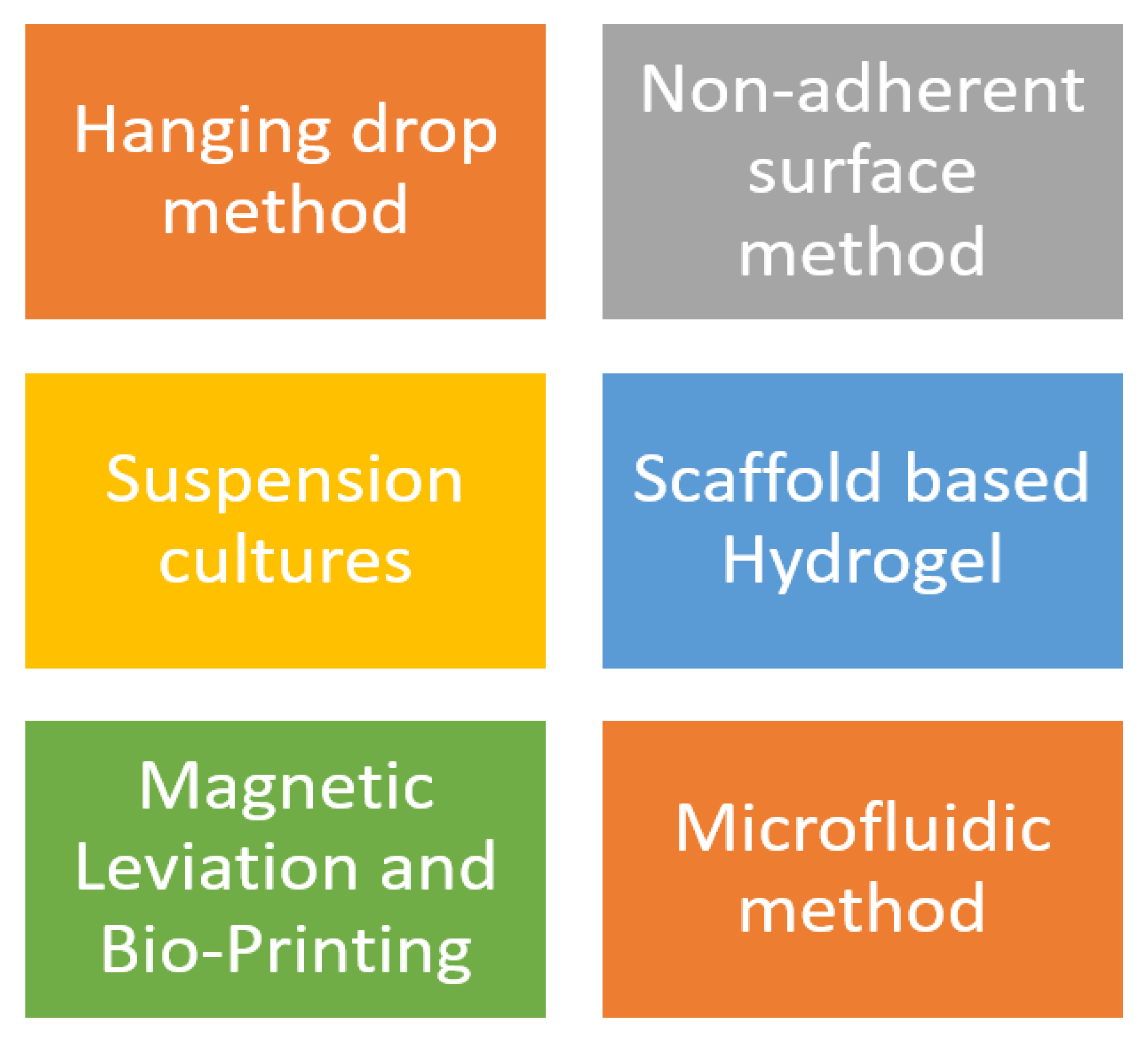
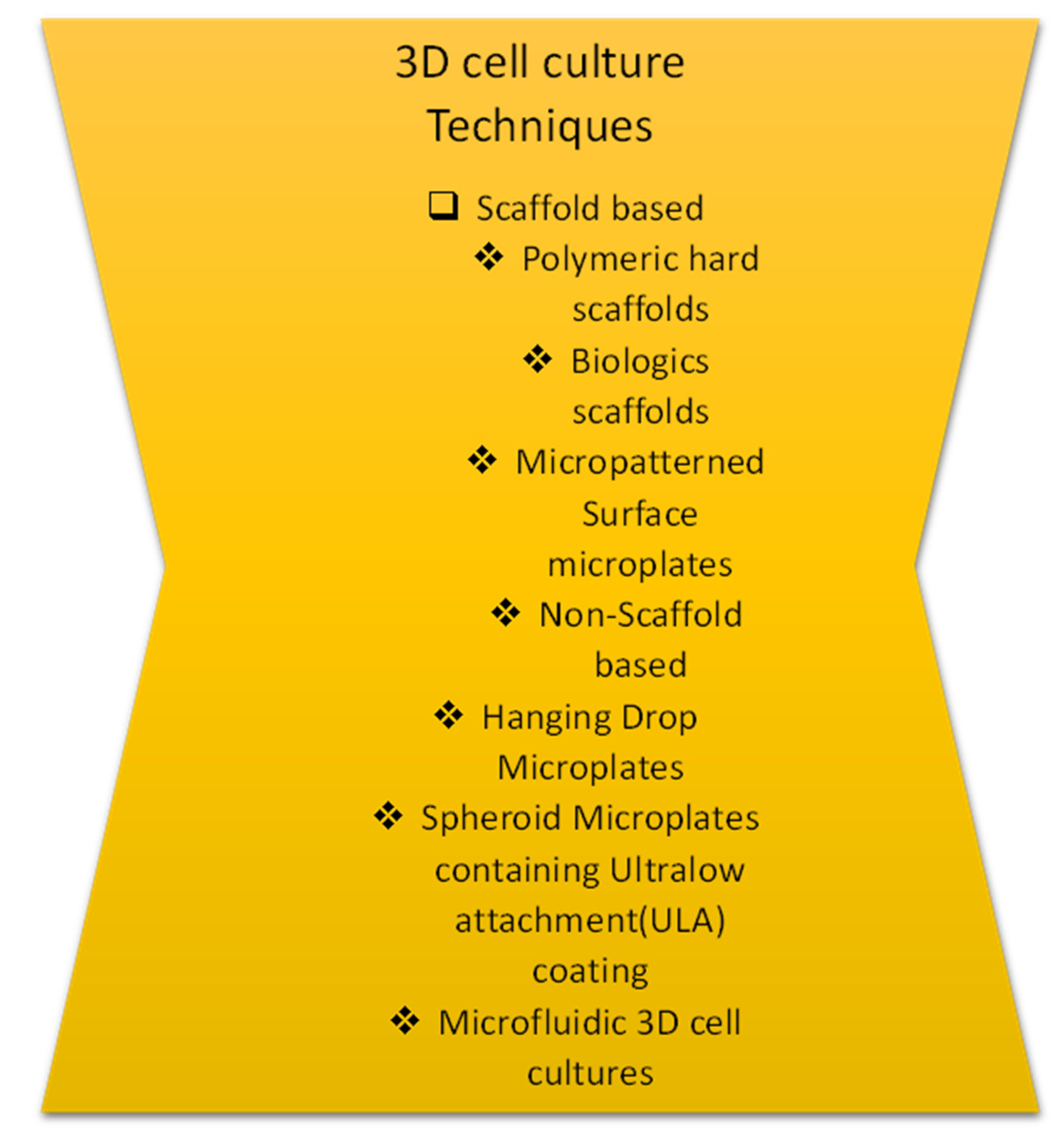
3. 3D Cell Culture Techniques

The aggregated spheroids grow in various modes and are summed up in the table describing their advantages and disadvantages. The categories in Figure 1 are listed below.

3.1. Hanging Drop Method

This method of studying bacteria in a controlled environment is a scaffold-free one that uses PBS as the medium. Cell suspensions are dropped onto a petri dish lid, where they aggregate due to surface tension and form spheroids [5,6] avoiding dehydration. A liquid-filled tray is kept at the bottom to maintain moisture and prevent drying out. Researchers have discovered that 50 µL is not enough to keep the cells attached, but the method has improved, allowing for the creation of more spheroids per plate. However, there is still a challenge in maintaining the stability of the spheroids, as any change in the medium can result in their disruption [7,8,9].
Different cell lines are thought to produce one spheroid per drop by a simple and consistent method [10]. HDPs were manipulated using liquid handling robots. The screening via a high throughput method opens a new door for replicating 3D in-vivo over a large set of drugs on distinct cell lines [10]. Thus, cell communications between cell-cell and cell-ECM can be maintained. Consequently, this approach promotes uniformly sized spheroids (mono and co-cultures). Moreover, spheroids can be cultured for several weeks for complex experiments within the droplet array.

3.2. Formation of Spontaneous Spheroid: Non-Adherent Surface Approaches

Sutherland et al. have recreated a 3D in-vitro system to exploit tumor cell response in radiotherapy. Substances such as agar or (poly-2 hydroxyethyl methyl acrylate)- poly-HEMA avert few cells to associate thus forming spheroids by adhering to the surface [10,11]. Subsequently, this method was developed further by Ivasku collaborators in 2006, from both cancerous and noncancerous cells with round and conical 96 well-bottomed plates [12]. These plates were coated with 0.5% poly-HEMA and dried for 2–3 days before the addition of cells. Nonetheless, the addition of about 2.5% redeveloped basement membrane formed the compact 3D spheroids to the suspensions upon 24h of centrifugation.
Precoated plates with characteristic features such as a neutral or a hydrophilic bottom, were formed into 3D spheroids upon suspension. Moreover, the coating formed over the process makes it a stable and non-degradable substance. The intrinsic factors such as dimensions, and composition along with the formation of small numbers of spheroids remain challenging. Despite that, it is possible to culture spheroids for extended periods and retrieve them after the culture process. Figure 2 listed below has represented techniques. here are some reasons to utilize 3D cultures.
  • Microenvironment conditions such as hypoxia, and nutrient gradients can be stimulated.
  • The cellular function can be differentiated.
  • Two or more different types-cultures can be exhibited.
  • Better proof diction in vivo responses upon drug treatment.
  • 3D cultures are known to mimic tissue-like structures.

3.3. Suspension Culture

Cells are suspended in a container either by agitation or by increasing the media viscosity (carboxymethyl cellulose can be added). The agitation methodology keeps the container in a gentle and rotary motion. Thus, continuous agitation of cells prevents them from adhering to the container walls and thus promotes cell-cell interactions. Spinner flasks and Bioreactors are the apparatuses used in this mode of culture.

3.4. Spinner Flasks

A stirring element works in continuous motion when present in the cell suspension. The constant motion of culture fluid, nutrients, and O2 is transported to spheroids and their wastes are removed. This production leads to larger yields which tend to easily change the culture medium.
The process begins by applying shearing force in spinner cells, for the modification of the physiology of the cell. The screening of drug assays becomes tough while handling a distinct range of spheroid sizes. Therefore, the use of ultra-low attachment plates of the appropriate sizes resolves this problem by allowing for their transfer back into spinner flasks [13].

3.5. Bioreactors

First created by NASA (National Aeronautics and Space Administration) in 1992, it was designed to culture cells and tissues during space flight [14]. The availability of discrete sizes for larger production gets enabled. Applying shearing forces to cells was not appropriate for bioreactors. Prior steps were followed, unlike using spinners, to choose spheroids of the same size.

3.6. Scaffold–Based Models: Hydrogels

The behavior of cell-ECM interactions keeps modifying the cellular organization and cell function during various therapies. Natural hydrogels, including Matrigel, collagen, alginate, and fibrin, as well as synthetic PEG and semi-synthetic hydrogels, are blended with synthetic and natural polymers, such as hyaluronan and polypeptides, to function effectively [15]. Nonetheless, either natural or synthetic hydrogels can be used [16]. The ideal condition for ECM is to recapitulate a 3D culture model in vivo. Initially, upon solidification, cells are placed on the top layer of the matrix using liquid hydrogel (matrix), after which the cells get embedded by gelatin in the matrix. Similarly, in both methods, cell cultures are coated with hydrogel prior to use. In the first approach, cells are added at 37 °C and agitated to ensure adherence, while in the second approach, cells are incubated at 37 °C while being surrounded by the hydrogel matrix. Clear differences between natural and synthetic properties matrices for 3D models are given below (Table 3).
The reorganization of 3D structures and cell attachment in natural scaffolds using the substances Matrigel and Collagen provides tensile strength for the functioning of cell migration, polarization, growth regulation, chemotherapeutic resistance, and adhesion [17]. However, substances are commercialized with mouse tumors of Engelbreth-Holm- Swarm (EHS). The most abundant fibrous protein is involved in cell migration and chemotaxis provides tensile strength and regulates cell adhesion [18,19,20,21].
This offers cell compatibility, amenability to cell adhesion, and a native viscoelastic environment. Yet substances such as matrigel, and collagen varies from batch to batch with low stiffness.

3.7. Magnetic Levitation

Initially, Souza et al. 2010 developed magnetic levitation using gold and magnetic iron oxide (MIO) nanoparticles. MIO hydrogels were used as a medium for cell growth of about 80% for about 1–2 days in a filamentous kind of bacteriophage. Treated cells were trypsinized in an ultra-low attachment plate. After cell aggregation, cells synthesize proteins, such as collagen, fibronectin, and laminin, that bind to each other [22]. Spheroids can be incubated for a few days for an ideal study. The speed of spheroid growth is much higher when compared to other methods such as forming intrinsic ECM, in the size range of mm; no specific medium is required. Nevertheless, this technique demands the use of expensive and toxic beads at higher concentrations with a limited number of cells produced [23].

3.8. Bioprinting

3D printing emerged over thirty years ago for mass production in varied fields [23]. Current 3D models allowed us to obtain different features in terms of spheroidal structures with limited vascularization potential, which is critical when studying tumor development [24]. This technology has provided numerous technical advancements, including the promotion of vascularization, the creation of scaffolds that more accurately reflect the heterogeneity of the tumor microenvironment, and improved 3D in vitro cancer models. The exploitation of imaging technology (MRI, CT scan, and X-ray) is needed for the relevant information on the structure of tissues and organs [25]. A detailed Table 4 includes important properties for the different types of bioprinting mentioned below. Figure 3 represents different stages of drug discovery and developmental processes of 3D cell culture techniques.

3.8.1. The Porosity of the Hydrogel

The movement of nutrients through the process of passive diffusion marks the early stage of the culture in metabolism. This interconnected network is established through cross-linking of the hydrogels, allowing for the flow of nutrients. Passive diffusion marks the initial stage of the culture for metabolism. Likewise, hydrogels promote ideal cell behavior [26]. The pore size makes the best appropriate cell size along with the target tissue to promote cell growth in 3D.

3.8.2. Physical Properties of Hydrogel

Mesenchymal stem cells which express bone morphogenic proteins (BMP) that are meant to transform beta growth factor were a best-suited example. From this, it is concluded that certain mechanical properties do control the behavior of cells. Therefore, distinct mechanical properties from distinct sources do affect distinct substrates. This shows the relationship is directly proportional to the factors of mechanical strength and degree of cross-linkage.

3.8.3. Biochemical Properties of Hydrogel

Hydrogels show good biocompatibility in contrast to noncytotoxic metabolites. The molecular chain has varied protein sites (fibronectin, collagen, or laminin) for adherence. The absence of adhering sites promotes growth in sodium alginate gels. Comparatively, hydrogels with functional groups can be linked with polypeptides by covalent bonds as seen in water-soluble drugs. This is improved and put to use for biological activity to regulate cell behavior [27].

3.9. Acellular Matrix-Cell Encapsulated Material

Various types of tissues and organs aims at the process of decellularization technology to obtain decellularized scaffolds [28]. An acellular scaffold in a target tissue retains the ultrastructure of the complex and mimics the nature of the physiological anatomy. Acellular scaffolds were exploited in huge numbers using decellularized scaffolds [29] such as in bone regeneration [30] liver [31] and lung [32] tissues in vitro.

Cell Supporting Materials

The soft and moist characteristics of the culture make it suitable for mimicking cornea, skin, and blood vessel tissue. The conventional approach is insufficient for the strong hydrogels to culture hard tissues such as bone and cartilage. Even though some hydrogels may have limited forming abilities, they can be shaped into spheres, blocks, or fibers to imitate the desired tissue. Despite that, few fibers and particles play a structural role in adherence to cells by improving the biological properties of the hydrogel. Figure 4 has a brief representation of 3D cell models in drug discovery.

3.10. Spheroid Models and Methods

The early 1970s marked the era of Spheroids by Sutherland and colleagues. Subsequently, varied models and techniques for spheroid formation have been utilized. The spontaneous aggregation of cells together with the binding of integrins to ECM leads to the formation of spheroids. Initially, cell-cell contact, upregulate E- cadherin accumulates onto the cell surface and thus the spheroid becomes a compact structure via strong intercellular E- cadherin interactions [6,33], driven by factors such as nutrients, oxygen, and growth factors [7].
Various spheroid models have been established based on the cellular origin. Multi-cellular tumors (MCTs) can be replicated by mimicking metabolic gradients and promoting cell proliferation, especially in response to chemotherapy. The clonal selection, along with its larger expansions, promises advancements with highly suitable throughput systems [8]. Tumor-derived spheroids have been created by breaking down the tumor into single cells and culturing them in serum or serum-free media. These spheroids have been developed and utilized in the brain [9] breast [10] lung [34] colon [35] prostate [36] Pancreas [37] and ovarian tumors [12].

Culturing of Spheroids

Tissue engineering and regenerative medicine exploit the best use of scaffold methods. Scaffold-free methods were commonly simple, inexpensive, and fast for generation of spheroids. Culturing of spheroids can be practiced with or without the support of ECM. Varied scaffold-free approaches have evolved. The cells are subjected to centrifugation to encourage cell-cell adhesion. The previously mentioned precautions have to be taken to prevent cell damage by shearing force when they are cultured in larger amounts, especially in the deep study of chondrogenesis, bone formation, and differentiation of mesenchymal stem cells [13,38]. Tumor spheroids were the simplest 3D cell culture models used in cell-cell and cell-ECM interactions, by emulating the properties of solid tumors in a few ways.

3.11. Organoid Methods and Models

Organoids have been cultured from embryonic stem cells (ESCs), induced pluripotent stem cells (PSCs), and adult stem cells (ASC). Certain developmental processes require ECM-forming basal lamina as a source of various cellular sources. Initially, Organoids from embryonic stem cells were expanded and later differentiated in a multi-step protocol, eventually developing into a full-fledged differentiated structure [11,38]. Thereby, derived organoids have been shown as a favorable model in various ways, e.g., pathology in genetics, organogenesis, and infectious disease devoid of regenerative capacity like the brain [18,19,20,21,39,40].
Air-Liquid Interface (ALI) was developed for the propagation of organoids for both stromal cells and epithelial cells [41]. This method is used for cell migration assays by exploiting Boyden chambers (cell culture inserts). The cells get embedded in ECM gels onto the upper surface via a porous membrane.
However, in this process, cells are embedded in ECM gels on an upper surface with a porous membrane by directly exposing them to oxygen, leading to a rise in oxygen supply when compared to an epithelial-only submerged organoid method. The process of diffusion takes place by obtaining nutrients, and certainly a few other growth factors across the porous membrane from the lower surface. Neonatal tissues utilize organoids using the ALI method without external niche factors because these factors are thought to be produced from stromal cells within organoids. Despite that, a distinct advantage of the ALI method is that it also retains the microenvironment for a longer period, including stromal cells [28].
Another approach involves embedding tissue samples into droplets of BMR and then transferring them into spinning bioreactors [42]. This method, which involves continuous agitation and oxygen and nutrient supply, is considered to be more promising than static methods. (e.g., cerebrum and retina) [29]. Some organoids, such as glioblastoma, are cultured in a well-defined medium through agitation, even in the presence of mitogens [43]. Interestingly, these cultures retain their histological and genetic characteristics.
A few interesting facts on 3D cell culture.
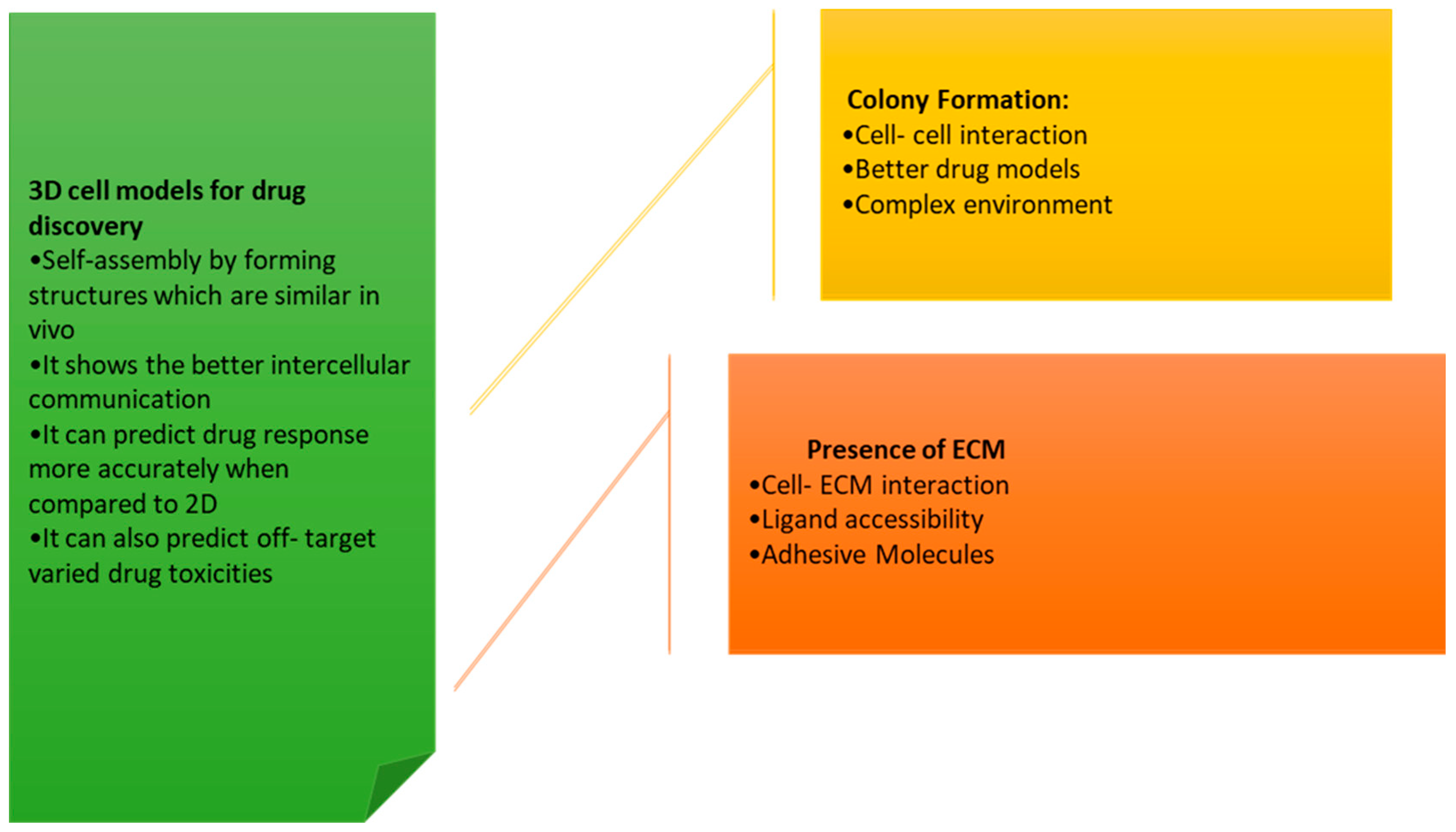
  • Compared to traditional 2D cell cultures, 3D cell cultures more accurately replicate the in vivo environment of cells, making them more effective for studying cellular behavior and biological processes.
  • The 3D cell cultures can be grown in a variety of different formats, including spheroids, organoids, and scaffold-based cultures.
  • The 3D cell cultures have been used extensively over a wide range of biological processes, including cancer progression, tissue development, and drug metabolism.
  • The 3D cell cultures have the potential to be used as an alternative to animal testing, as they can provide more accurate results and are more ethical.
  • The use of 3D cell cultures has been increasing in recent times due to advances in technology, such as the development of automated systems for growing and analyzing 3D cultures.

3.12. How Do 3D Cell Cultures Simulate the Structure?

The 3D structure can be simulated based on the physical and chemical properties of the cell microenvironment such as Molding, Microsphere, Microfibers, Channels, Composites.

3.12.1. Molding

The most convenient and simplest manufacturing method suits the production of hydrogels for small structures. While hydrogels are influenced by various factors such as light, and physical and chemical conditions, it is crucial that their preparation is done thoroughly in advance. Micro molding permits the construction of small structures by exploiting multiple and complex geometries. So, the high fluidity hydrogel and soft ultrafine fiber mold can be used to distribute liquid cells evenly through channels that are between 500 nanometers and 100 micrometers in size. During the process, the larger cells suffocate due to factors such as nutrients, metabolic waste, and internal channels.
Similarly, if cells are housed in large structures, they might miss out on internal channels, a source of nutrients, and a system for removing waste products, resulting in inadequate growth.

3.12.2. Microspheres

The hanging drop approach uses the principles of surface tension and viscosity of hydrogels in order to wrap cells. A cultured unit is defined by each microsphere under an electrostatic field. Subsequently, microlevel spheres are manufactured by high throughput culture and detection. The application has been used in drug delivery, tumor simulation, and stem culture as well.

3.12.3. Channels

Channel Structures are significant for blood vessels and respiratory tracts. Embedding the channels in the hydrogel culture units forms the basis of these kinds of structures. Manufacturing channel structures requires coaxial 3D printing, sacrificial template replication, and DLP printing. With advancements in manufacturing technology, coaxial printing can create tubes that can be stacked properly. The pipes remain non-bifurcated. Both DLP and sacrificial template replication ought to be bifurcated according to variated diameter.

3.12.4. Composites

The combination of different manufacturing techniques results in the creation of simulated human organs or tissues. Table 5 has listed various advantages and disadvantages of types of cell culture. This can be achieved by stacking tubes and layering fibers, creating blocks or sheets, as seen in the formation of bones, ears, and noses.

3.13. Applications of 3D Cell Culture

3.13.1. D Bioprinted Tissues/Organs for Transplantation

The tissues in 3D bioprinted bladders, tracheal grafts, bone, and cartilage have proved to be functional upon implantation and proper development, as observed in humans and animals. Liver and kidneys, with integrated vasculature, are in the process of being developed.
Subsequently, small diameters have been used in single or double layers, especially in human umbilical vein smooth muscle cells and skin fibroblasts of agarose rods. Apart from this, it can be observed in human umbilical vein endothelial cells, fibroblasts, or embryonic kidney cells. Significant efforts have been made to 3D print liver and kidneys with integrated blood vessels, using bio-printing technology.

3.13.2. 3D Cultures in Cell Therapy and Tissue Engineering

This approach has made a promising note in realizing the full potential of cell therapy and tissue engineering which meets the demand in quality, quantity, and robustness for commercialization and clinical trials. The production of 3D cells on a larger scale provides a new approach for treating different illnesses.
The idea of using spheroids has been applied to stem cells in the realm of tissue regeneration and repair processes. The methods of delivering it to target organs or tissues are limited, with loss of transplanted stem cells, low efficacy, and survival rate. In contrast with this, scaffolds, encapsulated materials, sheets, and platforms can be chosen as delivery modes. The method of construction of these platforms mimics the cell condition in vivo 3D cell-cell and cell- ECM interactions [57]. The use of spheroids, which are 3D structures formed from aggregates of cells, has garnered significant attention in the field of tissue engineering due to their potential to differentiate into various cell types and to mimic the properties of native tissues. These structures exhibit similar biological properties, such as size, cell density, viability, morphology, proliferative activity, and metabolic function, when compared to 3D cell culture systems, making them an attractive option for tissue repair and regeneration. In particular, spheroids are capable of differentiating into various lineages, including chondrogenic, osteogenic, adipogenic, and neurogenic cells, making them versatile tools for a wide range of tissue engineering applications. As a result, the study of spheroids and their applications in tissue engineering has gained significant momentum in recent years, and they are considered a promising approach for the restoration of damaged or diseased tissues.
Despite the growing effectiveness of stem cell therapies, there are still limitations due to the loss of transplanted stem cells. To address this issue and maximize the treatment efficacy of stem cells, researchers are striving to develop strategies for the efficient delivery of large numbers of stem cells, especially in cases where extensive or continuous treatment is required. To achieve this, it will be necessary to establish guidelines for the optimal number of stem cells to use in different therapeutic applications, taking into consideration the diverse nature of damaged human tissues and the varied mechanisms of diseases. The European Union [58] has established standards for the use of spheroids containing between 1 × 10⁵ and 2 × 10⁵ cells in clinical studies, with the implementation of fewer than 70 spheroids shown to be effective in treating defects in cartilage larger than 2 cm2. To further optimize the density and size of stem cell-based spheroids, it will be necessary to perform additional experimental studies. The use of stem cells can be more time consuming and costly, and technologies are being developed to maximize survival and minimize cell loss to improve treatment efficacy. In this regard, 3D cell cultures may still offer a superior alternative to 2D cultured stem cells [59].

3.13.3. Challenges and Future Perspectives

The 3D cell culturing methods unlock the limitations of 2D technologies. Although they are more expensive, 3D cell cultures have fewer hindrances. However, one factor is that the 3D culture method cannot replicate all the microenvironment conditions. Utilizing matrices that Constructing systems that contain heterogeneous components can be very labor-intensive, making it challenging to achieve [60]. Imaging turns out to be very tricky when using larger scaffolds in 3D. A study [61] revealed that anchorage-dependent cultures become incompatible while using microscopes and spheroids.
Although conventional analysis of cellular phenotype exploits the use of confocal fluorescence microscopy [61]. This becomes a great challenge over 3D to 2D, unlike 2D cell culture taken in a single XY plane of the axis. Therefore, higher magnification (40–60×) takes up large storage for high-throughput screening.
Likewise, a technique is used to count cells, and microorganisms by sorting cells, detecting biomarkers, protein engineering, and determining cell culture, characteristics, and functions [60]. The advent of 3D spheroids demands mechanical disruption of spheroids using the enzyme trypsin in a single-cell suspension [62] This is because spheroids have to be broken up into a single–cell suspension, which ultimately becomes an endpoint assay as the cells are meant to be disposed of by the end of the flow cytometry experiment [62].
The liquid handling suspension method throws a unique challenge for more viscous liquids such as collagen in suspension media and automated ultra-low attachment while the Matrigel-containing hydrogels too present a unique task [61]. Another important factor demands quick liquid handling and careful environmental control of temperature-sensitive polymerizations to avoid premature polymerization [63]. The automation approach can be achieved using 96- or 384-well plates for many 3D culturing techniques but further miniatured models find it more difficult in pipetting smaller volumes.
In automated cell therapy, certain bioreactors supported scaffold-based 3D progenitor cultures. These were constructed to control the internal environment.
The use of the microfluidic platform for 3D techniques, despite being expensive, has become a popular choice for creating tissue-like structures due to its ability to stimulate many standard 3D models. The utilization of 3D models in studying cell interactions with specific growth factors has proven to be crucial. In addition, spheroids and organoids have been marked as promising strategies over stem cell therapy. Nevertheless, 3D models have been targeted for therapeutics before any pre-clinical assessments.

4. Future Directions

Organoids which are miniature organ-like structures that can replicate the differentiated functions of diverse organs have made significant progress in replicating the functions of the nervous, vascular, and reproductive systems. However, there persists a significant gap in their ability to integrate and fuse with the host tissue at the site of transplantation. To optimize their therapeutic potential, there is still a need to develop technologies such as organ-on-a-chip, co-culture, and spatial control. Personalized stem cell organoids, which have the greater potential to reduce the risk of immune rejection in artificial organ transplantation, are also being explored as a promising approach. The integration of micro/nano engineering-based factors and systems will be necessary to enable the personalized method of organoid transplantation.
There are several directions that 3D cell culture research could take in the future. Some possible areas of focus engage in developing new and improved methods for creating and maintaining 3D cell cultures and understanding the impact of 3D cell cultures for various applications such as drug discovery and development, tissue engineering, and disease modeling. Another fascinating area of research may be the use of 3D cell cultures to study the effects of microgravity or other environmental factors on cells. Surprisingly, researchers may still look to employ various technologies, such as advanced imaging techniques and processes occurring within 3D cell cultures.
One possible arena for future research in the field of 3D cell culture could be the investigation of the effects of microgravity or other environmental factors on cells. This could be performed by using specialized bioreactors or other equipment to simulate different types of environments and to study the response of how cells behave with the conditions. This type of research could provide valuable insights into how cells function and adapt to different conditions, and could have potential applications in fields such as space exploration, medical research, and drug development. Strikingly, the study on 3D cell cultures helps us to understand how cells interact with each other and their surrounding environment, providing a more accurate representation of biological systems than traditional 2D cultures.

5. Conclusions

3D cell cultures offer a more realistic and physiological approach to studying cells compared to traditional 2D cell cultures. These 3D cell cultures can more closely mimic the in vivo microenvironment and provide a platform for studying various biological processes, including cell proliferation, differentiation, and migration. There are several different 3D culture systems available, including scaffold-based, matrix-based, and spheroid-based cultures. Each of these systems has its own unique set The use of 3D models in cell research has both benefits and drawbacks, but it offers a more accurate representation of real-life cell behavior.
Moreover, there are several other potential applications, such as to study the effects of microgravity or other environmental factors on cells, to develop novel drugs and therapies, and to study the behavior of cancer cells. Despite the many potential benefits of 3D cell cultures, several challenges need to be overcome to entirely realize their potential. These challenges include the need for standardized methods for generating and characterizing 3D cultures, as well as the need for a better understanding of various biological processes to lead the development of new and targeted therapies.

Author Contributions

S.K., A.T. and S.M.A. conceptualized the manuscript. S.M.A. collected information and prepared the manuscript. A.T. and S.K. have contributed to critically revising the draft and updating the manuscript for publication. All authors have read and agreed to the published version of the manuscript.

Funding

This research received no external funding.

Institutional Review Board Statement

Not applicable.

Informed Consent Statement

Not applicable.

Data Availability Statement

Not applicable.

Acknowledgments

The authors are also thankful to the GBPUA&T, Pantnagar provide valuable support.

Conflicts of Interest

Authors declare that there is no conflict of interest.

References

  1. Schutgens, F.; Clevers, H. Human Organoids: Tools for Understanding Biology and Treating Diseases. Annu. Rev. Pathol. Mech. Dis. 2020, 15, 211–234. [Google Scholar] [CrossRef]
  2. Fröhlich, E. Issues with Cancer Spheroid Models in Therapeutic Drug Screening. Curr. Pharm. Des. 2020, 26, 2137–2148. [Google Scholar] [CrossRef] [PubMed]
  3. Gilazieva, Z.; Ponomarev, A.; Rutland, C.; Rizvanov, A.; Solovyeva, V. Promising Applications of Tumor Spheroids and Organoids for Personalized Medicine. Cancers 2020, 12, 2727. [Google Scholar] [CrossRef]
  4. Simian, M.; Bissell, M.J. Organoids: A historical perspective of thinking in three dimensions. J. Cell Biol. 2017, 216, 31–40. [Google Scholar] [CrossRef] [PubMed]
  5. Sutherland, R.M.; McCredie, J.A.; Inch, W.R. Growth of Multicell Spheroids in Tissue Culture as a Model of Nodular Carcinomas. J. Natl. Cancer Inst. 1971, 46, 113–120. [Google Scholar] [CrossRef]
  6. Steinberg, M.S. Differential adhesion in morphogenesis: A modern view. Curr. Opin. Genet. Dev. 2007, 17, 281–286. [Google Scholar] [CrossRef] [PubMed]
  7. Bates, R.C. Spheroids and cell survival. Crit. Rev. Oncol. 2000, 36, 61–74. [Google Scholar] [CrossRef] [PubMed]
  8. Kunz-Schughart, L.A.; Freyer, J.P.; Hofstaedter, F.; Ebner, R. The Use of 3-D Cultures for High-Throughput Screening: The Multicellular Spheroid Model. J. Biomol. Screen. 2004, 9, 273–285. [Google Scholar] [CrossRef]
  9. Quereda, V.; Hou, S.; Madoux, F.; Scampavia, L.; Spicer, T.P.; Duckett, D. A Cytotoxic Three-Dimensional-Spheroid, High-Throughput Assay Using Patient-Derived Glioma Stem Cells. SLAS Discov. Adv. Life Sci. R D 2018, 23, 842–849. [Google Scholar] [CrossRef]
  10. Halfter, K.; Hoffmann, O.; Ditsch, N.; Ahne, M.; Arnold, F.; Paepke, S.; Grab, D.; Bauerfeind, I.; Mayer, B. Testing chemotherapy efficacy in HER2 negative breast cancer using patient-derived spheroids. J. Transl. Med. 2016, 14, 123. [Google Scholar] [CrossRef] [Green Version]
  11. Tomás-Bort, E.; Kieler, M.; Sharma, S.; Candido, J.B.; Loessner, D. 3D approaches to model the tumor microenvironment of pancreatic cancer. Theranostics 2020, 10, 5074–5089. [Google Scholar] [CrossRef]
  12. Raghavan, S.; Mehta, P.; Ward, M.R.; Bregenzer, M.E.; Fleck, E.M.A.; Tan, L.; McLean, K.; Buckanovich, R.J.; Mehta, G. Personalized Medicine–Based Approach to Model Patterns of Chemoresistance and Tumor Recurrence Using Ovarian Cancer Stem Cell Spheroids. Clin. Cancer Res. 2017, 23, 6934–6945. [Google Scholar] [CrossRef]
  13. Zhang, L.; Su, P.; Xu, C.; Yang, J.; Yu, W.; Huang, D. Chondrogenic differentiation of human mesenchymal stem cells: A comparison between micro mass and pellet culture systems. Biotechnol. Lett. 2010, 32, 1339–1346. [Google Scholar] [CrossRef]
  14. Timmins, N.E.; Nielsen, L.K. Generation of Multicellular Tumor Spheroids by the Hanging-Drop Method. In Tissue Engineering; Methods in Molecular MedicineTM; Humana Press: Totowa, NJ, USA, 2007; Volume 140, pp. 141–151. [Google Scholar] [CrossRef]
  15. Achilli, T.-M.; Meyer, J.; Morgan, J.R. Advances in the formation, use and understanding of multi-cellular spheroids. Expert Opin. Biol. Ther. 2012, 12, 1347–1360. [Google Scholar] [CrossRef]
  16. Costa, E.C.; de Melo-Diogo, D.; Moreira, A.F.; Carvalho, M.P.; Correia, I.J. Spheroids Formation on Non-Adhesive Surfaces by Liquid Overlay Technique: Considerations and Practical Approaches. Biotechnol. J. 2018, 13, 1700417. [Google Scholar] [CrossRef]
  17. Nunes, A.S.; Barros, A.S.; Costa, E.C.; Moreira, A.F.; Correia, I.J. 3D tumor spheroids as in vitro models to mimic in vivo human solid tumors resistance to therapeutic drugs. Biotechnol. Bioeng. 2019, 116, 206–226. [Google Scholar] [CrossRef] [PubMed]
  18. Takasato, M.; Er, P.X.; Chiu, H.S.; Maier, B.; Baillie, G.J.; Ferguson, C.; Parton, R.G.; Wolvetang, E.J.; Roost, M.S.; Chuva de Sousa Lopes, S.M.; et al. Kidney organoids from human iPS cells contain multiple lineages and model human nephrogenesis. Nature 2015, 526, 564–568. [Google Scholar] [CrossRef] [PubMed]
  19. Bartfeld, S.; Bayram, T.; van de Wetering, M.; Huch, M.; Begthel, H.; Kujala, P.; Vries, R.; Peters, P.J.; Clevers, H. In Vitro Expansion of Human Gastric Epithelial Stem Cells and Their Responses to Bacterial Infection. Gastroenterology 2015, 148, 126–136.e6. [Google Scholar] [CrossRef]
  20. Garcez, P.P.; Loiola, E.C.; Madeiro Da Costa, R.; Higa, L.M.; Trindade, P.; DelVecchio, R.; Nascimento, J.M.; Brindeiro, R.; Tanuri, A.; Rehen, S.K. Zika virus impairs growth in human neurospheres and brain organoids. Science 2016, 352, 816–818. [Google Scholar] [CrossRef] [PubMed] [Green Version]
  21. Takebe, T.; Sekine, K.; Enomura, M.; Koike, H.; Kimura, M.; Ogaeri, T.; Zhang, R.; Ueno, Y.; Zheng, Y.-W.; Koike, N.; et al. Vascularized and functional human liver from an iPSC-derived organ bud transplant. Nature 2013, 499, 481–484. [Google Scholar] [CrossRef]
  22. Huch, M.; Gehart, H.; van Boxtel, R.; Hamer, K.; Blokzijl, F.; Verstegen, M.M.; Ellis, E.; van Wenum, M.; Fuchs, S.A.; de Ligt, J.; et al. Long-Term Culture of Genome-Stable Bipotent Stem Cells from Adult Human Liver. Cell 2015, 160, 299–312. [Google Scholar] [CrossRef]
  23. Li, X.; Nadauld, L.; Ootani, A.; Corney, D.C.; Pai, R.K.; Gevaert, O.; Cantrell, M.A.; Rack, P.G.; Neal, J.T.; Chan, C.W.-M.; et al. Oncogenic transformation of diverse gastrointestinal tissues in primary organoid culture. Nat. Med. 2014, 20, 769–777. [Google Scholar] [CrossRef]
  24. Cruz, N.M.; Song, X.; Czerniecki, S.M.; Gulieva, R.E.; Churchill, A.J.; Kim, Y.K.; Winston, K.; Tran, L.M.; Diaz, M.A.; Fu, H.; et al. Organoid cystogenesis reveals a critical role of microenvironment in human polycystic kidney disease. Nat. Mater. 2017, 16, 1112–1119. [Google Scholar] [CrossRef] [PubMed]
  25. Pringle, S.; Maimets, M.; van der Zwaag, M.; Stokman, M.A.; van Gosliga, D.; Zwart, E.; Witjes, M.J.H.; de Haan, G.; van Os, R.; Coppes, R.P. Human Salivary Gland Stem Cells Functionally Restore Radiation Damaged Salivary Glands. Stem Cells 2016, 34, 640–652. [Google Scholar] [CrossRef] [PubMed]
  26. Kasagi, Y.; Chandramouleeswaran, P.M.; Whelan, K.A.; Tanaka, K.; Giroux, V.; Sharma, M.; Wang, J.; Benitez, A.J.; DeMarshall, M.; Tobias, J.W.; et al. The Esophageal Organoid System Reveals Functional Interplay Between Notch and Cytokines in Reactive Epithelial Changes. Cell. Mol. Gastroenterol. Hepatol. 2018, 5, 333–352. [Google Scholar] [CrossRef] [PubMed]
  27. Van de Wetering, M.; Francies, H.E.; Francis, J.M.; Bounova, G.; Iorio, F.; Pronk, A.; Van Houdt, W.; Van Gorp, J.; Taylor-Weiner, A.; Kester, L. Prospective derivation of a living organoid biobank of colorectal cancer patients. Cell 2015, 161, 933–945. [Google Scholar] [CrossRef]
  28. Neal, J.T.; Li, X.; Zhu, J.; Giangarra, V.; Grzeskowiak, C.L.; Ju, J.; Liu, I.H.; Chiou, S.-H.; Salahudeen, A.A.; Smith, A.R.; et al. Organoid Modeling of the Tumor Immune Microenvironment. Cell 2018, 175, 1972–1988.e16. [Google Scholar] [CrossRef]
  29. Qian, X.; Jacob, F.; Song, M.M.; Nguyen, H.N.; Song, H.; Ming, G.-L. Generation of human brain region–specific organoids using a miniaturized spinning bioreactor. Nat. Protoc. 2018, 13, 565–580. [Google Scholar] [CrossRef]
  30. Park, S.E.; Georgescu, A.; Huh, D. Organoids-on-a-chip. Science 2019, 364, 960–965. [Google Scholar] [CrossRef] [PubMed]
  31. Huh, D. A Human Breathing Lung-on-a-Chip. Ann. Am. Thorac. Soc. 2015, 12, S42–S44. [Google Scholar] [CrossRef]
  32. Shrestha, J.; Bazaz, S.R.; Es, H.A.; Azari, D.Y.; Thierry, B.; Warkiani, M.E.; Ghadiri, M. Lung-on-a-chip: The future of respiratory disease models and pharmacological studies. Crit. Rev. Biotechnol. 2020, 40, 213–230. [Google Scholar] [CrossRef] [PubMed]
  33. Lin, R.-Z.; Chou, L.-F.; Chien, C.-C.M.; Chang, H.-Y. Dynamic analysis of hepatoma spheroid formation: Roles of E-cadherin and β1-integrin. Cell Tissue Res. 2006, 324, 411–422. [Google Scholar] [CrossRef]
  34. Della Corte, C.M.; Barra, G.; Ciaramella, V.; Di Liello, R.; Vicidomini, G.; Zappavigna, S.; Luce, A.; Abate, M.; Fiorelli, A.; Caraglia, M.; et al. Antitumor activity of dual blockade of PD-L1 and MEK in NSCLC patients derived three-dimensional spheroid cultures. J. Exp. Clin. Cancer Res. 2019, 38, 253. [Google Scholar] [CrossRef]
  35. Jeppesen, M.; Hagel, G.; Glenthoj, A.; Vainer, B.; Ibsen, P.; Harling, H.; Thastrup, O.; Jørgensen, L.N.; Thastrup, J. Short-term spheroid culture of primary colorectal cancer cells as an in vitro model for personalizing cancer medicine. PLoS ONE 2017, 12, e0183074. [Google Scholar] [CrossRef]
  36. Linxweiler, J.; Hammer, M.; Muhs, S.; Kohn, M.; Pryalukhin, A.; Veith, C.; Bohle, R.M.; Stöckle, M.; Junker, K.; Saar, M. Patient-derived, three-dimensional spheroid cultures provide a versatile translational model for the study of organ-confined prostate cancer. J. Cancer Res. Clin. Oncol. 2018, 145, 551–559. [Google Scholar] [CrossRef] [PubMed]
  37. Bosnakovski, D.; Mizuno, M.; Kim, G.; Ishiguro, T.; Okumura, M.; Iwanaga, T.; Kadosawa, T.; Fujinaga, T. Chondrogenic differentiation of bovine bone marrow mesenchymal stem cells in pellet cultural system. Exp. Hematol. 2004, 32, 502–509. [Google Scholar] [CrossRef] [PubMed]
  38. McCauley, H.A.; Wells, J.M. Pluripotent stem cell-derived organoids: Using principles of developmental biology to grow human tissues in a dish. Development 2017, 144, 958–962. [Google Scholar] [CrossRef]
  39. Spence, J.R.; Mayhew, C.N.; Rankin, S.A.; Kuhar, M.F.; Vallance, J.E.; Tolle, K.; Hoskins, E.E.; Kalinichenko, V.V.; Wells, S.I.; Zorn, A.M.; et al. Directed differentiation of human pluripotent stem cells into intestinal tissue in vitro. Nature 2011, 470, 105–109. [Google Scholar] [CrossRef]
  40. Dutta, D.; Heo, I.; Clevers, H. Disease Modeling in Stem Cell-Derived 3D Organoid Systems. Trends Mol. Med. 2017, 23, 393–410. [Google Scholar] [CrossRef]
  41. Clevers, H. Modeling Development and Disease with Organoids. Cell 2016, 165, 1586–1597. [Google Scholar] [CrossRef]
  42. Ootani, A.; Li, X.; Sangiorgi, E.; Ho, Q.T.; Ueno, H.; Toda, S.; Sugihara, H.; Fujimoto, K.; Weissman, I.L.; Capecchi, M.R.; et al. Sustained in vitro intestinal epithelial culture within a Wnt-dependent stem cell niche. Nat. Med. 2009, 15, 701–706. [Google Scholar] [CrossRef] [PubMed]
  43. Lancaster, M.A.; Knoblich, J.A. Organogenesis in a dish: Modeling development and disease using organoid technologies. Science 2014, 345, 1247125. [Google Scholar] [CrossRef] [PubMed]
  44. Jacob, F.; Salinas, R.D.; Zhang, D.Y.; Nguyen, P.T.; Schnoll, J.G.; Wong, S.Z.H.; Thokala, R.; Sheikh, S.; Saxena, D.; Prokop, S.; et al. A patient-derived glioblastoma organoid model and biobank recapitulates inter-and intra-tumoral heterogeneity. Cell 2020, 180, 188–204.e22. [Google Scholar] [CrossRef] [PubMed]
  45. Zeitlin, B.D.; Dong, Z.; Nör, J.E. RAIN-Droplet: A novel 3D in vitro angiogenesis model. Lab. Investig. 2012, 92, 988–998. [Google Scholar] [CrossRef] [PubMed]
  46. Chwalek, K.; Tsurkan, M.V.; Freudenberg, U.; Werner, C. Glycosaminoglycan-based hydrogels to modulate heterocellular communication in in vitro angiogenesis models. Sci. Rep. 2014, 4, 4414. [Google Scholar] [CrossRef] [PubMed]
  47. Lee, D.W.; Yi, S.H.; Jeong, S.H.; Ku, B.; Kim, J.; Lee, M.-Y. Plastic pillar inserts for three-dimensional (3D) cell cultures in 96-well plates. Sens. Actuators B Chem. 2013, 177, 78–85. [Google Scholar] [CrossRef]
  48. Di, Z.; Klop, M.J.D.; Rogkoti, V.-M.; Le Dévédec, S.; Van De Water, B.; Verbeek, F.J.; Price, L.S.; Meerman, J.H.N. Ultra High Content Image Analysis and Phenotype Profiling of 3D Cultured Micro-Tissues. PLoS ONE 2014, 9, e109688. [Google Scholar] [CrossRef]
  49. Poincloux, R.; Collin, O.; Lizárraga, F.; Romao, M.; Debray, M.; Piel, M.; Chavrier, P. Contractility of the cell rear drives invasion of breast tumor cells in 3D Matrigel. Proc. Natl. Acad. Sci. USA 2011, 108, 1943–1948. [Google Scholar] [CrossRef]
  50. Gunness, P.; Mueller, D.; Shevchenko, V.; Heinzle, E.; Ingelman-Sundberg, M.; Noor, F. 3D Organotypic Cultures of Human HepaRG Cells: A Tool for In Vitro Toxicity Studies. Toxicol. Sci. 2013, 133, 67–78. [Google Scholar] [CrossRef]
  51. Mueller, D.; Krämer, L.; Hoffmann, E.; Klein, S.; Noor, F. 3D organotypic HepaRG cultures as in vitro model for acute and repeated dose toxicity studies. Toxicol. In Vitro 2014, 28, 104–112. [Google Scholar] [CrossRef]
  52. Thoma, C.R.; Stroebel, S.; Rösch, N.; Calpe, B.; Krek, W.; Kelm, J.M. A High-Throughput–Compatible 3D Microtissue Co-Culture System for Phenotypic RNAi Screening Applications. J. Biomol. Screen. 2013, 18, 1330–1337. [Google Scholar] [CrossRef] [PubMed]
  53. Li, Q.; Chen, C.; Kapadia, A.; Zhou, Q.; Harper, M.K.; Schaack, J.; Labarbera, D.V. 3D Models of Epithelial-Mesenchymal Transition in Breast Cancer Metastasis: High-Throughput Screening Assay Development, Validation, and Pilot Screen. J. Biomol. Screen. 2011, 16, 141–154. [Google Scholar] [CrossRef] [PubMed]
  54. Takayama, K.; Kawabata, K.; Nagamoto, Y.; Kishimoto, K.; Tashiro, K.; Sakurai, F.; Tachibana, M.; Kanda, K.; Hayakawa, T.; Furue, M.K.; et al. 3D spheroid culture of hESC/hiPSC-derived hepatocyte-like cells for drug toxicity testing. Biomaterials 2013, 34, 1781–1789. [Google Scholar] [CrossRef] [PubMed]
  55. Lutolf, M.P.; Doyonnas, R.; Havenstrite, K.; Koleckar, K.; Blau, H.M. Perturbation of single hematopoietic stem cell fates in artificial niches. Integr. Biol. 2009, 1, 59–69. [Google Scholar] [CrossRef]
  56. Håkanson, M.; Köbel, S.; Lutolf, M.P.; Textor, M.; Cukierman, E.; Charnley, M. Controlled Breast Cancer Microarrays for the Deconvolution of Cellular Multilayering and Density Effects upon Drug Responses. PLoS ONE 2012, 7, e40141. [Google Scholar] [CrossRef]
  57. Vonk, L.; Roël, G.; Hernigou, J.; Kaps, C.; Hernigou, P. Role of Matrix-Associated Autologous Chondrocyte Implantation with Spheroids in the Treatment of Large Chondral Defects in the Knee: A Systematic Review. Int. J. Mol. Sci. 2021, 22, 7149. [Google Scholar] [CrossRef]
  58. Antoni, D.; Burckel, H.; Josset, E.; Noel, G. Three-Dimensional Cell Culture: A Breakthrough In Vivo. Int. J. Mol. Sci. 2015, 16, 5517–5527. [Google Scholar] [CrossRef]
  59. Booij, T.H.; Price, L.S.; Danen, E.H.J. 3D Cell-Based Assays for Drug Screens: Challenges in Imaging, Image Analysis, and High-Content Analysis. SLAS Discov. Adv. Life Sci. R D 2019, 24, 615–627. [Google Scholar] [CrossRef]
  60. Picot, J.; Guerin, C.L.; Le Van Kim, C.; Boulanger, C.M. Flow cytometry: Retrospective, fundamentals and recent instrumentation. Cytotechnology 2012, 64, 109–130. [Google Scholar] [CrossRef] [Green Version]
  61. Gong, Y.; Fan, N.; Yang, X.; Peng, B.; Jiang, H. New advances in microfluidic flow cytometry. Electrophoresis 2019, 40, 1212–1229. [Google Scholar] [CrossRef]
  62. Lei, K.F.; Wu, M.H.; Hsu, C.W.; Chen, Y.D. Real-time and non-invasive impedimetric monitoring of cell proliferation and chemosensitivity in a perfusion 3D cell culture microfluidic chip. Biosens. Bioelectron. 2014, 51, 16–21. [Google Scholar] [CrossRef] [PubMed]
  63. De Bournonville, S.; Lambrechts, T.; Vanhulst, J.; Luyten, F.P.; Papantoniou, I.; Geris, L. Towards Self-Regulated Bioprocessing: A Compact Benchtop Bioreactor System for Monitored and Controlled 3D Cell and Tissue Culture. Biotechnol. J. 2019, 14, e1800545. [Google Scholar] [CrossRef] [PubMed]
Figure 1. 3D Cell Culture Techniques.
Futurepharmacol 03 00015 g001
Figure 2. Schematic representation of 3D cell culture technique.
Futurepharmacol 03 00015 g002
Figure 3. Representation of different stages of drug discovery and developmental processes of 3D cell culture techniques.
Futurepharmacol 03 00015 g003
Figure 4. Types of 3D cell models in drug discovery.
Futurepharmacol 03 00015 g004
Table 1. Brief differentiation between types of cell cultures with minimal properties.
Property2D2.5D3D
Culture MatrixFlat, inertA curved surface, bioactive in natureStereoscopic, bioactive
Cell PolarityNoyesyes
Biological factor diffusionFast, PassiveFast, PassiveSlow, active
MicroenvironmentStatic with the partial connection between cells,
Imperfect physiological functions
Dynamic, interconnected cells observed in the 2D microenvironmentDynamic, Reflects the interaction between cells, cells + ECM, cells, and tissues
Table 2. Representation of different characteristic features of Spheroids and Organoids.
CharacteristicsSpheroidsOrganoids
Source Primary cell lines in tumors, multicellular mixturesEmbryonic and adult stem cells, or induced pluripotent cells, tumor cells, and tissues
Organization(3D)Cell-cell in self-assembly, aggregation, and adhesion. In vivo models as self-organizationTo respond to physical and chemical cues in forming complex structures, organoids undergo self-organization and self-assembly
Organs PhysiologyThis shows various layers of proliferation in heterogeneous cells- necrotic tissues resembling 3D cellular organizationDiversified cell lineages that reflect the structure and function of the organ
culture conditions(3D)Extracellular matrix presence or absence of growth factors.Input as extracellular matrix along with a cocktail of growth factors
Table 3. Representation of natural and synthetic matrices for the 3D cell model purposes.
ModelNaturalSynthetic
BiocompatibilityHighMedium-High
BioactivityInherently bioactiveInert state
Cell Modification of ECMCannot be adjustedCan be adjusted
Endogenous factorsPresent naturallyNone
TunabilityLowHigh
ReproducibilityLowHigh
MicroenvironmentComplexSimple
Batch-batch variationsHighLow
Table 4. Representation of current 3D bioprinting techniques used for disease modeling and drug screening applications.
ParametersExtrusion BioprintingStereolithographyLaser-Assisted BioprintingInkjet Bioprinting
Resolution (um)ModerateHigh (100)High (50)High (50–300)
SpeedLowHighMediumHigh
Cell viability40–80%>85%>85%>85%
Cell densityHigh (spheroids)High106–107 cells/mL106–107 cells/mL
Ink viscosity (mPa/s)Up to 6 × 10⁷No limitation1–3003.5–12
AdvantagesSimple, Capable of printing, across biomaterialsNozzle-free technique, Printing time, Independent model, High accuracyDeposition in the solid or liquid phase, High spatial resolution, No issues with print head clottingAbility to print low-viscosity biomaterials, low volumes of solutions, and cells required
DisadvantagesApplicable for viscous liquidsUv light is toxic to cells, unable to print multiple cellsThermal damage due to laser irritationPoor functionality for vertical structures.
Table 5. Listed various advantages and disadvantages of types of cell culture.
Cell Cultures AdvantagesDisadvantagesReferences
(1) Hydrogel matrixCell-cell communications (cell-ECM) Upon 3D formation, the disposal of cells and changing growth media confers very low throughput and thus, makes it difficult for recovery.In vitro angiogenesis and drug testing [44]
Drug response study [45,46,47]
Cancer research [48]
Growth factors were incorporated quite handy
Microenvironment (in-vivo)
Uniformly spread spheroid
(2) Method of Hanging DropThe homogenous spheroids can be quite easily formed.This demands frequent growth in media change. However, analysis is required which demands labor and time. Cells were subjected to mechanical shocks very often. Hepatotoxicity testing with HepaRG cells ([49,50]; Target identification and validation using RNAi [51]
(3) Method of Liquid Overlay Quite easy to use and handy for long cultures. With extensive labor and time, centrifugation yields very low throughput, along heterogeneous cells were produced massively.Evaluation of the therapeutic response of anticancer drugs [52]
Identification of anticancer drugs [53] hepatoxicity testing with iPSC- derived hepatocytes [54]
(4) Method of Microwell PlatformHCL compatibility with different spheroid sizes.Cross-contamination takes place with microwells. Therefore, testing compounds turn out quite difficult.Study of self-renewal and differentiation process of stem cells [55]
Study of cancer and drug development [56]
Disclaimer/Publisher’s Note: The statements, opinions and data contained in all publications are solely those of the individual author(s) and contributor(s) and not of MDPI and/or the editor(s). MDPI and/or the editor(s) disclaim responsibility for any injury to people or property resulting from any ideas, methods, instructions or products referred to in the content.

