
Comprehensive Assessment of the Antioxidant and Anticancer Potential of Selected Ethnobotanical Plants

Abstract
:1. Introduction
- By using cell-free chemical-based methods using various free radical scavenging assays, such as DPPH, superoxide reduction, hydroxyl reduction, ABTS total reducing, and Fe2+ chelation
- whereas, Anticancer properties of plant extracts (PE) of selected five (CAN = Etlingera linguiformis, SES = Sesbania grandiflora, LEX = Smilax ovalifolia, DES = Desmodium triflorum, and CA = Chenopodium album) plants was investigated by using an MTT assay against breast cancer cell lines (MCF-7, MDA-MB-231, and MDA-MB-435S), and hepatic cancer cell lines (HepG2) in contrast to normal PBMCs.
2. Materials and Methods
- (a)
- General chemical and cell lines
- (b)
- Plant material collection
- (c)
- Extract preparation
- (d)
- Total phenolic and flavonoid content
- (e)
- ABTS radical scavenging activity
- (f)
- DPPH radical
- (g)
- Superoxide radical scavenging activity
- (h)
- Hydroxyl radical scavenging activity
- (i)
- Ferric chelation inhibitory activity
- (j)
- Total reducing power
- (k)
- PBMCs and Cell linesCulture condition
- (l)
- Cell viability using an MTT assay
- (m)
- Intracellular reactive oxygen species (ROS) measurement
- (n)
- Statistical analysis
3. Results
- (a)
- Total phenolic and flavonoid content
- (b)
- ABTS radical
- (c)
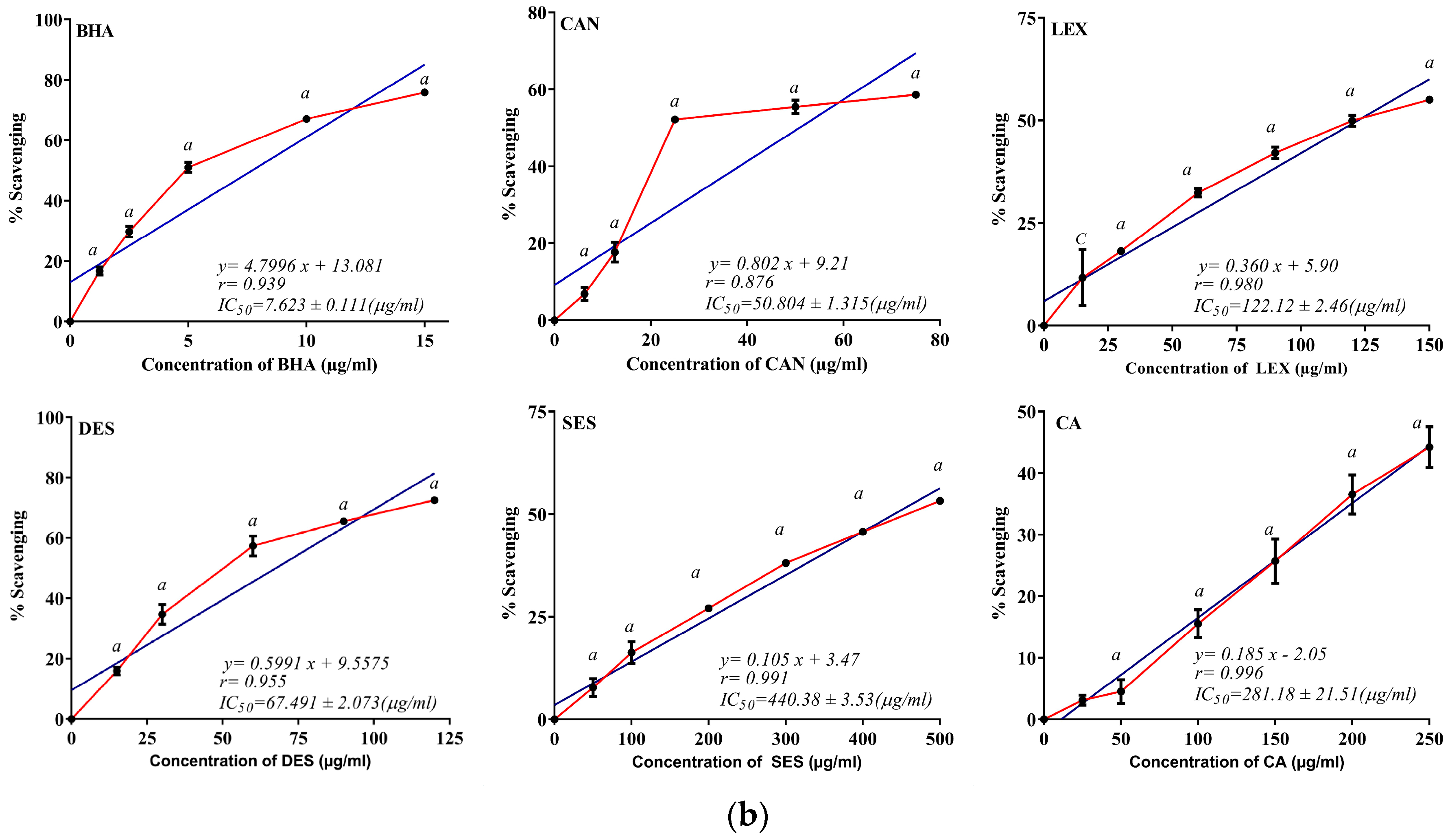
- DPPH
- (d)
- Superoxide radical scavenging activity (SO)
- (e)
- Hydroxyl radical
- (f)
- Ferric chelation (FC)
- (g)
- Total reducing power
- (h)
- Cell viability by MTT
- (i)
- Effect of plant extract on human Peripheral blood mononuclear cells (PBMCs)
- (j)
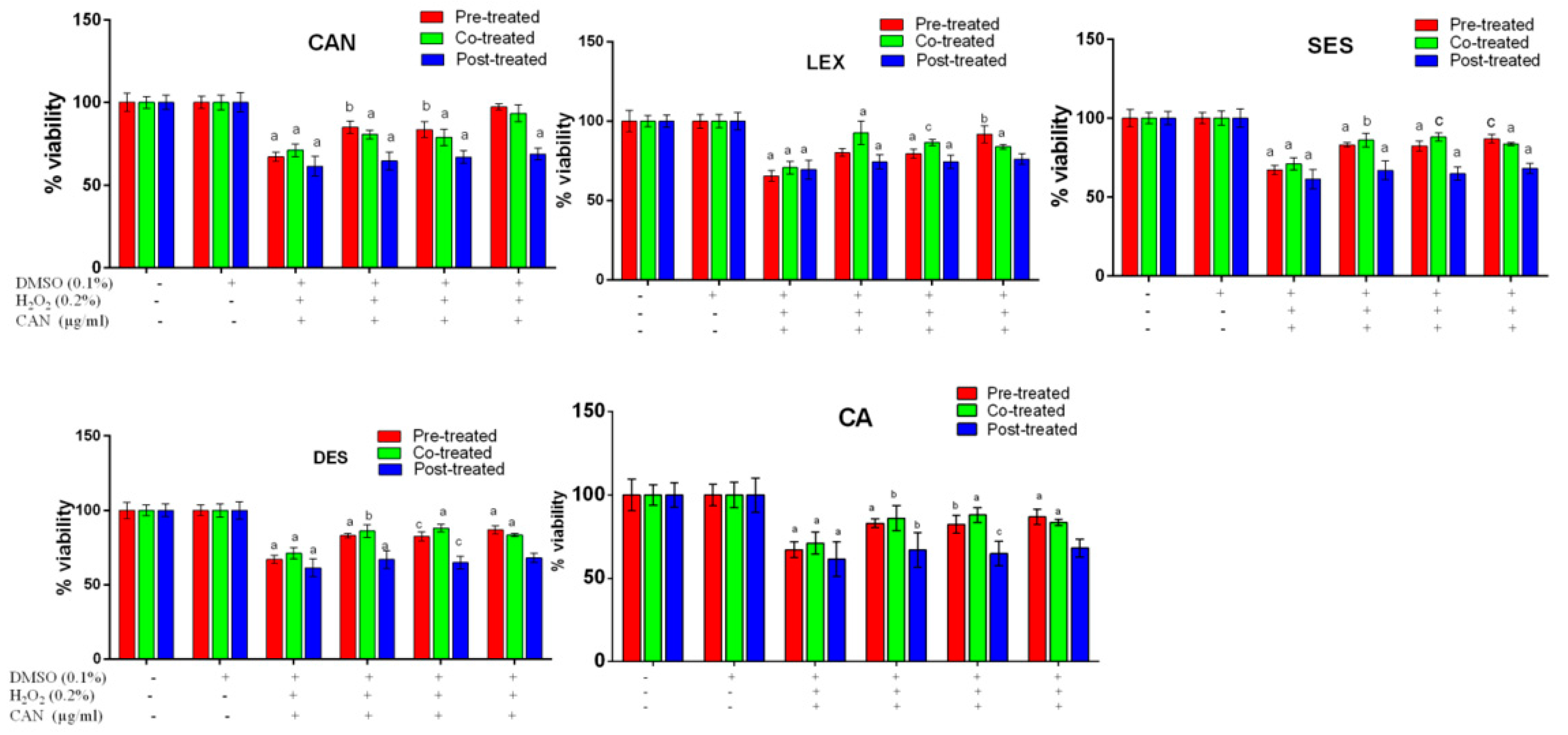
- Protective role of plants extracts against H2O2-induced cellular toxicity
- (k)
- Protective role of plants extracts against H2O2-induced Intracellular ROS level
4. Discussion
5. Conclusions
Supplementary Materials
Author Contributions
Funding
Institutional Review Board Statement
Informed Consent Statement
Data Availability Statement
Acknowledgments
Conflicts of Interest
Abbreviations
| ABTS | 2,2′-azino-di 3-ethylbenthiazolinesulfonate |
| DPPH | 2,2′-diphenyl-1-picrylhydrazy |
| MTT | 3-(4,5-dimethylthiazol-2-yl)-2,5-diphenyl tetrazolium bromide |
| DMSO | dimethyl sulfoxide |
| OD | optical density |
| ANOVA | analysis of variance |
| DMEM | Dulbecco’s modified Eagle’s medium |
| FBS | foetal bovine serum |
| IC50 | The half-maximal inhibitory concentration |
| ND | Not determined |
| PBS | phosphate buffer saline |
| SEM | standard error of the mean |
| BHA | beta Hydroxy Acids |
| TCA | trichloroacetic acid |
| FC | Folin–Ciocalteu Phenol reagent |
References
- Bodeker, G.; Ong, C.-K. WHO Global Atlas of Traditional, Complementary and Alternative Medicine; World Health Organization: Geneva, Switzerland, 2005; Volume 1. [Google Scholar]
- Sung, H.; Ferlay, J.; Siegel, R.L.; Laversanne, M.; Soerjomataram, I.; Jemal, A.; Bray, F. Global cancer statistics 2020: GLOBOCAN estimates of incidence and mortality worldwide for 36 cancers in 185 countries. CA Cancer J. Clin. 2021, 71, 209–249. [Google Scholar] [CrossRef] [PubMed]
- Bouali, N.; Hamadou, W.S.; Badraoui, R.; Lajimi, R.H.; Hamdi, A.; Alreshidi, M.; Adnan, M.; Soua, Z.; Siddiqui, A.J.; Noumi, E.; et al. Phytochemical Composition, Antioxidant, and Anticancer Activities of Sidr Honey: In Vitro and In Silico Computational Investigation. Life 2022, 13, 35. [Google Scholar] [CrossRef] [PubMed]
- Dyba, T.; Randi, G.; Martos, C.; Giusti, F.; Calvalho, R.; Neamtiu, L.; Nicholson, N.; Flego, M.; Dimitrova, N.; Bettio, M. 1501O Long-term estimates of cancer incidence and mortality for the EU and EFTA countries according to different demographic scenarios. Ann. Oncol. 2021, 32, S1102. [Google Scholar] [CrossRef]
- Calaf, G.M.; Urzua, U.; Termini, L.; Aguayo, F. Oxidative stress in female cancers. Oncotarget 2018, 9, 23824. [Google Scholar] [CrossRef] [PubMed]
- Kocyigit, A.; Guler, E.M.; Dikilitas, M. Role of Antioxidant Phytochemicals in Prevention, Formation and Treatment of Cancer; IntechOpen Limited: London, UK, 2018; pp. 21–45. [Google Scholar]
- Matowa, P.R.; Gundidza, M.; Gwanzura, L.; Nhachi, C.F. A survey of ethnomedicinal plants used to treat cancer by traditional medicine practitioners in Zimbabwe. BMC Complement. Med. Ther. 2020, 20, 278. [Google Scholar] [CrossRef]
- Wang, H.; Oo Khor, T.; Shu, L.; Su, Z.-Y.; Fuentes, F.; Lee, J.-H.; Tony Kong, A.-N. Plants vs. cancer: A review on natural phytochemicals in preventing and treating cancers and their druggability. Anti-Cancer Agents Med. Chem. 2012, 12, 1281–1305. [Google Scholar] [CrossRef]
- Gu, I.; Brownmiller, C.; Howard, L.; Lee, S.-O. Chemical Composition of Volatile Extracts from Black Raspberries, Blueberries, and Blackberries and Their Antiproliferative Effect on A549 Non-Small-Cell Lung Cancer Cells. Life 2022, 12, 2056. [Google Scholar] [CrossRef]
- Noumi, E.; Ahmad, I.; Bouali, N.; Patel, H.; Ghannay, S.; ALrashidi, A.A.; Abdulhakeem, M.A.; Patel, M.; Ceylan, O.; Badraoui, R.; et al. Thymus musilii Velen. Methanolic Extract: In Vitro and In Silico Screening of Its Antimicrobial, Antioxidant, Anti-Quorum Sensing, Antibiofilm, and Anticancer Activities. Life 2023, 13, 62. [Google Scholar] [CrossRef]
- Khan, M.; Khan, M.; Al-Hamoud, K.; Adil, S.F.; Shaik, M.R.; Alkhathlan, H.Z. Comprehensive Phytochemical Analysis of Various Solvent Extracts of Artemisia judaica and Their Potential Anticancer and Antimicrobial Activities. Life 2022, 12, 1885. [Google Scholar] [CrossRef]
- Ly, H.T.; Truong, T.M.; Nguyen, T.T.H.; Nguyen, H.D.; Zhao, Y.; Le, V.M. Phytochemical screening and anticancer activity of the aerial parts extract of Xanthium strumarium L. on HepG2 cancer cell line. Clin. Phytosci. 2021, 7, 14. [Google Scholar] [CrossRef]
- Wagh, A.S.; Butle, S.R. Phytochemical analysis and in-vitro anticancer activity of Duranta erecta L. (Verbenaceae). Int. J. Pharm. Sci. Res. 2019, 10, 2941–2946. [Google Scholar]
- Bhandari, J.; Muhammad, B.; Thapa, P.; Shrestha, B.G. Study of phytochemical, anti-microbial, anti-oxidant, and anti-cancer properties of Allium wallichii. BMC Complement. Altern. Med. 2017, 17, 102. [Google Scholar] [CrossRef] [PubMed]
- Al-Shehri, M.; Moustafa, M. Anticancer, Antibacterial, and Phytochemicals Derived From Extract of Aerva javanica (Burm.f.) Juss. ex Schult. Grown Naturally in Saudi Arabia. Trop. Conserv. Sci. 2019, 12, 194008291986426. [Google Scholar] [CrossRef]
- Jacobs, E.C. Potential Therapeutic Effects of Phytochemicals and Medicinal Herbs for Cancer Prevention and Treatment. Arch. Gen. Intern. Med. 2018, 2, 44–48. [Google Scholar] [CrossRef]
- Luo, H.; Vong, C.T.; Chen, H.; Gao, Y.; Lyu, P.; Qiu, L.; Zhao, M.; Liu, Q.; Cheng, Z.; Zou, J.; et al. Naturally occurring anti-cancer compounds: Shining from Chinese herbal medicine. Chin. Med. 2019, 14, 48. [Google Scholar] [CrossRef] [PubMed]
- Chaudhary, S.; Chandrashekar, K.S.; Pai, K.S.R.; Setty, M.M.; Devkar, R.A.; Reddy, N.D.; Shoja, M.H. Evaluation of antioxidant and anticancer activity of extract and fractions of Nardostachys jatamansi DC in breast carcinoma. BMC Complement. Altern. Med. 2015, 15, 50. [Google Scholar] [CrossRef]
- Ahmad, N.; Gupta, S.; Husain, M.M.; Heiskanen, K.M.; Mukhtar, H. Differential antiproliferative and apoptotic response of sanguinarine for cancer cells versus normal cells. Clin. Cancer Res. 2000, 6, 1524–1528. [Google Scholar]
- Hossan, M.A.; Ibrahim, M.; Ahsan, M.Q.; Aktar, F.; Kuddus, M.R.; Chowdhury, M.M.U.; Rashid, M.A. Pharmacological and phytochemical screenings of ethanol extract of Etlingera linguiformis (Roxb.) RM Sm. growing in Bangladesh. Bangladesh Pharm. J. 2013, 16, 33–37. [Google Scholar] [CrossRef]
- Ghasemzadeh, A.; Jaafar, H.Z.; Rahmat, A.; Ashkani, S. Secondary metabolites constituents and antioxidant, anticancer and antibacterial activities of Etlingera elatior (Jack) RM Sm grown in different locations of Malaysia. BMC Complement. Altern. Med. 2015, 15, 335. [Google Scholar] [CrossRef]
- Roy, A.; Bhoumik, D.; Sahu, R.K.; Dwivedi, J. Phytochemical screening and antioxidant activity of Sesbania grandiflora leaves extracts. Asian J. Res. Pharm. Sci. 2014, 4, 16–21. [Google Scholar]
- Pajaniradje, S.; Mohankumar, K.; Pamidimukkala, R.; Subramanian, S.; Rajagopalan, R. Antiproliferative and apoptotic effects of Sesbania grandiflora leaves in human cancer cells. BioMed Res. Int. 2014, 2014, 474953. [Google Scholar] [CrossRef] [PubMed]
- Shah, R.K.; Bioscience, A. Antioxidant activity and estimation of total phenols and flavonoids in extracts of Smilax ovalifolia leaves. Int. J. Pure Appl. Biosci. 2015, 3, 174–177. [Google Scholar]
- Malge, N.R.; Bandara, A.M.; Keerthirathna, W.L.; Dissanayake, D.M.; Perera, P.K.; Witharana, C.; Peiris, L.D. Antioxidant and Anti-proliferative Activities of Smilax Zeylanica Root and Rhizome Extract against Liver Carcinoma Cells. J. Herbs Spices Med. Plants 2021, 27, 188–199. [Google Scholar] [CrossRef]
- Singh, S.; Parmar, N.; Patel, B. A review on Shalparni (Desmodium gangeticum DC.) and Desmodium species (Desmodium triflorum DC. & Desmodium laxiflorum DC.)—Ethnomedicinal perspectives. J. Med. Plants 2015, 3, 38–43. [Google Scholar]
- Lai, S.-C.; Ho, Y.-L.; Huang, S.-C.; Huang, T.-H.; Lai, Z.-R.; Wu, C.-R.; Lian, K.-Y.; Chang, Y.-S. Antioxidant and antiproliferative activities of Desmodium triflorum (L.) DC. Am. J. Chin. Med. 2010, 38, 329–342. [Google Scholar] [CrossRef]
- Chamkhi, I.; Charfi, S.; Hachlafi, N.E.; Mechchate, H.; Guaouguaou, F.-E.; El Omari, N.; Bakrim, S.; Balahbib, A.; Zengin, G.; Bouyahya, A. Genetic diversity, antimicrobial, nutritional, and phytochemical properties of Chenopodium album: A comprehensive review. Food Res. Int. 2022, 154, 110979. [Google Scholar] [CrossRef]
- Al-Dabbagh, B.; Elhaty, I.A.; Al Hrout, A.; Al Sakkaf, R.; El-Awady, R.; Ashraf, S.S.; Amin, A. Antioxidant and anticancer activities of Trigonella foenum-graecum, Cassia acutifolia and Rhazya stricta. BMC Complement. Altern. Med. 2018, 18, 240. [Google Scholar] [CrossRef] [PubMed]
- Singleton, V.L.; Rossi, J.A. Colorimetry of Total Phenolics with Phosphomolybdic-Phosphotungstic Acid Reagents. Am. J. Enol. Vitic. 1965, 16, 144–158. [Google Scholar] [CrossRef]
- Jia, Z.; Tang, M.; Wu, J. The determination of flavonoid contents in mulberry and their scavenging effects on superoxide radicals. Food Chem. 1999, 64, 555–559. [Google Scholar] [CrossRef]
- Arnao, M.B.; Cano, A.; Acosta, M. The hydrophilic and lipophilic contribution to total antioxidant activity. Food Chem. 2001, 73, 239–244. [Google Scholar] [CrossRef]
- Kitts, D.D.; Wijewickreme, A.N.; Hu, C. Antioxidant properties of a North American ginseng extract. Mol. Cell. Biochem. 2000, 203, 1–10. [Google Scholar] [CrossRef] [PubMed]
- Shahidi, F.; Alasalvar, C.; Liyana-Pathirana, C.M. Antioxidant phytochemicals in hazelnut kernel (Corylus avellana L.) and hazelnut byproducts. J. Agric. Food Chem. 2007, 55, 1212–1220. [Google Scholar] [CrossRef] [PubMed]
- Srinivasan, R.; Chandrasekar, M.J.; Nanjan, M.J.; Suresh, B. Antioxidant activity of Caesalpinia digyna root. J. Ethnopharmacol. 2007, 113, 284–291. [Google Scholar] [CrossRef]
- Thakkar, K.; Prasad, A.; Nayak, J.; Iyer, S.V.; Kumar, S. Antioxidant and in vitro cytotoxic activity of extracts of aerial parts of Cocculus hirsutus (L) using cell line cultures (breast cell line). J. Phytopharm. 2014, 3, 395–399. [Google Scholar] [CrossRef]
- Halliwell, B.; Gutteridge, J.M. Formation of thiobarbituric-acid-reactive substance from deoxyribose in the presence of iron salts: The role of superoxide and hydroxyl radicals. FEBS Lett. 1981, 128, 347–352. [Google Scholar] [CrossRef] [PubMed]
- Hazra, B.; Sarkar, R.; Biswas, S.; Mandal, N. Comparative study of the antioxidant and reactive oxygen species scavenging properties in the extracts of the fruits of Terminalia chebula, Terminalia belerica and Emblica officinalis. BMC Complement. Altern. Med. 2010, 10, 20. [Google Scholar] [CrossRef]
- Dinis, T.C.; Maderia, V.M.; Almeida, L.M. Action of phenolic derivatives (acetaminophen, salicylate, and 5-aminosalicylate) as inhibitors of membrane lipid peroxidation and as peroxyl radical scavengers. Arch. Biochem. Biophys. 1994, 315, 161–169. [Google Scholar] [CrossRef]
- Zhu, K.-X.; Lian, C.-X.; Guo, X.-N.; Peng, W.; Zhou, H.-M. Antioxidant activities and total phenolic contents of various extracts from defatted wheat germ. Food Chem. 2011, 126, 1122–1126. [Google Scholar] [CrossRef]
- Das, A.J.; Das, M.K.; Singh, S.P.; Saikia, P.P.; Singh, N.; Islam, J.; Ansari, A.; Chattopadhyay, P.; Rajamani, P.; Miyaji, T.; et al. Synthesis of salicylic acid phenylethyl ester (SAPE) and its implication in immunomodulatory and anticancer roles. Sci. Rep. 2022, 12, 8735. [Google Scholar] [CrossRef]
- Manna, P.; Bhattacharyya, S.; Das, J.; Ghosh, J.; Sil, P.C. Phytomedicinal role of Pithecellobium dulce against CCl4-mediated hepatic oxidative impairments and necrotic cell death. Evid.-Based Complement. Altern. Med. 2011, 2011, 832805. [Google Scholar] [CrossRef]
- Chou, T.C.; Martin, N. CompuSyn for Drug Combinations: PC Software and User’s Guide: A Computer Program for Quantitation of Synergism and Antagonism in Drug Combinations, and the Determination of IC50 and ED50 and LD50 Values; ComboSyn: Paramus, NJ, USA, 2005. [Google Scholar]
- Salmerón-Manzano, E.; Garrido-Cardenas, J.A.; Manzano-Agugliaro, F. Worldwide research trends on medicinal plants. Int. J. Environ. Res. Public Health 2020, 17, 3376. [Google Scholar] [CrossRef] [PubMed]
- World Health Organization. WHO Guidelines on Good Agricultural and Collection Practices (GACP) for Medicinal Plants; World Health Organization: Geneva, Switzerland, 2003. [Google Scholar]
- Volenzo, T.; Odiyo, J. Integrating endemic medicinal plants into the global value chains: The ecological degradation challenges and opportunities. Heliyon 2020, 6, e04970. [Google Scholar] [CrossRef] [PubMed]
- Ralte, L.; Bhardwaj, U.; Singh, Y.T. Traditionally used edible Solanaceae plants of Mizoram, India have high antioxidant and antimicrobial potential for effective phytopharmaceutical and nutraceutical formulations. Heliyon 2021, 7, e07907. [Google Scholar] [CrossRef] [PubMed]
- Hostettmann, K.; Wolfender, J.-L.; Terreaux, C. Modern screening techniques for plant extracts. Pharm. Biol. 2001, 39, 18–32. [Google Scholar]
- Balamurugan, V.; Balakrishnan, V.; Robinson, J.P.; Ramakrishnan, M. Anti-cancer and apoptosis-inducing effects of Moringa concanensis using hepG2 cell lines. Bangladesh J. Pharmacol. 2014, 9, 604–609. [Google Scholar] [CrossRef]
- Robinson, J.P.; Suriya, K.; Subbaiya, R.; Ponmurugan, P. Antioxidant and cytotoxic activity of Tecoma stans against lung cancer cell line (A549). Braz. J. Pharm. Sci. 2017, 53. [Google Scholar] [CrossRef]
- Martemucci, G.; Costagliola, C.; Mariano, M.; D’andrea, L.; Napolitano, P.; D’Alessandro, A.G. Free Radical Properties, Source and Targets, Antioxidant Consumption and Health. Oxygen 2022, 2, 48–78. [Google Scholar] [CrossRef]
- Li, X.; Wu, X.; Huang, L. Correlation between antioxidant activities and phenolic contents of radix Angelicae sinensis (Danggui). Molecules 2009, 14, 5349–5361. [Google Scholar] [CrossRef]
- Matuszewska, A.; Jaszek, M.; Stefaniuk, D.; Ciszewski, T.; Matuszewski, Ł. Anticancer, antioxidant, and antibacterial activities of low molecular weight bioactive subfractions isolated from cultures of wood degrading fungus Cerrena unicolor. PLoS ONE 2018, 13, e0197044. [Google Scholar] [CrossRef]
- Tonisi, S.; Okaiyeto, K.; Mabinya, L.V.; Okoh, A.I. Evaluation of bioactive compounds, free radical scavenging and anticancer activities of bulb extracts of Boophone disticha from Eastern Cape Province, South Africa. Saudi J. Biol. Sci. 2020, 27, 3559–3569. [Google Scholar] [CrossRef]
- Aliyu, A.B.; Ibrahim, M.A.; Musa, A.M.; Musa, A.O.; Kiplimo, J.J.; Oyewale, A.O. Free radical scavenging and total antioxidant capacity of root extracts of Anchomanes difformis Engl. (Araceae). Acta Pol. Pharm. 2013, 70, 115–121. [Google Scholar]
- Ghasemzadeh, A.; Jaafar, H.Z. Profiling of phenolic compounds and their antioxidant and anticancer activities in pandan (Pandanus amaryllifolius Roxb.) extracts from different locations of Malaysia. BMC Complement. Altern. Med. 2013, 13, 341. [Google Scholar] [CrossRef] [PubMed]
- Puranik, S.I.; Ghagane, S.C.; Nerli, R.B.; Jalalpure, S.S.; Hiremath, M.B. Evaluation of in vitro antioxidant and anticancer activity of Simarouba glauca leaf extracts on T-24 bladder cancer cell line. Pharmacogn. J. 2017, 9, 906–912. [Google Scholar] [CrossRef]
- Maliyakkal, N.; Udupa, N.; Pai, K.; Rangarajan, A. Cytotoxic and apoptotic activities of extracts of Withania somnifera and Tinospora cordifolia in human breast cancer cells. Int. J. Appl. Res. Nat. Prod. 2013, 6, 1–10. [Google Scholar]
- Munro, B.; Vuong, Q.V.; Chalmers, A.C.; Goldsmith, C.D.; Bowyer, M.C.; Scarlett, C.J. Phytochemical, antioxidant and anti-cancer properties of Euphorbia tirucalli methanolic and aqueous extracts. Antioxidants 2015, 4, 647–661. [Google Scholar] [CrossRef]
- Nasser, M.; Damaj, R.; Merah, O.; Hijazi, A.; Trabolsi, C.; Wehbe, N.; Nasser, M.; Al-Khatib, B.; Damaj, Z. Potency of Combining Eucalyptus camaldulensis subsp. camaldulensis with Low-Dose Cisplatin in A549 Human Lung Adenocarcinomas and MCF-7 Breast Adenocarcinoma. Medicines 2020, 7, 40. [Google Scholar] [CrossRef]
- Chiu, Y.-W.; Lo, H.-J.; Huang, H.-Y.; Chao, P.-Y.; Hwang, J.-M.; Huang, P.-Y.; Huang, S.-J.; Liu, J.-Y.; Lai, T.-J. The antioxidant and cytoprotective activity of Ocimum gratissimum extracts against hydrogen peroxide-induced toxicity in human HepG2 cells. J. Food Drug Anal. 2013, 21, 253–260. [Google Scholar] [CrossRef]
- Baharum, Z.; Akim, A.M.; Taufiq-Yap, Y.H.; Hamid, R.A.; Kasran, R. In vitro antioxidant and antiproliferative activities of methanolic plant part extracts of Theobroma cacao. Molecules 2014, 19, 18317–18331. [Google Scholar] [CrossRef]
- Kokate, C.K.; Purohit, A.P.; Gokhale, S.B. Pharmacognosy; Nirali Prakashan: Pune, India, 2007. [Google Scholar]
- Jeet, K.; Thakur, R.; Sharma, A.K.; Shukla, A. Pharmacognostic and phytochemical investigation of whole aerial part of Argyreia nervosa. Int. J. Biol. Pharm. Res. 2012, 3, 713–717. [Google Scholar]


| Plants | TPC (mg GAE/gm) | TFC (mg ECE/gm) |
|---|---|---|
| CAN | 4.45 ± 0.09 | 29.29 ± 0.48 |
| LEX | 3.64 ± 0.04 | 23.51 ± 1.75 |
| SES | 1.87 ± 0.04 | 15.49 ± 0.48 |
| DES | 4.49 ± 0.07 | 34.50 ± 2.74 |
| CA | 19.75 ± 2.79 | 23.26 ± 2.31 |
| IC50 (µg/mL) | ||||||
|---|---|---|---|---|---|---|
| PE | DPPH | SO | HO | TR | FC | ABTS |
| CAN | 50.80 ± 1.31 | 69.32 ± 2.37 | 713.09 ± 18.32 | 467.83 ± 2.75 | 293.27 ± 7.93 | 48.48 ± 1.46 |
| LEX | 122.12 ± 2.40 | 117.46 ± 5.56 | 551.19 ± 18.74 | 887.00 ± 10.26 | 258.44 ± 7.49 | 42.57 ± 1.09 |
| DES | 67.49 ± 2.07 | 157.15 ± 5.53 | 384.98 ± 13.98 | 899.60 ± 6.55 | 284.77 ± 9.55 | 41.11 ± 1.09 |
| SES | 440.35 ± 3.53 | 157.15 ± 9.35 | 580.86 ± 14.90 | 951.00 ± 1.02 | 258.44 ± 7.49 | 64.62 ± 2.71 |
| CA | 281.18 ± 21.51 | 164.28 ± 2.13 | 1063.97 ± 27.30 | 15.68 ± 0.47 | 105.53 ± 7.11 | 26.28 ± 2.8 |
| STD | 7.62 ± 0.11 (BHA) | 14.57 ± 0.72 (AA) | 285.31 ± 4.20 (GA) | 28.18 ± 0.28 (AA) | 3.58 ± 0.14 (EDTA) | 1.55 ± 0.35 (BHA) |
| PE | CAN | LEX | SES | DES | CA | |
|---|---|---|---|---|---|---|
| Cells | ||||||
| HepG2 | 296.01 ± 1.23 | 657.02 ± 4.69 | 5531.20 ± 7.65 | 1684.68 ± 10.23 | 3151.91 ± 2.41 | |
| MCF-7 | 117.62 ± 2.54 | 104.81 ± 7.54 | 200.30 ± 6.54 | 231.60 ± 2.14 | 207.12 ± 2.51 | |
| MDA-MB-231 | 109.21 ± 3.51 | 132.02 ± 2.46 | 487.59 ± 3.45 | 490.35 ± 3.41 | 223.39 ± 2.31 | |
| MDA-MB-435S | 126.82 ± 2.35 | 218.20 ± 3.45 | 869.59 ± 4.52 | 262.13 ± 2.52 | 162.23 ± 2.15 | |
| Plants | CAN | LEX | ||||
|---|---|---|---|---|---|---|
| 12 h | 18 h | 36 h | 12 h | 18 h | 36 h | |
| Control | 100.00 ± 4.87 | 100.00 ± 4.68 | 100.00 ± 7.08 | 100.03 ± 5.53 | 100.01 ± 10.37 | 100.01 ± 11.44 |
| (0.1%) | 103.38 ± 5.38 | 104.26 ± 3.60 | 93.85 ± 8.09 | 102.77 ± 6.76 | 97.22 ± 2.67 | 98.90 ± 7.39 |
| 25 | 123.14 ± 2.03 b | 130.40 ± 8.46 b | 102.32 ± 10.02 | 103.98 ± 9.98 | 100.94 ± 6.04 | 96.22 ± 4.36 |
| 50 | 147.92 ± 3.71 a | 175.49 ± 9.33 a | 110.82 ± 12.49 | 103.69 ± 5.93 | 106.66 ± 3.03 | 104.34 ± 6.59 |
| 100 | 168.87 ± 12.50 a | 196.02 ± 46 a | 136.52 ± 8.14 c | 108.64 ± 4.61 | 104.04 ± 6.41 | 105.34 ± 5.53 |
| 150 | 189.43 ± 10.26 a | 190.92 ± 8.27 | 122.01 ± 12.64 c | 116.54 ± 6.97 a | 109.51 ± 6.47 c | 109.56 ± 7.25 a |
| 200 | 194.41 ± 12.26 a | 229.29 ± 15.86 a | 143.54 ± 17.73 a | 120.66 ± 4.61 a | 119.23 ± 5.77 | 113.76 ± 6.08 |
| 250 | 128.63 ± 3.56 b | 138.88 ± 3.9 | 184.21 ± 14.64 a | 130.88 ± 3.27 a | 131.14 ± 4.31 a | 128.27 ± 1.31 a |
| Plants | SES | DES | ||||
| 12 h | 18 h | 36 h | 12 h | 18 h | 36 h | |
| Control | 100.03 ± 3.51 | 100.00 ± 3.83 | 100.00 ± 3.05 | 99.96 ± 5.21 | 100.01 ± 14.10 | 99.99 ± 7.08 |
| (0.1%) | 91.15 ± 7.22 | 93.95 ± 3.94 | 102.55 ± 11.51 | 110.02 ± 5.80 | 97.60 ± 14.11 | 71.73 ± 2.98 |
| 25 | 96.43 ± 4.43 | 95.51 ± 2.92 | 97.21 ± 3.06 | 144.08 ± 3.63 | 108.00 ± 7.81 | 76.98 ± 1.89 |
| 50 | 102.12 ± 3.66 | 103.86 ± 3.68 | 118.15 ± 3.02 | 154.70 ± 2.16 | 101.69 ± 9.74 | 89.98 ± 9.92 |
| 100 | 113.82 ± 9.10 | 114.88 ± 3.71 | 139.76 ± 9.39 a | 164.76 ± 4.62 a | 91.59 ± 4.00 | 77.36 ± 11.05 |
| 150 | 118.75 ± 2.68 | 114.66 ± 4.33 a | 139.58 ± 13.46 | 173.26 ± 6.68 a | 91.16 ± 5.12 | 55.45 ± 6.42 b |
| 200 | 120.99 ± 4.20 | 119.06 ± 3.35 | 128.40 ± 8.64 a | 177.96 ± 8.38 a | 97.99 ± 1.64 | 56.47 ± 4.24 b |
| 250 | 129.48 ± 12.58 | 130.90 ± 9.51 | 126.68 ± 5.73 a | 184.81 ± 7.42 a | 104.73 ± 3.11 | 88.57 ± 4.55 b |
| Plants | CA | |||||
| 12 h | 18 h | 36 h | ||||
| Control | 99.97 ± 3.88 | 100.00 ± 5.18 | 100.00 ± 2.82 | |||
| (0.1%) | 93.36 ± 5.22 | 91.26 ± 2.01 | 97.70 ± 1.38 | |||
| 25 | 93.94 ± 5.63 | 96.52 ± 1.50 | 107.34 ± 10.16 | |||
| 50 | 95.44 ± 7.27 | 100.09 ± 8.15 | 106.68 ± 3.31 | |||
| 100 | 104.84 ± 7.67 | 101.39 ± 1.99 | 108.56 ± 4.16 | |||
| 150 | 107.63 ± 6.50 | 105.51 ± 6.01 | 119.04 ± 4.27 c | |||
| 200 | 109.07 ± 3.04 | 113.026 ± 4.83 c | 127.62 ± 8.31 a | |||
| 250 | 126.26 ± 7.04 | 121.54 ± 8.07 | 141.93 ± 4.62 a | |||
| Plants | CAN | LEX | ||||
|---|---|---|---|---|---|---|
| HepG2 | Pre | Post | Co | Pre | Post | Co |
| Control | 100.01 ± 5.50 | 100.00 ± 4.70 | 100.01 ± 3.53 | 100.02 ± 6.70 | 100.01 ± 3.76 | 100.01 ± 3.64 |
| (0.1%) | 99.98 ± 6.65 | 100.01 ± 5.89 | 99.98 ± 4.44 | 99.98 ± 4.35 | 100.01 ± 5.53 | 99.99 ± 4.08 |
| (0.2%) H2O2 | 67.07 ± 2.78 a | 61.37 ± 6.00 a | 71.08 ± 3.79 a | 65.52 ± 3.19 a | 69.45 ± 5.97 a | 70.68 ± 3.96 a |
| 25 | 84.91 ± 3.71 b | 64.52 ± 5.49 a | 80.60 ± 2.54 a | 80.18 ± 2.37 | 74.33 ± 4.50 a | 92.53 ± 7.36 a |
| 50 | 83.52 ± 4.84 b | 66.83 ± 3.85 a | 78.81 ± 5.04 a | 79.59 ± 2.86 a | 74.37 ± 4.25 a | 86.65 ± 1.91 b |
| 100 | 97.26 ± 1.84 | 68.75 ± 3.55 a | 93.37 ± 5.10 a | 91.66 ± 5.52 b | 76.06 ± 3.35 a | 83.90 ± 1.45 a |
| Plants | SES | DES | ||||
| HepG2 | Pre | Post | Co | Pre | Post | Co |
| Control | 99.98 ± 7.90 | 100.01 ± 3.26 | 100.01 ± 3.76 | 100.00 ± 5.50 | 100.00 ± 4.27 | 100.01 ± 3.53 |
| (0.1%) | 99.98 ± 5.05 | 100.02 ± 5.17 | 100.01 ± 3.73 | 99.98 ± 3.65 | 100.01 ± 5.89 | 99.98 ± 4.44 |
| (0.2%) H2O2 | 63.96 ± 3.60 a | 77.53 ± 5.93 a | 70.27 ± 4.13 a | 67.07 ± 2.78 a | 61.37 ± 6.01 a | 71.08 ± 3.79 a |
| 25 | 77.83 ± 3.14 c | 83.55 ± 3.14 a | 88.69 ± 13.37 b | 83.04 ± 1.5 a | 66.95 ± 6.03 a | 86.08 ± 4.33 b |
| 50 | 94.20 ± 8.20 a | 83.24 ± 4.13 a | 92.98 ± 11.37 c | 82.34 ± 3.10 c | 64.82 ± 4.22 c | 87.96 ± 2.57 a |
| 100 | 101.53 ± 1.15 c | 97.67 ± 3.50 a | 97.05 ± 6.00 a | 86.86 ± 2.72 | 68.11 ± 3.16 | 83.53 ± 1.02 |
| Plants | CA | |||||
| HepG2 | Pre | Post | Co | |||
| Control | 100.77 ± 6.92 | 100.01 ± 7.90 | 100.02 ± 13.12 | |||
| (0.1%) | 96.99 ± 4.20 | 97.58 ± 4.41 | 97.33 ± 5.23 | |||
| (0.2%) H2O2 | 63.59 ± 3.15 a | 77.42 ± 5.92 a | 60.59 ± 3.71 a | |||
| 25 | 67.81 ± 4.30 a | 80.14 ± 4.11 a | 71.90 ± 8.36 b | |||
| 50 | 72.76 ± 4.05 b | 82.92 ± 3.98 a | 73.30 ± 4.14 | |||
| 100 | 75.73 ± 4.11 c | 86.50 ± 5.36 a | 79.70 ± 3.83 a | |||
| Plants (µg/mL) | CAN | LEX | SES | DES | CA |
|---|---|---|---|---|---|
| Control | 1.00 ± 0.11 | 1.00 ± 0.12 | 1.00 ± 0.11 | 1.00 ± 0.14 | 1.00 ± 0.14 |
| H2O2 (0.2%) | 5.65 ± 1.00 a | 5.65 ± 1.01 a,x | 5.80 ± 0.43 a,x | 5.80 ± 0.04 a,x | 5.80 ± 0.10 a,x |
| 25 | 4.30 ± 1.46 a,x | 5.05 ± 0.58 a,x | 5.28 ± 0.31 a,x | 4.85 ± 0.05 a,x | 5.42 ± 0.18 a,x |
| 50 | 3.91 ± 1.16 a,x | 4.54 ± 1.58 a,x | 4.95 ± 0.44 a,x | 4.75 ± 0.11 a,x | 4.62 ± 0.77 a,x |
| 100 | 3.21 ± 1.68 a,x | 3.77 ± 0.23 a,x | 4.11 ± 0.96 a,x | 2.16 ± 1.21 a,x | 4.41 ± 0.53 a,x |
Disclaimer/Publisher’s Note: The statements, opinions and data contained in all publications are solely those of the individual author(s) and contributor(s) and not of MDPI and/or the editor(s). MDPI and/or the editor(s) disclaim responsibility for any injury to people or property resulting from any ideas, methods, instructions or products referred to in the content. |
© 2023 by the authors. Licensee MDPI, Basel, Switzerland. This article is an open access article distributed under the terms and conditions of the Creative Commons Attribution (CC BY) license (https://creativecommons.org/licenses/by/4.0/).
Share and Cite
Das, M.K.; Singh, N.; Rajamani, P. Comprehensive Assessment of the Antioxidant and Anticancer Potential of Selected Ethnobotanical Plants. Oxygen 2023, 3, 203-221. https://doi.org/10.3390/oxygen3020015
Das MK, Singh N, Rajamani P. Comprehensive Assessment of the Antioxidant and Anticancer Potential of Selected Ethnobotanical Plants. Oxygen. 2023; 3(2):203-221. https://doi.org/10.3390/oxygen3020015
Chicago/Turabian StyleDas, Monoj Kumar, Neelu Singh, and Paulraj Rajamani. 2023. "Comprehensive Assessment of the Antioxidant and Anticancer Potential of Selected Ethnobotanical Plants" Oxygen 3, no. 2: 203-221. https://doi.org/10.3390/oxygen3020015