Review

The Involvement of Lipid Mediators in the Mechanisms of Exercise-Induced Muscle Damage

by
Athanasios G. Gakis
1,
Tzortzis Nomikos
1,
Anastassios Philippou
2 and
Smaragdi Antonopoulou
1,*
1
Department of Nutrition and Dietetics, School of Health Sciences and Education, Harokopio University of Athens, 176 76 Kallithea, Greece
2
Department of Physiology, Medical School, National and Kapodistrian University of Athens, 115 27 Athens, Greece
*
Author to whom correspondence should be addressed.
Physiologia 2023, 3(2), 305-328; https://doi.org/10.3390/physiologia3020022
Submission received: 2 March 2023 / Revised: 1 April 2023 / Accepted: 8 May 2023 / Published: 10 May 2023
(This article belongs to the Special Issue Exercise Physiology and Biochemistry)

Abstract

:
Lipid mediators are a class of signaling molecules that play important roles in various physiological processes, including inflammation, blood pressure regulation, and energy metabolism. Exercise has been shown to affect the production and metabolism of several types of lipid mediators, including prostaglandins, leukotrienes, sphingolipids, platelet-activating factors and endocannabinoids. Eicosanoids, which include prostaglandins and leukotrienes, are involved in the regulation of inflammation and immune function. Endocannabinoids, such as anandamide and 2-arachidonoylglycerol, are involved in the regulation of pain, mood, and appetite. Pro-resolving lipid mediators are involved in the resolution of inflammation. Sphingolipids have a role in the function of skeletal muscle during and after exercise. There are many studies that have examined the effects of exercise on the production and release of these and other lipid mediators. Some of these studies have focused on the effects of exercise on inflammation and immune function, while others have examined the effects on muscle function and metabolism. However, much less is known about their involvement in the phenomenon of exercise-induced muscle damage that follows after intense or unaccustomed exercise.

1. Introduction

Inflammation is a dynamic tissue response mechanism against tissue injury and invasion by pathogens and is a critical component of appropriate host defense. All animals possess the biochemical machinery to deal with the invasion of pathogens and injury [1,2]. However, overwhelming and persistent inflammation can result in secondary cellular damage, promotes maladaptive tissue remodeling and leads to degenerative chronic inflammatory disease [3]. Typically, the inflammatory response is an acute and tightly regulated process that promotes resolution, restores tissue homeostasis and prevents the development of chronic disease. Though, accessing this process is difficult due to the multiple cells, mediators and pathways involved. This process and its resolution are driven by a complex set of mediators that regulates cellular events required to clear inflammatory cells from sites of infection and injury and the restoration of tissue function [4,5,6,7].
The health benefits of physical activity and exercise are well known. Even though exercise itself might act as an inflammatory stressor during and after its execution, it reduces the harmful effects of other stressors when performed correctly and at moderate intensities [8,9]. Skeletal muscle has the ability to adapt in response to external stress. High-intensity resistance exercise can lead to improved muscle strength by inducing changes in the muscle fiber composition, myofibrillar hypertrophy and neuromuscular mechanisms [10]. However, unaccustomed or high-demanding exercise (high intensity, long-duration, high frequency or combination of these) leads to exercise-induced muscle damage, a phenomenon that occurs as a consequence of the high mechanical and metabolic demands during that exercise and is accompanied by a number of other symptoms including the reduction of maximum strength and range of motion, muscle stiffness, swelling and increased sensitivity [11]. Moreover, acute exercise causes a temporary short-lasting activation of blood coagulation, platelet function and the prostaglandin system, the extent of these alterations being significantly less pronounced in well-trained athletes than in untrained individuals [12]. This inflammatory response mediates the restoration of muscle function and its adaptation to damaging external stimuli preventing by this way the magnitude of injury when repeated in the future. Interestingly, as a result, regular exercise training may be considered a long-lasting anti-inflammatory therapy after the acute inflammatory actions are resolved [13,14,15].
A complex crosstalk between cells exists and is regulated by numerous mediators, such as peptides (e.g., cytokines, chemokines), reactive oxygen species (e.g., superoxide anion, hydrogen peroxide), amino acid derivatives (e.g., histamine, nitric oxide), enzymes (e.g., matrix peroxidases) and lipid mediators (e.g., prostaglandins, leukotrienes, endocannabinoids, platelet-activating factor, sphingolipids) released by the resident cell populations depending on the cell types, anatomical site and inflammatory stimulus [16,17]. The latter, which are autocrine/paracrine signaling molecules, have a key role in muscle homeostasis and the resolution of inflammation caused by exercise [3,17].
The present review aims to provide an overview of the involvement of lipid mediators in the mechanisms of exercise-induced muscle damage.

2. Search Methodology

We searched the literature in MEDLINE and ScienceDirect databases until January 2023 using a combination of the following keywords: lipid mediator, exercise-induced muscle damage (EIMD), muscle damage, exercise, skeletal muscle, prostaglandin, PGE, PGF, COX, leukotriene, resolvin, maresin, sphingolipid, sphigosine, sphinganine, S1P, S1AP, ceramide, endocannabinoid, anandamide, 2-arachidonoylglycerol, AEA, 2-AG, phospholipids, platelet-activating factor (PAF). We included in vitro/ex vitro, animal and human studies that consisted entirely or partly (as a control group) of healthy subjects and supplement or drug interventions that included a placebo-controlled group. The studies that were selected included at least one lipid mediator assay or the levels of the key molecules of their metabolic pathways. Studies that did not include at least one of the established markers to evaluate EIMD or the exercise was not effective at altering their values significantly were discarded. The EIMD methods and markers include the pre-exercise and post-exercise comparison of perceived pain/discomfort ratings, muscle strength measurement, range of motion measurements, CK and LDH levels, biopsy analysis or other imaging methods.

3. Lipid Mediator Pathways

Lipid mediators include diverse bioactive lipids produced by a variety of cells under basal conditions or upon stimulation. They are derived from precursor lipid molecules of cellular membranes, mainly glycerol backbone phospholipids and sphingolipids. Briefly, by the action of phospholipases A2 (PLA2s), membrane glycerol-phospholipids are converted to their lyso-analogs, releasing the fatty acid esterified in the sn-2 position of the glycerol backbone. Arachidonic acid (AA; 20:4n-6), which is the most predominant polyunsaturated fatty acid, is esterified in the sn-2 position of the phosphatidylinositol and/or phosphatidylcholine and also regulates the physical properties of the membranes [3,18,19], is consequently enzymatically converted into a variety of eicosanoid mediators. The n-3 PUFA eicosapentaenoic acid (EPA, 20:5) and docosahexaenoic acid (DHA, 22:6) are also released and subsequently give rise to the less inflammatory and pro-resolving lipid mediators. In addition, the remodeling of the membrane glycerol-phospholipids leads to the production of potent phospholipid mediators, among them Platelet Activating Factor (PAF). On the other hand, the other integral structural components of cell membranes, the sphingolipids (SLs), along with their metabolites, ceramides, sphingosine and sphingosine-1-phosphate, are also bioactive lipids. Even though the lipid mediator pathways are not entirely independent from each other and synergistic interaction among different mediators cannot be excluded, below they are presented analytically for each class of lipid mediators.

3.1. Polyunsaturated Fatty Acid-Derived Lipid Mediator Pathways

PUFA are the main substrates for the synthesis of the majority of lipid mediators. After being released from the sn-2 position of the glycerol backbone phospholipids, AA and other PUFA can follow three enzymatic pathways that are described below.
The Cyclooxygenase (COX) pathway (Figure 1) includes the COX-1 and COX-2 isoforms responsible for the conversion of the free AA to prostaglandin G2 (PGG2) in the first step and prostaglandin H2 (PGH2) in the second step, followed by the conversion into five prostanoids, namely thromboxane A2 (TXA2) and prostaglandins D2, E2, F and I2 (PGD2, PGE2, PGF and PGI2, respectively). The COX enzymes can also use a broad range of 18–20C PUFA as substrates, though the two isoforms show different substrate selectivities. As a result, the products of the COX reactions are relatable to the substrate availability [19]. The prostanoids have several receptors on the muscle that regulate their actions through different mechanisms, contributing to processes such as myoblast proliferation, differentiation, fusion, migration and myotube hypertrophy [3].
The Lipoxygenase (LOX) pathway (Figure 1) includes the 5-LOX, 12-LOX, 12-LOXB, 15-LOX-1 and 15-LOX-2 enzymes expressed in specific cell types, which are responsible for the formation of specific h3ydroperoxyl-eicosatetraenes (HPETEs), which are subsequently reduced to hydroxy-eicosatetraenes (HETEs), 5(S)-HETE, 12(S)-HETE, 12(R)®-HETE, 15(S)-HETE and 8(S)-HETE, respectively. In the case of AA, 5-LOX is the key enzyme for the formation of leukotrienes (LTs). After the action of 5-LOX on AA, 5S-hydroperoxy-6t,8c,11c,14c-eicosatetraenoic acid (5-HPETE) is formed and through the aid of two proteins is converted into 5, 6-epoxy-7t,9t,11c,14c-eicosatetraenoic acid (5-HETE) or LTA4, an unstable molecule that can be hydrolyzed into LTB4, LTC4, LTD4 or LTE4 all bearing decisive role in acute and chronic inflammation and allergic diseases through a variety of biological effects. AA can also lead to the production of lipoxins (LXs) LXA4 and LXB4. These molecules, among others (resolvins, protectins and maresins synthesized by n-3 PUFAs) are characterized as specialized pro-resolving lipid mediators (SPMs) and are synthesized through the co-operation of different cell types, since the enzymes that are required for their formation are not present in a single cell type [3,19].
The cytochrome P450 epoxygenase pathway [3,18,19] can produce HETEs depending on the tissue, cell type and catalytic efficiency of the cytochrome P450 oxidase isoforms. In the case of AA, the cytochrome P450 oxidases can lead to the production of cis-epoxyeicosatrienoic acids (EETs) that can act in an autocrine and paracrine manner having beneficial effects (anti-hypertensive, fibrinolytic, anti-thrombotic) in the cardiovascular and renal systems. The EETs can be further metabolized into dihydroxyeicosatrienoic acids (DHETs) through the action of hydrolases, the biological role of which is still under investigation. Other n-3 PUFAs, such as EPA and DHA can also be used as substrates for the cytochrome P450 oxidases and their products can also have positive effects, indicating the advantages of dietary consumption of those fatty acids [19].

Pro-Resolving Lipid Mediators

The specialized pro-resolving lipid mediators (SMPs) as implied by their naming, have an important role in the resolution of inflammation. These include the above mentioned lipoxins synthesized through AA in the LOX pathway, as well as resolvins, protectins and maresins, which use n-3 PUFAs as their precursor molecules. Resolvins (Rv) can be synthesized by EPA, DHA and DPA n-3 PUFAs through the catalytic action of LOX enzymes (5- LOX and/or 15-LOX) and include RvE1, RvD1, RvD2, RvD3, RvD4, RvD5 and RvD6, the biosynthesis all of which have been detailly reviewed by Serhan et al. in 2017 [20,21]. Protectins are biosynthesized through the catalytic action of 15-LOX enzymes and include characteristic molecules of this class, such as PD1 and PD2 or PD1n-3 DPA and PD2n-3 DPA from DHA or n-3 DPA fatty acids, respectively. A detailed review of the protectins’ biosynthesis, metabolism and functions has been published by Hansen et al. in 2022 [22]. Finally, maresins (Macrophage mediator in resolving inflammation) are generated through the recently identified pathway that converts DHA into Maresin-1 and Maresin-2 through a multi-step catalytic action that involves 12-LOX (primarily in human M2 macrophages) and other enzymes. Their biosynthesis, structure, biological actions and their roles in diseases have been recently reviewed [23,24].

3.2. Platelet Activating Factor
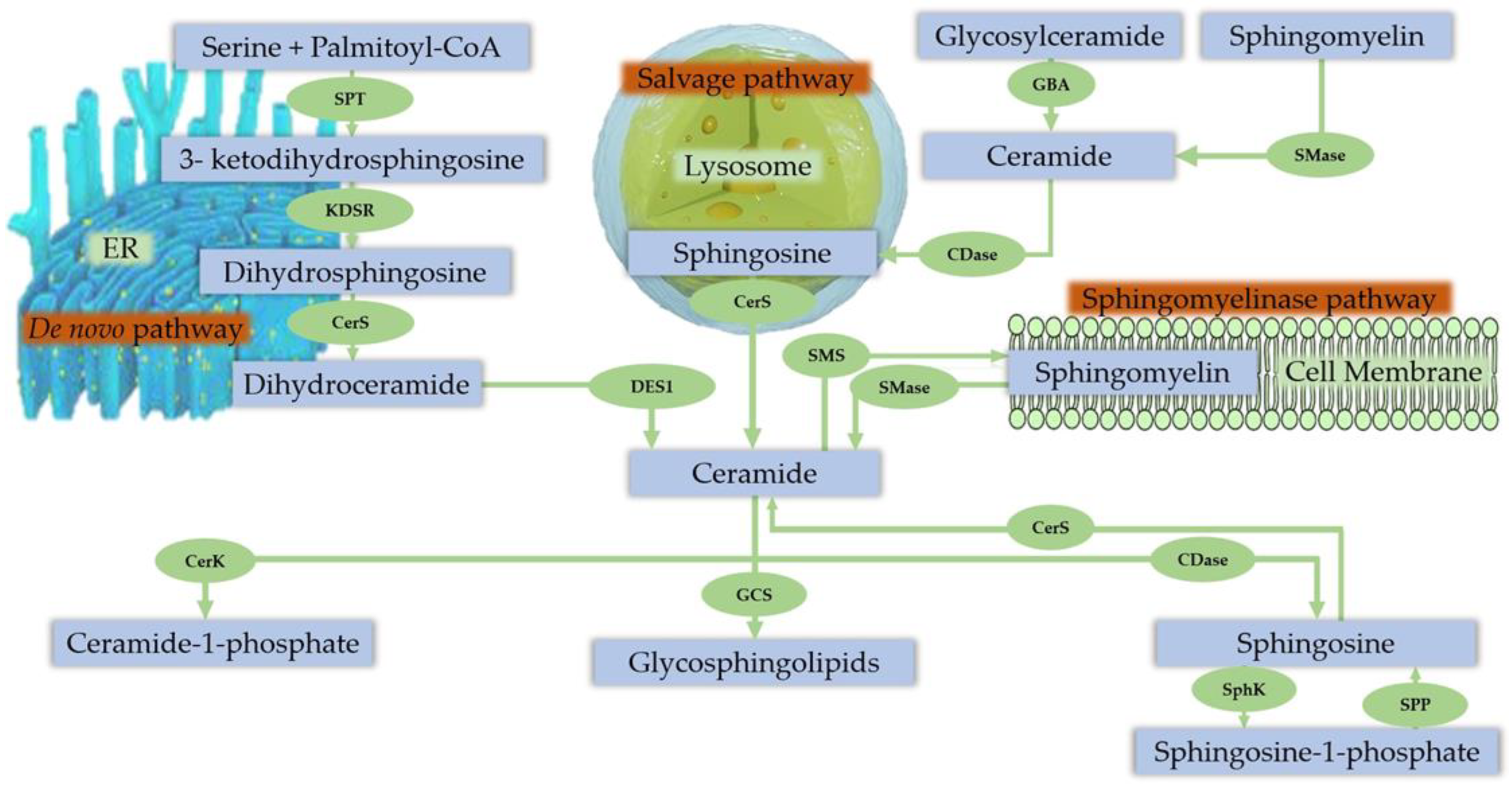
Platelet activating factor (PAF) is a glyceryl-ether phospholipid (1-O-alkyl-sn-glycero-3-phosphocholine) [25] and a potent inflammatory mediator that is involved in diverse physiological and pathological events including wound healing, inflammation, apoptosis, angiogenesis, anaphylaxis, reproduction, and fetal development [26,27]. While PAF’s primary role is to mediate intercellular interactions, it has been proposed to also have autocrine and intracrine effects [28]. There are two pathways that can lead to its synthesis: the remodeling and the de novo. The remodeling route, which is activated under inflammatory conditions and hypersensitivity responses, involves two steps. In the first step, the lyso-PAF intermediate is formed by the action of PLA2 on the membrane etherphosphatidylcholines mainly accompanied by the release of AA. In the second step, an acetyl-CoA:Lyso-PAF-acetyltransferase transfers an acetyl-group to the sn-2 position of the lyso-PAF in order to produce PAF [29]. The de novo pathway contributes to the non-pathological needs of PAF, for normal cellular function. The synthesis through this route involves three steps, catalyzed by three enzymes. In the first step, the acetyl-CoA:Alkylglycero-P-acetyltransferase transfers the acetyl residue onto an alkyl-lyso-glycerophosphate in order to form alkyl-acetyl-glycerophosphate. In the second step, the alkylacetylglycero-Phosphohydrolase catalyzes the dephosphorylation of alkyl-lyso-glycerophosphate to alkyl-acetyl-glycerol. The third and final step of the de novo pathway is performed by the CDP-Choline:Alkylacetylglycerol Cholinephosphotransferase that transfers CDP-choline to alkyl-acetyl-glycerol in order to form the PAF molecule [29].
PAF has a crucial role in specific syndromes and diseases, most of which are the result of increased PAF levels or of PAF-like activity due to impaired metabolism and/or to disbalanced oxidation process. PAF acetylhydrolases (PAF-Ahs) are the key enzymes in the regulation of its levels and activity. Three types of PAF-Ahs have been identified that belong in the PLA2 family of enzymes, one plasma known as Lipoprotein-associated PLA2 (Lp-PLA2) and two intracellular (types I and II). PAF-Ahs are involved in several pathological conditions including inflammation, cardiovascular disease, cancer and allergic diseases [26].

3.3. Sphingolipids

Sphingolipids (SL) were first described in 1884 by Thudichum [30] and several species have been discovered since then, defined by their sphingoid skeleton made up of 18 carbons with an amine and two alcohol groups [31,32]. SLs represent one of the major classes of eukaryotic bioactive phospholipids and are integral structural components of cell membranes, being responsible for several signaling pathways [17,33,34,35]. SLs and their metabolites, ceramides, sphingosine and sphingosine-1-phosphate (S1P) are involved in a plethora of cellular processes and functions, including cell growth, cell cycle, cell death, cell senescence, inflammation, immune responses, cell adhesion and migration, angiogenesis, nutrient uptake, metabolism, responses to stress stimuli and autophagy [17,34]. A mechanism named “sphingolipid rheostat” for explaining the opposite effects of S1P and ceramides was proposed by Cuvillier et al., in 1996 [36], suggesting that ceramides promote inflammatory and pro-apoptotic signaling, while accumulation of S1P promotes proliferation and cell survival. Later, Maceyka et al. in 2002 suggested that the relative and not the absolute amounts of these metabolites determine the result, backed-up by other authors, suggesting that this mechanism is crucial for several skeletal muscle functions [33,37,38,39,40,41].

Sphingolipid Metabolism in Skeletal Muscle

Ceramides, the central lipids for the SL biosynthesis, are mainly synthesized via three metabolic pathways [32,42] (Figure 2):
The de-novo synthesis pathway from saturated fatty acid, taking place in the endoplasmic reticulum (ER). This pathway begins with the connection of palmitate and serine that form 3-keto-dihydrosphingosine, a reaction catalyzed by the enzyme serine palmitoyltransferase (SPT), though SPT can also use other reactants producing different products [43,44]. The 3-keto-sphingosine is then reduced to dihydrosphingosine, which is acylated by ceramide synthases, producing dihydroceramide (Dcer). The final step of the ceramide synthesis is performed by the Dcer desaturase-1 (DES1), which desaturates the Dcer to ceramide. The ER-synthesized ceramide is immediately transported into the Golgi apparatus to generate SL via two types of ceramide transporters depending on the length of the FA chain (C14–20 or >C20) that is present in the ceramide.
The sphingomyelinase (Smase) pathway which converts the membrane boundsphingomyelin (SM) to ceramide.
The salvage/recycling pathway in the lysosome that produces sphingosine and ceramide after the breakdown of complex SLs and constitutes 50–90% of SL biosynthesis.
The synthesized ceramides can then follow two paths. They can be transformed into glycosphingolipids through the Golgi apparatus or the ER, producing glucosylceramides and then ceramide-1-phosphate (C1P) or galactosylceramides, respectively. Alternatively, ceramides can be deacylated by ceramidase (Cdase) to produce sphingosine which is then phosphorylated by sphingosine kinases to generate S1P [42]. C1P is synthesized through the phosphorylation of ceramide by the ceramide synthase.
There seems to be a connection between bioactive sphingolipids and the cytosolic PLA2–COX-2 pathway that generates bioactive prostaglandins. The Sphk1 and ceramide kinase 1, the enzymes responsible for the synthesis of S1P and C1P, respectively seem to be independently activated from cytokines, such as IL-1β and TNF-α and independently regulate the induction of COX-2 and the translocation/activation of cytosolic PLA2. Additionally, their products S1P and C1P also seem to have unique non-overlapping actions on COX-2 and cPLA2, respectively [17,45,46]. In addition, an interplay between PAF and bioactive sphingolipids may exist since acid sphingomyelinase is involved in PAF-mediated pulmonary edema and PAF is a potent stimulator of Smase activity [47].
Sphingosine-1-phosphate is one of the most active and most studied members of this lipid class and regulates a number of cellular processes including growth, differentiation, migration, proliferation, suppression of apoptosis and there is evidence of its involvement in the pathophysiology of several diseases [17,35]. Its degradation is regulated either by specific lipid phosphate phosphohydrolases that hydrolyze S1P to give sphingosine or by the S1P-lyase, splitting S1P into hexadecenal and phosphoethanolamine. It is a strong signal mediator that affects several intracellular functions, but it can also act as a ligand of the G protein-coupled receptors, called S1P receptors (S1PR) after being secreted into the extracellular environment [32,48]. S1P regulates several functions of the skeletal muscle including excitation–contraction coupling, satellite cell activity, and mitochondrial function. The current evidence suggests that the S1P levels in the circulation, muscle content and S1PR mRNA level increase after exercise, leading to fatigue resistance, increased mitochondrial respiration and biogenesis and activation of the satellite cells [35].

3.4. The Endocannabinoid System

The endocannabinoid system (ECS) is an endogenous signaling system that involves the encocannabinoids (eCBs) and their two transmembrane G-protein coupled receptors, known as cannabinoid receptors (CBRs) CB1 and CB2 (CB1R and CB2R, respectively). The CB1R while predominantly expressed in the brain structures, its presence has also been confirmed in the skeletal muscles, lungs, testes, gastrointestinal tract, pancreas and in adipocytes. The CB2R receptor is mainly expressed on the immune cells that are predominantly involved in inflammation and immune processes [49,50]. The eCB lipid ligands are biosynthesized on demand mainly from AA- containing phospholipid substrates of the cell membranes of most mammalian cells, such as neurons, adipocytes and skeletal muscle, in response to elevated cytoplasmic Ca2+ concentration. The two most studied eCBs are N-arachidonoylethanolamine (AEA) and arachidonoylglycerol (2-AG). AEA is a ligand for the CB1R while 2-AG is a ligand for both CB1 and CB2 receptors [49,50,51].
There are some studies that have investigated the influence of physical activity and/or exercise on the endocannabinoid signaling, though the mechanisms are still unclear. Physical activity depending on factors, such as type, frequency, intensity, duration, subject’s age and gender can lead to the activation of the ECS, increasing the levels of AEA and 2-AG in the circulation and altering the levels of the CBRs [50,51]. The CBS activation positively affects many systems and tissues, including the central nervous system (e.g., anti-depressive properties, memory and mood improvement, increased neurogenesis and neuroplasticity), cardiovascular system (e.g., reduction in blood pressure, increased endothelial function), respiratory system (bronchodilation), digestive system (e.g., increase in insulin sensitivity, reduction in insulin resistance, reduction in leptin resistance), as well as the function of adipocytes (e.g., increased lipolysis, reduced lipogenesis and increase in adiponectin). The effects of physical activity and exercise on CBS have been recently reviewed by Matei et al., in 2023 [52].

3.5. Exercise-Induced Muscle Damage

A muscle can contract in order to produce force by three ways: isometrically, concentrically and eccentrically. In an isometric contraction, the muscle retains its length while producing force, in a concentric contraction the muscle shortens while in eccentric contraction the muscle lengthens. Unaccustomed or high-intensity exercise can lead to the phenomenon of EIMD. EIMD is mainly induced by eccentric contractions, most probably because fewer motor units are recruited during an eccentric trial compared to the isometric or concentric contraction. Therefore, the mechanical stress on each motor unit is increased due to the increased force per unit. The pathophysiology of EIMD has been recently reviewed by Stožer et al. in 2020 [53]. In brief, the etiology EIMD comes down to the following factors:
Sarcomere overstretch, the structural and organizational disruption of the myofibrils while contracting due to increased force.
Imbalance in calcium homeostasis through the disruption of the T-tubule system due to membrane damage and uncontrolled Ca2+ release into the sarcoplasm. This increase leads to local injury contracture and increased passive muscle tension, resulting to the impairment of excitation–contraction coupling.
Disruption of the extracellular matrix and therefore the transmission of force between the exterior and the interior of the muscle cells, which possibly leads to the activation of several biochemical paths and the release of various growth factors and other molecules that guide the regeneration and adaptation to the damaging exercise. Moreover, this disruption also leads to increased permeability of the cell membrane allowing the release of different muscular proteins in plasma that are used as biomarkers of EIMD, such as creatine kinase (CK) and myoglobin (Mb) [53].
Inflammation follows immediately after the damaging exercise and is an important contributor to the EIMD. During the first 24 h after the exercise, there is a transient increase in neutrophil counts, followed by a transfer of fluid and plasma proteins into the damaged muscle tissue and the movement of other inflammatory cells [53]. This process is important for both the phagocytosis of the necrotic debris through the resident macrophages which are the key orchestrators of the inflammatory response [4,54], as well as the production of pro-inflammatory cytokines, such as IL-1β and TNF-α by the muscle cells that could persist for up to 5-days post-exercise. It is also a key mechanism in the differentiation of early macrophages through the pro-inflammatory M1 phenotype, to the anti-inflammatory M2 phenotype, in order to guide the regeneration and growth of the damaged muscle tissue [3,4,54].
Metabolic stress is induced through the depletion of glycogen stores and therefore low ATP levels which could lead to the activation of proteolytic and phospholipolytic pathways and the degradation of cytosolic proteins and membranes [53]. Other factors of metabolic stress could be the increased temperature and the produced reactive oxygen species that could damage several parts of the cells [53,55].

3.5.1. PUFA-Derived Lipid Mediators in EIMD Interventions in Humans

Numerous studies have used exercise as a stimulus in order to study their mechanisms and their molecular/biochemical pathways and interactions, as well as the effects of several supplements or drugs on their pathways. A large number of the studies has already been extensively reviewed by Markworth et al. [3] in 2016. In this review, we focused on the studies that provided enough data to prove that the exercise stimulus that was used, was effective in inducing EIMD on the human participants. Table 1 provides an overview of the relevant studies that were performed without any supplement and/or drug while Table 2 presents the studies where an intervention group with a supplement and/or drug is also included.
Most of the studies that provided adequate data, have used eccentric or resistance exercise as a method to induce EIMD on the subjects. In most of these cases, they noticed an increase in PGE2 with the selected muscle-damaging stimulus, immediately after exercise [10,56,57,58,59,60,61,62], in most cases peaking at 2 h after exercise. This increase was also followed by an elevation in PGF [10,56,57,63]. There are three cases where the PGE2 levels remained unaltered after the EIMD protocol [64,65,66]. In the first two, published by the same research group [64,65], the PGE2 levels did not change, despite the alterations on values, such as muscle soreness and CK indicating that the exercise stimulus that was used was effective in inducing muscle damage, though in one of them the exercise failed to reduce the maximum isometric force that the participants were able to produce, indicating that the EIMD wasn’t severe enough to alter that value [65]. In that same study, one group of volunteers were given piroxicam (20 g per day for 3 days). The administration of piroxicam did not alter the DOMS, exercise performance or the muscle enzyme leakage (CK and Mb) when compared to the control group [64]. In the third case, the two bouts of EC exercise on the elbow flexors were effective at altering markers of EIMD, such as muscle soreness, maximal isometric strength, CK and Mb after the first bout but not the second bout. However, PGE2 levels remained unaltered in both bouts [66].
One study used omega-3 fatty acid supplementation along with a placebo and control group [62]. The PGE2 levels were elevated after exercise in all groups, though this increase was attenuated in the omega-3 fatty acid supplemented group, a result that could be related to the strong anti-inflammatory properties of the supplement.
Fewer studies used aerobic exercise as an exercise stimulus. In a 42.2 marathon run, the researchers found an increase in the levels of PGE2, PGF and 6-keto-PGF1α 5 min after the trial. In another case, participants ran a 30 km distance and a 7.7-fold increase in PGE2 was recorded [59]. Finally, in a downhill running at 60% VO2peak, the EIMD caused an increase in PGF immediately after exercise for both the placebo and the American ginseng supplemented phase of this double-blind, randomized, crossover study, though the increase in the latter was not as intense [63].
There are a few cases where the researchers studied the COX enzymes in muscle biopsies [67,68,69] or in serum [70]. In the first case, the eccentric exercise stimulus that was used was able to elevate the COX-2 mRNA at 4 and 24 h after the exercise stimulus, but COX1b (b1,1, b1,2, b1,3) variants expression was low at any time point, remaining in very low abundance even when expressed, while the COX-1 (v1 and v2) variants mRNA that was most abundant in skeletal muscle remained unaltered by the exercise stimulus [67]. In two studies, COX-2 inhibitors were used. The researchers found an increased COX-2 mRNA when compared to the placebo groups, as an attempt to compensate for the anti-inflammatory blockade [68,69], though the protein levels of COX-2 remained unaltered [69]. The COX-1 levels remained unaffected by the COX-2 inhibitor, neither did its mRNA change after the exercise stimulus [68,69]. Finally, one more study measured the COX-2 levels in serum and noticed an increase after the isokinetic extension/flexion of the quadriceps muscles [70]. A 2019 study by Vella et al., used muscle biopsies in order to determine the levels of a large number of lipid mediators. The researchers found increased levels of PGE2, TXB2, 5,6-EpETrE, 11,12-DiHETrE, 14,15-DiHETrE and transiently increased levels of 12-Oxo LTB4, 20-COOH LTB4, 5-HETE, 12-HETE, 15-HETE, 4-HdoHE, 7-HdoHE, HdoHE, 2 h after the acute damaging resistance exercise of the m. vastus lateralis muscle [10].
Two studies used microdialysis as a method of PGE2 measurement [58,71]. In the first case, that studied the DOMS model, the microdialysis catheter was inserted in the muscle 24 h after the damaging exercise, and followed two pain stimulations of the exercised leg and the control leg. The PGE2 levels increased after the second pain stimulation for the exercised leg [58]. In the second study, the microdialysis catheter was inserted in the bicep muscle of the exercised arm and the control arm at 2–6 h after exercise and either 24 or 48 h. The PGE2 concentration did not differ in the exercised arm compared to the control arm [71].
Table 1. Human studies for PUFA-derived lipid mediators without drug/supplement intervention.
SubjectsDesign/InterventionBiological Samples/Time PointsVariablesEffect on Lipid Mediators and EIMD MarkersReference
10 ♂, healthy, moderately active (22 ± 0.4 years old) Intervention
Exercise: 8 × 5 maximal EC knee extensor and flexor at 60°/s angular velocity on both legs (eccentric and concentric trials)
Plasma/Serum: pre-ex,
10 min, 30 min, 24 h, 48 h post-exercise
PT (eccentric and concentric), DOMS
Plasma elastase, myeloperoxidase,
Serum CK, Mb,
Plasma PGE2
↔ PGE2
↑ CK post-ex for both concentric and eccentric. Peak CK at 48 h.
↑ CK in eccentric vs. concentric.
↑ DOMS in
Croisier et al., 1996 [64]
10 (9 ♂, 1 ♀) healthy, non-obese (25.7 years old) Exercise:
One side: 2 × 50 concentric/eccentric contractions of the calf muscles 24 h before the start of microdialysis (exercised leg)
Other side: Calf (untreated, control)
Biceps (normal saline)
Microdialysis 24 h after exercise for 80 min (legs and arms)
2 Pain stimulations during dialysis:
Legs (2 × 10 dorsal and plantar flexions of the foot, 10 min between sets).
Arms/biceps (normal saline and hypertonic saline injection, 5 × 200 μL 5.8 % NaCl, 2 min interval into the biceps muscle).
BC: pre-ex,
0 h, 24 h post-ex
Microdialysis: 20 min dialysate sample during pain stimulation on legs
Calf circumference, DOMS (legs and arms)
Serum: CK, Lactate
Microdialysis: PGE2, Nitric oxide, Substance P, Glutamate
No difference in PGE2 between exercised and control leg without pain stimulation
↑ PGE2 in exercised leg following second pain stimulation
↑ muscle soreness in the exercised leg pre-pains stimulation
↑ muscle soreness in exercised leg during pain stimulation
Tegeder et al., 2002 [58]
10 ♂, healthy, untrained (20.4 ± 2.0 years old) Intervention
Exercise: 2 bouts EC of the elbow flexors on the non-dominant arm separated by 4 weeks
Plasma: pre-ex,
6 h, 24 h, 48 h, 72 h, 96 h post-ex
IL-1β, IL-1ra, IL-4, IL-6, IL-8, IL-10, IL-12p40, TNF-α, G-CSF, MPO, HSP60 HSP70,
DOMS, Upper arm circumference (UAC), MIS (90°), ROM (FANG, RANG),
CK, Mb,
PGE2
↔ PGE2 after both bouts of exercise
↑ CK and Mb after first bout up to 4 days
↔ CK and Mb after second bout
↑ DOMS and UAC after first bout vs. second bout
↓ MIS and ROM after first bout vs. second bout
Hirose et al., 2004 [66]
12 ♂, healthy, recreationally active (28 ± 1.5 years old) Intervention
Exercise: Rebounds at 70% of maximal height until exhaustion
Serum: pre-ex,
0 h, 2 h, 2 d, 8 d post-ex
MIF (plantar flexor muscle), Leukocytes, CRP, IL-6, Substance P,
CK
PGE2
↑ PGE2 at 2 h post-ex. Complete recovery at 8-d
↑ CK post-ex (Peak at 2 d)
↓ MIF at 0 h and 2 d post-ex
Dousset et al., 2007 [60]
16 recreationally active (8 ♂: 26 ± 1 yr; 8 ♀: 24 ± 1 yr) Exercise: 10 × 10 EC with each leg at 120% of their concentric 1 RMMuscle biopsy: Pre-ex,
4 h, 24 h post-ex
Real-Time RT-PCR: COX-2
COX1b variants (COX-1b1, -1b2, and -1b3)
COX-1 (v1 and v2)
↑ COX-2 at 4 h and 24 h
↔ COX-1 and COX-1b variants
Weinheimer et al., 2007 [67]
40 ♂, army soldiers (19.1 ± 1.8 years old) Exercise: bench press at
50% 1-RM, 4 x ~20 (n = 8)
75% 1-RM, 5 x ~ 11 (n = 7)
90% 1-RM, 10 x ~4 (n = 7)
110% 1-RM, 8 x ~4 (n = 7)
control (no exercise, n = 6)
Plasma/Serum: pre-ex, 24 h, 48 h, 72 h post-exDOMS
IL-1β, IL-6, TNF-α,
CK
PGE2
↑ PGE2 for all exercise groups.
↑ PGE2 for the 110% group at 24 and 48 h vs. other groups.
↑ CK for all groups post-ex, peak at 24–48 h.
Correlation between peak DOMS and peak PGE2.
Correlation between peak CK and peak PGE2.
Uchida et al., 2009 [61]
17 ♀, healthy young (YW) (23.89 ± 2.03 years old)
Post-menopausal (PMW) (51.13 ± 5.08 years old)
5 × 6 maximal EC of the elbow flexorsSerum: pre-ex,
0 h, 24 h, 48 h, 72 h post-ex
Isometric Torque, DOMS, upper-arm circumference, ROM, IL-6, 10 IL-10, TNF-α,
CK,
PGE2
↑ PGE2 at 48 h and 72 h post-exercise in PWM vs. YW
Intra-groups PGE2 unaltered for both PWM and YW
↓ Isometric Torque and DOMS in 24 h, 48 h, 72 h post-ex in both PMW and YW
↔ Isometric Torque and DOMS in PMW vs. YW
↑ CK in 72 h post-ex in YW
↔ CK in PMW vs. YW
Positive correlation between age and PGE2
Negative correlations between age and DOMS at 48 h and 72 h post-ex
Conceição et al., 2012 [72]
8 ♂, young, resistance trained for 2 years, unaccustomed to bench press exercise 4 × 8 bench press EC at 70% of 1 RM.
2 bouts (2-week interval)
Serum: pre-ex,
24 h, 48 h, 72 h, 96 h post-ex
1 RM strength measurement, 1 RM eccentric strength measurement (1 Rmecc), DOMS,
CK,
PGE2
No significant interaction effect for PGE2
↑ peak plasma PGE2 at 48 and 72 h after first bout vs. second bout
↓ 1 RM at 24 h after first bout vs. second bout
↑ peak DOMS at 48 h after first bout vs. second bout
↑ CK at 72 h after first bout vs. second bout
Meneghel, Adilson J. 2014 [73]
12 ♂, untrained, recreationally active (18–25 years old) Acute bout of maximal ECC knee extensions, unilateralMuscle biopsy: pre-ex, 2 h, 4 h, 24 h post-exTXB2, 12(S) HHTrE, PGE2,
PGF, 15-Deoxy-Δ12,14-prostaglandin J3, 5-hydroperoxy-eicosatetranoic acid (5-HpETE), 12-Oxo-LTB4,
20-COOH-LTB4, 12-hydroxy-eicosatetraenoic acid (12-HETE), Tetranor 12-HETE,
12-hydroxy-eicosapentaenoic acid (12-HEPE), 15-hydroxy-eicosatetranoic acid (15-HETE), 15-hydroxy-eicosapentaenoic acid (15-HEPE), 4-hydroxy-docosahexanoic acid (4-HdoHE),
7-hydroxy-docosahexanoic acid (7-HdoHE), 14-hydroperxy-docosahexanoic acid (14-HpDoHE), 17-hydroxy-docosahexanoic acid (17-HdoHE), lipoxins (LXA4, LXB4, LXA5), E-series resolvins (RvE1 & RvE3)
D-series resolvins (RvD1, RvD2, RvD5, RvD6),
protectins (PD1 & 10S,17S-DiHDoHE),
maresins (MaR1),
epoxyeicosatrienoic acid (EpETrE),
dihydroxyeicosatrienoic acids (DiHETrEs),
CYP epoxidase enzyme products (9(10) -EpOME and 12(13) -EpOME,
9(10)-DiHOME and 12(13)-DiHOME)
TXB2 ↑ to 3.73 ng/g at 2 h from 1.12 ng/g at baseline.
12(S)-HHTrE ↑ to 13.50 ng/g at 2 h from 7.68 ng/g at baseline.
PGE2 ↑ to 2.84 ng/g at 2 h from 1.13 ng/g at baseline.
PGF ↑ to 1.20 ng/g at 2 h from 0.68 ng/g at baseline.
15-Deoxy-Δ12,14-prostaglandin J3 ↑ to 4.45 ng/g at 2 h from 3.09 ng/g at baseline.
5-HETE ↑ to 8.99 ng/g at 2 h from 3.38 ng/g at baseline.
12-Oxo-LTB4 and 20-COOH-LTB4 detected in resting muscle and ↑ at 2 h to 2.29 ng/g and 5.33 ng/g.
12-HETE to 63.81 ng/g ↑ at 2 h from 22.51 ng/g at baseline.
Tetranor 12-HETE ↑ to 3.97 ng/g at 2 h from 0.62 ng/g at baseline.
12 HEPE ↑ at 2 h after exercise.
7-HdoHE ↑ to 1.54 ng/g at 2 h from 0.98 ng/g at baseline.
14-HdoHE ↑ to 1.60 ng/g at 2 h from 0.68 ng/g at baseline.
5,6-EpETrE ↑ threefold to 14.12 ng/g at 2 h.
11,12-DiHETrE and 14,15-DiHETrE ↑ significantly at 2 h.
9(10)-and 12(13) -DiHOME were ↑ significantly at 2 h.
7-HdoHE ↑ to 1.54 ng/g at 2 h from 0.98 ng/g.
14-HdoHE ↑ to 1.60 ng/g at 2 h from 0.68 ng/g.
5,6-EpETrE ↑ 3-fold to 14.12 ng/g at 2 h.
11,12-DiHETrE and 14,15-DiHETrE ↑ at 2 h.
9(10)-and 12(13)-DiHOME ↑ at 2 h.
Vella et al., 2019 [10]
↑ Increased, ↓ Decreased, ↔ Unaltered/Not significantly altered/Not different, ♂: Males, ♀: Females, 1 RM: 1 Repetition Maximum, COX: Cyclooxygenase, CC: Concentric Contractions, CK: Creatine Kinase, CRP: C-Reactive Protein, DOMS: Delayed Onset Muscle Soreness, EC: Eccentric Contractions, ECC: Eccentric/Concentric Contractions, FANG: Flexed Angle, IL: Interleukin, Mb: Myoglobin, MIF/MIS/MIT: Maximal Isometric Force/Strength/Torque, PG: Prostaglandin, PT: Peak Torque, ROM: Range of Motion, RANG: Relaxed Angle, TNF-a: Tumor Necrosis Factor alpha.
Table 2. Human studies for PUFA-derived lipid mediators with drug/supplement intervention.
SubjectsDesign/InterventionBiological Samples/Time PointsVariablesEffect on Lipid Mediators and EIMD MarkersReference
20 ♂, college aged Randomized placebo-controlled trial
Aspirin (ASP, n = 10) (3 g/day) or PLA (n = 10) for 4 days, 1-day prior exercise.
Exercise: 3 × 10 reps squat (70% of 1 RM).
Plasma/Serum: pre-ex,
24 h, 48 h, 72 h post-ex
Muscle Soreness, Salicylate,
CK,
PGF, PGE2
↑ PGF and PGE2 at 24 h returning to baseline at 72 h for PLA
↔ PGF and PGE2 at ASP
↑ CK post-ex in ASP and PLA
↔ CK between ASP and PLA
No effect on muscle soreness
Boatwright et al., 1991 [56]
10 ♂, healthy, moderately active (22 ± 0.4 years old) Crossover placebo-controlled with piroxam (20 mg piroxicams/day, 3 days pre-ex)
Exercise: 8 × 5 maximal EC knee extensor and flexor at 60°/s angular velocity on both legs
Plasma/Serum: pre-ex,
10 min, 30 min, 24 h and 48 h after exercise
DOMS, PT,
Serum CK, Mb,
Plasma PGE2
↓ PGE2 at rest and after exercise in piroxam group
↓ PGE2 in piroxam vs. PLA
↔ PGE2 over time for both groups
No effect of piroxicam in DOMS, PT, CK and Mb
Croisier et al., 1996 [65]
24 ♂, recreationally active Double blind, placebo-controlled trial
Placebo (PLA, n = 8)
Ibuprofen (IBU 1200 mg, n = 8)
Acetaminophen (ACET, n = 8)
Exercise: Unilateral high intensity eccentric exercise 2 days after and ~24 h before muscle biopsies
Muscle biopsies: 2 d pre-ex, 24 h post-exPGE2, PGF↑ PGF in PLA (+77%) vs. IBU (−1%) and ACET (−14%)
↑ PGE2 in PLA (+64%) vs. ACET (−16%)
Trappe et al., 2001 [57]
34 ♂, athletes (25.5 ± 3.2 years old) Double-blind trial
CR (4 × 5 g creatine monohydrate + 60 g maltodextrine, n = 18)
PLA (carbohydrate, n = 16) 5 days.
Exercise: 30 km run
Plasma: 15 min pre-ex,
24 h post-ex
TNF-α, CK, LDH, Creatinine, PGE2↑ PGE2 in PLA and CR post-ex
↑ PGE2 in PLA vs. CR post-ex
↑ CK and TNF-α in PLA and CR post-ex
↑ LDH in PLA post-ex
↔ LDH in CR post-ex
Santos et al., 2004 [59]
29 ♂, recreationally active (22.55 ± 4.42 years old) Randomized, double-blinded, and placebo-controlled (cellulose) or proteolytic supplement (fungal proteases, bromelain, and papain, SUP).
Exercise: 3 × 5 Isokinetic extension/flexion of the quadriceps
Downhill running for 45 min at 60% VO2max 21 d after supplementation
Serum: baseline, pre-ex,
0 h, 3 h, 24 h, 48 h post-ex
PT, DOMS
CK, IgG, IgA, IgM, IL-6, IL-8, IL-10, IL-12, TNF-α, IL-1β, SOD, Circulating Leucocytes,
PGE2, 8-isoprostane (8-iso), COX-2
↑ COX2 at PLA vs. SUP
group × time interaction for COX2
↔ PGE2 between SUP and PLA
↔ PT and DOMS between SUP and PLA
↔ CK between SUP and PLA
Buford et al., 2009 [70]
16 ♂, recreationally active (23 ± 1 years old) Randomized placebo-controlled
COX-2 inhibitor (celecoxib 600 mg/d, n = 8)
Placebo (n = 8)
Exercise: 10 × 10 repetitions of high-intensity eccentric knee extensions with each leg at 120% of their concentric 1 RM
[2H5]phenylalanine infusion at arrival
Muscle biopsy: 2 h and 5 h
BC: 2.5 h, 3.5 h, 4.5 h
Fractional synthesis rate,
mRNA levels of COX-1 (1v1, 1v2) and COX-2,
protein levels of COX-1 (1v1, 1v2) and COX-2
↑ COX-2 mRNA (3.0 ± 0.9-fold) at COX-2 inhibitor group vs. PLA
↔ COX-1 mRNA and COX-1 protein levels in both groups
Burd et al., 2010 [68]
33 ♂, ♀, healthy, physically active (18–33 years old) Randomized double-blind, placebo-controlled with celecoxib (400 mg/d, CEL), 3-week wash-out period
Exercise: 14 × 5 eccentric unilateral contractions of the elbow flexors
Microdialysis in exercised arm and non-exercised arm (control) after the first bout.
Muscle biopsy: 1 h, 2 h, 4 h and 7 d after bout 1 and 1 h and 2 d after bout 2 (n = 24).
Serum: 1 h pre-ex,
1 h, 8 h, 20 h, 1 d, 2 d, 3 d, 4 d, 7 d, 9 d post-ex
Microdialysis: 2–6 h post-ex and either 24 h (n = 5) or 48 h (n = 5) post-ex.
PT (isometric and concentric), RANG, DOMS
Leucocyte number (CD68+), Number of satellite cells (CD56+),
CK (serum)
PGE2 (microdialysis)
↔ PGE2 after first bout in both CEL and PLA
↑ CK after first bout but not second bout.
↔ CK between CEL and PLA
↓ DOMS on CEL vs. PLA after both bouts
↓ PT (isometric and concentric) after both bouts in both CEL and PLA.
↔ PT (isometric and concentric) between CEL and PLA
Paulsen et al., 2010 [71]
45 ♂, healthy, untrained (29.7 ± 6.6 years old) Randomized, double-blinded, repeated measures trial
Groups: experimental (Omega-3 fish oil + 100 IU of d-a-tocopherol/dl-a-tocopherol acetate, EXP, n = 15)
PLA (soybean/corn oil mixture + 100 IU of d-a-tocopherol/dl-a-tocopherol acetate, n = 15)
Control (100 IU of d-a-tocopherol/dl-a-tocopherol acetate, CTL, n = 15)
Exercise: Bench stepping exercises with eccentric patterns for 40 min (5 min stepping, 1 rest).
Plasma: baseline, pre-ex,
0 h, 24 h, 48 h post-ex
IL-6, TNF-a,
CK, LDH, Mb,
PGE2
↑ PGE2, CK, LDH, Mb for all groups after exercise
↓ PGE2, CK, LDH, Mb in EXP vs. PLA or control
↓ PGE2 levels post-ex, 24 h and 48 h for EXP vs. PLA and control
↓ PGE2 levels pre-ex vs. baseline in EXP
↓ elevation of PGE2 post-ex, 24 h, 48 h for EXP
Tartibian et al., 2011 [62]
8 ♂, healthy (23 ± 3 years old Indomethacin (NSAID) microdialysis infusion on vastus lateralis of one leg.
Placebo microdialysis infusion on vastus lateralis of the other leg.
Exercise: 200 eccentric contractions in each leg (100 at 30°/s and 100 at 120°/s).
Muscle biopsy: Pre-ex,
5 h, 24 h, 28 h and 8 d post-ex
RNA extraction
Real-time PCR
Growth factor genes, extracellular matrix-related genes, PGC1α, PPARγ, MCP1
Gene expression of COX-1 and COX-2
↑ COX-2 expression (6-fold) at 5 h post-ex in NSAID vs. PLA
↔ COX-1 expression in both groups over time
↔ COX-1 expression NSAID vs. PLA
Mikkelsen et al., 2011 [69]
14 ♂, physically active (22.4 ± 1.7) Randomized, double-blinded, crossover, placebo-controlled with American ginseng supplement
30 day-supplementation with 1600 mg/d American ginseng extract (AG) or 1600 mg/d hydroxymethylcellulose (PLA)
Exercise: Downhill running at 60% VO2peak
Plasma: pre-supplementation, pre-ex,
0 h, 2 h, 24 h, 48 h and 72 h post-ex.
DOMS, TNF-α,
IL-1β, IL-4, IL-10,
CK,
8-iso-PGF
↑ 8-iso- PGF at 0 h post-ex vs. pre-ex in AG and PLA
↓ 8-iso- PGF at 0 h, 2 h and 24 h post-ex in AG vs. PLA
↑ CK at 0 h, 2 h, 24 h, 48 h, 72 h post-ex in PLA. Peak at 24 h
↑ CK at 0 h, 2 h, 24 h post-ex in AG. Peak at 24 h.
↑ DOMS at 0 h, 2 h, 24 h, 48 h, 72 h post-ex.
↔ DOMS between AG and PLA.
Lin et al., 2021 [63]
↑ Increased, ↓ Decreased, ↔ Unaltered/Not significantly altered/Not different, ♂: Males, ♀: Females, 1 RM: 1 Repetition Maximum, COX: Cyclooxygenase, CC: Concentric Contractions, CK: Creatine Kinase, DOMS: Delayed Onset Muscle Soreness, EC: Eccentric Contractions, Ig: Immunoglobulin, IL: Interleukin, LDH: Lactate Dehydrogenase, Mb: Myoglobin, MIF/MIS/MIT: Maximal Isometric Force/Strength/Torque, PG: Prostaglandin, PLA: Placebo, PT: Peak Torque, ROM: Range of Motion, RANG: Relaxed Angle, SOD: Superoxide Dismutase, TNF-a: Tumor Necrosis Factor alpha.

3.5.2. PUFA-Derived Lipid Mediators and EIMD in Animal and In-Vitro Studies

The animal and in vitro studies that focused on the PUFA-derived lipid mediators are limited and they are presented in Table 3.
Only one study with animals met the set criteria. The researchers focused on the effect of preconditioning contractions on muscle pain markers. The left leg of the Wistar rats was exercised 2 days before the EC protocol and the right leg was used as a control, receiving only the damaging EC stimulus. The COX-2 mRNA of the gastrocnemius muscle showed increased levels in both groups after the EC exercise, though the elevation was higher in the non-preconditioned leg and persisted through day 4 after the damaging EC only in that leg [74].
In the in vitro study, avian skeletal muscle cells have been mechanically stimulated for 48 h. PGE2 and PGF efflux rates increased within 4–5 h of stimulation, by 97% and 41%, respectively, while PGF efflux continued to increase up to 48 h. Ιndomethacin partially blocked stretch-induced cell damage indicating that this process cannot be totally attributed to prostaglandins [75].
Table 3. Animal and in vitro studies for PUFA-derived lipid mediators.
Animal ModelProtocolSampleVariablesEffect on Lipid Mediators and EIMD MarkersReference
36 ♂ Wistar rats (9-weeks old)Exercise: 100 repeated EC on plantar flexor muscles of left leg.
Right leg as control.
Precon group (n = 18): 10 repeated EC, 2 days pre-ex.
Non-Precon group (n = 18): no EC pre-ex
Plantar flexor muscle at 0 d, 2 d, 4 d post-exHistochemical analysis
mRNA levels: HGF, Pax7, MyoD, myogenin, BKB2, COX-2
↑ COX-2 mRNA at 2 d post-ex for both Precon and Non-Precon
↑ COX-2 mRNA at 4 d in non-Precon
↑ COX-2 mRNA in non-Precon vs. Precon
Nagahisa et al., 2018 [74]
Avian myoblastsMechanical stimulation: 5 × 20% substratum stretches and relaxations/20 s. 10 s rest. 3 times. (5 h, 24 h, 48 h)-Protein synthesis and degradation rate,
Proteinase,
CK,
PGE2, PGF, 6-keto-PGF
↑ PGE2 and PGF efflux by 97 and 41%, respectively within 4–5 h of mechanical stimulation.
↔ 6-keto-PGF1α
Vandenburgh et al., 1990 [75]
↑ Increased, ↓ Decreased, ↔ Unaltered/Not significantly altered/Not different, ♂: Males, COX: Cyclooxygenase, EC: Eccentric Contractions, ECC: Eccentric/Concentric contractions, HGF: Hepatocyte grown factor.

3.5.3. PAF and EIMD Interventions in Humans

One study measured the free and bound PAF levels. Milias et al. [76] used an eccentric exercise protocol of 6 sets of 6 repetition on the elbow flexors of the non-dominant hand of 13 healthy young males. An increase of free PAF in the circulation was observed at 48 h after the exercise while the bound PAF increased at 24 and 48 h. The total PAF levels were increased 24, 48 and 72 h after the eccentric exercise. The researchers also found a positive association between PAF levels and CK at 48 h, and an inverse correlation with maximal isometric torque at 48, 72 and 96 h, and arm circumference at 48 h, indicating that the muscle damage is influenced by the PAF increase after eccentric exercise.

3.5.4. Human Studies with Sphingolipid-Derived Lipid Mediators

Only one study on humans met the set criteria. In the study participated seven middle aged male runners in a 48-h ultramarathon race and the researchers measured the ceramides, sphingosine, sphinganine, S1P, SA1P levels before the run, at 24 h during the run and at 3 time points after the completion of the race. The levels of S1P, SA1P, sphingosine and sphinganine in plasma were decreased at 24 h during the marathon race and further reduced until 48 h after its completion, during the recovery period with minimum values at 24 h of recovery. The levels of sphingosine and S1P in erythrocytes were increased at 48 h of recovery and SA1P at 24 h of running. The ceramide levels were not altered during or after the completion of the ultramarathon race [77].

3.5.5. Animal Studies with Sphingolipid-Derived Lipid Mediators

Two studies measured sphingolipid-derived lipid mediators on animals (Table 4). Sphinganine and sphingosine levels were determined in different skeletal muscle types of male Wistar rats after two types of acute exercise, run until exhaustion and sciatic nerve stimulation with tetanic pulses. The results demonstrated that prolonged exercise increased sphinganine of approximately 6-fold in each muscle while sphingosine increased over 3-fold in the soleus and nearly 2-fold in the red and white sections of the gastrocnemius, implying that both de novo synthesis and catabolism of ceramide in skeletal muscles is activated during acute exercise [78]. Lee et al., measured the protein levels of key enzymes of the sphingolipid metabolism in gastrocnemiusmuscle biopsies of male mice. The researchers found increased levels of the acidic sphingomyelinase (A-Smase) immediately and 24 h after the treadmill running exercise they performed, but the neutral sphingomyelinase (N-Smase) remained unchanged. The SPT-1 remained unaltered. The authors concluded that the EIMD could contribute to the cellular ceramide production through the increased A-Smase, a key rate-limiting enzyme of the de novo synthesis, the suppression of which could alleviate the skeletal muscle damage in response to intensive exercise. Ceramides contribute to a number of biological processes and act as secondary messengers of the inflammatory response, promoting apoptosis by activating caspase-3 [79].
Table 4. Animal studies for Sphingolipid-derived lipid mediators.
Animal ModelStimuliSampleAssayEffect on Lipid Mediators and EIMD MarkersReference
30 ♂ Wistar rats3 groups:
1-Control (resting).
2-Exercise until exhaustion.
3-gastrocnemius muscle contraction through sciatic nerve stimulation
Electrical stimulation of sciatic nerve
Muscle samples: Soleus.
Red (slow-twitch oxidative fibers) and white (fast twitch glycolytic fibers) section of the gastrocnemius muscle
Sphinganine, sphingosine↑ sphinganine in soleus vs. red gastrocnemiusat rest.
↑ sphinganine in red gastrocnemiusvs. white gastrocnemiusat rest.
↑ sphinganine (~6-fold) in each muscle after prolonged exercise.
↑ sphingosine in soleus and red gastrocnemius s white gastrocnemius.
↑ sphingosine (3-fold) in the soleus and red and white gastrocnemius (~2-fold) after prolonged exercise.
Dobrzyń et al., 2002 [78]
Mice ♂ (7-week-old)Treadmill running at 5 m/min for 5 min, increasing to 10 m/min for 5 min, 15 m/min for 5 min, 20 m/min for 10 minSerum and
gastrocnemius muscle (white and red portion) at rest, 0 h and 24 h post-ex
IL-6, caspase-3,
Protein levels of Serine palmitoyltransferase-1 (palmitoyltransferase-1 (SPT-1),
acidic sphingomyelinase (A-Smase), neutral sphingomuelinase (N-Smase),
Serum CK
↑ levels of A-Smase at 0–24 h
↔ N-Smase levels
↔ SPT-1 levels
↑ CK at 24 h post-ex
Lee et al., 2019 [79]
↑ Increased, ↔ Unaltered/Not significantly altered/Not different, ♂: Males.

4. Synopsis

The majority of the studies focused on the prostaglandins PGE2 and PGF, mostly as markers of acute inflammation and their involvement in the perception of pain but also for their role in muscle regeneration and hypertrophy after damaging exercise. COX-2 enzyme is highly involved in muscle pain and the administration of COX-2 inhibitors before the lengthening contractions can reduce the muscle pain in animal models [80,81] and reduced DOMS in humans though it had no effect on recovery of muscle function after eccentric exercise of elbow flexors [71]. However, there is a case where piroxicam administration had no effect on DOMS after the EIMD protocol [65]. COX-2 is the key enzyme in the production of prostaglandins. An increase in the PGE2 levels also increases the sensitivity of the type III and IV afferent pain neurons. Due to edema from EIMD, movement or external palpation could further increase the small rises in intramuscular pressure leading to hypersensitivity of the pain receptors to the mechanical stimulus [80]. PGF plays an important role in muscle protein synthesis and hypertrophy [82,83]. Studies [56,57] found increased levels of the circulating PGF in the first 24 h of recovery and administration of non-steroidal anti-inflammatory drugs (NSAIDs) seems to block the increase in protein synthesis.
The understanding of PGs in EIMD remains incomplete. The PG response after eccentric exercise appears with an increase in their levels within 2 h after the damaging stimulus [60,63] lasting for up to 3 days [56,61,62,73] which comes in contrast to few studies that did not detect any significant changes in the PG levels [64,65,66,72] after the damaging exercise. These opposing results could be due to insufficient intensity of the exercise since it is suggested that greater mechanical stress is associated with greater increase in PGE2 levels [61].
The complete profile of PUFA derived lipid mediators of human skeletal muscle tissue after resistance exercise has been studied only recently by Vella et al. in 2019 [10]. Within the first 2 h of recovery after resistance exercise an increase was noticed in the classic AA-derived PGs, as well as the TXA2 production (through TXB2 and 12(S)-HHTrE) within the muscle tissue which is in agreement with previous results of increased TXB2 after aerobic and resistance exercise. 5-LOX derived LTB4 is usually measured in the circulation after exercise and was at low levels in the skeletal muscle tissue before the exercise stimulus, since 5-LOX’s is limited in bone marrow-derived cells though its levels increased 2 h after exercise. Similar to 5-LOX, the 12-LOX metabolites, 12-HETE, increased in the skeletal muscle tissue after exercise. Both LTB4 and 12-HETE and its downstream derivate tetranor 12-HETE are pro-inflammatory lipid mediators with chemotactic properties for PMNs [3,10]. The 15-LOX product, 15-HETE also increased within the muscle biopsies 2 h after exercise, but the lipoxins LXA4 and LXB4 that constitute its products were undetectable. The authors suggested that exercised myofibers could release lipoxins contributing to the systemic lipoxin response, also supported by previous findings by the same research group [10,84]. There was also a noticeable increase in the AA derived products of the CYP pathway 2 h after exercise, namely epoxyeicosatrienoic acid regioisomer 5-6-EpETrE and dihydroxyeicosatrienoic acids 11,12- and 14,15-DiHETrE, yet their role in skeletal muscle and EIMD remains unknown.
PAF is a potent inflammatory lipid mediator that besides its other roles, seems to be involved in the EIMD phenomenon, but is not yet studied thoroughly. The mechanism behind the increase in PAF levels could be hypothesized through the effects of muscle damage on the intracellular Ca2+ levels. PAF’s production during inflammatory conditions depends on the action of phospholipase A2. PLA2 is a Ca2+ dependent enzyme and as previously stated the Ca2+ levels increase due to EIMD. Thus, the increased activity of PLA2 could lead to increased free PAF in the circulation or bound PAF on the cell membranes [53,76]. In addition, PAF is reported to decrease skeletal muscle protein synthesis in animal studies [85] while Lp-PLA2 mutations in male Caucasians have been associated with a greater increase in lean muscle mass following training.
The studies that measured sphingolipid-derived lipid mediators after damaging exercise are limited both in humans, animal models and in vitro. The only human study that proved that EIMD was present, was conducted on male runners that performed a 48-h ultramarathon race. Since the subjects of the study were adapted to this type of exercise, only an extremely long 48-h run would be able to increase EIMD markers. The researchers found reduced levels of S1P during the marathon race and in the recovery period in plasma. The main source of plasma S1P is erythrocytes, although they are unable to synthesize sphingolipids de novo and this could be the reason behind the reduction in its levels. Erythrocytes use plasma sphingosine in order to produce S1P via phosphorylation and in that study the S1P and sphingosine levels in erythrocytes were stable throughout the exercise period with an increase at 48 h of recovery indicating that the synthesis in the erythrocytes met the requirements of the exercise [77]. S1P has a broad range of biological effects. In skeletal muscle particularly, there is evidence that sphingosine is able to reduce fatigue after its conversion to S1P, specifically by SphK in murine extensor digitorum longus muscle, positively affecting the excitation–contraction coupling [37]. Sphingosine, however, inhibits calcium release from the sarcoplasmic reticulum in skinned skeletal muscle fibers and murine skeletal muscle, having a direct effect on the ryanodine receptor which is responsible for the release of Ca2+ from intracellular stores during excitation–contraction coupling in both cardiac and skeletal muscle [86,87]. Since S1P and sphingosine have opposing effects on excitation–contraction coupling, the evidence points towards the importance of balance between them for normal contractile function [35]. Contrary to the results of that study, the majority of studies that used exercise noticed an increase or no alteration in the levels of S1P either in plasma or muscles, though the exercise stimulus varied and no data on EIMD markers were presented [35].

5. Limitations and Future Perspectives

An overview of the involvement of lipid mediators in the mechanisms of exercise-induced muscle damage is presented in this review. Although a significant number of human studies have dealt with EIMD, specific limitations are identified. Almost all studies concern young male population with a limited number of participants. Furthermore, each of these studies focuses on a specific class of lipid mediators without considering the changes that may occur in the other classes, as well as the possible cooperation of these mediators. There are also very few studies that have been performed with in vitro models or with knockout animals, that can provide more information on the mechanisms underlying EIMD. In addition, since first-phase inflammatory lipid mediators promote a shift to anti-inflammatory and pro-resolving mediators, classical anti-inflammatory treatments, such as non-steroidal anti-inflammatory drugs (NSAIDs) and/or blockade of enzymes activity implicated in the inflammatory phase should be replaced with carefully designed molecules that promote the M2 phenotype and the synthesis of pro-resolving lipid mediators. Lastly, since myalgias and generalized weakness have been reported to occur in one-quarter to one-half of symptomatic patients with COVID-19 [88] and it has also been reported that phospholipids were the most important family of compounds dysregulated both in the case of acute and post-COVID phase [89], common biochemical pathways between EIMD and musculoskeletal consequences of COVID-19 may exist [90].

6. Conclusions

In conclusion, the response of injured muscle during unaccustomed or high-intensity exercise shares common features with the inflammatory response of other tissues to trauma leading to synthesis of pro-inflammatory, as well as pro-resolving lipid mediators that are synthesized in a different time course. Since these bioactive lipids exert autocrine/paracrine action, further ex vivo studies, as well as studies performed with skeletal muscle biopsies and/or with knockout animals are needed to elucidate the role of skeletal muscle in lipid mediators’ metabolic pathways during unaccustomed or high-intensity exercise.

Author Contributions

Conceptualization, T.N., A.P. and S.A.; methodology, A.G.G.; investigation, A.G.G.; writing—review and editing, A.G.G., T.N., A.P. and S.A.; supervision, T.N. and S.A.; All authors have read and agreed to the published version of the manuscript.

Funding

This research received no external funding.

Informed Consent Statement

Not applicable.

Data Availability Statement

The data availability for this review is primarily based on publicly accessible information. No original data or specific datasets were collected or analyzed during the preparation of this review.

Conflicts of Interest

The authors declare no conflict of interest.

References

  1. Headland, S.E.; Norling, L.V. The Resolution of Inflammation: Principles and Challenges. Semin. Immunol. 2015, 27, 149–160. [Google Scholar] [CrossRef] [PubMed]
  2. Brennan, E.; Kantharidis, P.; Cooper, M.E.; Godson, C. Pro-Resolving Lipid Mediators: Regulators of Inflammation, Metabolism and Kidney Function. Nat. Rev. Nephrol. 2021, 17, 725–739. [Google Scholar] [CrossRef] [PubMed]
  3. Markworth, J.F.; Maddipati, K.R.; Cameron-Smith, D. Emerging Roles of Pro-Resolving Lipid Mediators in Immunological and Adaptive Responses to Exercise-Induced Muscle Injury. Exerc. Immunol. Rev. 2016, 22, 110–134. [Google Scholar]
  4. Arnold, L.; Henry, A.; Poron, F.; Baba-Amer, Y.; van Rooijen, N.; Plonquet, A.; Gherardi, R.K.; Chazaud, B. Inflammatory Monocytes Recruited after Skeletal Muscle Injury Switch into Antiinflammatory Macrophages to Support Myogenesis. J. Exp. Med. 2007, 204, 1057–1069. [Google Scholar] [CrossRef] [PubMed]
  5. Feehan, K.T.; Gilroy, D.W. Is Resolution the End of Inflammation? Trends Mol. Med. 2019, 25, 198–214. [Google Scholar] [CrossRef]
  6. Metsios, G.S.; Moe, R.H.; Kitas, G.D. Exercise and Inflammation. Best Pract. Res. Clin. Rheumatol. 2020, 34, 101504. [Google Scholar] [CrossRef]
  7. Bernard, C.; Zavoriti, A.; Pucelle, Q.; Chazaud, B.; Gondin, J. Role of Macrophages during Skeletal Muscle Regeneration and Hypertrophy-Implications for Immunomodulatory Strategies. Physiol. Rep. 2022, 10, e15480. [Google Scholar] [CrossRef]
  8. Deslandes, A.; Moraes, H.; Ferreira, C.; Veiga, H.; Silveira, H.; Mouta, R.; Pompeu, F.A.; Coutinho, E.S.; Laks, J. Exercise and Mental Health: Many Reasons to Move. Neuropsychobiology 2009, 59, 191–198. [Google Scholar] [CrossRef]
  9. Silveira, L.S.; Antunes, B.M.; Minari, A.L.A.; dos Santos, R.V.T.; Neto, J.C.R.; Lira, F.S. Macrophage Polarization: Implications on Metabolic Diseases and the Role of Exercise. Crit. Rev. Eukaryot. Gene Expr. 2016, 26, 115–132. [Google Scholar] [CrossRef]
  10. Vella, L.; Markworth, J.F.; Farnfield, M.M.; Maddipati, K.R.; Russell, A.P.; Cameron-Smith, D. Intramuscular Inflammatory and Resolving Lipid Profile Responses to an Acute Bout of Resistance Exercise in Men. Physiol. Rep. 2019, 7, e14108. [Google Scholar] [CrossRef]
  11. Methenitis, S.; Stergiou, I.; Antonopoulou, S.; Nomikos, T. Can Exercise-Induced Muscle Damage Be a Good Model for the Investigation of the Anti-Inflammatory Properties of Diet in Humans? Biomedicines 2021, 9, 36. [Google Scholar] [CrossRef] [PubMed]
  12. Sinzinger, H.; Virgolini, I. Effects of Exercise on Parameters of Blood Coagulation, Platelet Function and the Prostaglandin System. Sport. Med. 1988, 6, 238–245. [Google Scholar] [CrossRef]
  13. Gleeson, M.; Bishop, N.C.; Stensel, D.J.; Lindley, M.R.; Mastana, S.S.; Nimmo, M.A. The Anti-Inflammatory Effects of Exercise: Mechanisms and Implications for the Prevention and Treatment of Disease. Nat. Rev. Immunol. 2011, 11, 607–615. [Google Scholar] [CrossRef] [PubMed]
  14. Allen, J.; Sun, Y.; Woods, J.A. Chapter Fourteen—Exercise and the Regulation of Inflammatory Responses. In Progress in Molecular Biology and Translational Science; Bouchard, C., Ed.; Academic Press: Cambridge, MA, USA, 2015; Volume 135, pp. 337–354. ISBN 1877-1173. [Google Scholar]
  15. Cerqueira, É.; Marinho, D.A.; Neiva, H.P.; Lourenço, O. Inflammatory Effects of High and Moderate Intensity Exercise—A Systematic Review. Front. Physiol. 2020, 10, 1150. [Google Scholar] [CrossRef] [PubMed]
  16. Innes, J.K.; Calder, P.C. Omega-6 Fatty Acids and Inflammation. Prostaglandins Leukot Essent Fat. Acids 2018, 132, 41–48. [Google Scholar] [CrossRef] [PubMed]
  17. Hannun, Y.A.; Obeid, L.M. Sphingolipids and Their Metabolism in Physiology and Disease. Nat. Rev. Mol. Cell Biol. 2018, 19, 175–191. [Google Scholar] [CrossRef]
  18. Capra, V.; Rovati, G.E.; Mangano, P.; Buccellati, C.; Murphy, R.C.; Sala, A. Transcellular Biosynthesis of Eicosanoid Lipid Mediators. Biochim. Biophys. Acta 2015, 1851, 377–382. [Google Scholar] [CrossRef]
  19. Christie, W.W.; Harwood, J.L. Oxidation of Polyunsaturated Fatty Acids to Produce Lipid Mediators. Essays Biochem. 2020, 64, 401–421. [Google Scholar] [CrossRef]
  20. Serhan, C.N. Treating Inflammation and Infection in the 21st Century: New Hints from Decoding Resolution Mediators and Mechanisms. FASEB J. 2017, 31, 1273–1288. [Google Scholar] [CrossRef]
  21. Serhan, C.N. Discovery of Specialized Pro-Resolving Mediators Marks the Dawn of Resolution Physiology and Pharmacology. Mol. Asp. Med. 2017, 58, 1–11. [Google Scholar] [CrossRef]
  22. Vidar Hansen, T.; Serhan, C.N. Protectins: Their Biosynthesis, Metabolism and Structure-Functions. Biochem. Pharmacol. 2022, 206, 115330. [Google Scholar] [CrossRef] [PubMed]
  23. Gireddy, H.B.; Rajaram, H.; Koduganti, R.R.; Ambati, M.R.A.; Harika, T.S.L. Maresins: The Mainstay in Periodontal Resolution. Cureus 2022, 14, e21742. [Google Scholar] [CrossRef] [PubMed]
  24. Li, Q.F.; Hao, H.; Tu, W.S.; Guo, N.; Zhou, X.Y. Maresins: Anti-Inflammatory Pro-Resolving Mediators with Therapeutic Potential. Eur. Rev. Med. Pharmacol. Sci. 2020, 24, 7442–7453. [Google Scholar] [CrossRef] [PubMed]
  25. Demopoulos, C.A.; Pinckard, R.N.; Hanahan, D.J. Platelet-Activating Factor. Evidence for 1-O-Alkyl-2-Acetyl-Sn-Glyceryl-3-Phosphorylcholine as the Active Component (a New Class of Lipid Chemical Mediators). J. Biol. Chem. 1979, 254, 9355–9358. [Google Scholar] [CrossRef] [PubMed]
  26. Kono, N.; Arai, H. Platelet-Activating Factor Acetylhydrolases: An Overview and Update. Biochim. Biophys. Acta Mol. Cell Biol. Lipids 2019, 1864, 922–931. [Google Scholar] [CrossRef]
  27. Stafforini, D.M.; McIntyre, T.M.; Zimmerman, G.A.; Prescott, S.M. Platelet-Activating Factor, a Pleiotrophic Mediator of Physiological and Pathological Processes. Crit. Rev. Clin. Lab. Sci. 2003, 40, 643–672. [Google Scholar] [CrossRef]
  28. Prescott, S.M.; Zimmerman, G.A.; Stafforini, D.M.; McIntyre, T.M. Platelet-Activating Factor and Related Lipid Mediators. Annu. Rev. Biochem. 2000, 69, 419–445. [Google Scholar] [CrossRef]
  29. Snyder, F.; Fitzgerald, V.; Blank, M.L. Biosynthesis of Platelet-Activating Factor and Enzyme Inhibitors. Adv. Exp. Med. Biol. 1996, 416, 5–10. [Google Scholar] [CrossRef]
  30. Thudichum, J.L.W. A Treatise on the Chemical Constitution of the Brain: Based Throughout upon Original Researches. Glasg. Med. J. 1884, 22, 363–364. [Google Scholar]
  31. Merrill, A.H.; Sweeley, C.C. Chapter 12—Sphingolipids: Metabolism and Cell Signalling. In New Comprehensive Biochemistry; Vance, D.E., Vance, J.E., Eds.; Elsevier: Amsterdam, The Netherlands, 1996; Volume 31, pp. 309–339. ISBN 0167-7306. [Google Scholar]
  32. Tan-Chen, S.; Guitton, J.; Bourron, O.; Le Stunff, H.; Hajduch, E. Sphingolipid Metabolism and Signaling in Skeletal Muscle: From Physiology to Physiopathology. Front. Endocrinol. 2020, 11, 491. [Google Scholar] [CrossRef]
  33. Cordeiro, A.V.; Silva, V.R.R.; Pauli, J.R.; da Silva, A.S.R.; Cintra, D.E.; Moura, L.P.; Ropelle, E.R. The Role of Sphingosine-1-Phosphate in Skeletal Muscle: Physiology, Mechanisms, and Clinical Perspectives. J. Cell. Physiol. 2019, 234, 10047–10059. [Google Scholar] [CrossRef] [PubMed]
  34. Ohanian, J.; Ohanian, V. Sphingolipids in Mammalian Cell Signalling. Cell. Mol. Life Sci. 2001, 58, 2053–2068. [Google Scholar] [CrossRef] [PubMed]
  35. Hodun, K.; Chabowski, A.; Baranowski, M. Sphingosine-1-Phosphate in Acute Exercise and Training. Scand. J. Med. Sci. Sport. 2021, 31, 945–955. [Google Scholar] [CrossRef]
  36. Cuvillier, O.; Pirianov, G.; Kleuser, B.; Vanek, P.G.; Coso, O.A.; Gutkind, S.; Spiegel, S. Suppression of Ceramide-Mediated Programmed Cell Death by Sphingosine-1-Phosphate. Nature 1996, 381, 800–803. [Google Scholar] [CrossRef]
  37. Danieli-Betto, D.; Peron, S.; Germinario, E.; Zanin, M.; Sorci, G.; Franzoso, S.; Sandonà, D.; Betto, R. Sphingosine 1-Phosphate Signaling Is Involved in Skeletal Muscle Regeneration. Am. J. Physiol. Cell Physiol. 2010, 298, C550–C558. [Google Scholar] [CrossRef]
  38. Maceyka, M.; Milstien, S.; Spiegel, S. Shooting the Messenger: Oxidative Stress Regulates Sphingosine-1-Phosphate. Circ. Res. 2007, 100, 7–9. [Google Scholar] [CrossRef]
  39. Straczkowski, M.; Kowalska, I.; Baranowski, M.; Nikolajuk, A.; Otziomek, E.; Zabielski, P.; Adamska, A.; Blachnio, A.; Gorski, J.; Gorska, M. Increased Skeletal Muscle Ceramide Level in Men at Risk of Developing Type 2 Diabetes. Diabetologia 2007, 50, 2366–2373. [Google Scholar] [CrossRef] [PubMed]
  40. Bergman, B.C.; Brozinick, J.T.; Strauss, A.; Bacon, S.; Kerege, A.; Bui, H.H.; Sanders, P.; Siddall, P.; Kuo, M.S.; Perreault, L. Serum Sphingolipids: Relationships to Insulin Sensitivity and Changes with Exercise in Humans. Am. J. Physiol. Endocrinol. Metab. 2015, 309, E398–E408. [Google Scholar] [CrossRef]
  41. Nagata, Y.; Partridge, T.A.; Matsuda, R.; Zammit, P.S. Entry of Muscle Satellite Cells into the Cell Cycle Requires Sphingolipid Signaling. J. Cell Biol. 2006, 174, 245–253. [Google Scholar] [CrossRef]
  42. Gault, C.R.; Obeid, L.M.; Hannun, Y.A. An Overview of Sphingolipid Metabolism: From Synthesis to Breakdown. Adv. Exp. Med. Biol. 2010, 688, 1–23. [Google Scholar] [CrossRef]
  43. Duan, J.; Merrill, A.H. 1-Deoxysphingolipids Encountered Exogenously and Made de Novo: Dangerous Mysteries inside an Enigma. J. Biol. Chem. 2015, 290, 15380–15389. [Google Scholar] [CrossRef] [PubMed]
  44. Zuellig, R.A.; Hornemann, T.; Othman, A.; Hehl, A.B.; Bode, H.; Güntert, T.; Ogunshola, O.O.; Saponara, E.; Grabliauskaite, K.; Jang, J.H.; et al. Deoxysphingolipids, Novel Biomarkers for Type 2 Diabetes, Are Cytotoxic for Insulin-Producing Cells. Diabetes 2014, 63, 1326–1339. [Google Scholar] [CrossRef] [PubMed]
  45. Pettus, B.J.; Kitatani, K.; Chalfant, C.E.; Taha, T.A.; Kawamori, T.; Bielawski, J.; Obeid, L.M.; Hannun, Y.A. The Coordination of Prostaglandin E2 Production by Sphingosine-1-Phosphate and Ceramide-1-Phosphate. Mol. Pharmacol. 2005, 68, 330–335. [Google Scholar] [CrossRef] [PubMed]
  46. Gomez-Muñoz, A.; Presa, N.; Gomez-Larrauri, A.; Rivera, I.G.; Trueba, M.; Ordoñez, M. Control of Inflammatory Responses by Ceramide, Sphingosine 1-Phosphate and Ceramide 1-Phosphate. Prog. Lipid Res. 2016, 61, 51–62. [Google Scholar] [CrossRef]
  47. Göggel, R.; Winoto-Morbach, S.; Vielhaber, G.; Imai, Y.; Lindner, K.; Brade, L.; Brade, H.; Ehlers, S.; Slutsky, A.S.; Schütze, S.; et al. PAF-Mediated Pulmonary Edema: A New Role for Acid Sphingomyelinase and Ceramide. Nat. Med. 2004, 10, 155–160. [Google Scholar] [CrossRef]
  48. Fayyaz, S.; Japtok, L.; Kleuser, B. Divergent Role of Sphingosine 1-Phosphate on Insulin Resistance. Cell. Physiol. Biochem. 2014, 34, 134–147. [Google Scholar] [CrossRef]
  49. Heyman, E.; Gamelin, F.X.; Aucouturier, J.; Di Marzo, V. The Role of the Endocannabinoid System in Skeletal Muscle and Metabolic Adaptations to Exercise: Potential Implications for the Treatment of Obesity. Obes. Rev. 2012, 13, 1110–1124. [Google Scholar] [CrossRef]
  50. Charytoniuk, T.; Zywno, H.; Konstantynowicz-Nowicka, K.; Berk, K.; Bzdega, W.; Chabowski, A. Can Physical Activity Support the Endocannabinoid System in the Preventive and Therapeutic Approach to Neurological Disorders? Int. J. Mol. Sci. 2020, 21, 4211. [Google Scholar] [CrossRef]
  51. Charytoniuk, T.; Zywno, H.; Berk, K.; Bzdega, W.; Kolakowski, A.; Chabowski, A.; Konstantynowicz-Nowicka, K. The Endocannabinoid System and Physical Activity—A Robust Duo in the Novel Therapeutic Approach against Metabolic Disorders. Int. J. Mol. Sci. 2022, 23, 3083. [Google Scholar] [CrossRef]
  52. Matei, D.; Trofin, D.; Iordan, D.A.; Onu, I.; Condurache, I.; Ionite, C.; Buculei, I. The Endocannabinoid System and Physical Exercise. Int. J. Mol. Sci. 2023, 24, 1989. [Google Scholar] [CrossRef]
  53. Stožer, A.; Vodopivc, P.; Križančić Bombek, L. Pathophysiology of Exercise-Induced Muscle Damage and Its Structural, Functional, Metabolic, and Clinical Consequences. Physiol. Res. 2020, 69, 565–598. [Google Scholar] [CrossRef] [PubMed]
  54. Chazaud, B. Inflammation and Skeletal Muscle Regeneration: Leave It to the Macrophages! Trends Immunol. 2020, 41, 481–492. [Google Scholar] [CrossRef] [PubMed]
  55. He, F.; Li, J.; Liu, Z.; Chuang, C.C.; Yang, W.; Zuo, L. Redox Mechanism of Reactive Oxygen Species in Exercise. Front. Physiol. 2016, 7, 486. [Google Scholar] [CrossRef]
  56. Boatwright, D.; Byrd, R.; Mangum, M. Relationship between Serum Prostaglandin Formation, Creatine Kinase Activity, and Ratings of Perceived Soreness. Sport. Med. Train. Rehabil. 1991, 2, 85–88. [Google Scholar] [CrossRef]
  57. Trappe, T.A.; Fluckey, J.D.; White, F.; Lambert, C.P.; Evans, W.J. Skeletal Muscle PGF(2)(Alpha) and PGE(2) in Response to Eccentric Resistance Exercise: Influence of Ibuprofen Acetaminophen. J. Clin. Endocrinol. Metab. 2001, 86, 5067–5070. [Google Scholar] [CrossRef]
  58. Tegeder, L.; Zimmermann, J.; Meller, S.T.; Geisslinger, G. Release of Algesic Substances in Human Experimental Muscle Pain. Inflamm. Res. 2002, 51, 393–402. [Google Scholar] [CrossRef] [PubMed]
  59. Santos, R.V.; Bassit, R.A.; Caperuto, E.C.; Costa Rosa, L.F. The Effect of Creatine Supplementation upon Inflammatory and Muscle Soreness Markers after a 30 km Race. Life Sci. 2004, 75, 1917–1924. [Google Scholar] [CrossRef] [PubMed]
  60. Dousset, E.; Avela, J.; Ishikawa, M.; Kallio, J.; Kuitunen, S.; Kyröláinen, H.; Linnamo, V.; Komi, P.V. Bimodal Recovery Pattern in Human Skeletal Muscle Induced by Exhaustive Stretch-Shortening Cycle Exercise. Med. Sci. Sport. Exerc. 2007, 39, 453–460. [Google Scholar] [CrossRef] [PubMed]
  61. Uchida, M.C.; Nosaka, K.; Ugrinowitsch, C.; Yamashita, A.; Martins, E.; Moriscot, A.S.; Aoki, M.S. Effect of Bench Press Exercise Intensity on Muscle Soreness and Inflammatory Mediators. J. Sport. Sci. 2009, 27, 499–507. [Google Scholar] [CrossRef]
  62. Tartibian, B.; Maleki, B.H.; Abbasi, A. Omega-3 Fatty Acids Supplementation Attenuates Inflammatory Markers after Eccentric Exercise in Untrained Men. Clin. J. Sport Med. 2011, 21, 131–137. [Google Scholar] [CrossRef]
  63. Lin, C.H.; Lin, Y.A.; Chen, S.L.; Hsu, M.C.; Hsu, C.C. American Ginseng Attenuates Eccentric Exercise-Induced Muscle Damage via the Modulation of Lipid Peroxidation and Inflammatory Adaptation in Males. Nutrients 2021, 14, 78. [Google Scholar] [CrossRef] [PubMed]
  64. Croisier, J.L.; Camus, G.; Deby-Dupont, G.; Bertrand, F.; Lhermerout, C.; Crielaard, J.M.; Juchmès-Ferir, A.; Deby, C.; Albert, A.; Lamy, M. Myocellular Enzyme Leakage, Polymorphonuclear Neutrophil Activation and Delayed Onset Muscle Soreness Induced by Isokinetic Eccentric Exercise. Arch Physiol. Biochem. 1996, 104, 322–329. [Google Scholar] [CrossRef] [PubMed]
  65. Croisier, J.L.; Camus, G.; Monfils, T.; Deby-Dupon, G.; Fafchamps, M.; Venneman, I.; Crielaard, J.M.; Juchmès-Ferir, A.; Lhermerout, C.; Lamy, M.; et al. Piroxicam Fails to Reduce Myocellular Enzyme Leakage and Delayed Onset Muscle Soreness Induced by Isokinetic Eccentric Exercise. Mediat. Inflamm. 1996, 5, 230–234. [Google Scholar] [CrossRef] [PubMed]
  66. Hirose, L.; Nosaka, K.; Newton, M.; Laveder, A.; Kano, M.; Peake, J.; Suzuki, K. Changes in Inflammatory Mediators Following Eccentric Exercise of the Elbow Flexors. Exerc. Immunol. Rev. 2004, 10, 75–90. [Google Scholar] [PubMed]
  67. Weinheimer, E.M.; Jemiolo, B.; Carroll, C.C.; Harber, M.P.; Haus, J.M.; Burd, N.A.; LeMoine, J.K.; Trappe, S.W.; Trappe, T.A. Resistance Exercise and Cyclooxygenase (COX) Expression in Human Skeletal Muscle: Implications for COX-Inhibiting Drugs and Protein Synthesis. Am. J. Physiol. Regul. Integr. Comp. Physiol. 2007, 292, R2241-8. [Google Scholar] [CrossRef] [PubMed]
  68. Burd, N.A.; Dickinson, J.M.; Lemoine, J.K.; Carroll, C.C.; Sullivan, B.E.; Haus, J.M.; Jemiolo, B.; Trappe, S.W.; Hughes, G.M.; Sanders, C.E.; et al. Effect of a Cyclooxygenase-2 Inhibitor on Postexercise Muscle Protein Synthesis in Humans. Am. J. Physiol. Endocrinol. Metab. 2010, 298, E354–E361. [Google Scholar] [CrossRef]
  69. Mikkelsen, U.R.; Schjerling, P.; Helmark, I.C.; Reitelseder, S.; Holm, L.; Skovgaard, D.; Langberg, H.; Kjaer, M.; Heinemeier, K.M. Local NSAID Infusion Does Not Affect Protein Synthesis and Gene Expression in Human Muscle after Eccentric Exercise. Scand. J. Med. Sci. Sport. 2011, 21, 630–644. [Google Scholar] [CrossRef]
  70. Buford, T.W.; Cooke, M.B.; Redd, L.L.; Hudson, G.M.; Shelmadine, B.D.; Willoughby, D.S. Protease Supplementation Improves Muscle Function after Eccentric Exercise. Med. Sci. Sport. Exerc. 2009, 41, 1908–1914. [Google Scholar] [CrossRef]
  71. Paulsen, G.; Egner, I.M.; Drange, M.; Langberg, H.; Benestad, H.B.; Fjeld, J.G.; Hallén, J.; Raastad, T. A COX-2 Inhibitor Reduces Muscle Soreness, but Does Not Influence Recovery and Adaptation after Eccentric Exercise. Scand. J. Med. Sci. Sport. 2010, 20, e195–e207. [Google Scholar] [CrossRef]
  72. Conceição, M.S.; Libardi, C.A.; Nogueira, F.R.; Bonganha, V.; Gáspari, A.F.; Chacon-Mikahil, M.P.; Cavaglieri, C.R.; Madruga, V.A. Effects of Eccentric Exercise on Systemic Concentrations of Pro- and Anti-Inflammatory Cytokines and Prostaglandin (E2): Comparison between Young and Postmenopausal Women. Eur. J. Appl. Physiol. 2012, 112, 3205–3213. [Google Scholar] [CrossRef]
  73. Meneghel, A.J.; Verlengia, R.; Crisp, A.H.; Aoki, M.S.; Nosaka, K.; da Mota, G.R.; Lopes, C.R. Muscle Damage of Resistance-Trained Men after Two Bouts of Eccentric Bench Press Exercise. J. Strength Cond. Res. 2014, 28, 2961–2966. [Google Scholar] [CrossRef]
  74. Nagahisa, H.; Ikezaki, K.; Yamada, R.; Yamada, T.; Miyata, H. Preconditioning Contractions Suppress Muscle Pain Markers after Damaging Eccentric Contractions. Pain. Res. Manag. 2018, 2018, 3080715. [Google Scholar] [CrossRef] [PubMed]
  75. Vandenburgh, H.H.; Hatfaludy, S.; Sohar, I.; Shansky, J. Stretch-Induced Prostaglandins and Protein Turnover in Cultured Skeletal Muscle. Am. J. Physiol. 1990, 259, C232–C240. [Google Scholar] [CrossRef] [PubMed]
  76. Milias, G.A.; Nomikos, T.; Fragopoulou, E.; Athanasopoulos, S.; Antonopoulou, S. Effects of Eccentric Exercise-Induced Muscle Injury on Blood Levels of Platelet Activating Factor (PAF) and Other Inflammatory Markers. Eur. J. Appl. Physiol. 2005, 95, 504–513. [Google Scholar] [CrossRef] [PubMed]
  77. Baranowski, M.; Górski, J.; Klapcinska, B.; Waskiewicz, Z.; Sadowska-Krepa, E. Ultramarathon Run Markedly Reduces Plasma Sphingosine-1-Phosphate Concentration. Int. J. Sport. Nutr. Exerc. Metab. 2014, 24, 148–156. [Google Scholar] [CrossRef]
  78. Dobrzyń, A.; Górski, J. Effect of Acute Exercise on the Content of Free Sphinganine and Sphingosine in Different Skeletal Muscle Types of the Rat. Horm. Metab. Res. 2002, 34, 523–529. [Google Scholar] [CrossRef] [PubMed]
  79. Lee, Y.I.; Leem, Y.H. Acid Sphingomyelinase Inhibition Alleviates Muscle Damage in Gastrocnemius after Acute Strenuous Exercise. J. Exerc. Nutr. Biochem. 2019, 23, 1–6. [Google Scholar] [CrossRef]
  80. Murase, S.; Terazawa, E.; Hirate, K.; Yamanaka, H.; Kanda, H.; Noguchi, K.; Ota, H.; Queme, F.; Taguchi, T.; Mizumura, K. Upregulated Glial Cell Line-Derived Neurotrophic Factor through Cyclooxygenase-2 Activation in the Muscle Is Required for Mechanical Hyperalgesia after Exercise in Rats. J. Physiol. 2013, 591, 3035–3048. [Google Scholar] [CrossRef]
  81. Zhang, Y.; Shaffer, A.; Portanova, J.; Seibert, K.; Isakson, P.C. Inhibition of Cyclooxygenase-2 Rapidly Reverses Inflammatory Hyperalgesia and Prostaglandin E2 Production. J. Pharmacol. Exp. Ther. 1997, 283, 1069–1075. [Google Scholar]
  82. Rodemann, H.P.; Goldberg, A.L. Arachidonic Acid, Prostaglandin E2 and F2 Alpha Influence Rates of Protein Turnover in Skeletal and Cardiac Muscle. J. Biol. Chem. 1982, 257, 1632–1638. [Google Scholar] [CrossRef]
  83. Markworth, J.F.; Cameron-Smith, D. Prostaglandin F2α Stimulates PI3K/ERK/MTOR Signaling and Skeletal Myotube Hypertrophy. Am. J. Physiol. Cell Physiol. 2011, 300, C671–C682. [Google Scholar] [CrossRef] [PubMed]
  84. Markworth, J.F.; Vella, L.; Lingard, B.S.; Tull, D.L.; Rupasinghe, T.W.; Sinclair, A.J.; Maddipati, K.R.; Cameron-Smith, D. Human Inflammatory and Resolving Lipid Mediator Responses to Resistance Exercise and Ibuprofen Treatment. Am. J. Physiol. Regul. Integr. Comp. Physiol. 2013, 305, R1281–R1296. [Google Scholar] [CrossRef] [PubMed]
  85. Karlstad, M.D.; Buripakdi, D.; Carroll, R.C. Platelet-activating factor (PAF)-induced decreases in whole-body and skeletal muscle protein synthesis. Shock 2000, 14, 490–498. [Google Scholar] [CrossRef]
  86. Sabbadini, R.A.; Betto, R.; Teresi, A.; Fachechi-Cassano, G.; Salviati, G. The Effects of Sphingosine on Sarcoplasmic Reticulum Membrane Calcium Release. J. Biol. Chem. 1992, 267, 15475–15484. [Google Scholar] [CrossRef] [PubMed]
  87. Danieli-Betto, D.; Germinario, E.; Esposito, A.; Megighian, A.; Midrio, M.; Ravara, B.; Damiani, E.; Libera, L.D.; Sabbadini, R.A.; Betto, R. Sphingosine 1-Phosphate Protects Mouse Extensor Digitorum Longus Skeletal Muscle during Fatigue. Am. J. Physiol. Cell Physiol. 2005, 288, C1367–C1373. [Google Scholar] [CrossRef]
  88. Disser, N.P.; De Micheli, A.J.; Schonk, M.M.; Konnaris, M.A.; Piacentini, A.N.; Edon, D.L.; Toresdahl, B.G.; Rodeo, S.A.; Casey, E.K.; Mendias, C.L. Musculoskeletal Consequences of COVID-19. J. Bone Jt. Surg. 2020, 102, 1197–1204. [Google Scholar] [CrossRef]
  89. López-Hernández, Y.; Oropeza-Valdez, J.J.; García Lopez, D.A.; Borrego, J.C.; Murgu, M.; Valdez, J.; López, J.A.; Monárrez-Espino, J. Untargeted Analysis in Post-COVID-19 Patients Reveals Dysregulated Lipid Pathways Two Years after Recovery. Front. Mol. Biosci. 2023, 10, 1100486. [Google Scholar] [CrossRef]
  90. Antonopoulou, S.; Petsini, F.; Detopoulou, M.; Theoharides, T.C.; Demopoulos, C.A. Is There an Interplay between the SARS-CoV-2 Spike Protein and Platelet-Activating Factor? Biofactors 2022, 48, 1271–1283. [Google Scholar] [CrossRef]
Figure 1. Overview of the Cyclooxygenase and Lipoxygenase pathways. HETE: Hydroxy-eicosatetraene, HpETE: Hydroperoxy-eicosatetraene, PLA2: Phospholipase A2, PMN: Polymorphonuclear neutrophil, COX: Cyclooxygenase, LOX: Lipoxygenase, LT: Leukotriene, PG: Prostaglandin, TX: Thromboxane.
Physiologia 03 00022 g001
Figure 2. Overview of the Sphingolipid metabolism. ER: Endoplasmic reticulum, SPT: Serine palmitoyl transferase, KDSR: 3-keto-dihydrosphingosine, CerS: Ceramide synthase, DES: Dihydroceramide desaturase, Smase: Sphingomyelinase, Cdase: ceramidase, Smase: Sphingomyelinase, SMS: Sphingomyelin synthase, CerK: Ceramide kinase, GBA: acid β-glucosidase, GCS: Glucosyl ceramide synthase, SphK: Sphingosine kinase, SPP: lipid sphingosine phosphatase.
Physiologia 03 00022 g002
Disclaimer/Publisher’s Note: The statements, opinions and data contained in all publications are solely those of the individual author(s) and contributor(s) and not of MDPI and/or the editor(s). MDPI and/or the editor(s) disclaim responsibility for any injury to people or property resulting from any ideas, methods, instructions or products referred to in the content.

