Article

Curcumin Epigenetically Represses Histone Acetylation of Echinocandin B Producing Emericella rugulosa

1
Gaur Saudaryam, Greater Noida West 201306, Uttar Pradesh, India
2
DHLRI Wexner Medical Center, The Ohio State University Columbus, Columbus, OH 43201, USA
3
Civil Hospital Camp, Amroha 244221, Uttar Pradesh, India
4
Department of Biochemistry, Central University of Haryana, Mahendergarh 123031, Haryana, India
*
Author to whom correspondence should be addressed.
These authors contributed equally to this work.
Physiologia 2023, 3(2), 221-232; https://doi.org/10.3390/physiologia3020015
Submission received: 2 December 2022 / Revised: 25 February 2023 / Accepted: 4 April 2023 / Published: 11 April 2023

Abstract

:
Echinocandin B is a natural product that possesses potent antifungal property against a wide array of fungi. This antifungal agent is produced by Emericella rugulosa. The biosynthetic genes of echinocandin B are physically organized in two gene clusters (ecd and hty). The ecd gene cluster comprises 12 genes (ecdA–L). On the other hand, hty contains six genes (htyA–F). These gene clusters regulate Echinocandin B expression and play an essential role in chromatin modifications. The present study elucidates the epigenetic regulatory network of echinocandin B production using Histone acetyltransferase (HAT) inhibitor; curcumin using transcriptional gene expression analysis. The High-performance liquid chromatography (HPLC) analysis revealed suppression of the echinocandin B levels in the cells treated with curcumin. Curcumin was also found to repress the expression of different ecd genes by several folds. Taken together, we conclude that curcumin targets echinocandin B production by inhibiting histone acetylation as well as disrupting interspecies consortium communication, which eventually leads to a decrease in the echinocandin B synthesis.

1. Introduction

The emergence of drug resistance against most of the antifungal agents has triggered a prevalence of superficial and invasive Candidiasis and Aspergillosis ailments with a high rate of fatalities. In this light, Echinocandins are considered the first-line antifungals that exhibit both fungistatic and fungicidal activity against most of the fungal pathogens including azole-resistant strains. Echinocandin B is a non-competitive inhibitor of β-1, 3-Glucan synthetase, which restricts the synthesis of Glucan, a key component of the cell wall [1,2,3]. Echinocandin is known to have minimal toxic effects on the host than other counterparts like Amphotericin B. Echinocandin B is reported to be naturally synthesized in Emericella rugulosa NRRL11440 under specific conditions [4,5]. Due to the therapeutic and economical importance of echinocandin B, the genetic and physiological studies dealing with this secondary metabolite production have been the subject of intense research. The biosynthetic genes of echinocandin B are physically organized in two gene clusters (ecd and hty). The ecd gene cluster comprises 12 genes (ecdA-L) and hty contains six genes (htyA-F). For instance, the biosynthetic gene cluster of echinocandin (ecd) contains a six-module nonribosomal peptide synthetase (NRPS) encoded by ecdA, transcription factor ecdB, an acyl-AMP ligase ecdI and oxygenases ecdG, ecdH, and ecdK. ecdI activates the linoleate as linoleyl-AMP and installs it onto the first thiolation domain of ecdA for the maturation of the echinocandin B molecule [4,6,7].
It has been well established that the synthesis of the fungal secondary metabolite is strongly influenced by environmental factors and nutrients. These factors include carbon and nitrogen source, pH, light, redox, iron starvation, interspecies communication, etc. [8,9]. Regulation of secondary metabolite gene clusters is largely based on the modulation of a chromatin structure. This chromatin status restricts access to the underlying genetic material and thus results in reversible gene repression [10,11]. Accessibility to chromatin is mainly regulated at the level of post-translational modifications (PTMs) of histone proteins which define the degree of compactness ranging from a loose (euchromatin) to a very dense (heterochromatin). Protein modifications by acetylation, methylation, ubiquitination, and phosphorylation mainly represent on to different amino acids of N-terminal tails and globular domains of histone H2B, H3, and H4 proteins [11]. These post-translational modifications (PTMs) on histone proteins play an important role in chromatin-mediated regulation and various other biological processes [12,13]. The post-translational modifications of chromatin through acetylation and methylation of histone proteins serve as the signals for the recruitment or dismissal of silencing or activating factors for modulation of secondary metabolites biosynthesis. During a regulatory cycle, these markers can be altered again by deacetylases, demethylases, and phosphatases, or one modification can be replaced by another on the same residue. This is the case in lysine modification, which can either be acetylated or methylated on the same nitrogen atom. The PTM is a vital epigenetic mechanism that controls cell growth and other biological phenotypes without alteration of DNA sequence. Usually, the catalytic enzymes performing the specific addition or removal of the post-transcriptional modifications are part of specialized histone-modifying complexes. For instance, histone acetylation mainly linked to the activation of secondary metabolites synthesis is controlled by the action of histone acetyltransferases (HATs) and deacetylases (HDACs) [11]. However, histone methylation rendered by histone methyltransferases (HMT) opposes the activation of the secondary metabolites production [10]. These modifications are usually induced under nutrient-limiting conditions, humidity, temperature, UV irradiation, salt, or unfavorable pH [8]. In recent years, fungi have gained high attention to study epigenetic regulation as they are often involved in the synthesis of therapeutic-relevant secondary metabolites. However, histone modifiers have also been reported to have promising results in clinical practices, particularly in the regression of tumor growth cells with lower cytotoxicity [14,15,16]. Curcumin is an important compound found in turmeric that has been used for a long time in Asian foods [17]. This yellow color substance showed a variety of medicinal properties [18,19,20,21,22]. Earlier studies also reported that curcumin modifies the histones proteins to restrict acetylation and hence acts as the HAT inhibitor [18,19,20,21,22,23,24]. The present study is focused on investigating the role of curcumin in the production of echinocandin B antifungal in Emericella rugulosa NRRL11440.

2. Results

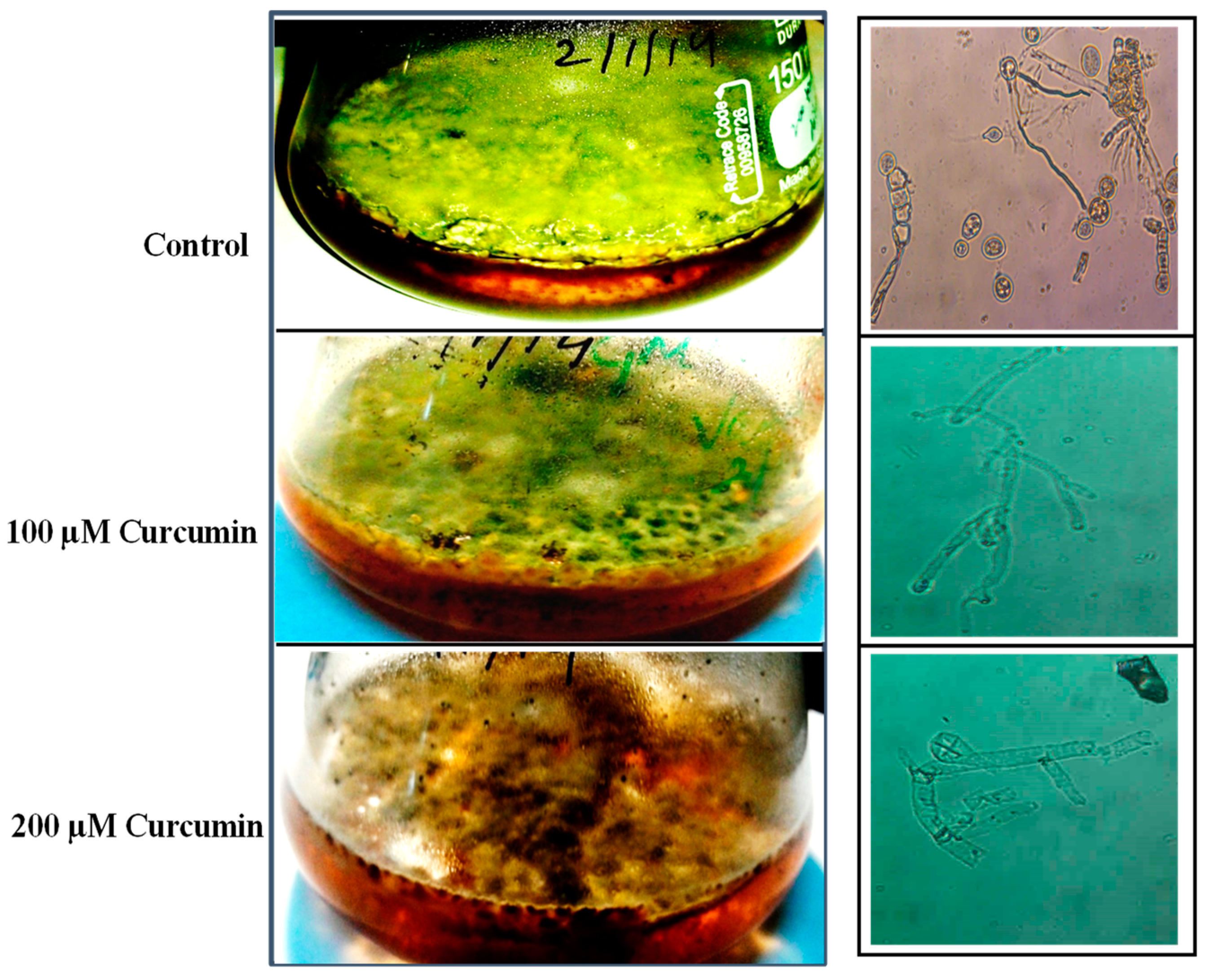
2.1. Effect of Curcumin on E. rugulosa Growth

In order to assess the effect of curcumin on E. rugulosa NRRL11440 growth, 10 days aged spores were grown in GMM-arginine medium in the absence and presence of 100 and 200 µM curcumin (dissolved in DMSO) at the static condition for further 10 days. Results showed that the growth of mycelial spores of E. rugulosa NRRL11440 was induced when it was treated with curcumin as compared to the control cells (Figure 1).

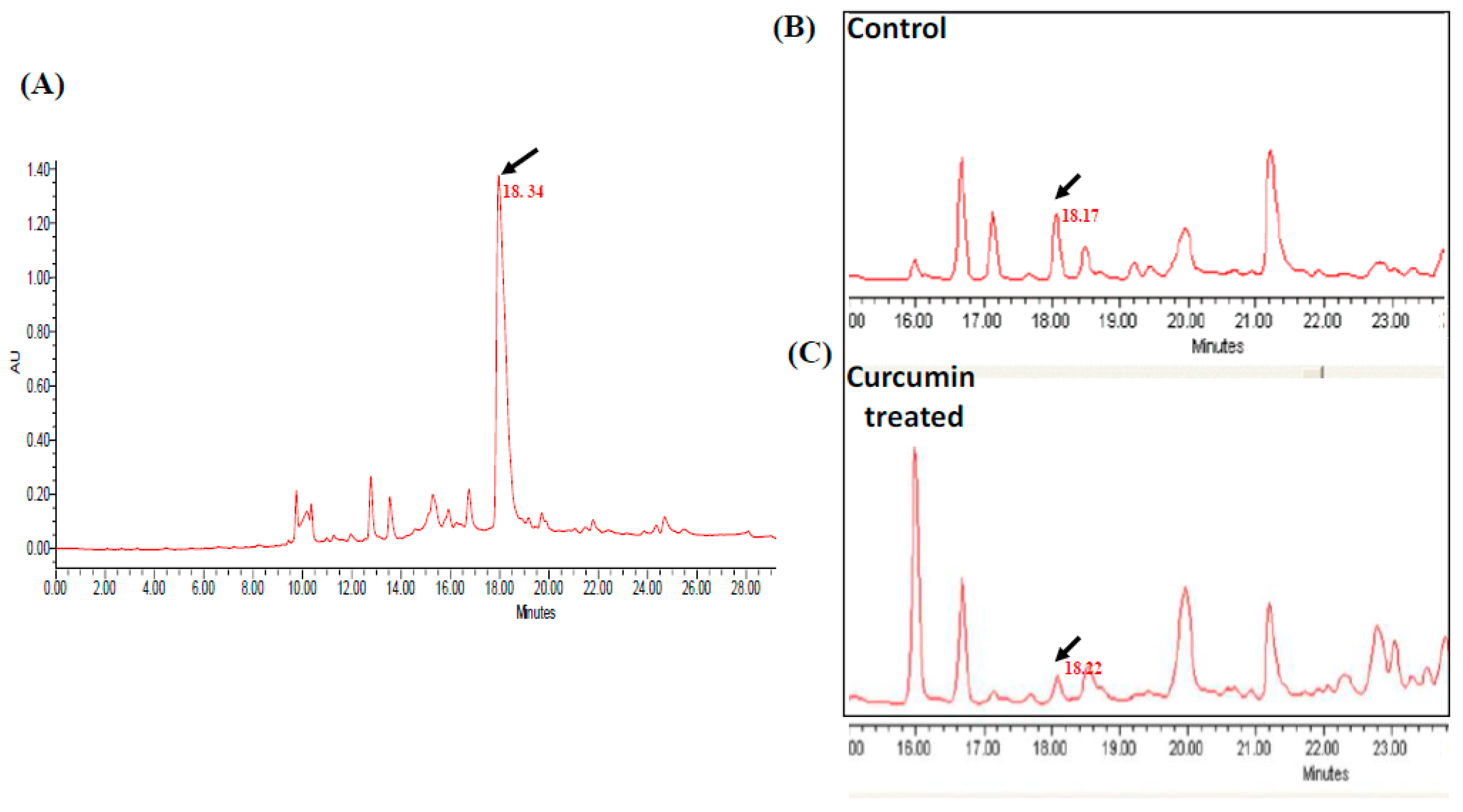
2.2. Effect of Curcumin on Echinocandin B Production

To check the effect of curcumin which is a known histone methyltransferase (HAT) inhibitor on echinocandin B antifungal production in E. rugulosa NRRL11440 strain.
The cell culture grown in echinocandin B producing medium (GMM-arginine) was treated with a fixed 100 µM concentration of curcumin for 10 days in static conditions. The HPLC analysis was performed with both the curcumin-treated and non-treated (Control) methanol extract samples using RP-C18 column (4.5 × 250 mm) for quantitative estimation of echinocandin B synthesis. Results show that the pure echinocandin B compound was used as a standard. It was detected at almost 18 min of retention time as shown in Figure 2A. The comparative analysis of echinocandin B production was also observed both in the control and curcumin-treated sample in particular to the observed peak with similar retention time both in the control and curcumin-treated sample (Figure 2B,C).
However, the echinocandin production in the curcumin-treated sample was sharply reduced, revealed by the peak intensity corresponding to ~18 min of retention time. These results indicate that the HAT inhibitor; curcumin inhibits echinocandin B production.

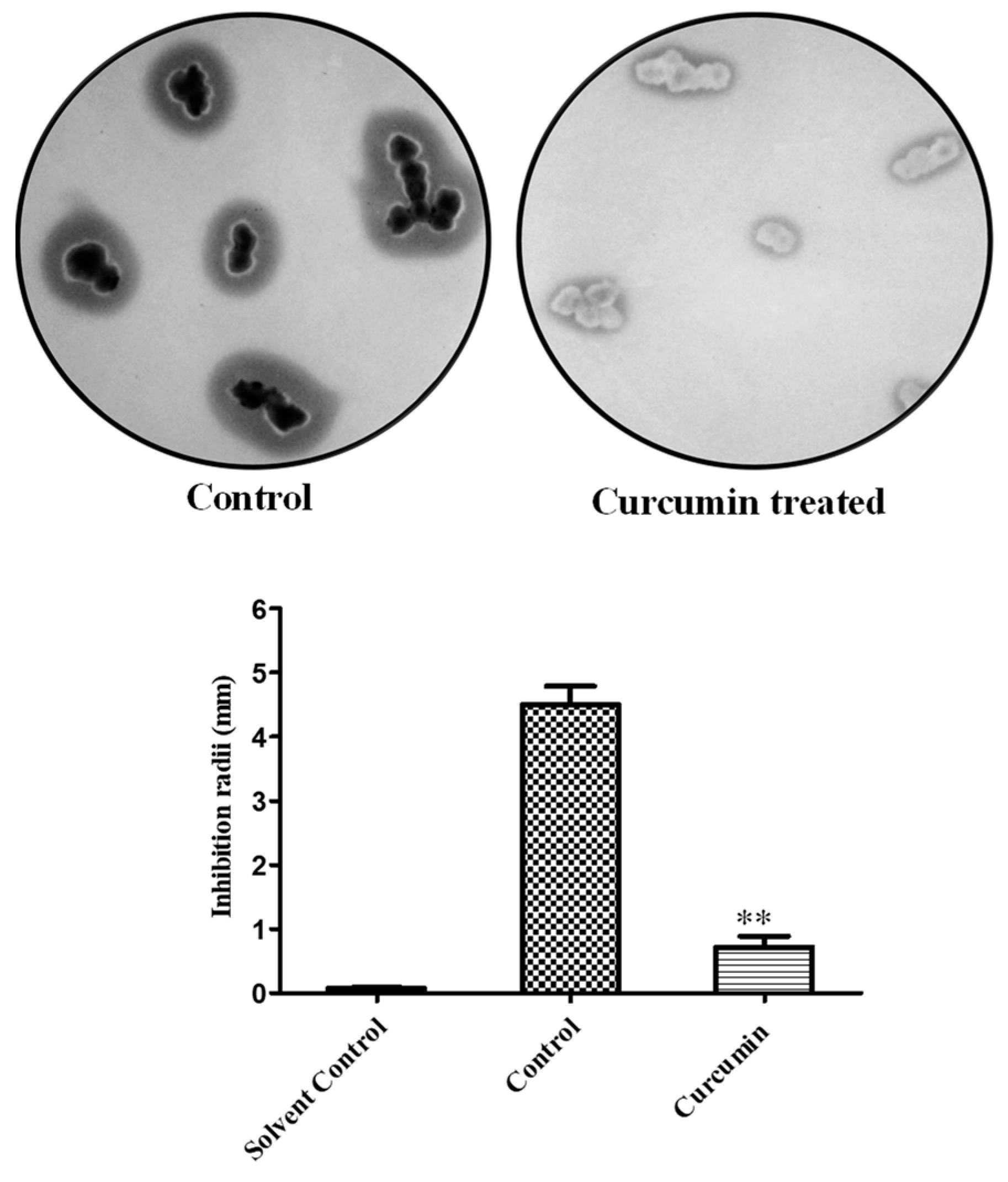
2.3. Cell Susceptibility Profiling of Curcumin-Treated Sample

To further evaluate the effect of curcumin on echinocandin B production, a cell susceptibility test was performed by confrontation assay using a mycelial mat of E. rugulosa NRRL11440, treated with and without curcumin (100 µM) sample for 10 days at static conditions. As expected that E. rugulosa NRRL11440 cells treated with curcumin severely suppressed the echinocandin production, resulting to show a very low cell susceptibility against C. albicans DSY294 as shown in Figure 3. These results indicate that curcumin limits the synthesis of echinocandin B in E. rugulosa NRRL11440.

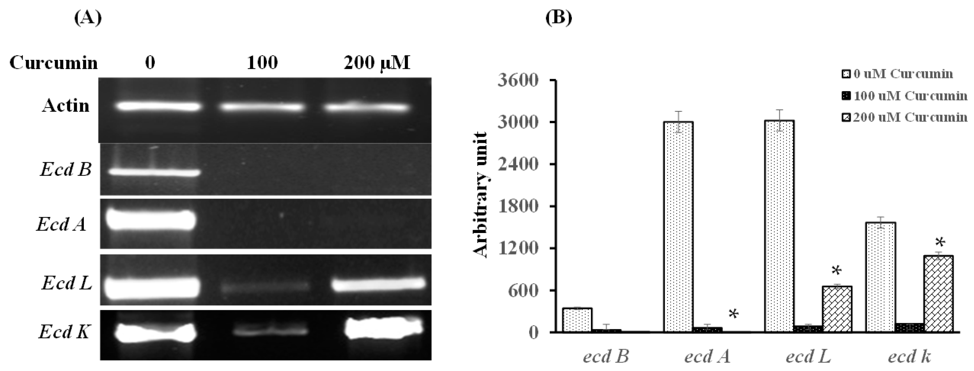
2.4. Curcumin Transcriptionally Represses Genes Present in the Echinocandin B Gene Cluster

To further evaluate the repression in cell susceptibility in the curcumin-treated sample was the result of post-translational modifications on histone proteins. For this, we analyzed the acetylation incurred at histone protein (H3) in the absence and presence of curcumin (100 and 200 µM) via Western blotting analysis using Anti-acetlyH3 Ab. Figure 4 illustrates that the acetylation on H3 histone proteins was found to be repressed in a dose-dependent manner when the sample was treated with curcumin. It was observed that the expression level of acetylation on histone (3 H) was repressed by 43% and 68% when samples were treated with 100 and 200 µM curcumin, respectively.
These results suggest that curcumin, a histone acetyltransferase (HAT) inhibitor is able to post-translationally suppress the acetylation process on a histone protein, resulting to repress the echinocandin B synthesis as revealed by western blot.

2.5. Curcumin Transcriptionally Represses Genes Present in the Echinocandin B Gene Cluster

To confirm the role of histone modifier, curcumin in echinocandin B production, we analyzed the gene expression pattern of different genes of the ecd gene cluster of echinocandin B. For this, gene expression profiling of ecd gene cluster genes, i.e., “ecdB which encodes for a transcription factor, ecdA encodes for an NRPS (Non-ribosomal Peptide synthetase), ecdL; for an ABC transporter, and ecdK encodes for a dioxygenase” were analyzed in the absence or 100 and 200 µM curcumin treatment. The expression of the actin gene was used as an internal reference.
Figure 5A,B shows that gene expression of transcription factor encoded “ecdB, and ecdA; an NRPS encoded gene” was completely repressed when cells were treated with curcumin which indicates that transcriptional factor encoded gene ecdB and NRPS genes present within the ecd gene cluster have a major role in the echinocandin B production. However, gene expression of ecdL and ecdK genes showed a dose-dependent expression which drastically repressed expression at 100 µM compared to 200 µM curcumin concentration as shown in Figure 5.

2.6. Interspecies Interactions Suppress the Inhibitory Effect of Curcumin on Echinocandin B Production

It is well established that interspecies interactions tend to induce the biosynthetic gene clusters (BGCs) of secondary metabolites which led to increased production of secondary metabolites. To assess the inhibitory effect of curcumin on echinocandin B production under interspecies interactions established between E. rugulosa NRRL11440 and C. albicans. For this, Emericella rugulosa NRRL11440 cell mat was grown in the absence or presence of 200 µM curcumin. On the 5th day, C. albicans cells were cocultured. to observe the interspecies effects on echinocandin B production.
Our results showed that the expression level of all the tested genes such as ecd B, transcription factor NPRS encoded; ecd A, ABC transporter encoded ecd L and ecd K genes were increased at different extents (Figure 6) when a consortium established with echinocandin B producing Emericella rugulosa NRRL11440 strain and C. albicans, suggesting to induce echinocandin B production. The induction of these gene expressions was repressed upon curcumin (200 µM) treatment to the consortium, leading to disturb interspecies interactions. These results suggest that curcumin is able to disrupt interspecies communication, leading to a decrease in the echinocandin B synthesis.

3. Discussion

Among the many remarkable discoveries in the genomics, the revelation of the fungal isolates possessing greater numbers of gene clusters encoded for the production of secondary metabolites is arguably the most terrific one. Echinocandin B is a secondary metabolite which is synthesized in E. rugulosa NRRL11440 strains under specialized conditions. This important antifungal entity is often used for the treatment of Candida infections, particularly for the treatment of drug-resistant fungal infections. The biosynthetic genes of echinocandin B are arranged into two (ecd and hty) gene clusters [4]. It has been established that the secondary metabolites’ clusters located distal to the chromosome ends are also regulated by a heterochromatin-dependent mechanism. Regulation by chromatin structure is a proficient mechanism for post-translational regulation of secondary metabolite production. Chromatin regulation largely depends on DNA and histone modifications such as acetylation, methylation, or phosphorylation which inhibit or induce the transcription of certain genomic regions under specific conditions. Many researchers have proved that fungal secondary metabolite biosynthesis clusters are controlled by chromatin-based mechanisms, histone modifiers, and proteins involved in heterochromatin formation. The acetylation of histones coordinated by histone acetyltransferase (HATs) is involved in the activation of gene expression, cellular metabolisms, and protein stability [25]. The HATs positively regulate the biosynthetic gene clusters by transferring the acetyl group to the N-terminal of histone lysine. Earlier studies revealed that GcnE and MYST-type histones acetyltransferases reported in A. nidulans are involved in regulating the activation of secondary metabolites synthesis [26]. However, disruption of such genes led to loss of secondary metabolites production [26,27]. To the best of our knowledge, this is the first evidence showing the effect of curcumin on the biosynthesis of echinocandin B. The curcumin treatment to E. rugulosa NRRL11440 showed severe impairment of the echinocandin B production as supported by the HPLC analysis (Figure 2). It is suggested that curcumin which acts as a HAT inhibitor restricts histone acetylation, resulting to affect the echinocandin B production. The suppression of echinocandin B production was further corroborated by the confrontation assay. The cell susceptibility effect was completely eliminated with no significant inhibition zone observed when E. rugulosa NRRL11440 cells were treated with curcumin. In order to investigate the effect of curcumin on the expression of genes present in the echinocandin B genes cluster, it was observed that expression of transcription factor encoded ecdB gene, and ecdA; NRPS encoded gene was repressed in curcumin-treated cells which indicates that ecdB and ecd A gene present within the ecd gene cluster of echinocandin B has an important role in the echinocandin B production. However, the expression of multi-drug ABC transporter encoded ecdL and α-ketoglutarate-dependent nonheme iron oxygenases encoded ecdK gene repressed more at 100 µM curcumin than 200 µM curcumin concentration (Figure 5). An earlier study described the role of ecdK in the maturation of echinocandin B synthesis [28]. It is expected that higher curcumin (200 µM) treatment which repressed echinocandin B production, is able to tailor the echinocandin B molecule in E. rugulosa NRRL11440. However, the role of ecdL is still to be characterized. Therefore, Curcumin transcriptionally regulates echinocandin B biosynthetic genes by modulating them epigenetically especially restricting the recruitment of the acetylation on histone protein as revealed by the western blot analysis shown in Figure 4. A different group of studies has revealed the effect of histone modifiers on the modulation of secondary metabolite production [10,29,30]. Epigenetic histone modifiers such as Histone deacetylase (HDAC) and DNA Methyltransferase (DNMT) inhibitors induced the expression of a secondary metabolite pathway in endophytic Nigrospora sphaerica [27,31]. Similar findings were also obtained when azacytidine; a DNA methyltransferase (DNMT) inhibitor epigenetically modulates the synthesis of antibacterial secondary metabolites endophytic Streptomyces coelicolor [29,32]. It is well described that interspecies communication led to stimulate the secondary metabolites production. The perseverance of such cell-cell communications for the cell defense against the surroundings or inhibition of the growth of competitors for their survival which led to regulating the activation of biosynthetic gene clusters for secondary metabolites production. We also found that the expression level of all the tested genes of echinocandin B biosynthetic gene clusters (BSCs) such as ecd B encoded for a transcription factor, NPRS encoded; ecd A, ABC transporter encoded ecd L and ecd K genes were increased at different extents when echinocandin B producing Emericella rugulosa NRRL11440 was grown with C. albicans, suggesting to induce echinocandin B production. This inducive effect was repressed after supplementation of curcumin (200 µM), a HAT inhibitor in the coculture of Emericella rugulosa NRRL11440 and C. albicans consortium, leading to upset interspecies interactions through chromatin modifications, resulting to decrease gene expression of echinocandin B biosynthetic genes.
In the present work, curcumin treatment to E. rugulosa NRRL11440 was found to suppress the echinocandin B antifungal biosynthesis by repression of gene expression of a transcriptional factor ecdB and an NRPS-coded ecdA which shows reduced cell susceptibility against C. albicans. Curcumin may turn out to be an effective histone acetyltransferase inhibitor for limiting acetylation on the histone proteins, that restrict echinocandin B production in E. rugulosa NRRL11440. This study signifies that the echinocandin B synthesis in E. rugulosa is regulated at an epigenetic level which may pave the way to further explore epigenetic regulation for the induction of echinocandin B production.

4. Materials and Methods

4.1. Chemicals, Strains, and Media

The echinocandin B producing Emericella rugulosa NRRL11440 strain was purchased from Fungal Genetics Stock Cente (FGSC), Manhattan, KS, USA, and it was maintained in YG medium at 30 °C. For echinocandin B production, Emericella rugulosa NRRL11440 strain was grown in GMM (Glucose minimal medium) supplemented with 10 mM of Arginine for 10 days in static conditions at 30 °C. For assessment of drug susceptibility analysis. Candida albicans DSY 294 strain was obtained as a kind gift from Prof. Dominique Sanglard, Centre Hospitalier Universitaire Vaudois (CHUV), Lausanne, Switzerland. Candida albicans DSY 294 strain culture and grown in the YEPD medium. The YG, YEPD, and GMM medium were purchased from the Hi-Media (Mumbai, India).

4.2. Production of Echinocandin B

Emericella rugulosa NRRL11440 strain was grown in YG medium at 30 °C at 220 rpm for three days. Spores formed were spread on YG plates and incubated at 30 °C for 10 days. Spores were harvested by drenching the plate with 5 mL of 0.1% Tween 20 and scraping off the mycelium with a sterile spreader without damaging the agar. The spore solution was agitated for 1 min. Spores were counted and inoculated 106 spores/mL in GMM (Glucose minimal medium) supplemented with 10 mM of Arginine as the sole nitrogenous source [4,5]. Further, to check the role of histone modification in echinocandin production, 100 and 200 µM Curcumin, a HAT (Histone acetyltransferase) inhibitor of histone modifiers was added to GMM-arginine medium at the time of adding spores. The cell culture was grown at 30 °C for 10 days in static conditions. The harvested culture was lyophilized and extracted with methanol at 30 °C for overnight for further use.

4.3. Measurement of Echinocandin B Production

The echinocandin B biosynthesis in E. rugulosa NRRL11440 in the absence and presence of curcumin was assessed by HPLC analysis. Samples for HPLC analysis were performed as per the method described earlier with minor modifications [5]. The methanol-extracted harvested sample of E. rugulosa NRRL11440 was filtered with a 0.22 µm filter. A 25 µL sample was injected into the analytical HPLC system (Waters, Milford, MA, USA) to run using an RP-C18 column (4.5 × 250 mm). The standard of echinocandin B (Santa Cruz Biotechnology, Dallas, TX, USA: SC-362020) was run and monitored at 222 nm. The echinocandin B concentration was calculated by peak(s) area and statistical analysis was applied using GraphPad Prism v5.01 (GraphPad Software, Inc., Boston, MA, USA).

4.4. Confrontation Assay

The E. rugulosa spores grown in 50 mL GMM-Arg medium were used for comparative analysis of echinocandin production with and without treatment of curcumin as per cell susceptibility methods described earlier [33,34,35,36,37]. The production of echinocandin B in the absence or presence of curcumin histone modifier was tested by confrontation assay (susceptibility) against the C. albicans DSY 204 strain. Briefly, a confrontation assay was done by placing the E. rugulosa mycelial mats on pre-inoculated C. albicans DSY 294 cells in the YEPD agar plate. The cell mat was recovered after 10 days of culture and washed with autoclaved water and cut into small pieces with a sterile blade. These small pieces were put on pre-inoculated C. albicans DSY 294 cells in YEPD agar plates and incubated at 30 °C for 24 h. The zone of inhibition formed by E. rugulosa NRRL11440 mat was measured.

4.5. RNA Isolation

For RNA isolation, a 100 mg fungal mycelial mat was ground up to a fine powder with liquid nitrogen. The RNA extraction was performed using TRIzol® (Life Technologies, Carlsbad, CA, USA). The ground tissue was homogenized with 1 mL Trizol buffer and incubated at room temperature for 5 min. 0.2 mL of chloroform was added to the sample and shaken vigorously for 15 s and incubated further at room temperature for 5 min. The sample was centrifuged at 12,000 rpm for 15 min at 4 °C. The top aqueous phase was taken out carefully and 0.5 mL of isopropanol was added and allowed to precipitate at room temperature for 1 h. The sample was centrifuged at 12,000 rpm for 15 min at 25 °C [38,39,40,41,42]. The precipitant pellet was washed twice with 1 mL of 75% ethanol at 12,000 rpm for 5 min. The concentration and purity were measured on a Nanodrop (Thermo Scientific, Waltham, MA, USA). RNA integrity was analyzed on 0.8% formaldehyde agarose gel prepared in MOPS buffer.

4.6. cDNA Synthesis

The cDNA was synthesized using 1 μg RNA from each sample which was reverse transcribed using ProtoScript II Reverse Transcriptase (NEB, Ipswich, MA, USA). Firstly, the RNA sample was mixed with 2 μL of oligo dT (50 μM) and 1 μL dNTPs (10 mM) and the volume was adjusted to 12 μL with nuclease-free water. This mix was heated at 65 °C for 5 min. It was spun briefly and placed promptly on ice. Then 4 µL of 5X ProtoScript II RT Reaction buffer, 2 µL DTT (0.1 M), 1 µL murine RNAse inhibitor (40 U/µL), and 1 µL ProtoScript II Reverse Transcriptase (200 U/ul). This was incubated at 42 °C for 1 h. The enzyme was inactivated at 80 °C for 5 min [5].

4.7. Transcription Gene Expression Profiling

To analyze the relative gene expression of the echinocandin genes RT-PCR was performed using 1.5 µL cDNA reaction mixtures in 25 µL PCR reaction mixture using gene-specific primers. The PCR was set up in a thermal cycler (Multiplex PCR, Applied Biosystems) with amplification for 35 cycles [5]. PCR amplification from different cDNA samples was used to determine the relative expression of the genes of the echinocandin B biosynthetic gene cluster in the presence of curcumin, a HAT inhibitor. Quickly, Genes ecdA, ecdB, ecdL, ecdK, and actin (internal control) were amplified from cDNA prepared from different samples. The 10 µL PCR products were run on 1% agarose gel and the image was captured in ChemiDoc Gel Imaging System (BioRad, Hercules, CA, USA). The relative gene expression was measured by band intensity which was evaluated using Image Lab v4.1 (BioRad, Hercules, CA, USA).

4.8. E. rugulosa and C. albicans Interspecies Interactions

To establish an interspecies consortium, the three days aged spores of E. rugulosa were harvested by drenching them with 0.1% Tween 20 (5 mL), and 106 spores/mL spores were inoculated in GMM-Arg (Glucose minimal medium-10 mM Arginine) medium [5]. Further in this, 200 µM Curcumin has supplemented at the time of spores inoculum. The cell culture was grown at 30 °C initially for 8 days in static conditions. On the 9th day, 1 × 10⁴ C. albicans cells were cultured with the existing culture and further grew for the next 2 days. The harvested culture mat was washed well (three times) with distilled water and further used for RNA isolation for gene expression analysis.

4.9. Crude Membrane Preparation

To prepare the crude membrane, E. rugulosa (106 spores/mL) spores were grown in GMM-Arg medium (50 mL) with and without treatment of different curcumin concentrations for 10 days, at 30 °C under static conditions. This assay was performed according to the method described earlier with some modifications [34]. The harvested culture mat was washed with PMSF (100 µM) containing distilled water. The cell mat was transferred into the prechilled mortar and homogenization buffer (10 mM Tris-Cl pH 7.5, 0.5 mM EDTA, 1 µg/mL TPCK, TLCK, and 100 µM PMSF) was added to crush the cell mat with pestle using liquid nitrogen. In order to remove cell debris, it was centrifuged at 2500 rpm for 5 min, 4 °C. The supernatants were then centrifuged for 60 min at 25,000 rpm, 4 °C to recover the pellet to dissolve in 3 mL, resuspension buffer (10 mM Tris-Cl pH 7.5, 150 mM NaCl, 10% Glycerol, 1 µg/mL TPCK, TLCK and 100 µM PMSF). The resuspension samples were quantified and aliquoted (100 µL) in tubes for further use.

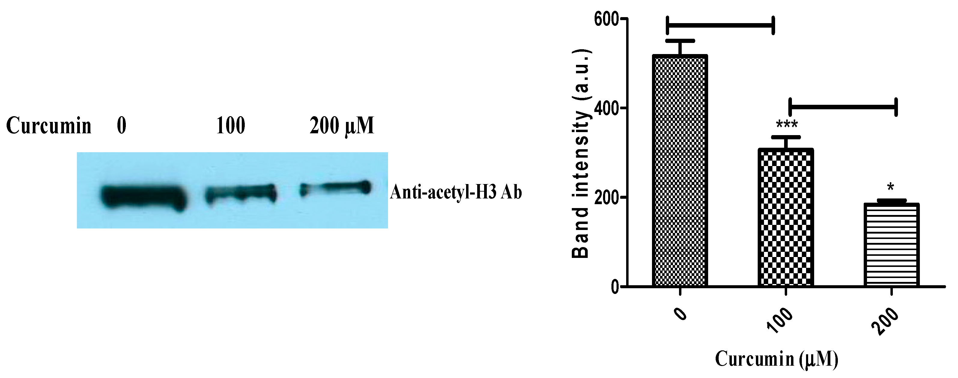
4.10. Immunodetection of Histone Acetylation

The Western blot analysis was carried out using an anti-acetyl H3 antibody (1:5000 dilution) as described in the previous studies [34].

4.11. Statistical Analyses

Statistical analyses were assessed using GraphPad prism v5.01 (Graph Pad Software, La Jolla, CA, USA). p values were calculated via Student t-test and ANOVA test. p < 0.05 was significant.

Author Contributions

A.K. and V.K. (Vandana Kumari) provided the conceptualization, A.K., V.K. (Vandana Kumari) and V.K. (Vinay Kumar) established the methodology, V.K. (Vandana Kumari), A.K. and V.K. (Vinay Kumar), investigated, and validated the results, and the original draft was prepared. A.K., M.K. and V.K. (Vandana Kumari). and V.K. (Vinay Kumar) performed review and editing. All authors have read and agreed to the published version of the manuscript.

Funding

This research received no external funding.

Institutional Review Board Statement

Not Applicable.

Informed Consent Statement

Not applicable.

Data Availability Statement

Data sharing is not applicable to this article.

Acknowledgments

We thank Rajendra Prasad for motivating us during the study. We acknowledge Central University of South Bihar, and Central University of Haryana for providing support for establishing this study.

Conflicts of Interest

The authors declare no conflict of interest.

References

  1. Szymański, M.; Chmielewska, S.; Czyżewska, U.; Malinowska, M.; Tylicki, A. Echinocandins-structure, mechanism of action and use in antifungal therapy. J. Enzym. Inhib. Med. Chem. 2022, 37, 876–894. [Google Scholar] [CrossRef]
  2. Patil, A.; Majumdar, S. Echinocandins in antifungal pharmacotherapy. J. Pharm. Pharmacol. 2017, 69, 1635–1660. [Google Scholar] [CrossRef] [Green Version]
  3. Fisher, M.C.; Alastruey-Izquierdo, A.; Berman; Bicanic, T.; Bignell, E.M.; Bowyer, P.; Bromley, M.; Brüggemann, R.; Garber, G.; Cornely, O.A.; et al. Tackling the emerging threat of antifungal resistance to human health. Nat. Rev. Microbiol. 2022, 20, 557–571. [Google Scholar] [CrossRef] [PubMed]
  4. Cacho, R.A.; Jiang, W.; Chooi, Y.H.; Walsh, C.T.; Tang, Y. Identification and characterization of the echinocandin B biosynthetic gene cluster from Emericella rugulosa NRRL 11440. J. Am. Chem. Soc. 2012, 134, 16781–16790. [Google Scholar] [CrossRef] [PubMed] [Green Version]
  5. Kumar, A.; Jaiswal, V.; Kumar, V.; Ray, A.; Kumar, A. Functional redundancy in Echinocandin B in-cluster transcription factor ecdB of Emericella rugulosa NRRL 11440. Biotechnol. Rep. 2018, 19, e00264. [Google Scholar] [CrossRef]
  6. Bera, K.; Rani, P.; Kishor, G.; Agarwal, S.; Kumar, A.; Singh, D.V. Structural elucidation of TMD0 Domain of EcdL: An MRP1 family of ABC transporter protein revealed by atomistic simulation. J. Biomol. Struct. Dyn. 2017, 36, 2938–2950. [Google Scholar] [CrossRef] [PubMed]
  7. Kumar, A.; Kumar, V.; Kumar, A. Functional characterization of host toxic EcdB transcription factor protein of echinocandin B biosynthetic gene cluster. Biotechnol. App. Biochem. 2019, 66, 626–633. [Google Scholar] [CrossRef]
  8. Hoffmeister, D.; Keller, N.P. Natural products of filamentous fungi: Enzymes, genes, and their regulation. Nat. Prod. Rep. 2007, 24, 393–416. [Google Scholar] [CrossRef]
  9. Yu, J.H.; Keller, N. Regulation of Secondary Metabolism in Filamentous Fungi. Ann. Rev. Phytopathol. 2005, 43, 437–458. [Google Scholar] [CrossRef]
  10. Collemare, J.; Seidl, M.F. Chromatin-dependent regulation of secondary metabolite biosynthesis in fungi: Is the picture complete? FEMS Microbiol. Rev. 2019, 43, 591–607. [Google Scholar] [CrossRef] [Green Version]
  11. Strauss, J.; Reyes-Dominguez, Y. Regulation of secondary metabolism by chromatin structure and epigenetic codes. Fungal Genet. Biol. 2011, 48, 62–69. [Google Scholar] [CrossRef] [PubMed] [Green Version]
  12. Brakhage, A. Regulation of fungal secondary metabolism. Nat. Rev. Microbiol. 2013, 11, 21–32. [Google Scholar] [CrossRef] [PubMed]
  13. Saavedra, F.; Marty-Lombardi, S.; Loyola, A. Characterization of Posttranslational Modifications on Histone Variants. Methods Mol. Biol. 2018, 1832, 21–49. [Google Scholar]
  14. Kim, J.; Lee, J.E.; Lee, J.S. Histone deacetylase-mediated morphological transition in Candida albicans. J. Microbiol. 2015, 53, 805–811. [Google Scholar] [CrossRef] [PubMed]
  15. Xu, H.; Wu, M.; Ma, X.; Huang, W.; Xu, Y. Function and mechanism of novel histone posttranslational modifications in health and disease. Biomed. Res. Int. 2021, 2021, 6635225. [Google Scholar] [CrossRef]
  16. Lopez, C.; Barnon, M.T.; Beacon, T.H.; Nardocci, G.; Davie, J.R. The key role of differential broad H3K4me3 and H3K4ac domains in breast cancer. Gene 2022, 826, 146463. [Google Scholar] [CrossRef]
  17. Jagganath, G. Turmeric, The Golden Spice: From Asia to Africa. Open Access J Arch Anthropol. 2020, 2, 1–6. [Google Scholar] [CrossRef]
  18. Joe, B.; Vijaykumar, M.; Lokesh, B.R. Biological Properties of Curcumin-Cellular and Molecular Mechanisms of Action. Crit. Rev. Food Sci. Nutr. 2004, 44, 97–111. [Google Scholar] [CrossRef]
  19. Hewlings, S.J.; Kalman, D.S. Curcumin: A Review of Its Effects on Human Health. Foods 2017, 6, 92. [Google Scholar] [CrossRef] [Green Version]
  20. Ojo, O.A.; Adeyemo, T.R.; Rotimi, D.; Batiha, G.E.; Mostafa-Hedeab, G.; Iyobhebhe, M.E.; Elebiyo, T.C.; Atunwa, B.; Adebola Busola Ojo, A.B.; Lima, C.M.G.; et al. Anticancer Properties of Curcumin Against Colorectal Cancer: A Review. Front. Oncol. 2022, 12, 881641. [Google Scholar] [CrossRef]
  21. Tanvir, E.M.; Hossen, S.; Hossain, F.; Afroz, R.; Gan, H.S.; Khalil, I.; Karim, N. Antioxidant Properties of Popular Turmeric (Curcuma longa) Varieties from Bangladesh. J. Food Qual. 2017, 2017, 8471785. [Google Scholar] [CrossRef] [Green Version]
  22. Jennings, M.R.; Parks, R.J. Curcumin as an Antiviral Agent. Viruses 2020, 12, 1242. [Google Scholar] [CrossRef] [PubMed]
  23. Hassan, F.; Saif-ur Rehman, M.; Khan, M.S.; Ali, M.A.; Javed, A.; Yang, A.C.N. Curcumin as an Alternative Epigenetic Modulator: Mechanism of Action and Potential Effects. Front. Genet. 2019, 10, 514. [Google Scholar] [CrossRef] [PubMed] [Green Version]
  24. Sunagawa, Y.; Funamoto, M.; Shimizu, K.; Shimizu, S.; Sari, N.; Katanasaka, Y.; Miyazaki, Y.; Kakeya, H.; Hasegawa, K.; Morimoto, T. Curcumin, an Inhibitor of p300-HAT Activity, Suppresses the Development of Hypertension-Induced Left Ventricular Hypertrophy with Preserved Ejection Fraction in Dahl Rats. Nutrients 2021, 13, 2608. [Google Scholar] [CrossRef] [PubMed]
  25. Li, Y.; Song, Z.; Wang, E.; Dong, L.; Bai, J.; Wang, D.; Zhu, J.; Zhang, C. Potential antifungal targets based on histones post-translational modifications against invasive aspergillosis. Front. Microbiol. 2022, 13, 980615. [Google Scholar] [CrossRef] [PubMed]
  26. Nutzmann, H.W.; Reyes-Dominguez, Y.; Scherlach, K.; Schroeckh, V.; Horn, F.; Gacek, A.; Schümann, J.; Hertweck, C.; Strauss, J.; Brakhage, A.A. Bacteria induced natural product formation in the fungus Aspergillus nidulans requires Saga/Ada-mediated histone acetylation. Proc. Natl. Acad. Sci. USA 2011, 108, 14282–14287. [Google Scholar] [CrossRef] [Green Version]
  27. Wang, J.; Xu, C.; Sun, Q.; Xu, J.; Chai, Y.; Berg, G.; Cernava, T.; Ma, Z.; Chen, Y. Post-translational regulation of autophagy is involved in intra-microbiome suppression of fungal pathogens. Microbiome 2021, 9, 131. [Google Scholar] [CrossRef]
  28. Wang, J.; Cacho, R.A.; Chiou, G.; Garg, N.K.; Tang, Y.; Walsh, C.T. EcdGHK are Three Tailoring Iron Oxygenases for Amino Acid Building Blocks of the Echinocandin Scaffold. J. Am. Chem. Soc. 2013, 135, 4457–4466. [Google Scholar]
  29. Guzman-Chavez, F.; Salo, O.; Samol, M.; Ries, M.; Kuipers, J.; Bovenberg, R.A.L.; Vreeken, R.J.; Driessen, A.J.M. Deregulation of secondary metabolism in a histone deacetylase mutant of Penicillium chrysogenum. Microbiologyopen 2018, 7, e00598. [Google Scholar] [CrossRef] [Green Version]
  30. Gómez-Rodríguez, E.Y.; Uresti-Rivera, E.E.; Patrón-Soberano, O.A.; Islas-Osuna, M.A.; Flores-Martínez, A.; Riego-Ruiz, L.; Rosales-Saavedra, M.T.; Casas-Flores, S. Histone acetyltransferase TGF-1 regulates Trichoderma atroviride secondary metabolism and mycoparasitism. PLoS ONE 2018, 13, e0193872. [Google Scholar] [CrossRef] [Green Version]
  31. Ramesha, K.P.; Mohana, N.C.; Nayaka, S.C.; Satish, S. Epigenetic Modifiers Revamp Secondary Metabolite Production in Endophytic Nigrospora sphaerica. Front. Microbiol. 2021, 21, 730355. [Google Scholar] [CrossRef] [PubMed]
  32. Kumar, J.; Sharma, V.K.; Singh, D.K.; Mishra, A.; Gond, S.K.; Verma, S.K.; Kumar, A.; Kharwar, R.N. Epigenetic Activation of Antibacterial Property of an Endophytic Streptomyces coelicolor Strain AZRA 37 and Identification of the Induced Protein Using MALDI TOF MS/MS. PLoS ONE 2016, 11, e0147876. [Google Scholar] [CrossRef] [PubMed] [Green Version]
  33. Kumar, S.; Kumar, A.; Kaushal, M.; Kumar, P.; Mukhopadhyay, K.; Kumar, A. Fungal-derived xenobiotic exhibits antibacterial and antibiofilm activity against S. aureus. Drug Discov. Ther. 2018, 12, 214–223. [Google Scholar] [CrossRef] [PubMed] [Green Version]
  34. Mandal, A.; Kumar, A.; Singh, A.; Lynn, A.M.; Kapoor, K.; Prasad, R. A key structural domain of the Candida albicans Mdr1 protein. Biochem. J. 2012, 445, 313–322. [Google Scholar] [CrossRef] [PubMed] [Green Version]
  35. Kumar, A.; Shukla, S.; Mandal, A.; Shukla, S.; Ambudkar, S.; Prasad, R. Divergent Signature motifs of Nucleotide Binding Domains of ABC multidrug transporter, CaCdr1p of pathogenic Candida albicans, are functionally asymmetric and non-interchangeable. BBA Biomemb. 2010, 1798, 1757–1766. [Google Scholar] [CrossRef] [Green Version]
  36. Rai, V.; Gaur, M.; Kumar, A.; Shukla, S.; Komath, S.S.; Prasad, R. Novel catalytic mechanism for ATP hydrolysis employed by the N-terminal nucleotide-binding domain of Cdr1p, a multidrug ABC transporter of C. albicans. BBA Biomemb. 2008, 1778, 2143–2153. [Google Scholar] [CrossRef] [Green Version]
  37. Kumari, N.; Singh, S.; Kumari, V.; Kumar, S.; Kumar, V.; Kumar, A. Ouabain potentiates the antimicrobial activity of aminoglycosides against Staphylococcus aureus. BMC Complement. Altern. Med. 2019, 19, 119. [Google Scholar] [CrossRef] [Green Version]
  38. Kumar, V.; Rosenzweig, R.; Asalla, A.; Nehra, S.; Prabhu, S.D.; Bansal, S.S. TNFR1 Contributes to Activation Induced Cell Death of Pathological Cd4+ T-Lymphocytes During Ischemic Heart Failure. JACC Basic Transl. Sci. 2022, 10, 1038–1049. [Google Scholar] [CrossRef]
  39. Irfan, M.; Kumar, P.; Kumar, V.; Datta, A. Fruit ripening specific expression of β-DN-acetylhexosaminidase (β-Hex) gene in tomato is transcriptionally regulated by Ethylene Response Factor SlERF. E4. Plant Sci. 2022, 323, 111380. [Google Scholar] [CrossRef]
  40. Kumar, V.; Irfan, M.; Ghosh, S.; Chakraborty, N.; Chakraborty, S.; Datta, A. Fruit ripening mutants reveal cell metabolism and redox state during ripening. Protoplasma 2016, 253, 581–594. [Google Scholar] [CrossRef]
  41. Irfan, M.; Ghosh, S.; Kumar, V.; Chakraborty, N.; Chakraborty, S.; Datta, A. Insights into transcriptional regulation of β-D-N-acetylhexosaminidase, an N-glycan-processing enzyme involved in ripening-associated fruit softening. J. Exp. Bot. 2014, 65, 5835–5848. [Google Scholar] [CrossRef] [PubMed] [Green Version]
  42. Ghosh, S.; Singh, U.K.; Meli, V.S.; Kumar, V.; Kumar, A.; Irfan, M.; Chakraborty, N.; Chakraborty, S.; Datta, A. Induction of senescence and identification of differentially expressed genes in tomato in response to monoterpene. PLoS ONE 2013, 8, e76029. [Google Scholar] [CrossRef] [PubMed] [Green Version]
Figure 1. Effect of curcumin on Emericella rugulosa growth: Ten-days aged Emericella rugulosa NRRL11440 mycelium grown in GMM-Arg medium. The mycelial growth was observed under a microscope in the absence and presence of 100 µM and 200 µM curcumin.
Physiologia 03 00015 g001
Figure 2. Estimation of echinocandin B biosynthesis: The methanolic extract sample recovered from the ten-days aged cell mat of E. rugulosa NRRL11440 was loaded the on RP-C18 column for assessment of Echinocandin B production. Arrow indicates the Echinocadin B peak on chromatogram. (A) Pure Echinocandin B sample as standard (1 mg/mL); (B). Methanolic extract of without curcumin treated (control) sample. (C). Methanolic extract of 100 µM curcumin treated sample.
Physiologia 03 00015 g002
Figure 3. Susceptibility test of curcumin-treated E. rugulosa NRRL1144: The cell extract was recovered from the curcumin-treated 10 days aged E. rugulosa NRRL11440 cells and the mycelial mat was put on the mid-log aged Candida albicans cells spread YEPD agar plate. The zone of inhibition was observed. Two-tailed Student t-test was performed for statistical analysis. p values of (** p ≤ 0.001) are considered significant. p values of control vs. curcumin: 0.0038.
Physiologia 03 00015 g003
Figure 4. Curcumin epigenetically represses the acetylation of histone protein: The crude membrane was prepared from Emericella rugulosa NRRL11440 grown in the absence and presence of 100 and 200 µM curcumin-treated cell cultures as described in Materials and Methods. The Western blot analysis was carried out using Anti-acetyl H3 antibody (1:5000 dilution). The histone acetylation was assessed by band intensity using Image J software. Two-tailed Student t-test was performed for statistical analysis. p values of (* p ≤ 0.05) (*** p ≤ 0.0001) are considered as significant. Detailed p values of control vs. 100 µM curcumin: 0.0008, 100 µM curcumin vs. 200 µM curcumin: 0.024.
Physiologia 03 00015 g004
Figure 5. Curcumin transcriptionally represses echinocandin B genes: (A).Transcriptional expression of ecdA, ecdB, ecdL and ecdK genes of echinocandin B biosynthetic pathway was tested in the absence or presence of curcumin treatment. The expression pattern of the actin gene was measured as a reference. (B). Bar diagram representation of gene expression of the tested genes. Statistical analyses were done using the two-way ANOVA test. p values of (* p ≤ 0.05) are considered as significant in comparison to control groups.
Physiologia 03 00015 g005
Figure 6. Interspecies interactions suppress curcumin effect on echinocandin B production: Transcriptional expression of ecdA, ecdB, ecdL, and ecdK genes of the echinocandin B biosynthetic gene cluster was tested in E. rugulosa the absence or presence of C. albicans. (1). Gene expression of different genes of echinocandin B biosynthesis gene cluster (BGC) in E. rugulosa without coculture C. albicans, as control. (2). Gene expression pattern of different genes of echinocandin B BSC in E. rugulosaC. albicans cocultured treated with curcumin (200 µM). (3). Gene expression pattern of different genes of echinocandin B BSCs in E. rugulosaC. albicans cocultured treated without curcumin (200 µM), as a reference.
Physiologia 03 00015 g006
Disclaimer/Publisher’s Note: The statements, opinions and data contained in all publications are solely those of the individual author(s) and contributor(s) and not of MDPI and/or the editor(s). MDPI and/or the editor(s) disclaim responsibility for any injury to people or property resulting from any ideas, methods, instructions or products referred to in the content.

