Article

The Effectiveness of Benzalkonium Chloride as an Active Compound on Selected Foodborne Pathogens Biofilm

1
Department of Microbiology and Parasitology, Faculty of Medicine, University of Rijeka, Braće Branchetta 20, 51000 Rijeka, Croatia
2
Saponia d.d., Matije Gupca 2, 31000 Osijek, Croatia
3
Faculty of Health Studies, University of Mostar, 88000 Mostar, Bosnia and Herzegovina
*
Author to whom correspondence should be addressed.
Hygiene 2022, 2(4), 226-235; https://doi.org/10.3390/hygiene2040020
Submission received: 30 September 2022 / Revised: 18 November 2022 / Accepted: 23 November 2022 / Published: 28 November 2022

Abstract

:
Benzalkonium chloride (BAC) is a chlorine-based chemical compound with proven antimicrobial properties against bacteria, viruses, and fungi, depending on the length of the alkyl chain. It can be used as a biocide, as a cationic surfactant, and as a phase transfer agent. The aim of this study was to investigate the effectiveness of commercial cleaning agents for sanitary areas Bis duo Active (BDA) with BAC as an active compound in working concentrations of 5% and 20% on the destruction of bacterial biofilm formed on ceramic tiles. A biofilm of Staphylococcus aureus, Salmonella enterica serotype Typhimurium, and Listeria monocytogenes were grown on ceramic tiles with dimensions of 2.5 × 2.5 cm over 24 h. These plates were then treated with working concentrations of BAC for 10 min. After washing, ATP bioluminescence was measured with a luminometer, CFU/cm2 was determined and the total biomass reduction was measured after crystal-violet staining. Both working concentrations of benzalkonium chloride proved to be very effective in destroying the created bacterial biofilm on ceramic tiles. Both treatments caused a reduction in total bacteria number of up to 3.12 log10CFU/cm2, followed by a reduction in total biomass up to circa (ca.) 64%. Inhibition rates varied from ca. 28% to ca. 99%, depending on the method and concentration used. Mechanical cleaning prior or after treatment with BAC is essential to improve biofilm destruction. All methods used in this study are effective for the determination of the anti-biofilm activity of BAC. Further analyses are needed.

1. Introduction

Benzalkonium chloride (BAC) is a chlorine-based chemical compound with proven antimicrobial properties against bacteria, viruses, and fungi [1]. Structurally, it is made of a mixture of alkylbenzyldimethylammonium chloride, in which the alkyl chain has a different number of carbon atoms, most often n-C12H25 (dodecyl), n-C14H29 (tetradecyl), and n-C16H33 (hexadecyl) [2]. Antimicrobial activity depends on the length of the alkyl chain. The C12-homolog is effective against fungi and molds, C14 acts on Gram-positive bacteria, and C16 on Gram-negative bacteria [3]. Alkylbenzyldimethylammonium chlorides can be used in three main categories: as a biocide, as a cationic surfactant, and as a phase transfer agent [4]. It is commonly used as a disinfectant in the food industry and industrial and domestic areas [5], but additional tests are needed before it can be used as a biocide [6]. Aside from that, the spectrum of benzalkonium chloride usage is wide, from preservatives in certain consumer products to skin antiseptics and active ingredients in surgical disinfection [3,7]. Although widely used by the industry, there is certain evidence about its toxic effects on the pulmonary system, causing ailments such as bronchoconstriction in asthmatic patients [8]. From a toxicological point of view, in humans, doses thought to be fatal are 100–400 mg/kg of BAC taken orally or 5–15 mg/kg of BAC taken parenterally [9]. It is considered that up to 0.1% concentrations of BAC are safe to use as an antimicrobial agent [10]. BAC is commonly used in combination with other quaternary ammonium compounds and in small working concentrations [1,11]. One of the biggest problems in the food industry is biofilm formation and development, as well as preventive measures against it [12,13]. Certain food industries, such as dairy factories, raw milk tanks, pipelines, cheese tanks, butter centrifuges, and pasteurizers, act as surface substrates for biofilm formation in different environmental factors such as temperature and air humidity [13,14]. It is easy to conclude that the main locations for biofilm development depend on factory type, but it is important to emphasize that they can include water, all types of pipelines, tables, employee gloves, storage silos for raw materials and additives, etc. [15]. Biofilm is a highly structured cluster of microorganisms that are attached to a surface and/or to each other and incorporated in a self-produced matrix [16] made mostly from the exopolysaccharide substance (EPS). EPS forms certain shields over bacteria in the biofilm matrix and protects them from environmental factors, including disinfection procedures [17]. Chlorine preparations, such as BAC, belongs to the chemical method of disinfection [18]. After disinfection, bacteria in the biofilm can secrete more exopolysaccharide (EPS) to better protect themselves, especially if a disinfecting agent such as BAC is used in sublethal concentrations or if disinfectant is used not according to manufacturer’s instructions (not properly diluted) [19], therefore weakening disinfecting action [20]. S. aureus, S. Typhimurium, and L. monocytogenes are well-known foodborne pathogens that can cause foodborne outbreaks and individual food poisoning with a different set of symptoms and the severity of the disease. All three species can form biofilm on the live or inanimate surfaces which presents an issue regarding cleaning and disinfection efficacy. Furthermore, not every disinfecting agent is equally efficient on bacterial biofilm due to the resistance feature of biofilm [21]. Bacteria within a biofilm have several advantages, among which stands out that they are 150 to 3000 times more resistant to chlorine preparations [20,22], so a wise choice of efficient disinfectant is crucial for efficient biofilm removal. Scientific data on the antimicrobial BAC effect on S. aureus, S. Typhimurium, and L. monocytogenes biofilm are scarce. Due to that the aim of this research paper was to investigate the anti-biofilm effect of benzalkonium chloride on bacterial biofilm of selected foodborne pathogens, as well as to determine the effectiveness of ATP bioluminescence, crystal violet and CFU/cm2 as measurement methods of BAC effectiveness on biofilm.

2. Materials and Methods

2.1. Chemical Reagents and Bacterial Strains

For this research paper, the chemical reagent Bis duo Active (BDA) with benzalkonium chloride (BAC) as an active ingredient was provided by Saponia d.d., Osijek, Croatia. Working concentrations of 5% BDA (0.05% BAC) and 20% BDA (0.2% BAC) were made. Apart from that, neutralizing reagents at pH 9 with thiosulphate and saline were used. Bacterial strains Staphylococcus aureus ATCC 29213, Salmonella Typhimurium ATCC 14028, and Listeria monocytogenes ATCC 19115 were provided by the culture collection of the Department of Microbiology and Parasitology, Faculty of Medicine, University of Rijeka, Croatia. Strains were held at −80 °C in 10% glycerol broth. Before use in the experiment, the bacteria were plated on Mueller-Hinton agar and cultivated at 37 °C for 24 h.

2.2. Preparation of Ceramic Tiles

The top surfaces of ceramic tiles with dimensions 2.5 cm × 2.5 cm were used as a ceramic surface for bacterial adherence and biofilm formation. The method for biofilm formation was adopted from and previously described by Ivanković et al. [23] and modified by Piletić et al. [24]. The biofilm was formed on the top surface of the tiles which were previously thoroughly brushed and sterilized in an autoclave at 121 °C for 15 min. Ceramic tiles were placed in plastic Petri dishes and covered with liquified agar (2%, v/v), leaving the top surface of the tiles free and not touched by the agar. Ceramic tiles were used because they are a very common material found in sanitary facilities in various industries and as such, serve as a surface for biofilm development. The bacterial suspensions of 106 CFU/mL for S. aureus, S. Typhimurium and L. monocytogenes in Mueller Hinton broth were poured over the top surface of the ceramic tiles and then incubated in an orbital shaker at 40 rpm at 30 ± 5 °C for 24 h. After incubation, the ceramic tiles were washed with a sterile saline solution and dried out in a laboratory safety cabinet for 1 min. Then, the tiles were removed from agar and washed out with a sterile saline solution and placed in Petri dishes containing either a 5% solution of Bis duo Active (5% BDA) or 20% solution of Bis duo Active (20% BDA). Both working concentrations of benzalkonium chloride were used on all bacterial strains, making sure that the surface of the tiles was completely covered with disinfectant. After 10 min of exposure, the tiles were moved in new Petri dishes containing neutralizing agent, 10% sodium thiosulphate solution (Kemika, Zagreb, Croatia). After 10 min of neutralization, ceramic tiles were moved to sterile tubes. Control tiles (tiles that were not treated) were directly put from the agar in sterile tubes with 10 mL of sterile saline solution.

2.3. Determining Total Bacteria Number

To determine the number of cultivable bacteria, the ceramic tiles were put in sterile tubes which were filled with 10 mL of sterile saline solution and then sonicated in an ultrasound bath (Bandelin-BactoSonic, Berlin, Germany) at a frequency of 40 kHz for 1 min to enhance the release of the adhered cells from the tiles. Using a vortex, samples were homogenized one more time to further enhance the detachment of the remaining cells from the biofilm. Afterwards, ten-fold serial dilutions were made and later inoculated onto Mueller Hinton agar and incubated for 24 h at 35 ± 2 °C. Inhibition percentages were calculated using the formula given:
%   i n h i b i t i o n = 1 N t r e a t m e n t N c o n t r o l 100
where Ntreatment is the mean value of all CFU/cm2 or RLU or CV values after treatment, and Ncontrol is the mean value of CFU/cm2 or RLU or CV values without treatment. The experiment was performed three times in triplicates.

2.4. Determining RLU Values by ATP Bioluminescence

The surface of the ceramic tiles with formed biofilm was wiped with a 3MTM Clean-TraceTM Surface ATP Test Swab UXL100 (3M, Saint Paul, MN, USA) swab preimmersed for 1 min in luciferine/luciferase reagent. After one minute, the swab was put in a chamber of the 3MTM Clean-TraceTM luminometer (3M, Saint Paul, MN, USA). The amount of light produced was read from the luminometer and was expressed as RLU. Non–treated biofilm served as the control. Inhibition percentages were calculated using the previously given formula. The experiment was performed three times in triplicates.

2.5. Determining Biomass Reduction by Crystal-Violet Staining

The controls and treated ceramic tiles were rinsed with sterile saline solution and then fixated for 30 min at 80 °C in a dry heat sterilizer (ST-01/02, Instrumentaria, Zagreb, Croatia). After fixation, the tiles were stained with 0.1% crystal violet for 20 min. This step is followed by 15 min of intense mixing and rinsing with 95% ethanol. Afterwards, the optical density (OD) was measured on a spectrophotometer (Eppendorf, Biophotometer, model #6131, Hamburg, Germany) at a wavelength of 600 nm. Inhibition percentages were calculated using the previously given formula. The experiment was performed three times in triplicates.

2.6. Statistical Analysis and Graphing

To determine if there was any statistically significant difference between control and treated ceramic tiles, Wilcoxon signed-rank test was performed (p < 0.05). To determine if there was any statistically significant difference between treatments, the Kruskal–Wallis multiple comparison test (p < 0.05) was performed. To see if there was any statistically significant difference between the two treatments within one bacterium, the Mann–Whitney H test was performed (p < 0.05). Statistical analysis was done using the software TIBCO Statistica 14.0.0. (StatSoft Inc., Tulsa, OK, USA). Graphing was done using GraphPad Prism version 9.4.1 (GraphPad Software, San Diego, CA, USA).

3. Results

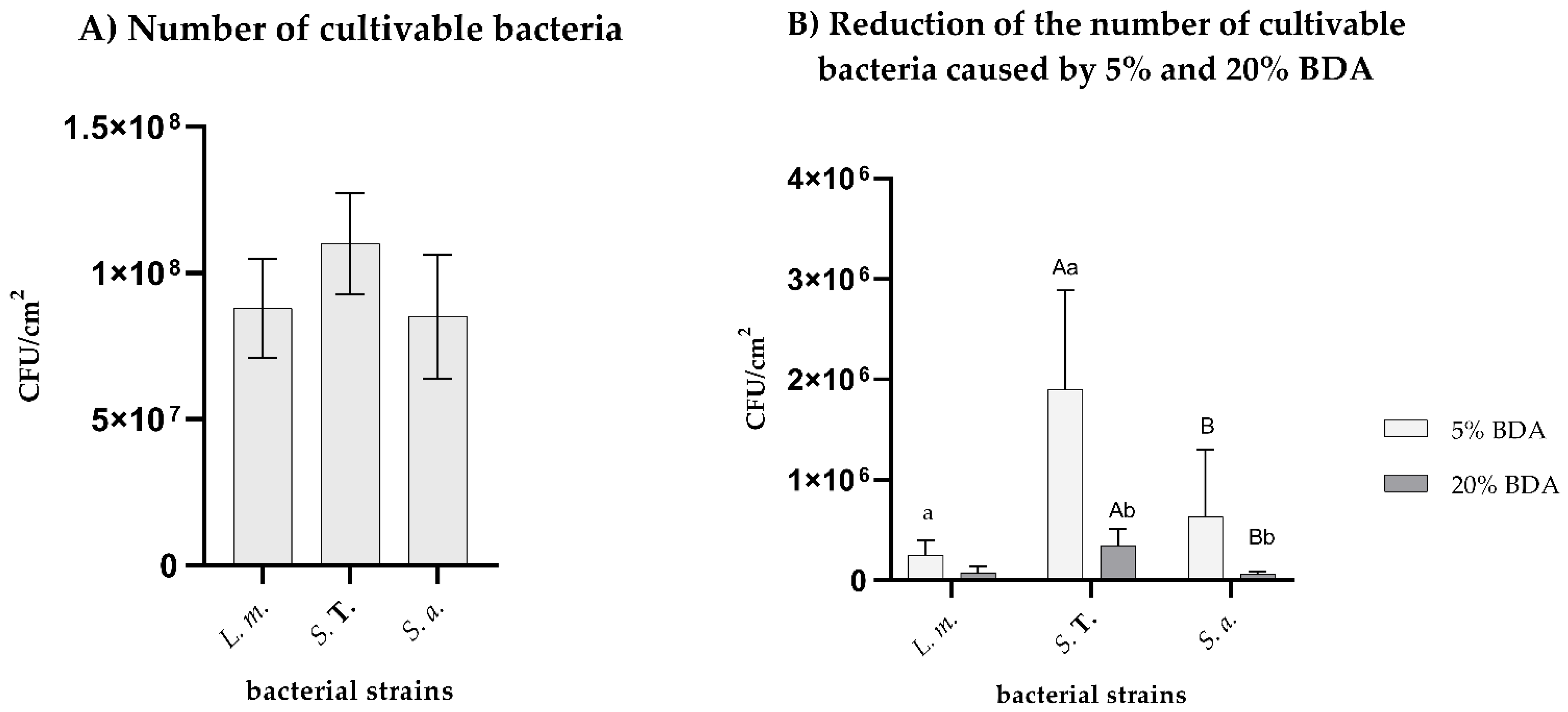
3.1. Reduction of the Number of Cultivable Bacteria Caused by 5% and 20% BDA

All tested bacteria formed biofilm on ceramic tile surfaces, and the number of bacteria varied from 7.0 × 10⁷ CFU/cm2 to 1.3 × 10⁸ CFU/cm2 (Figure 1A). A disinfection treatment with both 5% and 20% solution of Bis duo Active significantly reduced the number of cultivable bacteria in the biofilm. Numbers of cultivable bacteria are shown in Figure 1A (control groups) and the reduction values after treatment with working concentrations of BDA are shown in Figure 1B.
Considering the calculated log10CFU/cm2 values, the working concentration of 5% BDA reduced the number of bacteria for approximately 1.7 log10CFU/cm2 to 2.5 log10CFU/cm2 depending on the bacterial strain, L. monocytogenes being most sensitive strain. S. aureus was the most sensitive strain to the 20% working concentration of BDA, which caused reductions for 3.12 log10CFU/ cm2, while S. Typhimurium was the most resistant strain for both working concentrations.

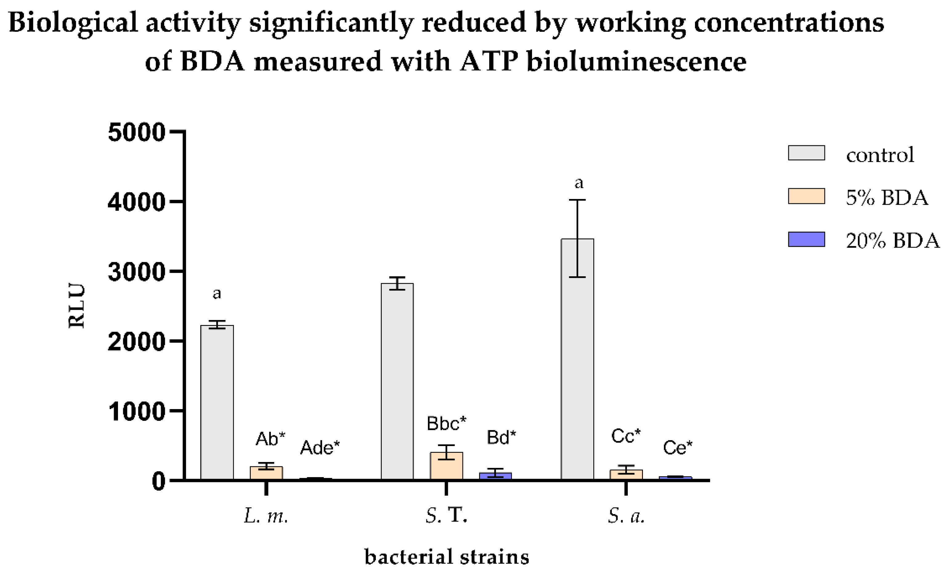
3.2. Biological Activity Significantly Reduced by Working Concentrations of BDA Measured with ATP Bioluminescence

Biological activity measured using the ATP bioluminescence method showed a statistically significant reduction in biomass after disinfection treatments with both working solutions of BDA in comparison to the control group. A significant difference between different bacteria was observed (p < 0.05) (Figure 2).
Considering the ATP bioluminescence method, the RLU values on non-treated ceramic tiles varied from approximately 2250 to 3500 RLU depending on the bacterial strain, while after 5% BDA disinfection treatment, measured values were far under 500 RLU and after 20% BDA disinfection treatment the values are under 120 RLU for all treated bacterial strains. Again, the most resistant strain was S. Typhimurium, while the most sensitive was S. aureus.

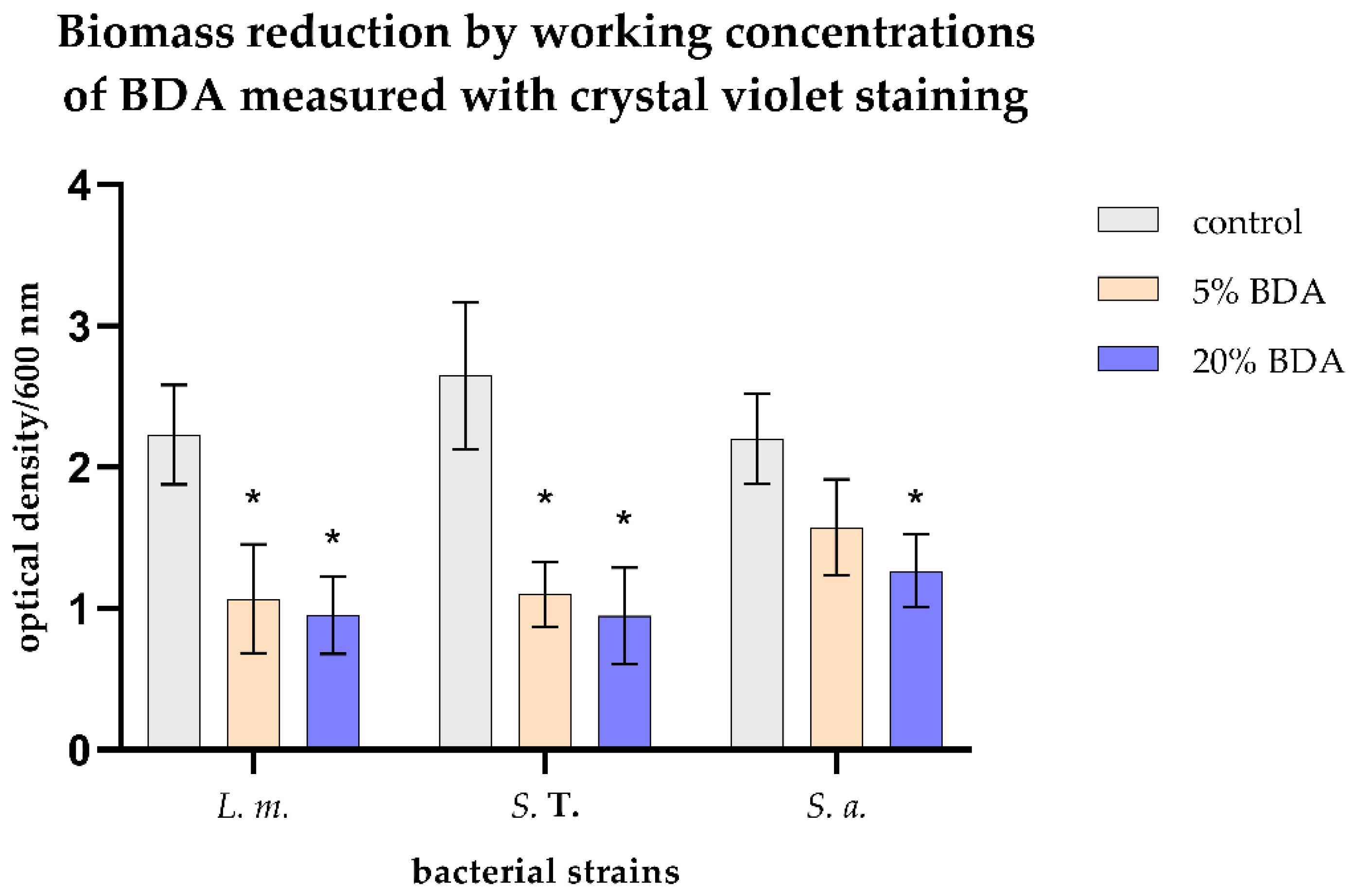
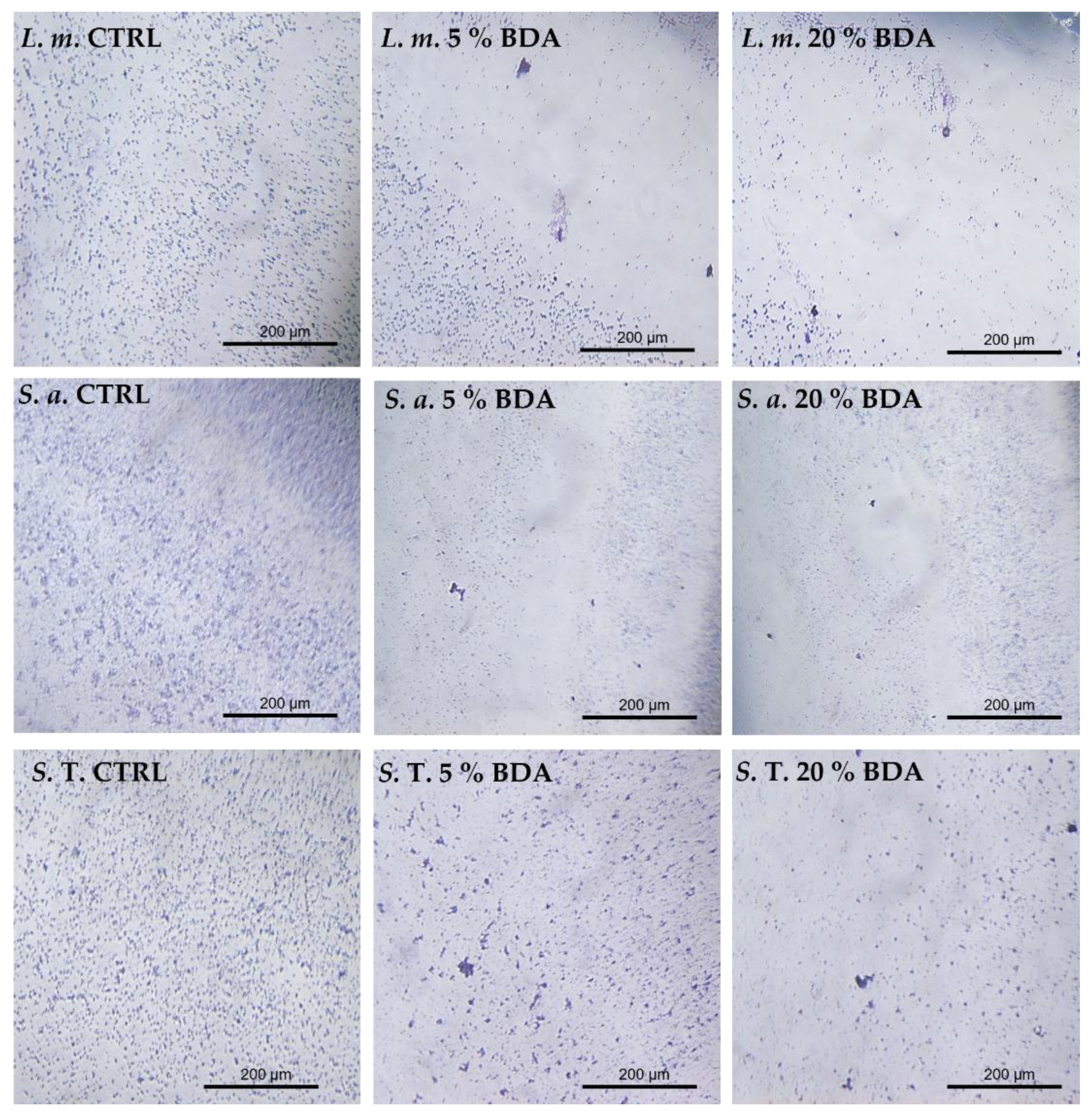
3.3. Biomass Reduction by Working Concentrations of BAC Measured with Crystal Violet Staining

The total biomass reduction was observed for all tested bacteria after disinfection treatment with 5% and 20% working solution of BDA. The reduction was statistically significant in comparison to the control group for all tested bacteria except for the S. aureus biofilm treated with the 5% solution of BDA (p < 0.05) (Figure 3).
There was no statistically significant difference between a 5% BDA and 20% BDA disinfection protocol within the same bacterium strain for all tested strains. There was a statistically significant difference between either 5% or 20% BDA compared to the control group for each strain, except for the S. aureus ceramic tile treated with 5% BDA (p < 0.05). Standard deviations are larger, which is not strange considering that this staining is not very precise.
To determine changes in biofilm morphology after treatment with 5% and 20% BDA, light microscopy was performed. Morphological changes in biofilm density on ceramics were found for all tested bacterial strains in comparison with the control (Figure 4). Biofilm destruction and detachment from ceramic tiles were pronounced after treatment with 20% BDA for all tested strains.

3.4. Biofilm Inhibition Rates Varied Using Different Detection Methods

The inhibition percentages were calculated for both 5% and 20% Bis duo Active working solutions in comparison to the control group separately for each method used. Inhibition rates varied from circa (ca.) 28% to ca. 99% depending on the used method (Table 1).

4. Discussion

Benzalkonium chloride (BAC) is a chlorine-based chemical compound with proven antimicrobial properties against bacteria, viruses, and fungi. It is considered that concentrations of BAC up to 0.1% are safe for human use [9]. Appropriate disinfecting protocols for infection prevention are crucial almost everywhere, especially in the food industry. One of the most common foodborne pathogens in food industries are Staphylococcus aureus, Salmonella Typhimurium, and Listeria monocytogenes. Forming biofilm on different surfaces, they become more resistant to environmental factors and are better attached to the substrate surface which means they are harder to remove [25]. Furthermore, scientific data on this topic are scarce, so, considering those facts, anti-biofilm activity of BAC on S. aureus, S. Typhimurium, and L. monocytogenes was investigated with working concentrations of 5% and 20% BAC during 10 min of exposure. According to the results from this investigation, BAC is efficient as a disinfecting agent against formed biofilm, however, not enough for full biofilm eradication. Even when used in working concentrations, BAC was unable to fully eradicate biofilm, once again corresponding to previous studies on biofilm resistance to disinfectants [19,20,21,22,24,25,26]. Numerous mechanisms of biofilm resistance are still ongoing issues in scientific community but some of the determined factors worth mentioning are EPS overproduction and frequent usage of the same disinfectants in diluted concentrations (sub-lethal) [26]. Furthermore, BAC treatment in different concentrations can result in the formation of the viable but not cultivable state of bacteria (VBNC); therefore, to better determine its anti-biofilm effect, authors also investigated the effectiveness of crystal violet staining and ATP bioluminescence as additional methods for bacterial biomass determination.
All used methods (total viable bacteria count, ATP bioluminescence, and crystal violet staining) showed efficiency in the determination of the anti-biofilm effect of BAC on the formed biofilm of chosen pathogens, but with certain variations and limitations. The total bacterial count showed the greatest inhibition rates, followed by ATP bioluminescence and crystal violet for all tested pathogens. This variation in inhibition rates between methods can be explained by the very nature of each method and its measuring focus. Both ATP bioluminescence and crystal violet can be used as additional methods for biomass determination, enumerating live cells, dead cells, EPS, and VBNC cells; therefore, inhibition rates measured with these methods are smaller when compared to total viable bacteria count. The same results were previously described by Piletic et al. for the gaseous ozone anti-biofilm effect on K. pneumoniae biofilm [24]. Furthermore, not every method is evenly precise.
For example, crystal-violet staining is not a very precise method for detecting total biofilm biomass. This staining is one of the most extensively used methods for the quantification of biofilm biomass, but it has some limitations, such as unspecific binding to negatively charged molecules and low reproducibility due to uneven dye extraction or differential removal of biofilm biomass throughout the washing steps [27]. Due to that, some studies suggest using fluorescence staining instead [27]. On the other hand, CFU is considered and used as a gold standard method which has the advantage that only viable bacteria are counted as the CFU excludes dead bacteria and debris. The disadvantage of this method is that sometimes clusters of bacteria can be miscounted as single colonies and, consequently, the results can be wrong and misinterpreted [28]. ATP bioluminescence is a fast, easy and highly sensitive method which gives an insight into the amount of light produced by biofilm by virtue of ATP reaction with luciferine and luciferase [29,30]. In addition to ATP from microorganisms, ATP from product residues on the surface is also measured. ATP bioluminescence shows good results in the detection of biofilm on ceramic tiles of all three tested bacteria and, after treatment with a cleaning agent, shows a very good reduction in value. The negative side of this method is the lack of determined standards, or reference values, especially for microbiological compliance of food, feed, and microbiological standards for surface cleanliness. Another disadvantage is the possibility of a false positive result. When taking a swab, it may happen that a surface that is not of interest is wiped, and certain microorganisms from that surface may react with the luciferin reagent and give a result that is not relevant to research [31]. Although certain variations in used methods were observed, keeping in mind advantages and disadvantages, all methods are effective for the determination of the disinfecting agents anti-biofilm activity, especially in the determination number of viable, dead and VBNC bacterial cells. Because this disinfecting agent is a combined agent for cleaning also sanitary facilities, such as toilets, bathrooms, and wardrobes, the authors recommend adequate cleaning protocols for changing rooms and sanitary facilities in food industries to prevent the fecal–oral route of transmission of foodborne pathogens.

5. Conclusions

Though our results indicate that working concentrations (5% and 20%) of Bis duo Active are not sufficient to fully mitigate S. aureus, S. Typhimurium, and L. monocytogenes biofilm from ceramic surfaces, it indeed significantly reduced the number of bacteria present (p < 0.05), and 20% BDA proved to be more effective for biofilm destruction than 5% BDA.
Since most efficacy standards prescribe testing on planktonic forms of bacteria, we recommend that biofilm on different surfaces should be tested for benzalkonium chloride efficiency. A longer time of exposure (more than 10 min) should be tested because of the resistance of bacteria in biofilm, but that is something for further investigation. We would also like to emphasize the importance of mechanical cleaning, which is crucial for the destruction of biofilms.

Author Contributions

Conceptualization, B.K. and I.G.; methodology, B.K.; formal analysis, B.K., N.K.G. and K.P.; investigation, B.K.; resources, I.G.; writing—original draft preparation, B.K, K.P.; writing—review and editing, I.G. and N.K.G.; supervision, I.G.; funding acquisition, I.G. All authors have read and agreed to the published version of the manuscript.

Funding

This research was funded by the University of Rijeka, grant number UNIRI-biomed 18-171.

Institutional Review Board Statement

Not applicable.

Informed Consent Statement

Not applicable.

Data Availability Statement

Not applicable.

Acknowledgments

The authors would like to express gratitude to NOACK d.o.o. from Zagreb, Croatia for the donation of the luminometer.

Conflicts of Interest

The authors declare no conflict of interest.

Abbreviations

BACbenzalkonium chloride
BDABis duo Active
ca. circa
CFU/cm2colony forming unit per square centimeter
CVcrystal violet
EPSexopolysaccharide
L. m.Listeria monocytogenes
ODoptical density
RLUrelative light units
S. T.Salmonella Typhimurium
S. a.Staphylococcus aureus
VBNCviable but not cultivable state of bacteria

References

  1. Merchel Piovesan Pereira, B.; Tagkopoulos, I. Benzalkonium Chlorides: Uses, Regulatory Status, and Microbial Resistance. Appl. Environ. Microbiol. 2019, 85, e00377-19. [Google Scholar] [CrossRef] [PubMed] [Green Version]
  2. Rowe, R.C.; Sheskey, P.J.; Quinn, M.E. Handbook of Pharmaceutical Excipients, 6th ed.; Pharmaceutical Press: London, UK, 2009; ISBN 978-0-85369-792-3. [Google Scholar]
  3. Kuca, K.; Marek, J.; Stodulka, P.; Musilek, K.; Hanusova, P.; Hrabinova, M.; Jun, D. Preparation of Benzalkonium Salts Differing in the Length of a Side Alkyl Chain. Molecules 2007, 12, 2341–2347. [Google Scholar] [CrossRef] [PubMed] [Green Version]
  4. Lackner, M.; Guggenbichler, J.P. Antimicrobial Surfaces. In Ullmann’s Encyclopedia of Industrial Chemistry; Wiley-VCH Verlag GmbH & Co. KGaA: Weinheim, Germany, 2013; pp. 1–13. ISBN 978-3-527-30673-2. [Google Scholar]
  5. Lavorgna, M.; Russo, C.; D’Abrosca, B.; Parrella, A.; Isidori, M. Toxicity and Genotoxicity of the Quaternary Ammonium Compound Benzalkonium Chloride (BAC) Using Daphnia Magna and Ceriodaphnia Dubia as Model Systems. Environ. Pollut. 2016, 210, 34–39. [Google Scholar] [CrossRef] [PubMed]
  6. European Chemicals Agency. Guidance on the Biocidal Products Regulation. Volume IV, Environment. Part A: Information Requirements; European Chemicals Agency: Helsinki, Finland, 2018.
  7. Choi, H.-Y.; Lee, Y.-H.; Lim, C.-H.; Kim, Y.-S.; Lee, I.-S.; Jo, J.-M.; Lee, H.-Y.; Cha, H.-G.; Woo, H.J.; Seo, D.-S. Assessment of Respiratory and Systemic Toxicity of Benzalkonium Chloride Following a 14-Day Inhalation Study in Rats. Part. Fibre Toxicol. 2020, 17, 5. [Google Scholar] [CrossRef] [PubMed]
  8. Prabhakaran, S.; Abu-Hasan, M.; Hendeles, L. Benzalkonium Chloride: A Bronchoconstricting Preservative in Continuous Albuterol Nebulizer Solutions. Pharmacother. J. Hum. Pharmacol. Drug Ther. 2017, 37, 607–610. [Google Scholar] [CrossRef]
  9. European Medicines Agency. Benzalkonium Chloride Used as an Excipient; Comittee for Human Medicinal Products (CHMP): Amsterdam, The Netherlands, 2017; p. 14.
  10. Marple, B.; Roland, P.; Benninger, M. Safety Review of Benzalkonium Chloride Used as a Preservative in Intranasal Solutions: An Overview of Conflicting Data and Opinions. Otolaryngol. Neck Surg. 2004, 130, 131–141. [Google Scholar] [CrossRef]
  11. McKeen, L. Introduction to Food Irradiation and Medical Sterilization. In The Effect of Sterilization on Plastics and Elastomers; Elsevier: Amsterdam, The Netherlands, 2012; pp. 1–40. ISBN 978-1-4557-2598-4. [Google Scholar]
  12. Vickery, K. Special Issue: Microbial Biofilms in Healthcare: Formation, Prevention and Treatment. Materials 2019, 12, 2001. [Google Scholar] [CrossRef] [PubMed] [Green Version]
  13. Galié, S.; García-Gutiérrez, C.; Miguélez, E.M.; Villar, C.J.; Lombó, F. Biofilms in the Food Industry: Health Aspects and Control Methods. Front. Microbiol. 2018, 9, 898. [Google Scholar] [CrossRef] [PubMed]
  14. Mizan, M.F.R.; Jahid, I.K.; Ha, S.-D. Microbial Biofilms in Seafood: A Food-Hygiene Challenge. Food Microbiol. 2015, 49, 41–55. [Google Scholar] [CrossRef]
  15. Camargo, A.C.; Woodward, J.J.; Call, D.R.; Nero, L.A. Listeria monocytogenes in Food-Processing Facilities, Food Contamination, and Human Listeriosis: The Brazilian Scenario. Foodborne Pathog. Dis. 2017, 14, 623–636. [Google Scholar] [CrossRef]
  16. Vestby, L.K.; Grønseth, T.; Simm, R.; Nesse, L.L. Bacterial Biofilm and Its Role in the Pathogenesis of Disease. Antibiotics 2020, 9, 59. [Google Scholar] [CrossRef] [Green Version]
  17. Nkemngong, C.A.; Voorn, M.G.; Li, X.; Teska, P.J.; Oliver, H.F. A Rapid Model for Developing Dry Surface Biofilms of Staphylococcus Aureus and Pseudomonas Aeruginosa for in Vitro Disinfectant Efficacy Testing. Antimicrob. Resist. Infect. Control 2020, 9, 134. [Google Scholar] [CrossRef]
  18. Curran, E.T.; Wilkinson, M.; Bradley, T. Chemical Disinfectants: Controversies Regarding Their Use in Low Risk Healthcare Environments (Part 1). J. Infect. Prev. 2019, 20, 76–82. [Google Scholar] [CrossRef]
  19. Yu, T.; Ma, M.; Sun, Y.; Xu, X.; Qiu, S.; Yin, J.; Chen, L. The Effect of Sublethal Concentrations of Benzalkonium Chloride on the LuxS/AI-2 Quorum Sensing System, Biofilm Formation and Motility of Escherichia coli. Int. J. Food Microbiol. 2021, 353, 109313. [Google Scholar] [CrossRef]
  20. Stewart, P.S.; William Costerton, J. Antibiotic Resistance of Bacteria in Biofilms. Lancet 2001, 358, 135–138. [Google Scholar] [CrossRef]
  21. Bridier, A.; Briandet, R.; Thomas, V.; Dubois-Brissonnet, F. Resistance of Bacterial Biofilms to Disinfectants: A Review. Biofouling 2011, 27, 1017–1032. [Google Scholar] [CrossRef] [PubMed]
  22. Allard, M.W.; Bell, R.; Ferreira, C.M.; Gonzalez-Escalona, N.; Hoffmann, M.; Muruvanda, T.; Ottesen, A.; Ramachandran, P.; Reed, E.; Sharma, S.; et al. Genomics of Foodborne Pathogens for Microbial Food Safety. Curr. Opin. Biotechnol. 2018, 49, 224–229. [Google Scholar] [CrossRef]
  23. Ivanković, T.; Goić-Barišić, I.; Hrenović, J. Reduced Susceptibility to Disinfectants of Acinetobacter Baumannii Biofilms on Glass and Ceramic. Arch. Ind. Hyg. Toxicol. 2017, 68, 99–108. [Google Scholar] [CrossRef] [Green Version]
  24. Piletić, K.; Kovač, B.; Perčić, M.; Žigon, J.; Broznić, D.; Karleuša, L.; Lučić Blagojević, S.; Oder, M.; Gobin, I. Disinfecting Action of Gaseous Ozone on OXA-48-Producing Klebsiella Pneumoniae Biofilm In Vitro. Int. J. Environ. Res. Public Health 2022, 19, 6177. [Google Scholar] [CrossRef]
  25. Zheng, S.; Bawazir, M.; Dhall, A.; Kim, H.-E.; He, L.; Heo, J.; Hwang, G. Implication of Surface Properties, Bacterial Motility, and Hydrodynamic Conditions on Bacterial Surface Sensing and Their Initial Adhesion. Front. Bioeng. Biotechnol. 2021, 9, 643722. [Google Scholar] [CrossRef]
  26. Piletić, K.; Kovač, B.; Planinić, M.; Vasiljev, V.; Karačonji, I.B.; Žigon, J.; Gobin, I.; Oder, M. Combined Biocidal Effect of Gaseous Ozone and Citric Acid on Acinetobacter Baumannii Biofilm Formed on Ceramic Tiles and Polystyrene as a Novel Approach for Infection Prevention and Control. Processes 2022, 10, 1788. [Google Scholar] [CrossRef]
  27. Amador, C.I.; Stannius, R.O.; Røder, H.L.; Burmølle, M. High-Throughput Screening Alternative to Crystal Violet Biofilm Assay Combining Fluorescence Quantification and Imaging. J. Microbiol. Methods 2021, 190, 106343. [Google Scholar] [CrossRef] [PubMed]
  28. Hazan, R.; Que, Y.-A.; Maura, D.; Rahme, L.G. A Method for High Throughput Determination of Viable Bacteria Cell Counts in 96-Well Plates. BMC Microbiol. 2012, 12, 259. [Google Scholar] [CrossRef] [PubMed] [Green Version]
  29. Arroyo, M.G.; Ferreira, A.M.; Frota, O.P.; Rigotti, M.A.; de Andrade, D.; Brizzotti, N.S.; Peresi, J.T.M.; Castilho, E.M.; de Almeida, M.T.G. Effectiveness of ATP Bioluminescence Assay for Presumptive Identification of Microorganisms in Hospital Water Sources. BMC Infect. Dis. 2017, 17, 458. [Google Scholar] [CrossRef] [Green Version]
  30. Monica, S.; Bancalari, E.; Castellone, V.; Rijkx, J.; Wirth, S.; Jahns, A.; Bottari, B. ATP Bioluminescence for Rapid and Selective Detection of Bacteria and Yeasts in Wine. Appl. Sci. 2021, 11, 4953. [Google Scholar] [CrossRef]
  31. Griffith, C. Surface Sampling and the Detection of Contamination. In Handbook of Hygiene Control in the Food Industry; Elsevier: Amsterdam, The Netherlands, 2016; pp. 673–696. ISBN 978-0-08-100155-4. [Google Scholar]
Figure 1. L. m. represents Listeria monocytogenes, S. T. Salmonella Typhimurium, and S. a. Staphylococcus aureus. (A) Number of cultivable bacteria for each strain (control groups). There is no statistically significant difference between each strain on untreated ceramic tiles (p > 0.05). (B) CFU/cm2 reduction results presented for 5% and 20% working concentration of BDA (Bis duo Active). Results are shown as a mean value with standard deviation. The lowercase letters, a and b, express the statistically significant difference between treated bacteria within same concentration of BAC (Kruskal–Wallis H test, p < 0.05). The capital letters, A and B, mark the statistically significant difference between 5% and 20% solution of BDA treatment within same bacterium (capital letter A for S. Typhimurium and capital letter B for S. aureus) (Mann–Whitney U test, p < 0.05).
Hygiene 02 00020 g001
Figure 2. L. m. represents Listeria monocytogenes, S. T. Salmonella Typhimurium, and S. a. Staphylococcus aureus. RLU values get by ATP bioluminescence method for all bacterial strains. Results are expressed as a mean value with standard deviation. Lowercase letters, a–e, express statistically significant difference between treated bacteria within control group or same concentration of BDA (Kruskal–Wallis H test, p < 0.05). The capital letters, A–C, mark the statistically significant difference between 5% and 20% solution of BDA treatment within same bacterium (capital letter A for L. monocytogenes, capital letter B for S. Typhimurium, and capital letter C for S. aureus) (Mann–Whitney U test, p < 0.05). Asterisk (*) shows a statistically significant difference between the control group and either 5% or 20% treatment for specific bacteria (Wilcoxon signed-rank test, p < 0.05).
Hygiene 02 00020 g002
Figure 3. L. m. represents Listeria monocytogenes, S. T. Salmonella Typhimurium, and S. a. Staphylococcus aureus. Optical density values at 600 nm before and after treatments. Results are expressed as a mean value with standard deviation. Asterisk (*) expresses a statistically significant difference between the control group and either 5% or 20% BDA treatment for specific bacteria (Wilcoxon signed-rank test, p < 0.05).
Hygiene 02 00020 g003
Figure 4. L. m. represents Listeria monocytogenes, S. T. Salmonella Typhimurium, and S. a. Staphylococcus aureus. Crystal violet staining of biofilm before (control-CTRL) and after treatments with 5% and 20% Bis duo Active (BDA). The dark blue stains present the biomass particles stained with crystal violet. Magnification 20×. Visualizing was done using inverted microscope.
Hygiene 02 00020 g004
Table 1. Inhibition percentages for used bacteria for 5% and 20% BDA. Results are expressed as %.
L. monocytogenesS. TyphimuriumS. aureus
5% BDA20% BDA5% BDA20% BDA5% BDA20% BDA
CFU/cm2999998999999
RLU919886969598
CV525758642843
Publisher’s Note: MDPI stays neutral with regard to jurisdictional claims in published maps and institutional affiliations.

