Article

A Study into the Identity, Patterns of Infection and Potential Pathological Effects of Rumen Fluke and the Frequency of Co-Infections with Liver Fluke in Cattle and Sheep

1
School of Biodiversity, One Health & Veterinary Medicine, University of Glasgow, Glasgow G61 1QH, UK
2
Moredun Research Institute, Pentlands Science Park, Midlothian EH26 0PZ, UK
3
Finn Pathologists, One Eyed Lane, Norfolk IP21 5TT, UK
4
Department of Livestock and One Health, Institute of Infection, Veterinary and Ecological Sciences, University of Liverpool, Liverpool CH64 7TE, UK
*
Author to whom correspondence should be addressed.
Ruminants 2023, 3(1), 27-38; https://doi.org/10.3390/ruminants3010004
Submission received: 25 November 2022 / Revised: 17 January 2023 / Accepted: 20 January 2023 / Published: 25 January 2023
(This article belongs to the Special Issue Disease Diagnostics and Surveillance in Ruminants)

Abstract

:
Rumen fluke (RF) are trematode parasites that have increased in prevalence within European ruminant livestock since the 1990s. Morbidity and mortality can result from the development of juvenile flukes in the duodenum, however, evidence for significant impacts of adult fluke in the rumen of the final host is equivocal. The presence of rumen fluke in Scotland had not previously been quantified, so the purpose of this study was to use historical coprological data collected between 2008 and 2018 at the School of Veterinary Medicine in Glasgow to evaluate the frequency of rumen fluke infection and to compare this with the presence of liver fluke. This analysis showed evidence of adult rumen fluke in 6.6% of samples submitted, with a substantial and significant increase in positive diagnoses from 2016, following which positive rumen fluke diagnoses equaled or slightly outnumbered those of liver fluke. A prospective post-mortem examination study was also carried out to determine the presence of rumen and liver fluke in cattle and sheep, to quantify adult rumen fluke burdens and to assess any pathological changes in the reticulorumen and proximal duodenum associated with infection. The presence of rumen fluke in post-mortem cases was 26.9% (n = 18/67), the majority (66.7%, n = 12/18) with burdens of less than 100 adult rumen flukes. There was no significant difference in mean ruminal papillar density and length in animals with and without adult rumen fluke and no significant gross pathology was observed in the rumen or reticulum. Examination of animals with adult rumen fluke provided no evidence of any consistent associations with acute or chronic inflammatory changes in the duodenum. All rumen fluke collected at necropsy were identified by PCR and sequencing as Calicophoron daubneyi.

1. Introduction

Trematodes are important livestock parasites with a worldwide distribution that are responsible for serious economic losses and compromised animal welfare. Two of the most significant trematodes in temperate regions are rumen fluke (RF) and liver fluke, Fasciola hepatica. RF are digenean trematodes belonging to the family Paramphistomidae, which includes several genera such as Calicophoron and Paramphistomum, which commonly infect ruminants worldwide [1]. Adult RF are conically shaped, pink or red in colour and measure between 5–12 mm in length and 2–4 mm in width, and are found primarily within the reticulum and rumen. Juveniles develop in the duodenum and measure 1–2 mm in length [2]. There are over 10 species of paramphistomes known to infect domestic ruminants, however, the species Calicophoron daubneyi has been confirmed as the most prevalent in Europe. Historically, Paramphistomum cervi was believed to be the dominant RF species in United Kingdom (UK) and Irish livestock, but DNA sequencing of RF specimens from cattle and sheep from recent samples has confirmed that C. daubneyi is the dominant RF species across the UK and Ireland [3,4]. Like liver fluke, RF require a molluscan intermediate host, most commonly freshwater snails, and a final ruminant host to allow completion of its lifecycle. It has been confirmed that, in Europe, the intermediate host for liver fluke, Galba truncatula, also acts as a host for C. daubneyi [5].
Identifying paramphistomes in livestock is largely based upon post-mortem examination (PME) and faecal egg counts (FECs). FECs are useful in detecting infections with adult RF, but not during the pre-patent period when juvenile flukes only are present; that said, juvenile fluke can be detected in faecal samples during acute paramphistomosis [6]. PME of animals for evidence of haemorrhagic duodenitis with immature flukes confirmed on microscopic examination of intestinal scrapes [7] or recovery of adult RF from the rumen and reticulum are regarded as conclusive proof of RF infection [8].
RF have been present in the UK and Ireland for many years, however, their previously low prevalence and perceived low pathogenicity have meant they have received limited attention from veterinary parasitologists and livestock owners [8]. In recent years, the prevalence of RF in continental Europe, Ireland and the UK has increased [9,10], with reports of herd-level prevalence in the UK reaching 59% [11]. Furthermore, serious consequences have been associated with the presence of juveniles in the duodenum, where heavy burdens can result in clinical disease and mortality in both sheep and cattle [12,13]. In juvenile infections there is evidence of erosion, petechiation and necrosis, leading to haemorrhagic enteritis and duodenitis [7]. Little is known, however, about any long-term effects on the duodenal mucosa caused by juvenile RF infection. In endemic areas, it is quite possible that ruminants could play host to both RF and liver fluke simultaneously if they graze on pastures contaminated with metacercariae of both species. Co-infection with C. daubneyi and F. hepatica could also confound the interpretation of clinical, pathological and parasitological observations [14].
While adult RF are still considered largely non-pathogenic, it is known that infections can result in focal chronic ulcerative ruminitis, ruminal papillar atrophy and ulceration at the point of attachment of the adults [15]. The rumen is a complex fermentative organ and infection with C. daubneyi has been shown to change an animal’s volatile fatty acid profile, potentially affecting nutrient absorption and utilisation [16]. Additionally, alterations in papillar shape and length have been observed in clinical cases associated with adult RF [15].
Based on the increase in RF diagnoses in the UK and the relative lack of evidence of the subclinical effects of RF, the aims of this study were the following: (1) to analyse available historical parasitological data and establish the frequency and pattern of RF infection in Central and South West Scotland; (2) to investigate animals submitted for PME for the presence and burden of RF and liver fluke infection; (3) to determine the species of RF most prevalent in samples submitted to the laboratory; (4) to assess anatomical changes in the rumen and duodenum of cattle and sheep affected by adult RF; and (5) to provide a preliminary assessment of the frequency of rumen and liver fluke co-infections.

2. Materials and Methods

2.1. Parasitological Data

At the University of Glasgow Veterinary Diagnostic Service, faecal samples from ruminants, collected for routine analysis of University associated farms (n = 6) and from farm animal clinical cases referred from first opinion veterinary practitioners [17], are routinely tested for evidence of trematode eggs. Faecal samples are tested for both RF and liver fluke eggs using a modified Boray sedimentation technique as follows. A total of 3 grams of faeces were homogenised with 42 mL of tap water and the fluid suspension was passed through a course sieve (250 microns) into a 1 L measuring cylinder and allowed to sediment for two minutes. The supernatant was removed and two drops of 5% methylene blue was added to the sediment material. The resultant solution was placed in a lined petri dish and examined using a stereo microscope at 12× magnification. The total number of eggs in the sample for both RF and liver fluke was recorded from 0 to >25 eggs per gram (epg). After counting 25 eggs, the samples were discarded and a value of >25 epg was recorded. Differentiation of the eggs of rumen and liver fluke was through morphological features and the colour of the eggs.

2.2. Post-mortem Examination (PME)

2.2.1. Presence and Burden of Adult Rumen Fluke

An 8-week study was conducted for two consecutive years (January and February 2018 and 2019). All ovine and bovine cases submitted to the University of Glasgow Veterinary Diagnostic Service for PME during these time periods were inspected for the presence of RF and liver fluke as follows: the forestomachs were thoroughly inspected for the presence of adult RF by gross examination of the contents and the mucosa. If RF were present, the infection burden was classified between 0–4 [18]: 0—no fluke, 1—between 1 and 10, 2—between 11 and 100, 3—between 100 and 200, 4—more than 200 adult rumen fluke. The liver and gall bladder of each case were also examined by longitudinal section for the presence of adult liver fluke. The presence of liver fluke was recorded as either present or absent. All the animals submitted for PME were over 8 months old.

2.2.2. Rumen Fluke Species Identification

During the first year of the project (2018), for each positive case, up to 10 individual adult RF were collected from the rumen and reticulum and stored in 100% ethanol and subsequently processed for species identification [19]. A Qiagen DNEasy Blood and Tissue kit (QIAGEN, Hilden, Germany) was used as detailed in the manufacturing instructions to extract DNA from each individual adult RF [20,21]. The PCR programme was performed using a GeneAMP PCR system 2720 thermal cylinder (Applied Biosystems, Foster City, CA, USA) and the gel was viewed on an Alphamagel Imaging System (Alpha Innotech, San Leandro, CA, USA). The purification of the resultant PCR products achieved using the QIAquick PCR Purification Kit as described by the manufacturer (QIAGEN, Hilden, Germany) and the purified products were sent for Direct Nucleotide Sequencing (Eurofins MWG Operon, Ebersberg, Germany). The resultant sequences were assessed with a Lasergene 10 core suite Software SeqMan Pro (DNASTAR, Madison, WI, USA) and compared to reference sequences from the GenBank database using BLASTn at the European Bioinformatics Institute website (http://www.ebi.ac.uk/, accessed on 22 February 2018).

2.2.3. Anatomical Changes

For the second year of the project, the rumen and duodenum of cattle and sheep submitted for PME were assessed macroscopically (gross examination of ruminal papillae) and microscopically (histopathology of duodenum samples). A 10 cm2 square sample of the atrium ruminis (Figure 1A) was obtained from all cases. If the sample contained adult RF, the flukes were carefully removed to permit a blinded analysis. All samples were stapled to a numbered cardboard during fixation in a 10% buffered formalin solution for a minimum of 48 hours. Ruminal papillae measurements carried out were: density (average number of papillae within a plastic template of three squares of 3 × 1 cm size—Figure 1B) and length (average of 10 randomly selected papillae throughout the sample measured in millimetres using a ruler as adapted from [22]).
To examine the duodenum for microscopic changes, a 2.5 cm sample from the proximal duodenum (approximately 5 cm distal to the pylorus) was collected and stored for a minimum of 24 hours in 10% buffered formalin, embedded in paraffin and sectioned at 2 μm, and stained with haematoxylin and eosin for histopathological analysis. The stained slides were then examined at 200× and 400× magnification using an Olympus BX 43 microscope by two veterinary pathologists. The pathologists examining the sections did not have access to the PME findings.
Following initial histopathological examination of the sections, a scoring system was developed, based on standards used in small animal medicine [23] to assess several non-specific microscopic features present: inflammation of the mucosa and submucosa, and evidence of chronic changes such as fibrosis or granulomas (Appendix A).

2.3. Statistical Analysis

All data analysis in this study was performed using R version 4.1.1 and RStudio version 2021.09.0 (R Core Team, 2022; RStudio Team, 2020, Vienna, Austria). Statistical significance was defined at p-value ≤ 0.05.
The relationship between the number of positive RF infections and year was analysed using a logistic regression model. This was then repeated for liver fluke. A Pearson’s Chi-squared test for independence was used to establish whether a difference existed in the overall number of RF infections between bovine and ovine samples. The Pearson’s Chi-squared test was also used to establish whether there was a significant seasonal pattern in RF and liver fluke diagnoses, cumulative across all years.
For the PME data, a generalised linear regression was used to test for a relationship between bovine/ovine and papillar density, then papillar length. Furthermore, the same method was also utilised to test for a relationship between adult RF presence (adult rumen fluke burden score 1–4 combined) and papillar length, then adult RF presence and papillar density.
A binomial generalised linear model with rumen fluke presence (adult rumen fluke burden score 1–4 combined) and absence (score 0) as the explanatory variable was used to assess the relationship between adult RF presence and each of the seven categories of histopathological changes (Appendix A). For category ‘a’ (inflammation of the lamina propria with lymphocytes and plasma cells), classification 1 (0–5 cells between crypts) was compared to classification 2–4 combined (5 or more cells). For all statistical analysis, co-infected animals were included in all analyses where RF presence/absence was tested.

3. Results

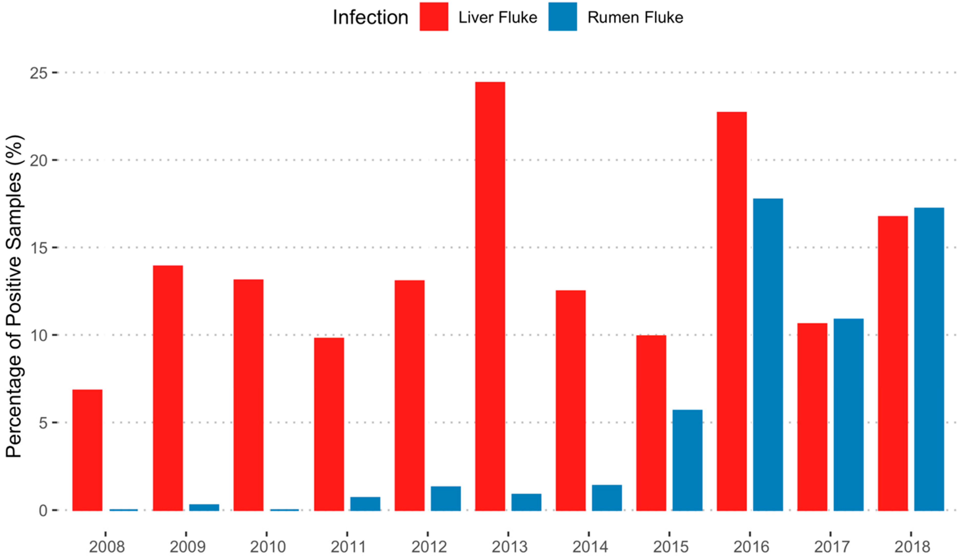
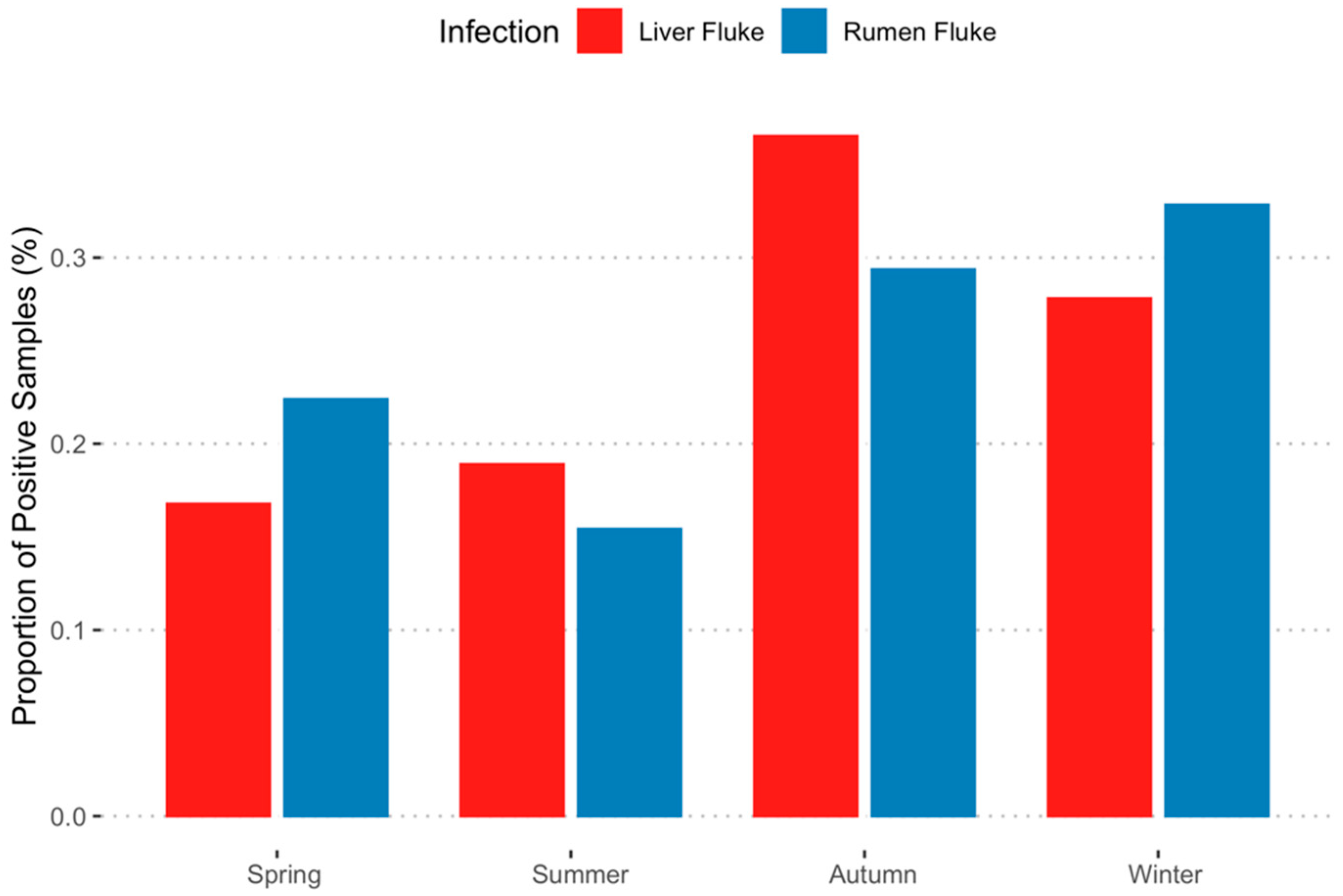
3.1. Parasitological Data

The first positive RF egg count recorded was in August 2009 (data recording for trematodes started in 1979), while only F. hepatica eggs were found in previous faecal samples. FECs data were therefore analysed from January 2008 to December 2018. In this period, 3068 ruminant faecal samples were submitted for FECs analysis. A total of 595 samples (19.4%) contained trematode eggs: 124 had only RF eggs (4%) and 394 (12.8%) contained only liver fluke eggs, with 77 (2.5%) positive for both trematodes, giving an overall presence of adult rumen fluke infections of 6.6% (n = 201/3068). Of the 595 trematode egg positive samples, 437 (73.5%) were of known bovine or ovine origin, the remainder were not specified (but were either bovine or ovine). Over the 10-year study, a total of 192 (n = 192/437; 43.9%) known bovine samples and 245 (n = 245/437; 56.1%) known ovine samples submitted were positive for RF.
Considering the total number of samples submitted (n = 3068), diagnoses of rumen fluke increased yearly, with the highest percentage of positive RF diagnoses being recorded in 2016 (n = 104/586; 17.8%). In 2017, the occurrence of RF infection fell to 10.9% (n = 42/386), rising back in 2018 to 17.2% (n = 36/209). There were two notable peaks in liver fluke diagnoses, one in 2013, when the proportion of liver fluke positive samples reached 24.4% (n = 83/340), and another in 2016, reaching 22.7% (n = 133/586). From 2017, the percentage of positive samples for RF exceeded that of liver fluke (Figure 2). There was no statistical difference in RF infection between bovine and ovine samples (p = 0.20; x2 = 1.66).
RF seasonal variation in positive samples (Figure 3) displayed the highest proportion of positive samples in winter (33%), and lowest in summer (15%); however, there was no significant statistical difference between number of positive diagnoses made each season (p = 0.14; x2 = 5.5). The highest proportion of samples with liver fluke present occurred in autumn (37%). This decreased in winter (28%) and the lowest occurrence was observed in spring (17%). A significant difference in the number of positive liver fluke diagnoses made per season was observed (p = 0.001; x2 = 15.8).

3.2. Post-Mortem Examination (PME)

3.2.1. Presence and Burden of Adult Rumen Fluke

A total of 67 animals (38 in year 1 and 29 in year 2) were submitted for PME. Of these animals, 18 (26.9%: 11 in year 1 and seven in year 2) had adult RF present within their forestomach: thirteen bovine (13/30; 43.3%) and five ovine (5/37; 13.5%). Eleven animals (11/67; 16.4%) were positive for liver fluke across both years. Five of these had co-infection with RF (5/67; 7.5%) (four in year 1 and one in year 2). Different burdens of adult RF were found in the RF positive animals (n = 18) during the PME study, with most (12/18; 66.7%) showing scores below 100 adult RF (Table 1).
Upon inspection of the animals at PME, adult RF were identified morphologically. The highest densities of RF were generally located in the passage between the rumen and reticulum (Figure 4) and in the atrium ruminis. Adult flukes were attached via their ventral sucker (acetabulum) to ruminal and reticular mucosa, with animals exhibiting higher burdens (score 3 and 4) also presenting with RF within the ruminal and reticular contents. No gross pathological changes were visible.

3.2.2. Rumen Fluke Species Identification

Thirty-three adult RF collected from seven animals (twenty-five samples from five cattle and eight samples from two sheep) during the first year of the study were used for species identification. All 33 RF samples were confirmed to be C. daubneyi by PCR and sequencing (Genbank OQ102004–OQ102036). A total of 31 out of 33 samples matched as 99–100% identity to C. daubneyi from Ireland (Genbank KP201674), two bovine RF samples had an identity match of 98% to C. daubneyi.

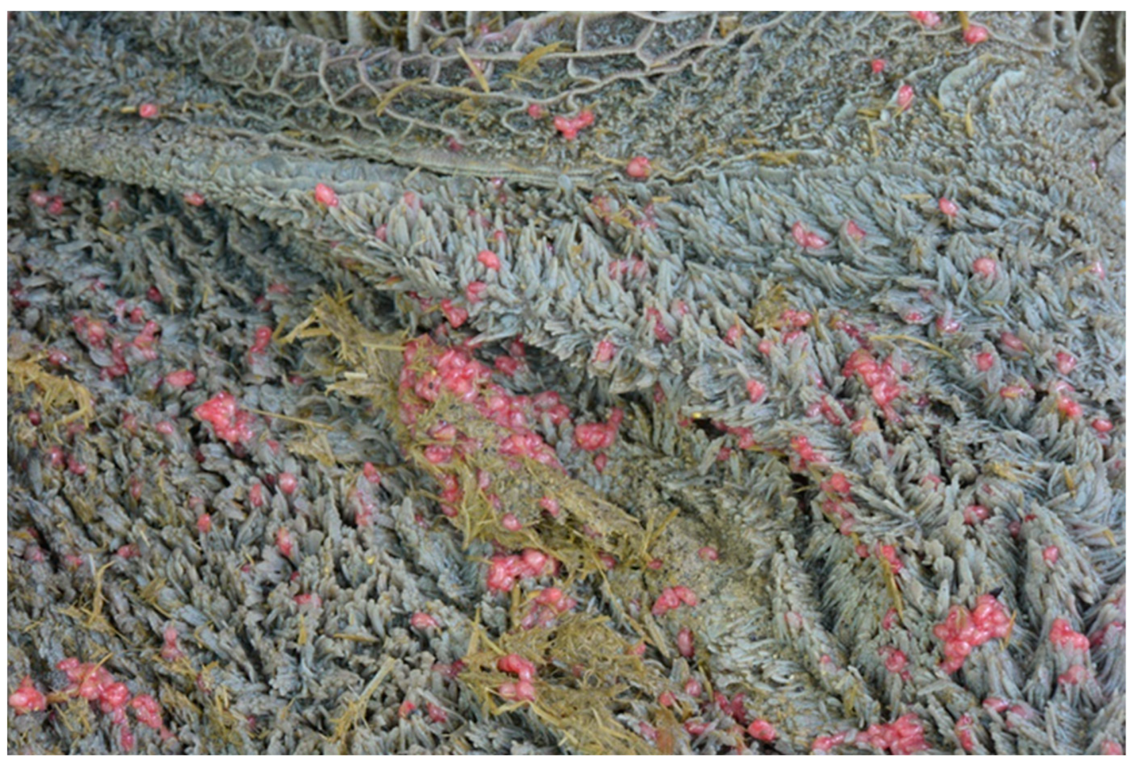
3.2.3. Anatomical Changes

During the second year, 28 rumen and 27 duodenal samples were collected and evaluated. The average papillar density was 21.8 papillae/cm2 and 19.9 papillae/cm2 for ovine and bovine samples, respectively. There was no significant difference between species (p = 0.32l; OR = 6.9; CI = 0.16–288.2). Additionally, no significant relationship was found between adult RF presence and papillar density (p = 0.38; OR = 7.0; CI = 0.1–511.1). The average papillar length was 3.3 mm for ovine samples and 8.6 mm for bovine samples, with a significant difference (p < 0.0001; OR = 0.005; CI = 0.002–0.02) between species. However, no significant difference was found between adult RF presence and papillar length when the bovine/ovine difference was taken into account (p = 0.06; OR = 0.27; CI = 0.08–0.98).
All but one of the twenty-seven duodenal samples had at least one non-specific microscopic change noted (see Appendix A for details of these changes). Immature rumen flukes were not observed in any of the sections examined. There was no significant relationship between any of the seven categories of microscopic duodenal lesions and the presence/absence of adult RF (Table 2).

4. Discussion

In this study, the trends seen in the presence of RF eggs in routine faecal samples are similar to those seen in other surveys in the UK [1] and Ireland [9,24], where the first significant detection of eggs occurred in 2007–2008, followed by a rapid increase in positive diagnoses. A similar trend was observed in France in the 1990s [25], but it is not known where the original introduction of C. daubneyi occurred, nor how it spread so rapidly in Europe. The original isolation and identification of C. daubneyi was in the Kenyan highlands [26] and it seems likely that spread to other countries was caused by movement of livestock; however, long distance movement of snails can also occur [27]. This study also suggests C. daubneyi is the predominant, if not the only, species of RF present in cattle and sheep within Central and Southwest Scotland.
The number of PMEs carried out over the two-year period was relatively small and therefore interpretation of the results should be guarded. The observed percentage of positive RF cases in bovine animals (43.3%) is somewhat higher than previous studies on finished cattle examined at abattoirs (25% in England and Wales [18] and 29% in Scotland [1]). This could be explained by the different source of samples in these studies. Abattoir samples are collected from a healthy, mainly young population of animals, while cases submitted to the University of Glasgow for PME are primarily animals with undiagnosed or chronic diseases. In contrast, the percentage of sheep that had fluke in the rumen was relatively low (13.5%) compared with other recent surveys in Ireland and Wales [5,9,11], though the values were higher than a study conducted in the Netherlands [28]. Whether these are true differences or the consequence of our small sample size cannot be determined from these data.
Over the 10 years of the FECs study, co-infections with RF and liver fluke in sheep and cattle were quite low (2.5%), despite the fact that all infected animals presumably grazed in habitats containing their intermediate host, G. truncatula. Co-infection of snails is considered a rare occurrence (<1%), both experimentally and naturally [2,29]; this points to competition of RF and liver fluke within the intermediate hosts. It has also been speculated that duodenal pathology resulting from immature RF could impede the migration of juvenile liver fluke through the intestinal wall into the abdominal cavity en route to the liver [27]. A potential confounder of these observations is that animals sampled or submitted may have been previously treated with flukicides (other than oxyclozanide) and their liver fluke burdens reduced or eliminated.
Adult RF found at PME were observed at highest densities within the atrium ruminis, surrounding the reticular groove and within the reticulum and, in heavy burdens, flukes were also often observed within the ruminal contents. These observations are in line with current literature citing the location of infection [1,30]. Parasite burdens were predominantly below 100 adult RF, which agrees with findings from a previous abattoir study in the UK, where most parasitized cattle were found to have ≤100 adult RF at necropsy [18], lower than those reported in Spain [31], Belgium [8] and the Netherlands [28]. No gross pathology was observed in the rumen and reticulum of parasitized animals, nor was there any significant association between the density or length of papillae and RF infection status. Previous studies have found shortening, ulceration and necrosis of parasitized papillae [15] and papillar atrophy [15,32], which could suggest either a different response between healthy cattle and this study cohort, or might reflect a different spectrum of host responses to adult rumen fluke, ranging from apparently healthy tissue through to local inflammatory changes around the attachment sites.
No juvenile RF were observed in the duodenum of any parasitized animals and there was no association between duodenal histopathology and the presence of adult RF. This suggests that previous colonisation of the duodenum by juvenile C. daubneyi did not have a long-term impact on duodenal morphology or function. Collectively, these data indicate that infections with adult rumen fluke are not consistently associated with pathological changes in the rumen or duodenum and therefore lend support to the observed relative lack of pathogenicity of adult paramphistomosis in Europe [33,34].

5. Conclusions

This study has demonstrated that, based on coprological methods, the appearance and increasing prevalence of rumen fluke since the late 2000s, which has been observed in other parts of the UK and in Ireland, has also occurred within Central and Southwest Scotland and, furthermore, that the species responsible is predominantly, if not exclusively, C. daubneyi. The frequency of rumen fluke at post-mortem within the source population (diseased animals) was different compared with those reported elsewhere in abattoir surveys and epidemiological studies. Co-infection with liver fluke in sheep and cattle appeared to be uncommon and this may have implications for the epidemiology of these trematodes in regions where they share the same molluscan intermediate hosts, G. truncatula. No evidence could be found for current or historic pathological changes as a direct result of rumen fluke in the rumen or duodenum, supporting evidence for a lack of severe pathological impact in the final host of moderate infections with adult C. daubneyi.

Author Contributions

Conceptualization, V.B., E.G. and A.F.; methodology, E.G., G.R., G.M., P.S., K.W. and C.M., software, E.G.; formal analysis, E.G.; writing—original draft preparation, V.B.; writing—review and editing, V.B., E.G., G.M., P.S., K.W., C.M. and A.F.; visualization, E.G.; supervision, V.B.; project administration, V.B. All authors have read and agreed to the published version of the manuscript.

Funding

This research received no external funding.

Institutional Review Board Statement

The study was conducted in accordance with the University of Glasgow Ethics Committee regulations (Ref 49a/18).

Data Availability Statement

Data are available upon request to the corresponding author.

Acknowledgments

The authors are very grateful to James McGoldrick, Richard Irvine, Alexander Gray and all the post-mortem room staff and the rest of the Pathology team for allowing access to collect samples and for their valued assistance throughout the study.

Conflicts of Interest

The authors declare no conflict of interest.

Appendix A

Table A1. Classification of microscopic duodenal lesions.
Microscopic FeatureDescriptionFeatures AssessedClassification Used
Lymphocytes and plasma cellsInflammation of the lamina propriaThe number of lymphocytes and plasma cells between crypts1 (0–5 cells between crypts)
2 (5–10 cells between crypts)
3 (10–20 cells between crypts)
4 (>20 cells between crypts)
EosinophilsInflammation of lamina propriaThe number of eosinophils in the lamina propria per high-power field (hpf)1 (<5 eosinophils per hpf)
2 (>5 eosinophils per hpf)
Lymphocytes and plasmacells/eosinophilsInflammation of the submucosa/Brunner’s glandsThe presence of lymphocytes, plasma cells and/or eosinophils within the submucosa or between Brunner’s glandsPresent (clusters of lymphocytes, plasma cells and/or eosinophils)
Absent (Rare scattered leucocytes, or complete absence of leucocytes)
GranulomaSubmucosal granulomasPresence of granulomas within the submucosaPresent
Absent
Crypt hyperplasiaHyperplasia of the crypt epitheliumThe length and arrangement of crypts, level of epithelial proliferation and the number of mitosesPresent
Absent
Dilation or hyperplasia of Brunner’s glandsDilation or hyperplasia of Brunner’s glandsGland size and width, and gland epithelial proliferationPresent
Absent
FibrosisFibrosis of the submucosa/between Brunner’s glandsThe presence of greater than two fibrocyte layers separating Brunner’s glands within lobules, or nodules of collagen and fibrocytes surrounding Brunner’s glands.Present
Absent

References

  1. Sargison, N.; Francis, E.; Davison, C.; Bronsvoort, B.M.d.; Handel, I.; Mazeri, S. Observations on the biology, epidemiology and economic relevance of rumen flukes (Paramphistomidae) in cattle kept in a temperate environment. Vet. Parasitol. 2016, 219, 7–16. [Google Scholar] [CrossRef] [PubMed] [Green Version]
  2. Rojo-Vázquez, F.A.; Meana, A.; Valcárcel, F.; Martínez-Valladares, M. Update on trematode infections in sheep. Vet. Parasitol. 2012, 189, 15–38. [Google Scholar] [CrossRef] [PubMed]
  3. Gordon, D.K.; Roberts, L.C.P.; Lean, N.; Zadoks, R.N.; Sargison, N.D.; Skuce, P.J. Identification of the rumen fluke, Calicophoron daubneyi, in GB livestock: Possible implications for liver fluke diagnosis. Vet. Parasitol. 2013, 195, 65–71. [Google Scholar] [CrossRef] [PubMed]
  4. Martinez-Ibeas, A.M.; Munita, M.P.; Lawlor, K.; Sekiya, M.; Mulcahy, G.; Sayers, R. Rumen fluke in Irish sheep: Prevalence, risk factors and molecular identification of two paramphistome species. BMC Vet. Res. 2016, 12, 143. [Google Scholar] [CrossRef] [PubMed] [Green Version]
  5. Jones, R.A.; Williams, H.W.; Dalesman, S.; Brophy, P.M. Confirmation of Galba truncatula as an intermediate host snail for Calicophoron daubneyi in Great Britain, with evidence of alternative snail species hosting Fasciola hepatica. Parasites Vectors 2015, 8, 656. [Google Scholar] [CrossRef] [Green Version]
  6. Duignan, G.; Fagan, J.; Joyce, C. Diagnosing acute larval paramphistomosis in ruminants. Vet. Rec. 2017, 180, 618. [Google Scholar] [CrossRef]
  7. Millar, M.; Colloff, A.; Scholes, S. Disease associated with immature paramphistome infection. Vet. Rec. 2012, 171, 509–510. [Google Scholar] [CrossRef]
  8. Malrait, K.; Verschave, S.; Skuce, P.; Van Loo, H.; Vercruysse, J.; Charlier, J. Novel insights into the pathogenic importance, diagnosis and treatment of the rumen fluke (Calicophoron daubneyi) in cattle. Vet. Parasitol. 2015, 207, 134–139. [Google Scholar] [CrossRef]
  9. Toolan, D.P.; Mitchell, G.; Searle, K.; Sheehan, M.; Skuce, P.J.; Zadoks, R.N. Bovine and ovine rumen fluke in Ireland-Prevalence, risk factors and species identity based on passive veterinary surveillance and abattoir findings. Vet. Parasitol. 2015, 212, 168–174. [Google Scholar] [CrossRef]
  10. Huson, K.M.; Oliver, N.A.M.; Robinson, M.W. Paramphistomosis of Ruminants: An Emerging Parasitic Disease in Europe. Trends Parasitol. 2017, 33, 836–844. [Google Scholar] [CrossRef] [Green Version]
  11. Jones, R.A.; Brophy, P.M.; Mitchell, E.S.; Williams, H.W. Rumen fluke (Calicophoron daubneyi) on Welsh farms: Prevalence, risk factors and observations on co-infection with Fasciola hepatica. Parasitology 2017, 144, 237–247. [Google Scholar] [CrossRef] [Green Version]
  12. Mason, C.; Stevenson, H.; Cox, A.; Dick, I.; Rodger, C. Disease associated with immature paramphistome infection in sheep. Vet. Rec. 2012, 170, 343–344. [Google Scholar] [CrossRef]
  13. O’Shaughnessy, J.; Garcia-Campos, A.; McAloon, C.G.; Fagan, S.; de Waal, T.; McElroy, M.; Casey, M.; Good, B.; Mulcahy, G.; Fagan, J.; et al. Epidemiological investigation of a severe rumen fluke outbreak on an Irish dairy farm. Parasitology 2018, 145, 948–952. [Google Scholar] [CrossRef]
  14. Hoyle, R.C.; Rose Vineer, H.; Duncan, J.S.; Williams, D.J.L.; Hodgkinson, J.E. A survey of sheep and/or cattle farmers in the UK shows confusion over the diagnosis and control of rumen fluke and liver fluke. Vet. Parasitol. 2022, 312, 109812. [Google Scholar] [CrossRef]
  15. Fuertes, M.; Perez, V.; Benavides, J.; Gonzalez-Lanza, M.C.; Mezo, M.; Gonzalez-Warleta, M.; Giraldez, F.J.; Fernandez, M.; Manga-Gonzalez, M.Y.; Ferreras, M.C. Pathological changes in cattle naturally infected by Calicophoron daubneyi adult flukes. Vet. Parasitol. 2015, 209, 188–196. [Google Scholar] [CrossRef]
  16. Huson, K.M.; Morphew, R.M.; Allen, N.R.; Hegarty, M.J.; Worgan, H.J.; Girdwood, S.E.; Jones, E.L.; Phillips, H.C.; Vickers, M.; Swain, M. Polyomic tools for an emerging livestock parasite, the rumen fluke Calicophoron daubneyi; identifying shifts in rumen functionality. Parasites Vectors 2018, 11, 617. [Google Scholar] [CrossRef] [Green Version]
  17. Bruguera Sala, A. Caseload of a Farm Animal Veterinary Teaching Hospital as a Form of Passive Surveillance with Particular Reference to Bovine Viral Diarrhoea Virus. Master’s Thesis, University of Glasgow, Glasgow, Scotland, 2017. Available online: https://theses.gla.ac.uk/8000/1/2016BrugueraSala.MVMpdf.pdf (accessed on 1 January 2018).
  18. Bellet, C.; Green, M.J.; Vickers, M.; Forbes, A.; Berry, E.; Kaler, J. Ostertagia spp., rumen fluke and liver fluke single- and poly-infections in cattle: An abattoir study of prevalence and production impacts in England and Wales. Prev. Vet. Med. 2016, 132, 98–106. [Google Scholar] [CrossRef] [Green Version]
  19. Mitchell, G.; Zadoks, R.N.; Skuce, P.J. A Universal Approach to Molecular Identification of Rumen Fluke Species Across Hosts, Continents, and Sample Types. Front. Vet. Sci. 2021, 7, 605259. [Google Scholar] [CrossRef]
  20. Itagaki, T.; Tsumagari, N.; Tsutsumi, K.-I.; Chinone, S. Discrimination of three amphistome species by PCR-RFLP based on rDNA ITS2 markers. J. Vet. Med. Sci. 2003, 65, 931–933. [Google Scholar] [CrossRef] [Green Version]
  21. Rinaldi, L.; Perugini, A.G.; Capuano, F.; Fenizia, D.; Musella, V.; Veneziano, V.; Cringoli, G. Characterization of the second internal transcribed spacer of ribosomal DNA of Calicophoron daubneyi from various hosts and locations in southern Italy. Vet. Parasitol. 2005, 131, 247–253. [Google Scholar] [CrossRef]
  22. Kern, R.; Lindholm-Perry, A.; Freetly, H.; Kuehn, L.; Rule, D.; Ludden, P. Rumen papillae morphology of beef steers relative to gain and feed intake and the association of volatile fatty acids with kallikrein gene expression. Livest. Sci. 2016, 187, 24–30. [Google Scholar] [CrossRef] [Green Version]
  23. Day, M.J.; Bilzer, T.; Mansell, J.; Wilcock, B.; Hall, E.J.; Jergens, A.; Minami, T.; Willard, M.; Washabau, R. Histopathological Standards for the Diagnosis of Gastrointestinal Inflammation in Endoscopic Biopsy Samples from the Dog and Cat: A Report from the World Small Animal Veterinary Association Gastrointestinal Standardization Group. J. Comp. Pathol. 2008, 138, S1–S43. [Google Scholar] [CrossRef] [PubMed]
  24. Zintl, A.; Garcia-Campos, A.; Trudgett, A.; Chryssafidis, A.L.; Talavera-Arce, S.; Fu, Y.; Egan, S.; Lawlor, A.; Negredo, C.; Brennan, G.; et al. Bovine paramphistomes in Ireland. Vet. Parasitol. 2014, 204, 199–208. [Google Scholar] [CrossRef] [PubMed]
  25. Mage, C.; Bourgne, H.; Toullieu, J.M.; Rondelaud, D.; Dreyfuss, G. Fasciola hepatica and Paramphistomum daubneyi: Changes in prevalences of natural infections in cattle and in Lymnaea truncatula from central France over the past 12 years. Vet. Res. 2002, 33, 439–447. [Google Scholar] [CrossRef] [PubMed] [Green Version]
  26. Dinnik, J. Paramphistomum daubneyi sp. nov. from cattle and its snail host in the Kenya Highlands. Parasitology 1962, 52, 143–151. [Google Scholar] [CrossRef]
  27. Forbes, A. Rumen fluke: Past, present and future. Livestock 2018, 23, 227–231. [Google Scholar] [CrossRef]
  28. Ploeger, H.W.; Ankum, L.; Moll, L.; van Doorn, D.C.K.; Mitchell, G.; Skuce, P.J.; Zadoks, R.N.; Holzhauer, M. Presence and species identity of rumen flukes in cattle and sheep in the Netherlands. Vet. Parasitol. 2017, 243, 42–46. [Google Scholar] [CrossRef]
  29. Iglesias-Piñeiro, J.; González-Warleta, M.; Castro-Hermida, J.A.; Córdoba, M.; González-Lanza, C.; Manga-González, Y.; Mezo, M. Transmission of Calicophoron daubneyi and Fasciola hepatica in Galicia (Spain): Temporal follow-up in the intermediate and definitive hosts. Parasites Vectors 2016, 9, 610. [Google Scholar] [CrossRef] [Green Version]
  30. Ferreras, M.C.; González-Lanza, C.; Pérez, V.; Fuertes, M.; Benavides, J.; Mezo, M.; González-Warleta, M.; Giráldez, J.; Martínez-Ibeas, A.M.; Delgado, L.; et al. Calicophoron daubneyi (Paramphistomidae) in slaughtered cattle in Castilla y León (Spain). Vet. Parasitol. 2014, 199, 268–271. [Google Scholar] [CrossRef] [Green Version]
  31. Gonzalez-Warleta, M.; Lladosa, S.; Castro-Hermida, J.A.; Martinez-Ibeas, A.M.; Conesa, D.; Munoz, F.; Lopez-Quilez, A.; Manga-Gonzalez, Y.; Mezo, M. Bovine paramphistomosis in Galicia (Spain): Prevalence, intensity, aetiology and geospatial distribution of the infection. Vet. Parasitol. 2013, 191, 252–263. [Google Scholar] [CrossRef] [Green Version]
  32. Devos, J.; Vassiloglou, B.; Amenna-Bernard, N.; Marcotty, T. Paramphistomosis in sheep; natural infection of lambs by Calicophoron daubneyi. Rev. Med. Vet. 2013, 11, 0035–1555. [Google Scholar]
  33. Atcheson, E.; Lagan, B.; McCormick, R.; Edgar, H.; Hanna, R.E.B.; Rutherford, N.H.; McEvoy, A.; Huson, K.M.; Gordon, A.; Aubry, A.; et al. The effect of naturally acquired rumen fluke infection on animal health and production in dairy and beef cattle in the UK. Front. Vet. Sci. 2022, 9, 968753. [Google Scholar] [CrossRef]
  34. Delafosse, A. Rumen fluke infections (Paramphistomidae) in diarrhoeal cattle in western France and association with production parameters. Vet. Parasitol. Reg. Stud. Rep. 2022, 29, 100694. [Google Scholar] [CrossRef]
Figure 1. (A) Opened rumen and reticulum. Black outline represents sampling area (atrium ruminis); (B) Plastic template of 3 squares 3 × 1 cm size.
Ruminants 03 00004 g001
Figure 2. The percentage of faecal samples testing positive for rumen fluke eggs and liver fluke eggs during the study period (2008–2018).
Ruminants 03 00004 g002
Figure 3. Seasonal distribution of positive faecal samples for rumen fluke (n = 201) and liver fluke (n = 471).
Ruminants 03 00004 g003
Figure 4. Adult RF in the reticulo-rumen of a bovine with burden score of 4.
Ruminants 03 00004 g004
Table 1. Number of animals (total, bovines and ovines) in relation to adult RF burden.
Adult Rumen Fluke Burden ScoreNumber of Adult Rumen FlukeTotal Number of Infected Animals (Percentage %)Total Number of Infected Bovines (Percentage %)Total Number of Infected Ovines (Percentage %)
11–105 (27.8%)1 (7.7%)4 (80%)
211–1007 (38.9%)6 (46.2%)1 (20%)
3101–2003 (16.6%)3 (23.1%)0
4201+3 (16.6%)3 (23.1%)0
Table 2. Estimate and p-value output from the generalised linear models examining the relationship between adult rumen fluke presence and the histopathological changes of the microscopic duodenal lesions (n = 27).
Histopathological ChangesEstimateStandard Errorp-ValueOdds Ratio (OR)95% Confidence Interval
Inflammation of lamina propria (Lymphocytes and plasma cells)1.181.180.323.230.32–32.48
Inflammation of lamina propria (Eosinophils)−1.140.910.210.320.05–1.90
Inflammation of submucosa/Brunner’s glands (Lymphocytes and plasma cells/eosinophils)−0.120.890.900.890.16–5.08
Inflammation of submucosa/Brunner’s glands (Granuloma)0.821.050.432.270.29–17.58
Crypt hyperplasia−2.031.320.130.130.01–1.76
Dilation or hyperplasia of Brunner’s glands−1.391.000.170.250.04–1.77
Fibrosis submucosa/Brunner’s glands−0.220.940.810.800.13–5.09
Disclaimer/Publisher’s Note: The statements, opinions and data contained in all publications are solely those of the individual author(s) and contributor(s) and not of MDPI and/or the editor(s). MDPI and/or the editor(s) disclaim responsibility for any injury to people or property resulting from any ideas, methods, instructions or products referred to in the content.

