Article

Anatomical Distribution of Ultraviolet Radiation Depends on Phototherapy Unit Design and on Personal Height and Body Mass

by
Hans Christian Wulf
*,
Peter A. Philipsen
and
Jakob Heydenreich
Department of Dermatology, Bispebjerg University Hospital, 2400 Copenhagen NV, Denmark
*
Author to whom correspondence should be addressed.
Int. J. Transl. Med. 2023, 3(1), 66-80; https://doi.org/10.3390/ijtm3010006
Submission received: 18 November 2022 / Revised: 15 December 2022 / Accepted: 16 January 2023 / Published: 20 January 2023

Abstract

:
Phototherapy using ultraviolet radiation (UVR) treatment units of various designs is common in dermatology. The anatomical distribution of UVR should be even, regardless of individual body shapes. Using electronic dosimeters, we measured the irradiance at 31 body sites on 12 persons of different heights and body mass (BMI). Five different treatment unit designs were tested: cabinet units with standing patients, units with patients lying down, and a unit where patients rotated in front of flatly arranged UVR tubes. In treatment units with short tubes, persons taller than 170 cm received low irradiance on the face, neck, and shoulders. In cabinet-type units, higher BMI lowered the irradiance on the chest and belly. The relative standard deviation (RSD) of irradiance was smallest for the rotating unit, and for the unit with patients lying down while irradiated from above only. A higher RSD was found in the unit designs where patients stood inside cabinets, and where patients lay down and were simultaneously irradiated from both sides. In general, longer tubes lower the overall RSD. The irradiance of the different body areas is about 60% of the measured calibration values, but to avoid provoking any erythema, the treatment dose can only be increased by 10%.

1. Introduction

Phototherapy has been in use for a very long time to treat various skin diseases [1,2,3,4]. The efficacy of full-body phototherapy depends on the type and severity of the skin disease, on the appropriate body distribution of ultraviolet radiation (UVR), on the sensitivity of different skin areas, and on the level of pigmentation. Individual body mass index (BMI) may highly influence patients’ proximity to the UVR tubes, and variations in height may lead to some patients having areas of their body outside the length of the tubes. Therefore, the phototherapy unit designs may be of vast importance to the anatomical distribution of UVR intensity.
The phototherapy units are constructed as cabinets containing various lengths of vertically arranged fluorescing UVR tubes surrounding a standing person, or tubes arranged horizontally irradiating a lying person from above or from both sides simultaneously (Figure 1). As an alternative, we constructed a device where the person rotates while standing in front of a narrow flat tube panel (Figure 1b).
The anatomical uniformity of irradiance attracted attention early when PUVA treatment units equipped with fluorescent tubes became common [5]. Measurements were performed with film badges mounted on a manikin. The irradiance differed greatly with only low intensities reaching the insides of legs and body areas shadowed by the arms.
Diffey et al. [6] also investigated the UV distribution on a manikin rotating in front of a filtered medium-pressure mercury arc source and found very low irradiance to the face and legs.
The aim of this study was to investigate which treatment unit design could provide the most even distribution of UVR to all skin areas (except scalp and soles) in persons of different heights and BMI. Additionally, we aimed to determine how the UVR dose to different anatomical areas related to the calibration. We investigated the anatomical irradiance distribution of 12 volunteers in five different phototherapy units (Figure 1). No treatment interventions were performed.

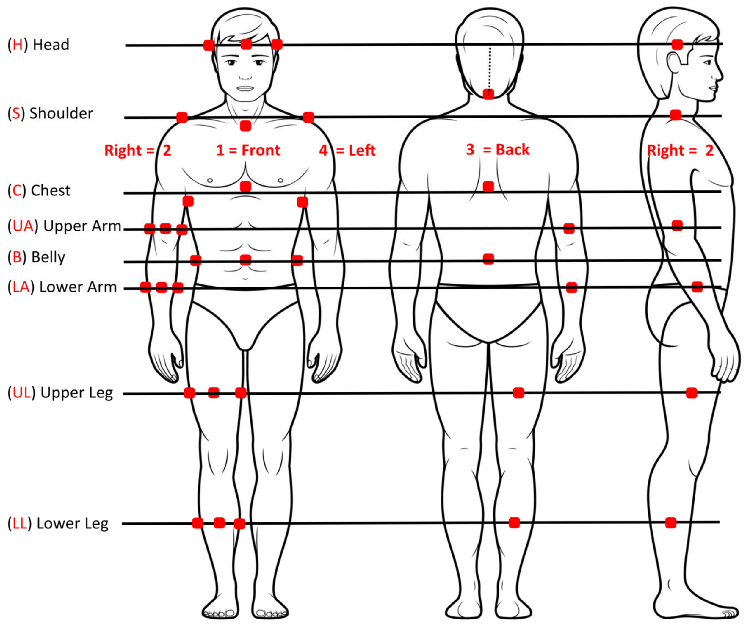
2. Materials and Methods
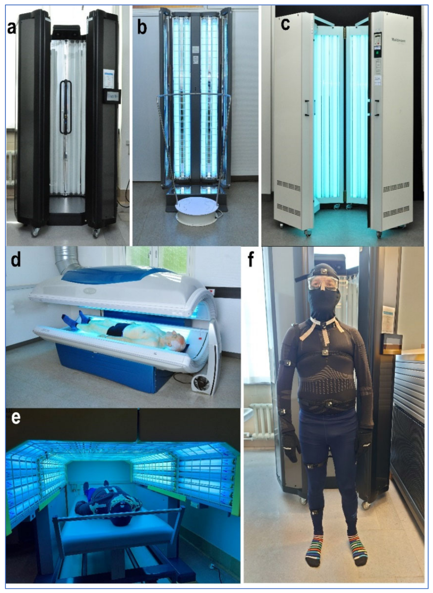
This is an observational prospective quality control study without treatment intervention. Such studies do not, according to the Danish National Ethics Center (https://nationaltcenterforetik.dk/ansoegerguide/overblik/hvad-skal-jeg-anmelde, accessed on 13 January 2023), require an ethics committee approval.
Informed written consent was obtained from all 12 subjects involved in this study concerning participation as well as the publication of this paper.
The height of each participant was given in meter and the body weight in kg. The BMI (kg/m2) was calculated and used in the calculations of the relation between height, BMI, and irradiance in the 5 treatment units. All participants were equipped with tight-fitting clothes including a balaclava and proctective, yellow-tinted, UVR-absorbing PUVA eyewear (Figure 1f). The penetration of UVR through the clothing was measured and found to be less than 1 permille of the UVR intensity in the treatment units, leaving the participants virtually unirradiated during the procedure. The participants were equipped with 31 individually calibrated dosimeters mounted on their clothes (Figure 1f). The positions are seen in Figure 2. The UVR dose was measured in the standard erythema dose (SED). The dosimeters can measure a maximum of 25 SED/hour (0.007 SED/s). The tubes in each unit were chosen to accommodate this. Measurements of the SED [7] were performed with the well-described electronic dosimeters (SunSaver, Bispebjerg University Hospital, Copenhagen, Denmark) [8,9]. Their sensitivity corresponded to the erythema action spectrum, and each dosimeter was set to measure every second. The dosimeter detectors had close to ideal cosine responses.
The calibration of all dosimeters was performed using a double monochromator (Bentham DM150, Bentham Instruments, Ltd., Newbury, UK). Measurements by the monochromator were performed at every nanometer in the UVR range in the middle of the tubes at a 30 cm distance in units equipped with Philips (Amsterdam, The Netherlands) TL01, TL09, and TL10 tubes. The 31 dosimeters were calibrated in the exact same position in all treatment units. Subsequently, a mosaic of nine (3 × 3) SunSaver dosimeters was used to calibrate other treatment units. Calibration was performed according to the regulations [10,11,12]. All light sources were turned on 5 min before the start of the study to stabilize their intensity [10]. Measurements were performed every second, and the average irradiance during the total irradiation period was calculated from these.
I = 1 n   S S t n   ( SED / hour )
where I = intensity; n = number of measurements; SS = sum of all SunSaver measurements; t = number of seconds.
The five phototherapy units were as follows (Figure 1).

2.1. Unit A

Unit A was the PUVA Daavlin cabinet Series 3, NeoLux (Daavlin, Bryan OH, USA) with the test persons in a standing position and irradiated from all sides (Figure 1a). The cabinet was equipped with 40 tubes placed in an octagon which measured 85 cm from side to side and 72 cm from door to between the tubes in the back of the cabinet. The Philips TL09 tubes measured 2 m in length. A total of 180 readings of one-second intervals were performed by every dosimeter. The average intensity was calculated from the equation:
I = 1 180   S S t 180
A part of the measurement string for dosimeter C3 (upper back of one person) in treatment unit A is shown here ((SED/h) × 10): 47 46 46 46 46 46 47 47 46 46 46 46 46 46 46 46 45 46 46 46 46 46 46 45 46 46 46 46 46 47.
The measurement string was relatively stable (range 43–48, n = 180).

2.2. Unit B

This phototherapy unit was constructed for this study purpose (Figure 1b). It consisted of a rotating platform (40 cm in diameter) on which the persons were standing while holding on to a 105 cm high railing to keep the arms lifted in a fixed position. In front of this, a flat light source, Daavlin cabinet Series 7 × 311, 4D (Daavlin, Bryan, OH, USA), mounted with 4 Philips TL01 tubes with a length of 180 cm, irradiated the person during 10 rotations of 26 s. A total of 261 readings with one-second intervals were performed by every dosimeter. The average intensity was:
I = 1 261   S S t 261
A part of the measurement string for dosimeter C3 (upper back of one person) in treatment unit B is shown here ((SED/h) × 10): 0 0 33 190 395 437 622 676 540 303 282 85 0 0 0 0 0 0 0 0 0 0 0 0 0 0 0 0 0 0 0 0 0 0 0 0 0 0 0 0 56 214 434 636 643 669 512 294 265 75 0 0.
The zeroes illustrate the moment when the dosimeters pointed away from the light source during the patient’s rotation. Accordingly, the exposure intensity greatly varied (range 0–690, n = 261).
As the TL01 tubes could not be exchanged to UVA tubes, we reduced the erythrogenic dose by covering the tubes with a density filter.

2.3. Unit C

Unit C was the UV1000L cabinet (Waldmann, Villingen-Schwenningen, Germany) equipped with Philips TL09 tubes measuring 180 cm in length (Figure 1c). The illumination length was 160–165 cm as the tops of the tubes were covered by a ventilator and the bottoms by a 10 cm metal plate. The cabinet contained 26 tubes placed in a hexagon. The distance between tubes from side to side was 80, and 65 cm from the door to the back of the cabinet. The persons were irradiated from all sides in a standing position with 170 readings of one-second intervals by every dosimeter. The average intensity was:
I = 1 170 S S t 170

2.4. Unit D

Unit D was Sunvision (Alisun, Odense, Denmark) equipped with 40 Philips TL10 tubes below and above the person (Figure 1d). The persons, with their heads resting on a fixed pillow so the face only received indirect irradiation, were placed lying on an 80 cm wide plexiglass plate on top of the bench tubes, and then the upper part of the treatment unit was lowered to a standard position. The head was always in the same position, independent of the test person’s height. The tube length was 170 cm. A total of 180 readings of one-second intervals were performed by every dosimeter. The average intensity was:
I = 1 180 S S t 180

2.5. Unit E

Unit E was the PUVA 4000 phototherapy unit (Waldmann, Germany) with 40 tubes above and on both sides of the test persons (Figure 1e). The persons were irradiated for about 3 min lying on their back, then turned over and irradiated face-down for about 3 min. The unit was equipped with Philips Cleo performance tubes with a total length of 240 cm. The total number of readings was 170 × 2 of one-second intervals by every dosimeter. The average intensity was:
I = 1 340   S S t 340

2.6. Statistics

A proper sample size could not be calculated as no information about the standard deviation of data from the investigated treatment units was available. However, we measured a very low relative standard deviation (RSD) of 3.6% based on five consecutive measurement series of 23 uncovered dosimeters attached to a test person entering and exiting treatment unit A. The low RSD of 3.6% and an expected body shape effect of 5.4% would require 8 persons to complete the study, assuming α = 0.05 and β = 0.80. The SED/sec was given as the mean. Standard deviation (SD) and RSD (SD/mean × 100) were used to compare the individual treatment units. Pearson’s correlation (r) was used to evaluate the relation between dosimeter readings, height, and BMI of all participants. Straight lines illustrate the relation between irradiance and height and BMI (Figure 3). SPSS statistics for Windows version 25 (IBM, Armonk NY, USA) was used. p-values < 0.05 were considered significant.

3. Results

Twelve subjects volunteered to participate in this study (Table 1). The 12 participants had an average height of 173 cm (160–205 cm) and an average BMI of 27.2 (18–43). Each person was equipped with 31 electronic dosimeters distributed on the entire body (Figure 1f and Figure 2) and subsequently placed in the five different treatment units.

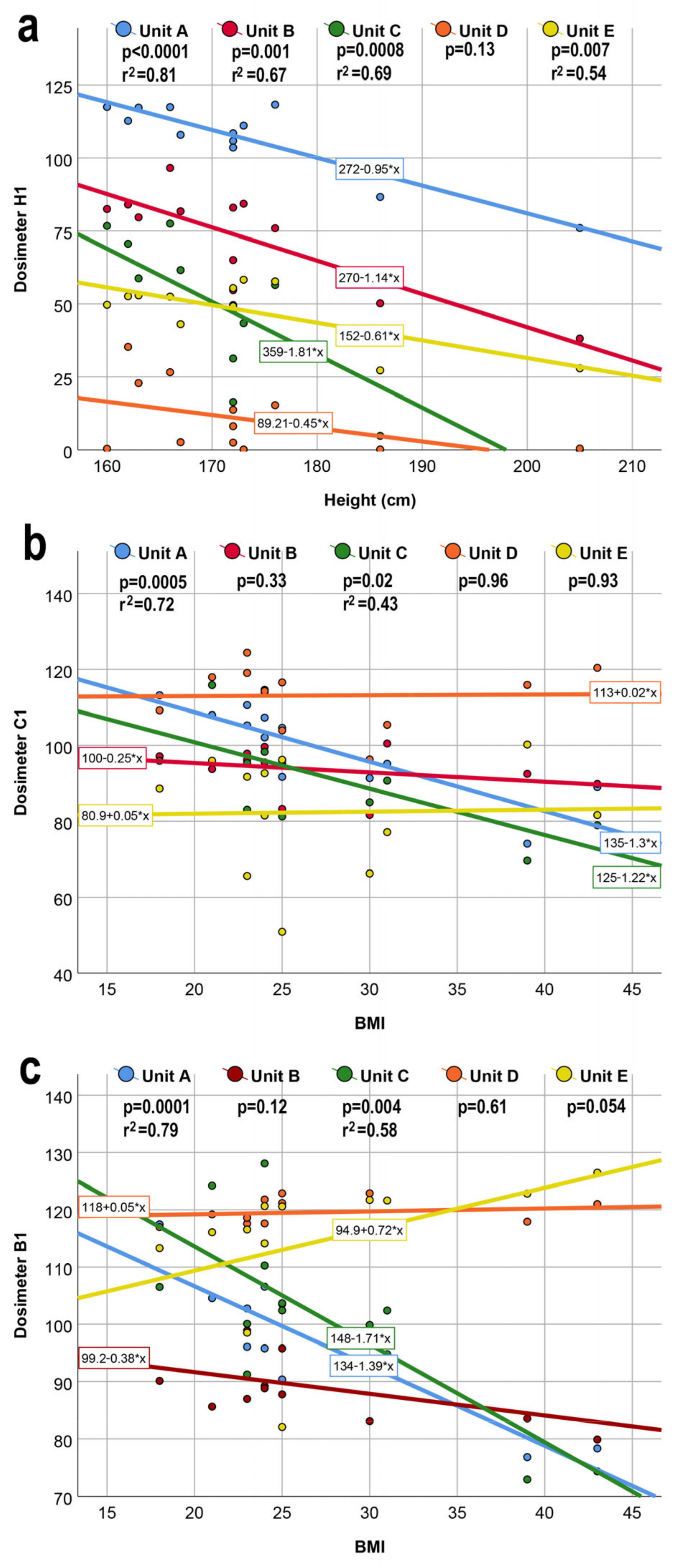
3.1. Importance of Body Shape

Individual variations of irradiance depended primarily on the person’s height as several cabinets were relatively short. Figure 3a shows a negative correlation to individual height in unit A, unit B, unit C, and unit E, indicating the importance of unit design, especially for patients taller than 170 cm (H1 measurements). A negative correlation between height and measured irradiance was mainly seen for the head (H1) and shoulders (S1, S2, and S4) (correlation coefficients r = −0.6 to r = −0.9, p < 0.05). A negative relation for unit E was found on the trunk front (C1) and back (C3) (r = −0.6, p < 0.05). All of the unit D measurements had no relation to height (p > 0.05), except UA4 (r = −0.6, p = 0.04), probably because the head was in a fixed position independent of tube length. In general, there was no significant correlation between height and irradiance on arms and legs. This was found for 78 out of 80 correlations.
Having a high BMI usually indicates a voluminous belly, and the BMI is primarily related to the measurement of B1 (center of abdomen), C1 (breast), and B3 (lumbar region). The irradiance decreased with increasing BMI (Figure 3b,c), but only significantly for unit A and unit C constructed as cabinets (r = −0.7 to r = −0.8, p < 0.05). The sides of the belly were negatively correlated to irradiance (B2 and B4, r = −0.6 to r = −0.7, p < 0.05) for unit A and unit C, but not to unit B, unit D, and unit E. Unit E had a positive correlation for B2 and B3 (r = 0.6, p = 0.03). Generally, there was no significant correlation between BMI and irradiance on arms and legs (71 of 80 correlations), the head (0 of 20 correlations), and shoulders (2 of 15 correlations).

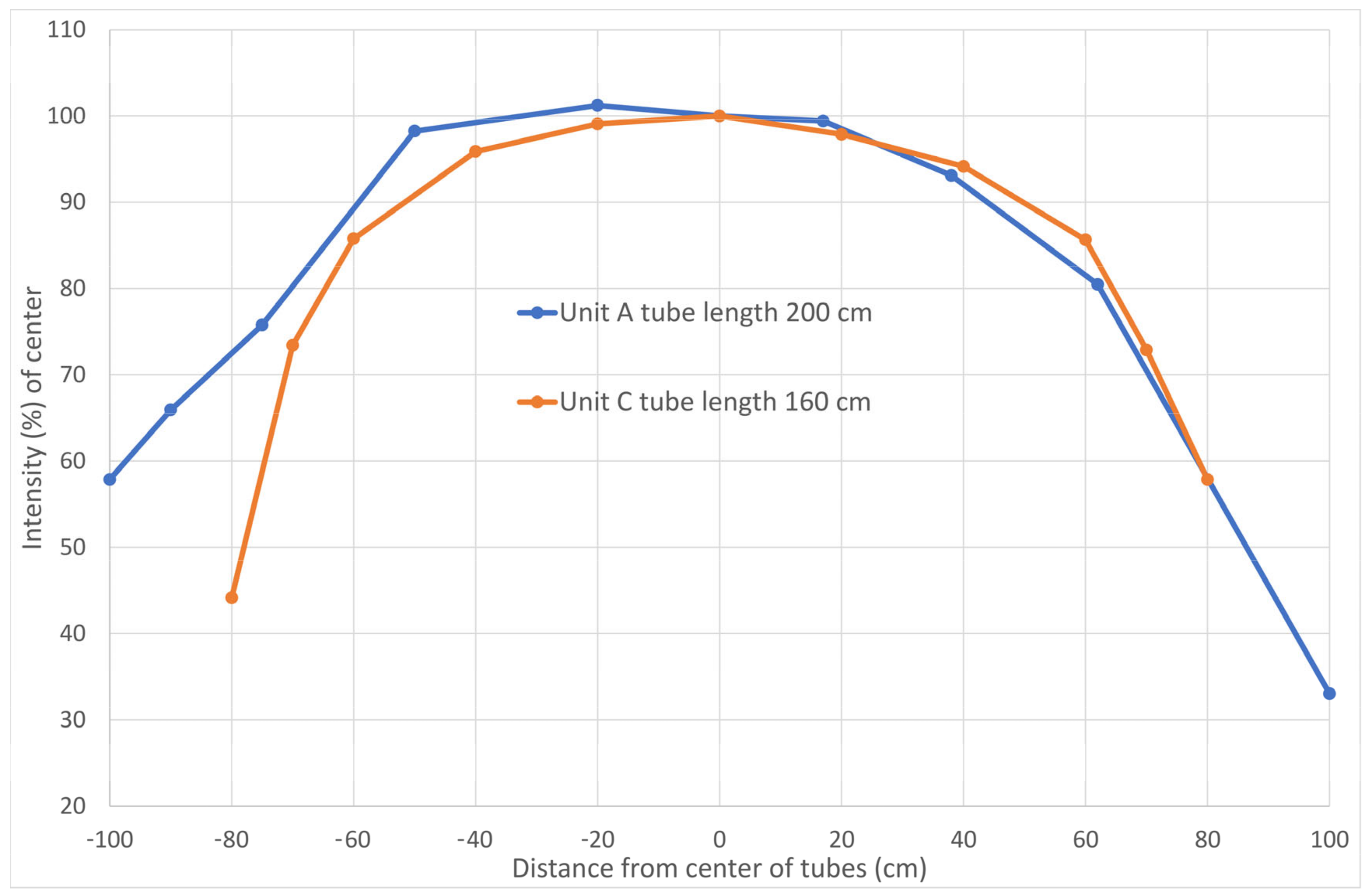
3.2. Importance of Phototherapy Unit Design

The irradiance measured at the 31 body locations was most stable on the upper back, corresponding to dosimeter C3, and this area was chosen for normalization to 100%. All other dosimeter measurements are given in % of C3 in Tables S1a–S5a. In this way, the cabins were comparable independent of UVR intensity.
A cabinet must be able to handle a wide range of body shapes (height and BMI). We calculated the average irradiance and SD of each dosimeter for all 12 participants combined (Table 2). It also included the relative standard deviation (RSD = SD/average × 100), which allows us to compare the performance of the treatment units. The dosimeters measured particularly low values on the side of the body (C2 and C4), on the inside of the arm (UA4 and LA4), and the inside of the thigh (UL4), often <25% of C3 intensity. Likewise, the upper part of the shoulders (S2 and S4) and the face (H2 and H4) were poorly exposed on tall persons (Tables S1a–S5a). To demonstrate which unit distributed the irradiance most uniformly (Table 2, light grey color), we compared the RSD for each body part, showing which part presented the lowest value (most even distribution). The percentage irradiance ranges in the different units were as follows: unit A: 7–125; unit B: 12–100; unit C: 7–119; unit D: 7–131; unit E: 20–114. Overall, the rotating unit B performed best (average RSD % = 22.4), followed by unit E (23.4), unit A (31.3), unit C (42.2), and unit D (50.0). When the RSD for unit B was normalized to 1.0, the numbers for the other units were: unit E: 1.04; unit A: 1.40; unit C: 1.88; unit D: 2.23. The irradiance distribution was clearly best in unit B and unit E.
As the length of the tubes might be of special significance to the irradiation of the face (H) and shoulders (S), we have examined how the RSD % performed without H and S dosimeter measurements (Table 2). Unit B still performed best with RSD % (20.5); unit E (23.5); unit D (33.6); unit A (37.3); and unit C (42.8). The long tubes in unit A resulted in a clearly reduced RSD when H and S dosimeter measurements were included, and long tubes should, ideally, be used in treatment units universally.

3.3. Received UV Irradiance of Calibration

First, each of the unoccupied phototherapy units was calibrated in an area with the highest UVR intensity at the middle of the tubes. Then, we investigated how the irradiance was affected by placing the test persons in the treatment units, resulting in a different distance between the tube and body site compared to the calibration distance. This was of particular interest when calculating the irradiance and treatment dose. Tables S1b–S5b show the relation between the absolute calibration intensity and the individual measurements, demonstrating to what extent a correction should be performed to avoid a sunburn when treating skin diseases. On average, our test persons received between 52 and 64% of the calibration value, due to the differences in doses received to the face and shoulders (Table 3). Hardly any differences in dose were found on the rest of the body (61–65%) for all treatment units. Theoretically, the exposure could be increased by a factor of 1.6, but as the SD for an individual person was substantial, only an increase of about 10% could be tolerated. Tables S1b–S5b show that only a few dosimeters received more than 100% of the calibration value.

4. Discussion

We chose five very different phototherapy units for this study to investigate which design is advantageous for delivering an even, full-body irradiance. Some older unit designs are now discontinued, mainly based on how much space they occupy, not on investigations into irradiation properties. We made sure that our participants represented very different body shapes (height and BMI), as the treatment units must be able to accommodate all shapes. Even though we used tubes producing low erythrogenic UVR doses, we still protected the participants with full-body clothes, gloves, balaclavas, and goggles (Figure 1f), and none developed skin erythema even after sequential exposure to UVR in all 5 units on the same day.
In many parts of the world, home treatment is preferred to save patients’ time and travel costs [13,14]. We constructed a space-saving unit (unit B) where the patient rotated while standing up. This was developed for home treatment as well as for use in office-based clinics. Achieving an even UVR distribution may be very difficult if the patients themselves must turn around in front of a narrow UVR source. Even distribution is easily obtainable when the patient stands on a slowly rotating platform, and UVR doses can be regulated by the number of rotations. Unit B was very usable particularly for short exposure with TL01, whereas treatment with UVA (TL09 or TL10) may last too long. All other tested units were unsuitable for home use. Narrowband UVB (TL01) is the most widely used in phototherapy, and the treatment takes seconds to minutes only. It could be acceptable to prolong the treatment, as in the rotating unit. This may also improve the dose accuracy as the UVR irradiance delivered by the units may vary, particularly in the first minutes after turning on the lamps.
UVR intensity depends on the temperature of the light tubes, and devices may deliver up to 10% higher intensity during the first minutes if they have been used a short while ago. When the tubes are cold, the intensity increases within the first five minutes after turning on the unit [15]. To counter this, we used a burn-in time of 5 min shortly before exposing each person [10,15]. This may further indicate an advantage of longer exposure times in the daily settings. Some units are equipped with UVR dose measurement devices to accommodate for the differences in intensity over time [15].
The increasing height of our population will cause a low grade or lack of UV exposure to the face, neck, and shoulders when treating patients taller than 170 cm in units with shorter tubes. Using units with longer tubes (units A and E) will address this issue. In unit D, the head was always in a fixed position, and lower legs and feet may not be irradiated in tall patients. This, however, was not addressed in our set-up, as the lowest-positioned dosimeters were placed in the middle of the lower leg. Longer tubes would improve the distribution of UVR (decreasing RSD) to the benefit of all patients.
The space inside the stand-up cabinets (units A and C) was rather narrow, and patients with a high BMI were positioned with their belly circumference (B1–B4 dosimeters) very close to the tubes, presumably exposing their body to an increased intensity of direct UVR. In units A and C, however, we observed that persons with a high BMI received less irradiance to the belly and chest area, probably because a more voluminous body will shade the diffuse irradiation (Figure 3). Direct irradiation was only affected to a limited degree due to the almost linearity between the intensity and distance from the skin level to tube level, as the lighting area was large. However, UVR from the periphery was diminished which resulted in a lower total intensity = self-shielding [16]. With patients lying down, the belly will flatten, minimizing this problem. The importance of BMI for the UVR distribution is only significant in treatment units A and C (Figure 3).
The calibration of units is performed without occupants, and the measured irradiance is different when the unit is occupied by a person. This will lower the exposure dose compared to the calibration value [16,17], as the distance from a person to the tubes will not be the same as at calibration. This would be the case for unit A, C, and D, but not necessarily for units B and E, where the irradiation was one-sided. Calibration was conducted at a 30 cm distance from the center of the tubes where intensity was maximal [16,18]. In units where the patient was resting on a transparent plate and irradiated from below, the calibration was performed directly on the plate, as described in the Danish Standard of 2015 regarding the control measurement of sunbeds [11]. In this case, the back of the person will receive the calibrated dose. The intensity will always be lower close to the tube ends where the UV only reaches a person from one side, as on the feet and face/scalp (Figure 4). This, particularly, was the case for unit D, in which the head was in a fixed position and the lower legs and feet of tall people may extend outside the tube area. The irradiance emitted by the tube ends may be as low as 40% of the irradiance in the middle of the tubes, measured in treatment units A and C. With a person occupying units A and C, the irradiance was higher for dosimeters LL1–LL4 because the dosimeters were placed about 30 cm from the tube ends (Figure 4, Table 3 and Tables S1b and S3b). The problem associated with low intensity at the tube ends could be addressed by placing the tubes horizontally across to provide equal intensity to the patient’s full height. If only a limited area is to be treated, e.g., the head or lower legs, a section of tubes could be turned off. The future use of UVR LEDs will further solve this problem.
The intensity reaching the sides of the body was generally low but might be increased by instructing the patients to change position while irradiated (overriding the instruction provided in the manual). This was partly managed in unit B with the arms in a raised position, and in unit E with the persons lying on their back on a wide couch. Positioning the arms without shading the body sides may be particularly difficult in cabinet-shaped units where space is limited.
Although the irradiation in most cases was lower than the calibration value, it was limited by how much the dose can be increased to avoid erythema in parts of the body as described by Martin et al. [19]. The level of increase was estimated to about 10% in this study, and dose regulation may not be performed.
A limitation of the measurement technique is that while the dosimeter positions on the body were easy to maintain when the person was standing up as in units A, B, and C, it was more difficult to ensure that the dosimeters will stay in place while lying down and turning over inside a unit. In unit D, the persons were simultaneously irradiated from both sides with different intensities from the lower and upper part. The upper part gave about 9% higher intensity than the lower part. We found that UVR intensity in the center of treatment units A and C differed by up to 15% when measuring between 0 and 360° directions in a horizontal plane in the middle of the tubes. Calibration intentionally is performed where UVR intensity is highest. However, the rather low mean % of the calibration value suggests substantial variations in light intensity of the different parts of the treatment units.
Even if the intensities reaching the different body parts were identical, this might not be ideal, as various levels of UVR sensitivity are found depending on skin sites. The construction of new treatment units may take this issue into account by intentionally having different intensities of, e.g., the legs and the rest of the body, or by measuring skin pigmentation and adjusting the dose accordingly [20].

5. Conclusions

In conclusion, irradiation in phototherapy units with TL01 tubes, where treatment time is short, may be performed standing up. When the irradiation takes longer, it may be preferable to use a unit where the person lies down. When placed along the body, as in all our tested treatment units, the tubes must be at least 200 cm and preferably longer. As the intensity is rather low at the end of the tubes, this could be compensated by placing the tubes horizontally across the body. In treatment unit B, the tube length could be just 60 cm. Here, it would be easy to place the tubes across the patient at any preferred height and turn off parts of the tubes when not performing the full-body treatment. As the arms must be kept away from the body as in unit B, and unit E, this requires some space which will also accommodate differences in BMI. Suitable for home treatment, unit B is convenient and saves space while securing good irradiation distribution properties. Unit B and unit E had the lowest RSD and the most even distribution of irradiance. These observations might be useful when constructing new phototherapy units.

Supplementary Materials

The following supporting information can be downloaded at: https://www.mdpi.com/article/10.3390/ijtm3010006/s1. Table S1. Mean irradiance (of dosimeter C3 values in %) measured by each of the 31 dosimeters on each of the 12 participants with different body shapes in treatment unit A (a). Percent of calibration value for each dosimeter and each person in treatment unit A (b). Person 1 is the shortest and person 12 the tallest. Table S2. Mean irradiance (of dosimeter C3 values in %) measured by each of the 31 dosimeters on each of the 12 participants with different body shapes in treatment unit B (a). Percent of calibration value for each dosimeter and each person in treatment unit B (b). Person 1 is the shortest and person 12 the tallest. Table S3. Mean irradiance (of dosimeter C3 values in %) measured by each of the 31 dosimeters on each of the 12 participants with different body shapes in treatment unit C (a). Percent of calibration value for each dosimeter and each person in treatment unit C (b). Person 1 is the shortest and person 12 the tallest. Table S4. Mean irradiance (of dosimeter C3 values in %) measured by each of the 31 dosimeters on each of the 12 participants with different body shapes in treatment unit D (a). Percent of calibration value for each dosimeter and each person in treatment unit D (b). Person 1 is the shortest and person 12 the tallest. Table S5. Mean irradiance (of dosimeter C3 values in %) measured by each of the 31 dosimeters on each of the 12 participants with different body shapes in treatment unit E (a). Percent of calibration value for each dosimeter and each person in treatment unit E (b). Person 1 is the shortest and person 12 the tallest.

Author Contributions

Conceptualization, resources, supervision, administration, and original draft preparation, H.C.W.; visualization, methodology, validation, analysis, review writing, H.C.W., P.A.P., and J.H. All authors have read and agreed to the published version of the manuscript.

Funding

This research received no external funding.

Institutional Review Board Statement

Not applicable.

Informed Consent Statement

Written informed consent has been obtained from the patients to publish this paper.

Data Availability Statement

Not applicable.

Acknowledgments

We wish to thank all the volunteers, photographer Nis Kentorp, medical secretary Louise Holbæk Kaihøi, and engineer Klaus Thøgersen, MSc. for their assistance.

Conflicts of Interest

The authors declare no conflict of interest.

References

  1. Parrish, J.A.; Fitzpatrick, T.B.; Tanenbaum, L.; Pathak, M.A. Photochemotherapy of psoriasis with oral methoxsalen and longwave ultraviolet light. N. Engl. J. Med. 1974, 291, 1207–1211. [Google Scholar] [CrossRef]
  2. Myers, E.; Kheradmand, S.; Miller, R. An update on narrowband ultraviolet B therapy for the treatment of skin diseases. Cureus 2021, 13, E19182. [Google Scholar] [CrossRef]
  3. Musters, A.H.; Mashayekhi, S.; Harvey, J.; Axon, E.; Lax, S.J.; Flohr, C.; Drucker, A.M.; Gerbens, L.; Ferguson, J.; Ibbotson, S.; et al. Phototherapy for atopic eczema. Cochrane Database Syst. Rev. 2021, 10, CD013870. [Google Scholar] [CrossRef] [PubMed]
  4. Ibbotson, S.H.; Bilsland, D.; Cox, N.H.; Dawe, R.S.; Diffey, B.; Edwards, C.; Farr, P.M.; Ferguson, J.; Hart, G.; Hawk, J.; et al. An update and guidance on narrowband ultraviolet B phototherapy: A British Photodermatology Group Workshop report. Br. J. Dermatol. 2004, 151, 283–297. [Google Scholar] [CrossRef] [PubMed]
  5. Diffey, B.L.; Harrington, T.R.; Challoner, A.V.J. A comparison of the anatomical uniformity of irradiation in two different photochemotherapy units. Br. J. Dermatol. 1978, 99, 361–363. [Google Scholar] [CrossRef] [PubMed]
  6. Diffey, B.L.; Harrington, T.R.; Davis, A. The anatomical distribution of ultraviolet radiation in photochemotherapy. Phys. Med. Biol. 1977, 22, 1014–1016. [Google Scholar] [CrossRef] [PubMed]
  7. Wulf, H.C.; Lock-Andersen, J.; The Scandinavian Photodermatology Research Group. Standard erythema dose. Skin. Res. Technol. 1996, 4, 192. [Google Scholar]
  8. Heydenreich, J.; Wulf, H.C. Miniature personal electronic UVR dosimeter with erythema response and time-stamped readings in a wristwatch. Photochem. Photobiol. 2005, 81, 1138–1144. [Google Scholar] [CrossRef] [PubMed]
  9. Heydenreich, J.; Wulf, H.C. Personal electronic UVR dosimeter measurements: Specific and general uncertainties. Photochem. Photobiol. Sci. 2019, 18, 1461–1470. [Google Scholar] [CrossRef] [PubMed]
  10. Moseley, H. Scottish UV dosimetry guidelines, “ScUViDo”. Photodermatol. Photoimmunol. Photomed. 2001, 17, 230–233. [Google Scholar] [CrossRef] [PubMed]
  11. Departmental order regarding control measurement of sunbeds in solaria and regarding digital registration (in Danish): Erhvervsministeriets Bekendtgørelse om kontrolmåling af solarier i solcentre og om digital registrering. BEK nr. 657 af 12/05/2015. Erhvervs-og Vækstministeriet, Sikkerhedsstyrelsen, Jounalnummer 170-01-00008. Available online: https://www.retsinformation.dk/eli/lta/2015/657 (accessed on 11 November 2022).
  12. Taylor, D.K.; Anstey, A.V.; Coleman, A.J.; Diffey, B.L.; Farr, P.M.; Ferguson, J.; Ibbotson, S.; Langmack, K.; Lloyd, J.J.; McCann, P.; et al. Guidelines for dosimetry and calibration in ultraviolet radiation therapy: A report of a British Photodermatology Group workshop. Br. J. Dermatol. 2002, 146, 755–763. [Google Scholar] [CrossRef] [PubMed] [Green Version]
  13. Cameron, H.; Yule, S.; Moseley, H.; Dawe, R.S.; Ferguson, J. Taking treatment to the patient: Development of a home TL-01 ultraviolet B phototherapy service. Br. J. Dermatol. 2002, 147, 957–965. [Google Scholar] [CrossRef] [PubMed]
  14. Jacob, J.; Pona, A.; Cline, A.; Feldman, S. Home UV Phototherapy. Dermatol. Clin. 2020, 38, 109–126. [Google Scholar] [CrossRef] [PubMed]
  15. Currie, G.D.; Evans, A.L.; Smith, D.; Martin, C.J.; McCalman, S.; Bilsland, D. An automated dosimetry system for testing whole-body ultraviolet phototherapy cabinets. Phys. Med. Biol. 2001, 46, 333–346. [Google Scholar] [CrossRef] [PubMed]
  16. Langmack, K.A. An insight into the contributions of self-shielding and lamp reflectors to patient exposure in phototherapy units. Phys. Med. Biol. 1998, 43, 207–214. [Google Scholar] [CrossRef] [PubMed]
  17. Clarkson, D.M.; Franks, L. The use of a simulated body shape for determination of patient dosimetry within whole body ultraviolet treatment cabinets. Phys. Med. Biol. 2006, 51, N51–N58. [Google Scholar] [CrossRef] [PubMed]
  18. Fulljames, C.A.; Welsh, A.D. Measurement of patient dose in ultraviolet therapy using a phantom. Br. J. Dermatol. 2000, 142, 748–751. [Google Scholar] [CrossRef] [PubMed]
  19. Martin, J.A.; Laube, S.; Edwards, C.; Gambles, B.; Anstey, A.V. Rate of acute adverse events for narrow-band UVB and Psoralen-UVA phototherapy. Photodermatol. Photoimmunol. Photomed. 2007, 23, 68–72. [Google Scholar] [CrossRef] [PubMed]
  20. Wulf, H.C.; Heydenreich, J.; Philipsen, P.A. Variables in full-body ultraviolet B treatment of skin diseases. Photodermatol. Photoimmunol. Photomed. 2010, 26, 165–169. [Google Scholar] [CrossRef] [PubMed]
Figure 1. The five different treatment units (ae) used in this study are described in the Methods section. A test person (f) covered in clothes with attached dosimeters and protective, yellow-tinted, UVR-absorbing PUVA eyewear was irradiated in all five treatment units.
Ijtm 03 00006 g001
Figure 2. The figure shows the location of the 31 dosimeters. H: head; S: shoulder/neck; C: upper chest; B: belly level; UA: upper arm; LA: lower arm; UL: thigh; LL: lower leg. In general, the dosimeters covering the standing person are placed pointing in four directions in a horizontal plane. (1 = front; 2 = right; 3 = back; and 4 = left).
Ijtm 03 00006 g002
Figure 3. The figure shows how the irradiance of each treatment unit, measured by the H1 dosimeter, depends on the person’s height (a). The linear relation is significant, except for treatment unit E. The irradiance dependence on BMI is shown in (b) measured by dosimeter C1, and in (c) measured by dosimeter B1. The relation is only significant for unit A and unit C. r2 values are given when relations are significant.
Ijtm 03 00006 g003
Figure 4. Light intensity in % of the intensity in the middle of the tubes. Measurements are performed at the center of the treatment unit. More than 80% intensity is obtained in the middle 133 cm of the tubes in unit A and in the middle 129 cm of the tubes in unit C. Some of the differences may be caused by measuring the tubes in their full length (200 cm) in treatment unit A, but only measuring 160 cm of total tube length (180 cm) in unit C where approximately 10 cm of both tube ends are covered by metal.
Ijtm 03 00006 g004
Table 1. Study participants’ sex, height, and weight characteristics.
ParticipantSexHeight cmWeight kgBMI
1F1606325
2F1626023
3F1634818
4F1666524
5F1678731
6F1726121
7M1729030
8F17212743
9F17311739
10F1767524
11M1868525
12M2059623
Mean9 F/3 M17381.227.2
Range 160–20548–12718–43
Table 2. Mean dosimeter readings in % of C3 dosimeter readings for all participants combined. Values for 31 dosimeters on various body locations as measured in five different treatment units. To compare the UV distribution in the five treatment units, SD was divided by the mean of measurements (relative standard deviation = RSD). The lower the RSD, the more even the distribution. Pale grey indicates the unit with the lowest RSD in a specific area. Individual measurements of all participants are shown in Supplementary Material, Tables S1a–S5a.
UnitUnitUnitUnitUnit
A B C D E
Dosimeter LocationMeanRSDMean RSDMeanRSDMean RSDMeanRSDMean RSDMeanRSDMean RSDMeanRSDMean RSD
Head/ShoulderH110712 7323 4657 11108 4717
H210713 6325 5347 984 438
H39514 5229 6232 2999 5345
H411311126820245942448112101461421
S11085 9111 8426 7233 6617
S27220 2945 4943 9127 2629
S4701814304132464137913498232925
TrunkC19911 946 9214 1137 8213
C213163 5918 22101 7116 6022
C31007 1005 1008 10013 1007
C472181005825147199801213066472717
B19711 896 10116 1202 1146
B24651 4724 4653 3960 7417
B3988 999 9613 7924 1057
B4437035601915319644325134702213
ArmUA11257 9410 1158 5825 729
UA21174 6522 1108 10313 2468
UA38925 6511 9623 7835 7327
UA414124402162261213844119642206743
LA11226 6622 1079 9616 868
LA21193 948 10915 10114 2065
LA38023 4732 8717 9214 8119
LA41850212625221311840227630563331
LegUL11255 8810 1144 1313 1075
UL21126 806 1169 1029 5631
UL31085 957 1117 1224 1024
UL42547161292291511434574816573719
LL110611 6916 1057 1265 939
LL21079 6912 1197 11013 5324
LL310410 8015 1127 1233 9410
LL4552413312918513514852913712517
Mean total 31.3 22.4 42.2 50.0 23.4
Normalized1.40 1.00 1.88 2.23 1.04
Mean
(H+S values 37.3 20.5 42.8 33.6 23.5
excluded)
Table 3. Mean dosimeter readings for all participants combined in % of calibration irradiance. Values for 31 dosimeters on various body locations as measured in five different treatment units. Individual measurements of all participants are shown in Supplementary Material, Tables S1b–S5b.
UnitUnitUnitUnitUnit
ABCDE
Dosimeter LocationMeanMean GroupMeanMean GroupMeanMean GroupMeanMean GroupMeanMean Group
Head/ShoulderH181 68 35 8 43
H281 58 41 7 38
H372 49 47 23 48
H48680636046426114243
S182 85 65 57 60
S255 27 38 7 23
S45363284735467242135
TrunkC175 87 71 89 74
C210 55 17 6 55
C376 93 77 79 91
C464254725429464366
B173 83 78 94 104
B235 44 36 31 67
B374 92 74 62 95
B432545669245325536482
ArmUA195 88 89 46 66
UA288 60 85 81 22
UA368 61 74 61 66
UA4116520579649491843
LA193 62 83 75 78
LA290 87 84 79 18
LA361 44 67 73 73
LA414642454106118615055
LegUL195 82 88 103 97
UL285 75 89 80 51
UL382 88 86 96 92
UL419701164126945815273
LL180 64 81 100 84
LL281 64 92 87 48
LL379 75 86 97 85
LL442712958397466876470
Mean total64 61 57 52 59
Mean6163616365
(H+S values excluded)
Disclaimer/Publisher’s Note: The statements, opinions and data contained in all publications are solely those of the individual author(s) and contributor(s) and not of MDPI and/or the editor(s). MDPI and/or the editor(s) disclaim responsibility for any injury to people or property resulting from any ideas, methods, instructions or products referred to in the content.

