Review

Radiation and Diabetic Retinopathy: A Dark Synergy

by
Tom A. Gardiner
1,*,
Desmond B. Archer
2,
Giuliana Silvestri
3 and
Winfried M. Amoaku
4
1
School of Medicine, Dentistry & Biomedical Sciences, Queen’s University Belfast, Belfast BT9 7BL, UK
2
Wellcome-Wolfson Institute for Experimental Medicine, School of Medicine, Dentistry & Biomedical Sciences, Queen’s University Belfast, Belfast BT9 7BL, UK
3
Department of Ophthalmology, Belfast Health & Social Care Trust, Belfast BT12 6BA, UK
4
Academic Ophthalmology and Visual Sciences, DCN, University of Nottingham and University Hospitals, Nottingham NG7 2UH, UK
*
Author to whom correspondence should be addressed.
Int. J. Transl. Med. 2023, 3(1), 120-159; https://doi.org/10.3390/ijtm3010011
Submission received: 5 January 2023 / Revised: 22 February 2023 / Accepted: 1 March 2023 / Published: 7 March 2023

Abstract

:
Exacerbation of the vascular pathology in radiation retinopathy as a result of pre-existing diabetes has been recognized for many years, as reflected by clinical reports and a few early experimental studies. However, the underlying pathogenetic mechanisms for the synergistic interaction of radiation retinopathy (RR) and diabetic retinopathy (DR) have not been compared and evaluated for insight on this phenomenon. The present work draws attention to the roles of reactive oxygen species (ROS) and reactive nitrogen species (RNS) as common mediators of both conditions and sources of ongoing cellular injury in the radiation-induced bystander effect (RIBE) and the senescence-associated secretory phenotype (SASP). Chronic hyperglycemia-mediated oxidative stress and depleted antioxidant defense in diabetes, together with impaired DNA damage sensing and repair mechanisms, were identified as the primary elements contributing to the increased severity of RR in diabetic patients. We conclude that apart from strategic genetic mutations affecting the DNA damage response (DDR), diabetes represents the most significant common risk factor for vascular injury as a side effect of radiotherapy.

Graphical Abstract

1. Introduction

In a response to several clinical reports [1,2,3,4], experimental studies in the early 1990s demonstrated the increased severity of the compound insult inflicted on the retinal vasculature by a combination of diabetes and ionizing radiation (IR), compared to IR alone [5,6,7,8]. The major pathophysiological mechanisms underlying the selective nature of the injury exerted by each of these factors in vascular cells have been significantly elucidated over the past 30 years; however, a unified paradigm of vascular pathology covering both phenomena has not been described. In this review we will summarize the known pathogenetic mechanisms common to both radiation retinopathy (RR) and diabetic retinopathy (DR) and how they converge to exacerbate retinal disease when combined in patients and experimental models. Particular attention will be paid to oxidative and nitrositive stress as shared mechanisms of cell injury in DR and RR, combined with compromise of DNA repair and the DNA damage response (DDR) caused by diabetes. Additionally, the contribution of the radiation-induced bystander effect and the senescence-associated secretory phenotype as complementary components of chronic cellular dysfunction will be discussed. Finally, factors contributing to the selective vulnerability imposed by the unique biology of vascular endothelial cells (ECs) in DR and RR will be considered. Firstly, we will consider EC generation of cytotoxic reactive nitrogen species (RNS) during oxidative stress. Secondly, we will investigate difficulties in the repair of DNA damage in the heterochromatin that characterizes the nuclear architecture in ECs.
As changes in clinical practice in relation to RR have been addressed by several recent open access reviews [9,10,11,12,13], this work will be devoted to basic pathogenetic mechanisms in DR and RR and how pre-existing diabetes potentiates RR. At the core of diabetes and its complications lies insulin and glucose: one is the pivotal anabolic regulator in mammalian metabolism, and the other is the most important energy substrate. Therefore, deregulation of both such central elements in metabolism and growth is bound to have far-reaching and complex sequalae, as indeed is the case in DR. This is beyond the scope of a single review. In comparison, the impact of IR on the retina, while complicated, is relatively simple and in this work will serve as the discriminator of which pathogenetic mechanisms of diabetes to compare and discuss.

2. Vascular Histopathology in Radiation and Diabetic Retinopathies: Similar Yet Distinctive

Early clinical and experimental studies were quick to recognize the vulnerability of the retinal vasculature to IR and the similarity of the vascular pathology in RR to that already described for other ischemic retinopathies such as DR, venous occlusion, and hypertensive retinopathy [14,15,16]. Fluorescein angiographic studies revealed that the retinal vascular pathology in RR is characterized by dilated and telangiectatic vessels, microaneurysms and progressive capillary nonperfusion, resulting in vascular leakage and inner retinal ischemia [3,5,17,18] (Figure 1).
Archer explained the common structural pathology of ischemic retinopathies during adulthood as a consequence of a limited range of responses to diverse physical or metabolic insults [6]. In contrast with the preferential loss of pericytes that represents a hallmark of DR [19], we have documented widespread loss of microvascular endothelial cells in the presence of surviving pericytes using vascular digests from both clinical RR and animal models [5,6] (Figure 2A). Pericyte survival in the presence of endothelial cell loss was initially observed in vascular digest specimens from experimental studies of RR in the primate retina [18], although the authors were unsure of the change as they were unable to find corroborative ultrastructural evidence. However, using electron microscopy, we were able to show well-preserved pericytes within the walls of basement membrane tubes that were devoid of endothelium (Figure 2B), a result confirmed in the study by Webster et al. [8,20].
We have previously proposed that the vascular changes in RR are consistent with an initial seeding of acutely damaged endothelial cells (ECs), whose death precipitates the reparative but untimely migration and division of a secondary population of sub-lethally damaged ECs, which undergo cell death when forced to divide [6,21]. This initiates a vicious cycle, with successive episodes of EC death and migration culminating in progressive capillary occlusion. Capillary loss in the microvascular bed will then expose the residual network to hemodynamic forces that can either stimulate collateralization and recanalization, or pathological remodeling in the form of telangiectasia and microaneurysm development (Figure 3), depending on the regenerative potential of the system. We will seek to update this schema in light of current knowledge and how diabetes impacts each of the stages outlined above.

3. Telangiectasia as a Distinctive Vascular Change in Radiation Retinopathy

Although the term telangiectasia is imprecise, authors tend to apply it to irregular, tortuous, and dilated vessels, some with fusiform microaneurysmal distensions (Figure 1 and Figure 3). In the context of severe RR or the classic retinal telangiectasia of Coats’ disease, such vessels are associated with gross leakage, resulting in funduscopically visible retinal exudates [2,3,22,23]. Additionally, as in Coats’ disease, the exudates contain cholesterol crystals that can be internalized by the local glial cells (Figure 4). Interestingly, the descriptor telangiectatic does not tend to be applied to the vascular pathology of DR but is consistently used to characterize it in RR. Indeed, ultrastructural studies of the telangiectatic vessels in RR or Coats’ disease demonstrate clear distinctions from the vascular pathology in DR [24,25]. Our previous studies [5,6] have shown that, like the retinal telangiectasia of Coats’ disease [24], the vascular leakage in RR can, at least in part, be explained by a switch of the endothelium to a fenestrated phenotype in the affected vessels (Figure 4). Although endothelial fenestration has been reported in long-term DR [26], it does not appear to be as common as in RR.
In addition to the phenotypic switch in the endothelium, telangiectatic retinal vessels have a sparse covering of pericytes and a grossly expanded Virchow–Robin space containing a homogeneous expanse of fine collagen filaments (Figure 4), a feature that may represent the clinical phenomenon of the fundoscopically opaque “sheathing” described in the larger vessels in RR [3,27]. The reasons for development of the fibrous pseudoadventitia in telangiectatic retinal vessels is unknown, but the phenomenon is not unique to the retinal vasculature, as Stitt et al., found vessels with the same feature in the choriocapillaris of irradiated diabetic rats [8]. Furthermore, is not clear which cells produce the matrix proteins, although the significant development of the rough endoplasmic reticulum in the ECs of telangiectatic vessels suggests them as one possible source (Figure 4). A similar expansion of the perivascular extracellular matrix has been demonstrated in Coats’ telangiectasia [24,25,27] and described by the general histopathological term, hyalinization [24], but appears to represent the same phenomenon as observed in RR. These changes are distinct from those in DR, where obvious thickening and reduplication of the vascular basal lamina produces a dense laminated basement membrane conglomerate [28].
Overall, telangiectasia presents as a form of pathological remodeling that is distinct from intraretinal microvascular abnormalities (IRMA), the only distinctive vascular remodeling observed in DR [19]. Recanalization with telangiectatic change is common in RR, while in DR delayed recanalization tends to permit ingress of space-filling glial processes to the vascular basement membrane (BM) tubes, precluding any subsequent opening of the channel [5].
The ultrastructural pathology in the retinal vasculature in DR and RR is summarized in Table 1 below.

3.1. Telangiectasia: Endothelial Phenotype and Paracrine Signaling from VEGF to Norrin

Archer (1993) previously proposed that loss of the barrier endothelium in telangiectatic retinal vessels in RR may be related to distancing of the endothelium from the glia limitans by creation and expansion of the matrix-filled perivascular space [6]. Normal retinal capillaries do not have a perivascular space, as the vascular and glial basal laminae are fused in a common basement membrane [29,30,31], an arrangement that facilitates paracrine crosstalk between the cells of the neurovascular unit (NVU) [32,33,34]. Therefore, disruption of the NVU by physical separation of the retinal vascular endothelium from the glia limitans may deprive the endothelium of the paracrine stimulation essential for the induction and maintenance of the barrier phenotype. Following the seminal study of Janzer and Raff (1987) implicating astrocytes in paracrine induction of the blood–brain barrier [35], Benjamin suggested that pericytes had a central role in achieving vascular maturity in the retina [36]. Although later studies questioned the need for the physical presence of pericytes if recombinant angiopoietin-1 was provided exogenously [37], further investigations reasserted a crucial role for the physical presence and direct EC communication with pericytes in induction of the blood–brain barrier [38,39].

3.2. VEGF: Mitogen and Morphogen in Vascular Development

Although we now have considerable agreement that tissue specialization and functional requirements dictate the vascular phenotype through conditioning of the paracrine microenvironment, we still have not characterized the molecular profiles responsible for morphological aberrations such as telangiectasia. However, much progress has been made in the past 30 years, starting with the identification of vascular endothelial growth factor/vascular permeability factor (VEGF) [40,41] and subsequent elucidation of its actions [42,43,44]. We now have increasing insight into the pervasive role that VEGF exerts on all aspects of vascular biology in health and disease, not least in terms of its power to initiate growth and control vascular morphology and organization during retinal development [45]. Initially defined as an endothelial-specific mitogen and modulator of vascular permeability, demonstration of the necessity for precise modulation of the VEGF signaling gradient in sprouting angiogenesis [46] highlighted its importance as a morphogen [47]. Indeed, VEFG also has important roles in neural development and health in adulthood [43,44] and is involved in branching morphogenesis beyond that in the vasculature [48,49]. In the present context, the change from a continuous barrier endothelium to a fenestrated type in telangiectatic retinal vessels can reasonably be attributed to unopposed VEGF signaling, as its ability to rapidly effect such a switch was proven within the first decade after its discovery [50,51]. Subsequent studies have shown the precision with which polarized local VEGF secretion can maintain fenestration in specialized vascular beds, notably in maintenance of the abundantly fenestrated retinal aspect of the choriocapillaris [52]. This study showed that fenestrated endothelium requires a continued maintenance level of VEGF signaling, and that fenestration indicates endothelial specialization rather than a default phenotype [52].

3.3. Vascular Stability and the Barrier Phenotype: Angiopoietin/Tie-2 and Norrin

While VEGF initiates and drives vascular development and remodeling after injury or disease, it is dependent on the same paracrine and juxtracrine morphogenic organizers [45,47] that determine cell fate and form tissue architecture in the embryonic development of all other tissues [53,54]. In the retinal vasculature, stimulation of endothelial Tie-2 receptor signaling by pericyte and glia-derived angiopoietin-1 (Ang-1) is essential for initial stabilization of the vessels and control of permeability in pathological situations [55] but does not appear to directly modulate the barrier phenotype [45]. However, Wnt signaling has been shown to play a major role in the development of CNS vessels [56] and induction of the blood–brain/blood–retinal barrier [57,58], with vascular Wnt signaling in the retina attributable to the unconventional Wnt ligand Norrin [59,60]. Mutations in any of the genes that encode Norrin, its receptor or coreceptors have been identified as causative in Norrie’s disease, a devastating X-linked genetic condition characterized by pathological vascular development and exudative retinopathy [58,60,61]. Norrin is a matrix-associated paracrine protein [62] secreted by Muller cells in the retina [63] that binds the frizzled-4 Wnt receptor expressed by the retinal ECs [59,64].

3.4. Norrin: A Role in Telangiectasia?

Interestingly, the NDP gene that codes for Norrin was associated with Coats’ telangiectasia prior to identification of the protein product Norrin [65]. Although no report of the ultrastructural changes in the retinal vessels in human Norrie’s disease is available for comparison with those in Coats’ or radiation-induced telangiectasia, an early mouse model of the condition showed fenestration in the retinal vessels [66]. Later studies of Norrie mouse models showed impaired sprouting angiogenesis during retinal vascular development. Failed sprouting resulted in a complete absence of the deep and intermediate retinal vascular plexuses in the most affected variants [60,67] with tortuosity and abnormal crossings of the large retinal vessels [68]. Although the initial superficial vascular plexus develops in these mice, the microvascular beds are sparse and characterized by dilated capillaries with blunt-ended hypercellular protrusions suggestive of aborted attempts at sprouting angiogenesis [60]. Some studies have interpreted these structures as microaneurysms; however, their homogeneous distribution, consistent hypercellularity and investment with a large concentration of mural cells/pericytes [67,68,69] is not congruent with the life cycle of true microaneurysms, as observed in DR [70,71]. Indeed, the early recruitment of excessive numbers of pericytes, which is a feature of retinal angiogenesis in these models, may be responsible for the failure of vertical sprouting [60], as pericyte recruitment leads to increased vascular stability [36] and suppression of endothelial cell proliferation [72].
So, in respect to the possible relevance of Norrin to the loss of a barrier endothelium in RR, at present, we can only speculate. Muller cell loss or dysfunction that impairs Norrin expression could result in telangiectasia, and indeed, experiments in transgenic mice with a conditional ablation of Muller cells show retinal telangiectasia and blood–retinal barrier breakdown [73]. Alternatively, following secretion by Muller cells at the glia limitans, the strong affinity of Norrin for the extracellular matrix [62] could trap it within the expanded fibrous sheath in telangiectatic vessels and weaken its barrier-inducing signaling at the endothelium. Such a scenario then presents the issue of what causes the excessive collagen deposition in the perivascular space of telangiectatic vessels [5,6,27].

3.5. The Vascular Wall in Telangiectasia: TGFβ in Control of the Extracellular Matrix

As pathological fibrosis in diverse situations involves transforming growth factor beta (TGFβ), the role of this pleiotropic cytokine must be considered in relation to the distinctive fibrous sheath that typifies the vascular wall in telangiectatic vessels. TGFβ plays a central role in vascular development and remodeling [47] and is activated in radiation injury [74,75]. As the protein is secreted in a latent form requiring proteolysis for activation [76], the particular tissue environment and cell sources of the necessary proteases are crucial. Unfortunately, radiation-induced fibrosis tends to be studied in peripheral tissues, where the cell players are significantly different to those in the retina [77]. However, in the retina, Muller cells express TGFβ and its receptors [78], permitting autoactivation of TGFβ-mediated responses that may include increased expression of matrix proteins, including collagen [79]. In mammals, TGFβ promotes reactive gliosis in Muller cells and upregulated plasminogen activator inhibitor (PAI-1), which inhibits cell-associated proteolysis and promotes matrix accumulation [80]. Intriguingly, reactive oxygen species (ROS) generated by IR have been shown to activate latent TGFβ independently of enzymatic or cell-associated factors [74,75]. Coincidentally, ROS can also induce expression of PAI-1 [81], although this phenomenon has not been investigated in Muller cells. Therefore, matrix-bound TGFβ within the vascular basement membrane could be activated by radiation-induced ROS, and in turn activate ECs or Muller cells at the vascular interface to initiate matrix secretion coupled with positive feedback for further TGFβ and matrix protein expression. Alternatively, TGFβ could be released from senescent vascular cells as part of the senescence associated secretory phenotype [82] or the radiation-induced bystander effect [83] discussed below.
In summary, it appears that the thickened perivascular sheath in telangiectatic retinal vessels could represent both a direct result of IR combined with hemodynamic stress due to adjacent vascular occlusion and increased collateral blood flow [6].
The above discussion highlights the remaining uncertainties and gaps in our knowledge relating to retinal telangiectasia, either in RR or other disease situations, and suggests a need for more detailed studies examining the role of Norrin in these pathologies. Hopefully, if a lack of Norrin due to Muller cell loss or dysfunction were to prove responsible for the morphological changes in IR-induced telangiectasia, it may be possible to use therapeutic Norrin [84] or a novel Frizzled-4 receptor agonist [85] to prevent telangiectasia following RR.

4. Reactive Oxygen and Reactive Nitrogen Species in RR

4.1. Reactive Oxygen Species: Initiators and Source of Chronic Cell Injury in RR

Cell damage caused by IR occurs via two recognized mechanisms: direct radiation damage to DNA, membrane lipids and protein, and secondary oxidative damage mediated by reactive oxygen species (ROS), initially generated through the ionization of water [86]. The level of injury accountable to ROS is estimated to comprise approximately 60% of the total insult, with DNA representing the most critical target [74]. Importantly, mitochondrial ROS generation beyond the initial injury can extend the oxidative insult, as described below in relation to the radiation-induced bystander effect. The mechanism of mitochondrial generation of ROS is discussed in relation to mitochondrial dysfunction in DR below.

4.2. Reactive Nitrogen Species: Products and Effectors of Oxidative Stress

Closely allied with ROS generation in the post-irradiation period is the production of reactive nitrogen species (RNS). The chief source of RNS in biological tissues is the gaseous physiological signaling molecule nitric oxide (NO), generated by the three nitric oxide synthase enzymes, two of which are expressed constitutively: neuronal NOS (nNOS/NOS-1) and endothelial NOS (eNOS/NOS-3), while the third (iNOS/NOS-2) is induced by pro-inflammatory signals [87]. In contrast with the constitutive NOS isoforms, iNOS can be expressed by a wide variety of cells on exposure to bacterial LPS and pro-inflammatory cytokines, so extending the possibility of RNS generation to many cell types during inflammation [88].
In our previous studies of RR, we proposed that part of the selective vulnerability of retinal microvascular ECs, compared to pericytes, was EC expression of eNOS and the generation of the highly reactive RNS, peroxynitrite [21]. At that time, NO’s potential to combine with superoxide to yield peroxynitrite had been recognized, although it appeared that the presence of superoxide dismutase (SOD) would prevent such unfavorable reactions [89]. However, subsequent studies showed that under conditions of oxidative stress, the reaction between NO and superoxide to yield peroxynitrite occurs more quickly than the dismutation of superoxide by SOD [88,90]. Therefore, in pathological situations in which antioxidant defenses are stressed, NO may become a threat to the cells that normally utilize it as a signaling molecule [91]: specifically neurons and vascular ECs [88,92].

4.3. NOS Uncoupling: Source of ROS and RNS

While NO can react with superoxide from various sources, during oxidative stress, NOS itself has the capacity for internal generation of ROS and RNS. Under physiological conditions, NOS generates NO using L-arginine as a substrate and NADPH, flavin adenine dinucleotide (FAD), flavin mononucleotide (FMN), and tetrahydrobiopterin (BH4) as cofactors, with molecular oxygen as an electron acceptor [87]. This complex enzyme accommodates separate C-terminal reductase and N-terminal oxidase sites and forms a functional dimer with physical interaction at the heme site of the oxygenase domains [87]. An internal electron transport chain (ETC) shuttles electrons from the reductase site of one monomer and the oxidase site of its partner. Binding of BH4 at the heme site stabilizes the dimer and facilitates the transfer of electrons to the opposing monomer [87]. The generation of NO from L-Arginine is a two-step process which requires the enzyme to cycle twice with the intermediate of Hydroxy-L-Arginine (HLArg) produced in the first stage [93]. In the second step, the HLArg, which has remained bound to the enzyme, is oxidized to NO and L-Citrulline [93]. However, under oxidative stress or suboptimal concentrations of L-arginine and/or BH4, the ETC can become uncoupled and generate superoxide, as well as NO and their reaction product, peroxynitrite [94]. Peroxynitrite then causes a loss of enzyme activity through BH4 oxidation and irreversible destruction of the heme site [94,95]. In relation to the issue of eNOS uncoupling due to depletion of BH4, it should also be appreciated that in inflammatory situations such as those that occur in the diabetic retina [96,97], or the radiation-induced bystander effect (discussed below) [98], iNOS may outcompete eNOS for the available BH4 [99].

4.4. S-Glutathionylation of eNOS

eNOS has multiple physiological roles and is subject to a host of regulatory post-translational modifications [87]. However, in 2010, a ground-breaking study by Chen et al. [100] showed that under conditions of oxidative stress, eNOS may undergo reversible S-glutathionylation. This modification occurred at two critical cysteine residues within the reductase domain of the protein, causing uncoupling of the enzyme with a 70% reduction in NOS activity and a 5-fold increase in superoxide generation [100]. Importantly, glutathionylation requires the oxidized form of glutathione (GSSH), so eNOS uncoupling may occur simultaneously with the other negative effects of oxidative stress and the diabetes-related GSH depletion described below.

5. The Radiation-Induced Bystander Effect

It has become increasing clear that ROS generation at the time of irradiation represents only a portion of the total oxidative insult, as ROS are generated by mitochondria in the post irradiation period, a response that may persist indefinitely due to mitochondrial DNA damage [101,102,103]. Such mitochondrial ROS generation is now recognized as a major component of the radiation-induced bystander effect (RIBE) [104,105], an umbrella term for a host of deleterious “off target” effects observed in nonirradiated cells in response to soluble factors released by the irradiated population [105,106,107]. Irradiated cells release ROS/RNS, along with proinflammatory cytokines that incite further ROS/RNS production in the bystander cells [103,105], a mechanism that requires the transcriptional activity of nuclear factor kappa-B (NFkB) [108]. Crucial to the generation of RNS in the RIBE is the expression of iNOS [98], an enzyme that, when induced by proinflammatory signals, occurs at much higher levels than the other NOS isoforms and generates significantly more NO and superoxide than either of the constitutive enzymes [87]. It should also be appreciated that iNOS can be induced in vascular ECs as a direct effect of IR exposure [109], which may be considered an early component of the RIBE [98]. In addition to ROS/RNS and cytokines, it has been shown that the release of specific micro-RNA species (miRs) by irradiated cells represents a potentially powerful arm of the RIBE with the ability to genetically program RIBE effects in target cells [110,111]. miRs may be released from irradiated cells and internalized in a simple paracrine fashion by bystander cells [111]. Alternatively, miRs may be transferred by one or more of the more recently discovered avenues for cellular exchange, such as direct cytoplasmic transfer through tunnelling nanotubes [112] or extracellularly by exosomes or related microvesicles [113,114]. Obviously, miR exchange via nanotubes is limited to cells in the immediate environment of irradiated individuals [114], while those in solution or in extracellular vesicles may operate over great distances, the latter receiving direct protection from RNAase degradation [115,116].

Role of Cellular Senescence in the RIBE

Although one of the recognized cellular responses to IR is the induction of cellular senescence [117], it is not clear what component of the RIBE phenomenon is attributable to the generation of ROS and pro-inflammatory cytokines by senescent cells [117]. This phenomenon, known as the senescence-associated secretory phenotype (SASP) [118], is not unique to IR-induced senescent cells, but is a feature of senescence induced by a variety of different stimuli [119]. In many situations, SASP appears to be executed by the Ataxia Telangiectasia Mutated kinase (ATM)-p53-p21 pathway, an extension of DDR signaling coordinated by p53 [119,120]. Senescence represents an alternative fate decision to apoptosis following p53-mediated cell cycle arrest after cells have incurred severe toxic, inflammatory or metabolic stress—insults that challenge the integrity of the genome [121]. In spite of the fact that the RIBE can have a long-distance [105] or even systemic impact, there is obvious crossover between the paracrine effects of RIBE and SASP, such as persistent DNA damage and the associated DDR [122,123]. Likewise, both phenomena involve the generation of pro-fibrotic TGF-β [82,83] and inflammatory cytokines, and ROS/RNS mediated by a NFkB transcriptional program [108,118]. Indeed, the deleterious effects of SASP on healthy cells, due to the presence of senescent neighbors, has been termed a senescent cell bystander effect [124] and should also be considered a source of ROS and inflammatory mediators in DR [125,126]. Recent studies have revealed that, like the RIBE, a SASP-like phenotype may be propagated by microvesicular transfer of specific miRs in endothelial cells [127,128], although on a positive note, miRs have been identified that specifically downregulate the SASP-associated inflammation [129].
Importantly, senescent cells are resistant to apoptosis [121] and, if not cleared by normal immune mechanisms [130], may propagate paracrine senescence to healthy cells in their locality [131]. Therefore, with reference to RR, it may be fortunate that senescent vascular ECs appear to be more vulnerable to apoptosis than other cell types, such as fibroblasts [121], and may open to clearance by senolytic drugs, as will be discussed later [132].
Although bystander effects do not appear to have been specifically investigated in relation to RR in clinical practice, one study discovered a relationship between the severity of RR and the area of retina involved in the radiation field [133]. This result suggests that a greater irradiated volume of retinal cells may produce higher local concentrations of ROS and inflammatory cytokines, thus generating a more intense RIBE.
The mechanisms of chronic cellular dysfunction that converge in the RIBE and SASP pathologies provide some explanation of the variable latency period observed in our clinical observations of RR [3,6,21] (for consideration of RR latency in relation to treatment modalities, and risk factors, including diabetes and concomitant chemotherapy, see the discussion by Archer [6]).

6. ROS and Impaired Antioxidant Defense in the Pathogenesis of DR

Pathological ROS generation also represents a central pathway of cell damage in diabetes and has been shown to drive several avenues of glucose-mediated metabolic insult [134,135,136]. As is the case for RR, a major source of metabolic ROS in diabetes is the mitochondrial electron transport chain (ETC) [135,137,138,139]. Under conditions of nutrient saturation in a high glucose environment, the flow of reducing equivalents from the citric acid cycle is maximal and increased generation of superoxide is provided by the ETC [140]. Although superoxide is short-lived and confined by phospholipid membranes, it undergoes conversion to hydrogen peroxide (H2O2) through the action of manganese-dependent superoxide dismutase (MnSOD) within the mitochondrial matrix [141]. H2O2 is less reactive than superoxide but has a much longer half-life, and unlike superoxide, readily crosses cell membranes. In normoglycemia, H2O2 is efficiently converted to oxygen and water, primarily by glutathione peroxidase (GSX), two isoforms of which are located within the mitochondria [141,142]. Indeed, under normal conditions, mitochondria have the capacity to buffer exogenous H2O2 to maintain a physiological redox balance [142,143,144]. However, in the high glucose environment of poorly controlled diabetes, mitochondrial GSX activity may be impaired if the availability of its hydrogen donor, reduced glutathione (GSH), is limited. In this regard, the activity of the aberrant polyol pathway in DR is significant, as it consumes NADPH [140], an important cofactor required in order for glutathione reductase to regenerate oxidized glutathione (GSSH) to its reduced antioxidant form [145]. GSH is crucial to antioxidant defenses in two important ways. Firstly, it functions in a nonenzymatic manner to scavenge ROS, RNS, and toxic electrophiles [146]. Secondly, as mentioned above, it serves as the hydrogen donor for GSX, an enzyme with the ability to detoxify a broad range of peroxides, including organic peroxides derived from lipid peroxidation [147,148]. Although firm evidence of the functional status of mitochondrial GSX in diabetic cells and tissues appears to be lacking, cytoplasmic depletion of its hydrogen donor GSH will eventually impact its enzymatic capacity [149]. GSH has been shown to be reduced in the retina of diabetic rats [150] and multiple studies of antioxidant status show a systemic reduction in human subjects [151,152]. Crucially, GSX has been shown to be vulnerable to inactivation by peroxynitrite [153].

6.1. NADPH Oxidase as a Source of ROS in DR

A second major source of ROS in diabetes [154,155,156], with particular importance in the vasculature [157,158], is NADPH oxidase. NADPH oxidase has multiple subunits and isoforms [159], but NOX2 appears to be the most significant in the retinal vasculature and during diabetes [154], more so because its location in the caveolae microdomains of the plasma membrane facilitates close access to endothelial nitric oxide synthase (eNOS/NOS-3) [160]. Under physiological conditions, this arrangement favors the propagation of ROS-dependent signaling, but in a pro-oxidant environment, it may contribute to uncoupling of the internal electron transport chain of eNOS with generation of superoxide and RNS [160]. NADH oxidase further exacerbates eNOS dysfunction in diabetes through induction of arginase, an enzyme that reduces the bioavailability of L-arginine, an additional inducer of NOS uncoupling [161]. NADPH oxidase has also been proposed as the initial source of aberrant ROS generation in DR [162]. In this scheme, ROS generation by NADPH oxidase precedes mitochondrial dysfunction and may cause it, although the precise mechanism of NADPH oxidase activation in DR is unclear [162]. Finally, NADPH oxidase-derived superoxide has been shown to cause EC senescence on exposure to high glucose in vitro [126], thereby propagating SASP-mediated inflammation. Interestingly, intermittent rather than chronic high glucose produced a higher population of senescent cells, suggesting that the variable glucose levels experienced by diabetic patients may be more injurious to vascular ECs than constant hyperglycemia [126]. In contrast, fluctuating glucose levels are associated with cell death in microvascular pericytes, suggesting a basic difference in cell fate decisions of these closely associated cells in response to a common stressor [163].

6.2. Role of iNOS in Vascular Dysfunction in DR and RR

Impaired NO-mediated vascular functions are well characterized in diabetes and other vascular diseases [99] with inflammation-driven iNOS suppressing physiological eNOS activity and exacerbating oxidative/nitrositive stress. While the impact of iNOS expression is difficult to assess in patients, work from animal models of DR suggests that iNOS has a central role in vascular dysfunction in DR [164]. Likewise, iNOS can be induced in vascular ECs, both as a direct effect of radiation [109], and as a component of the RIBE [98].

6.3. Nitrositive Stress in Neuro-Glial Pathology in DR and RR

Neuronal and glial dysfunction in DR now represents a major research focus [14,165,166], although the aberrant effects of nitrositive stress does not appear to have been investigated in vulnerable neurons such as nitric oxide-dependent amacrine cells [167]. Amacrine cells appear to be the main source of nitric oxide in the inner retina [167] and although this is not implicated in diabetes-related functional abnormalities [168], an early study showed loss of NO expressing amacrine cells in diabetic rats [169]. Intriguingly, the amacrine cell loss in this study was reversed by treatment with aminoguanidine (AG), a benefit the authors attributed solely to inhibition of advanced glycation end-products (AGEs) [169]. However, if iNOS had been induced as a secondary effect of nNOS-related nitrositive stress, the AG treatment could have exerted additional benefit in its role as a selective inhibitor of iNOS [170,171]. Clearly, RNS injury in amacrine cells during diabetes and following exposure to IR requires further work in animal models.
In contrast with DR, neuronal dysfunction in RR is largely unexplored. Although our original studies of RR revealed an unexpected radiosensitivity of the rod photoreceptor cells in the rat, no cell death or ultrastructural abnormalities were recorded in the inner retinal neurons in the same time period [172]. However, at 6 months post-irradiation, swollen axons containing typical cytoid bodies and mitochondrial aggregates were present in the nerve fiber layer [173]. Cytoid bodies in the ganglion cell axons of the nerve fiber layer are the ultrastructural signature of “cotton wool spots” and evidence of impaired axoplasmic transport leads to significant vascular insufficiency and local tissue hypoxia [174].

7. Chromatin Structure: A Determinant of Radiation Sensitivity

As mentioned above, our early studies on radiation damage to the retina showed that the rod photoreceptor cells were by far the most radiosensitive retinal cells in the rat, in sharp contrast with the radioresistance of the inner retinal neurons [172]. This observation was surprising in view of the apparent radioresistance of the human retina [6] and drew our attention to the dense heterochromatin (HC) of the rat rod cells in relation to their extreme radiosensitivity [6,21,172]. The nuclear chromatin in rod cells of nocturnal mammals such as rats and mice is dramatically different than in human rods [6] and has an inverted arrangement [6,21,172,175] to the conventional pattern. The vast majority of mammalian cells show a variable amount of densely clumped heterochromatin at the nuclear periphery and finely dispersed euchromatin in the central nucleoplasm, as is the norm in the inner retinal neurons [6,21]. In contrast, rod cells in nocturnal rodents have a central mass of dense heterochromatin and a thin peripheral margin of euchromatin [175], an inverted arrangement (Figure 5) that appears to enhance vision in low light levels [176].
We hypothesized that other cells with a high proportion of heterochromatin (HC), such as retinal vascular ECs, may be similarly vulnerable to radiation-induced DNA damage (Figure 5). We suggested that unrepaired DNA damage may trigger cell death if ECs harboring such damage were forced to re-enter the cell cycle, in order to cover defects created by loss of adjacent cells within the EC monolayer [6,21]. Although the influence of chromatin conformation on mechanisms of DNA repair were largely unknown in the early 1990s, previous studies by Wheeler and Wierowski showed that DNA damage was more efficiently repaired in dividing tumor cells than in terminally differentiated CNS neurons [177]. These authors concluded that the difference in DNA repair efficiency was due to easier access of the repair enzymes to lesion sites in dividing cells [177]. Although their “Accessibility Hypothesis” did not embrace chromatin accessibility [177], the concept prompted our consideration of the need for bulky enzymes to penetrate a crowded macromolecular environment to access DNA break sites, and condensed chromatin as a potential barrier to the repair apparatus, as compared to the open conformation of euchromatin [6,21].
Since our original proposal that DNA lesions lying within HC may not be as efficiently repaired as those within euchromatin [6,21], a great deal has been learned about the diverse and overlapping mechanisms of DNA repair [178,179,180,181]. In the context of IR-induced injury, the repair of double-strand breaks is of particular importance and represents a major research focus (Box 1). This is also true of the complex damage sensing and transduction pathways that constitute the mammalian DDR [182,183]. Many of the genes with roles in the DDR have been identified because of the diseases that result from dysfunction in the sensor or effector proteins they encode [184]. This was the case for the ATM gene, named for Ataxia Telangiectasia, Mutated [185], a rare autosomal recessive condition manifesting with cerebellar neuropathy, immunodeficiency, and cutaneous telangiectasia in conjunctiva or sun-exposed skin [186,187], and considered by some experts as the most radiosensitive human condition [188].
The ATM gene encodes the ATM kinase, a serine/threonine protein kinase of the phosphoinositide-3 kinase superfamily [189] that acts as the central transducer of the DDR with multiple substrates that include the cell cycle checkpoint kinases Chk1 and Chk2, as well as p53 [182,190,191]. ATM is normally resident in the cell nucleus, where it is maintained as an inactive dimer or multimer [192]. However, chromatin changes in the vicinity of double-strand DNA breaks (DSBs) cause rapid autophosphorylation by adjacent enzyme molecules at serine residue-1981 (S1981), thereby releasing active ATM monomers [192]. Monomeric ATM is then recruited to the DSB site by the triple protein MRN complex, which, in combination with exposure to linear DNA, fully activates ATM activity and increases its substrate affinity [193,194]. As mentioned above in relation to cell senescence, the outcome of the transduction cascade initiated by ATM may be cell cycle arrest to permit time for DNA repair, followed by either apoptosis or senescence if satisfactory repair cannot be achieved [121].
Box 1. Double-strand break repair.
The most significant form of DNA damage arising from exposure to IR is dou-ble-strand DNA breaks (DSBs), as failure to repair such lesions seriously threatens the integrity of the genome. Beyond triggering cell death, DSBs may cause chromosomal breakage and, if incorrectly repaired, may induce highly mutagenic chromosomal translocations [195]. Most DSBs are restored by one of two main classes of repair mechanism: homologous recombination (HR) or nonhomologous end-joining (NHEJ) [179]. HR is a high-fidelity mechanism that operates in the late-S and G2 phases of the cell cycle and is possible when homologous sequences of the region around the break site are available on either a sister chromatid or a homologous chromosome [196]. DSBs at replication forks are most amenable to HR because of the unwound nature of the DNA strand and proximity of the sister chromatid as a correct template. In con-trast, DSBs caused by IR are “accidental” and may occur in a restrictive chromatin environment that limits the efficiency of a homology search and causes it to default to an NHEJ option for repair [196]. Compared to HR, NHEJ operates at all stages of the cell cycle, although it is considered an error-prone mechanism as it requires processing of the DNA ends at the break site and may cause deletions or insertions that are poten-tially mutagenic [195]. On the other hand, NEJM is relatively simple, efficient, and, as most somatic cells in mammals do not cycle, only DSBs within the coding region of a regularly transcribed gene will be detrimental to cell function. This system appears to be expedient in most situations, as NHEJ is the predominant DSB repair mechanism, accounting for the repair of up to 80% of DSBs in human cells [195].

7.1. DSB Repair in Heterochromatin

In relation to our original thesis, considerable experimental evidence now supports the notion that a dense chromatin conformation may impede the repair of enclosed DNA lesions. Ground-breaking studies conducted over 10 years ago showed that DSBs within HC are repaired two-fold more slowly than those within euchromatin, and that the slow components have a specific requirement for ATM signaling and the Artemis nuclease [197,198,199]. Interestingly, these studies highlighted a need for DSB sites within HC to relocate to the interface with euchromatin at the periphery of the chromobodies (dense HC aggregates) and a need for ATM in chromatin modification in the region of the break site. The investigators concluded that DSBs repaired by slow kinetics did so because of the HC environment, rather than lesion complexity, suggesting that HC was refractory to expansion of the signaling complex formed at DSB sites [199,200,201]. These conclusions are congruent with previous and later investigations comparing DSB repair in HC and euchromatin [202,203,204,205].
Such studies rely on technical advances derived from the identification of radiation-induced foci (RIF) [206,207], a multiprotein complex recruited to DSB sites and initially characterized by a megabase-scale accumulation of the histone variant γH2AX around the break site [208]. Phosphorylation of H2AX at lysine-139 mediated by ATM, or the closely related DNA-dependent protein kinase (DNA-PK) [209], promotes recruitment and location of the MRN sensor-repair apparatus, and a complex array of DNA repair and DDR signal transduction enzymes, including ATM. The extent of γH2AX modified chromatin and the size of RIF in comparison to the minute scale of a DSB is remarkable. Yet, in relation to the importance of DSBs in the continued survival of the cell and organism, RIFs represent an appropriate signaling platform for amplification of the DDR. Phospho-specific antibodies for γH2AX have been of enormous value in locating DSB sites in immunostained cells and tissues, and for microscopy-based identification of the various protein participants colocalized at RIF. Readers are recommended to refer to the excellent and comprehensive assessment of RIF formation and relationship to DSBs by Chiolo et al. [207].

7.2. Heterochromatin: Constitutive or Facultative

While the studies cited above represent a great advance in recognizing the importance of HC in DNA repair [210], there is still much work required to evaluate DSB repair in relation to the different types of HC involved. Only a few authors are explicit that in their work, they assume heterochromatin to be constitutive-HC (cHC), while others fail to make the distinction or recognize the existence of facultative-HC (fHC). fHC is qualitatively distinct from cHC, in that it is rich in conditionally silenced genes relating to cell differentiation [211,212], rather than permanently repressed repetitive elements and transposons that characterize cHC [213]. fHC comprises a significant proportion of the clumped chromatin that gives differentiated cells their characteristic nuclear chromatin patterns [214], that undoubtedly reflect the portions of the genome that are transcriptionally inactive in particular types of differentiated cells [211]. In comparison, cHC is less diverse and contains few genes but plays central roles in genome stability, such as in silencing repetitive sequences that are subject to inappropriate recombination with DSBs [213]. Beyond insulating potentially dangerous regions of the genome, recent studies attribute important roles for cHC in chromosome architecture and the overall organization of the genome [215]. Therefore, unrepaired DSBs in cHC or fHC domains may have quite different consequences in cycling and noncycling cells. However, both are likely to be relevant in retinal vascular ECs, which may be quiescent at the time when DNA damage occurs, but are later forced to re-enter the cycle, as we have discussed previously [6,21].
The role of the HC environment in DNA repair and DDR signaling raises questions about how to study these processes in vascular ECs. ECs from various tissues, including the retina [216], may be conveniently propagated in vitro, and monolayers are routinely employed for studies of DNA repair [217]. However, when placed in culture, the nuclei of vascular ECs show a dramatic reduction in HC compared to ECs in vivo (Figure 6), raising doubts as to whether their radiosensitivity in culture reflects that in situ within their tissue of origin. Such ultrastructural alterations in chromatin probably reflect reactivation of conditionally silenced genes held within fHC and a movement into the euchromatin [211,212]. Such regions would necessitate inclusion of cycle-related genes, as ECs in culture are continually cycling, while those in vivo only divide in response to vascular repair or remodeling and angiogenic stimulation. Similarly, in relation to the present discussion, ECs in telangiectatic vessels from irradiated retinas show an altered and highly euchromatic chromatin distribution, suggesting a radically different gene-expression profile to that of normal retinal ECs (Figure 4A,B,F and Figure 6E). The observed chromatin changes in ECs can be explained by recent studies of distinct chromosomal regions that physically interact with the nuclear lamina, called lamina-associated domains (LADs) [215,218,219]. Elegant studies have shown that LADs within fHC contain large numbers of silenced genes that, when activated, move away from the lamina and reassociate when expression of the contained genes ceases [220,221,222].
Retinal vascular ECs in vivo are particularly stable, with only ≈0.05% engaged in the cell cycle at any given time [223], but readily re-enter the cycle when placed in suitable culture conditions in vitro [216]. Interestingly, cultured ECs that are induced to form a tubular network with a covering of extracellular matrix gel undergo nuclear changes that produce a chromatin architecture similar to that observed in vivo [224] (Figure 6). Therefore, there is a need for detailed studies of RIF kinetics in retinal vascular cells following IR exposure in vivo. Flat mounted retinas represent the model of choice for studies of developmental angiogenesis and could be conveniently exploited for studies of DNA repair in RR.
Figure 6. Electron micrographs comparing vascular endothelial cell (EC) chromatin from the following sources: (A) EC nucleus from normal primate retinal capillary shows a thick layer of densely stained HC associated with the nuclear envelope (arrows). Finely dispersed euchromatin (Eu) occupies more central regions of the nucleoplasm. (B) EC nucleus from normal rat retinal capillary shows a typical chromatin distribution similar to that in (A). (C) Typica nucleus from human umbilical vein EC (HUVEC) in monolayer culture is largely occupied by finely dispersed euchromatin (Eu). Only a thin margin of HC is present in close association with the nuclear lamina/envelope complex. Nucleolus—N. (D) EC nucleus from bovine retinal capillary EC tube in 3-D Matrigel culture. The layer of peripheral HC is thicker than that of the HUVEC nucleus in monolayer culture, but is thinner than that of retinal ECs in vivo, shown in (A,B). The collapsed lumen of the EC tube is marked L. Nucleolus—N. (E) EC nucleus from telangiectatic vessel in irradiated diabetic human retina (18 months after 55Gy) is remarkably euchromatic compared to those of normal retinal ECs in vivo (A,B) or that from a retinal capillary of a diabetic rat (F). (Adapted for Archer et al., 1991 [5]; Archer 1993 [6]; Amoaku et al., 1992, [173]; Stitt et al., 2005 [224]).
Ijtm 03 00011 g006

7.3. DNA Damage: Chromatin Sensitivity vs. Cellular Sensitivity

It should be fully appreciated that the difficulties of repairing DNA damage occurring within HC, discussed above, does not mean that DNA within HC is more vulnerable to damage than when located within euchromatin. Importantly, the reverse appears to be the case, with transcriptionally inactive DNA within HC incurring fewer DSBs than active euchromatic regions [201,225]. However, the same study showed that DSBs in transcriptionally active DNA were repaired more efficiently [225], so DSBs within HC, although less numerous, could represent a greater threat to cell survival, as suggested by our studies of irradiated rod photoreceptor cells in the rat retina (Figure 5) [6,21,172].

8. Telangiectasia in ATM: DNA Damage and/or Oxidative Stress

Studies over the past 20 years have shown that in addition to its multiple actions in DNA repair, ATM activation is also central to the sensing and coordination of cellular responses to oxidative stress [226,227]. Oxidant-mediated activation of ATM results in a disulfide cross-linked dimer with kinase activity, in contrast with ATM autophosphorylation and release of functional monomers, as described above in relation to DNA damage sensing [228]. Mutations in a specific cysteine residue was found to abrogate the ATM response to oxidative stress while leaving its DDR activities intact [226].
Early studies showed that ATM-deficient cells demonstrated impaired antioxidant defenses (discussed by Guo [226], specifically in reduced levels of GSH, an effect that was most likely related to the reduced levels of NADPH required for regeneration of oxidized glutathione, as discussed above. This notion was supported by later studies showing that ATM stimulates the pentose monophosphate shunt, an important source of NADPH [229]. Intriguingly, the impaired antioxidant defense in DR is linked to NADPH deficiency resulting from polyol pathway activity [140], a feature that may further associate the vascular pathology in DR with that in clinical ataxia telangiectasia.
In spite of the extensive studies on the actions of ATM in basic DDR signaling and the downstream sequalae in cerebellar degeneration and immune dysfunction, little attention has been paid to the pathogenesis of the vascular phenotype that is characteristic of the eponymous human disease [186,187]. However, as the known vascular changes occur in the conjunctiva and sun-exposed skin, it is likely that the telangiectasia has a degenerative rather than a developmental etiology. Indeed, the tortuosity of telangiectatic vessels replicates the morphology of the preferential flow path defined in a capillary bed by collateral remodeling in response to local vascular occlusion [230]. Such a scenario involving progressive capillary closure was described by Archer in the retinal vasculature during radiation retinopathy [6]. Reduction in capillary density would impose additional hemodynamic stress on surviving vessels with resultant dilatation and vascular leakage, exacerbated by local tissue hypoxia (Figure 3). It is currently unknown whether such vascular pathology in ATM patients is attributable to DNA damage or oxidative stress, but as both factors tend to occur in unison [231], any attempt to dissect the downstream effects may be difficult. That said, some clues can be obtained from studies on related DNA repair disorders. Notably, Nijmegen break syndrome (NBS) causes a similar DNA repair deficiency as ATM but without the telangiectasia, suggesting that impairment of the antioxidant functions of ATM is at least partially responsible for the vascular damage [232].
Interestingly, retinal telangiectasia has not been described in ATM patients [233], although certain ATM alleles have been associated with increased risk of idiopathic and radiation-induced telangiectasia [234,235]. These studies cite pre-existing diabetes as a risk factor for telangiectasia, although the possible role of ATM dysfunction in the development of both conditions was not discussed. Nevertheless, such dysfunction is a real possibility, as ATM has also been associated with diabetes and glucose intolerance [236,237], conditions that may be related to impaired antioxidant actions in ATM deficiency.
Further insight into the vulnerability of the retinal vasculature to ATM dysfunction can be gained from the occurrence of blood–retinal barrier dysfunction in ATM-null mice [238], and manifestation of a DR-like vasodegeneration in chimeric mice after an ATM-null bone marrow transplant [239]. In the latter study, the chimeras that received ATM-null hematopoietic stem cells showed a higher level of retinal capillary fallout compared to the equivalent control wild type (WT) mice receiving WT donor cells. ATM-null bone marrow chimeras also showed exacerbated DR when diabetes was induced with streptozotocin (STZ) [239]. Surprisingly, the authors observed increased capillary occlusion in the nondiabetic ATM-null chimeras compared to wild type controls, a change attributed to impairment of endothelial repair by the ATM-null marrow-derived precursors [239]. This result was in accordance with a previous study showing loss of antioxidant potential and failure of self-renewal by hematopoietic stem cells in ATM-null mice [240]. However, it is difficult to attribute the vasodegeneration in nondiabetic ATM-null bone marrow chimeras solely to deficiencies in the vascular precursors [239]. As mentioned above, the retinal vascular ECs are an extremely stable population [223] that, under normal circumstances, should have a modest need for marrow-derived progenitors. However, this may not be the case in retinopathy observed following the total body irradiation (TBI) applied before a bone marrow transplant. Indeed, it seems likely that the observed retinopathy may have been initiated by retinal endothelial damage during the TBI necessary for bone marrow ablation prior to the transplant [239]. The gamma radiation dose of 8–9 Gy in TBI approximates to the threshold for lethal radiation damage in vascular endothelial cells of mice or humans [241]. This was also the dose that produced a measurable increase in acellular capillaries in the retina of normal rats [7] and should have been sufficient to increase EC death in the retinal vasculature of the chimeric mice, thereby exposing the deficiency in ATM-null marrow-derived endothelial progenitors [15].

9. DNA Damage and Repair in Diabetes: Roles for RAGE and miR200

The vulnerability of vascular ECs to high-glucose-induced DNA damage was demonstrated >30 years ago, although the generation of ROS/RNS under diabetic conditions was not fully appreciated at that time [242]. Subsequently, evidence of oxidative DNA damage in diabetes has steadily increased [243,244,245,246,247], facilitated by the identification of 8-hydroxydeoxy-guanosine (8-OHdG), a soluble oxidation product of the DNA base guanine as a biomarker [248]. Likewise, evidence of impaired DNA repair and disruption of the DDR in diabetes has also been advanced [246,249,250,251], including significant recent evidence of a hitherto unknown role of the receptor for advanced glycation end-products (RAGE) in DSB repair [252,253,254]. RAGE is a cell surface mediator of inflammation induced by various proinflammatory signals [255], notably as a result of exposure to advanced glycation end-products, moieties with well-characterized roles in the complications of diabetes [256,257]. Kumar et al., have shown RAGE to be a previously unrecognized substrate of ATM that, following phosphorylation, is recruited to DSB sites [252,253], where it colocalizes with the MRE11 subunit of the MRN damage sensor complex discussed above [258]. Experiments with RAGE-null mice showed that loss of RAGE from the DSB repair sensor complex resulted in persistent DDR signaling and cellular senescence, with release of the cytokine markers of SASP described above [252]. Subsequent studies revealed that chronic exposure to diabetic conditions compromises DNA repair, specifically nonhomologous end-joining (NHEJ), that can be corrected by treatment with a mimetic of phosphorylated RAGE [253]. As RAGE is a known inducer of oxidative stress via NADPH oxidase [259], its actions in DNA repair initially appear paradoxical, but may be compensatory.
Until recently, it was unknown how diabetes and the associated oxidative stress exerted its repressive effects on DNA repair and DDR signaling at the level of gene expression. However, an important recent study by Bhatt et al., offers valuable insights by comparing groups of type-1 diabetic patients, with and without clinical evidence of complications after more than 50 years of diabetes [260]. Using cultured fibroblasts and induced pluripotent stem cells (iPS) from these patients, the study provided evidence of aberrant suppression of DNA damage signaling by the regulatory RNA miR200. Fibroblasts from patients without complications demonstrated normal DDR protein expression, while those from patients with complications showed downregulation of ATM and several other downstream elements of the DDR. Importantly, patients with complications showed higher circulating levels of miR200 than those without complications or nondiabetic controls. Additionally, overexpression of miR200 in iPS-derived human neurons and vascular ECs caused downregulation of ATM protein accompanied by increases in DNA damage markers and apoptosis, changes that were reversed by restoration of ATM. It is interesting that a number of DDR genes were shown to represent targets of miR200, yet singular restoration of ATM effectively reversed the pathology. This result raises the possibility that in addition to its DDR signaling roles, the antioxidant functions of ATM may also have been involved in the beneficial effects [260]. It appears that several members of the miR200 family were included in the analysis and the overexpression and knockdown experiments [260], so it is not clear which member/s of the family [261] mediated the observed effects. However, previous studies have shown that oxidative stress significantly increases expression of miR200c in vascular ECs and that its overexpression induced growth arrest, apoptosis, and senescence [262]. Notably, the same study observed that blockade of miR200c prevented oxidant-induced senescence, a result that suggests greater oxidative stress may have been involved in the diabetic patients with complications in the study by Bhatt et al. [260].

Oxidant-Induced Single-Strand Breaks and Poly-(ADP-Ribose) Polymerase in DR

Much discussion concerning oxidative DNA damage in diabetes has focused on SSBs, as poly-(ADP-ribose) polymerase (PARP), the primary sensor of single-strand breaks (SSBs) has been implicated in an unusual pathological mechanism resulting in necrotic cell death [178]. Although the presence of an undamaged sister strand enables high-fidelity repair of SSBs, they are several orders of magnitude more common than DSBs and a frequent result of oxidative damage [178]. The intensity of oxidant-induced SSBs in some pathophysiological conditions, including diabetes, can induce necrosis [263,264] through uncontrolled activation of PARP [178]. The enzymatic activity of PARP is triggered within seconds of binding to SSBs leading to generation of poly-ADP-ribose (PAR) using NAD+ as substrate [178]. PAR is a branched polymer that coats PARP itself and many other DNA-associated proteins, facilitating recruitment of repair enzymes and remodeling of the chromatin environment [265]. However, overwhelming numbers of strand breaks and/or the persistence of unrepaired DNA lesions can induce futile hyperactivation of PARP, with cell death following exhaustion of cellular NAD+ and ATP [263,264,266]. Additionally, independent of cellular energy status, it has been shown that PAR accumulation on the surface of the mitochondria can also trigger cell death through the release of apoptosis-inducing factor [266].
Significantly, PARP is induced in the retinal vasculature of diabetic rats [267] and PARP inhibition prevented hyperglycemia-induced death of retinal ECs in vitro [267]. PARP-mediated cell death has also been shown following peroxynitrite-mediated DNA damage in vascular ECs in diabetes [268,269]. Indeed, much of the severe ROS/RNS-mediated DNA damage in the retinal vasculature during both DR and RR involves the powerful oxidant peroxynitrite [270,271], with the purine base guanine as the most vulnerable target [272]. As both mechanisms for repair of such damage, base-excision repair (BER) and nucleotide excision repair (NER) are depressed in diabetes [249,250], many such lesions can progress to SSBs, and eventually some may progress to DSBs [273]. A dramatic illustration of the prevalence of oxidant-induced DNA strand breaks in the diabetic retina is provided in a 2011 study by McVicar et al. [274]. This study showed intense nick-end labeling of broken DNA strands in a majority of retinal cells after 7 months of diabetes, a defect that was significantly corrected by a 1-month period of treatment with a cytoprotective erythropoietin mimetic peptide [274]. Importantly, the cytoprotective actions of erythropoietin signaling include stimulation of major antioxidant pathways [275,276].

10. Vascular Repair in DR and RR: Role of Endothelial Progenitor Cells

Diabetes is characterized by depressed wound healing [277], vascular collateralization [278], and angiogenesis [224], and studies over the past 25 years have added dysfunction of endothelial progenitor cells (EPCs) to this list [15]. EPCs originate from hematopoietic stem cells in the bone marrow, although other disseminated repositories may also be found in perivascular niches of the large blood vessels (see the discussion by Stitt et al., 2011) [15]. We have proposed that accelerated cell death [279] by retinal ECs in diabetes, leads to exhaustion of local repair capacity by differentiated ECs in situ [223], a situation that, under physiological conditions, should trigger replenishment by EPCs and subsequent regeneration. Indeed, it is likely that successful recruitment of EPCs may be responsible for the observed vascular recovery in RR (Figure 7).
However, the systemic nature of diabetes means that the local demand for vasoreparative EPCs by the retina is compromised, both by depressed numbers of circulating EPCs and diabetes-related cellular dysfunction [15,280]. In spite of such negative constraints on vascular repair in the diabetic retina, the fact that preretinal neovascularization (NV) continues as a sight-threatening complication of the condition attests to the qualitative difference between reparative or remodeling processes and neo-angiogenesis. It is currently unclear at which point in the vasodegenerative phase of DR are the conditions that trigger neo-angiogenesis and the progression to proliferative diabetic retinopathy (PDR) created. However, it appears to be closely linked to the extent of ischemic hypoxia in the neural retina, as a reduction in retinal oxygen consumption by pan-retinal photocoagulation [281,282,283] still provides an effective and lasting treatment for PDR [284] in comparison to anti-VEGF therapy [285], which does nothing to address the underlying tissue hypoxia. The hypoxic retina is able to generate both local and systemic distress signals, and recruitment of the stem-like replicative potential of EPCs to sites of sprouting angiogenesis may provide an explanation for how a moribund circulation with such deficient powers of regeneration can suddenly generate the florid growth observed in preretinal-NV [15]. This remarkable change was well illustrated in our rat model of IR on a background of STZ diabetes (discussed below). In this model, the retinal capillaries showed the typical degenerative changes of DR with thickened capillary basement membranes and pericyte loss (Figure 8), including the recently recognized phenomenon of autophagy-associated cell death in the mural cells [28].
In contrast, the preretinal-NV observed in these animals possessed healthy organelle-rich endothelial cells and pericytes, in every way equivalent to those of the developing retina (Figure 9). It is particularly interesting that these vessels were replete with healthy pericytes, as we have previously demonstrated in preretinal-NV in nondiabetic primates following branch vein occlusion [230]. Although pericytes continue to divide following recruitment to the endothelial tubes in retinal vascular development, they show almost no turnover in the mature retinal circulation and no attempt to replace those lost during DR [223]. These facts pose the question of where the new pericytes come from in our model. While pericytes in the developing retinal vasculature appear to derive from the neural crest [286,287], in adulthood, mesenchymal stem cells (MSCs) from similar repositories as EPCs appear to have the potential to generate pericytes [288] and the bone marrow presents as a likely source, although detection of circulating MSCs is problematic [289]. Interestingly, for many years, pericytes were proposed to represent a local source of MSCs, as pericytes from many different tissues were able to generate multiple differentiated cell types in vitro or after transplantation [290,291]. However, a recent study employing lineage-tracing of the transcription factor TBX18, shared by pericytes and vascular smooth muscle cells, has challenged this notion [291]. The authors attribute the results of previous studies showing the multipotentiality of pericytes to genetic and epigenetic aberrations induced by in vitro manipulation [291]. Regardless of their in vitro potential, the pericytes of the retinal vasculature appear to have little in vivo potential beyond the possibility of further development into fully differentiated vascular smooth muscle cells [292]. In the context of our model the devastating impact of diabetes almost certainly precludes any possibility of an endogenous pericyte contribution to the observed preretinal-NV and makes recruitment of marrow-derived MSCs the likely source of mural cells, although the recruitment process has not been studied with as much vigor as that of EPCs. EPC recruitment is complex (see the discussion by Stitt et al., 2011 [15]), but the switch from background to PDR appears to occur relatively quickly once retinal hypoxia has progressed beyond a critical level.

Micro-RNAs in Vascular Repair

Beyond direct cellular additions and interactions by EPCs, the capacity for long-range control of vascular repair by circulating miRs, which are either naked or enclosed by microvesicles, adds a new dimension to some of our previous work. A 2005 study from our laboratory examined the ability of sera from diabetic patients to support in vitro angiogenesis compared to nondiabetic controls [224]. Sera from diabetic patients exerted a strong antiangiogenic effect compared to nondiabetic sera. Furthermore, sera from diabetic patients with poor glycemic control (HbA1C > 11) was significantly more antiangiogenic than that from diabetic patients with good control (HbA1C < 6). This study showed that a significant component of the antiangiogenic effect was mediated by advanced glycation end-products, levels of which were higher in the sera from diabetic patients with poor glycemic control [224]. However, other soluble factors may also have been involved, such as inhibitory miRs as shown for diabetes-related DNA damage responses [260] or SASP [129]. Future studies will be required to examine the profile of miRs in the circulation of diabetic patients and test the pro- or antiangiogenic activity of those miRs with known vascular targets [293,294] in suitable models. Hopefully the negative effects mediated by miRs may be overcome through judicial use of the same class of molecules. Indeed, it appears that much of the positive input from EPCs in vascular repair is mediated by miRs [295,296].

11. Addition of Radiation to DR Provides an Animal Model with Unique Features

Unfortunately, studies of EPC involvement in retinal NV have only been possible in neonatal models of oxygen-induced retinopathy [297], as suitable diabetic models are not available [298,299,300,301]. The discrete nature of the scattered capillary closure that occurs in the commonly employed streptozotocin (STZ)-induced rodent models of DR fails to produce sufficiently confluent regions of retinal ischemia to promote neovascularization, and even more complex transgenic models only produce subretinal rather than the preretinal-NV that occurs in diabetic patients [302]. Preretinal-NV has been reported in diabetic monkeys, but only after many years of diabetes [299,301,302], which is impractical for research purposes and presents significant ethical and financial barriers. In contrast, we have shown that the superimposition of RR on a background of STZ diabetes in the rat produces confluent sectors of microvascular closure in the inner retina within a matter of months [6,8,173]. The results of these experiments were assessed using trypsin digests and electron microscopy, yet even with the random and limited tissue sampling of the latter, we were able to demonstrate preretinal-NV (Figure 9). Importantly, these studies also revealed widespread foci of cytoid bodies (discussed above), the ultrastructural correlates of “cotton wool” spots (Figure 10)—another feature of human DR that has only been reproduced in primate models [299,301,302].
Electron microscopy reveals that cytoid bodies identified in this model consist of swollen axons, occurring either singly or in groups, containing the products of failed axonal transport, chiefly “log-jams” of deranged mitochondria that are normally associated with microtubular motor proteins (Figure 10). The states of preservation of the mitochondria ranged from aggregates with normal morphology to others resembling the heterogeneous membranous contents of autophagic vacuoles (Figure 10), although it remains to be proven whether the various signaling components of the autophagic apparatus can access cytoid bodies. Mitochondria themselves have been shown to represent important sites of autophagic membrane biogenesis [303], but essential enzymes and signaling molecules may only be available in the cell soma, proximal to the nucleus [304]. Curiously, the contents of some cytoid bodies consisted almost entirely of microvesicles with the size and morphology of synaptosomes, while others were filled with swirling bundles of neurofilaments (Figure 10). Such clumps of neurofilaments were observed in the seminal study that documented the nature of “cotton wool” spots [174] but it has yet to be determined whether the filaments involved represent aggregation of the original axonal contents or polymerization of neurofilament subunits being transported at the time of interruption [305]. The observed diversity of the contents of adjacent axons within the cytoid body probably reflects the relative proximity of their ganglion cell bodies to the site of vascular insufficiency and tissue hypoxia causing the ATP deficiency and microtubular transport failure.
It can be argued that the superimposition of ionizing radiation to DR in our model simply introduces additional oxidative stress and greater intensity of the shared inflammatory factors of the RIBE and SASP, discussed above. As such, the addition of a moderate level of ionizing radiation (single dose of 15Gy) in STZ diabetic rats, and probably mice as well, offers a cleaner and more clinically relevant model of DR than transgenic approaches.

12. Possible Therapeutic Interventions

Clearly, maximal tumor cell lethality represents the primary goal in patients receiving therapeutic radiation for head and neck cancer, so any radioprotective measure for adjacent normal tissues must not conflict with that priority. Again, any systemic measure aimed at preservation of vascular function could compromise that goal if applied in advance of treatment. A longstanding opinion is that a significant proportion of the treatment effect in radiotherapy is attributable to destruction of the tumor vasculature [241,306], although recent studies have challenged this view [307]. Arguably, such considerations can be overcome by treatments applied in the post-irradiation period, or in the case of the retina, by exploiting intraocular delivery of cytoprotective agents with antioxidant properties prior to irradiation [274]. Alternatively, significant preservation of the retinal vasculature may be possible with post-irradiation therapies aimed at the persistent bystander effects, including SASP-related inflammation and ROS/RNS generation. One such option may be the use of senolytic drugs. These are currently undergoing evaluation to determine the possible benefits of reducing the inflammatory burden of senescent cells in relation to natural aging and fibrotic disease [132,308]. At a suitable time-point in the post-radiation period, systemic use of senolytic drugs designed to trigger apoptosis in vascular ECs with a SASP profile may reduce chronic vascular pathology in the retina and other bystander organs. Such approaches would require exhaustive evaluation in suitable models, as removal of senescent cells may not always be of benefit [309]. Again, such strategies could possibly exacerbate vascular closure in diabetic patients with impaired endothelial regeneration and EPC responses.
Use of recombinant Norrin or Frizzled-4 receptor agonists was suggested above as a possible treatment for blood–retinal barrier dysfunction in radiation-induced telangiectasia, although a broad-spectrum approach to neuroprotection targeting all the components of the neurovascular unit appears more promising. Arguably, the preservation of normal Muller cell function, including endogenous Norrin expression, could prevent all the features of telangiectatic pathology. In this regard, suitable neuroprotective agents may be available from those under evaluation for the treatment of ischemic stroke [310,311].
The pervasive role of the powerful oxidant peroxynitrite in much of the radiation and diabetes-related pathology discussed in this review suggests peroxynitrite as a suitable drug target. Indeed, several studies have already revealed the efficacy of peroxynitrite decomposition catalysts for both vascular and neuroprotection in DR [312,313,314] and the endogenous antioxidants uric acid [311] and curcumin [315,316] have exhibited peroxynitrite scavenging properties. Interestingly, like some synthetic radioprotectants, curcumin exerts radioprotection in normal tissues and radiosensitization in some tumors [317].
Synthetic radioprotectants have been under development for many years, but only amifostine appears to have been tested in the retina [318]. The authors reported that rats pre-treated with systemic amifostine showed less apoptosis than irradiated controls. However, as the rats were not tumor bearing, no information was available on the relative level of radioprotection afforded to normal retina compared to tumor. Unfortunately, there is much controversy regarding whether or not amifostine also protects the target tumors in radiotherapy [319].
A more novel compound RRx-001, also called ABDNAZ (1-bromoacetyl-3,3-dinitroazetidine), is reported to exert differential radioprotection through induction of the master antioxidant transcription factor Nrf2 [320,321], although the study reporting protection of normal tissues only examined cells in vitro [320]. Previous work showed that ABDNAZ exerts its antitumor effects via generation of ROS and RNS [322], suggesting that the upregulation of Nrf2 in normal cells may be due to its oxidative challenge, a feature of concern in diabetic individuals, in light of the role of ROS/RNS in DR discussed above. It was also shown that the radiosensitivity of normal cells was enhanced by ABDNAZ, and more so under hypoxia [322]. Again, this is a potentially problematic property for the diabetic retina that may suffer a generalized functional hypoxia with discrete regions of frank ischemic hypoxia [19]. Indeed, the unique metabolism [323] and polarized tissue oxygen profile of the retina [324] need to be considered in the use of any potential radiosensitizers. Specifically, the use of aerobic glycolysis, or the “Warburg Effect” by many retinal cells [323] may make them vulnerable to drugs targeting the same metabolism in cancer cells [325].
Useful avenues of radioprotection may also be achieved through the repurposing of well-characterized drugs, as shown by a very recent study of the antidiabetic drug metformin [326]. Metformin is widely prescribed to treat insulin resistance in type-2 diabetic patients and has established neuroprotective actions following ischemic stroke and spinal cord injury [326]. The authors found that pre-treatment with metformin enhanced recovery of neurogenesis with functional benefits in recovery of olfactory memory following irradiation with 8Gy of X-rays. The treatment effect was attributed to a reduction in neuroinflammation via inhibition of NF-kB, and although vascular benefits were not explored, metformin has previously demonstrated improvement in post-ischemic vascular recovery and angiogenesis [327]. For radioprotection in diabetic patients, metformin may hold additional benefits, as it has been shown to reduce nitrositive stress via reduction of peroxynitrite [328].
Whether any of the suggested approaches to radioprotection for the retina reach the clinic will depend on critical evaluation in experimental models that are already available.
In relation to radioprotection, it should also be appreciated that since our original studies of RR, the clinical practice of radiotherapy has advanced considerably with the adoption of hyperfractionation [329] and intensity-modulated radiation therapy [330,331] These techniques offer, respectively, inter-fraction recovery of normal tissues and sharper cut-offs in the radiation field with reduction in the area of the irradiated penumbra. However, much remains to be understood in relation to the retina, especially the trade-off in improved recovery of irradiated tissue versus a greater RIBE in adjacent tissue. In vitro studies by Mothersill et al., have shown that the strength of RIBE signaling elicited by irradiated cells does not decrease for subsequent fractions, so the overall impact may be greater [332,333], although the effects of repeated exposure to such signals by bystander tissues in vivo have not been investigated.

13. Conclusions: Combined Vascular Insult of RR on a Background of DR

Since our early clinico-pathological studies of RR in diabetic patients and experimental studies in animal models, knowledge of IR-induced cell damage and DR has expanded enormously and birthed new concepts and mechanisms of tissue injury in both conditions. At that time, the role of oxidative damage to cells and tissues was firmly established in RR, but was poorly appreciated in DR, and the cytotoxicity of RNS was yet to be elucidated in either condition. Therefore, our conjecture of the threat posed to the retinal vasculature by peroxynitrite was purely intuitive. Now, extensive literature details the activity of the NOS enzymes and attests to the reality of nitrositive stress, with peroxynitrite confirmed to be its most destructive species.
Clearly, the combined oxidative/nitrositive insult of an initial radiation injury and the ongoing cytotoxic effects of the RIBE can only be exacerbated when superimposed on a diabetic background of impaired antioxidant defense and depressed DNA repair. In the retinal vasculature, both IR and diabetes can induce endothelial senescence with a common legacy of SASP-mediated chronic inflammation. However, from a broader biological perspective, it is obvious that diabetes induces distinctive and more insidious effects than radiation insult alone, not least impairment of DNA repair and the signaling mechanisms of the DDR: Longitudinal clinical studies on nondiabetic patients with RR provide definitive evidence of vascular recovery in capillary beds previously obliterated by radiation [15], while RR in diabetic individuals is inexorably progressive with limited recanalization, passive remodeling, and telangiectasia. Similarly, diabetic patients show a more rapid onset of RR and more extensive vaso-occlusion [3,17], consistent with impaired vascular recovery, as demonstrated by investigations of various tissues, including the retina [224,278].
In summary, it is obvious that pre-existing diabetes, whether manifested as DR or not, represents a major threat to the vasculature of both irradiated and bystander tissues. New research on IR and the RIBE in DR are needed to understand their combined impact on all the cellular components of the neurovascular unit and to evaluate novel radioprotection strategies.

Funding

This research received no external funding.

Conflicts of Interest

The authors declare no conflict of interest.

Abbreviations

AGaminoguanidine
ATataxia telangiectasia mutated
BH4tetrahydrobiopterin
BLbasal lamina
BMbasement membrane
cHCconstitutive heterochromatin
DRdiabetic retinopathy
DSBsdouble-strand DNA breaks
ECendothelial cell
eNOSendothelial nitric oxide synthase
EPCendothelial progenitor cell
ETCelectron transport chain
FADflavin adenine dinucleotide
fHCfacultative heterochromatin
γH2AXphospho-histone H2AX
GSHreduced glutathione
GSSHoxidised glutathione
GSXglutathione peroxide
H2O2hydrogen peroxide
HbA1Chemoglobin-A1C
HCheterochromatin
HLArghydroxy-L-arginine
IL-1interleukin-1
IL-6interleukin-6
IL-8interleukin-8
iNOSinducible nitric oxide synthase
IRionizing radiation
IRMAintraretinal microvascular abnormalities
LADslamina-associated domains
miRmicroRNA
miRmicroRNA
MnSODmanganese superoxide dismutase
MRNMRE11-Rad50-Nbs1 complex
MSCsmesenchymal stem cells
NAD+oxidised nicotinamide adenine dinucleotide
NADPHreduced nicotinamide adenine dinucleotide phosphate
NF-kBnuclear factor kappa-B
NHEJNon-homologous end joining
nNOSneuronal nitric oxide synthase
NOnitric oxide
NOX2-NADPH-oxidase-2
NVUneurovascular unit
P21cyclin-dependent kinase inhibitor-1
P53tumor suppressor protein-53
PARpoly-ADP-ribose
PARPpoly-ADP-ribose polymerase
PDRproliferative diabetic retinopathy
RAGEreceptor for advanced glycation end-products
RIBEradiation-induced bystander effect
RIFradiation-induced foci
RNSreactive nitrogen species
ROSreactive oxygen species
RRradiation retinopathy
SASP-senescence-associated secretory phenotype
SSBssingle-strand DNA breaks
STZstreptozotocin
TBItotal body irradiation
TGFβtransforming growth factor beta
TNFαtumor necrosis factor alpha,
VEGFvascular endothelial growth factor
8-OHdG8-hydroxydeoxy-guanosine

References

  1. Dhir, S.P.; Joshi, A.V.; Banerjee, A.K. Radiation Retinopathy in Diabetes Mellitus Report of a Case. Acta Radiol. Oncol. 1982, 21, 111–113. [Google Scholar] [CrossRef]
  2. Brown, G.C.; Shields, J.A.; Sanborn, G.; Augsburger, J.J.; Savino, P.J.; Schatz, N.J. Radiation Retinopathy. Ophthalmology 1982, 89, 1494–1501. [Google Scholar] [CrossRef] [PubMed]
  3. Amoaku, W.M.K.; Archer, D.B. Cephalic radiation and retinal vasculopathy. Eye 1990, 4, 195–203. [Google Scholar] [CrossRef] [PubMed]
  4. Viebahn, M.; Barricks, M.E.; Osterloh, M.D. Synergism between diabetic and radiation retinopathy: Case report and review. Br. J. Ophthalmol. 1991, 75, 629–632. [Google Scholar] [CrossRef] [PubMed]
  5. Archer, D.B.; Amoaku, W.M.K.; Gardiner, T.A. Radiation retinopathy—Clinical, histopathological, ultrastructural and experimental correlations. Eye 1991, 5, 239–251. [Google Scholar] [CrossRef]
  6. Archer, D.B. Responses of retinal and choroidal vessels to ionising radiation. Eye 1993, 7, 1–13. [Google Scholar] [CrossRef]
  7. Gardiner, T.A.; Amoaku, W.M.K.; Archer, D.B. The Combined Effect of Diabetes and Ionising Radiation on the Retinal Vasculature of the Rate. Curr. Eye Res. 1993, 12, 1009–1014. [Google Scholar] [CrossRef]
  8. Stitt, A.W.; Anderson, H.R.; Gardiner, T.A.; McIntyre, I.; Archer, D.B. The combined effects of diabetes and ionising radiation on the rat retina: An ultrastructural study. Curr. Eye Res. 1994, 13, 79–86. [Google Scholar] [CrossRef]
  9. García-O’Farrill, N.; Pugazhendhi, S.; Karth, P.A.; Hunter, A.A. Radiation retinopathy intricacies and advances in management. Semin. Ophthalmol. 2022, 37, 417–435. [Google Scholar] [CrossRef]
  10. Sahoo, N.K.; Ranjan, R.; Tyagi, M.; Agrawal, H.; Reddy, S. Radiation Retinopathy: Detection and Management Strategies. Clin. Ophthalmol. 2021, 15, 3797–3809. [Google Scholar] [CrossRef]
  11. Yu, H.J.; Schefler, A.C.; Houston, H.R.C.O. Radiation Retinopathy—A Review of Past and Current Treatment Strategies. US Ophthalmic Rev. 2020, 13, 34–39. [Google Scholar] [CrossRef]
  12. Souto, E.B.; Campos, J.R.; Da Ana, R.; Fangueiro, J.F.; Martins-Gomes, C.; Durazzo, A.; Lucarini, M.; López, E.S.; Espina, M.; García, M.L.; et al. Diabetic Retinopathy and Ocular Melanoma: How Far We Are? Appl. Sci. 2020, 10, 2777. [Google Scholar] [CrossRef]
  13. Powell, B.E.; Chin, K.J.; Finger, P.T. Early anti-VEGF treatment for radiation maculopathy and optic neuropathy: Lessons learned. Eye 2022, 36, 1–9. [Google Scholar] [CrossRef] [PubMed]
  14. Stitt, A.W.; Curtis, T.M.; Chen, M.; Medina, R.J.; McKay, G.J.; Jenkins, A.; Gardiner, T.A.; Lyons, T.J.; Hammes, H.-P.; Simó, R.; et al. The progress in understanding and treatment of diabetic retinopathy. Prog. Retin. Eye Res. 2016, 51, 156–186. [Google Scholar] [CrossRef]
  15. Stitt, A.W.; O’Neill, C.L.; O’Doherty, M.T.; Archer, D.B.; Gardiner, T.A.; Medina, R.J. Vascular stem cells and ischaemic retinopathies. Prog. Retin. Eye Res. 2011, 30, 149–166. [Google Scholar] [CrossRef]
  16. Garner, A.; Ashton, N.; Tripathi, R.; Kohner, E.M.; Bulpitt, C.J.; Dollery, C.T. Pathogenesis of hypertensive retinopathy. An experimental study in the monkey. Br. J. Ophthalmol. 1975, 59, 3–44. [Google Scholar] [CrossRef] [Green Version]
  17. Amoaku, W.M.K.; Archer, D.B. Fluorescein angiographic features, natural course and treatment of radiation retinopathy. Eye 1990, 4, 657–667. [Google Scholar] [CrossRef]
  18. Irvine, A.R.; Alvarado, J.A.; Wara, W.M.; Morris, B.W.; Wood, I.S. Radiation retinopathy: An experimental model for the ischemic--proliferative retinopathies. Trans. Am. Ophthalmol. Soc. 1981, 79, 103–122. [Google Scholar]
  19. Curtis, T.M.; Gardiner, T.A.; Stitt, A.W. Microvascular lesions of diabetic retinopathy: Clues towards understanding pathogenesis? Eye 2009, 23, 1496–1508. [Google Scholar] [CrossRef] [Green Version]
  20. Webster, A.R.; Anderson, J.R.; Richards, E.M.; Moore, A.T. Ischaemic retinopathy occurring in patients receiving bone marrow allografts and campath-1G: A clinicopathological study. Br. J. Ophthalmol. 1995, 79, 687–691. [Google Scholar] [CrossRef] [Green Version]
  21. Archerz, D.B.; Gardiner, T.A. Ionizing radiation and the retina. Curr. Opin. Ophthalmol. 1994, 5, 59–65. [Google Scholar] [CrossRef] [PubMed]
  22. Sigler, E.J.; Randolph, J.C.; Calzada, J.I.; Wilson, M.W.; Haik, B.G. Current management of Coats disease. Surv. Ophthalmol. 2014, 59, 30–46. [Google Scholar] [CrossRef]
  23. Shields, J.A.; Shields, C.L.; Honavar, S.; Demirci, H. Clinical variations and complications of Coats disease in 150 cases: The 2000 Sanford Gifford Memorial Lecture. Am. J. Ophthalmol. 2001, 131, 561–571. [Google Scholar] [CrossRef] [PubMed]
  24. McGettrick, P.M.; Loeffler, K.U. Bilateral Coats’ disease in an infant (a clinical, angiographic, light and electron microscopic study). Eye 1987, 1, 136–145. [Google Scholar] [CrossRef] [Green Version]
  25. Tripathi, R.; Ashton, N. Electron microscopical study of Coat’s disease. Br. J. Ophthalmol. 1971, 55, 289–301. [Google Scholar] [CrossRef] [Green Version]
  26. Ishibashi, T.; Inomata, H. Ultrastructure of retinal vessels in diabetic patients. Br. J. Ophthalmol. 1993, 77, 574–578. [Google Scholar] [CrossRef] [Green Version]
  27. Darche, M.; Verschueren, A.; Farias, D.C.; Borella, Y.; Paques, M. Confocal microscopy of telangiectatic capillaries (Tel Caps) and other features of microvascular remodeling following branch retinal vein occlusion. J. Anat. 2022. [Google Scholar] [CrossRef]
  28. Gardiner, T.A.; Stitt, A.W. Pericyte and Vascular Smooth Muscle Death in Diabetic Retinopathy Involves Autophagy. Int. J. Transl. Med. 2022, 2, 26–40. [Google Scholar] [CrossRef]
  29. Ashton, N. Vascular basement membrane changes in diabetic retinopathy. Montgomery lecture, 1973. Br. J. Ophthalmol. 1974, 58, 344–366. [Google Scholar] [CrossRef] [Green Version]
  30. Ishikawa, T. Fine structure of retinal vessels in man and the macaque monkey. Investig. Ophthalmol. 1963, 2, 1–15. [Google Scholar]
  31. Hogan, M.J.; Feeney, L. The ultrastructure of the retinal vessels: II. The small vessels. J. Ultrastruct. Res. 1963, 9, 29–46. [Google Scholar] [CrossRef] [PubMed]
  32. McConnell, H.L.; Kersch, C.N.; Woltjer, R.L.; Neuwelt, E.A. The Translational Significance of the Neurovascular Unit. J. Biol. Chem. 2017, 292, 762–770. [Google Scholar] [CrossRef] [PubMed] [Green Version]
  33. Metea, M.R.; Newman, E.A. Signalling within the neurovascular unit in the mammalian retina. Exp. Physiol. 2007, 92, 635–640. [Google Scholar] [CrossRef]
  34. Ward, N.L.; Lamanna, J.C. The neurovascular unit and its growth factors: Coordinated response in the vascular and nervous systems. Neurol. Res. 2004, 26, 870–883. [Google Scholar] [CrossRef] [PubMed]
  35. Janzer, R.C.; Raff, M.C. Astrocytes induce blood–brain barrier properties in endothelial cells. Nature 1987, 325, 253–257. [Google Scholar] [CrossRef]
  36. Benjamin, L.E.; Hemo, I.; Keshet, E. A plasticity window for blood vessel remodelling is defined by pericyte coverage of the preformed endothelial network and is regulated by PDGF-B and VEGF. Development 1998, 125, 1591–1598. [Google Scholar] [CrossRef]
  37. Uemura, A.; Ogawa, M.; Hirashima, M.; Fujiwara, T.; Koyama, S.; Takagi, H.; Honda, Y.; Wiegand, S.J.; Yancopoulos, G.D.; Nishikawa, S.-I. Recombinant angiopoietin-1 restores higher-order architecture of growing blood vessels in mice in the absence of mural cells. J. Clin. Investig. 2002, 110, 1619–1628. [Google Scholar] [CrossRef]
  38. Daneman, R.; Zhou, L.; Kebede, A.A.; Barres, B.A. Pericytes are required for blood–brain barrier integrity during embryogenesis. Nature 2010, 468, 562–566. [Google Scholar] [CrossRef] [Green Version]
  39. Armulik, A.; Genové, G.; Mäe, M.; Nisancioglu, M.H.; Wallgard, E.; Niaudet, C.; He, L.; Norlin, J.; Lindblom, P.; Strittmatter, K.; et al. Pericytes regulate the blood & ndash; brain barrier. Nature 2010, 468, 557–561. [Google Scholar] [CrossRef] [Green Version]
  40. Leung, D.W.; Cachianes, G.; Kuang, W.-J.; Goeddel, D.V.; Ferrara, N. Vascular Endothelial Growth Factor Is a Secreted Angiogenic Mitogen. Science 1989, 246, 1306–1309. [Google Scholar] [CrossRef]
  41. Senger, D.R.; Galli, S.J.; Dvorak, A.M.; Perruzzi, C.A.; Harvey, V.S.; Dvorak, H.F. Tumor Cells Secrete a Vascular Permeability Factor That Promotes Accumulation of Ascites Fluid. Science 1983, 219, 983–985. [Google Scholar] [CrossRef] [PubMed]
  42. Ferrara, N.; Davis-Smyth, T. The Biology of Vascular Endothelial Growth Factor. Endocr. Rev. 1997, 18, 4–25. [Google Scholar] [CrossRef] [PubMed]
  43. Carmeliet, P.; Storkebaum, E. Vascular and neuronal effects of VEGF in the nervous system: Implications for neurological disorders. Semin. Cell Dev. Biol. 2002, 13, 39–53. [Google Scholar] [CrossRef] [PubMed]
  44. Lange, C.; Storkebaum, E.; de Almodovar, C.R.; Dewerchin, M.; Carmeliet, P. Vascular endothelial growth factor: A neurovascular target in neurological diseases. Nat. Rev. Neurol. 2016, 12, 439–454. [Google Scholar] [CrossRef]
  45. Selvam, S.; Kumar, T.; Fruttiger, M. Retinal vasculature development in health and disease. Prog. Retin. Eye Res. 2018, 63, 1–19. [Google Scholar] [CrossRef] [PubMed]
  46. Gerhardt, H.; Golding, M.; Fruttiger, M.; Ruhrberg, C.; Lundkvist, A.; Abramsson, A.; Jeltsch, M.; Mitchell, C.; Alitalo, K.; Shima, D.; et al. VEGF guides angiogenic sprouting utilizing endothelial tip cell filopodia. J. Cell Biol. 2003, 161, 1163–1177. [Google Scholar] [CrossRef] [PubMed]
  47. Holderfield, M.T.; Hughes, C.C. Crosstalk Between Vascular Endothelial Growth Factor, Notch, and Transforming Growth Factor-β in Vascular Morphogenesis. Circ. Res. 2008, 102, 637–652. [Google Scholar] [CrossRef] [Green Version]
  48. Marlier, A.; Schmidt-Ott, K.M.; Gallagher, A.-R.; Barasch, J.; Karihaloo, A. Vegf as an epithelial cell morphogen modulates branching morphogenesis of embryonic kidney by directly acting on the ureteric bud. Mech. Dev. 2009, 126, 91–98. [Google Scholar] [CrossRef]
  49. Shinkai, M.; Shinkai, T.; Montedonico, S.; Puri, P. Effect of VEGF on the branching morphogenesis of normal and nitrofen-induced hypoplastic fetal rat lung explants. J. Pediatr. Surg. 2006, 41, 781–786. [Google Scholar] [CrossRef]
  50. Roberts, W.; Palade, G. Increased microvascular permeability and endothelial fenestration induced by vascular endothelial growth factor. J. Cell Sci. 1995, 108, 2369–2379. [Google Scholar] [CrossRef]
  51. Esser, S.; Wolburg, K.; Wolburg, H.; Breier, G.; Kurzchalia, T.; Risau, W. Vascular Endothelial Growth Factor Induces Endothelial Fenestrations In Vitro. J. Cell Biol. 1998, 140, 947–959. [Google Scholar] [CrossRef] [PubMed] [Green Version]
  52. Blaauwgeers, H.G.; Holtkamp, G.M.; Rutten, H.; Witmer, A.N.; Koolwijk, P.; Partanen, T.A.; Alitalo, K.; Kroon, M.E.; Kijlstra, A.; van Hinsbergh, V.W.; et al. Polarized Vascular Endothelial Growth Factor Secretion by Human Retinal Pigment Epithelium and Localization of Vascular Endothelial Growth Factor Receptors on the Inner Choriocapillaris: Evidence for a Trophic Paracrine Relation. Am. J. Pathol. 1999, 155, 421–428. [Google Scholar] [CrossRef] [PubMed]
  53. Sonnen, K.F.; Janda, C.Y. Signalling dynamics in embryonic development. Biochem. J. 2021, 478, 4045–4070. [Google Scholar] [CrossRef] [PubMed]
  54. Perrimon, N.; Pitsouli, C.; Shilo, B.-Z. Signaling Mechanisms Controlling Cell Fate and Embryonic Patterning. Cold Spring Harb. Perspect. Biol. 2012, 4, a005975. [Google Scholar] [CrossRef] [Green Version]
  55. Saharinen, P.; Eklund, L.; Alitalo, K. Therapeutic targeting of the angiopoietin–TIE pathway. Nat. Rev. Drug Discov. 2017, 16, 635–661. [Google Scholar] [CrossRef]
  56. Daneman, R.; Agalliu, D.; Zhou, L.; Kuhnert, F.; Kuo, C.J.; Barres, B.A. Wnt/β-catenin signaling is required for CNS, but not non-CNS, angiogenesis. Proc. Natl. Acad. Sci. USA 2009, 106, 641–646. [Google Scholar] [CrossRef] [Green Version]
  57. Daneman, R.; Prat, A. The Blood–Brain Barrier. Cold Spring Harb. Perspect. Biol. 2015, 7, a020412. [Google Scholar] [CrossRef] [Green Version]
  58. Zhou, Y.; Wang, Y.; Tischfield, M.; Williams, J.; Smallwood, P.M.; Rattner, A.; Taketo, M.M.; Nathans, J. Canonical WNT signaling components in vascular development and barrier formation. J. Clin. Investig. 2014, 124, 3825–3846. [Google Scholar] [CrossRef] [Green Version]
  59. Xu, Q.; Wang, Y.; Dabdoub, A.; Smallwood, P.M.; Williams, J.; Woods, C.; Kelley, M.W.; Jiang, L.; Tasman, W.; Zhang, K.; et al. Vascular Development in the Retina and Inner Ear: Control by Norrin and Frizzled-4, a High-Affinity Ligand-Receptor Pair. Cell 2004, 116, 883–895. [Google Scholar] [CrossRef] [Green Version]
  60. Ye, X.; Wang, Y.; Nathans, J. The Norrin/Frizzled4 signaling pathway in retinal vascular development and disease. Trends Mol. Med. 2010, 16, 417–425. [Google Scholar] [CrossRef] [Green Version]
  61. Black, G.; Redmond, R.M. The molecular biology of Norrie’s disease. Eye 1994, 8, 491–496. [Google Scholar] [CrossRef] [PubMed]
  62. Perez-Vilar, J.; Hill, R.L. Norrie Disease Protein (Norrin) Forms Disulfide-linked Oligomers Associated with the Extracellular Matrix. J. Biol. Chem. 1997, 272, 33410–33415. [Google Scholar] [CrossRef] [PubMed] [Green Version]
  63. Ye, X.; Smallwood, P.; Nathans, J. Expression of the Norrie disease gene (Ndp) in developing and adult mouse eye, ear, and brain. Gene Expr. Patterns 2011, 11, 151–155. [Google Scholar] [CrossRef] [PubMed] [Green Version]
  64. Wang, Y.; Rattner, A.; Zhou, Y.; Williams, J.; Smallwood, P.M.; Nathans, J. Norrin/Frizzled4 Signaling in Retinal Vascular Development and Blood Brain Barrier Plasticity. Cell 2012, 151, 1332–1344. [Google Scholar] [CrossRef] [PubMed] [Green Version]
  65. Black, G.C.M.; Perveen, R.; Bonshek, R.; Cahill, M.; Clayton-Smith, J.; Lloyd, I.C.; McLeod, D. Coats’ disease of the retina (unilateral retinal telangiectasis) caused by somatic mutation in the NDP gene: A role for norrin in retinal angiogenesis. Hum. Mol. Genet. 1999, 8, 2031–2035. [Google Scholar] [CrossRef] [Green Version]
  66. Richter, M.; Gottanka, J.; May, C.A.; Welge-Lüssen, U.; Berger, W.; Lütjen-Drecoll, E. Retinal vasculature changes in Norrie disease mice. Investig. Opthalmology Vis. Sci. 1998, 39, 2450–2457. [Google Scholar]
  67. Luhmann, U.F.O.; Lin, J.; Acar, N.; Lammel, S.; Feil, S.; Grimm, C.; Seeliger, M.W.; Hammes, H.-P.; Berger, W. Role of the Norrie Disease Pseudoglioma Gene in Sprouting Angiogenesis during Development of the Retinal Vasculature. Investig. Opthalmology Vis. Sci. 2005, 46, 3372–3382. [Google Scholar] [CrossRef]
  68. Beck, S.C.; Feng, Y.; Sothilingam, V.; Garrido, M.G.; Tanimoto, N.; Acar, N.; Shan, S.; Seebauer, B.; Berger, W.; Hammes, H.-P.; et al. Long-term consequences of developmental vascular defects on retinal vessel homeostasis and function in a mouse model of Norrie disease. PLoS ONE 2017, 12, e0178753. [Google Scholar] [CrossRef] [Green Version]
  69. Zuercher, J.; Fritzsche, M.; Feil, S.; Mohn, L.; Berger, W. Norrin stimulates cell proliferation in the superficial retinal vascular plexus and is pivotal for the recruitment of mural cells. Hum. Mol. Genet. 2012, 21, 2619–2630. [Google Scholar] [CrossRef] [Green Version]
  70. Stitt, A.W.; Gardiner, T.A.; Archer, D.B. Histological and ultrastructural investigation of retinal microaneurysm development in diabetic patients. Br. J. Ophthalmol. 1995, 79, 362–367. [Google Scholar] [CrossRef] [Green Version]
  71. Gardiner, T.A.; Archer, D.B.; Curtis, T.M.; Stitt, A.W. Arteriolar Involvement in the Microvascular Lesions of Diabetic Retinopathy: Implications for Pathogenesis. Microcirculation 2007, 14, 25–38. [Google Scholar] [CrossRef] [PubMed]
  72. Orlidge, A.; D’Amore, P.A. Inhibition of capillary endothelial cell growth by pericytes and smooth muscle cells. J. Cell Biol. 1987, 105, 1455–1462. [Google Scholar] [CrossRef] [PubMed] [Green Version]
  73. Shen, W.; Fruttiger, M.; Zhu, L.; Chung, S.H.; Barnett, N.L.; Kirk, J.K.; Lee, S.R.; Coorey, N.J.; Killingsworth, M.C.; Sherman, L.S.; et al. Conditional Muller Cell Ablation Causes Independent Neuronal and Vascular Pathologies in a Novel Transgenic Model. J. Neurosci. 2012, 32, 15715–15727. [Google Scholar] [CrossRef] [PubMed] [Green Version]
  74. Barcellos-Hoff, M.H.; Park, C.; Wright, E.G. Radiation and the microenvironment—Tumorigenesis and therapy. Nat. Rev. Cancer 2005, 5, 867–875. [Google Scholar] [CrossRef] [PubMed] [Green Version]
  75. Barcellos-Hoff, M.H.; Dix, T.A. Redox-mediated activation of latent transforming growth factor-beta 1. Mol. Endocrinol. 1996, 10, 1077–1083. [Google Scholar] [CrossRef] [Green Version]
  76. Prud’Homme, G.J. Pathobiology of transforming growth factor β in cancer, fibrosis and immunologic disease, and therapeutic considerations. Lab. Investig. 2007, 87, 1077–1091. [Google Scholar] [CrossRef] [Green Version]
  77. Straub, J.M.; New, J.; Hamilton, C.D.; Lominska, C.; Shnayder, Y.; Thomas, S.M. Radiation-induced fibrosis: Mechanisms and implications for therapy. J. Cancer Res. Clin. Oncol. 2015, 141, 1985–1994. [Google Scholar] [CrossRef] [Green Version]
  78. Bringmann, A.; Iandiev, I.; Pannicke, T.; Wurm, A.; Hollborn, M.; Wiedemann, P.; Osborne, N.N.; Reichenbach, A. Cellular signaling and factors involved in Müller cell gliosis: Neuroprotective and detrimental effects. Prog. Retin. Eye Res. 2009, 28, 423–451. [Google Scholar] [CrossRef]
  79. Ponsioen, T.L.; Van Luyn, M.J.; Van Der Worp, R.J.; Pas, H.H.; Hooymans, J.M.; Los, L.I. Human retinal Müller cells synthesize collagens of the vitreous and vitreoretinal interface in vitro. Mol. Vis. 2008, 14, 652–660. [Google Scholar]
  80. Conedera, F.M.; Pousa, A.M.Q.; Presby, D.M.; Mercader, N.; Enzmann, V.; Tschopp, M. Diverse Signaling by TGFβ Isoforms in Response to Focal Injury is Associated with Either Retinal Regeneration or Reactive Gliosis. Cell Mol. Neurobiol. 2020, 41, 43–62. [Google Scholar] [CrossRef] [Green Version]
  81. Görlach, A.; Berchner-Pfannschmidt, U.; Wotzlaw, C.; Cool, R.H.; Fandrey, J.; Acker, H.; Jungermann, K.; Kietzmann, T. Reactive oxygen species modulate HIF-1 mediated PAI-1 expression: Involvement of the GTPase Rac1. Thromb. Haemost. 2003, 89, 926–935. [Google Scholar] [CrossRef] [PubMed]
  82. Tominaga, K.; Suzuki, H.I. TGF-β Signaling in Cellular Senescence and Aging-Related Pathology. Int. J. Mol. Sci. 2019, 20, 5002. [Google Scholar] [CrossRef] [PubMed] [Green Version]
  83. Shao, C.; Folkard, M.; Prise, K.M. Role of TGF-β1 and nitric oxide in the bystander response of irradiated glioma cells. Oncogene 2008, 27, 434–440. [Google Scholar] [CrossRef] [PubMed] [Green Version]
  84. Díaz-Coránguez, M.; Lin, C.-M.; Liebner, S.; Antonetti, D.A. Norrin restores blood-retinal barrier properties after vascular endothelial growth factor–induced permeability. J. Biol. Chem. 2020, 295, 4647–4660. [Google Scholar] [CrossRef] [PubMed] [Green Version]
  85. Chidiac, R.; Abedin; Macleod, G.; Yang, A.; Thibeault, P.E.; Blazer, L.L.; Adams, J.J.; Zhang, L.; Roehrich, H.; Jo, H.; et al. A Norrin/Wnt surrogate antibody stimulates endothelial cell barrier function and rescues retinopathy. EMBO Mol. Med. 2021, 13, e13977. [Google Scholar] [CrossRef]
  86. Desouky, O.; Ding, N.; Zhou, G. Targeted and non-targeted effects of ionizing radiation. J. Radiat. Res. Appl. Sci. 2015, 8, 247–254. [Google Scholar] [CrossRef] [Green Version]
  87. Förstermann, U.; Sessa, W.C. Nitric oxide synthases: Regulation and function. Eur. Heart J. 2012, 33, 829–837. [Google Scholar] [CrossRef] [Green Version]
  88. Pacher, P.; Beckman, J.S.; Liaudet, L. Nitric Oxide and Peroxynitrite in Health and Disease. Physiol. Rev. 2007, 87, 315–424. [Google Scholar] [CrossRef] [Green Version]
  89. Beckman, J.S.; Beckman, T.W.; Chen, J.; Marshall, P.A.; Freeman, B.A. Apparent hydroxyl radical production by peroxynitrite: Implications for endothelial injury from nitric oxide and superoxide. Proc. Natl. Acad. Sci. USA 1990, 87, 1620–1624. [Google Scholar] [CrossRef] [Green Version]
  90. Indo, H.P.; Yen, H.-C.; Nakanishi, I.; Matsumoto, K.-I.; Tamura, M.; Nagano, Y.; Matsui, H.; Gusev, O.; Cornette, R.; Okuda, T.; et al. A mitochondrial superoxide theory for oxidative stress diseases and aging. J. Clin. Biochem. Nutr. 2015, 56, 1–7. [Google Scholar] [CrossRef] [Green Version]
  91. Lundberg, J.O.; Weitzberg, E. Nitric oxide signaling in health and disease. Cell 2022, 185, 2853–2878. [Google Scholar] [CrossRef] [PubMed]
  92. Radi, R. Oxygen radicals, nitric oxide, and peroxynitrite: Redox pathways in molecular medicine. Proc. Natl. Acad. Sci. USA 2018, 115, 5839–5848. [Google Scholar] [CrossRef] [PubMed] [Green Version]
  93. Stuehr, D.; Pou, S.; Rosen, G.M. Oxygen Reduction by Nitric-oxide Synthases. J. Biol. Chem. 2001, 276, 14533–14536. [Google Scholar] [CrossRef] [PubMed] [Green Version]
  94. Kuzkaya, N.; Weissmann, N.; Harrison, D.G.; Dikalov, S. Interactions of Peroxynitrite, Tetrahydrobiopterin, Ascorbic Acid, and Thiols: Implications for Uncoupling Endothelial Nitric-Oxide Synthase. J. Biol. Chem. 2003, 278, 22546–22554. [Google Scholar] [CrossRef] [PubMed] [Green Version]
  95. Chen, W.; Druhan, L.J.; Chen, C.-A.; Hemann, C.; Chen, Y.-R.; Berka, V.; Tsai, A.-L.; Zweier, J.L. Peroxynitrite Induces Destruction of the Tetrahydrobiopterin and Heme in Endothelial Nitric Oxide Synthase: Transition from Reversible to Irreversible Enzyme Inhibition. Biochemistry 2010, 49, 3129–3137. [Google Scholar] [CrossRef] [Green Version]
  96. Joussen, A.M.; Poulaki, V.; Mitsiades, N.; Kirchhof, B.; Koizumi, K.; Döhmen, S.; Adamis, A.P. Nonsteroidal anti-inflammatory drugs prevent early diabetic retinopathy via TNF-α suppression. FASEB J. 2002, 16, 438–440. [Google Scholar] [CrossRef]
  97. Adamis, A.P.; Berman, A.J. Immunological mechanisms in the pathogenesis of diabetic retinopathy. Semin. Immunopathol. 2008, 30, 65–84. [Google Scholar] [CrossRef]
  98. Yakovlev, V.A. Role of nitric oxide in the radiation-induced bystander effect. Redox Biol. 2015, 6, 396–400. [Google Scholar] [CrossRef] [Green Version]
  99. Gunnett, C.; Lund, D.; McDowell, A.; Faraci, F.; Heistad, D. Mechanisms of Inducible Nitric Oxide Synthase–Mediated Vascular Dysfunction. Arter. Thromb. Vasc. Biol. 2005, 25, 1617–1622. [Google Scholar] [CrossRef] [Green Version]
  100. Chen, C.-A.; Wang, T.-Y.; Varadharaj, S.; Reyes, L.A.; Hemann, C.; Hassan Talukder, M.A.; Chen, Y.-R.; Druhan, L.J.; Zweier, J.L. S-glutathionylation uncouples eNOS and regulates its cellular and vascular function. Nature 2010, 468, 1115–1118. [Google Scholar] [CrossRef] [Green Version]
  101. Yoshida, T.; Goto, S.; Kawakatsu, M.; Urata, Y.; Li, T.-S. Mitochondrial dysfunction, a probable cause of persistent oxidative stress after exposure to ionizing radiation. Free Radic. Res. 2012, 46, 147–153. [Google Scholar] [CrossRef] [PubMed] [Green Version]
  102. Ricci, C.; Pastukh, V.; Leonard, J.; Turrens, J.; Wilson, G.; Schaffer, D.; Schaffer, S.W. Mitochondrial DNA damage triggers mitochondrial-superoxide generation and apoptosis. Am. J. Physiol. Cell Physiol. 2008, 294, C413–C422. [Google Scholar] [CrossRef] [PubMed] [Green Version]
  103. Azzam, E.I.; Jay-Gerin, J.-P.; Pain, D. Ionizing radiation-induced metabolic oxidative stress and prolonged cell injury. Cancer Lett. 2012, 327, 48–60. [Google Scholar] [CrossRef] [Green Version]
  104. Havaki, S.; Kotsinas, A.; Chronopoulos, E.; Kletsas, D.; Georgakilas, A.; Gorgoulis, V.G. The role of oxidative DNA damage in radiation induced bystander effect. Cancer Lett. 2015, 356, 43–51. [Google Scholar] [CrossRef]
  105. Prise, K.M.; O’Sullivan, J.M. Radiation-induced bystander signalling in cancer therapy. Nat. Rev. Cancer 2009, 9, 351–360. [Google Scholar] [CrossRef] [PubMed]
  106. Mothersill, C.; Seymour, C. Radiation-Induced Bystander Effects: Past History and Future Directions. Radiat. Res. 2001, 155, 759–767. [Google Scholar] [CrossRef] [PubMed]
  107. Hei, T.K.; Zhou, H.; Ivanov, V.; Hong, M.; Lieberman, H.B.; Brenner, D.J.; Amundson, S.A.; Geard, C.R. Mechanism of radiation-induced bystander effects: A unifying model. J. Pharm. Pharmacol. 2008, 60, 943–950. [Google Scholar] [CrossRef] [PubMed] [Green Version]
  108. Zhou, H.; Ivanov, V.N.; Lien, Y.-C.; Davidson, M.; Hei, T.K. Mitochondrial Function and Nuclear Factor-κB–Mediated Signaling in Radiation-Induced Bystander Effects. Cancer Res. 2008, 68, 2233–2240. [Google Scholar] [CrossRef] [Green Version]
  109. Hong, C.-W.; Kim, Y.-M.; Pyo, H.; Lee, J.-H.; Kim, S.; Lee, S.; Noh, J.M. Involvement of inducible nitric oxide synthase in radiation-induced vascular endothelial damage. J. Radiat. Res. 2013, 54, 1036–1042. [Google Scholar] [CrossRef] [Green Version]
  110. Koturbash, I.; Zemp, F.J.; Kutanzi, K.; Luzhna, L.; Loree, J.; Kolb, B.; Kovalchuk, O. Sex-Specific microRNAome deregulation in the shielded bystander spleen of cranially exposed mice. Cell Cycle 2008, 7, 1658–1667. [Google Scholar] [CrossRef] [Green Version]
  111. Xu, S.; Ding, N.; Pei, H.; Hu, W.; Wei, W.; Zhang, X.; Zhou, G.; Wang, J. MiR-21 is involved in radiation-induced bystander effects. RNA Biol. 2014, 11, 1161–1170. [Google Scholar] [CrossRef] [PubMed] [Green Version]
  112. Haimovich, G.; Ecker, C.M.; Dunagin, M.C.; Eggan, E.; Raj, A.; Gerst, J.E.; Singer, R.H. Intercellular mRNA trafficking via membrane nanotube-like extensions in mammalian cells. Proc. Natl. Acad. Sci. USA 2017, 114, E9873–E9882. [Google Scholar] [CrossRef] [PubMed] [Green Version]
  113. Xu, S.; Wang, J.; Ding, N.; Hu, W.; Zhang, X.; Wang, B.; Hua, J.; Wei, W.; Zhu, Q. Exosome-mediated microRNA transfer plays a role in radiation-induced bystander effect. RNA Biol. 2015, 12, 1355–1363. [Google Scholar] [CrossRef] [Green Version]
  114. Nawaz, M.; Fatima, F. Extracellular Vesicles, Tunneling Nanotubes, and Cellular Interplay: Synergies and Missing Links. Front. Mol. Biosci. 2017, 4, 50. [Google Scholar] [CrossRef] [PubMed] [Green Version]
  115. Raposo, G.; Stoorvogel, W. Extracellular vesicles: Exosomes, microvesicles, and friends. J. Cell Biol. 2013, 200, 373–383. [Google Scholar] [CrossRef] [PubMed] [Green Version]
  116. Shu, Z.; Tan, J.; Miao, Y.; Zhang, Q. The role of microvesicles containing microRNAs in vascular endothelial dysfunction. J. Cell Mol. Med. 2019, 23, 7933–7945. [Google Scholar] [CrossRef]
  117. Li, M.; You, L.; Xue, J.; Lu, Y. Ionizing Radiation-Induced Cellular Senescence in Normal, Non-transformed Cells and the Involved DNA Damage Response: A Mini Review. Front. Pharmacol. 2018, 9, 522. [Google Scholar] [CrossRef] [PubMed]
  118. Childs, B.G.; Durik, M.; Baker, D.J.; Van Deursen, J.M. Cellular senescence in aging and age-related disease: From mechanisms to therapy. Nat. Med. 2015, 21, 1424–1435. [Google Scholar] [CrossRef] [Green Version]
  119. Kumari, R.; Jat, P. Mechanisms of Cellular Senescence: Cell Cycle Arrest and Senescence Associated Secretory Phenotype. Front. Cell Dev. Biol. 2021, 9, 645593. [Google Scholar] [CrossRef]
  120. Huang, T.T.; Wuerzberger-Davis, S.M.; Wu, Z.-H.; Miyamoto, S. Sequential Modification of NEMO/IKKγ by SUMO-1 and Ubiquitin Mediates NF-κB Activation by Genotoxic Stress. Cell 2003, 115, 565–576. [Google Scholar] [CrossRef] [Green Version]
  121. Childs, B.G.; Baker, D.J.; Kirkland, J.L.; Campisi, J.; Van Deursen, J.M. Senescence and apoptosis: Dueling or complementary cell fates? EMBO Rep. 2014, 15, 1139–1153. [Google Scholar] [CrossRef] [PubMed] [Green Version]
  122. Hagelstrom, R.T.; Askin, K.F.; Williams, A.J.; Ramaiah, L.; Desaintes, C.; Goodwin, E.H.; Ullrich, R.L.; Bailey, S.M. DNA-PKcs and ATM influence generation of ionizing radiation-induced bystander signals. Oncogene 2008, 27, 6761–6769. [Google Scholar] [CrossRef] [PubMed] [Green Version]
  123. Campisi, J.; d’Adda di Fagagna, F. Cellular senescence: When bad things happen to good cells. Nat. Rev. Mol. Cell Biol. 2007, 8, 729–740. [Google Scholar] [CrossRef]
  124. Nelson, G.; Wordsworth, J.; Wang, C.; Jurk, D.; Lawless, C.; Martin-Ruiz, C.; von Zglinicki, T. A senescent cell bystander effect: Senescence-induced senescence. Aging Cell 2012, 11, 345–349. [Google Scholar] [CrossRef] [Green Version]
  125. Bertelli, P.M.; Pedrini, E.; Hughes, D.; McDonnell, S.; Pathak, V.; Peixoto, E.; Guduric-Fuchs, J.; Stitt, A.W.; Medina, R.J. Long term high glucose exposure induces premature senescence in retinal endothelial cells. Front. Physiol. 2022, 13, 929118. [Google Scholar] [CrossRef]
  126. Maeda, M.; Hayashi, T.; Mizuno, N.; Hattori, Y.; Kuzuya, M. Intermittent High Glucose Implements Stress-Induced Senescence in Human Vascular Endothelial Cells: Role of Superoxide Production by NADPH Oxidase. PLoS ONE 2015, 10, e0123169. [Google Scholar] [CrossRef] [PubMed]
  127. Guerrero, F.; Carmona, A.; Obrero, T.; Jiménez, M.J.; Soriano, S.; Moreno, J.A.; Martín-Malo, A.; Aljama, P. Role of endothelial microvesicles released by p-cresol on endothelial dysfunction. Sci. Rep. 2020, 10, 10657. [Google Scholar] [CrossRef]
  128. Terlecki-Zaniewicz, L.; Lämmermann, I.; Latreille, J.; Bobbili, M.R.; Pils, V.; Schosserer, M.; Weinmüllner, R.; Dellago, H.; Skalicky, S.; Pum, D.; et al. Small extracellular vesicles and their miRNA cargo are anti-apoptotic members of the senescence-associated secretory phenotype. Aging 2018, 10, 1103–1132. [Google Scholar] [CrossRef]
  129. Colpani, O.; Spinetti, G. MicroRNAs orchestrating senescence of endothelial and vascular smooth muscle cells. Vasc. Biol. 2019, 1, H75–H81. [Google Scholar] [CrossRef] [Green Version]
  130. Kale, A.; Sharma, A.; Stolzing, A.; Desprez, P.-Y.; Campisi, J. Role of immune cells in the removal of deleterious senescent cells. Immun. Ageing 2020, 17, 16. [Google Scholar] [CrossRef]
  131. Acosta, J.C.; Banito, A.; Wuestefeld, T.; Georgilis, A.; Janich, P.; Morton, J.P.; Athineos, D.; Kang, T.-W.; Lasitschka, F.; Andrulis, M.; et al. A complex secretory program orchestrated by the inflammasome controls paracrine senescence. Nature 2013, 15, 978–990. [Google Scholar] [CrossRef] [PubMed]
  132. Gasek, N.S.; Kuchel, G.A.; Kirkland, J.L.; Xu, M. Strategies for targeting senescent cells in human disease. Nat. Aging 2021, 1, 870–879. [Google Scholar] [CrossRef]
  133. Takeda, A.; Shigematsu, N.; Suzuki, S.; Fujii, M.; Kawata, T.; Kawaguchi, O.; Uno, T.; Takano, H.; Kubo, A.; Ito, H. Late retinal complications of radiation therapy for nasal and paranasal malignancies: Relationship between irradiated-dose area and severity. Int. J. Radiat. Oncol. 1999, 44, 599–605. [Google Scholar] [CrossRef] [PubMed]
  134. Baynes, J.W. Role of Oxidative Stress in Development of Complications in Diabetes. Diabetes 1991, 40, 405–412. [Google Scholar] [CrossRef] [PubMed]
  135. Nishikawa, T.; Edelstein, D.; Du, X.L.; Yamagishi, S.-I.; Matsumura, T.; Kaneda, Y.; Yorek, M.A.; Beebe, D.J.; Oates, P.J.; Hammes, H.-P.; et al. Normalizing mitochondrial superoxide production blocks three pathways of hyperglycaemic damage. Nature 2000, 404, 787–790. [Google Scholar] [CrossRef]
  136. Brownlee, M. The Pathobiology of Diabetic Complications: A Unifying Mechanism. Diabetes 2005, 54, 1615–1625. [Google Scholar] [CrossRef] [Green Version]
  137. Kowluru, R.A.; Abbas, S.N. Diabetes-Induced Mitochondrial Dysfunction in the Retina. Investig. Opthalmology Vis. Sci. 2003, 44, 5327–5334. [Google Scholar] [CrossRef] [Green Version]
  138. Kowluru, R.A. Diabetic Retinopathy: Mitochondrial Dysfunction and Retinal Capillary Cell Death. Antioxid. Redox Signal. 2005, 7, 1581–1587. [Google Scholar] [CrossRef]
  139. Kowluru, R.A.; Kowluru, V.; Xiong, Y.; Ho, Y.-S. Overexpression of mitochondrial superoxide dismutase in mice protects the retina from diabetes-induced oxidative stress. Free Radic. Biol. Med. 2006, 41, 1191–1196. [Google Scholar] [CrossRef]
  140. Giacco, F.; Brownlee, M. Oxidative Stress and Diabetic Complications. Circ. Res. 2010, 107, 1058–1070. [Google Scholar] [CrossRef] [Green Version]
  141. Handy, D.; Loscalzo, J. Redox Regulation of Mitochondrial Function. Antioxid. Redox Signal. 2012, 16, 1323–1367. [Google Scholar] [CrossRef] [PubMed] [Green Version]
  142. Mailloux, R.J. Mitochondrial Antioxidants and the Maintenance of Cellular Hydrogen Peroxide Levels. Oxidative Med. Cell Longev. 2018, 2018, 7857251. [Google Scholar] [CrossRef] [PubMed]
  143. Ježek, P.; Hlavatá, L. Mitochondria in homeostasis of reactive oxygen species in cell, tissues, and organism. Int. J. Biochem. Cell Biol. 2005, 37, 2478–2503. [Google Scholar] [CrossRef] [PubMed]
  144. Starkov, A.A.; Andreyev, A.Y.; Zhang, S.F.; Starkova, N.N.; Korneeva, M.; Syromyatnikov, M.; Popov, V.N. Scavenging of H2O2 by mouse brain mitochondria. J. Bioenerg. Biomembr. 2014, 46, 471–477. [Google Scholar] [CrossRef] [PubMed] [Green Version]
  145. Gaucher, C.; Boudier, A.; Bonetti, J.; Clarot, I.; Leroy, P.; Parent, M. Glutathione: Antioxidant Properties Dedicated to Nanotechnologies. Antioxidants 2018, 7, 62. [Google Scholar] [CrossRef] [Green Version]
  146. Griffith, O.W. Biologic and pharmacologic regulation of mammalian glutathione synthesis. Free Radic. Biol. Med. 1999, 27, 922–935. [Google Scholar] [CrossRef]
  147. Handy, D.E.; Loscalzo, J. The role of glutathione peroxidase-1 in health and disease. Free Radic. Biol. Med. 2022, 188, 146–161. [Google Scholar] [CrossRef]
  148. Flohé, L.; Toppo, S.; Orian, L. The glutathione peroxidase family: Discoveries and mechanism. Free Radic. Biol. Med. 2022, 187, 113–122. [Google Scholar] [CrossRef]
  149. Mikkelsen, R.B.; Wardman, P. Biological chemistry of reactive oxygen and nitrogen and radiation-induced signal transduction mechanisms. Oncogene 2003, 22, 5734–5754. [Google Scholar] [CrossRef] [Green Version]
  150. Agardh, C.-D.; Agardh, E.; Qian, Y.; Hultberg, B. Glutathione levels are reduced in diabetic rat retina but are not influenced by ischemia followed by recirculation. Metabolism 1998, 47, 269–272. [Google Scholar] [CrossRef]
  151. Sekhar, R.V.; McKay, S.V.; Patel, S.G.; Guthikonda, A.P.; Reddy, V.T.; Balasubramanyam, A.; Jahoor, F. Glutathione Synthesis Is Diminished in Patients with Uncontrolled Diabetes and Restored by Dietary Supplementation With Cysteine and Glycine. Diabetes Care 2011, 34, 162–167. [Google Scholar] [CrossRef] [PubMed] [Green Version]
  152. Lutchmansingh, F.K.; Hsu, J.W.; Bennett, F.I.; Badaloo, A.; McFarlane-Anderson, N.; Gordon-Strachan, G.M.; Wright-Pascoe, R.A.; Jahoor, F.; Boyne, M.S. Glutathione metabolism in type 2 diabetes and its relationship with microvascular complications and glycemia. PLoS ONE 2018, 13, e0198626. [Google Scholar] [CrossRef] [PubMed] [Green Version]
  153. Padmaja, S.; Squadrito, G.L.; Pryor, W.A. Inactivation of Glutathione Peroxidase by Peroxynitrite. Arch. Biochem. Biophys. 1998, 349, 1–6. [Google Scholar] [CrossRef] [PubMed]
  154. Kowluru, R. Diabetic Retinopathy and NADPH Oxidase-2: A Sweet Slippery Road. Antioxidants 2021, 10, 783. [Google Scholar] [CrossRef]
  155. Al-Shabrawey, M.; Rojas, M.; Sanders, T.; Behzadian, A.; El-Remessy, A.; Bartoli, M.; Parpia, A.K.; Liou, G.; Caldwell, R. Role of NADPH Oxidase in Retinal Vascular Inflammation. Investig. Opthalmology Vis. Sci. 2008, 49, 3239–3244. [Google Scholar] [CrossRef]
  156. Al-Shabrawey, M.; Bartoli, M.; El-Remessy, A.B.; Ma, G.; Matragoon, S.; Lemtalsi, T.; Caldwell, R.W. Role of NADPH Oxidase and Stat3 in Statin-Mediated Protection against Diabetic Retinopathy. Investig. Opthalmology Vis. Sci. 2008, 49, 3231–3238. [Google Scholar] [CrossRef] [Green Version]
  157. Fortuño, A.; José, G.S.; Moreno, M.U.; Díez, J.; Zalba, G. Oxidative stress and vascular remodelling. Exp. Physiol. 2005, 90, 457–462. [Google Scholar] [CrossRef]
  158. Inoguchi, T.; Sonta, T.; Tsubouchi, H.; Etoh, T.; Kakimoto, M.; Sonoda, N.; Sato, N.; Sekiguchi, N.; Kobayashi, K.; Sumimoto, H.; et al. Protein Kinase C–Dependent Increase in Reactive Oxygen Species (ROS) Production in Vascular Tissues of Diabetes. J. Am. Soc. Nephrol. 2003, 14, S227–S232. [Google Scholar] [CrossRef] [Green Version]
  159. Drummond, G.R.; Sobey, C.G. Endothelial NADPH oxidases: Which NOX to target in vascular disease? Trends Endocrinol. Metab. 2014, 25, 452–463. [Google Scholar] [CrossRef]
  160. Bubb, K.J.; Birgisdottir, A.B.; Tang, O.; Hansen, T.; Figtree, G.A. Redox modification of caveolar proteins in the cardiovascular system—Role in cellular signalling and disease. Free Radic. Biol. Med. 2017, 109, 61–74. [Google Scholar] [CrossRef]
  161. Caldwell, R.B.; Zhang, W.; Romero, M.J. Vascular dysfunction in retinopathy—An emerging role for arginase. Brain Res. Bull. 2010, 81, 303–309. [Google Scholar] [CrossRef] [PubMed] [Green Version]
  162. Kowluru, R.A.; Kowluru, A.; Veluthakal, R.; Mohammad, G.; Syed, I.; Santos, J.M.; Mishra, M. Tiam1-Rac1 signalling axis-mediated activation of NADPH oxidase-2 initiates mitochondrial damage in the development of diabetic retinopathy. Diabetologia 2014, 57, 1047–1056. [Google Scholar] [CrossRef] [PubMed]
  163. Li, W.; Liu, X.; Yanoff, M.; Cohen, S.; Ye, X. Cultured retinal capillary pericytes die by apoptosis after an abrupt fluctuation from high to low glucose levels: A comparative study with retinal capillary endothelial cells. Diabetologia 1996, 39, 537–547. [Google Scholar] [CrossRef] [PubMed]
  164. Zheng, L.; Du, Y.; Miller, C.; Gubitosi-Klug, R.A.; Kern, T.S.; Ball, S.; Berkowitz, B.A. Critical role of inducible nitric oxide synthase in degeneration of retinal capillaries in mice with streptozotocin-induced diabetes. Diabetologia 2007, 50, 1987–1996. [Google Scholar] [CrossRef] [Green Version]
  165. Antonetti, D.A.; Silva, P.S.; Stitt, A.W. Current understanding of the molecular and cellular pathology of diabetic retinopathy. Nat. Rev. Endocrinol. 2021, 17, 195–206. [Google Scholar] [CrossRef]
  166. Simó, R.; Stitt, A.W.; Gardner, T.W. Neurodegeneration in diabetic retinopathy: Does it really matter? Diabetologia 2018, 61, 1902–1912. [Google Scholar] [CrossRef] [Green Version]
  167. Vielma, A.H.; Retamal, M.A.; Schmachtenberg, O. Nitric oxide signaling in the retina: What have we learned in two decades? Brain Res. 2012, 1430, 112–125. [Google Scholar] [CrossRef]
  168. Eggers, E.D.; Carreon, T.A. The effects of early diabetes on inner retinal neurons. Vis. Neurosci. 2020, 37, E006. [Google Scholar] [CrossRef]
  169. Roufail, E.; Soulis, T.; Boel, E.; Cooper, M.E.; Rees, S. Depletion of nitric oxide synthase-containing neurons in the diabetic retina: Reversal by aminoguanidine. Diabetologia 1998, 41, 1419–1425. [Google Scholar] [CrossRef] [Green Version]
  170. Corbett, J.A.; McDaniel, M.L. Selective inhibition of inducible nitric oxide synthase by aminoguanidine. Methods Enzymol. 1996, 268, 398–408. [Google Scholar] [CrossRef]
  171. Nejad, P.; Farhad, A.; Razavi, S. The use of aminoguanidine, a selective inducible nitric oxide synthase inhibitor, to evaluate the role of nitric oxide on periapical healing. Dent. Res. J. 2011, 8, 197–202. [Google Scholar] [CrossRef]
  172. Amoaku, W.M.K.; Frew, L.; Mahon, G.J.; Gardiner, T.A.; Archer, D.B. Early ultrastructural changes after low-dose X-irradiation in the retina of the rat. Eye 1989, 3, 638–646. [Google Scholar] [CrossRef] [PubMed] [Green Version]
  173. Amoakul, W.M.K.; Mahon, G.J.; Gardiner, T.A.; Frew, L.; Archer, D.B. Late ultrastructural changes in the retina of the rat following low-dose X-irradiation. Graefe’s Arch. Clin. Exp. Ophthalmol. 1992, 230, 569–574. [Google Scholar] [CrossRef] [PubMed]
  174. McLeod, D.; Marshall, J.; Kohner, E.M.; Bird, A.C. The role of axoplasmic transport in the pathogenesis of retinal cotton-wool spots. Br. J. Ophthalmol. 1977, 61, 177–191. [Google Scholar] [CrossRef] [Green Version]
  175. Carter-Dawson, L.D.; Lavail, M.M. Rods and cones in the mouse retina. I. Structural analysis using light and electron microscopy. J. Comp. Neurol. 1979, 188, 245–262. [Google Scholar] [CrossRef] [PubMed]
  176. Solovei, I.; Kreysing, M.; Lanctôt, C.; Kösem, S.; Peichl, L.; Cremer, T.; Guck, J.; Joffe, B. Nuclear Architecture of Rod Photoreceptor Cells Adapts to Vision in Mammalian Evolution. Cell 2009, 137, 356–368. [Google Scholar] [CrossRef] [Green Version]
  177. Wheeler, K.T.; Wierowski, J.V. DNA Accessibility: A Determinant of Mammalian Cell Differentiation? Radiat. Res. 1983, 93, 312–318. [Google Scholar] [CrossRef] [PubMed]
  178. Caldecott, K.W. Single-strand break repair and genetic disease. Nat. Rev. Genet. 2008, 9, 619–631. [Google Scholar] [CrossRef] [PubMed]
  179. Lieber, M.R. The Mechanism of Double-Strand DNA Break Repair by the Nonhomologous DNA End-Joining Pathway. Annu. Rev. Biochem. 2010, 79, 181–211. [Google Scholar] [CrossRef] [Green Version]
  180. Iyama, T.; Wilson, D.M., III. DNA repair mechanisms in dividing and non-dividing cells. DNA Repair 2013, 12, 620–636. [Google Scholar] [CrossRef] [Green Version]
  181. Frock, R.L.; Sadeghi, C.; Meng, J.; Wang, J.L. DNA End Joining: G0-ing to the Core. Biomolecules 2021, 11, 1487. [Google Scholar] [CrossRef]
  182. Ciccia, A.; Elledge, S.J. The DNA Damage Response: Making It Safe to Play with Knives. Mol. Cell 2010, 40, 179–204. [Google Scholar] [CrossRef] [PubMed] [Green Version]
  183. Harper, J.W.; Elledge, S.J. The DNA Damage Response: Ten Years after. Mol. Cell 2007, 28, 739–745. [Google Scholar] [CrossRef] [PubMed]
  184. Lam, F.C. The DNA damage response—From cell biology to human disease. J. Transl. Genet. Genom. 2022, 6, 204–222. [Google Scholar] [CrossRef]
  185. Taylor, A.M.R.; Harnden, D.G.; Arlett, C.F.; Harcourt, S.A.; Lehmann, A.R.; Stevens, S.; Bridges, B.A. Ataxia telangiectasia: A human mutation with abnormal radiation sensitivity. Nature 1975, 258, 427–429. [Google Scholar] [CrossRef] [PubMed]
  186. Rothblum-Oviatt, C.; Wright, J.; Lefton-Greif, M.A.; McGrath-Morrow, S.A.; Crawford, T.O.; Lederman, H.M. Ataxia telangiectasia: A review. Orphanet J. Rare Dis. 2016, 11, 159. [Google Scholar] [CrossRef] [Green Version]
  187. McKinnon, P.J. ATM and ataxia telangiectasia. EMBO Rep. 2004, 5, 772–776. [Google Scholar] [CrossRef] [Green Version]
  188. Riballo, E.; Kühne, M.; Rief, N.; Doherty, A.; Smith, G.C.; Recio, M.-J.; Reis, C.; Dahm, K.; Fricke, A.; Krempler, A.; et al. A Pathway of Double-Strand Break Rejoining Dependent upon ATM, Artemis, and Proteins Locating to γ-H2AX Foci. Mol. Cell 2004, 16, 715–724. [Google Scholar] [CrossRef]
  189. Savitsky, K.; Bar-Shira, A.; Gilad, S.; Rotman, G.; Ziv, Y.; Vanagaite, L.; Tagle, D.A.; Smith, S.; Uziel, T.; Sfez, S.; et al. A Single Ataxia Telangiectasia Gene with a Product Similar to PI-3 Kinase. Science 1995, 268, 1749–1753. [Google Scholar] [CrossRef]
  190. Shiloh, Y. ATM and related protein kinases: Safeguarding genome integrity. Nat. Rev. Cancer 2003, 3, 155–168. [Google Scholar] [CrossRef]
  191. Helt, C.E.; Cliby, W.A.; Keng, P.C.; Bambara, R.A.; O’Reilly, M.A. Ataxia Telangiectasia Mutated (ATM) and ATM and Rad3-related Protein Exhibit Selective Target Specificities in Response to Different Forms of DNA Damage. J. Biol. Chem. 2005, 280, 1186–1192. [Google Scholar] [CrossRef] [PubMed] [Green Version]
  192. Bakkenist, C.J.; Kastan, M.B. DNA damage activates ATM through intermolecular autophosphorylation and dimer dissociation. Nature 2003, 421, 499–506. [Google Scholar] [CrossRef] [PubMed]
  193. Lee, J.-H.; Paull, T.T. ATM Activation by DNA Double-Strand Breaks Through the Mre11-Rad50-Nbs1 Complex. Science 2005, 308, 551–554. [Google Scholar] [CrossRef] [PubMed]
  194. Lee, J.-H.; Paull, T.T. Activation and regulation of ATM kinase activity in response to DNA double-strand breaks. Oncogene 2007, 26, 7741–7748. [Google Scholar] [CrossRef] [PubMed] [Green Version]
  195. Stinson, B.M.; Loparo, J.J. Repair of DNA Double-Strand Breaks by the Nonhomologous End Joining Pathway. Annu. Rev. Biochem. 2021, 90, 137–164. [Google Scholar] [CrossRef] [PubMed]
  196. Sonoda, E.; Hochegger, H.; Saberi, A.; Taniguchi, Y.; Takeda, S. Differential usage of non-homologous end-joining and homologous recombination in double strand break repair. DNA Repair 2006, 5, 1021–1029. [Google Scholar] [CrossRef]
  197. Noon, A.T.; Shibata, A.; Rief, N.; Löbrich, M.; Stewart, G.; Jeggo, P.A.; Goodarzi, A.A. 53BP1-dependent robust localized KAP-1 phosphorylation is essential for heterochromatic DNA double-strand break repair. Nature 2010, 12, 177–184. [Google Scholar] [CrossRef]
  198. Goodarzi, A.A.; Noon, A.T.; Deckbar, D.; Ziv, Y.; Shiloh, Y.; Löbrich, M.; Jeggo, P.A. ATM Signaling Facilitates Repair of DNA Double-Strand Breaks Associated with Heterochromatin. Mol. Cell 2008, 31, 167–177. [Google Scholar] [CrossRef]
  199. Goodarzi, A.A.; Jeggo, P.; Lobrich, M. The influence of heterochromatin on DNA double strand break repair: Getting the strong, silent type to relax. DNA Repair 2010, 9, 1273–1282. [Google Scholar] [CrossRef]
  200. Goodarzi, A.A.; Jeggo, P.A. The Heterochromatic Barrier to DNA Double Strand Break Repair: How to Get the Entry Visa. Int. J. Mol. Sci. 2012, 13, 11844–11860. [Google Scholar] [CrossRef] [Green Version]
  201. Falk, M.; Lukasova, E.; Kozubek, S. Higher-order chromatin structure in DSB induction, repair and misrepair. Mutat. Res. Mol. Mech. Mutagen. 2010, 704, 88–100. [Google Scholar] [CrossRef] [PubMed]
  202. Kim, J.-A.; Kruhlak, M.; Dotiwala, F.; Nussenzweig, A.; Haber, J.E. Heterochromatin is refractory to γ-H2AX modification in yeast and mammals. J. Cell Biol. 2007, 178, 209–218. [Google Scholar] [CrossRef] [PubMed] [Green Version]
  203. Cowell, I.G.; Sunter, N.J.; Singh, P.; Austin, C.; Durkacz, B.W.; Tilby, M.J. γH2AX Foci Form Preferentially in Euchromatin after Ionising-Radiation. PLoS ONE 2007, 2, e1057. [Google Scholar] [CrossRef] [PubMed]
  204. Costes, S.V.; Ponomarev, A.; Chen, J.L.; Nguyen, D.; Cucinotta, F.A.; Barcellos-Hoff, M.H. Image-Based Modeling Reveals Dynamic Redistribution of DNA Damage into Nuclear Sub-Domains. PLoS Comput. Biol. 2007, 3, e155. [Google Scholar] [CrossRef] [Green Version]
  205. Lorat, Y.; Schanz, S.; Schüler, N.; Wennemuth, G.; Rübe, C.; Rübe, C.E. Beyond Repair Foci: DNA Double-Strand Break Repair in Euchromatic and Heterochromatic Compartments Analyzed by Transmission Electron Microscopy. PLoS ONE 2012, 7, e38165. [Google Scholar] [CrossRef]
  206. Rogakou, E.P.; Pilch, D.R.; Orr, A.H.; Ivanova, V.S.; Bonner, W.M. DNA Double-stranded Breaks Induce Histone H2AX Phosphorylation on Serine. J. Biol. Chem. 1998, 273, 5858–5868. [Google Scholar] [CrossRef] [Green Version]
  207. Chiolo, I.; Tang, J.; Georgescu, W.; Costes, S.V. Nuclear dynamics of radiation-induced foci in euchromatin and heterochromatin. Mutat. Res. Mol. Mech. Mutagen. 2013, 750, 56–66. [Google Scholar] [CrossRef] [Green Version]
  208. Rogakou, E.P.; Boon, C.; Redon, C.; Bonner, W.M. Megabase Chromatin Domains Involved in DNA Double-Strand Breaks In Vivo. J. Cell Biol. 1999, 146, 905–916. [Google Scholar] [CrossRef] [Green Version]
  209. Stiff, T.; O’Driscoll, M.; Rief, N.; Iwabuchi, K.; Löbrich, M.; Jeggo, P.A. ATM and DNA-PK Function Redundantly to Phosphorylate H2AX after Exposure to Ionizing Radiation. Cancer Res. 2004, 64, 2390–2396. [Google Scholar] [CrossRef] [Green Version]
  210. Misteli, T.; Soutoglou, E. The emerging role of nuclear architecture in DNA repair and genome maintenance. Nat. Rev. Mol. Cell Biol. 2009, 10, 243–254. [Google Scholar] [CrossRef]
  211. Trojer, P.; Reinberg, D. Facultative Heterochromatin: Is There a Distinctive Molecular Signature? Mol. Cell 2007, 28, 1–13. [Google Scholar] [CrossRef] [PubMed]
  212. Beisel, C.; Paro, R. Silencing chromatin: Comparing modes and mechanisms. Nat. Rev. Genet. 2011, 12, 123–135. [Google Scholar] [CrossRef] [PubMed]
  213. Colmenares, S.U.; Karpen, G.H. Heterochromatin: Guardian of the Genome. Annu. Rev. Cell Dev. Biol. 2018, 34, 265–288. [Google Scholar] [CrossRef] [Green Version]
  214. Grigoryev, S.A.; Bulynko, Y.A.; Popova, E.Y. The end adjusts the means: Heterochromatin remodelling during terminal cell differentiation. Chromosom. Res. 2006, 14, 53–69. [Google Scholar] [CrossRef]
  215. van Steensel, B.; Belmont, A.S. Lamina-Associated Domains: Links with Chromosome Architecture, Heterochromatin, and Gene Repression. Cell 2017, 169, 780–791. [Google Scholar] [CrossRef] [Green Version]
  216. Gitlin, J.D.; D’Amore, P.A. Culture of retinal capillary cells using selective growth media. Microvasc. Res. 1983, 26, 74–80. [Google Scholar] [CrossRef]
  217. Cervelli, T.; Panetta, D.; Navarra, T.; Andreassi, M.G.; Basta, G.; Galli, A.; Salvadori, P.A.; Picano, E.; Del Turco, S. Effects of single and fractionated low-dose irradiation on vascular endothelial cells. Atherosclerosis 2014, 235, 510–518. [Google Scholar] [CrossRef]
  218. Guelen, L.; Pagie, L.; Brasset, E.; Meuleman, W.; Faza, M.B.; Talhout, W.; Eussen, B.H.; de Klein, A.; Wessels, L.; de Laat, W.; et al. Domain organization of human chromosomes revealed by mapping of nuclear lamina interactions. Nature 2008, 453, 948–951. [Google Scholar] [CrossRef]
  219. Nazer, E. To be or not be (in the LAD): Emerging roles of lamin proteins in transcriptional regulation. Biochem. Soc. Trans. 2022, 50, 1035–1044. [Google Scholar] [CrossRef]
  220. Peric-Hupkes, D.; Meuleman, W.; Pagie, L.; Bruggeman, S.W.; Solovei, I.; Brugman, W.; Gräf, S.; Flicek, P.; Kerkhoven, R.M.; van Lohuizen, M.; et al. Molecular Maps of the Reorganization of Genome-Nuclear Lamina Interactions during Differentiation. Mol. Cell 2010, 38, 603–613. [Google Scholar] [CrossRef]
  221. Meuleman, W.; Peric-Hupkes, D.; Kind, J.; Beaudry, J.-B.; Pagie, L.; Kellis, M.; Reinders, M.; Wessels, L.; van Steensel, B. Constitutive nuclear lamina–genome interactions are highly conserved and associated with A/T-rich sequence. Genome Res. 2013, 23, 270–280. [Google Scholar] [CrossRef] [PubMed] [Green Version]
  222. Poleshko, A.; Shah, P.P.; Gupta, M.; Babu, A.; Morley, M.P.; Manderfield, L.J.; Ifkovits, J.L.; Calderon, D.; Aghajanian, H.; Sierra-Pagán, J.E.; et al. Genome-Nuclear Lamina Interactions Regulate Cardiac Stem Cell Lineage Restriction. Cell 2017, 171, 573–587.e14. [Google Scholar] [CrossRef] [PubMed] [Green Version]
  223. Sharma, N.; Gardiner, T.; Archer, D. A Morphologic and Autoradiographic Study of Cell Death and Regeneration in the Retinal Microvasculature of Normal and Diabetic Rats. Am. J. Ophthalmol. 1985, 100, 51–60. [Google Scholar] [CrossRef] [PubMed]
  224. Stitt, A.W.; McGoldrick, C.; Rice-McCaldin, A.; McCance, D.R.; Glenn, J.V.; Hsu, D.K.; Liu, F.-T.; Thorpe, S.R.; Gardiner, T.A. Impaired Retinal Angiogenesis in Diabetes: Role of Advanced Glycation End Products and Galectin-3. Diabetes 2005, 54, 785–794. [Google Scholar] [CrossRef] [PubMed] [Green Version]
  225. Falk, M.; Lukášová, E.; Kozubek, S. Chromatin structure influences the sensitivity of DNA to γ-radiation. Biochim. Biophys. Acta BBA Mol. Cell Res. 2008, 1783, 2398–2414. [Google Scholar] [CrossRef] [PubMed] [Green Version]
  226. Guo, Z.; Deshpande, R.; Paull, T.T. ATM activation in the presence of oxidative stress. Cell Cycle 2010, 9, 4805–4811. [Google Scholar] [CrossRef] [PubMed] [Green Version]
  227. Herrup, K.; Li, J.; Chen, J. The role of ATM and DNA damage in neurons: Upstream and downstream connections. DNA Repair 2013, 12, 600–604. [Google Scholar] [CrossRef] [Green Version]
  228. Guo, Z.; Kozlov, S.; Lavin, M.F.; Person, M.D.; Paull, T.T. ATM Activation by Oxidative Stress. Science 2010, 330, 517–521. [Google Scholar] [CrossRef] [Green Version]
  229. Cosentino, C.; Grieco, D.; Costanzo, V. ATM activates the pentose phosphate pathway promoting anti-oxidant defence and DNA repair. EMBO J. 2011, 30, 546–555. [Google Scholar] [CrossRef] [Green Version]
  230. Archer, D.B.; Gardiner, T.A.; Stitt, A.W. Functional Anatomy, Fine Structure and Basic Pathology of the Retinal Vasculature. In Retinal Vascular Disease; Joussen, A.M., Gardner, T.W., Kirchhof, B., Ryan, S.J., Eds.; Springer: Berlin/Heidelberg, Germany, 2007; pp. 3–23. [Google Scholar]
  231. Barzilai, A.; Yamamoto, K.-I. DNA damage responses to oxidative stress. DNA Repair 2004, 3, 1109–1115. [Google Scholar] [CrossRef]
  232. Gilad, S.; Chessa, L.; Khosravi, R.; Russell, P.; Galanty, Y.; Piane, M.; Gatti, R.A.; Jorgensen, T.J.; Shiloh, Y.; Bar-Shira, A. Genotype-Phenotype Relationships in Ataxia-Telangiectasia and Variants. Am. J. Hum. Genet. 1998, 62, 551–561. [Google Scholar] [CrossRef] [Green Version]
  233. Gatti, R.A.; Becker-Catania, S.; Chun, H.H.; Sun, X.; Mitui, M.; Lai, C.-H.; Khanlou, N.; Babaei, M.; Cheng, R.; Clark, C.; et al. The Pathogenesis of Ataxia-Telangiectasia. Learning from a Rosetta Stone. Clin. Rev. Allergy Immunol. 2001, 20, 87–108. [Google Scholar] [CrossRef] [PubMed]
  234. Mauget-Fay¨sse, M.; Vuillaume, M.; Quaranta, M.; Moullan, N.; Ange’le, S.; Friesen, M.D.; Hall, J. Idiopathic and Radiation-Induced Ocular Telangiectasia: The Involvement of the ATM Gene. Investig. Opthalmology Vis. Sci. 2003, 44, 3257–3262. [Google Scholar] [CrossRef] [PubMed] [Green Version]
  235. Barbazetto, I.A.; Room, M.; Yannuzzi, N.A.; Barile, G.R.; Merriam, J.E.; Bardal, A.M.C.; Freund, K.B.; Yannuzzi, L.A.; Allikmets, R. ATMGene Variants in Patients with Idiopathic Perifoveal Telangiectasia. Investig. Opthalmology Vis. Sci. 2008, 49, 3806–3811. [Google Scholar] [CrossRef] [PubMed]
  236. Donath, H.; Hess, U.; Kieslich, M.; Theis, M.; Ohlenschläger, U.; Schubert, R.; Woelke, S.; Zielen, S. Diabetes in Patients with Ataxia Telangiectasia: A National Cohort Study. Front. Pediatr. 2020, 8, 317. [Google Scholar] [CrossRef] [PubMed]
  237. Nissenkorn, A.; Levy-Shraga, Y.; Banet-Levi, Y.; Lahad, A.; Sarouk, I.; Modan-Moses, D. Endocrine abnormalities in ataxia telangiectasia: Findings from a national cohort. Pediatr. Res. 2016, 79, 889–894. [Google Scholar] [CrossRef] [PubMed] [Green Version]
  238. Raz-Prag, D.; Galron, R.; Segev-Amzaleg, N.; Solomon, A.S.; Shiloh, Y.; Barzilai, A.; Frenkel, D. A Role for Vascular Deficiency in Retinal Pathology in a Mouse Model of Ataxia-Telangiectasia. Am. J. Pathol. 2011, 179, 1533–1541. [Google Scholar] [CrossRef]
  239. Bhatwadekar, A.D.; Duan, Y.; Chakravarthy, H.; Korah, M.; Caballero, S.; Busik, J.V.; Grant, M.B. Ataxia Telangiectasia Mutated Dysregulation Results in Diabetic Retinopathy. Stem Cells 2016, 34, 405–417. [Google Scholar] [CrossRef] [Green Version]
  240. Ito, K.; Hirao, A.; Arai, F.; Matsuoka, S.; Takubo, K.; Hamaguchi, I.; Nomiyama, K.; Hosokawa, K.; Sakurada, K.; Nakagata, N.; et al. Regulation of oxidative stress by ATM is required for self-renewal of haematopoietic stem cells. Nature 2004, 431, 997–1002. [Google Scholar] [CrossRef]
  241. Fuks, Z.; Kolesnick, R. Engaging the vascular component of the tumor response. Cancer Cell 2005, 8, 89–91. [Google Scholar] [CrossRef] [Green Version]
  242. Lorenzi, M.; Montisano, D.F.; Toledo, S.; Barrieux, A. High glucose induces DNA damage in cultured human endothelial cells. J. Clin. Investig. 1986, 77, 322–325. [Google Scholar] [CrossRef] [PubMed] [Green Version]
  243. Dandona, P.; Thusu, K.; Cook, S.; Snyder, B.; Makowski, J.; Armstrong, D.; Nicotera, T. Oxidative damage to DNA in diabetes mellitus. Lancet 1996, 347, 444–445. [Google Scholar] [CrossRef] [PubMed]
  244. Leinonen, J.; Lehtimäki, T.; Toyokuni, S.; Okada, K.; Tanaka, T.; Hiai, H.; Ochi, H.; Laippala, P.; Rantalaiho, V.; Wirta, O.; et al. New biomarker evidence of oxidative DNA damage in patients with non-insulin-dependent diabetes mellitus. FEBS Lett. 1997, 417, 150–152. [Google Scholar] [CrossRef] [PubMed] [Green Version]
  245. Hinokio, Y.; Suzuki, S.; Hirai, M.; Chiba, M.; Hirai, A.; Toyota, T. Oxidative DNA damage in diabetes mellitus: Its association with diabetic complications. Diabetologia 1999, 42, 995–998. [Google Scholar] [CrossRef] [Green Version]
  246. Blasiak, J.; Arabski, M.; Krupa, R.; Woźniak, K.; Zadrozny, M.; Kasznicki, J.; Zurawska, M.; Drzewoski, J. DNA damage and repair in type 2 diabetes mellitus. Mutat. Res. Mol. Mech. Mutagen. 2004, 554, 297–304. [Google Scholar] [CrossRef]
  247. Giovannini, C.; Piaggi, S.; Federico, G.; Scarpato, R. High levels of γ-H2AX foci and cell membrane oxidation in adolescents with type 1 diabetes. Mutat. Res. Mol. Mech. Mutagen. 2014, 770, 128–135. [Google Scholar] [CrossRef]
  248. Nishikawa, T.; Sasahara, T.; Kiritoshi, S.; Sonoda, K.; Senokuchi, T.; Matsuo, T.; Kukidome, D.; Wake, N.; Matsumura, T.; Miyamura, N.; et al. Evaluation of Urinary 8-Hydroxydeoxy-Guanosine as a Novel Biomarker of Macrovascular Complications in Type 2 Diabetes. Diabetes Care 2003, 26, 1507–1512. [Google Scholar] [CrossRef] [Green Version]
  249. Simone, S.; Gorin, Y.; Velagapudi, C.; Abboud, H.E.; Habib, S.L. Mechanism of Oxidative DNA Damage in Diabetes: Tuberin Inactivation and Downregulation of DNA Repair Enzyme 8-Oxo-7,8-Dihydro-2’-Deoxyguanosine-DNA Glycosylase. Diabetes 2008, 57, 2626–2636. [Google Scholar] [CrossRef] [Green Version]
  250. Ciminera, A.K.; Shuck, S.C.; Termini, J. Elevated glucose increases genomic instability by inhibiting nucleotide excision repair. Life Sci. Alliance 2021, 4, e202101159. [Google Scholar] [CrossRef]
  251. Zhong, A.; Chang, M.; Yu, T.; Gau, R.; Riley, D.J.; Chen, Y.; Chen, P.-L. Aberrant DNA Damage Response and DNA Repair Pathway in High Glucose Conditions. J. Cancer Res. Updates 2018, 7, 64–74. [Google Scholar] [CrossRef] [Green Version]
  252. Kumar, V.; Fleming, T.; Terjung, S.; Gorzelanny, C.; Gebhardt, C.; Agrawal, R.; Mall, M.A.; Ranzinger, J.; Zeier, M.; Madhusudhan, T.; et al. Homeostatic nuclear RAGE–ATM interaction is essential for efficient DNA repair. Nucleic Acids Res. 2017, 45, 10595–10613. [Google Scholar] [CrossRef] [PubMed]
  253. Kumar, V.; Agrawal, R.; Pandey, A.; Kopf, S.; Hoeffgen, M.; Kaymak, S.; Bandapalli, O.R.; Gorbunova, V.; Seluanov, A.; Mall, M.A.; et al. Compromised DNA repair is responsible for diabetes-associated fibrosis. EMBO J. 2020, 39, e103477. [Google Scholar] [CrossRef] [PubMed]
  254. Tsai, K.; Tullis, B.; Breithaupt, K.; Fowers, R.; Jones, N.; Grajeda, S.; Reynolds, P.; Arroyo, J. A Role for RAGE in DNA Double Strand Breaks (DSBs) Detected in Pathological Placentas and Trophoblast Cells. Cells 2021, 10, 857. [Google Scholar] [CrossRef]
  255. Gorroño, L.E.; López-Díez, R.; Yepuri, G.; Ramirez, L.S.; Reverdatto, S.; Gugger, P.; Shekhtman, A.; Ramasamy, R.; Schmidt, A.M. Receptor for Advanced Glycation End Products (RAGE) and Mechanisms and Therapeutic Opportunities in Diabetes and Cardiovascular Disease: Insights from Human Subjects and Animal Models. Front. Cardiovasc. Med. 2020, 7, 37. [Google Scholar] [CrossRef] [PubMed]
  256. Glenn, J.V.; Stitt, A.W. The role of advanced glycation end products in retinal ageing and disease. Biochim. Biophys. Acta BBA Gen. Subj. 2009, 1790, 1109–1116. [Google Scholar] [CrossRef] [PubMed]
  257. McVicar, C.M.; Ward, M.; Colhoun, L.M.; Guduric-Fuchs, J.; Bierhaus, A.; Fleming, T.; Schlotterer, A.; Kolibabka, M.; Hammes, H.-P.; Chen, M.; et al. Role of the receptor for advanced glycation endproducts (RAGE) in retinal vasodegenerative pathology during diabetes in mice. Diabetologia 2015, 58, 1129–1137. [Google Scholar] [CrossRef]
  258. Qiu, S.; Huang, J. MRN complex is an essential effector of DNA damage repair. J. Zhejiang Univ. B 2021, 22, 31–37. [Google Scholar] [CrossRef]
  259. Daffu, G.; Del Pozo, C.H.; O’Shea, K.M.; Ananthakrishnan, R.; Ramasamy, R.; Schmidt, A.M. Radical Roles for RAGE in the Pathogenesis of Oxidative Stress in Cardiovascular Diseases and Beyond. Int. J. Mol. Sci. 2013, 14, 19891–19910. [Google Scholar] [CrossRef] [Green Version]
  260. Bhatt, S.; Gupta, M.K.; Khamaisi, M.; Martinez, R.; Gritsenko, M.A.; Wagner, B.K.; Guye, P.; Busskamp, V.; Shirakawa, J.; Wu, G.; et al. Preserved DNA Damage Checkpoint Pathway Protects against Complications in Long-Standing Type 1 Diabetes. Cell Metab. 2015, 22, 239–252. [Google Scholar] [CrossRef] [Green Version]
  261. Magenta, A.; Ciarapica, R.; Capogrossi, M.C. The Emerging Role of miR-200 Family in Cardiovascular Diseases. Circ. Res. 2017, 120, 1399–1402. [Google Scholar] [CrossRef]
  262. Magenta, A.; Cencioni, C.; Fasanaro, P.; Zaccagnini, G.; Greco, S.; Sarra-Ferraris, G.; Antonini, A.; Martelli, F.; Capogrossi, M.C. miR-200c is upregulated by oxidative stress and induces endothelial cell apoptosis and senescence via ZEB1 inhibition. Cell Death Differ. 2011, 18, 1628–1639. [Google Scholar] [CrossRef] [PubMed] [Green Version]
  263. Charron, M.J.; Bonner-Weir, S. Implicating PARP and NAD+ depletion in type I diabetes. Nat. Med. 1999, 5, 269–270. [Google Scholar] [CrossRef] [PubMed]
  264. Burkart, V.; Wang, Z.-Q.; Radons, J.; Heller, B.; Herceg, Z.; Stingl, L.; Wagner, E.F.; Kolb, H. Mice lacking the poly(ADP-ribose) polymerase gene are resistant to pancreatic beta-cell destruction and diabetes development induced by streptozocin. Nat. Med. 1999, 5, 314–319. [Google Scholar] [CrossRef]
  265. Chaudhuri, A.R.; Nussenzweig, A. The multifaceted roles of PARP1 in DNA repair and chromatin remodelling. Nat. Rev. Mol. Cell Biol. 2017, 18, 610–621. [Google Scholar] [CrossRef]
  266. Heeres, J.T.; Hergenrother, P.J. Poly(ADP-ribose) makes a date with death. Curr. Opin. Chem. Biol. 2007, 11, 644–653. [Google Scholar] [CrossRef]
  267. Zheng, L.; Szabó, C.; Kern, T.S. Poly(ADP-Ribose) Polymerase Is Involved in the Development of Diabetic Retinopathy via Regulation of Nuclear Factor-κB. Diabetes 2004, 53, 2960–2967. [Google Scholar] [CrossRef] [PubMed] [Green Version]
  268. Soriano, F.; Virág, L.; Szabó, C. Diabetic endothelial dysfunction: Role of reactive oxygen and nitrogen species production and poly(ADP-ribose) polymerase activation. J. Mol. Med. 2001, 79, 437–448. [Google Scholar] [CrossRef]
  269. Soriano, F.G.; Virág, L.; Jagtap, P.; Szabó, E.; Mabley, J.G.; Liaudet, L.; Marton, A.; Hoyt, D.G.; Murthy, K.G.K.; Salzman, A.L.; et al. Diabetic endothelial dysfunction: The role of poly(ADP-ribose) polymerase activation. Nat. Med. 2001, 7, 108–113. [Google Scholar] [CrossRef]
  270. Szabó, C.; Ohshima, H. DNA Damage Induced by Peroxynitrite: Subsequent Biological Effects. Nitric Oxide 1997, 1, 373–385. [Google Scholar] [CrossRef]
  271. Islam, B.U.; Habib, S.; Ahmad, P.; Allarakha, S.; Moinuddin; Ali, A. Pathophysiological Role of Peroxynitrite Induced DNA Damage in Human Diseases: A Special Focus on Poly(ADP-ribose) Polymerase (PARP). Indian J. Clin. Biochem. 2015, 30, 368–385. [Google Scholar] [CrossRef] [Green Version]
  272. de la Lastra, J.M.P.; Juan, C.A.; Plou, F.J.; Pérez-Lebeña, E. The Nitration of Proteins, Lipids and DNA by Peroxynitrite Derivatives-Chemistry Involved and Biological Relevance. Stresses 2022, 2, 5. [Google Scholar] [CrossRef]
  273. Tamir, S.; Burney, S.; Tannenbaum, S.R. DNA Damage by Nitric Oxide. Chem. Res. Toxicol. 1996, 9, 821–827. [Google Scholar] [CrossRef] [PubMed]
  274. McVicar, C.M.; Hamilton, R.; Colhoun, L.M.; Gardiner, T.A.; Brines, M.; Cerami, A.; Stitt, A.W. Intervention With an Erythropoietin-Derived Peptide Protects Against Neuroglial and Vascular Degeneration During Diabetic Retinopathy. Diabetes 2011, 60, 2995–3005. [Google Scholar] [CrossRef] [Green Version]
  275. Bailey, D.M.; Lundby, C.; Berg, R.M.G.; Taudorf, S.; Rahmouni, H.; Gutowski, M.; Mulholland, C.W.; Sullivan, J.L.; Swenson, E.R.; McEneny, J.; et al. On the antioxidant properties of erythropoietin and its association with the oxidative-nitrosative stress response to hypoxia in humans. Acta Physiol. 2014, 212, 175–187. [Google Scholar] [CrossRef] [PubMed]
  276. Katavetin, P.; Tungsanga, K.; Eiam-Ong, S.; Nangaku, M. Antioxidative effects of erythropoietin. Kidney Int. 2007, 72, S10–S15. [Google Scholar] [CrossRef] [PubMed] [Green Version]
  277. Browning, A.C.; Alibhai, A.; McIntosh, R.S.; Rotchford, A.P.; Bhan, A.; Amoaku, W.M. Effect of diabetes mellitus and hyperglycemia on the proliferation of human Tenon’s capsule fibroblasts: Implications for wound healing after glaucoma drainage surgery. Wound Repair Regen. 2005, 13, 295–302. [Google Scholar] [CrossRef] [PubMed]
  278. Waltenberger, J. Impaired collateral vessel development in diabetes: Potential cellular mechanisms and therapeutic implications. Cardiovasc. Res. 2001, 49, 554–560. [Google Scholar] [CrossRef] [Green Version]
  279. Mizutani, M.; Kern, T.S.; Lorenzi, M. Accelerated death of retinal microvascular cells in human and experimental diabetic retinopathy. J. Clin. Investig. 1996, 97, 2883–2890. [Google Scholar] [CrossRef]
  280. Loomans, C.J.; de Koning, E.J.; Staal, F.J.; Rookmaaker, M.B.; Verseyden, C.; de Boer, H.C.; Verhaar, M.C.; Braam, B.; Rabelink, T.J.; van Zonneveld, A.-J. Endothelial Progenitor Cell Dysfunction: A Novel Concept in the Pathogenesis of Vascular Complications of Type 1 Diabetes. Diabetes 2004, 53, 195–199. [Google Scholar] [CrossRef] [Green Version]
  281. Stefánsson, E. Ocular Oxygenation and the Treatment of Diabetic Retinopathy. Surv. Ophthalmol. 2006, 51, 364–380. [Google Scholar] [CrossRef]
  282. Stefánsson, E.; Hatchell, D.L.; Fisher, B.L.; Sutherland, F.S.; Machemer, R. Panretinal Photocoagulation and Retinal Oxygenation in Normal and Diabetic Cats. Am. J. Ophthalmol. 1986, 101, 657–664. [Google Scholar] [CrossRef] [PubMed]
  283. Stefansson, E.; Landers, M.B., 3rd; Wolbarsht, M.L. Increased Retinal Oxygen Supply Following Pan-Retinal Photocoagulation and Vitrectomy and Lensectomy. Trans. Am. Ophthalmol. Soc. 1981, 79, 307–334. [Google Scholar] [PubMed]
  284. Evans, J.R.; Michelessi, M.; Virgili, G. Laser photocoagulation for proliferative diabetic retinopathy. Cochrane Database Syst. Rev. 2014, 11, CD011234. [Google Scholar] [CrossRef]
  285. Gross, J.G.; Glassman, A.R.; Liu, D.; Sun, J.K.; Antoszyk, A.N.; Baker, C.W.; Bressler, N.M.; Elman, M.J.; Ferris, F.L.; Gardner, T.W.; et al. Five-Year Outcomes of Panretinal Photocoagulation vs. Intravitreous Ranibizumab for Proliferative Diabetic Retinopathy: A Randomized Clinical Trial. JAMA Ophthalmol. 2018, 136, 1138–1148. [Google Scholar] [CrossRef] [Green Version]
  286. Gage, P.J.; Rhoades, W.; Prucka, S.K.; Hjalt, T. Fate Maps of Neural Crest and Mesoderm in the Mammalian Eye. Investig. Opthalmology Vis. Sci. 2005, 46, 4200–4208. [Google Scholar] [CrossRef]
  287. Trost, A.; Schroedl, F.; Lange, S.; Rivera, F.J.; Tempfer, H.; Korntner, S.; Stolt, C.C.; Wegner, M.; Bogner, B.; Kaser-Eichberger, A.; et al. Neural Crest Origin of Retinal and Choroidal Pericytes. Investig. Opthalmology Vis. Sci. 2013, 54, 7910–7921. [Google Scholar] [CrossRef] [PubMed] [Green Version]
  288. Melero-Martin, J.M.; De Obaldia, M.E.; Kang, S.-Y.; Khan, Z.A.; Yuan, L.; Oettgen, P.; Bischoff, J. Engineering Robust and Functional Vascular Networks In Vivo With Human Adult and Cord Blood–Derived Progenitor Cells. Circ. Res. 2008, 103, 194–202. [Google Scholar] [CrossRef] [Green Version]
  289. Velden, D.L.; Houthuijzen, J.M.; Roodhart, J.M.; Werkhoven, E.; Voest, E.E. Detection of endogenously circulating mesenchymal stem cells in human cancer patients. Int. J. Cancer 2018, 143, 2516–2524. [Google Scholar] [CrossRef] [Green Version]
  290. Trost, A.; Lange, S.; Schroedl, F.; Bruckner, D.; Motloch, K.A.; Bogner, B.; Kaser-Eichberger, A.; Strohmaier, C.; Runge, C.; Aigner, L.; et al. Brain and Retinal Pericytes: Origin, Function and Role. Front. Cell Neurosci. 2016, 10, 20. [Google Scholar] [CrossRef] [Green Version]
  291. Guimarães-Camboa, N.; Cattaneo, P.; Sun, Y.; Moore-Morris, T.; Gu, Y.; Dalton, N.D.; Rockenstein, E.; Masliah, E.; Peterson, K.L.; Stallcup, W.B.; et al. Pericytes of Multiple Organs Do Not Behave as Mesenchymal Stem Cells In Vivo. Cell Stem Cell 2017, 20, 345–359.e5. [Google Scholar] [CrossRef] [Green Version]
  292. Volz, K.S.; Jacobs, A.H.; Chen, H.I.; Poduri, A.; McKay, A.S.; Riordan, D.P.; Kofler, N.; Kitajewski, J.; Weissman, I.; Red-Horse, K. Pericytes are progenitors for coronary artery smooth muscle. Elife 2015, 4, e10036. [Google Scholar] [CrossRef] [PubMed]
  293. Sun, L.; Li, W.; Lei, F.; Li, X. The regulatory role of micro RNA s in angiogenesis-related diseases. J. Cell Mol. Med. 2018, 22, 4568–4587. [Google Scholar] [CrossRef] [PubMed] [Green Version]
  294. Zeng, J.; Xiong, Y.; Li, G.; Liu, M.; He, T.; Tang, Y.; Chen, Y.; Cai, L.; Jiang, R.; Tao, J. MiR-21 is Overexpressed in Response to High Glucose and Protects Endothelial Cells from Apoptosis. Exp. Clin. Endocrinol. Diabetes 2013, 121, 425–430. [Google Scholar] [CrossRef] [PubMed]
  295. Zhang, M.; Malik, A.B.; Rehman, J. Endothelial progenitor cells and vascular repair. Curr. Opin. Hematol. 2014, 21, 224–228. [Google Scholar] [CrossRef] [PubMed] [Green Version]
  296. Hu, H.; Wang, B.; Jiang, C.; Li, R.; Zhao, J. Endothelial progenitor cell-derived exosomes facilitate vascular endothelial cell repair through shuttling miR-21-5p to modulate Thrombospondin-1 expression. Clin. Sci. 2019, 133, 1629–1644. [Google Scholar] [CrossRef]
  297. Medina, R.J.; O’Neill, C.L.; Humphreys, M.W.; Gardiner, T.A.; Stitt, A.W. Outgrowth endothelial cells: Characterization and their potential for reversing ischemic retinopathy. Investig. Ophthalmol. Vis. Sci. 2010, 51, 5906–5913. [Google Scholar] [CrossRef] [Green Version]
  298. Quiroz, J.; Yazdanyar, A. Animal models of diabetic retinopathy. Ann. Transl. Med. 2021, 9, 1272. [Google Scholar] [CrossRef]
  299. Olivares, A.M.; Althoff, K.; Chen, G.F.; Wu, S.; Morrisson, M.A.; DeAngelis, M.M.; Haider, N. Animal Models of Diabetic Retinopathy. Curr. Diabetes Rep. 2017, 17, 93. [Google Scholar] [CrossRef] [Green Version]
  300. Cai, X.; McGinnis, J.F. Diabetic Retinopathy: Animal Models, Therapies, and Perspectives. J. Diabetes Res. 2016, 2016, 3789217. [Google Scholar] [CrossRef] [Green Version]
  301. Lai, A.K.W.; Lo, A.C.Y. Animal Models of Diabetic Retinopathy: Summary and Comparison. J. Diabetes Res. 2013, 2013, 106594. [Google Scholar] [CrossRef]
  302. Jiang, X.; Yang, L.; Luo, Y. Animal Models of Diabetic Retinopathy. Curr. Eye Res. 2015, 40, 761–771. [Google Scholar] [CrossRef] [PubMed]
  303. Hailey, D.W.; Rambold, A.S.; Satpute-Krishnan, P.; Mitra, K.; Sougrat, R.; Kim, P.K.; Lippincott-Schwartz, J. Mitochondria Supply Membranes for Autophagosome Biogenesis during Starvation. Cell 2010, 141, 656–667. [Google Scholar] [CrossRef] [PubMed] [Green Version]
  304. Evans, C.S.; Holzbaur, E.L. Degradation of engulfed mitochondria is rate-limiting in Optineurin-mediated mitophagy in neurons. Elife 2020, 9, e50260. [Google Scholar] [CrossRef] [PubMed]
  305. Yuan, A.; Rao, M.V.; Veeranna; Nixon, R.A. Neurofilaments and Neurofilament Proteins in Health and Disease. Cold Spring Harb. Perspect. Biol. 2017, 9, a018309. [Google Scholar] [CrossRef] [Green Version]
  306. Garcia-Barros, M.; Paris, F.; Cordon-Cardo, C.; Lyden, D.; Rafii, S.; Haimovitz-Friedman, A.; Fuks, Z.; Kolesnick, R. Tumor Response to Radiotherapy Regulated by Endothelial Cell Apoptosis. Science 2003, 300, 1155–1159. [Google Scholar] [CrossRef] [Green Version]
  307. Moding, E.J.; Castle, K.D.; Perez, B.A.; Oh, P.; Min, H.D.; Norris, H.; Ma, Y.; Cardona, D.M.; Lee, C.-L.; Kirsch, D.G. Tumor cells, but not endothelial cells, mediate eradication of primary sarcomas by stereotactic body radiation therapy. Sci. Transl. Med. 2015, 7, 278ra34. [Google Scholar] [CrossRef] [Green Version]
  308. Chaib, S.; Tchkonia, T.; Kirkland, J.L. Cellular senescence and senolytics: The path to the clinic. Nat. Med. 2022, 28, 1556–1568. [Google Scholar] [CrossRef]
  309. Grosse, L.; Wagner, N.; Emelyanov, A.; Molina, C.; Lacas-Gervais, S.; Wagner, K.-D.; Bulavin, D.V. Defined p16High Senescent Cell Types Are Indispensable for Mouse Healthspan. Cell Metab. 2020, 32, 87–99.e6. [Google Scholar] [CrossRef]
  310. Ghozy, S.; Reda, A.; Varney, J.; Elhawary, A.S.; Shah, J.; Murry, K.; Sobeeh, M.G.; Nayak, S.S.; Azzam, A.Y.; Brinjikji, W.; et al. Neuroprotection in Acute Ischemic Stroke: A Battle Against the Biology of Nature. Front. Neurol. 2022, 13, 870141. [Google Scholar] [CrossRef]
  311. Chamorro, Á.; Dirnagl, U.; Urra, X.; Planas, A.M. Neuroprotection in acute stroke: Targeting excitotoxicity, oxidative and nitrosative stress, and inflammation. Lancet Neurol. 2016, 15, 869–881. [Google Scholar] [CrossRef]
  312. El-Remessy, A.B.; Tawfik, H.E.; Matragoon, S.; Pillai, B.; Caldwell, R.B. Peroxynitrite Mediates Diabetes-Induced Endothelial Dysfunction: Possible Role of Rho Kinase Activation. Exp. Diabetes Res. 2010, 2010, 247861. [Google Scholar] [CrossRef] [PubMed] [Green Version]
  313. Al-Gayyar, M.M.; Abdelsaid, M.A.; Matragoon, S.; Pillai, B.A.; El-Remessy, A.B. Neurovascular Protective Effect of FeTPPs in N-Methyl-D-Aspartate Model: Similarities to Diabetes. Am. J. Pathol. 2010, 177, 1187–1197. [Google Scholar] [CrossRef]
  314. Ali, T.K.; Matragoon, S.; Pillai, B.A.; Liou, G.I.; El-Remessy, A.B. Peroxynitrite Mediates Retinal Neurodegeneration by Inhibiting Nerve Growth Factor Survival Signaling in Experimental and Human Diabetes. Diabetes 2008, 57, 889–898. [Google Scholar] [CrossRef] [PubMed] [Green Version]
  315. Liu, W.; Fan, Z.; Han, Y.; Lu, S.; Zhang, D.; Bai, X.; Xu, W.; Li, J.; Wang, H. Curcumin attenuates peroxynitrite-induced neurotoxicity in spiral ganglion neurons. Neurotoxicology 2011, 32, 150–157. [Google Scholar] [CrossRef]
  316. Goel, A.; Aggarwal, B.B. Curcumin, the Golden Spice from Indian Saffron, Is a Chemosensitizer and Radiosensitizer for Tumors and Chemoprotector and Radioprotector for Normal Organs. Nutr. Cancer 2010, 62, 919–930. [Google Scholar] [CrossRef] [PubMed]
  317. Zoi, V.; Galani, V.; Tsekeris, P.; Kyritsis, A.P.; Alexiou, G.A. Radiosensitization and Radioprotection by Curcumin in Glioblastoma and Other Cancers. Biomedicines 2022, 10, 312. [Google Scholar] [CrossRef]
  318. Yildirim, B.A.; Çetin, E.; Topkan, E.; Ozyigit, G.; Cengiz, M.; Surucu, S.; Usubutun, A.; Akyol, F. Prevention of radiation-induced retinopathy with amifostine in wistar albino rats. Retina 2015, 35, 1458–1464. [Google Scholar] [CrossRef]
  319. Brizel, D.M. Does amifostine have a role in chemoradiation treatment? Lancet Oncol. 2003, 4, 378–380. [Google Scholar] [CrossRef]
  320. Oronsky, B.; Scribner, C.; Aggarwal, R.; Cabrales, P. RRx-001 protects normal tissues but not tumors via Nrf2 induction and Bcl-2 inhibition. J. Cancer Res. Clin. Oncol. 2019, 145, 2045–2050. [Google Scholar] [CrossRef]
  321. Vomund, S.; Schäfer, A.; Parnham, M.J.; Brüne, B.; Von Knethen, A. Nrf2, the Master Regulator of Anti-Oxidative Responses. Int. J. Mol. Sci. 2017, 18, 2772. [Google Scholar] [CrossRef] [Green Version]
  322. Ning, S.; Bednarski, M.; Oronsky, B.; Scicinski, J.; Saul, G.; Knox, S.J. Dinitroazetidines Are a Novel Class of Anticancer Agents and Hypoxia-Activated Radiation Sensitizers Developed from Highly Energetic Materials. Cancer Res. 2012, 72, 2600–2608. [Google Scholar] [CrossRef] [PubMed] [Green Version]
  323. Rajala, R.V.S. Aerobic Glycolysis in the Retina: Functional Roles of Pyruvate Kinase Isoforms. Front. Cell Dev. Biol. 2020, 8, 266. [Google Scholar] [CrossRef] [PubMed]
  324. Yu, D.-Y.; Cringle, S.J. Oxygen Distribution and Consumption within the Retina in Vascularised and Avascular Retinas and in Animal Models of Retinal Disease. Prog. Retin. Eye Res. 2001, 20, 175–208. [Google Scholar] [CrossRef] [PubMed]
  325. Chen, X.-S.; Li, L.-Y.; Guan, Y.-D.; Yang, J.-M.; Cheng, Y. Anticancer strategies based on the metabolic profile of tumor cells: Therapeutic targeting of the Warburg effect. Acta Pharmacol. Sin. 2016, 37, 1013–1019. [Google Scholar] [CrossRef]
  326. Derkach, D.; Kehtari, T.; Renaud, M.; Heidari, M.; Lakshman, N.; Morshead, C.M. Metformin pretreatment rescues olfactory memory associated with subependymal zone neurogenesis in a juvenile model of cranial irradiation. Cell Rep. Med. 2021, 2, 100231. [Google Scholar] [CrossRef]
  327. Liu, Y.; Tang, G.; Zhang, Z.; Wang, Y.; Yang, G.-Y. Metformin promotes focal angiogenesis and neurogenesis in mice following middle cerebral artery occlusion. Neurosci. Lett. 2014, 579, 46–51. [Google Scholar] [CrossRef] [PubMed]
  328. Sambe, T.; Mason, R.P.; Dawoud, H.; Bhatt, D.L.; Malinski, T. Metformin treatment decreases nitroxidative stress, restores nitric oxide bioavailability and endothelial function beyond glucose control. Biomed. Pharmacother. 2018, 98, 149–156. [Google Scholar] [CrossRef]
  329. Monroe, A.T.; Bhandare, N.; Morris, C.G.; Mendenhall, W.M. Preventing radiation retinopathy with hyperfractionation. Int. J. Radiat. Oncol. 2005, 61, 856–864. [Google Scholar] [CrossRef]
  330. Wiegner, E.A.; Daly, M.E.; Murphy, J.D.; Abelson, J.; Chapman, C.H.; Chung, M.; Yu, Y.; Colevas, A.D.; Kaplan, M.J.; Fischbein, N.; et al. Intensity-Modulated Radiotherapy for Tumors of the Nasal Cavity and Paranasal Sinuses: Clinical Outcomes and Patterns of Failure. Int. J. Radiat. Oncol. 2012, 83, 243–251. [Google Scholar] [CrossRef]
  331. Askoxylakis, V.; Hegenbarth, P.; Timke, C.; Saleh-Ebrahimi, L.; Debus, J.; Röder, F.; Huber, P.E. Intensity modulated radiation therapy (IMRT) for sinonasal tumors: A single center long-term clinical analysis. Radiat. Oncol. 2016, 11, 17. [Google Scholar] [CrossRef] [Green Version]
  332. Mothersill, C.; Seymour, C.B. Bystander and Delayed Effects after Fractionated Radiation Exposure. Radiat. Res. 2002, 158, 626–633. [Google Scholar] [CrossRef] [PubMed]
  333. Mothersill, C.; Moriarty, M.; Seymour, C. Radiotherapy and the potential exploitation of bystander effects. Int. J. Radiat. Oncol. 2004, 58, 575–579. [Google Scholar] [CrossRef] [PubMed]
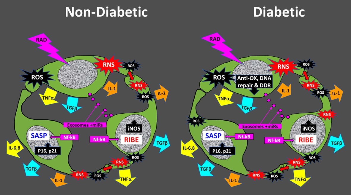
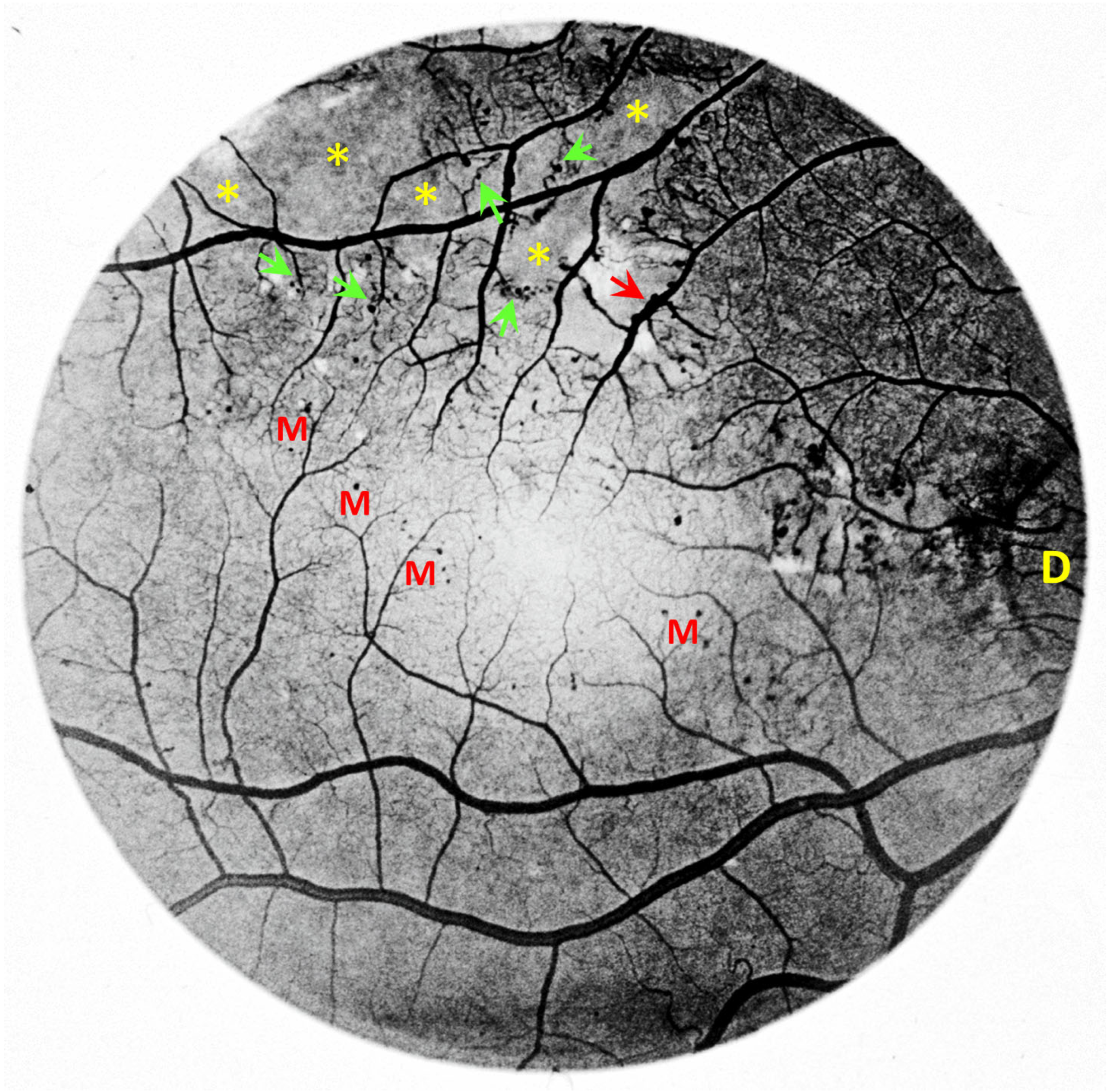
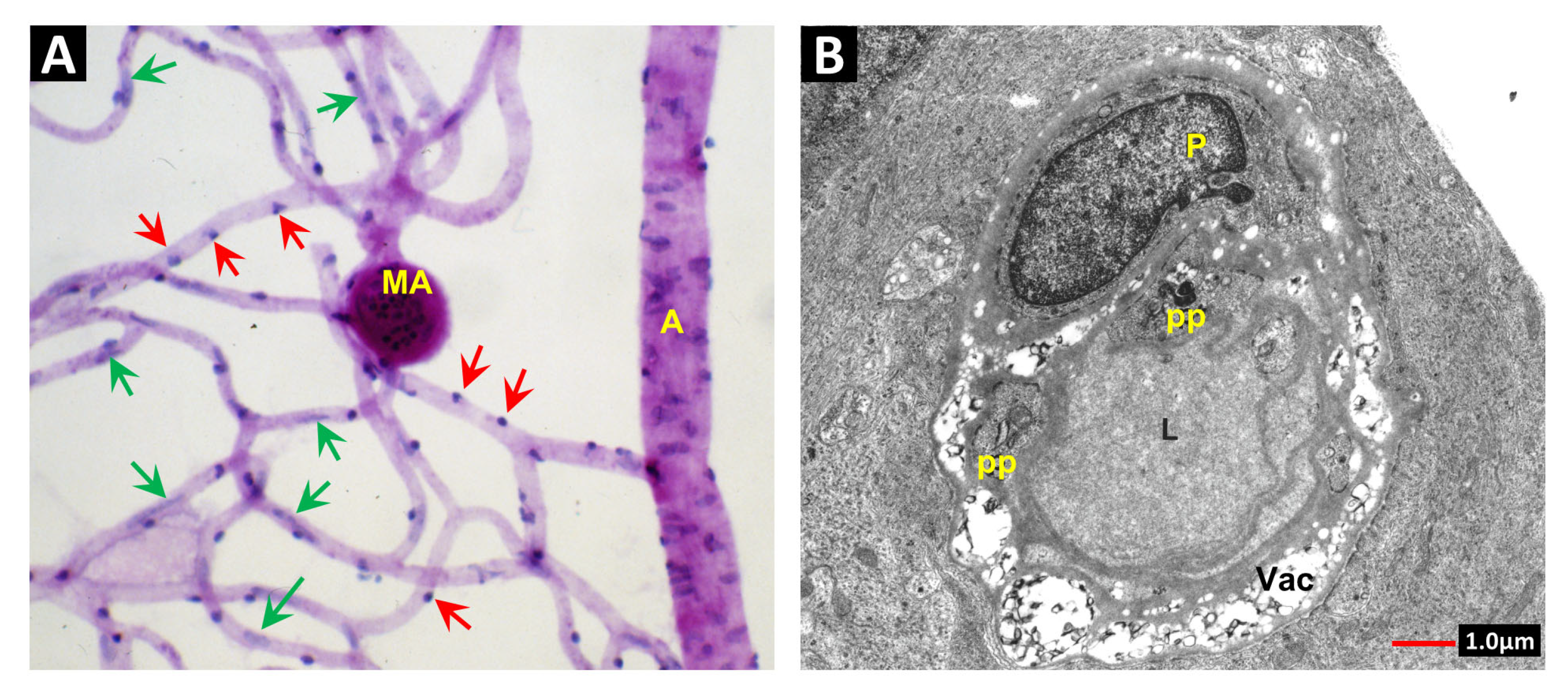
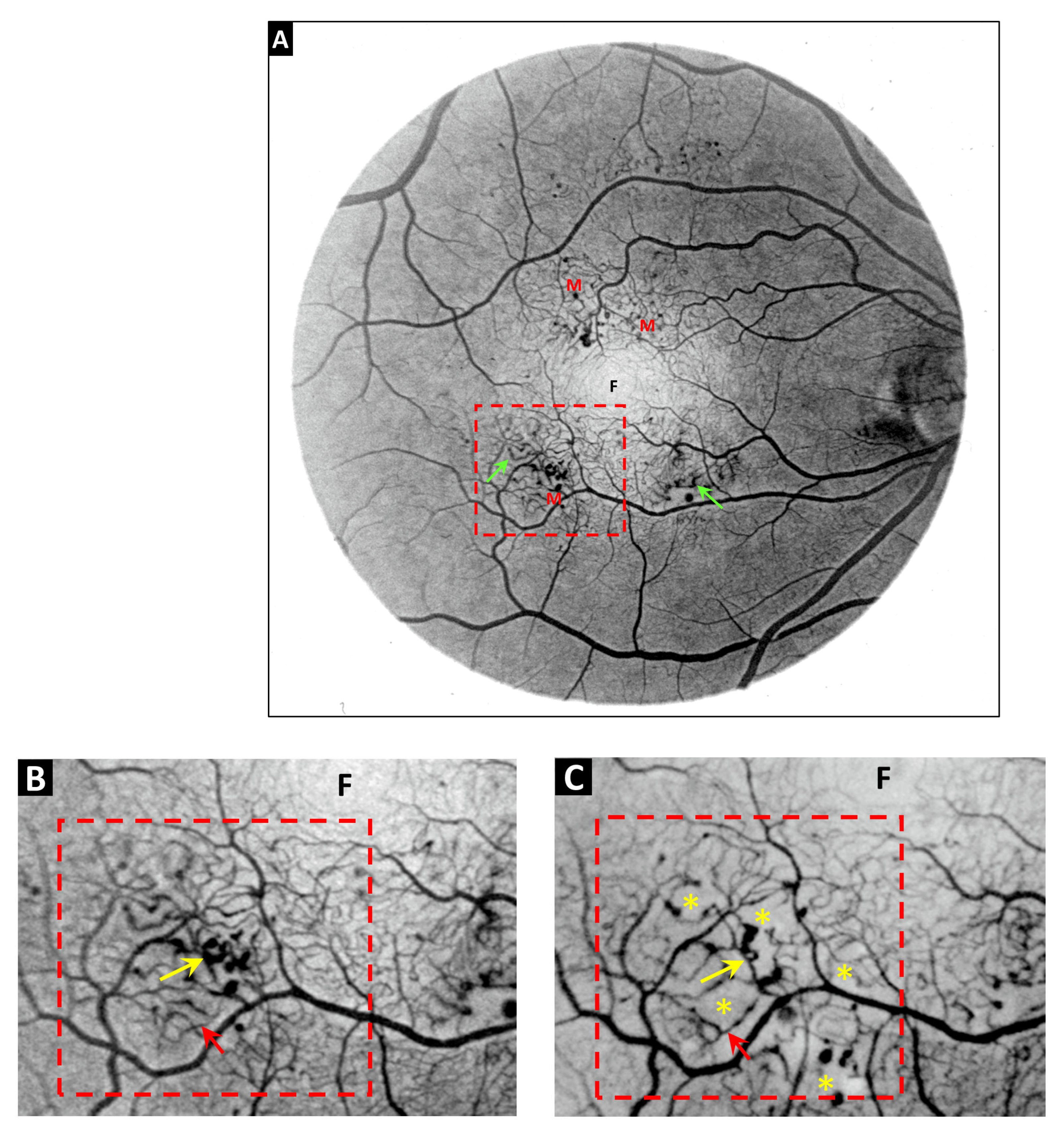
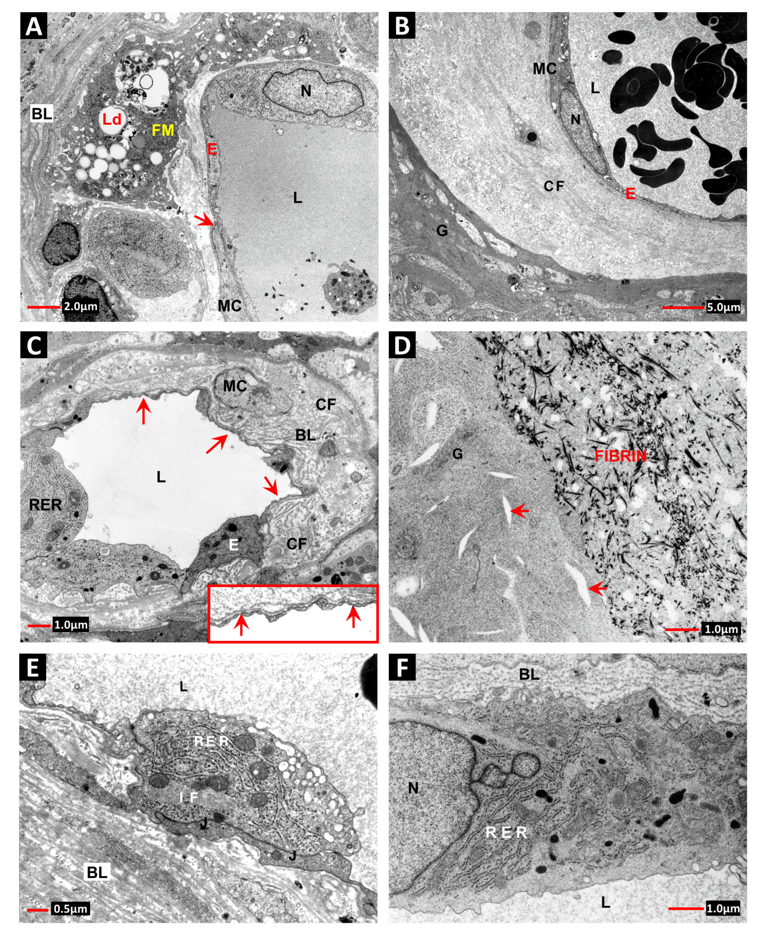
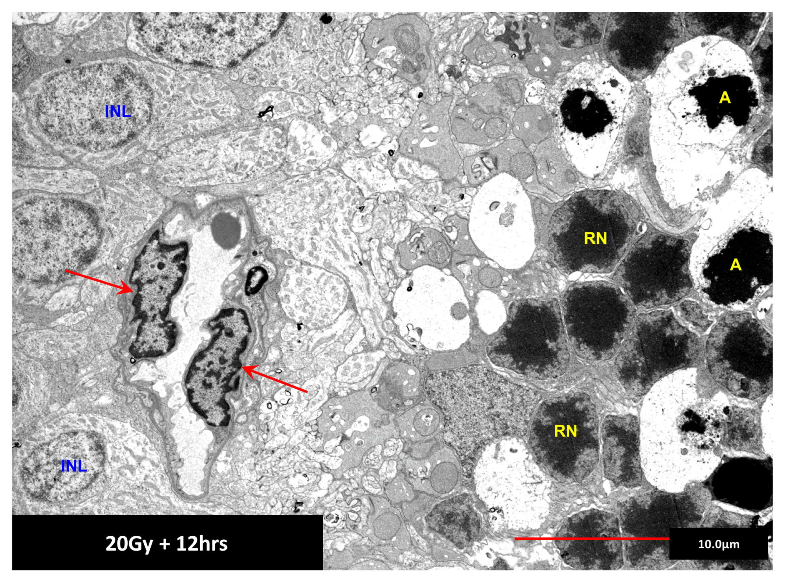
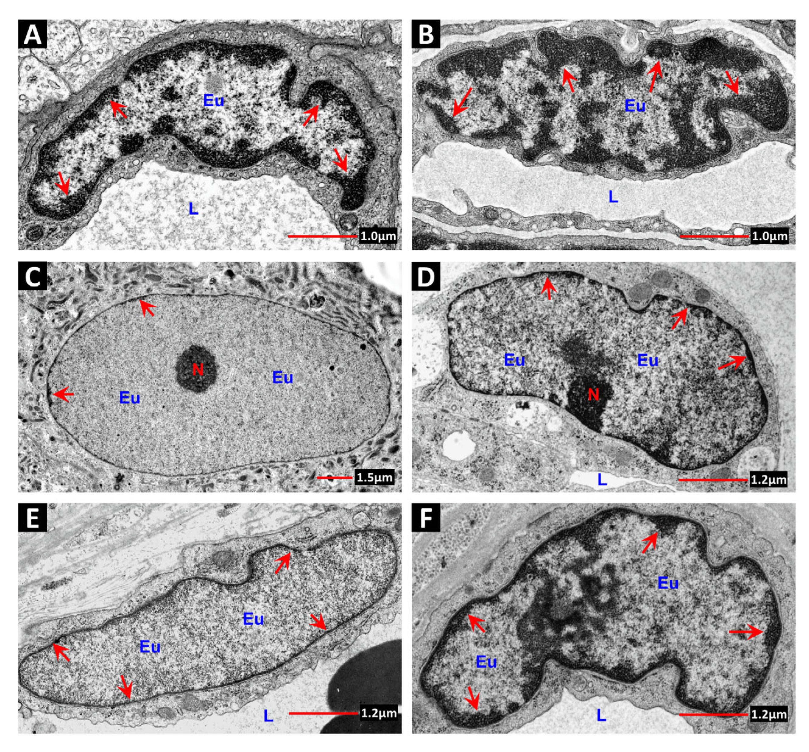
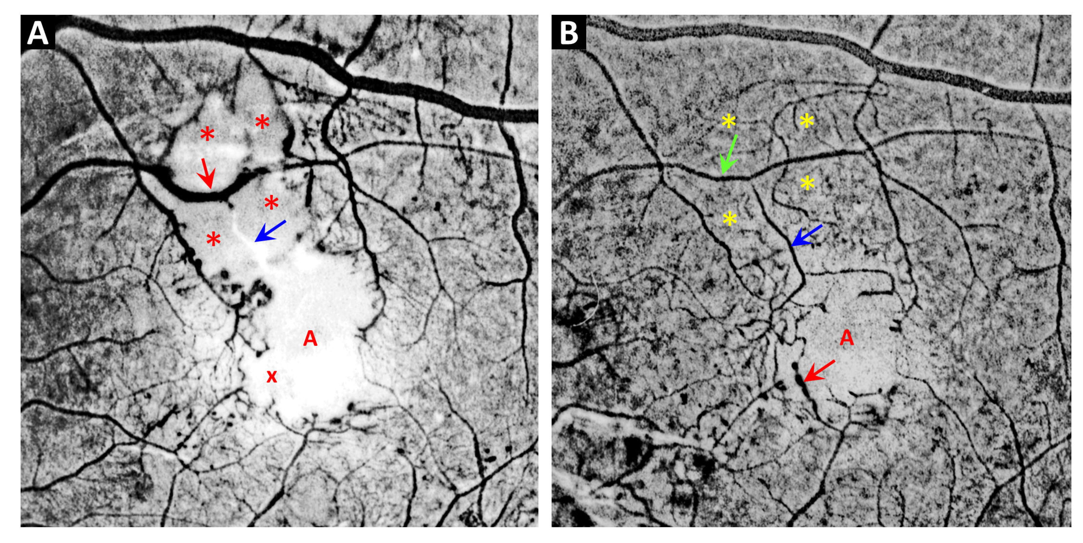
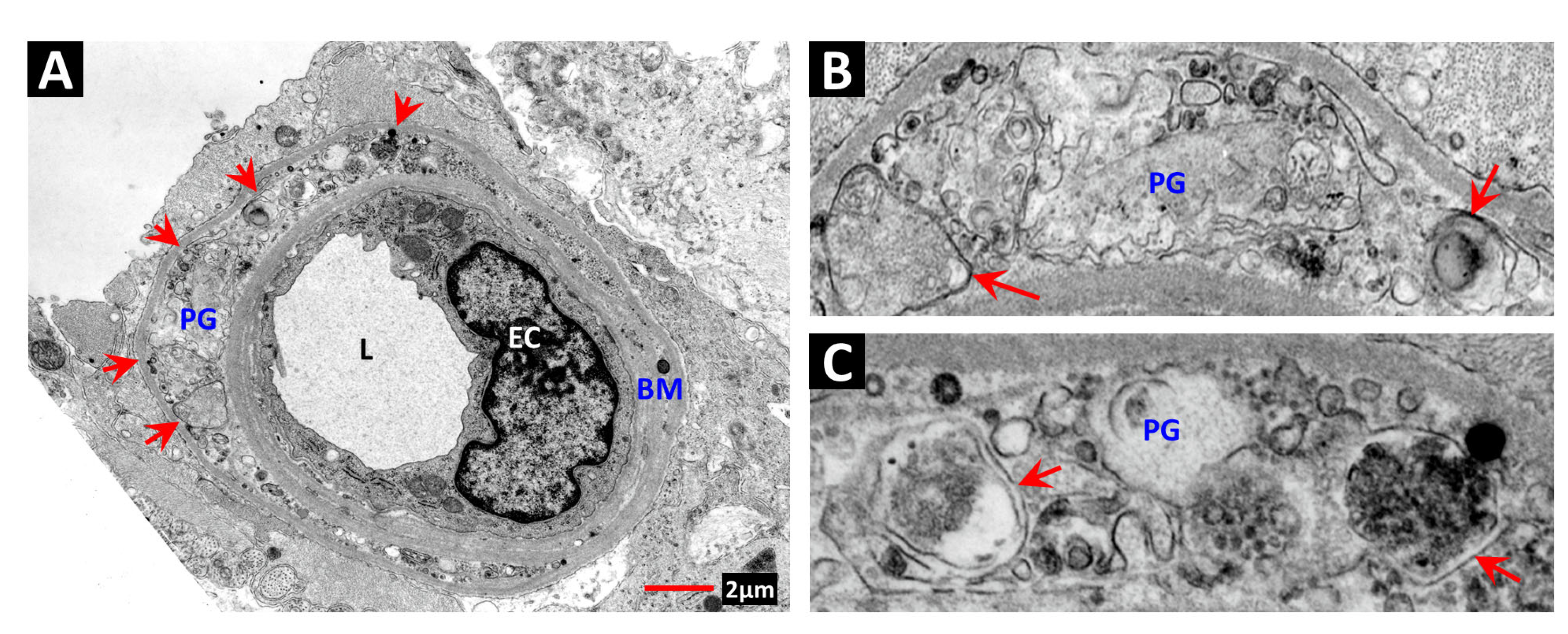
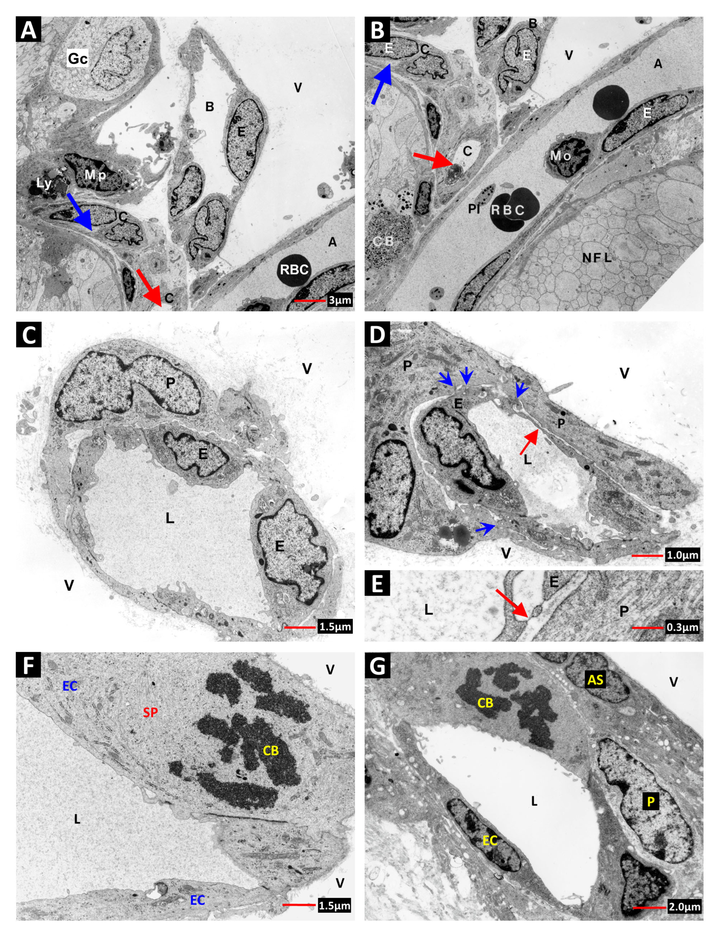
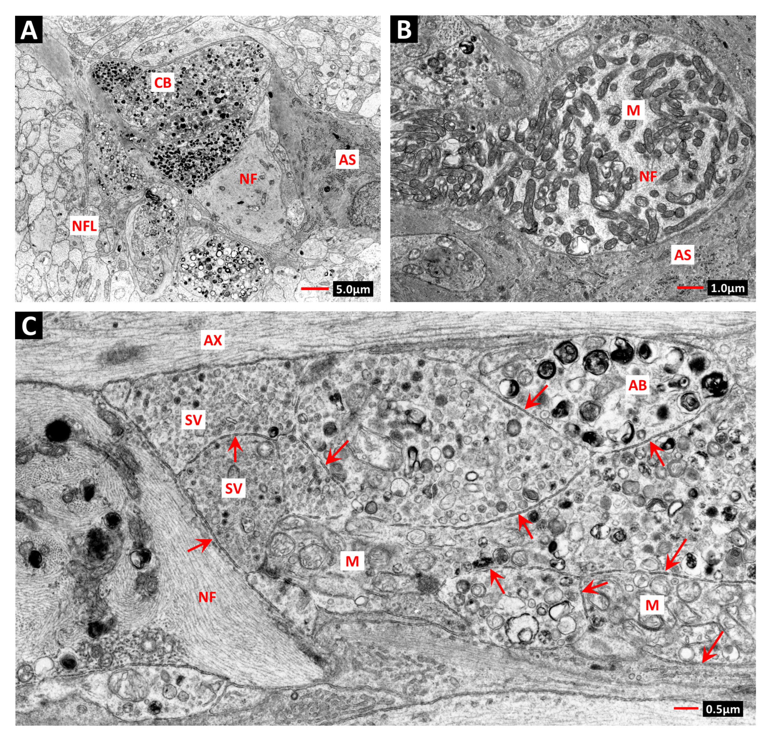
Figure 1. Venous phase fluorescein angiogram of the retinal vasculature in a diabetic patient with a 5-year duration of diabetes, prior to radiotherapy for a parieto-temporal astrocytoma (45Gy of photon X-rays (LINAC) in 20 fractions). A horizontal cut-off in the vascular pathology is obvious due to RR changes in the superior fundus that fell within the radiation field. The angiogram shows widespread areas of confluent capillary nonperfusion (asterisks), multiple microaneurysms (M) and telangiectatic vessels (green arrows). Some larger vessels appear “beaded” but may indicate sites of occluded branches, rather than dilatations (red arrow). The parafoveal capillaries have normal morphology; therefore, the MAs at the same level and inferior to the fovea may have been the result of DR and predated irradiation. Optic disc—D. (From Amoaku et al., 1993, [17]).
Ijtm 03 00011 g001
Figure 2. (A) Trypsin digest preparation of human retinal vasculature 6 months after 45Gy of photon X-rays in 20 fractions shows a microaneurysm (MA) adjacent to a small arteriole (A). The aneurysm is infiltrated by acute inflammatory cells: predominantly polymorphonuclear leucocytes, as identified by their intensely stained multi-lobed nuclei. Some of the capillaries in the region of the MA are acellular, while others show only viable pericytes (red arrows). The typically pale elongated nuclei of surviving endothelial cells are indicated by green arrows. (B) Electron micrograph shows a surviving pericyte cell body and nucleus (P) and pericyte processes (pp) within the basement membrane tube of a retinal capillary from a diabetic patient 18 months after 55GY of photon X-rays in 25 fractions. No endothelium is present. The basement membrane shows the age-related vacuolation characteristic of human retinal capillaries (Vac). Position of original vascular lumen—L, stain—PAS-hematoxylin. (From Archer et al., 1991, [5]).
Ijtm 03 00011 g002
Figure 3. (A) Venous phase fluorescein angiogram of the retinal vasculature in a patient who received 50Gy of radiation in 25 fractions for a tumor of the nasopharynx 8 years prior to ophthalmic assessment. A illustrates the predilection of RR for the macular region of the retina, especially the capillary beds proximal to the fovea (F); microaneurysms—M, telangiectatic vessels—green arrows. (From Amoaku et al., 1993, [17]). (B,C) show enlargement of box outlined in (A). (C) is taken from an angiogram recorded 6 months after that shown in B and shows expansion of discrete regions of capillary nonperfusion (asterisks). The region of telangiectasia indicated by yellow arrow in B shows remodeling in (C). Vessel indicated by red arrow in (B) shows telangiectatic change in (C), probably in response to increased hemodynamic stress due to closure of adjacent vessels and local tissue hypoxia. (From Amoaku et al., 1993, [17]).
Ijtm 03 00011 g003
Figure 4. Electron micrographs of telangiectatic retinal blood vessels from 65-year-old female patient diagnosed diabetic 1 year prior to treatment with 55Gy of X-rays for lacrimal sac tumor. (A) Previously occluded retinal arteriole showing recanalization by telangiectatic endothelium (E). The endothelial cells are extremely attenuated, and their nuclei are remarkably euchromatic (N). The vessel wall contains “foamy” macrophages (FM) containing multiple lipid droplets (Ld) and multiple layers of basal lamina. A mural cell process (MC) shows a junction with an endothelial cell (arrow). (B) Typical large telangiectatic vessel shows attenuated endothelial cell (E) with highly euchromatic nucleus (N) and adjacent mural cell process (MC). The wall shows the pseudoadventitia, or sheath of fine collagen fibrils (CF), that typifies many large telangiectatic vessels. (C) Typical small telangiectatic retinal vessel shows large expanses of endothelial cell fenestration (arrows). Insert shows that the fenestrations are covered by diaphragms (arrows). The nonfenestrated portions of endothelial cytoplasm often contained multiple cisternae of rough endoplasmic reticulum (RER). The vessel wall contains bundles of fine collagen fibrils (CF) and fragmented segments of basal lamina (BL). Lumen—L. (D) Large pools of proteinaceous fibrin-rich exudate were found in the vicinity of the fenestrated telangiectatic retinal vessels. Cholesterol crystals were not obvious within the exudate, but the characteristic residual spaces that remain after extraction of the cholesterol during tissue processing (arrows) were obvious within the cytoplasm of adjacent glial cells (G). (E,F): The significant development of the rough endoplasmic reticulum (RER) in the endothelial cells of small (E) and large telangiectatic vessels (F) suggests that the endothelium may be responsible for synthesis of the excessive collagen in the vessel walls. IF—intermediate filament bundles. (From Archer et al., 1991, [5]).
Ijtm 03 00011 g004
Figure 5. Shows an electron micrograph of the outer and inner nuclear layers of the rat retina, fixed 12 h after irradiation with 20Gy of X-rays. Note the high proportion of heterochromatin (HC) and the inverted chromatin conformation in the rod photoreceptor cell nuclei (RN). Many of the rod cells are in apoptosis and show typical apoptotic chromatin condensation (A). In comparison, the predominantly euchromatic inner retinal neurons appear undamaged. Note that the vascular endothelial cells (arrows) have significantly more heterochromatin than the cells of the inner nuclear layer (INL) but show no evidence of acute injury at this time. (From Amoaku et al., 1989, [172]; Archer & Gardiner 1994, [21]).
Ijtm 03 00011 g005
Figure 7. Retinal fluorescein angiograms from a patient who received 55Gy of photon X-rays for treatment of an anterior dural myxosarcoma. (A) shows closure of the capillary bed superior to the foveal avascular zone 9 months after treatment. A large vessel (red arrow) traversing the ischemic region (red asterisks) shows dilation and leakage. (B) Angiogram of the same region 6 months later shows reperfusion of the occluded capillary bed (yellow asterisks), including a large vessel (blue arrows). The large vessel traversing the previously ischemic bed (green arrow) has regained competence to fluorescein and is no longer dilated. NB* An occluded perifoveal vessel marked with a red X in angiogram-A shows reperfusion in angiogram-B, although the recanalized vessel shows evidence of telangiectatic change (red arrow). A-indicates the foveal avascular zone. (From Archer & Gardiner 1994, [21]).
Ijtm 03 00011 g007
Figure 8. Electron micrographs of intra-retinal capillary in a moderately hyperglycemic STZ diabetic rat, 1 year after induction of diabetes and 6 months after 15Gy of X-irradiation. (A) The capillary basement membrane (BM) is grossly thickened and contains the profile of a pericyte “ghost” (PG). Although the ghost cell lacks a continuous plasma membrane, it retains many profiles of endomembranes consistent with intense autophagic activity (arrows). (B,C) Enlarged images of the PG regions indicated by arrows in image-A show heterogeneous cell debris either enclosed or partially enclosed by double membranes (arrows). (From Stitt et al., 1994, [8]).
Ijtm 03 00011 g008
Figure 9. Electron micrographs of pre-retinal neovascularization in a moderately hyperglycemic 1-year STZ diabetic rat 6 months after 15Gy of X-irradiation. (A) Site of entry of new retinal vessels to the pre-retinal region of the vitreous body (V). Vessel-A shows a formed lumen and is perfused by red blood cells (RBC). Vessel-B has a formed lumen but appears empty, while the lumen of the tortuous capillary vessel-C appears in 2 profiles marked by arrows (also in image-(B)). The profile marked by the red arrow has opened, while the blue arrow indicates the slit-like lumen typical of capillary sprouts proximal to the tip. The adjacent retina shows a macrophage (Mp) engorged with secondary lysosomes. Ganglion cell—Gc. (B) Montage extension of location depicted in image-(A) shows passage of vessel through the nerve fiber layer (NFL). CB indicates a cytoid body within distended ganglion cell axon. Vessel lumen shows a platelet and a mononuclear cell (Mo) adherent to the endothelium. (C) A new vessel within the vitreous body shows healthy endothelial cells (E) and pericytes (P) rich in intracellular organelles. Lumen—L. (D) A new capillary vessel within the vitreous body shows a large organelle-rich pericyte (P) enveloping an endothelial cell (E). Several pericyte–endothelial junctions are indicated by blue arrows. Lumen—L, vitreous—V. Red arrow—see legend of image-(E). (E) Serial section of the attenuated portion of the EC cytoplasm (E) indicated by the red arrow in image-(D) shows endothelial fenestrae covered by diaphragms (arrow). Lumen—L, pericyte—P. (F) A new vessel in the vitreous body (V) shows the typical site of endothelial mitosis at a site with a formed lumen (L), as shown in normal intraretinal vascular development in (G). Condensed chromosomal bodies (CB) are located within the mitotic spindle (SP). (G) Endothelial mitosis in developing retinal vasculature at postnatal day-5. Condensed chromosomal bodies are located in EC which forms part of the vessel lumen (L). Pericyte—P, astrocyte—AS, vitreous—V, CB—chromosomal body, SP—mitotic spindle, L—vascular lumen. (Adapted for Archer et al., 1991, [5]; Archer 1993, [6]; Stitt et al., 1994, [8]).
Ijtm 03 00011 g009
Figure 10. Electron micrographs of cytoid bodies in the nerve fiber layer (NFL) of the retina in a moderately hyperglycemic STZ diabetic rat, 1 year after induction of diabetes and 6 months after 15Gy of X-irradiation. (A) Cytoid bodies (CB) consisting of swollen ganglion cell axons, occurring either singly or in groups, are filled with intact mitochondria (M) and multiple membrane-bound bodies enclosing heterogenous cell debris and remnants of endomembranes. Some individual axons are almost entirely filled by neurofilaments (NF). AS—astrocyte. (B) Single axon distended by an aggregate of intact mitochondria surrounded by neurofilaments (NF). AS—cytoplasm of astrocyte. (C) Cytoid body composed of several swollen axons showing the axolemmal boundaries (arrows) and distinctive contents of certain axons: Those marked SV are almost exclusively filled with small vesicles with the size and morphology of synaptosomes, while others are filled with recognizable mitochondria, and some are filled with autophagic bodies (AB). AX - normal axon filled with neurotubules. (From Stitt et al., 1994, [8]).
Ijtm 03 00011 g010
Table 1. Ultrastructural changes in the retinal vasculature in DR and RR.
Vascular PathologyDiabetic
Retinopathy
Radiation
Retinopathy
Loss of pericytes or vascular smooth muscle cells in vessels with surviving endothelial cellsYesNo
Loss of endothelial cells in vessels with surviving pericytes or vascular smooth muscle cellsNoYes
Generalized thickening of capillary basement membranesYesNo
Vessels with fenestrated endothelial cellsOnly in end-stage diseaseYes
Vascular occlusion and inner retinal ischaemiaYesYes
Vascular recovery No—Space-filling of BM tubes by glial cells is permanentLimited recovery
Vascular remodellingIRMATelangiectasia
Telangiectatic vessels with expanded pseudoadventitia and fenestrated endothelial cells with euchromatic nucleiNoYes
Disclaimer/Publisher’s Note: The statements, opinions and data contained in all publications are solely those of the individual author(s) and contributor(s) and not of MDPI and/or the editor(s). MDPI and/or the editor(s) disclaim responsibility for any injury to people or property resulting from any ideas, methods, instructions or products referred to in the content.

