Article

Microplastic Contamination in Cultured Mussels and Pearl Oysters in Greece

by
Stefania M. Manolaki
1,*,
Dimitra Chatzivasileiou
1,
Maria Lampa
1,
Panagiotis D. Dimitriou
1,
Aggelos Philippidis
2,
Ioannis Karakassis
1 and
Nafsika Papageorgiou
3
1
Department of Biology, University of Crete, GR 70013 Heraklion, Greece
2
Institute of Electronic Structure and Laser, Foundation for Research and Technology-Hellas (IESL-FORTH), GR 71110 Heraklion, Greece
3
Department of Agricultural Development, Agrofood & Management of Natural Resources, National and Kapodistrian University of Athens, GR 34400 Athens, Greece
*
Author to whom correspondence should be addressed.
Microplastics 2023, 2(2), 168-181; https://doi.org/10.3390/microplastics2020013
Submission received: 30 November 2022 / Revised: 14 February 2023 / Accepted: 27 March 2023 / Published: 3 April 2023
(This article belongs to the Collection Current Opinion in Microplastics)

Abstract

:
Τhis study aims to measure the abundance of microplastic (MP) particles in the soft tissue of mussel (Mytilus galloprovincialis) and pearl oyster (Pinctada imbricata radiata) specimens. Samples were collected at four sites in Greece (Sagiada, Malesina, Elounda, Rhodes) from wild and farmed populations. The identification of MPs was accomplished by Raman spectroscopy. Comparisons were made between the two different species where the two species co-existed (Malesina), between the four study sites (five sampling stations) in relation to P. imbricata radiata individuals, and also in every station for the different MP types found. For the specimens from Malesina, M. galloprovincialis had more MPs in their soft tissue compared to P. imbricata radiata. Microfibers were found in abundance in M. galloprovincialis, while microfragments were found in P. imbricata radiata specimens. The main MP type found in P. imbricata radiata specimens was microfragments in all five sampling stations, and ranged between 1.54 ± 0.63 (Rhodes-baskets) and 3.56 ± 0.35 (Sagiada) MP particles/g. While the samples of mussels and pearl oysters were similar in age, the differences found in the concentrations of MPs appears to be due to their different farming methods and location characteristics concerning the five sampling stations of pearl oysters. This study indicates that the culturing system does not affect MP concentration in bivalves, and further investigation is needed to find the most appropriate method to limit and reduce MPs that end up in the farmed organisms.

Graphical Abstract

1. Introduction

During the past decades, microplastics (MPs) have caused major concerns for the public and the scientific community, as they have rapidly increased in the oceans, with various ecological consequences [1]. The term microplastics has been used by the National Oceanic and Atmospheric Administration (NOAA) for plastic particles less than 5 mm in size [2]. MPs enter the marine environment through many ways, for example, from the terrestrial environment through surface water runoff, plastic degradation from human coastal activities, where they remain for long periods of time [3], or from wastewater treatment plants, which are considered an important source of MPs (mostly fibers) in the marine environment [4].
MP analysis can be classified according to physical and chemical characterization. In physical characterization, several physical factors such as the size distribution, shape and color of the microplastics, are evaluated. In particular, MPs are divided into categories such as microfibers, microfragments, microbeads, microfoams, and microfilms based on their shape [5]. During physical characterization, the use of the stereoscope is the most widespread, as it provides a direct visual assessment of the number of MPs in the samples, their morphology and their size. Due to the low magnification factor of the stereoscope, visual identification may be limited and always depends on the researcher. The accuracy of this method can be improved by using various precautionary measures; however, it is still time consuming and cannot predict polymer types [6].
The composition of MPs is investigated through chemical characterization, which has the ability to accurately determine their composition. So far, chemical analysis methods are divided into destructive and non-destructive spectroscopic techniques, with Raman spectroscopy being the most commonly used method for MP samples with sizes <1 μm [6], as in the case of this study.
The ingestion of MPs is thought to be higher in filter-feeding organisms such as mussels and pearl oysters [7,8] due to the effective uptake and assimilation of particles suspended in the water column [9]. As a result, the measurement of the average body load of MPs in these organisms is being used as an indicator of MP environmental pollution [8,9,10]. Both field and laboratory studies have indicated that MPs can be detected globally in bivalves, causing, among others, adverse effects on the physiological responses of the organisms and their immune and antioxidant systems, as well as histological changes [11].
The study of MPs in mussels (Mytilus galloprovincialis) and pearl oysters (Pinctada imbricata radiata) in the Mediterranean region is very important. They are an essential link between the coastal, benthic and pelagic zones, since their ability to filter and remove particles from the water column provides benthic organisms with pelagic resources (food, nutrients) that would otherwise be unavailable [12,13]. In addition, Mediterranean mussels are farmed for human consumption, and pearl oysters are considered to be a very good candidate for cultivation [14] as they are an invasive but well-established species in the Mediterranean region that over time has become part of the local population’s diet [15,16].
It is known that fish farming releases large amounts of nutrients and organic waste that are capable of causing eutrophication phenomena in nearby coastal and aquatic systems [17,18,19]. Therefore, there has been an increased interest in alternative sustainable practices such as integrated multitrophic aquaculture (IMTA) [20]. IMTA focuses on the sustainability of aquaculture through the integrated production of species coming from different trophic levels, which can minimize energy loss and environmental degradation [21]. To this end, co-cultured species often include fish as a central crop, and filter-feeding species exploiting suspended organic matter (e.g., bivalves) [22]. MPs have been found in a variety of aquaculture environments, such as fish farms, and rice–fish co-culture systems [23,24]
The present study examined whether culturing techniques and fish farming activities affected the MPs abundance in the soft tissue of mussels (M. galloprovincialis) and pearl oysters (P. imbricata radiata). The main objective of the study was to compare MP concentrations in the soft tissues of bivalves originating from wild and IMTA-cultured populations. In addition, the difference in MP concentrations in the two bivalve species was investigated for the sites where these two species co-existed.

2. Materials and Methods

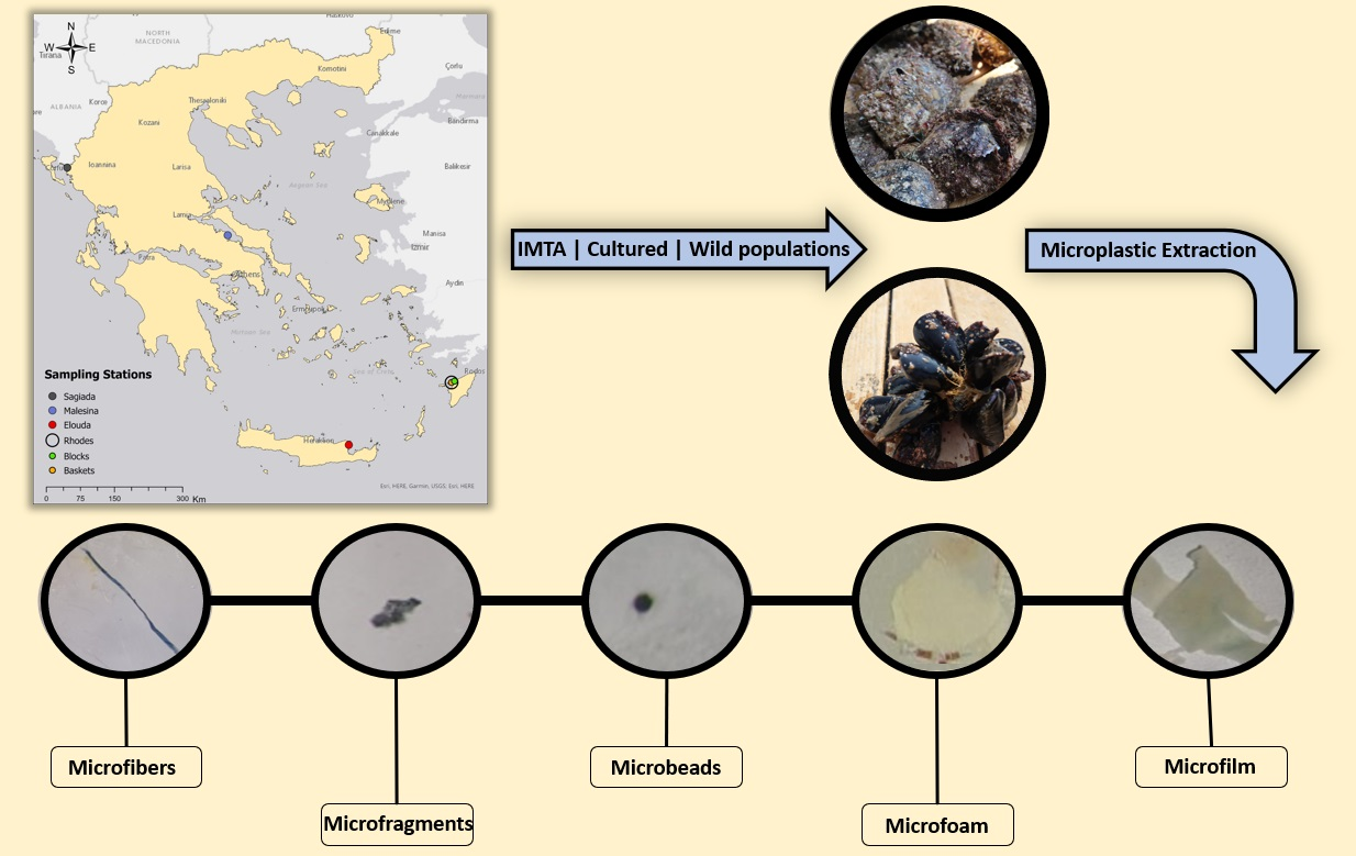
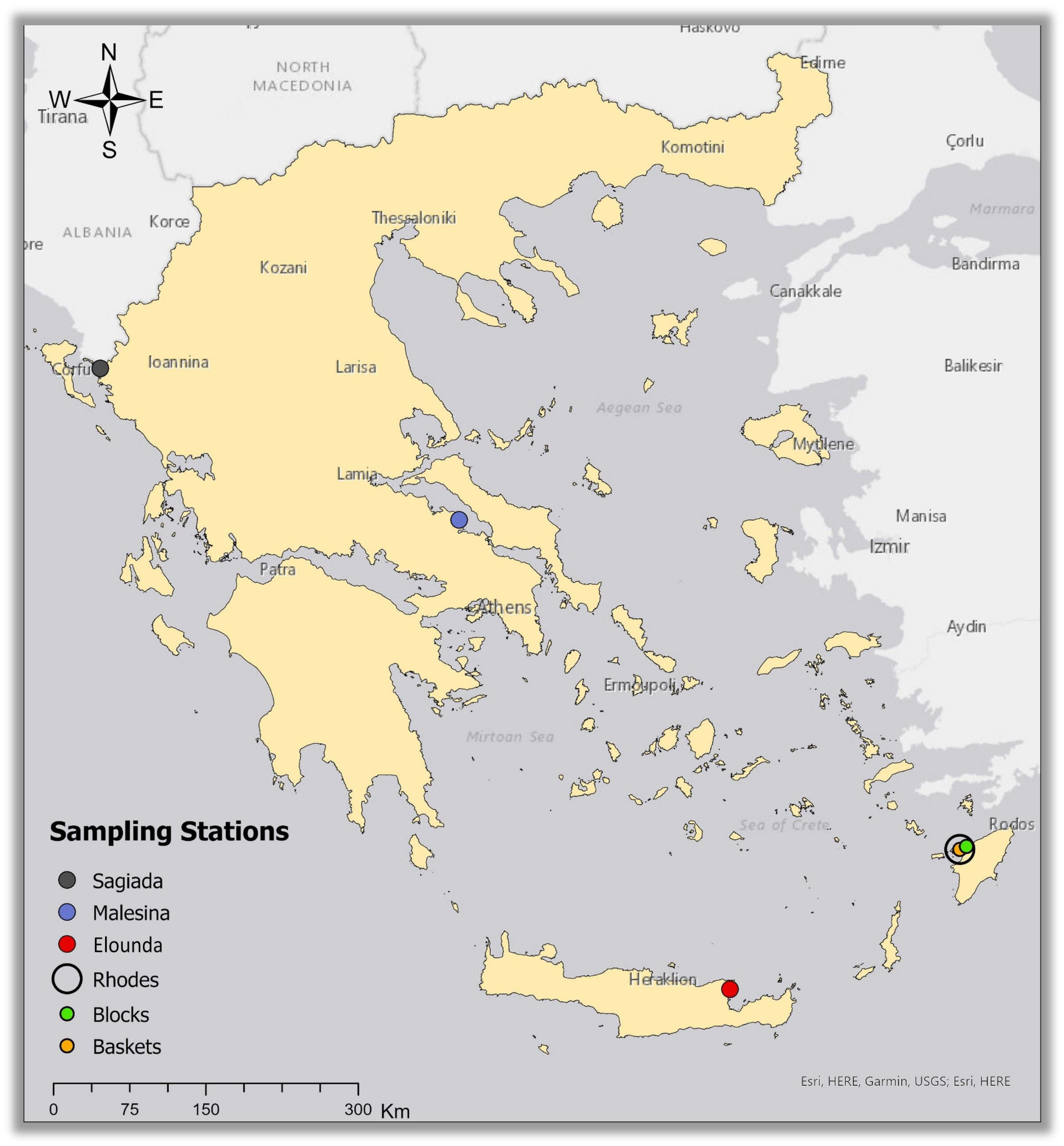
2.1. Study Sites and Sampling Stations

The study included five stations with bivalve populations from four areas in Greece (Figure 1): Sagiada station in the northern Ionian Sea codenamed “Sagiada”, Malesina station in North Evoikos Gulf codenamed “Malesina”, Elounda Bay station in Crete codenamed “Elounda”, and two stations in northern Rhodes in the Aegean Sea codenamed “Rhodes-baskets” and “Rhodes-blocks”.
In Sagiada station, P. imbricata radiata were farmed using the mussel farming methodology (longline-pergolaris) deployed within the cages of a fish farm as an experimental IMTA. Malesina station included mussels (M. galloprovincialis) and pearl oysters (P. imbricata radiata) farmed near fish cages (pilot IMTA, Figure 2a and Figure 3a). The IMTA culture methodology was based on the typical mussel culture methodology reported in [14]: mussel seeds were collected from mooring ropes within the fish farm and placed in elongated plastic cylindrical tubing nets (pergolaris) of 6 m in length and net eye of 80 mm. The pergolaris were made using polyvinylchloride cylindrical tubes with a diameter ranging between 4 and 7 cm, which were then deployed around a fish cage in the center of the fish farm (Figure 2b).
Pearl oyster juveniles were also collected from fish farm mooring ropes and were farmed in baskets made from polypropylene with carbon for UV stabilization, used in oyster culture and manufactured by SEAPA© (Figure 3b). Although these baskets are designed for longline oyster farming, they can be adapted to suit a range of alternative farming systems and methods such as IMTA. The baskets were tied to ropes around a fish cage in the center of the farm. Cultivation duration was 9 months during which both bivalves reached commercial size.
Elounda station supported a wild population of P. imbricata radiata close to the coast at 1.5–2 m depth near a highly developed touristic area (Figure 3d). The Rhodes-baskets station included only cultured pearl oysters (pilot IMTA) because M. galloprovincialis cannot survive in these ultra-oligotrophic conditions close to the Levantine Sea. Pearl oyster juveniles were collected from the mooring ropes around the fish cages and were farmed in SEAPA© baskets, like those at Malesina station. Finally, the pearl oyster wild population inhabiting the mooring blocks of the fish farm at 30 m depth was named ‘Rhodes-block’ station (Figure 3c). Two of the locations, Malesina and Rhodes, were pilot IMTA cultures developed in the framework of the research project, Innovative Development of Multitrophic Aquaculture (IDMA–www.idma.uoc.gr (accessed on 10 February 2023)).

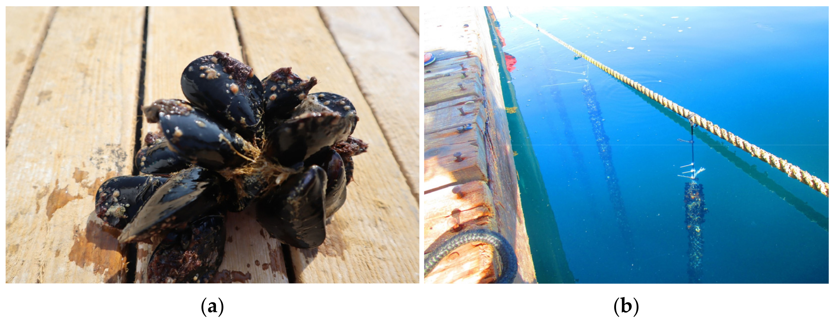
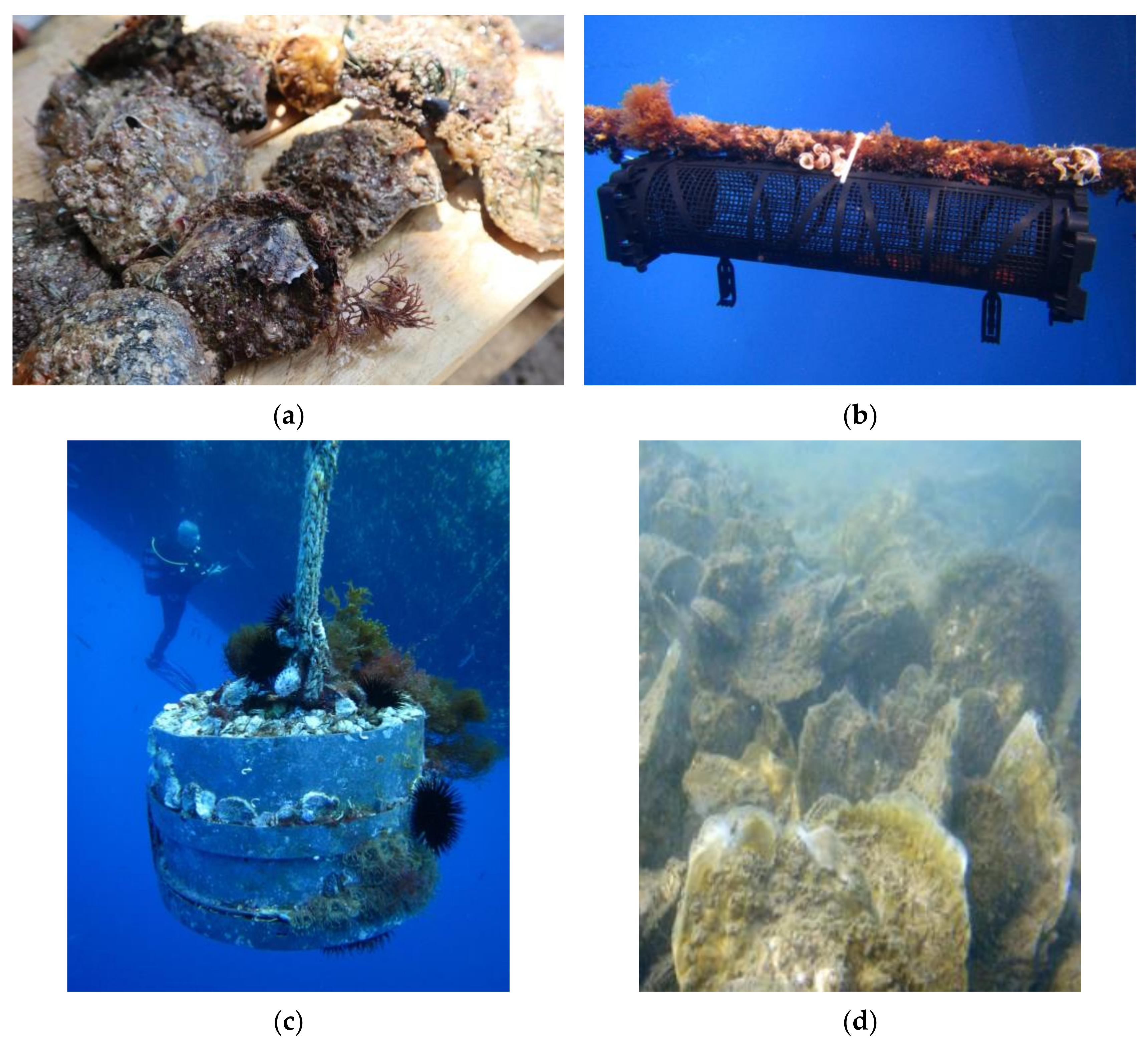
2.2. Sample Collection

All the collected individuals in every station were of a similar size (commercial size) and were therefore approximately the same age class. From Sagiada station, a total of 6 individuals of P. imbricata radiata with a total of 53.12 g of flesh weight were collected. At Malesina station, a total of 12 individuals of farmed P. imbricata radiata with a total flesh weight of 231.04 g, and 30 individuals of farmed M. galloprovincialis with a total flesh weight of 145.09 g were collected. The wild P. imbricata radiata population of Elounda station consisted of 14 individuals with a total of 85.09 g flesh weight. Finally, 23 pearl oyster individuals with 116.65 g of flesh weight were collected from Rhodes-blocks station, and in Rhodes-baskets, 19 individuals of farmed P. imbricata radiata were collected reaching a total of 88.17 g of flesh weight. All bivalves collected were opened in the field in indoor and well-protected rooms to avoid extensive airborne contamination. Their soft tissue was removed, weighed, rinsed with ultrapure water, and placed in glass jars that had been well cleaned with HCl (10% w/v) solution. They were then refrigerated to prevent distortion until further analysis.

2.3. Mussel and Oyster Sample Preparation and Digestion

The procedure to extract MP particles from the soft tissue of mussels and pearl oysters was based on published methods [25,26,27,28] and was the same for both species. The samples were rinsed with ultrapure water and the wet flesh weight/individual was measured. Individuals of every station and species were sorted according to their weight into 5 to 12 replicated samples of approximately 10 g. The number of replicates was specified from the total flesh weight/station. Each sample was placed in a glass conical flask and digested with a 200 mL filtered KOH (10%, 1:20 w/v) and 2 mL H2O2 (30%) mixed solution. The samples were covered with aluminum foil and placed in 60 °C for 48 h, with a regular 40 sec shaking every 8 h (Figure 4a). After 48 h, 2 mL of H2O2 (30%) solution was added approximately every 4 h, after the foam had settled and the samples had been stirred for 40 sec. This procedure was repeated until there was no more organic material present and the solution was clear yellow in the case of mussels and clear green in the case of pearl oysters.
A filtered saline solution (NaCl) with 1.2 g cm−3 density was then added to each sample in concentrations twice as high as the KOH (400 mL saline solution), to induce flotation of the microplastic particles contained in the sample. The samples were well stirred and left for 24 h, allowing any organic material remaining in the conical flask to settle. The supernatant solution was then transferred to a new clean conical flask. At the end of the above procedure, the samples were filtered with 0.8 μm diameter Whatman™ cellulose nitrate membrane filters with a glass/metal filtering system and transferred to glass Petri dishes with lids where they were stored until further analysis.

2.4. Microscopic Inspection of Microplastics

The filtered samples were analyzed visually using a stereoscope (magnification ×1.0 to ×5.0), to identify and count the type of MPs larger than 0.8 μm according to their physical characteristics in each replicated sample [29]. The confirmation of the presence of MPs was performed by the ‘needle test’. This method involves the use of a red-hot needle, which, upon coming into contact with the presumed microplastic, melts it [30]. The smallest MPs found were 0.8 μm, while the biggest were 1500 μm. All MPs found were classified into 5 types (microfibers, microfragments, microbeads, microfoams and microfilms) according to the standardized size and color sorting (SCS) system [31]. In order to ensure the correct count of the MPs, each filter was marked near them with the tip of a needle.

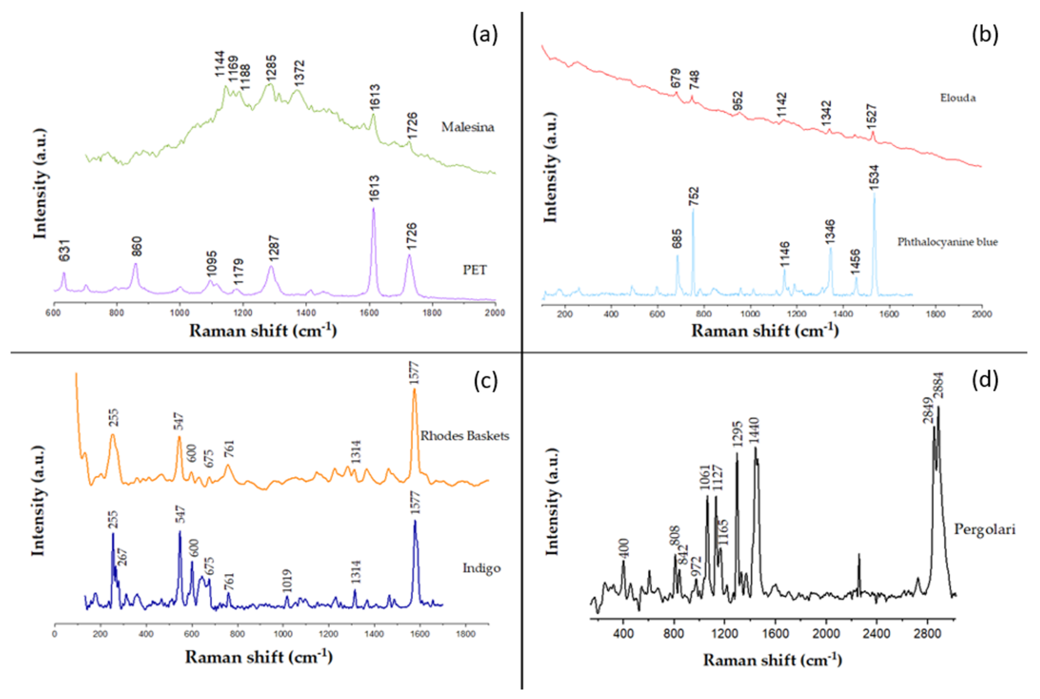
2.5. Raman Spectroscopy

A subset of microplastic particles was randomly selected from each sampling station to determine their chemical type. Due to the small size of the samples and the difficulty in handling them, only microfibers were selected to be analyzed by Raman spectroscopy. A mobile Raman spectrometer (HE 785, JY Horiba) with excitation from a cw diode laser at λexc = 785 nm was used in the current study as previously described [32]. Measurements were made through a ×20 objective lens and the typical exposure time was 30–45 s per scan, with a minimum of 2 scans averaged per measurement. The spectrograph (Exemplar Plus, B&W Tek) provided spectral coverage from 98 cm−1 up to 3362 cm−1 at a spectral resolution of about 8–10 cm−1. Laser power (PL) values were in the range of 13–53 mW, measured on the sample surface. In addition, Origin Pro 2023 software was used to preprocess (smoothing, subtraction) some spectra. Raman spectroscopy was performed on 22 microfiber samples from all sampling stations, but also on a sample of the pergolari fibers used in some of the farms. The aim of the spectroscopic analysis was to indicate whether the particles detected during the experiment corresponded to microplastics.

2.6. Data Analysis

The measured numbers of MPs for each sample (total and each subcategory) were transformed to counts per 1 g of biomass. The data from the study were used to (a) compare MP accumulation in the two bivalve species (M. galloprovincialis and P. imbricata radiata) in Malesina, (b) examine the difference in pearl oyster MP concentration at the five different sampling stations (Sagiada, Malesina, Elounda, Rhodes-blocks, Rhodes-baskets), and (c) examine the difference in the MP types in every station separately.
A T-Test was used to check if there were significant differences in the counted MPs between the species at Malesina station, and a one-way analysis of variance (ANOVA) between the five different stations (Sagiada, Malesina, Elounda, Rhodes-blocks, Rhodes-baskets) and between the MP types of every station, for the P. imbricata radiata species. The requirements of parametric analysis were assessed using the Shapiro–Wilk test of normality and Levene’s test for homogeneity of variances, while boxplots were used to check for outliers. The values of the results represent the average ± standard deviation (average ± SD). All statistical analyses were performed with the “IBM SPSS Statistics v.26” software.

2.7. Quality Control and Contamination Precautions

All of the equipment used during sampling and in the experiment was either glass or metal and properly rinsed with HCl (10% w/v) solution before use. In the laboratory, all windows and doors were closed and all surfaces were cleaned regularly with acetone. The samples were covered with aluminum foil throughout the analyses to avoid airborne contamination of MPs. Laboratory personnel used protective cotton laboratory robes during the entire experiment, while hands were scrubbed to control self-contamination from skin, hair or dirt. All liquid solutions (KOH, H2O2, NaCl) used were filtered with 0.8 μm diameter Whatman™ cellulose nitrate membrane filters before use, to minimize their MP content. While conducting the experiment, three blind samples were created for every sample batch, to estimate and remove any MP contamination from the laboratory background.

3. Results

3.1. Identification of MPs

According to the physical characterization, the visual assessment of the stereoscopic analysis showed that the MPs found in the soft tissue of the two species were classified as microfibers, microfragments, microbeads, microfoams, and microfilms. The MP concentration for both species, but also for all the sampling stations, was higher in microfibers (6.46–47.74%) and microfragments (42.96–91.25%).
According to the chemical characterization, the microfibers in the oyster samples from the stations of Malesina and Rhodes-baskets exhibit Raman spectra bands assigned to polyethylene terephthalate (PET) (Figure 4a). Furthermore, the spectra obtained from the Elounda station indicated Raman peaks corresponding to the organic pigment “phthalocyanine blue” [33] (Figure 4b). At the stations of Sagiada and Rhodes-baskets, the Raman spectra peaks appeared to match the organic pigment “indigo” [33] (Figure 4c). Finally, the Raman bands spectra from the pergolari sample corresponded to polyethylene (PE) and polypropylene (PP) (Figure 4d) [34]. No similar peaks were found in the spectra obtained from the bivalve microfiber samples.
At wavenumbers higher than 1800 cm−1, samples do not exhibit any Raman bands and for this reason only the 100–1800 cm−1 spectral region is shown for all spectra with the exception of the pergolari sample, which also shows Raman bands in the 2800–3000 cm−1 spectral region (Figure 4). Apart from pergolaris, Raman spectroscopy was also performed on samples from SEAPA baskets, and fish farm nets and ropes used to support pergolaris, but they did not correspond to the spectral coverage provided by the spectrograph.

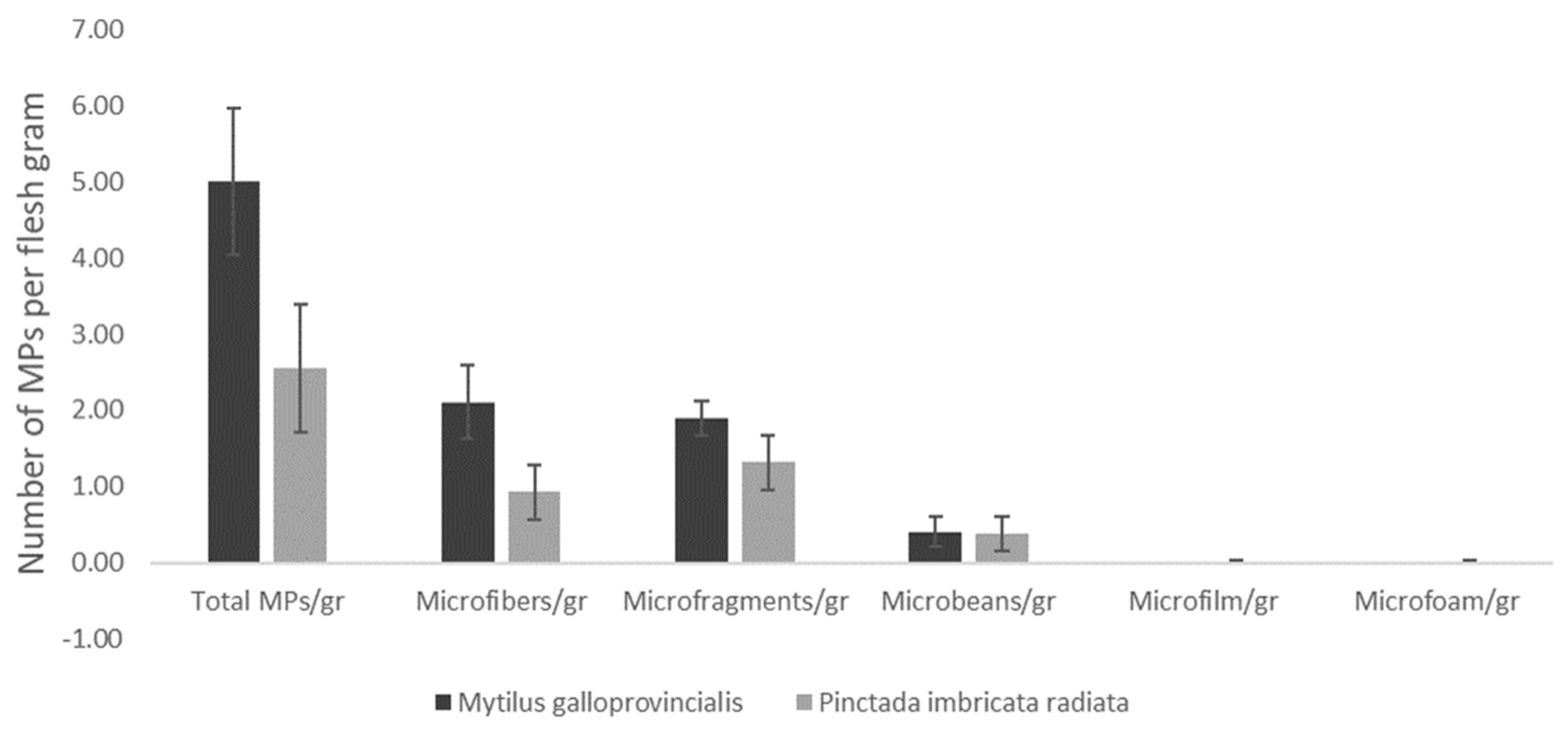
3.2. Differences of MPs between the Two Bivalve Species

The comparison of MPs for the two bivalve species co-cultured at Malesina station showed significant differences in total MPs and microfibers between mussels and pearl oysters (t-Test: F = 6.223, p < 0.05 and F = 9.298, p < 0.01, respectively). MP counts showed that M. galloprovincialis had more total MPs in their tissue (5.00 ± 0.96 MPs/g flesh) compared to P. imbricata radiata (2.55 ± 0.83 MPs/g flesh) (Figure 5). Furthermore, fibers were more abundant in mussels (2.11 ± 0.48 vs. 0.93 ± 0.36 fibers/g flesh). No significant differences were detected for microfragments, microbeads, microfoam or microfilm (t-Test: p > 0.05).
As shown in Table 1, total mussel MPs consisted mainly of microfibers (47.74%), while total pearl oyster MPs consisted mainly of microfragments (49.89%) (Table 1). In addition, pearl oysters contained more microbeads than mussels. Furthermore, pearl oysters contained some microfoam and microfilm particles that were not present in mussels.

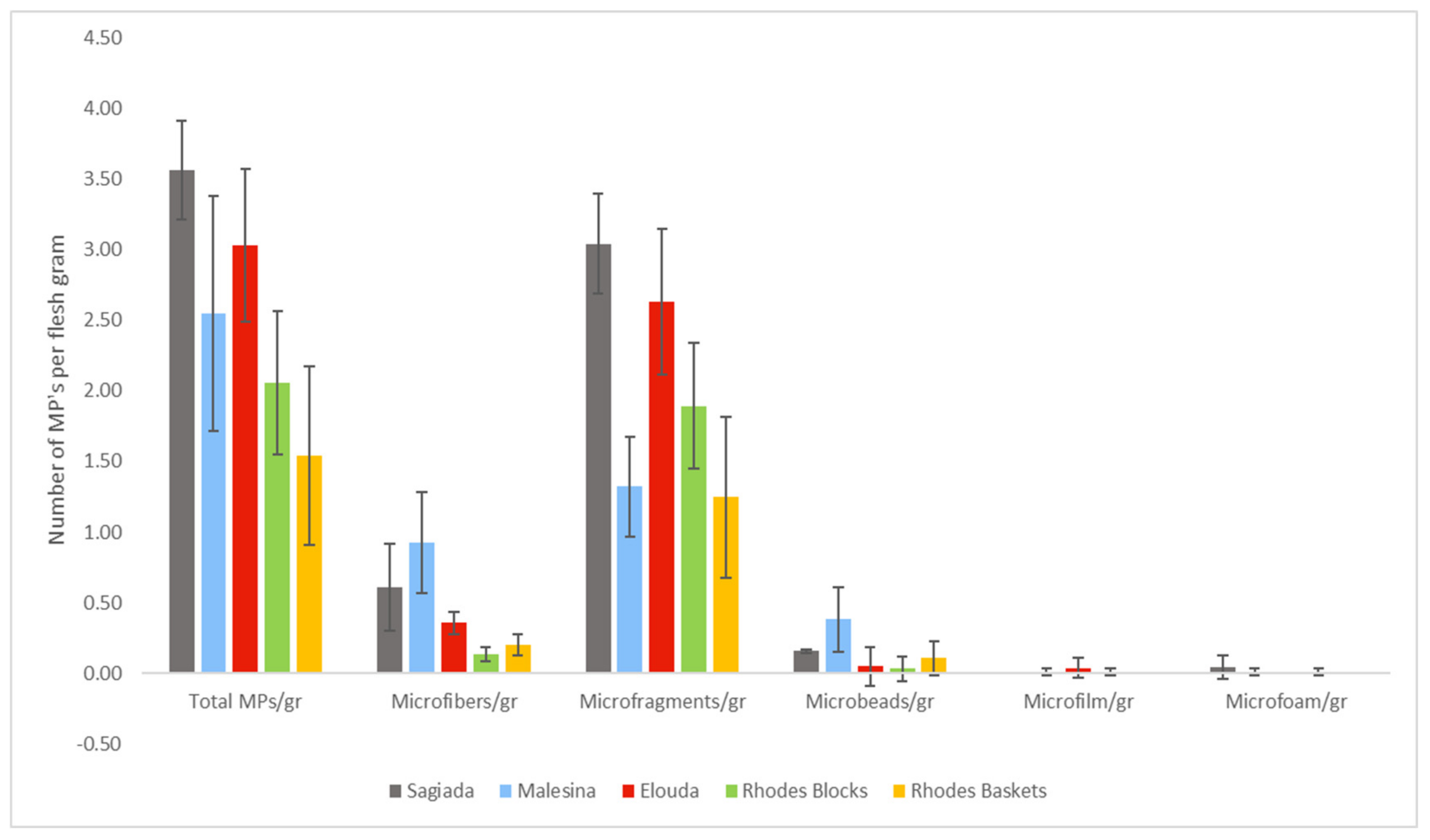
3.3. Differences of MPs between Stations

For the comparison between stations, we used P. imbricata radiata specimens collected from the five different sampling stations of the study (shown from west to east in Figure 6). The total MP particles per gram of soft tissue of P. imbricata radiata for Sagiada station were 3.56 ± 0.35, for Malesina station they reached 2.55 ± 0.83, for Elounda they were 3.03 ± 0.54, for Rhodes-blocks they were 2.06 ± 0.51, while for Rhodes-baskets they were 1.54 ± 0.63 (Figure 6). ANOVA results confirmed that there were significant differences in total MPs, microfiber, microfragment, and microbead categories between the five stations of the collected samples (F = 3.652, p < 0.05, F = 7.672, p < 0.05, F = 5.095, p < 0.05 and F = 5.122, p < 0.05, respectively). The dominant type of MP at all sampling stations was microfragments (Figure 6), and consequently they affected the overall pattern of total MPs.
The post hoc analysis indicated that regarding microfibers, Malesina had significantly more particles than Elounda, Rhodes-baskets and blocks stations, and more microbeads than Rhodes-blocks and Elounda stations. On the other hand, specimens from Elounda and Sagiada stations had significantly higher microfragments than those collected from Malesina and Rhodes-baskets stations. The overall comparison, for all the particles, showed that the total number of MPs was higher in Sagiada and lower in Rhodes-basket, and the values found for other stations were between these two extremes, without significant differences.

3.4. Differences of MPs in Every Station

For the comparison in every station, we used the MP types that we found in P. imbricata radiata specimens from the five different sampling stations of the study (Figure 6). ANOVA results confirmed that there were significant differences in the MP types in every station (Sagiada: F = 103.838, p < 0.05; Malesina: F = 23.029, p < 0.05; Elounda: F = 57.732, p < 0.05; Rhodes-blocks: F = 30.061, p < 0.05; Rhodes-baskets: F = 13.416, p < 0.05).
The post hoc analysis indicated that regarding Sagiada, Elounda, Rhodes-blocks, and Rhodes-baskets, there were significantly more microfragments than the other MP types found in each station (microfragment type ranging from 79.06% to 91.25%, Table 2). On the other hand, Malesina station had significantly more microfibers (34.98%) and microfragments (49.89%) found in its specimens (Table 2).

4. Discussion

This study provides information on microplastic pollution and its widespread presence in the soft tissue of marine cultured organisms. The presence of MPs in the soft tissue of mussels and pearl oysters indicates that the MPs released from (or discharged in the site of) an aquaculture farm may end up in the food chain, especially in the case of filter feeding organisms. Benthic filter-feeding organisms, such as mussels and pearl oysters can give in situ data related to the concentration and bioavailability of the seawater pollutants [35], and thus they are widely considered as biological indicators [36].
From the first comparison, the main MP type found in mussels was microfibers, whereas microplastic fragments were found mainly in pearl oysters. Microplastic fibers could not have originated from the pergolaris since the Raman analysis showed no matching of the pergolari peaks with the corresponding bands of the bivalve samples. On the other hand, it is unclear if the SEAPA oyster baskets correspond to any of the analyzed samples, as the Raman spectroscopy did not show an outcome in the spectral coverage of the spectrograph. However, the SEAPA baskets are made of sturdy plastic, which is less likely to break into smaller pieces. Breaking microplastic from these SEAPA baskets is more likely to form fragments than fibers. Furthermore, Raman spectra, characteristic bands assigned to PET were found in three analyzed samples; in contrast, none of the Raman peaks in all the microfiber samples corresponded to PE and PP. This may lead us to the conclusion that this kind of cultivation method does not affect the cultivated bivalves. The two pigments found in some of the samples indirectly imply that these particles have an anthropogenic origin. In particular, the pigment “phthalocyanine blue” is a synthetic widely used in boat dyes and also as an antifouling agent [37]. Similarly, the use of indigo pigment is mainly for dyeing polyester clothing and cotton fibers [38]. However, identification was not successful for all of the samples. This may be due to the small size of the samples, the fact that their content is too low and close to the Raman spectroscopy limits, or even the fact that some of them may not in fact be plastic.
The MP content in mussels found in the present study is similar to others conducted in the Aegean Sea [Izmir Bay] [28] and in the Ionian Sea [39] (Table 3), where fibers were the main type of MPs found in their soft tissues. From the present study, it is observed that the values of MPs found in cultured mussels of Malesina station are similar to the ones found in wild populations (Table 3). However, results of other studies, indicate a variety of different MP concentrations for M. galloprovincialis. This difference in the MP concentrations is related to the methodology used in every study, as well as filter pore size (Table 3), since the smaller the pore size, the more MP particles are identified. To our knowledge, there is quite a limited number of comparable datasets on MPs found in the case of P. imbricata radiata. In the present study, the MPs found are not very different to those found in the wild population of the Persian Gulf [40] (Table 3).
The results of the comparison of MP content between the five different sampling stations confirmed the preference of P. imbricata radiata for microfragments. The population of Sagiada and the wild population of Elounda were found to be enriched with more MP fragments than the farmed populations of Malesina and Rhodes stations.
The higher and lower abundances of total MP content in P. imbricata radiata corresponds to the different water quality of the farming sites and the use of plastic material for the farming practices.
In the case of Sagiada, this may be due to the fact that the sampling station was in the vicinity of the discharge area of Kalamas River. Upstream estuaries can transport significant amounts of anthropogenic litter and discharge them into the marine environment [50].
Furthermore, the P. imbricata radiata individuals were collected from an experimental longline culture that are more likely to generate MP debris, as it breaks into smaller pieces more easily than the sturdy material of the SEAPA baskets. On the other hand, regarding total MPs, values from the Rhodes-baskets station were significantly lower than those from Sagiada, which might be due to the fact that the Rhodes-baskets station was located in an oligotrophic area far away from urban centers and any other MP pollution hot spots.
In the case of Elounda station, the results (i.e., high levels of microfragments) can be attributed to the characteristics of the study site, since the collected specimens inhabited the hard substrate of a shallow enclosed bay, next to a highly developed touristic area. Thus, the pearl oysters of Elounda station were exposed to plastic debris that was sinking to the bottom from different sources (visitors on the sea side, boats, etc.).
The pearl oyster populations at Malesina and Rhodes-baskets stations were farmed with the same method in areas with different water circulation conditions. Thus, the placement of Malesina station in a semi-enclosed area could explain the higher concentration of MPs in relation to the open sea area of Rhodes-baskets.
In Malesina station, the torrents seasonally discharging freshwater and agricultural wastes and plastic debris in the vicinity of the farm could be a potential source of enrichment with MPs in the area. Furthermore, the pergolari method used for mussel farming may have acted as an additional source of the MPs ingested by the oyster population.
Finally, the comparison of the MPs contained in the soft tissue of the specimens has indicated variations depending inter alia on the species, the site, and the extraction method used, as confirmed by recent studies on other members of the Ostreida order (Table 3). Nevertheless, the results of the current study for all five sampling stations are within the range reported so far for their conspecifics (Table 3).

5. Conclusions

Most of the existing regulations around the world are specific to macroplastics; there is no specific regulatory framework to contain MPs. However, there have been many attempts by various institutions/countries to introduce regulatory actions to reduce microplastics in water. In 2019, the European Chemicals Agency (ECHA) suggested banning products containing intentionally added microplastics from the European market [4]. The same year, the European Directive 2019/904 was adopted to limit the use of single-use plastics in order to reduce marine pollution [51]. In 2021, the European Union released the European Water Framework Directive (EWFD), which aimed to establish a methodology to quantify microplastics in water so they could be included in the Watch List [4].
The present study confirms that high or low concentrations of MPs are widely distributed in the natural ecosystem. Further research is needed to find ways to reduce or control MP pollution and set safety limits for human consumption, as at the moment the effects on human health, the marine organisms and generally the marine ecosystem are rather uncertain.
Most of the MPs found in the samples of the present study are microfibers and microfragments, which can easily end up in the marine environment, especially in areas of high anthropogenic activity. Raman spectroscopy is a useful analytical tool for the identification of MPs contained in mussel and pearl oyster specimens. More investigation is needed to determine whether the lower concentration of MPs found in the samples of the multitrophic aquaculture is due to the culturing method used for these organisms.
Regarding the P. imbricata radiata species, only one similar study was found in the literature (Table 3, [40]), and therefore further analyses should be conducted on wild and farmed populations to augment the available information on this issue and determine whether culture is a factor that affects the concentration of microplastic particles in the soft tissue of these organisms.

Author Contributions

Conceptualization, N.P., P.D.D. and I.K.; Methodology, S.M.M. and N.P.; Formal Analysis, S.M.M., P.D.D. and A.P.; Investigation, S.M.M., D.C. and M.L.; Resources, N.P., P.D.D. and I.K.; Writing—Original Draft Preparation, S.M.M.; Writing—Review and Editing, S.M.M., N.P., P.D.D. and I.K.; Supervision, N.P.; Funding Acquisition, I.K. All authors have read and agreed to the published version of the manuscript.

Funding

The present work is part of the project “Innovative Development of Multitrophic Aquaculture” funded by the “Innovation in Fisheries” EU–Greece Operational Program of Fisheries and Maritime, EPAL 2014–2020 (grant number 5029294).

Institutional Review Board Statement

Not applicable.

Informed Consent Statement

Not applicable.

Data Availability Statement

The data from this paper will be made available in the IDMA project report and will be hosted at www.idma.uoc.gr in the publication section.

Acknowledgments

We thank the COs and personnel of the fish farms for providing facilities and equipment as well as the scientific divers Ioulios Glampedakis and Anastasios Baltadakis for their numerous dives. We also thank John Theodorou for the Sagiada sample collection.

Conflicts of Interest

The authors declare no conflict of interest. The sponsors had no role in the design, execution, interpretation, or writing of the study.

References

  1. Andrady, A.L. Microplastics in the marine environment. Mar. Pollut. Bull. 2011, 62, 1596–1605. [Google Scholar] [CrossRef] [PubMed]
  2. Wright, S.L.; Thompson, R.C.; Galloway, T.S. The physical impacts of microplastics on marine organisms: A review. Environ. Pollut. 2013, 178, 483–492. [Google Scholar] [CrossRef] [PubMed]
  3. Dris, R.; Gasperi, J.; Saad, M.; Mirande, C.; Tassin, B. Synthetic fibers in atmospheric fallout: A source of microplastics in the environment? Mar. Pollut. Bull. 2016, 104, 290–293. [Google Scholar] [CrossRef] [PubMed]
  4. Khan, N.A.; Khan, A.H.; Maldonado, E.A.L.; Alam, S.S.; López, J.R.L.; Herrera, P.F.M.; Mohamed, B.A.; Mahmoud, A.E.D.; Abutaleb, A.; Singh, L. Microplastics: Occurrences, treatment methods, regulations and foreseen environmental impacts. Environ. Res. 2022, 215, 114224. [Google Scholar] [CrossRef] [PubMed]
  5. Wu, Y.; Guo, P.; Zhang, X.; Zhang, Y.; Xie, S.; Deng, J. Effect of microplastics exposure on the photosynthesis system of freshwater algae. J. Hazard. Mater. 2019, 374, 219–227. [Google Scholar] [CrossRef]
  6. Sun, J.; Dai, X.; Wang, Q.; Van Loosdrecht, M.C.; Ni, B.-J. Microplastics in wastewater treatment plants: Detection, occurrence and removal. Water Res. 2019, 152, 21–37. [Google Scholar] [CrossRef]
  7. Setälä, O.; Norkko, J.; Lehtiniemi, M. Feeding type affects microplastic ingestion in a coastal invertebrate community. Mar. Pollut. Bull. 2016, 102, 95–101. [Google Scholar] [CrossRef]
  8. Thushari, G.G.N.; Senevirathna, J.D.M.; Yakupitiyage, A.; Chavanich, S. Effects of microplastics on sessile invertebrates in the eastern coast of Thailand: An approach to coastal zone conservation. Mar. Pollut. Bull. 2017, 124, 349–355. [Google Scholar] [CrossRef]
  9. Ward, J.E.; Shumway, S.E. Separating the grain from the chaff: Particle selection in suspension-and deposit-feeding bivalves. J. Exp. Mar. Biol. Ecol. 2004, 300, 83–130. [Google Scholar] [CrossRef]
  10. Avio, C.G.; Gorbi, S.; Regoli, F. Plastics and microplastics in the oceans: From emerging pollutants to emerged threat. Mar. Environ. Res. 2017, 128, 2–11. [Google Scholar] [CrossRef]
  11. Li, L.-L.; Amara, R.; Souissi, S.; Dehaut, A.; Duflos, G.; Monchy, S. Impacts of microplastics exposure on mussel (Mytilus edulis) gut microbiota. Sci. Total Environ. 2020, 745, 141018. [Google Scholar] [CrossRef] [PubMed]
  12. Harris, L.S.; Gill, H.; Carrington, E. Microplastic changes the sinking and resuspension rates of marine mussel biodeposits. Mar. Pollut. Bull. 2021, 165, 112165. [Google Scholar] [CrossRef] [PubMed]
  13. Ward, J.E.; Rosa, M.; Shumway, S.E. Capture, ingestion, and egestion of microplastics by suspension-feeding bivalves: A 40-year history. Anthr. Coasts 2019, 2, 39–49. [Google Scholar] [CrossRef] [Green Version]
  14. Chatzivasileiou, D.; Dimitriou, P.D.; Theodorou, J.; Kalantzi, I.; Magiopoulos, I.; Papageorgiou, N.; Pitta, P.; Tsapakis, M.; Karakassis, I. An IMTA in Greece: Co-Culture of Fish, Bivalves, and Holothurians. J. Mar. Sci. Eng. 2022, 10, 776. [Google Scholar] [CrossRef]
  15. Moutopoulos, D.K.; Ramfos, A.; Theodorou, J.A.; Katselis, G. Biological aspects, population and fishery dynamics of the non-indigenous pearl oyster Pinctada imbricata radiata (Leach, 1814) in the Eastern Mediterranean. Reg. Stud. Mar. Sci. 2021, 45, 101821. [Google Scholar] [CrossRef]
  16. Theodorou, J.A.; Makri, M.; Douvi, X.; Ramfos, A.; Spinos, E. Seasonal variation in the biochemical composition, condition index, and meat yield of the non-indigenous pearl oyster Pinctada imbricata radiata (Leach, 1814) from the West of the Aegean Sea, Greece. Aquac. Fish. 2021, 8, 451–456. [Google Scholar] [CrossRef]
  17. Kalantzi, I.; Karakassis, I. Benthic impacts of fish farming: Meta-analysis of community and geochemical data. Mar. Pollut. Bull. 2006, 52, 484–493. [Google Scholar] [CrossRef]
  18. Papageorgiou, N.; Kalantzi, I.; Karakassis, I. Effects of fish farming on the biological and geochemical properties of muddy and sandy sediments in the Mediterranean Sea. Mar. Environ. Res. 2010, 69, 326–336. [Google Scholar] [CrossRef]
  19. Pitta, P.; Tsapakis, M.; Apostolaki, E.T.; Tsagaraki, T.; Holmer, M.; Karakassis, I. ‘Ghost nutrients’ from fish farms are transferred up the food web by phytoplankton grazers. Mar. Ecol. Prog. Ser. 2009, 374, 1–6. [Google Scholar] [CrossRef]
  20. Alexander, K.; Angel, D.; Freeman, S.; Israel, D.; Johansen, J.; Kletou, D.; Meland, M.; Pecorino, D.; Rebours, C.; Rousou, M.; et al. Improving sustainability of aquaculture in Europe: Stakeholder dialogues on integrated multi-trophic aquaculture (IMTA). Environ. Sci. Policy 2016, 55, 96–106. [Google Scholar] [CrossRef]
  21. Chopin, T.; Cooper, J.A.; Reid, G.; Cross, S.; Moore, C. Open-water integrated multi-trophic aquaculture: Environmental biomitigation and economic diversification of fed aquaculture by extractive aquaculture. Rev. Aquac. 2012, 4, 209–220. [Google Scholar] [CrossRef]
  22. Freeman, S.; Angel, D.; Freeman, S. Integrated aquaculture (INTAQ) as a tool for an ecosystem approach to the marine farming sector in the Mediterranean Sea. Integr. Maric. 2019, 529, 133–183. [Google Scholar]
  23. Chen, G.; Li, Y.; Wang, J. Occurrence and ecological impact of microplastics in aquaculture ecosystems. Chemosphere 2021, 274, 129989. [Google Scholar] [CrossRef] [PubMed]
  24. Piperagkas, O.; Papageorgiou, N.; Karakassis, I. Qualitative and quantitative assessment of microplastics in three sandy Mediterranean beaches, including different methodological approaches. Estuar. Coast. Shelf Sci. 2019, 219, 169–175. [Google Scholar] [CrossRef]
  25. Munno, K.; Helm, P.A.; Jackson, D.A.; Rochman, C.; Sims, A. Impacts of temperature and selected chemical digestion methods on microplastic particles. Environ. Toxicol. Chem. 2018, 37, 91–98. [Google Scholar] [CrossRef] [PubMed]
  26. Patterson, J.; Jeyasanta, K.I.; Laju, R.; Edward, J.P. Microplastic contamination in Indian edible mussels (Perna perna and Perna viridis) and their environs. Mar. Pollut. Bull. 2021, 171, 112678. [Google Scholar] [CrossRef]
  27. Wakkaf, T.; El Zrelli, R.; Kedzierski, M.; Balti, R.; Shaiek, M.; Mansour, L.; Tlig-Zouari, S.; Bruzaud, S.; Rabaoui, L. Microplastics in edible mussels from a southern Mediterranean lagoon: Preliminary results on seawater-mussel transfer and implications for environmental protection and seafood safety. Mar. Pollut. Bull. 2020, 158, 111355. [Google Scholar] [CrossRef]
  28. Yozukmaz, A. Investigation of microplastics in edible wild mussels from Izmir Bay (Aegean Sea, Western Turkey): A risk assessment for the consumers. Mar. Pollut. Bull. 2021, 171, 112733. [Google Scholar] [CrossRef]
  29. Hidalgo-Ruz, V.; Gutow, L.; Thompson, R.C.; Thiel, M. Microplastics in the marine environment: A review of the methods used for identification and quantification. Environ. Sci. Technol. 2012, 46, 3060–3075. [Google Scholar] [CrossRef]
  30. De Witte, B.; Devriese, L.; Bekaert, K.; Hoffman, S.; Vandermeersch, G.; Cooreman, K.; Robbens, J. Quality assessment of the blue mussel (Mytilus edulis): Comparison between commercial and wild types. Mar. Pollut. Bull. 2014, 85, 146–155. [Google Scholar] [CrossRef]
  31. Crawford, C.B.; Quinn, B.; Crawford, C.; Quinn, B. Microplastic identification techniques. Microplastic Pollut. 2017, 10, 219–267. [Google Scholar]
  32. Philippidis, A.; Mikallou, A.; Anglos, D. Determining optimum irradiation conditions for the analysis of vermilion by Raman spectroscopy. Eur. Phys. J. Plus 2021, 136, 1194. [Google Scholar] [CrossRef]
  33. Caggiani, M.C.; Cosentino, A.; Mangone, A. Pigments Checker version 3.0, a handy set for conservation scientists: A free online Raman spectra database. Microchem. J. 2016, 129, 123–132. [Google Scholar] [CrossRef]
  34. da Silva, D.J.; Parra, D.F.; Wiebeck, H. Applying confocal Raman spectroscopy and different linear multivariate analyses to sort polyethylene residues. Chem. Eng. J. 2021, 426, 131344. [Google Scholar] [CrossRef]
  35. Li, J.; Lusher, A.L.; Rotchell, J.M.; Deudero, S.; Turra, A.; Bråte, I.L.N.; Sun, C.; Hossain, M.S.; Li, Q.; Kolandhasamy, P.; et al. Using mussel as a global bioindicator of coastal microplastic pollution. Environ. Pollut. 2019, 244, 522–533. [Google Scholar] [CrossRef]
  36. Beyer, J.; Green, N.W.; Brooks, S.; Allan, I.J.; Ruus, A.; Gomes, T.; Bråte, I.L.N.; Schøyen, M. Blue mussels (Mytilus edulis spp.) as sentinel organisms in coastal pollution monitoring: A review. Mar. Environ. Res. 2017, 130, 338–365. [Google Scholar] [CrossRef]
  37. Strokov, K.; Schäfer, A.H.; Dobrindt, U.; Galstyan, A. Facile fabrication of silicon (IV) phthalocyanine-embedded poly (vinyl alcohol)-based antibacterial and antifouling interfaces. ACS Appl. Bio Mater. 2020, 3, 3751–3760. [Google Scholar] [CrossRef]
  38. Turner, S.; Horton, A.A.; Rose, N.L.; Hall, C. A temporal sediment record of microplastics in an urban lake, London, UK. J. Paleolimnol. 2019, 61, 449–462. [Google Scholar] [CrossRef]
  39. Digka, N.; Tsangaris, C.; Torre, M.; Anastasopoulou, A.; Zeri, C. Microplastics in mussels and fish from the Northern Ionian Sea. Mar. Pollut. Bull. 2018, 135, 30–40. [Google Scholar] [CrossRef]
  40. Naji, A.; Nuri, M.; Vethaak, A.D. Microplastics contamination in molluscs from the northern part of the Persian Gulf. Environ. Pollut. 2018, 235, 113–120. [Google Scholar] [CrossRef]
  41. Gedik, K.; Eryaşar, A.R.; Gözler, A.M. The microplastic pattern of wild-caught Mediterranean mussels from the Marmara Sea. Mar. Pollut. Bull. 2022, 175, 113331. [Google Scholar] [CrossRef]
  42. Leslie, H.; Brandsma, S.; Van Velzen, M.; Vethaak, A. Microplastics en route: Field measurements in the Dutch river delta and Amsterdam canals, wastewater treatment plants, North Sea sediments and biota. Environ. Int. 2017, 101, 133–142. [Google Scholar] [CrossRef] [PubMed]
  43. Li, J.; Green, C.; Reynolds, A.; Shi, H.; Rotchell, J.M. Microplastics in mussels sampled from coastal waters and supermarkets in the United Kingdom. Environ. Pollut. 2018, 241, 35–44. [Google Scholar] [CrossRef] [PubMed]
  44. Li, J.; Qu, X.; Su, L.; Zhang, W.; Yang, D.; Kolandhasamy, P.; Li, D.; Shi, H. Microplastics in mussels along the coastal waters of China. Environ. Pollut. 2016, 214, 177–184. [Google Scholar] [CrossRef]
  45. Li, H.-X.; Ma, L.-S.; Lin, L.; Ni, Z.-X.; Xu, X.-R.; Shi, H.-H.; Yan, Y.; Zheng, G.-M.; Rittschof, D. Microplastics in oysters Saccostrea cucullata along the Pearl River estuary, China. Environ. Pollut. 2018, 236, 619–625. [Google Scholar] [CrossRef]
  46. Van Cauwenberghe, L.; Janssen, C.R. Microplastics in bivalves cultured for human consumption. Environ. Pollut. 2014, 193, 65–70. [Google Scholar] [CrossRef]
  47. Saldaña-Serrano, M.; Bastolla, C.L.; Mattos, J.J.; Lima, D.; Freire, T.B.; Nogueira, D.J.; De-la-Torre, G.E.; Righetti, B.P.; Zacchi, F.L.; Gomes, C.H.; et al. Microplastics and linear alkylbenzene levels in oysters Crassostrea gigas driven by sewage contamination at an important aquaculture area of Brazil. Chemosphere 2022, 307, 136039. [Google Scholar] [CrossRef] [PubMed]
  48. Dang, T.T.; Le, X.T.T.; Nguyen, D.T.; Phung, T.V.; Vu, D.N.; Pham, H.V. Abundance of microplastics in cultured oysters (Crassostrea gigas) from Danang Bay of Vietnam. Mar. Pollut. Bull. 2022, 180, 113800. [Google Scholar]
  49. Waite, H.R.; Donnelly, M.J.; Walters, L.J. Quantity and types of microplastics in the organic tissues of the eastern oyster Crassostrea virginica and Atlantic mud crab Panopeus herbstii from a Florida estuary. Mar. Pollut. Bull. 2018, 129, 179–185. [Google Scholar] [CrossRef]
  50. Aslam, S.; Tzoraki, O.; Krasakopoulou, E. Anthropogenic litter in freshwater bodies and their estuaries: An empirical analysis in Lesvos, Greece. Environ. Sci. Pollut. Res. 2022, 29, 16563–16575. [Google Scholar] [CrossRef]
  51. Conti, I.; Simioni, C.; Varano, G.; Brenna, C.; Costanzi, E.; Neri, L.M. Legislation to limit the environmental plastic and microplastic pollution and their influence on human exposure. Environ. Pollut. 2021, 288, 117708. [Google Scholar] [CrossRef] [PubMed]
Figure 1. Map of the sampling stations.
Microplastics 02 00013 g001
Figure 2. (a) M. galloprovincialis collected samples, (b) M. galloprovincialis cultivating method using pergolaris.
Microplastics 02 00013 g002
Figure 3. (a) Collected P. imbricata radiata within SEAPA© baskets, (b) P. imbricata radiata farmed in SEAPA© baskets near to the fish cages, (c) Blocks from where samples of P. imbricata radiata were collected in Rhodes, and (d) P. imbricata radiata individuals from the seabed of Elounda bay.
Microplastics 02 00013 g003
Figure 4. Raman spectra of microfibers. (a) Sample from Malesina station (green line) in comparison with a reference of PET, from our personal library (purple line). (b) Sample from Elounda station (red line) and the pure phthalocyanine blue, for reference (light blue line). (c) Sample from Rhodes-baskets station (yellow line) and pure indigo (blue line) for comparison. (d) Sample from pergolari with characteristic Raman bands correspond to PE and PP.
Microplastics 02 00013 g004
Figure 5. Average (±SD) number of MPs (total and subcategories) in 1 g of mussel and pearl oyster flesh in Malesina station.
Microplastics 02 00013 g005
Figure 6. Average (±SD) number of MPs (total and subcategories) in 1 g of pearl oyster flesh in all stations.
Microplastics 02 00013 g006
Table 1. Percentage of each MP type found within all mussel and pearl oyster specimens from Malesina station.
Study SiteMP TypeMytilus galloprovincialis (%)Pinctada imbricata radiata (%)
MalesinaMicrofibers47.7434.98
Microfragments42.9649.89
Microbeads9.2914.44
Microfoam-0.34
Microfilm-0.34
Table 2. Percentage of each MP category found within pearl oyster samples for all stations.
Study SiteMicrofibers (%)Microfragments (%)Microbeads (%)Microfoam (%)Microfilm (%)
Sagiada15.8279.064.051.07-
Malesina34.9849.8914.440.340.34
Elounda11.5785.691.56-1.18
Rhodes-blocks6.4691.251.470.420.40
Rhodes-baskets13.0180.056.94--
Table 3. Range of microplastic concentrations (particles/g of flesh) in different types of marine bivalves found in the scientific literature.
SpeciesMP ConcentrationFilter Pore Size (μm)Study SiteReference
Mytilus galloprovincialis5.3 ± 0.51.2Ionian Sea (wild)[39]
5.0 ± 0.960.8Evoikos Gulf, Malesina (farmed)This study
2.81 to 4.980.7Izmir Bay (wild)[28]
2.5 ± 0.31.2Ionian Sea (farmed)[39]
1.121.2Marmara Sea (wild)[41]
Mytilus edulis13.20.7Dutch North Sea Coast (wild)[42]
0.7 to 2.95U.K. (wild)[43]
2.75China (wild)[33,44,45]
1.65China (farmed)[44]
0.36 ± 0.070.8North Sea, Germany (farmed)[46]
Pinctada imbricata radiata1.54 to 3.560.8Sagiada, Malesina, Rhodes, Greece (farmed)This study
2.06 to 3.030.8Elounda bay, Rhodes, Greece (wild)This study
0.2 to 2.20.45Persian Gulf, Iran (wild)[40]
Saccostrea cucullata1.5 to 7.220Pearl River, Estuary, China (wild)[45]
Crassostrea gigas0.27–0.640.6Santa Caterina Island, Brazil[47]
0.47 ± 0.160.8Brittany, France, Atlantic Ocean (commercial)[46]
1.88 ± 1.580.6Danang Bay, Vietnam[48]
Crassostrea virginica3.84 ± 3.390.45Mosquito Lagoon, Indian River, Florida[49]
Disclaimer/Publisher’s Note: The statements, opinions and data contained in all publications are solely those of the individual author(s) and contributor(s) and not of MDPI and/or the editor(s). MDPI and/or the editor(s) disclaim responsibility for any injury to people or property resulting from any ideas, methods, instructions or products referred to in the content.

