Article

Effect of Polystyrene Microplastics in Different Diet Combinations on Survival, Growth and Reproduction Rates of the Water Flea (Daphnia magna)

by
Melek Isinibilir
1,
Kamil Mert Eryalçın
2,* and
Ahmet Erkan Kideys
3
1
Department of Marine and Freshwater Resources Management, Faculty of Aquatic Science, Istanbul University, Istanbul 34134, Turkey
2
Phytoplankton-Zooplankton Culture Laboratory, Department of Aquaculture and Fish Diseases, Faculty of Aquatic Science, Istanbul University, Istanbul 34134, Turkey
3
Institute of Marine Sciences, Middle East Technical University, Erdemli-Mersin 33731, Turkey
*
Author to whom correspondence should be addressed.
Microplastics 2023, 2(1), 27-38; https://doi.org/10.3390/microplastics2010002
Submission received: 17 September 2022 / Revised: 15 October 2022 / Accepted: 18 November 2022 / Published: 23 December 2022
(This article belongs to the Collection Current Opinion in Microplastics)

Abstract

:

Highlights

Potential effects of microplastics (in 6 micron size) were investigated on the survival, growth and reproduction of the Daphnia magna during 21 days of laboratory experiments.
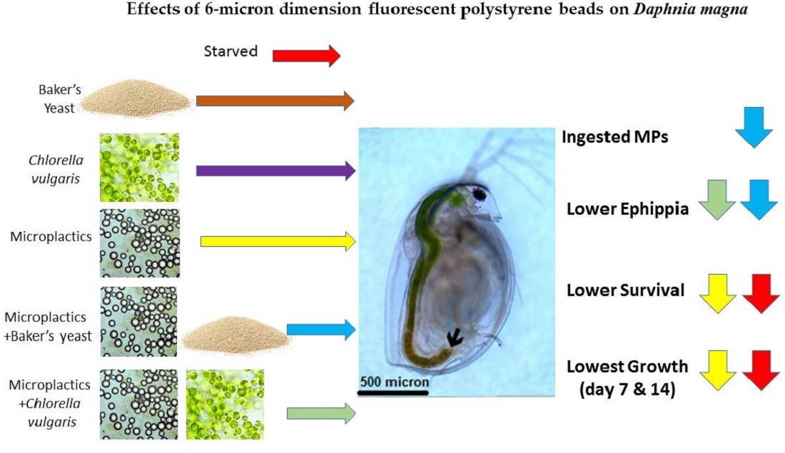
  • Microplastics MPs were ingested alone or along with either microalgae Chlorella vulgaris (Cv) or baker’s yeast (By)
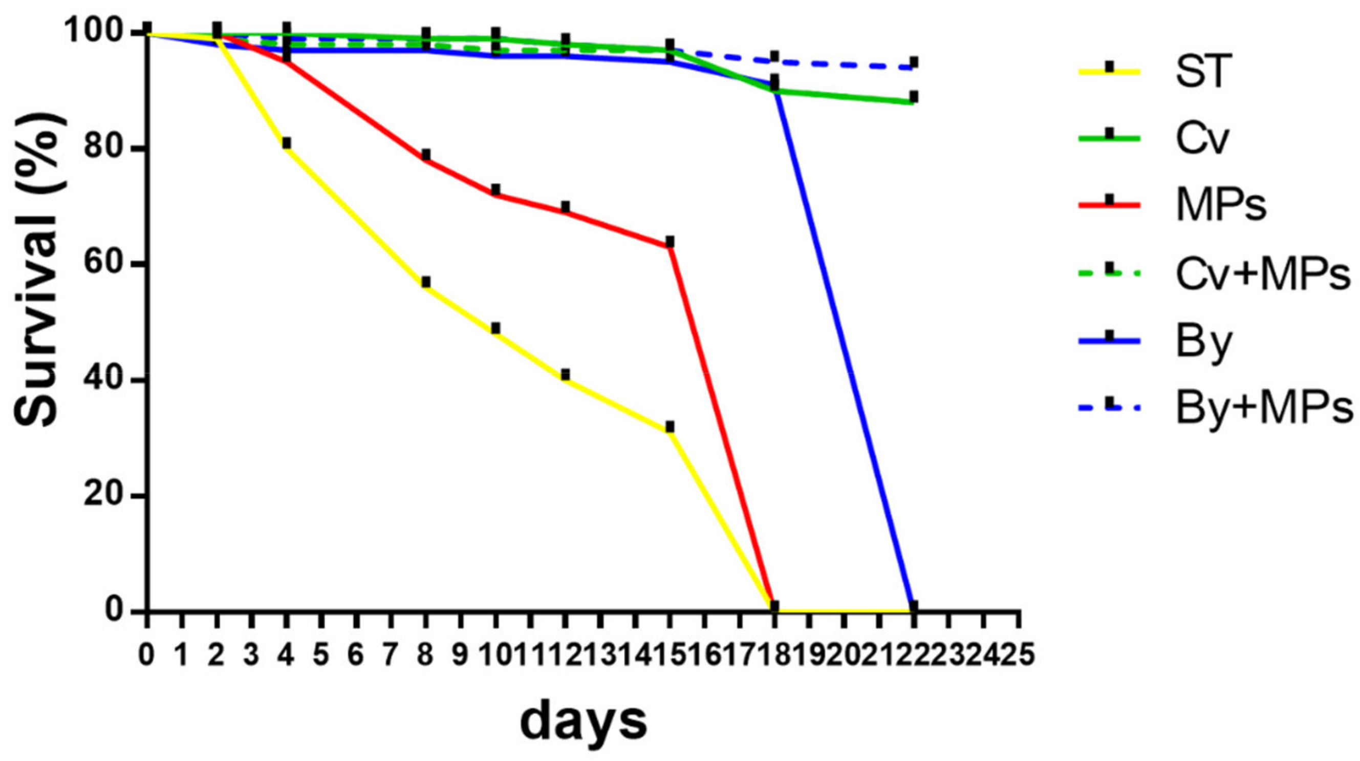
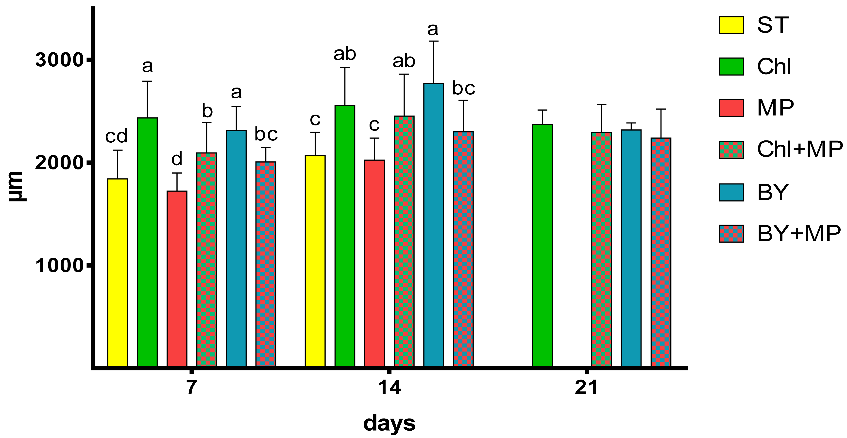
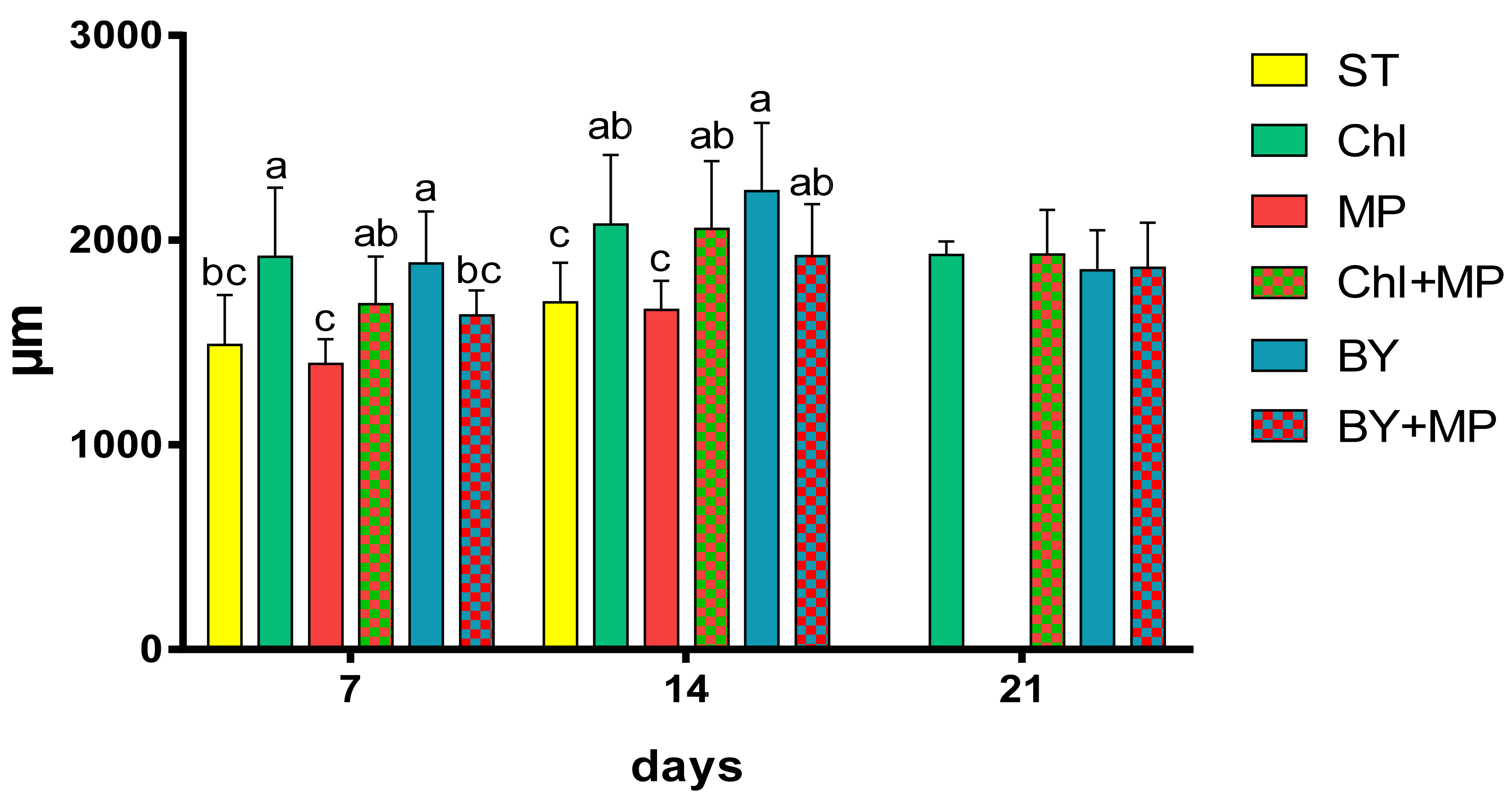
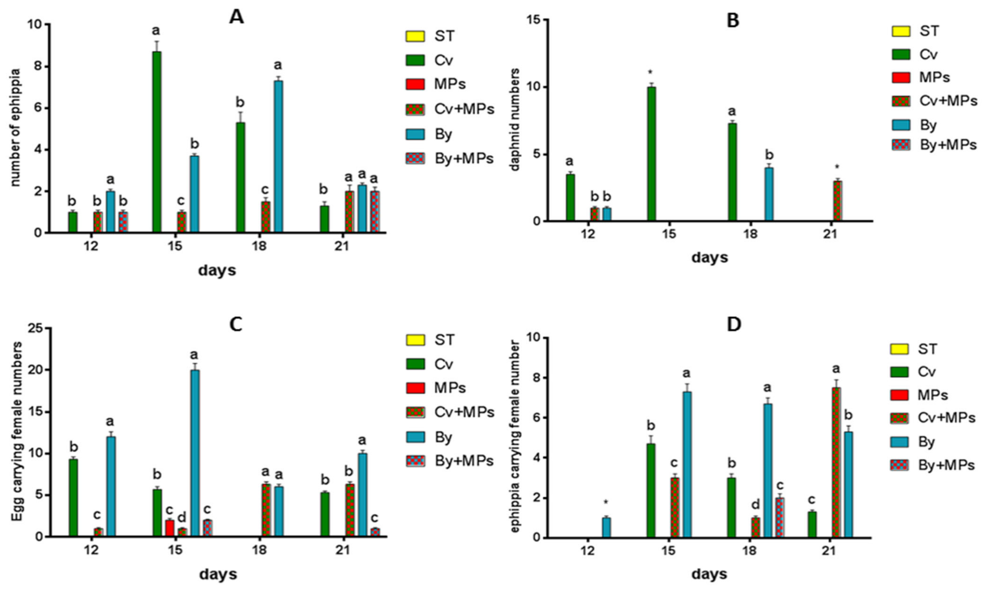
  • Microplastics decreased survival, growth and reproduction of Daphnia
  • High concentrations of microplastics adversely affect Daphnia magna populations

Abstract

Microplastic pollution is a problem not only in the marine environment but also in freshwater ecosystems. Water flea (Daphnia magna) is one of the most common omnivorous cladocerans in freshwater ecosystems. In this study, the potential effects of microplastics (fluorescent polystyrene beads with dimensions of 6 microns) on the survival, growth and reproduction of Daphnia magna were examined during 21 days of laboratory experiments. Microplastics (MPs) were observed to be ingested alone or along with either the microalgae Chlorella vulgaris (Cv) or baker’s yeast (By). D. magna fed exclusively with microplastics showed a drastic decline in survival similar to that in the starving group. The least growth in total length or width was observed in Daphnia specimens fed only MPs and the starved groups. Daphia fed with a mixture of MPs/Cv or MPs/By produced a significantly (p < 0.05) lower number of ephippia. Our results show that high concentrations of microplastics adversely affect Daphnia magna populations.

Graphical Abstract

1. Introduction

With the ever-increasing usage of plastics, particularly since the 1950s, we are now in the plastic age. Globally, plastic production has increased linearly over the last 10 years, reaching 368 million tons in 2019 [1]. Primary microplastics are defined as plastics produced with a microscopic size [2]. Secondary microplastics are small plastic particles produced during the degradation of larger plastics [3]. Polyethylene plastic is the most abundant source of plastics in the world [4]. Microplastics (MPs) consist of a variety of plastics, polyethylene and polystyrene being the most common types found in environmental samples [5,6]. The main sources of microplastics are wastewater and landfill leachate [7] through wastewater effluent and rivers [8].
Although the majority of early investigations into microplastic pollution focused on the prevalence and harmful effects on marine species, microplastics have lately been documented in freshwater environments, with toxicity issues involving invertebrates and fish [9,10,11,12,13,14]. Microplastics have been reported to interact with aquatic organisms via feeding, cutaneous uptake and respiration [4,15,16,17].
A number of studies have shown that microplastics can be efficiently ingested by at least 160 marine [18] and 39 freshwater species [19], including invertebrates such as cladocerans [20], rotifers [21] and molluscs [22]. The exposure to MPs and the resulting consequences in zooplanktonic filter-feeder species, which absorb MPs indiscriminately during their regular swimming and feeding activities, are of special concern [23]. Several studies have shown that the presence of microplastics (MPs) in the digestive tracts of several zooplanktonic species may have negative consequences. Microplastics can be a subtrate for the proliferation of microalgae on their surface and this lead to Daphnia finding sufficient food. Consequently, reproduction and survival may not be affected by these MPs [20]. The toxicity of MPs on D. magna was also linearly correlated with the light intensity and temperature elevation in their habitat [24]. Long-term studies with a combination of algae and different sizes of MPs showed the harmful effects on the survival of D.magna populations [25].
The diverse cladoceran Daphnia magna (Straus, 1820) is a major zooplankton species utilized in environmental toxicology studies [26]. Its ecological importance in freshwater is linked to the transmission of energy to higher trophic levels and filtration of phytoplankton, bacteria and natural particles [27]. Because of its tiny size (less than 5 mm) and brief life cycle (usually less than two months), Daphnia magna has been used as an indicator species since the 1940s [28].
Studies on Daphnia magna have shown that the ingestion of microplastics are influenced by the particles’ type, size and shape [2,29]. Furthermore, it has been observed that when daphnids ingest large concentrations of polyethylene (PE) microbeads 1 and 100 µm in size, their immobilization rates increased in a dosage- and time-dependent manner [30]. In microplastic studies, the life cycle of D. magna (egg; offspring for less than 24 h of life; juvenile; adult after the first spawning) is often assessed using traditional endpoints such as survival and number of offspring [31].
To the best of our knowledge, the effect of a combination of small microplastic particles with common diets (microalgae and baker’s yeast) in D.magna are scarce. Therefore, the aim of this study was to establish the impact of microplastics on the survival, growth and reproduction of the water flea D. magna when offered alone or with either the green algae Chlorella vulgaris or baker’s yeast.

2. Materials and Methods

2.1. Daphnia Magna Culture

Stock cultures of Daphnia magna were isolated from Ömerli Lake, Turkey. The culture water was filtered through 0.22 μm membrane filters with tap water. Stock cultures were maintained at 20 °C at a 16L:8D photoperiod in 20 L polyethylene buckets. The culture water weas renewed daily with fresh suspended feed. Daphnia magna were fed daily with Cv at a density of 1 × 10⁶ cells/mL. Experiments were conducted using third-clutch neonates which were a maximum of 12 h old.

2.2. Microalgae Culture and Stock Concentrations of Microplastics

Microalgae (Chlorella vulgaris, strain number: 211/109) were provided by the CCAP (Culture Collection of Algae and Protozoa, Scotland, UK). The freshwater microalgae Chlorella vulgaris was cultured in 3N-BBM + V medium (NaNO3 + CaCl2.H2O + MgSO4.7H2O + K2HPO4.3H2O + KH2PO4 + NaCl + trace minerals + Vitamin B1 and Vitamin B12). In order to calculate growth numbers, the microalgae were counted daily using a Neubauer hemocytometer. Thermo Scientific fluorescent polystyrene microplastic particles (6 micron size) were used for the microplastic experiments. The stock microplastic solution contained 2.5 million beads/mL.

2.3. Experimental Diets

The experimental groups, in addition to the starving group (i.e., 6 groups in total), were as follows: (1) microplastics only diet (2) Cv only (3) By only (4) Cv + MPs and (5) By + MPs. Each group was tested in triplicate in 500 mL beakers containing 100 adult D. magna. individuals. The experimental food concentrations were as follows: Group 1 (microplastics only) was fed with 6 mL of microplastics from the stock solution, Group 2 (Cv) was fed at a concentration of 2 × 10⁶ cells/mL/day, Group 3 (baker’s yeast, Pakmaya®, Ankara, Turkey) was fed 3 g/L every 2 days, Group 4 (Cv and MPs) was fed with 2 × 10⁶ cells/mL/day + 3 mL of microplastics from the stock solution, and Group 5 (By and MPs) was fed with 3 g/L every 2 days By and 3 mL of microplastics from main MP stocks. The experimental period was 21 days.

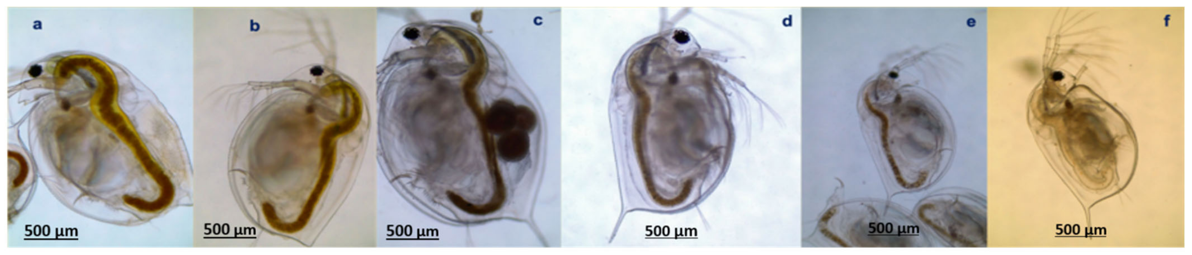
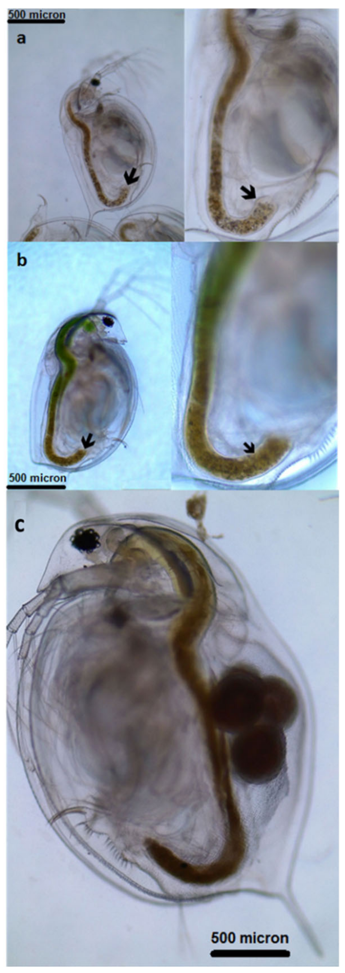
2.4. Observation of Microplastic Ingestion

To verify that Daphnia ingested the microplastics offered, five specimens (n = 5) were randomly sampled from the beaker containing microplastics only on experimental Day 2. Each specimen was individually examined on a microscope slide, where organic substances were removed by adding a few drops of hydrogen peroxide. In order to estimate the rate of microplastic consumption, the number of microplastic beads consumed by each Daphnia magna was counted under the microscope (Leica DM-1000, Danaher Cooperation, Germany) with a computer attachment on Days 7 and 21.

2.5. Survival, Growth and Reproductive Performance of Daphnia magna

Survival was determined by counting dead Daphnia individuals every day. Measurements for determining the growth performance, total length, body length and width of Daphnia magna were carried out every 7 days during the experimental period (21 days in total). For these measurements, randomly sampled individuals (n = 5) from each beaker were placed on glass slides for microscopic examination. Reproductive performance was determined by counting the numbers of females, eggs and ephippia numbers from each individual for each replicate every 3 days between Day 12 and Day 21 of culture.

2.6. Statistical Analysis

All data were statistically evaluated using SPSS Statistical Software System 15.0 (SPSS, www.spss.com). The significant level for all analysis was 5%, and the results are given as the mean and standard deviation values. All variables were additionally checked for normality and homogeneity of variance using Levene’s test. To compare the means, the group data were statistically tested using one-way ANOVA. Where the variances were not homogeneous, a non-parametric Kruskal–Wallis test was applied.

3. Results

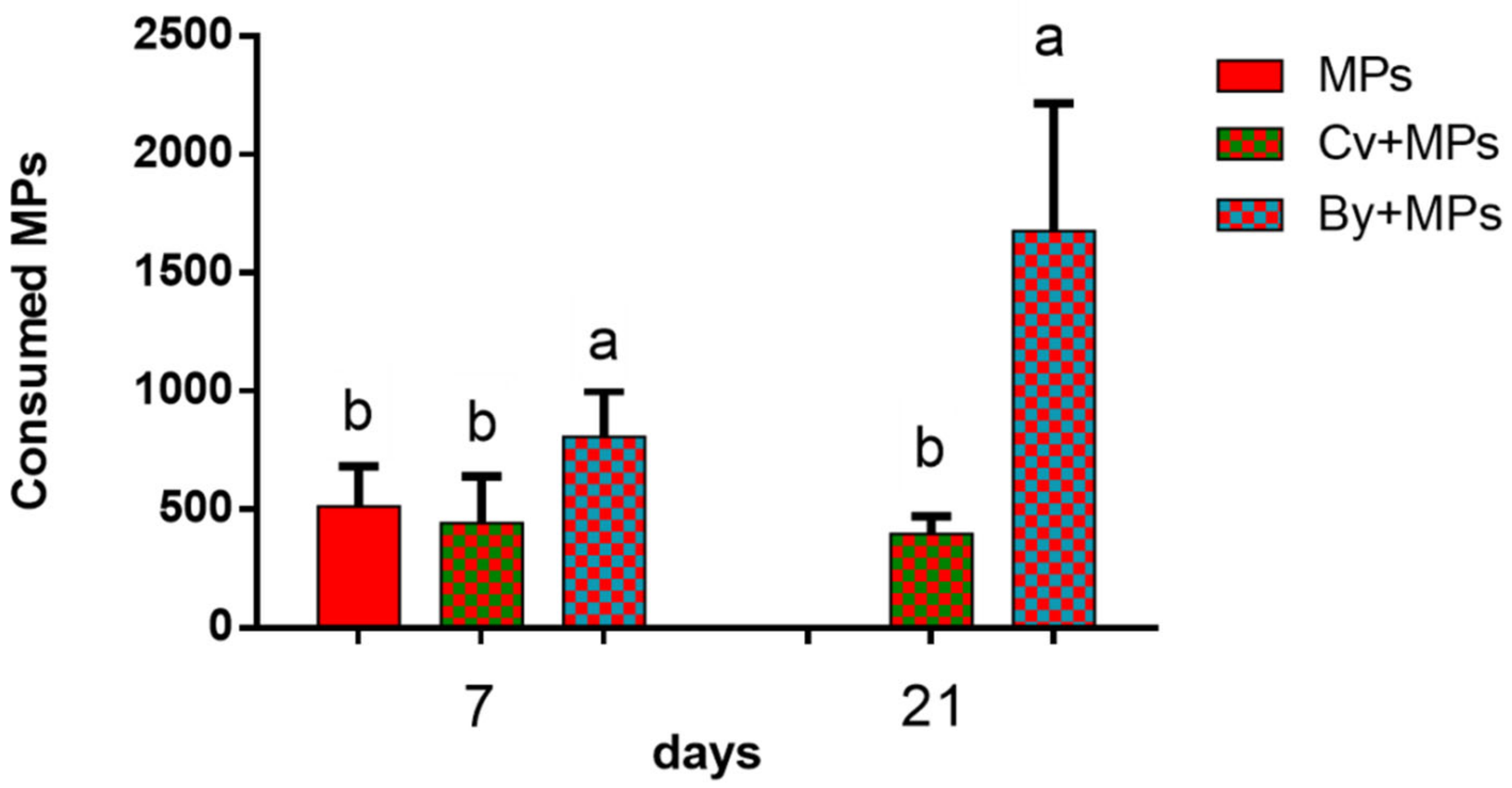
3.1. Ingestion of Microplastics and Microalgae

Micrographs revealed that all diets (i.e., green microalgae, By, MPs and their combinations) were accepted by Daphnia magna individuals. Thus, microplastics were observed to be ingested either alone or with microalgae Cv or By (Figure 1 and Figure 2).
It was striking that after only 7 days from the start of the experiment, Daphnia specimens in the microplastics only group had consumed a significant number of beads (approx. 500 beads/ind., Figure 3). Daphnia fed on By + MPs showed significantly higher instantenous microplastic ingestion on Days 7 and 21 of the experiment compared with the MP only or Cv + MP groups (p < 0.05) (Figure 3).

3.2. Survival of Daphnia magna

Daphnia individuals fed only on MPs and the starved groups displayed drastic declines in their final survival rates (Figure 4). Daphnia fed on MPs and By showed higher survival rates (94%) until the end of the experiment. The Cv diets also induced high survival (88%) in Daphnia. The survival rate of Daphnia fed solely on baker’s yeast continued positively up to Day 19, after which, total mortality occurred in this group (Figure 4).

3.3. Effects of Microplastics on the Growth and Reproduction (Total Length and Width, Female Numbers, Egg and Ephippia Numbers, and Dry Weight)

The least growth in terms of total length was obtained for the groups fed only MPs and for starved individuals on Day 7 (p < 0.05; Figure 5). Daphnia fed solely on Cv microalgae or on By produced similar results in terms of growth in length and width, and were significantly higher than those of other groups on Day 7 (p < 0.05). A similar positive effect of baker’s yeast on total length was detected on Day 14 of the culture (p < 0.05). The total length of Daphnia fed on By, Cv and on By + MPs showed similar results; their average lengths were significantly higher than those of the starved or MPs only groups (p < 0.05) (Figure 5). There was no significant difference among the treatments on Day 21 when the starved and MP-fed Daphnia specimens died.
Similar to the results for total length, the body length measurements of Daphnia also displayed positive trends in the groups fed on By, Cv and on Cv + MPs on Day 7. Daphnia fed with Cv, baker’s yeast (By) and Cv + MPs exhibited similar body lengths, significantly higher than the body lengths of starved individuals (ST) and Daphnia fed exclusively with MPs or a mixture of By + MPs on Day 7 (p < 0.05) (Figure 6). However, on Day 14, the lowest body length measurements were obtained for both the ST and MP groups. There was no significant difference on Day 21 of culture among the groups.
The average widths of Daphnia fed on By and Cv were found to be similar but were greater than width measurements of the other groups on Day 7. Interestingly, on Day 14, the widths of Daphnia fed on Cv + MPs were the second highest, following the group fed on By (p < 0.05) (Figure 7).
Daphnia fed on baker’s yeast produced significantly higher numbers of ephippia on Days 12 and 18. However, on Day 15 of the experiment, the Cv-fed groups displayed higher ephippia numbers (p < 0.05) (Figure 8A). Starving individuals or those fed with MPs only produced no ephippia. Daphnia fed a mixture of MPs/Cv or MPs/By produced significantly (p < 0.05) lower numbers of ephippia.
During the entire experimental period, daphnid production was not observed (p < 0.05) in the diet groups ST, MPs or By + MPs but was highest for the individuals fed solely on Chlorella vulgaris. Interestingly, daphnid numbers were significantly higher in the Cv + MPs group during the final phase of the experiment (p < 0.05) (Figure 8B). Egg production by females was significantly high in Daphnia fed on baker’s yeast during the entire experiment (Figure 8C). The combination of Cv and microplastics (Cv + MPs) also resulted in high egg production on Days 18 and 21 of the experiment (p < 0.05) (Figure 8C). Ephippia-carrying female numbers also increased in the By and Cv + MPs groups, and a lower number of ephippia was observed on Day 12 (p < 0.05) (Figure 8D).

4. Discussion

Microplastics are widely distributed in marine and freshwater environments in different concentrations, colors, shapes and sizes. The concentration of MPs (smaller than 10 µm) in drinking water is assumed to be between 1.5 and 35 particles/L [32,33]. According to Chen et al. (2020), this concentration is expected to increase by 83–150-fold by 2060 [34].
Daphnia belong to the herbivorous zooplankton that feed through filtering a variety of particles suspended in the water column [35]. Therefore, this species is generally unable to distinguish MPs from natural food items when offered together [34]. Canniff and Hoang (2018) showed the effects of larger plastic microbeads (63–75 µm) on D. magna’s survival and reproductive performance. In marine and freshwater environments, plastics tend to become smaller as time passes. In our laboratory study, we used smaller (6 µm) polystyrene MPs in order to see the effect on D. magna’s growth and reproduction. For that purpose, Daphnia fed a diet of By and MPs had a greater accumulation of MPs in their body compared with individuals fed Cv + MP diets. Rehse et al. (2016) showed that the ingestion of 1 µm PE particles in Daphnia magna resulted in the immobilization of animals, in a correlation with the time and dose. Similarly, 6 µm PE microplastics size ingested at a higher rate with a combination of diets in our study. This could be related to their similarity to yeast particles in terms of size and color. In another study, An et al. (2021) also mentioned that the size of MPs is strongly correlated to their accumulation in the body [36]. A greater amount of small MP fragments (17.23 µm) accumulated compared with MP beads (39.50 µm) and large MPs (34.43 µm). Our MP beads were 6 µm in size, which is somewhat smaller than those mentioned in other studies. The food particle preferences of D. magna are mostly related to the size of their natural diet (1–30 µm) and, as a result, as MPs become smaller, the ingestion rate could increase whether the materials are beads or fragments. The number of ingested beads is also related to the exposure time of particle concentrations.
High concentrations of polystyrene MPs were reported to decrease the survival of both Daphnia magna [37,38] and Daphnia pulex populations [39]. Moreover, Liu et al. (2019) concluded that the expression of stress defense genes in D. pulex was negatively affected by the utilization of MPs. On the other hand, Aljaibachi and Callagahan (2018) reported no negative effects of MPs on the Daphnia population when a plentiful supply of microalgae was available, stating that this species avoided consuming polystyrene MPs [40]. In fact, in another study, survival was higher when algae were presented together with polystyrene microplastics [5]. Similarly, in our study, groups fed the algae + polystyrene MPs and baker’s yeast + polystyrene MPs combinations had improved survival until 18 days of culture in our study. After that period, a sudden decline was recorded that might be related to insufficient nutrients for further stages of the life cycle. Zhang et al. (2019) also mentioned the impact of polystyrene plastics on D. magna and reported that 48 h of exposure to 1 µm and 10 µm microplastics led to higher mortality and increased immobilization rates in Daphnia individuals [41]. In our study, the size of the polystyrene particles was 6 µm and Daphnia’s survival was also negatively affected in individuals fed solely on MPs, and after 15 days of culture, the entire population collapsed. Besides the plastic type, the age of Daphnia affects the mortality rate and immobilization responses after exposure to both fasting and different feeding conditions. Starving individuals are much weaker to resist physical stress conditions. Survival was found to be negatively affected by the ingestion of MP fragments [42]. These results were attributed to the longer retention times of irregular MPs fragments in the digestive tracts. Fragmented polyethylene microplastics (17.23 and 34.43 µm) were ingested efficiently compared with commercial microplastic beads, and survival (20–60%) was affected by the utilization of fragmented MPs compared with MP beads (90%) in Daphnia individuals [36]. In other short-term feeding trials, Daphnia exhibited high mortality rates related to elevated exposure concentrations. However, Daphnia individuals pre-fed with microalgae displayed similar mortality among groups [2]. Daphnia exposed to irregularly shaped microplastics displayed no increased mortality or malformations in adults and juveniles [43]. These results were also attributed to the different types, sizes, shapes and ages of MPs. It is also assumed that multiple stress factors play important roles in microplastic contamination in the natural environment [43].
In our study, the number of egg-carrying female Daphnia individuals was relatively high for the By diet group at the end of the experiment. This could be related to baker’s yeast being rich in Vitamin B12, thus increasing zooplankton production. Interestingly, the number of ephippia-carrying females was found to be higher in the group fed on Cv + MPs at the end of the experiment. However, feeding solely on MPs resulted in a continuous negative effect on the Daphnia population. This result is supported by Aljaibachi and Callagahan’s (2018) findings that if there is additional natural food available, mainly algae, the Daphnia population will not be significantly affected by MPs in the environment [5,40]. In our trial, the number of ephippia-carrying females was high for the Cv + MP groups at the end of the experiment. This might be related to the negative effects of MPs on the reproductive capability of daphnids due to physiological stress. De Felice et al. (2019) concluded that body length and reproductive performance in D. magna were not negatively affected by 1–10 µm polystyrene MPs [44]. Additionally, exposure of D. magna to two concentrations of polystyrene MPs (0.05 µg/mL and 0.5 µg/mL) resulted in an increasing level of energy contents on Day 21 [45]. This result can be attributed to the high stress level of D. magna and also to secondary production such as growth and reproduction. In agreement with this finding, in our study, 6 µm polystyrene MPs did not negatively affect either the body length or the total length of Daphnia due to simultaneous exposure to food such as By and Cv. This result could be due to the ability of Daphnia individuals to metabolize sufficient energy from their diets even when MPs were present. On the other hand, our results for body length were lower than those of De Felice et al. (2019) obtained after 21 days of the experiment. In our study, Daphnia’s growth stopped after 14 days of culture, which might be related to stress conditions or not enough energy gain from diets with a combination of MPs [44].
In conclusion, our results shows that the presence of polystyrene MPs could lead to more ephippia numbers in females when foods such as the microalgae Chlorella vulgaris were available in this trial. In freshwater ecosystems, Daphnia individuals can ingest 6 micron MPs and survive without negative effects on their basal metabolism. This means this microplastics can be transferred via the food chain to higher invertebrates. Different types of MP have been studied in various zooplankton such as Artemia, Rotifer and Daphnia. MPs have negative effects on the growth of Artemia by changing the gut microbiota, and this led to slow growth [46]. Further studies are needed to understand effect of MPs on stress-related genes, the gut microbiota and nutrient metabolism.

Author Contributions

Conceptualization, M.I. and K.M.E.; methodology, K.M.E.; M.I. and A.E.K.; software, K.M.E.; validation, M.I., A.E.K. and K.M.E.; formal analysis, K.M.E.; investigation, K.M.E.; resources, K.M.E.; data curation, K.M.E.; A.E.K.; writing—original draft preparation, M.I., K.M.E. and A.E.K.; writing—review and editing, M.I., K.M.E. and A.E.K.; visualization, K.M.E.; supervision, M.I., K.M.E. and A.E.K.; project administration, K.M.E.; funding acquisition, K.M.E. All authors have read and agreed to the published version of the manuscript.

Funding

This research was funded by [Istanbul University, Research Foundation] grant number [FBA-2020-33325].

Institutional Review Board Statement

Not applicable.

Informed Consent Statement

Not applicable.

Data Availability Statement

All data relevant to this study have been included in the paper and if it is request, all data of this study are promised to provide by the corresponding author.

Acknowledgments

We thank Ezgi Emiş Türkeri for laboratory assistance.

Conflicts of Interest

The authors declare that they have no conflict of interest.

References

  1. Europe, P. Plastics–the facts. PlasticEurope 2020, 1, 1–64. [Google Scholar]
  2. Andrady, A.L. Microplastics in the marine environment. Marine Pollut. Bull. 2011, 62, 1596–1605. [Google Scholar] [CrossRef]
  3. Jemec, A.; Horvat, P.; Kunej, U.; Bele, M.; Kržan, A. Uptake and effects of microplastic textile fibers on freshwater crustacean Daphnia magna. Environ. Pollut. 2016, 219, 201–209. [Google Scholar] [CrossRef] [PubMed]
  4. Cole, M.; Lindeque, P.; Fileman, E.; Halsband, C.; Goodhead, R.; Moger, J.; Galloway, T.S. Microplastic ingestion by zooplankton. Environ. Sci. Technol. 2013, 47, 6646–6655. [Google Scholar] [CrossRef] [PubMed]
  5. Aljaibachi, R.; Laird, W.B.; Stevens, F.; Callaghan, A. Impacts of polystyrene microplastics on Daphnia magna: A laboratory and a mesocosm study. Sci. Total Environ. 2020, 705, 135800. [Google Scholar] [CrossRef] [PubMed]
  6. Güven, O.; Gökdağ, K.; Jovanović, B.; Kıdeyş, A.E. Microplastic litter composition of the Turkish territorial waters of the Mediterranean Sea, and its occurrence in the gastrointestinal tract of fish. Environ. Pollut. 2017, 223, 286–294. [Google Scholar] [CrossRef]
  7. Browne, M.A.; Galloway, T.S.; Thompson, R.C. Spatial patterns of plastic debris along estuarine shorelines. Environ. Sci. Technol. 2010, 44, 3404–3409. [Google Scholar] [CrossRef]
  8. Özgüler, Ü.; Demir, A.; Kayadelen, G.C.; Kıdeyş, A.E. Riverine Microplastic Loading to Mersin Bay, Turkey on the North-eastern Mediterranean. Turk. J. Fish. Aquat. Sci. 2021, 22, TRJFAS20253. [Google Scholar] [CrossRef]
  9. Dris, R.; Imhof, H.K.; Löder, M.G.; Gasperi, J.; Laforsch, C.; Tassin, B. Microplastic contamination in freshwater systems: Methodological challenges, occurrence and sources. In Microplastic Contamination in Aquatic Environments; Elsevier: Amsterdam, The Netherlands, 2018; pp. 51–93. [Google Scholar] [CrossRef]
  10. Shim, W.J.; Hong, S.H.; Eo, S. Marine microplastics: Abundance, distribution, and composition. In Microplastic Contamination in Aquatic Environments; Zeng, E.Y., Ed.; Elsevier: Amsterdam, The Netherlands, 2018; pp. 1–26. [Google Scholar]
  11. Murphy, F.; Quinn, B. The effects of microplastic on freshwater Hydra attenuata feeding, morphology & reproduction. Environ. Pollut. 2018, 234, 487–494. [Google Scholar] [CrossRef] [Green Version]
  12. Windsor, F.M.; Tilley, R.; Tyler, C.R.; Ormerod, S.J. Microplastic ingestion by riverine macroinvertebrates. Sci. Total Environ. 2019, 646, 68–74. [Google Scholar] [CrossRef]
  13. Hurley, R.R.; Woodward, J.C.; Rothwell, J.J. Ingestion of microplastics by freshwater tubifex worms. Environ. Sci. Technol. 2017, 51, 12844–12851. [Google Scholar] [CrossRef] [PubMed]
  14. Phillips, M.B.; Bonner, T.H. Occurrence and amount of microplastic ingested by fishes in watersheds of the Gulf of Mexico. Mar. Pollut. Bull. 2015, 100, 264–269. [Google Scholar] [CrossRef] [PubMed]
  15. Lambert, S.; Wagner, M. Microplastics are contaminants of emerging concern in freshwater environments: An overview. Freshw. Microplastics 2018, 58, 1–23. [Google Scholar] [CrossRef] [Green Version]
  16. Isinibilir, M.; Svetlichny, L.; Mykitchak, T.; Türkeri, E.E.; Eryalçın, K.M.; Doğan, O.; Can, G.; Yüksel, E.; Kideys, A.E. Microplastic consumption and its effect on respiration rate and motility of Calanus helgolandicus from the Marmara Sea. Front. Mar. Sci. 2020, 7, 603321. [Google Scholar] [CrossRef]
  17. Svetlichny, L.; Isinibilir, M.; Mykitchak, T.; Eryalçın, K.M.; Türkeri, E.E.; Yuksel, E.; Kideys, A.E. Microplastic consumption and physiological response in Acartia clausi and Centropages typicus: Possible roles of feeding mechanisms. Reg. Stud. Mar. Sci. 2021, 43, 101650. [Google Scholar] [CrossRef]
  18. Lusher, A. Marine anthropogenic litter. In Microplastics in the Marine Environment: Distribution, Interactions and Effects; Bergmann, M., Gutow, L., Klages, M., Eds.; Springer: Cham, Switzerland, 2015; pp. 245–307. [Google Scholar] [CrossRef] [Green Version]
  19. Scherer, C.; Brennholt, N.; Reifferscheid, G.; Wagner, M. Feeding type and development drive the ingestion of microplastics by freshwater invertebrates. Sci. Rep. 2017, 7, 17006. [Google Scholar] [CrossRef] [Green Version]
  20. Canniff, P.M.; Hoang, T.C. Microplastic ingestion by Daphnia magna and its enhancement on algal growth. Sci. Total Environ. 2018, 633, 500–507. [Google Scholar] [CrossRef] [Green Version]
  21. Jeong, C.B.; Won, E.J.; Kang, H.M.; Lee, M.C.; Hwang, D.S.; Hwang, U.K.; Zhou, B.; Soussi, S.; Lee, S.J.; Lee, J.S. Microplastic size-dependent toxicity, oxidative stress induction, and p-JNK and p-p38 activation in the monogonont rotifer (Brachionus koreanus). Environ. Sci. Technol. 2016, 50, 8849–8857. [Google Scholar] [CrossRef]
  22. Imhof, H.K.; Laforsch, C. Hazardous or not–Are adult and juvenile individuals of Potamopyrgus antipodarum affected by non-buoyant microplastic particles? Environ. Pollut. 2016, 218, 383–391. [Google Scholar] [CrossRef]
  23. Gorokhova, E. Screening for microplastic particles in plankton samples: How to integrate marine litter assessment into existing monitoring programs? Mar. Pollut. Bull. 2015, 99, 271–275. [Google Scholar] [CrossRef]
  24. Guilhermino, L.; Martins, A.; Cunha, S.; Fernandes, J.O. Long-term adverse effects of microplastics on Daphnia magna reproduction and population growth rate at increased water temperature and light intensity: Combined effects of stressors and interactions. Sci. Total Environ. 2021, 784, 147082. [Google Scholar] [CrossRef] [PubMed]
  25. Kelpsiene, E.; Torstensson, O.; Ekvall, M.T.; Hansson, L.A.; Cedervall, T. Long-term exposure to nanoplastics reduces life-time in Daphnia magna. Sci. Rep. 2020, 10, 5979. [Google Scholar] [CrossRef] [PubMed] [Green Version]
  26. Bownik, A. Daphnia swimming behaviour as a biomarker in toxicity assessment: A review. Sci. Total Environ. 2017, 601, 194–205. [Google Scholar] [CrossRef] [PubMed]
  27. Faithfull, C.; Huss, M.; Vrede, T.; Karlsson, J.; Bergström, A.K. Transfer of bacterial production based on labile carbon to higher trophic levels in an oligotrophic pelagic system. Can. J. Fish. Aquat. Sci. 2012, 69, 85–93. [Google Scholar] [CrossRef]
  28. Anderson, B.G. The toxicity thresholds of various substances found in industrial wastes as determined by the use of Daphnia magna. Sew. Work. J. 1944, 16, 1156–1165. [Google Scholar]
  29. Frydkjær, C.K.; Iversen, N.; Roslev, P. Ingestion and egestion of microplastics by the cladoceran Daphnia magna: Effects of regular and irregular shaped plastic and sorbed phenanthrene. Bull. Environ. Contaminat. Toxicol. 2017, 99, 655–661. [Google Scholar] [CrossRef]
  30. Rehse, S.; Kloas, W.; Zarfl, C. Short-term exposure with high concentrations of pristine microplastic particles leads to immobilisation of Daphnia magna. Chemosphere 2016, 153, 91–99. [Google Scholar] [CrossRef]
  31. OECD. Test No. 211: Daphnia magna Reproduction Test. In OECD Guidelines for the Testing of Chemicals, Section 2; OECD Publishing: Paris, France, 2012; Volume 2. [Google Scholar] [CrossRef] [Green Version]
  32. Oßmann, B.E.; Sarau, G.; Holtmannspötter, H.; Pischetsrieder, M.; Christiansen, S.H.; Dicke, W. Small-sized microplastics and pigmented particles in bottled mineral water. Water Res. 2018, 141, 307–316. [Google Scholar] [CrossRef] [PubMed]
  33. Pivokonsky, M.; Cermakova, L.; Novotna, K.; Peer, P.; Cajthaml, T.; Janda, V. Occurrence of microplastics in raw and treated drinking water. Sci. Total Environ. 2018, 643, 1644–1651. [Google Scholar] [CrossRef]
  34. Chen, Q.; Li, Y.; Li, B. Is color a matter of concern during microplastic exposure to Scenedesmus obliquus and Daphnia magna? J. Hazard. Mater. 2020, 383, 121224. [Google Scholar] [CrossRef]
  35. Turcihan, G.; Isinibilir, M.; Zeybek, Y.G.; Eryalçın, K.M. Effect of different feeds on reproduction performance, nutritional components and fatty acid composition of cladocer water flea (Daphnia magna). Aquac. Res. 2022, 53, 2420–2430. [Google Scholar] [CrossRef]
  36. An, D.; Na, J.; Song, J.; Jung, J. Size-dependent chronic toxicity of fragmented polyethylene microplastics to Daphnia magna. Chemosphere 2021, 271, 129591. [Google Scholar] [CrossRef] [PubMed]
  37. Bosker, T.; Olthof, G.; Vijver, M.G.; Baas, J.; Barmentlo, S.H. Significant decline of Daphnia magna population biomass due to microplastic exposure. Environ. Pollut. 2019, 250, 669–675. [Google Scholar] [CrossRef] [Green Version]
  38. Schür, C.; Zipp, S.; Thalau, T.; Wagner, M. Microplastics but not natural particles induce multigenerational effects in Daphnia magna. Environ. Pollut. 2020, 260, 113904. [Google Scholar] [CrossRef] [PubMed]
  39. Liu, Z.; Yu, P.; Cai, M.; Wu, D.; Zhang, M.; Huang, Y.; Zhao, Y. Polystyrene nanoplastic exposure induces immobilization, reproduction, and stress defense in the freshwater cladoceran Daphnia pulex. Chemosphere 2019, 215, 74–81. [Google Scholar] [CrossRef]
  40. Aljaibachi, R.; Callaghan, A. Impact of polystyrene microplastics on Daphnia magna mortality and reproduction in relation to food availability. PeerJ 2018, 6, e4601. [Google Scholar] [CrossRef] [PubMed] [Green Version]
  41. Zhang, P.; Yan, Z.; Lu, G.; Ji, Y. Single and combined effects of microplastics and roxithromycin on Daphnia magna. Environ. Sci. Pollut. Res. 2019, 26, 17010–17020. [Google Scholar] [CrossRef]
  42. Ogonowski, M.; Schür, C.; Jarsén, Å.; Gorokhova, E. The effects of natural and anthropogenic microparticles on individual fitness in Daphnia magna. PLoS ONE 2016, 11, e0155063. [Google Scholar] [CrossRef] [Green Version]
  43. Imhof, H.K.; Rusek, J.; Thiel, M.; Wolinska, J.; Laforsch, C. Do microplastic particles affect Daphnia magna at the morphological, life history and molecular level? PLoS ONE 2017, 12, e0187590. [Google Scholar] [CrossRef]
  44. De Felice, B.; Sabatini, V.; Antenucci, S.; Gattoni, G.; Santo, N.; Bacchetta, R.; Ortenzi, M.A.; Parolini, M. Polystyrene microplastics ingestion induced behavioral effects to the cladoceran Daphnia magna. Chemosphere 2019, 231, 423–431. [Google Scholar] [CrossRef]
  45. De Felice, B.; Sugni, M.; Casati, L.; Parolini, M. Molecular, biochemical and behavioral responses of Daphnia magna under long-term exposure to polystyrene nanoplastics. Environ. Int. 2022, 164, 107264. [Google Scholar] [CrossRef] [PubMed]
  46. Li, H.; Chen, H.; Wang, J.; Li, J.; Liu, S.; Tu, J.; Chen, Y.; Zong, Y.; Zhang, P.; Wang, Z.; et al. Influence of microplastics on the growth and the intestinal microbiota composition of brine shrimp. Front. Microbiol. 2021, 12, 717272. [Google Scholar] [CrossRef] [PubMed]
Figure 1. Images of Daphnia magna individuals on Day 14 of the feeding experiment: (a) Daphnia fed on Cv; (b) Daphnia fed on MPs + Cv; (c) Daphnia fed on By; (d) Daphnia fed on MPs + By; (e) Daphnia fed on MPs only; (f) starved Daphnia.
Microplastics 02 00002 g001
Figure 2. Images of (a) Daphnia magna individuals fed MPs exclusively, (b) Daphnia magna individuals fed Cv + MPs and (c) Daphnia fed By, all taken on Day 21 of the experiment. Arrows show ingested microplastic particles in the gut of Daphnia specimens.
Microplastics 02 00002 g002
Figure 3. Average ingested microplastics, microalgae and baker’s yeast particles (number/ind.) in the gut of Daphnia magna on Days 7 and 21 (n = 10, vertical error bars show the standard deviations, different superscript letters show significant differences, p ≤ 0.05).
Microplastics 02 00002 g003
Figure 4. Survival percentage of Daphnia magna.
Microplastics 02 00002 g004
Figure 5. Average total length of Daphnia magna fed different diets during the 21-day experiment (ST; starved, Cv; Chlorella vulgaris, MPs; microplastics, Cv + MPs; Chlorella vulgaris + microplastics, By; baker’s yeast, By + MPs; baker’s yeast + microplastics). Low-case letters (a, b, bc, c, cd, d) denote significant variable differences among means of different treatments from Duncan’s Multiple Range test, p < 0.05.
Microplastics 02 00002 g005
Figure 6. Average body lengths of Daphnia magna fed different diet combinations during the experiment. Low-case letters (a, ab, bc, c,) denote significant variable differences among means of different treatments from Duncan’s Multiple Range test, p < 0.05.
Microplastics 02 00002 g006
Figure 7. Average widths of Daphnia magna fed different diet combinations during the 21-day experiment. Low-case letters (a, b, bc, cd, d) denote significant variable differences among means of different treatments from Duncan’s Multiple Range test, (p < 0.05).
Microplastics 02 00002 g007
Figure 8. Growth parameters of Daphnia fed different diet combinations with MPs: (A) number of ephippia, (B) daphnia numbers, (C) egg-carrying females and (D) ephippia-carrying females (* indicates a significant appearance of a group that existed on that experimental day). Low-case letters (a, b, c, d) denote significant variable differences among means of different treatments from Duncan’s Multiple Range test, p < 0.05.
Microplastics 02 00002 g008
Disclaimer/Publisher’s Note: The statements, opinions and data contained in all publications are solely those of the individual author(s) and contributor(s) and not of MDPI and/or the editor(s). MDPI and/or the editor(s) disclaim responsibility for any injury to people or property resulting from any ideas, methods, instructions or products referred to in the content.

