Entry

Primary Progenitor Tenocytes: Cytotherapeutics and Cell-Free Derivatives

by
Alexis Laurent
1,2,3,*,
Corinne Scaletta
2,4,5,
Philippe Abdel-Sayed
2,4,5,6,
Wassim Raffoul
4,5,
Nathalie Hirt-Burri
2,4,5 and
Lee Ann Applegate
2,4,5,7
1
Applied Research Department, LAM Biotechnologies SA, CH-1066 Epalinges, Switzerland
2
Regenerative Therapy Unit, Lausanne University Hospital, University of Lausanne, CH-1066 Epalinges, Switzerland
3
Faculty of Biology and Medicine, University of Lausanne, CH-1015 Lausanne, Switzerland
4
Lausanne Burn Center, Lausanne University Hospital, University of Lausanne, CH-1011 Lausanne, Switzerland
5
Plastic, Reconstructive, and Hand Surgery Service, Lausanne University Hospital, University of Lausanne, CH-1011 Lausanne, Switzerland
6
DLL Bioengineering, STI School of Engineering, Ecole Polytechnique Fédérale de Lausanne, CH-1015 Lausanne, Switzerland
7
Center for Applied Biotechnology and Molecular Medicine, University of Zurich, CH-8057 Zurich, Switzerland
*
Author to whom correspondence should be addressed.
Encyclopedia 2023, 3(1), 340-361; https://doi.org/10.3390/encyclopedia3010021
Submission received: 2 February 2023 / Revised: 2 March 2023 / Accepted: 15 March 2023 / Published: 16 March 2023
(This article belongs to the Section Biology & Life Sciences)

Definition

:
Primary progenitor tenocytes are diploid cells that may be cultured in vitro and therapeutically used for allogeneic musculoskeletal regenerative medicine. Firstly, technical aspects of cell banking, biotechnological manufacturing, and extensive preclinical characterization data have confirmed that FE002-Ten primary progenitor tenocytes may be safely considered for human cytotherapeutic use (e.g., in tissue engineering products, standardized transplants). Parallelly, lyophilized progenitor tenocyte extracts (e.g., stabilized cells or cell-free derivatives) were shown to optimally act as potent hyaluronan-based hydrogel functionalizing agents, useful for stability enhancement against oxidative product degradation. Therefore, primary progenitor tenocytes (e.g., FE002-Ten cell source) may potentially be used in diverse clinical presentations of tendon-related pathologies, ranging from volumetric tissue replacement (i.e., for the promotion of enhanced graft bio-integration) to local management of tissular inflammation and pain (i.e., ancillary action of the cellular extracts for the functional enhancement of injectable hyaluronan-based preparations). Overall, the primary progenitor tenocytes investigated under the Swiss progenitor cell transplantation program were shown to represent highly standardized biotechnological materials with a versatility of potential therapeutic uses after formulation into an array of cytotherapeutic preparations or cell-free devices.

Graphical Abstract

1. Introduction

While numerous translational applications have been investigated for diverse tissue engineering and cytotherapeutic approaches, it is probable that cutaneous and musculoskeletal affections will be most rapidly addressed at large clinical scale [1,2,3]. Notably, tendon tissue traumatic defects or degenerative affections are highly prevalent, yet few effective therapeutic interventions enable rapid and high-quality restoration of tissular structures and functions [4,5,6]. Therein, injectable hyaluronan-based hydrogels have been successfully clinically used for mild to moderate tendinopathy cases, providing some anti-inflammatory properties, tissular lubrication or gliding enhancement, and local immunomodulatory properties [7,8,9]. Biological-based regenerative approaches to tendon injuries and acute pathologies have notably comprised the injection use of bone marrow or adipose-derived stem cells, amniotic cells, placenta cells, tenocytes, tendon sheath fibroblasts, and platelet derivatives (e.g., platelet-rich plasma, PRP) [10,11,12,13,14,15,16]. Therein, clinical outcomes and success of the intervention were reported to be highly dependent on the retained therapeutic indication and on the related treatment protocols [11,17,18,19].
Primary progenitor tenocytes (i.e., FE002-Ten cell source) have been studied under the Swiss progenitor cell transplantation program as a potential ad hoc cytotherapeutic material source for optimal homologous and allogeneic management of tendinous tissue disorders [20,21,22]. Firstly, this specific approach was technologically based on primary diploid cell biobanking concepts from the 1960s, which enabled the development of the global vaccine industry (e.g., WI-38, MRC-5 cell types) [23,24]. Secondly, the renewed and optimized application of these early biotechnology concepts was enabled by modern technical advancements in the cell manufacturing and bioengineering fields, for enhanced process standardization and biological material quality [21,25]. Based on these robust conceptual and technical elements, clinical-grade primary progenitor tenocytes (i.e., FE002-Ten cells) were procured and established as a cryopreserved cellular source for allogeneic musculoskeletal regenerative medicine applications under modern manufacturing quality requirements [21].
The primary progenitor tenocytes investigated under the Swiss progenitor cell transplantation program (i.e., FE002-Ten cells) were shown to represent highly standardized biotechnological materials with high versatility in potential therapeutic uses [20,21,22,23,26,27]. Primary progenitor tenocytes are cultured diploid cells (i.e., characterized by a finite in vitro lifespan during serial cell cultivation) that may be clinically used for allogeneic musculoskeletal regenerative medicine [20,22]. Notably, primary progenitor cells are inherently pre-terminally differentiated, display pure and monomodal phenotypes in vitro, and are highly robust during serial monolayer cell expansion under consistent manufacturing parameters and technical specifications [20,21,28]. Importantly, primary progenitor tenocytes are characterized by high biocompatibility, an inherent immune privilege, and by the absence of in vitro and in vivo tumorigenic behaviors [22,26].
Following in vitro manufacturing under defined multi-tiered progenitor cell banking workflows, viable cellular lots may be used for off-the-freezer preparation of cell-seeded tendon allografts (i.e., using synthetic tendon/ligament scaffolds or decellularized biological tendon matrices) for tissue engineering (Figure 1A) [22,26].
Alternatively, viable FE002-Ten tenogenic cells may be combined with hyaluronan-based hydrogels for the local delivery of cytotherapeutic payloads to tendon lesion sites in view of promoting or supporting tissular repair and/or regeneration (Figure 1A) [9]. In particular, various technical aspects of cell banking and extensive characterization data have confirmed that FE002-Ten primary progenitor tenocytes may be safely considered for human cytotherapeutic use [21,22]. Furthermore, primary progenitor tenocytes may be used as biological starting materials for the standardized preparation of an array of stabilized cellular derivatives (Figure 1B) [27,29]. Specifically, it was shown that cytotherapy-inspired lyophilized preparations containing FE002 primary progenitor tenocyte derivatives or extracts present significant intrinsic antioxidant functions (e.g., Trolox equivalent antioxidant capacity, TEAC) [29]. In detail, the lyophilized progenitor tenocyte extracts (e.g., stabilized cell-free fractions) were shown to act as potent hyaluronan-based hydrogel functionalizing agents for injectable product stability enhancement against oxidative degradation (Figure 1C) [27]. Such stabilized and sterilizable extracts may be used for the significant stability enhancement of various types of hyaluronan-based hydrogels, which may be clinically applied in mild to moderate cases of tendinopathy [27].
Overall, the translational qualification of FE002-Ten cells, performed over the past decade in Switzerland, has confirmed the applicability of such standardized biological materials as cellular active ingredients or as starting materials within the development of therapeutic products and devices for human use [22,29]. In detail, a growing compilation of peer-reviewed scientific reports has been constituting the multifaceted body of knowledge available around the FE002-Ten primary progenitor tenocyte source of interest (Table 1).
Therefore, primary progenitor tenocytes may potentially be used in diverse clinical presentations of tendon-related pathologies, ranging from volumetric tissue replacement (i.e., for the promotion of enhanced graft bio-integration) to the local management of tissular inflammation and pain (i.e., ancillary action of the cellular extracts for the enhancement of injectable hyaluronan-based preparations, Figure 1) [22,27]. Overall, primary progenitor FE002-Ten cellular materials were shown to be robust and well-adapted for processing in highly standardized manufacturing workflows, with a high versatility of potential therapeutic uses after formulation into a wide range of tissue engineering and biotechnological preparations (Figure 1 and Table 1) [9,22,27].

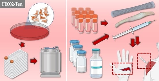
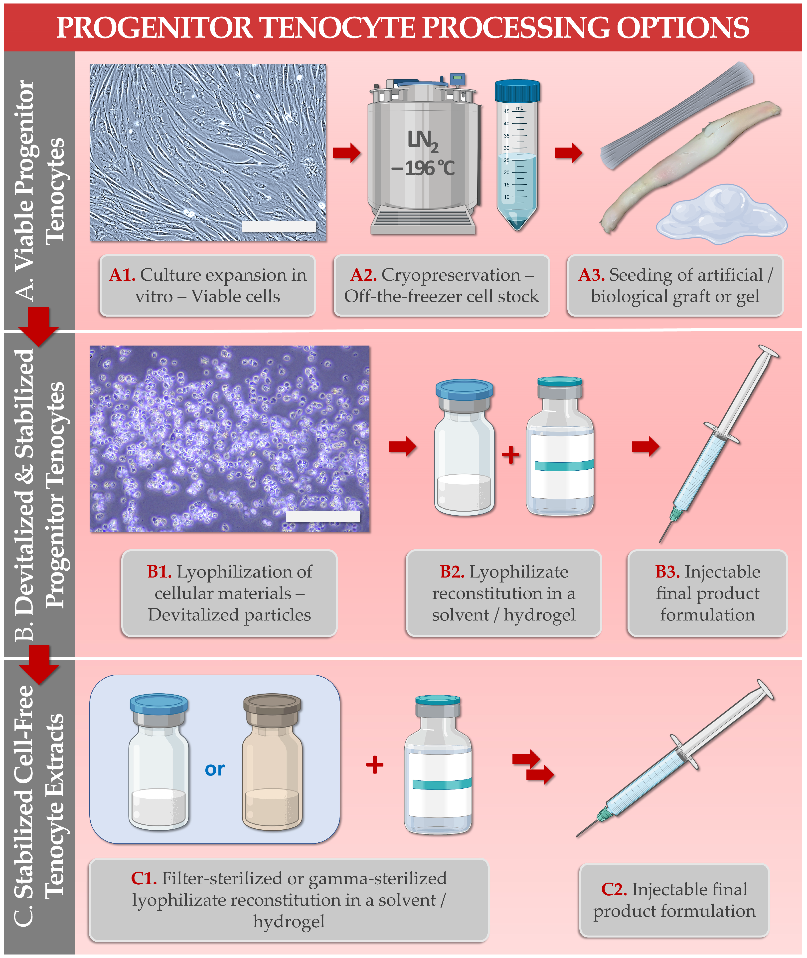
2. Biological Material Procurement and Cell Source Establishment Methodology: FE002 Primary Progenitor Tenocytes for Clinical Applications

In order to comply with modern material traceability and quality requirements for clinical tissue engineering, the primary progenitor FE002-Ten cell source was established following the stringent regulatory standards applicable for advanced therapy medicinal products (ATMP) and notably good manufacturing practices (GMP) [21,31]. An iterative therapeutic cell source selection optimization process, devised and carried out over three decades in Western Switzerland, led to the retention of specific pre-natal tendon tissue donations for the subsequent establishment of primary progenitor tenocyte cell types in adherent in vitro culture [3,21]. For strict compliance with applicable medical and ethical guidelines as well as local traceability requirements, an ad hoc regulated progenitor cell transplantation program was set up [21,32]. This national operational platform then enabled standardized whole-cell bioprocessing from the original starting tissue material and the establishment of progeny cryopreserved parental cell banks (PCB) and master cell banks (MCB) in a multi-tiered biobanking system (Figure 2) [21].
A prime technical advantage of the retained in vitro cell processing methodology and processing workflows consists in the technical possibility to sustainably and scalably manufacture several million therapeutic cellular lots from a single cryopreserved primary progenitor cell source, without the need for renewed tissue procurement phases [21]. Importantly, manufacturing process robustness is a key parameter of the adopted biotechnological approach, enabling the obtention of extensive, homogeneous, and consistent cellular harvests during GMP manufacturing campaigns [22,28]. This in turn constitutes an important quality prerequisite in order to subsequently guarantee cytotherapeutic product quality, safety, and efficacy for further formulation purposes or for direct clinical use (Figure 1).

3. Primary Progenitor Tenocytes for Allogeneic Tissue Engineering Applications and for Cytotherapies

Tendon injuries are sustained by various patient populations, notably in acute settings (e.g., blunt-force or sharp-force trauma, accidental secondary ruptures) or in chronic settings (i.e., overuse injuries in athletes, ruptures in elderly patients) [4,19,33]. Additionally, many disorders drastically impact tendinous tissues, such as calcification, systemic drug-related iatrogenesis, lipoid degeneration, and tendinosis [6,10]. Tendon tissue physiologic repair processes following destructive afflictions are long and complex, wherein fibrotic structure formation may easily lead to scar tissue build-up and debilitating adhesions [10,34]. As the successful management of many tendon tissue affections requires specialized care and long-term follow-up or re-education, the incurred financial burdens may be substantial [33]. Therefore, various tissue engineering therapeutic approaches have been investigated as promising contenders to supplement the surgical management of tendon injuries (e.g., Achilles tendon, rotator cuff, knee and hand tendons/ligaments) [26,30,35,36,37]. Such approaches are conceptually appealing, given the relative slowness of tendon tissue metabolism, delayed inflammation mechanisms and effector recruitment, or slow tissular matrix deposition and architectural reorganisation during repair [34].
Various autologous (i.e., vestigial tissues), allogeneic (i.e., cadaveric grafts), xenogeneic (e.g., decellularized equine tendons), or synthetic scaffolds may be considered for effective tendon tissue bioengineering and/or grafting strategies [26,36,37,38,39]. Due to the adverse nature of the implantation environment (i.e., on-going healing processes, high mechanical strains) in the host, many tissue grafts often rapidly degenerate and do not enable the restoration of tensile strength or structural elasticity and mobility recovery. When focusing on the therapeutic cellular component of ideal tendinous tissue engineering products, primary progenitor tenocytes (e.g., FE002-Ten cell source) display clear advantages [10,26]. As these types of cells display high stability of tenogenic properties and low propensity for de-differentiation, their use appears to be appropriate within a homologous biological-based therapeutic approach [20,28]. Furthermore, progenitor tenocytes are known to produce as much or more extracellular matrix (ECM, e.g., collagen, scleraxis, tenomodulin) than primary adult tenocytes in vitro [20]. Based on the nature and the known attributes of progenitor cells, these may potentially mediate scarless tissue repair or regeneration [20,40].
Of note, it has been previously set forth that the FE002-Ten primary progenitor cell source could potentially be used to obtain >1014 cells of appropriate quality for clinical therapeutic use, or for the preparation of >108 tissue engineering treatment units [22]. Finally, it was shown that in addition to scaffold-based tendon tissue engineering approaches, highly versatile formulation adaptation could be performed using hyaluronan-based hydrogels (i.e., commercialized class III medical devices) to prepare injectable cytotherapeutic hydrogel products (Figure 1 and Figure 3) [9,20].
This parallel approach presents several advantages for the local management of tendon degenerative diseases, small hand tendon injuries, ligamentary fissures, or partial tendon ruptures (Figure 3) [9]. Such standardized transplant products were shown to possess attributes that were well-adapted for clinical administration, such as injectability, cellular payload viability maintenance, system rheological behaviour, etc. [9]. Specifically, this approach may be of interest as a complement to conservative surgical approaches or to classical tissue engineering strategies for tendon repair promotion.

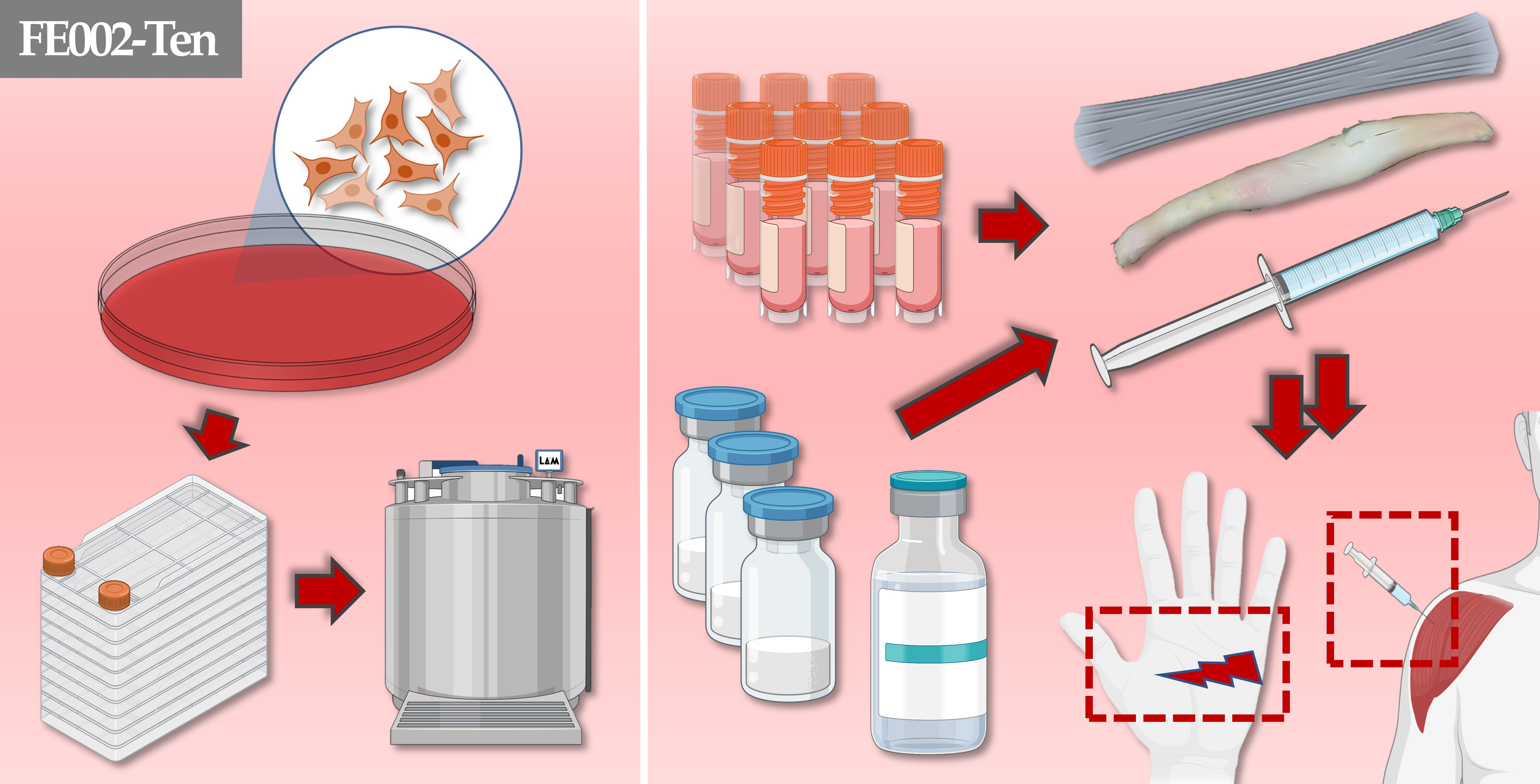
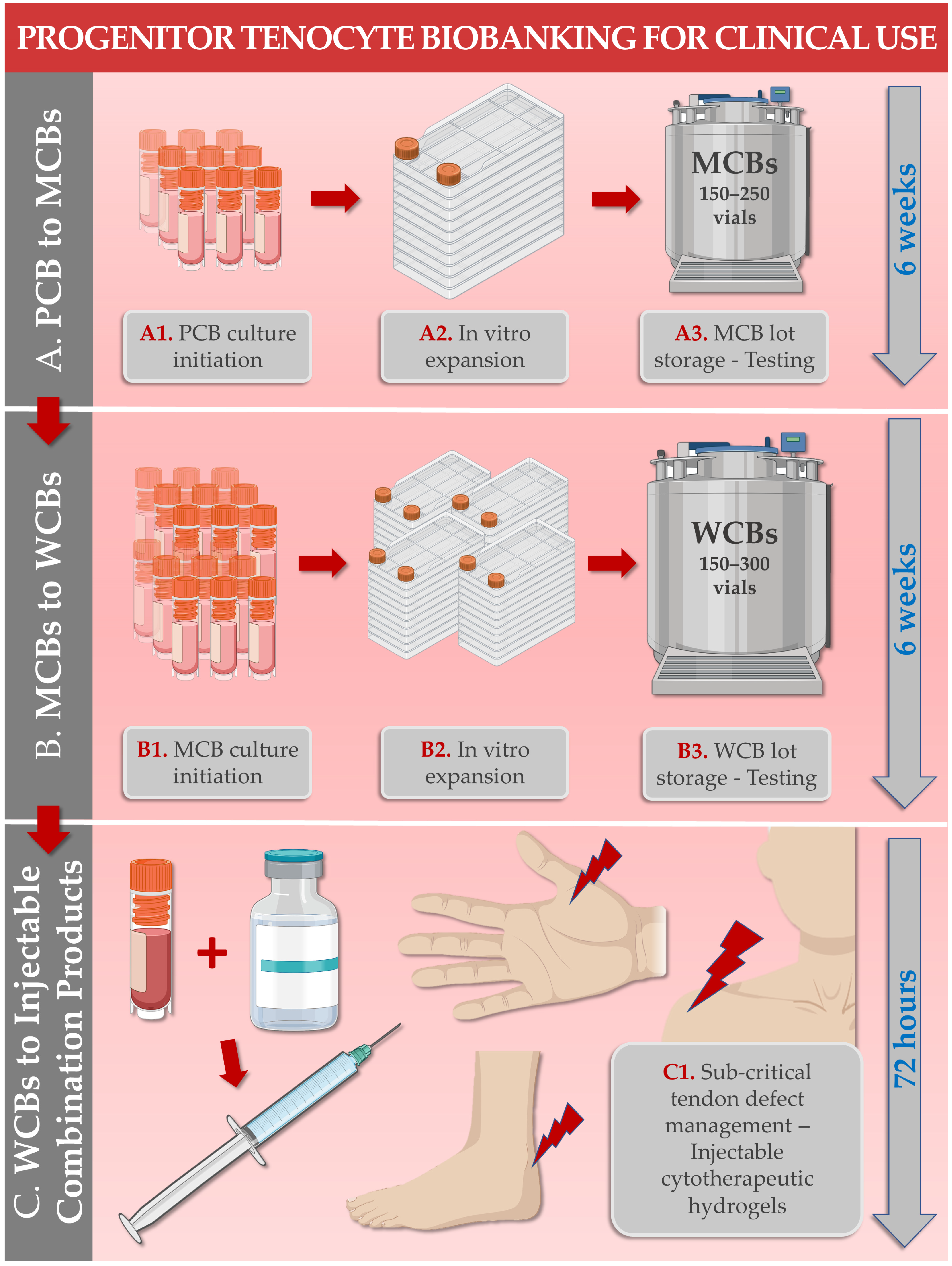
3.1. Primary Progenitor Tenocyte Manufacturing Processes

Appropriate devising and application of the described primary progenitor tenocyte bioprocessing methodologies are essential for the maintenance of both the high quality and the sustainability of the considered biological source (Figure 2 and Figure 3) [21,22]. Following the initial progenitor cell culture initiation procedure from the donated tendon tissue, all manufacturing activities are performed following conservative best practices of adherent primary diploid cell culture [21,25]. Therein, serial cell banking and multi-tiered cryopreserved vial lot manufacture in view of eventual clinical use enable both time and cost rationalization (Figure 3). With regard to the GMP-compliant manufacturing of primary progenitor cells and the related multi-tiered biobanking systems, the various cell bank tiers are defined by the in vitro passage level or by the in vitro population doubling level of a considered cellular population [21]. In the order of increasing in vitro passage levels along the progenitor cell manufacturing workflow, the following cell bank tiers are considered:
  • Parental cell banks (PCB), at early in vitro passage levels;
  • Master cell banks (MCB), at intermediate in vitro passage levels;
  • Working cell banks (WCB), at in vitro passage levels appropriate for clinical use;
  • End of production cell banks (EOPCB), at in vitro passage levels beyond those appropriate for clinical use.
In addition to these four types of manufacturing cell banks, pilot cell banks of the appropriate tiers may be used for preliminary material and contact-process consumable qualification, as well as technical specification optimization [21]. For each in vitro cellular expansion phase within the defined progenitor cell banking workflow, used to incrementally generate cell lots of superior passage levels in the cell bank system, the following individual process steps are sequentially performed:
  • Retrieval and initiation of the cryopreserved starting materials;
  • Qualitative and quantitative assessments of the initiated cellular suspension;
  • Serial seeding of an appropriate in vitro cell culture system using predefined technical specifications;
  • Incubation of the cell culture system with periodical culture medium exchanges and monitoring;
  • Endpoint enzymatic harvest of the expanded and confluent cell population;
  • Qualitative and quantitative assessments of the harvested cell suspension;
  • Cellular bulk formulation and dilution in a cryoprotectant solution and serial dispensing in storage vessels;
  • Controlled freezing of the conditioned cellular bulk lot;
  • Cryogenic storage of the conditioned cellular bulk lot.
Following the described manufacturing activities and in addition to specified in-process controls, appropriate post-process controls (i.e., characterization, qualification, and/or release testing) are carried out on the constituted cell bank lots [22]. Both the extent and the frequency of the control activities are based on compendial and regulatory requirements, yet a margin of appreciation exists for the manufacturer [21]. The latter is based on the appreciation of risks and on the effectiveness of the implemented biosafety testing scheme, principally for the insurance of material innocuity [28]. The various types of assays and testing activities to be carried out at the various stages of progenitor cell banking (i.e., as specified and justified by the manufacturer) are related to primary cell type identity and purity, to extraneous agent detection, and to cell type tumorigenicity study [21]. Individual characterization/testing assays comprise the following:
  • Cellular morphology and adherent behavior in microscopy following recovery;
  • Cell type identification by genetic, biochemical, or immunological means;
  • Cell type karyotype establishment;
  • Cell type in vitro lifespan determination;
  • Testing for bacterial and fungal contamination;
  • Testing for mycobacteria and mycoplasmas;
  • Electron microscopy for elucidation of cellular structures;
  • Testing for extraneous agents in cell cultures;
  • Testing for viruses and for retroviruses;
  • Safety/toxicity testing in small animals or in chicken eggs;
  • Tumorigenicity assays (i.e., in vitro, in vivo).
The described quality-driven manufacturing and testing activities correspond to standard risk-based industrial biotechnology practices, which are to be integrated in primary progenitor tenocyte production technical specifications and workflows [22,28]. In the reported studies of FE002-Ten primary progenitor tenocytes, conservative cell culture parameters and specifications have been consistently used [21,22,28]. Noteworthy technical specification elements pertaining to in vitro FE002-Ten cell culture activities for manufacturing purposes are listed hereafter:
  • Use of consistent technical specifications, targets, and acceptance criteria;
  • Use of thoroughly qualified contact-process consumables, materials, and reagents;
  • Use of 10% v/v fetal bovine serum (FBS) as a cell proliferation medium supplement;
  • Use of a humidified 5% CO2 incubation atmosphere at 37 °C, with 21% O2 or 2% O2;
  • Use of FBS-based and dimethyl sulfoxide-based cryopreservation medium;
  • Use of constant-rate freezing devices prior to cell bank lot cryogenic storage.
While modern regulatory guidelines tend to orient manufacturers toward the use of synthetic media, reagents, and materials for GMP cellular production activities, high attention must be paid to benchmarking such items against historically validated and qualified options. Importantly, manufacturing process designs should be pragmatically considered during transposition to GMP settings, for rationalization of the overall fixed costs incurred before therapy/product registration and commercialization [21,28].
Notably, regulators have been increasingly favoring the use of defined or human-sourced growth supplements (e.g., human platelet lysate) over the use of FBS for cell manufacturing activities in alleviating the risks associated with xenogeneic components [41,42]. Despite on-going validation work on various culture medium supplement alternatives for the in vitro expansion of FE002-Ten primary progenitor tenocytes, no suitable options have been identified to date, based on quality aspects of the produced cell populations (i.e., key and critical attributes of the harvest cell pools) [21,22]. Furthermore and importantly, parallel clinical work on the topical cytotherapeutic use of primary progenitor dermal fibroblasts within the Lausanne University Hospital (i.e., over three decades) has comprised the use of FBS for clinical-grade cell lot manufacture, with no reported adverse events linked to the use of xenogeneic materials during production [3,21]. Therein, successive regulatory reviews and approvals of technical specifications (i.e., including the use of FBS) by national and international bodies (i.e., FDA, TFDA, PMDA, Swissmedic) have confirmed that some regulatory leeway remains around this practice until a better option (i.e., of equal or superior safety and functionality for cellular expansion) is identified [21].

3.2. Primary Progenitor Tenocyte In Vitro Characterization Data

During the past decade, various preliminary and applied multicentric qualification studies performed on the FE002-Ten primary progenitor tenocyte cellular source have enabled the gathering of robust characterization data in view of their further translational use (Table 1). The main investigated parameters are listed hereafter, along with the corresponding literature references:
  • Tenogenic phenotype identity [20,28];
  • Phenotype stability in chemical induction media [20,28];
  • In vitro cellular lifespan in monolayer culture [20,22,28];
  • Impacts of hypoxia on key and critical cellular attributes [28];
  • Cellular proliferation stimulation potency in co-cultures with irradiated primary adult tenocytes [20];
  • Cellular genetic stability at in vitro passages levels of 3–12 [20];
  • Cell surface markers [20,28];
  • Cellular production of ECM (e.g., collagens, fibronectin, GAGs, elastin) [20,28];
  • Proteomic contents [28];
  • Biocompatibility with solid and commercial hyaluronan-based hydrogel scaffolds, or with decellularized equine tendons [9,26];
  • Applicability in bio-printed tissue engineering [30];
  • Cellular survival at 4 °C in hydrogels and hydrogel stability, maintenance of attachment and proliferation capacities [9];
  • Cellular survival after extrusion through clinical administration systems [9];
  • Absence of tumorigenic behavior in vitro (i.e., soft agar transformation assays) [22].
Overall, the existing and published body of knowledge around the FE002-Ten primary progenitor cell source has confirmed that biological and technical aspects of such primary cells were well-adapted for the standardized manufacture of tissue engineering products and cell therapies for human clinical use (Table 1) [22].

3.3. Primary Progenitor Tenocyte Preclinical Safety Evidence

In addition to the in vitro characterization data and safety evidence described hereabove for the FE002-Ten primary progenitor cell source, several assays and studies were further performed to confirm the safety of such primary cells for further translational use. The main investigated parameters are listed hereafter, along with the corresponding literature references:
  • Limited in vitro cellular lifespan of the primary cells in monolayer culture [20,22];
  • No observed embryotoxicity or angiotoxicity in ovo (i.e., standardized chorioallantoic membrane model) [22];
  • No observed significant adverse reactions (i.e., local or systemic) in vivo (i.e., GLP study with a lagomorph model of patellar tendon partial-thickness defect) [22].
Notably, the absence of toxicity or tumorigenicity of the considered primary progenitor cellular source was demonstrated within the retained experimental setups [9,20,22]. In vitro, the studied FE002-Ten primary progenitor tenocytes were not found to be capable of non-adherent proliferation (i.e., in agarose-based or hyaluronan-based hydrogels), contrasting with the behavior of known cancerous cell lines (e.g., HeLa cells, positive controls) [9,22]. In ovo, the studied progenitor tenocyte cellular materials did not exert acute embryotoxicity and did not display angiotoxic effects within the study period [22]. In the six-week GLP study in white rabbits, viable primary progenitor tenocytes delivered in hyaluronan-based hydrogels were shown to elicit no product rejection, immune reactions, or tumor formation [22]. Specifically, xenogeneic implantation of the cytotherapeutic hydrogel product within the hindleg tendon midsubstance enabled the study of recipient biological reactions over the initial phases (i.e., proliferative and remodeling stages) of tissular repair, with no reported adverse events [22]. While normal and local cellular reactions to suture materials were observed at the time of the study endpoint, no systemic markers were recorded as being abnormal [22].
Overall, the available in vitro and in vivo data for the FE002-Ten primary progenitor cell source have documented a good safety profile and did not yield any evidence of toxicity, immunogenicity, or tumorigenicity in the considered studies (Table 1). The aggregation of the available and published safety evidence has enabled a positive assessment of the applicability of FE002-Ten primary progenitor tenocytes for further translational investigation and for pilot human clinical trials.

3.4. Regulatory Considerations and Limitations for Tissue Engineering Products Containing Viable Primary Progenitor Tenocytes

The cytotherapeutic use of viable primary progenitor tenocytes in injectable form (e.g., in hyaluronan-based hydrogels) or in tissue engineering products (e.g., cells delivered on a synthetic tendon scaffold) falls under the category of a standardized transplant product (TrSt) in Switzerland (Figure 1) [9,10,22]. In Europe, this type of approach is considered as a combined advanced therapy medicinal product (cATMP) [21,31]. While the regulatory classification of such products is straightforward, few private and institutional sponsors currently choose to pursue the development of TrSt products or ATMPs due to burdensome fixed manufacturing costs and evolutive constraints entailed by GMPs [2,31,43,44,45]. Although some translational leeway exists, notably in university research centers and in the context of cytotherapy investigational use in clinical trials, the economic viability of classical cytotherapeutic approaches is difficult to materialize and sustain [2,3,45]. Several specialized examples of continued cytotherapy investigation and implementation exist (e.g., oncology, immunology, orthopedics), yet high rates of attrition and retraction of novel protocols are currently the norm [2,3,46,47].
In addition to the listed challenges (i.e., regulatory, financial) that characterize the classical cytotherapy ecosystem, several technological and technical elements of cell therapies must be carefully considered during the early development phases. Specifically, high logistical challenges (i.e., cryogenic storage and cold chains) and low reported rates of exogeneous cellular material engraftment have historically prompted the development of alternative cytotherapy-inspired approaches [3,25,27,48]. Such has notably been the case around primary progenitor tenocytes under the Swiss progenitor cell transplantation program, wherein parallel research was conducted on tissue engineering approaches as well as on off-the-shelf progenitor cell-derived preparations (Table 1) [9,22,27,29]. Specifically, the use of temperature-stabilized and sterilizable cell-free primary progenitor tenocyte extracts as hyaluronan-based hydrogel functionalization agents has opened promising perspectives for the further translational study of simple and potentially widely-available cytotherapy-inspired injectable preparations [27,29].

3.5. Economic Considerations around Cell Therapy Production and Applicability in Modern Healthcare Systems

As previously mentioned, large scale monolayer progenitor cell expansion for cytotherapeutic uses incurs elevated costs, especially under GMPs. However, some technical elements may be implemented to reduce the direct costs of cellular active substance manufacture, such as the use of multilayer cell culture vessels (e.g., Nunc Cell Factories, Corning HyperFlasks, or Greiner CellDiscs) [22,25]. Such vessels have been qualified for the in vitro expansion of the considered primary progenitor tenocytes and allow for the obtention of high harvest cell yields while limiting operator manipulations to a minimum and thereby reducing the direct costs of manufacture.
From a practical standpoint and based on cell manufacturing workflow optimization, the overall costs of cytotherapy manufacture may be competitive as compared to commercial products and standards of care [49]. Despite relatively high fixed costs of operation of a GMP platform, previous research has shown that the PBB preparation (i.e., allogeneic dermal progenitor fibroblasts on a collagen scaffold for burn wounds) was less expensive per cm2 of treatment than commercial comparators (i.e., tissue engineering skin substitutes, cadaveric skin) [49]. Therefore, and based on 30 years of clinical experience in topical cytotherapies for cutaneous wound management, it is possible to set forward important resource management optimization in order to keep the costs of therapeutic interventions under control [3,49,50].
Notably, stratified autologous keratinocyte sheets prepared by cellular expansion for burn victims are reimbursed by basic health insurance in Switzerland. These dispositions have been set in place following the implementation of such cytotherapeutic protocols in the Lausanne University Hospital since the 1980s [50,51]. During the review of the therapeutic intervention by national healthcare administrations, the costs of therapy manufacture have been factored into the assessment and have been assessed as acceptable in the cost-benefit analysis [50,51]. Therefore, it may be assessed overall that, despite the elevated fixed costs of GMP cell manufacture, clinical results and practical experience have enabled the justification of the expenses, especially in underserved medical conditions [3,50,51]. While specific cost-benefit assessments remain to be performed on the various possible therapeutic interventions using primary progenitor tenocytes, it is possible to set forth that manufacturing costs are in line with those already implemented for several decades at a clinical level, confirming their feasibility in routine practice [50].

4. Lyophilized Primary Progenitor Tenocyte Derivatives for Potent Functionalization of Hyaluronan-Based Hydrogels

Hyaluronan or hyaluronic acid (HA) has been widely used in medical and aesthetic practices over the past several decades [52]. Extensive preclinical and clinical data are available for topical and injectable hyaluronan-based preparations, and this polymer is known for its high biocompatibility and relative ease of biotechnological manufacturing [52,53,54,55,56]. Quality- and safety-related aspects of the FE002-Ten primary progenitor tenocytes used as biological starting materials for stabilized cellular extract obtention are considered to be covered within the previously discussed tissue engineering approach (Table 1) [29]. Therefore, both of the main components of the considered complex hydrogel preparations may be thoroughly assessed from a risk-based and quality-oriented point-of-view [27,29]. In addition, the use of appropriate pharmaceutical processes such as two-step lyophilization enables the obtention of the potential widespread and off-the-shelf availability of progenitor tenocyte extracts or complex hydrogel preparations [27,48]. As these components were shown to be compatible with localized injection administration modalities, they could be considered as a replacement or as an adjuvant to the use of corticoid injections for tendon tissue affections [9,27,29].
Due to the aforementioned specificities of cytotherapy translational development and several of the incurred burdens, high interest was directed toward the further obtention of stabilized cell-derived preparations characterized by conserved functional attributes (Figure 1) [27,29]. This approach is in line with current reported efforts around the use of biologically-derived acellular materials and complexes for managing diverse tendinous tissue affections [57,58,59,60,61,62,63]. Although such approaches enable the avoidance of several technical issues (i.e., cryogenics-related cell damages, logistical burdens), specific and sometimes complex additional requirements are incurred [64,65,66]. Notwithstanding, several pharmaceutical technology processes were applied for the obtention and stabilization of FE002-Ten primary progenitor tenocyte extracts. In particular, two-step lyophilization processing was shown to optimally maintain key and critical characteristics/attributes of the biological materials of interest, such as the intrinsic antioxidant activity and hyaluronan-based hydrogel viscosity modulation capacities [27,29].
At the end of the technical processing phase for stabilized progenitor tenocyte extract preparation, the materials of interest are made available in lyophilizate form, usually in glass vials corresponding to a unitary therapeutic dose of extract (Figure 1) [29]. The cellular extract lyophilizates are designed to be reconstituted in hyaluronan-based hydrogels compatible with standard injection materials for the local treatment of tendon tissue-related affections [29]. Due to the manufacturing process of the extracts, all cellular viability or proliferation capacity of the remaining biological materials is removed following lyophilization [27]. In some cases (e.g., 0.22 µm filter-sterilized extracts), only a small acellular fraction of the biological starting materials is present in the final lyophilizate form (Figure 1) [27]. Therefore, hydrogels containing such purified cellular extracts are closer to the definition of a medical device (MD) than that of an ATMP [27,67]. Such considerations are also based on the functional attributes of the extracts and the mechanisms of action that they deploy in a hyaluronan-based hydrogel system (e.g., antioxidant and viscomodulating effects) [27,29,62].
Stabilized hyaluronan-based hydrogels may be manufactured and obtained via an array of processes and have been studied or developed notably for the management of tendinopathies, osteoarthritis, ophthalmology, and skin defects [68,69,70,71,72]. Chemical (e.g., crosslinking, grafting) or formulation means (e.g., incorporation of carbohydrates) have been successfully employed for HA-based hydrogel product stability enhancement against oxidative and/or enzymatic degradation [52,68,69,70,73]. As the main mechanism of action of hyaluronan is physical (i.e., exertion of a cushioning and gliding enhancement effect), all technical and technological means of strengthening the rheological properties of the hydrogel are considered as beneficial for the improvement of its therapeutic properties [29,73,74,75]. Therefore, primary progenitor tenocyte extracts were employed for their intrinsic antioxidant attributes (e.g., reactive oxygen species scavenging) and their ability to conserve or to enhance the complex viscosity of hyaluronan-based hydrogel preparations, especially in highly oxidative environments [27,29]. Of note, such useful behavior of the reconstituted extracts was shown to be dose-dependent, highly robust, and stable over time [27]. Finally, the critical processing step of cellular extract sterilization was experimentally investigated, wherein it was validated that the critical and key functional parameters of the cell-based and cell-derived cell-free lyophilizates were partly or fully conserved following 5–50 kGy 60Co gamma irradiation [27]. The technical possibility of including a terminal sterilization step in the manufacturing process of the considered primary progenitor tenocyte extracts enables an overall enhancement of therapeutic material quality, based on an improved risk-benefit profile [27].

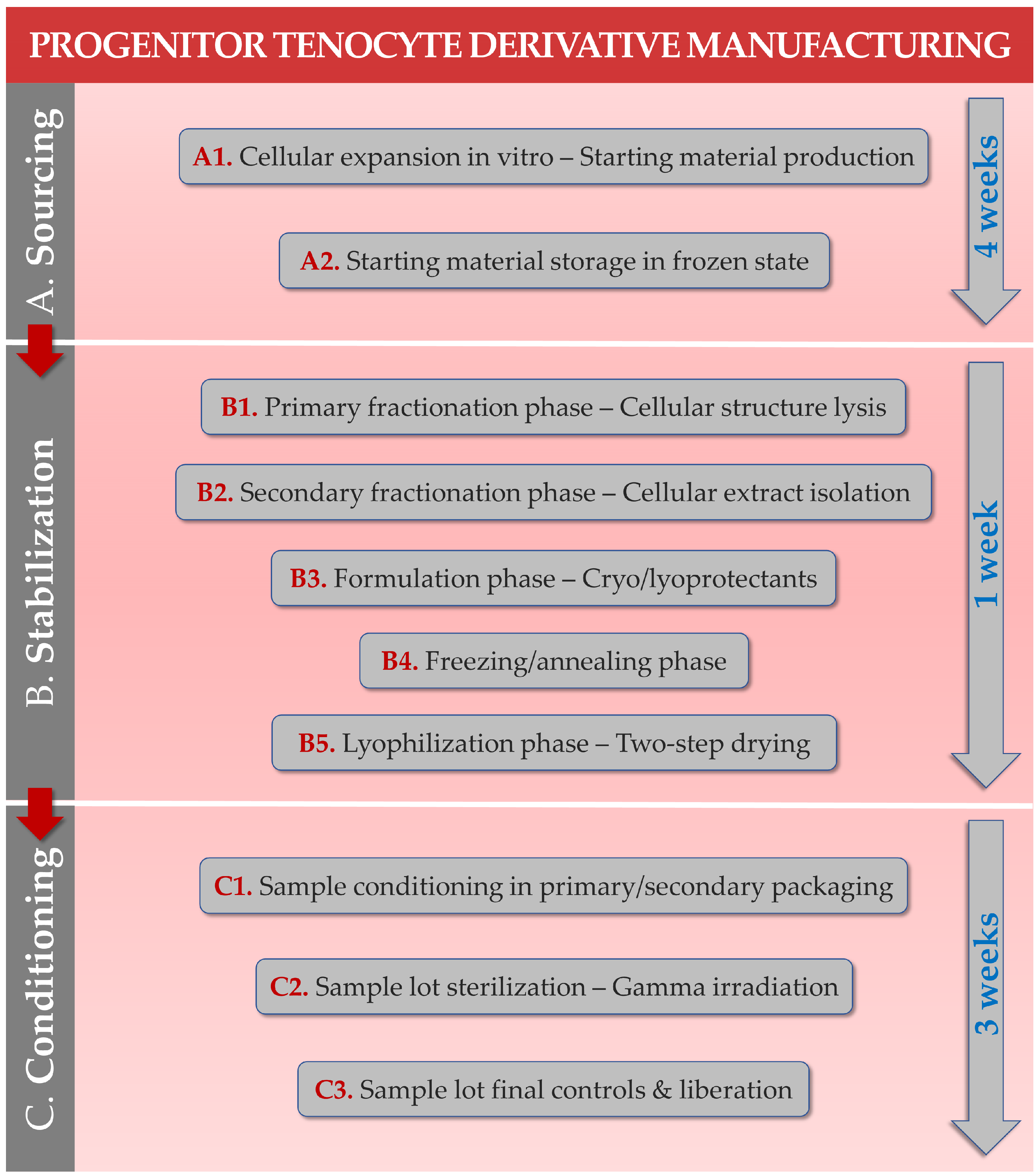
4.1. Primary Progenitor Tenocyte Stabilized Derivative Manufacturing Processes

In terms of progenitor tenocyte cellular extract manufacturing process, simple physical means of fractionation and extraction are employed (i.e., thermal cell lysis, differential centrifugation, single or serial submicron filtration) [27,29]. Therein, processing simplicity was always designed as a means of robustness enhancement, which is especially important in the handling of inherently variable biological starting materials [29,48]. Sugar-based (e.g., saccharose/dextran) excipient formulas were used to prepare the samples for freezing and drying. Then, conservative parameters of two-step lyophilization were employed in order to optimally preserve the considered biological structures and entities and to obtain temperature-stable cell-derived extracts [27,29]. A general step-by-step workflow applicable for the manufacture of such stabilized cytotherapy-derived progenitor tenocyte extracts is presented in Figure 4.
Following the stabilized cellular extract manufacturing phase, the control activities related to the conditioned progenitor tenocyte extract lots are derived from classical characterization assays relative to the pharmaceutical lyophilizate form [29]. Cellular extract lot technical specifications for qualification and release testing may include such control assays, along with predetermined targets and acceptance criteria. Common control types are provided as examples hereafter:
  • Descriptive and photographic controls of the lyophilizate vial lots;
  • pH value determination of the reconstituted lyophilizates;
  • Osmolality determination of the reconstituted lyophilizates;
  • Study of sample particulate population distribution (i.e., except in cell-free extracts);
  • Determination of lyophilizate resuspension time;
  • Determination of lyophilizate residual moisture levels.
Overall, the use of simple and well-known pharmaceutical technology processes and control assays has enabled the specification of tight tolerances and acceptance criteria for progenitor cellular extracts derived from biological starting materials [27,48]. Such methodological elements are of key importance as regards cellular extract lot quality insurance levels and standardization, which are important prerequisites for regulatory evaluations and for tangible subsequent clinical evaluation of therapeutic intervention efficacy. Available data and hindsight indicate that primary progenitor tenocytes may be effectively used for the industrial-scale development of cytotherapy-inspired and cell-free complex hydrogels for potential therapeutic management of diverse tendon tissue disorders (e.g., subcritical structural defects, adjuvant post-surgical use) [27,29].

4.2. Primary Progenitor Tenocyte Stabilized Derivative Characterization Data

Based on the functional attributes and on the known physical mechanisms of action of the considered primary progenitor tenocyte extracts, robust and quantitative quality control assays (e.g., rheology, antioxidant capacity) were implemented in the ad hoc manufacturing processes (Figure 4) [27,29]. Furthermore, various characterization studies were performed in order to gather additional evidence of translational relevance (e.g., hydrogel injectability, ex vivo bioadhesivity, cytocompatibility) [27]. The various types of characterization assays and testing activities are to be carried out at the end of the cellular extract manufacturing process, wherein possible individual assays comprise the following:
  • Lyophilizate total proteomic contents;
  • Lyophilizate stability in storage over time;
  • Lyophilizate intrinsic antioxidant activity;
  • Lyophilizate cytocompatibility with primary patient cells;
  • Hyaluronan-based hydrogel viscosity modulation function of the lyophilizates;
  • Hyaluronan-based hydrogel viscosity modulation function of the lyophilizates in oxidative environments (e.g., H2O2 challenge assays);
  • Complex hydrogel preparation injectability evaluation in vitro/ex vivo;
  • Complex hydrogel preparation bioadhesivity evaluation in vitro/ex vivo;
  • Complex hydrogel preparation tribology characterization in vitro/ex vivo.
The various assays described hereabove have notably enabled the confirmation of the compatibility of the various product ingredients, as well as formulation stability over time following lyophilizate reconstitution in the hydrogels [29]. Results have shown that lyophilized cell-free extracts were equivalent in terms of antioxidant capacity (i.e., TEAC) as compared to freshly harvested and viable primary progenitor tenocytes [27]. Of note, the considered cellular extracts were reported to contain low amounts of total proteinic materials (i.e., ≤2.5 ng total proteins/vial), to present excellent stability over time, and to possess conserved significant antioxidant capacities [29]. Importantly, in vitro experiments have confirmed that the lyophilized and filter-sterilized or gamma-sterilized cellular extracts retained their significant antioxidant properties and hyaluronan-based hydrogel viscosity modulating effects [27]. Finally, it was shown that the considered complex hydrogel preparations procured significant friction modulation effects in vitro and tendon bioadhesivity ex vivo, with improved system behavior as compared to simple hyaluronan gels in oxidative environments in particular [27].
These elements confirmed the technical possibility of obtaining serial off-the-shelf and functional progenitor tenocyte extracts characterized by facilitated logistical requirements as compared to off-the-freezer cytotherapeutic cellular material lots (Figure 3). Once reconstituted in linear hyaluronan-based hydrogels, the primary progenitor tenocyte extracts were shown to exert significant protective effects on the viscoelastic properties of the system, especially in strongly oxidative environments (i.e., H2O2 challenge assays) [27,29]. Such functional data were considered positively from a formula functionalization or stability enhancement point-of-view. Importantly, safety aspects related to in vitro cellular extract cytocompatibility and technical aspects related to complex hydrogel ex vivo injectability have confirmed the applicability of such materials in further translational studies and in view of clinical administration [27].

4.3. Technical Development and Rationale for the Use of Stabilized Progenitor Tenocyte Derivatives as Hyaluronan-Based Hydrogel Stability Enhancement Agents

As regards the inception of the use of stabilized progenitor tenocyte derivatives as hyaluronan-based hydrogel stability enhancement agents, accelerated hydrogel degradation studies were performed [29,76]. As hyaluronan-based hydrogels are mostly degraded by the action of enzymes (i.e., hyaluronidases) and oxidative stress mediators, laboratory assays to mimic such degradation in accelerated settings comprised the use of enzymatic or H2O2 challenges [29,77,78,79,80]. The initial goal of these investigations was to exclude an intrinsic negative effect of the cellular derivatives on the stability of the considered hydrogel systems. In this context, it was rapidly evidenced that the hyaluronan-based hydrogels containing the stabilized progenitor tenocyte derivatives were in fact significantly protected against oxidative stress (i.e., maintenance or enhanced stability of rheological parameters) [27,29]. Thereafter, the stabilizing effects of the cellular extracts were benchmarked against alternative options described for hyaluronan hydrogel stability enhancement (i.e., inclusion of sugars, chemical modification of the polymer), confirming the significant nature of the described effects [27,29,68,73,76].
As regards specification of the internal mechanism of action of the various stabilized cellular extracts and their effects on the hydrogel system, experimental work is currently on-going. Based on previous reports, it appears that a complex effect is exerted by the cellular extracts within the system [27,29]. The first component of this complex effect is that of a direct antioxidant action of the cellular extracts in oxidative settings, with scavenging of the oxidative species and limitation of their negative effects on the attributes of the hydrogel [27]. The second component of this complex effect probably consists in a type of cross-linking of the hyaluronan polymer by the protein components of the extracts in an oxidative environment [29]. This kind of indirect system stabilization mechanism is necessary to explain the documented rise in complex viscosity of the H2O2-challenged combination formulas and the overall ancillary effect of stability enhancement [27,29].

4.4. Important Logistical Advantages of Stabilized Progenitor Tenocyte Derivatives over Classical Cytotherapies

The major advantage procured by two-step lyophilization processing consists in the availability of room-temperature-stable cellular extracts, which maintain specific functions over several months [29,81,82,83,84]. This aspect is a clear logistical advantage in comparison to the classical ultra-low cold chain necessary for cell therapies. Indeed, usual practices for cell therapies warrant storage in liquid nitrogen vessels and shipping in liquid nitrogen or dry ice containers, which are costly and must be performed rapidly [85,86]. Additionally, once the preparations are shipped from the manufacturing site to the clinical administration site, appropriate local storage possibilities must be available (e.g., liquid nitrogen storage, ultra-low temperature freezers) [3]. Overall, the elevated costs and limited availability of appropriate technical means and infrastructure for classical cell therapies highly restrict the number of patients potentially benefiting from the related therapeutic interventions. Conversely, room temperature or refrigerated shipping and storage of the lyophilized cellular derivatives limit the costs and widen the pool of potential clinical users of the described preparations [27,29,48]. Specifically, the described cellular extracts may be used similarly to lyophilized antibiotic or antifungal drugs, which are classically appropriately reconstituted extemporaneously before clinical administration [87].

4.5. Regulatory Considerations for the Therapeutic Use of Stabilized Primary Progenitor Tenocyte Derivatives

High plasticity in the regulatory context and classification options for the described cell-based and cell-derived preparations is currently reported [27,29,43,44,45]. Numerous academic studies have undertaken the description of the registration processes for cell-based and cell-derived preparations for human clinical use [2,19,43,44,45,88]. Notwithstanding, the perspectives presented in this study (i.e., and in the previously published original reports on the subject) correspond to the latest and updated knowledge of the area, following and based on specific interactions with the appropriate specialists and regulatory authorities, in addition to the scientific understanding of the process [20,21,22,27,29]. The regulatory classification of the described stabilized primary progenitor tenocyte extracts and of the related hydrogels may be considered as borderline and depends on a number of factors, among which are product composition and modes of action [27,29]. Based on the existing and published information on these formulas, consideration of class III medical device classification may technically be pursued for progenitor tenocyte extracts included in a hyaluronan-based hydrogel preparation [29].
Firstly and as regards the contents of the stabilized extracts, it is determined that cellular viability is excluded following the physical processing and lyophilization phases. The contents of the stabilized extracts are therefore in majority related to the proteinic constituents of the bulk biological starting material, which are present in low overall quantities [28,29]. Although it is not excluded that individual growth factors and cytokines may retain some form of biological activity after lyophilization, the low amounts of individual entities make it highly unlikely that a significant pharmacological, immunological, or metabolic mechanism of action would be exerted by the preparation and be therapeutically useful. Furthermore, it was shown that the extract’s functional attributes of interest were conserved even in filter-sterilized preparations, where most of the proteinic constituents were removed during processing [27]. On the other hand, the inherent antioxidant attributes (i.e., mediating a physical/chemical action) of the biological constituents of the stabilized extracts were determined to be significant and repeatably measurable [27,29].
Secondly, as hyaluronan hydrogel-based preparations are currently commercially proposed for injectable use in various types of tendinopathy (e.g., Ostenil Tendon, Biolevox HA Tendon), it is considered that the principal mechanism of action (i.e., necessary and sufficient) is physical and is related to the properties of hyaluronan [9,29]. Using hyaluronan for tendon affections and repair enhancement is based on its viscoelastic properties, which can promote tendon gliding and reduce adhesions for improved architectural organization of healing tendons with mechanically reduced friction and pain [9,10]. Additionally, the inclusion of stabilized progenitor tenocyte extracts is known to procure specific and significant hydrogel system stability enhancement, especially in oxidative environments [27].
The ancillary use of carbohydrates for hyaluronan-based hydrogel stability enhancement has been previously reported, yet experimental research on the considered stabilized progenitor tenocyte extracts has shown that the cellular derivatives were more effective than saccharose (i.e., used as a lyoprotectant in the cellular extract manufacturing process) alone [27,68,80]. Therefore, key advantages of such a device (i.e., containing the cellular extract) over commercially available HA hydrogels for tendinopathies comprise comparatively enhanced resistance toward oxidative degradation, and therefore a postulated prolonged time of exertion of the principal mechanism of action of the hydrogel in situ [27].
Overall, it may be assessed that the stabilized progenitor tenocyte extracts exert ancillary mechanisms of action on the hydrogel itself (i.e., antioxidant and viscomodulation properties of the extracts) for system stability enhancement. Such activities are not necessary nor sufficient for the deployment of the principal mechanism of action of the system, confirming their ancillary nature [27,29]. Furthermore, it is possible that the stabilized extracts and the hyaluronan polymer both exert direct ancillary antioxidant functions/effects in vivo, potentially attenuating the effects of free radicals as triggers of inflammatory reactions.

5. Conclusions and Prospects

Compilation and critical review of the existing body of knowledge around primary progenitor tenocytes (i.e., FE002-Ten cell source) enables the assessment of the high versatility of potential uses for such biological materials within translational approaches of tendon tissue affection therapeutic management. Preclinical research conducted under the Swiss progenitor cell transplantation program has demonstrated that viable progenitor tenocytes could be safely and consistently used in allogeneic tissue engineering products and off-the-freezer cytotherapies for potentially optimized tendon defect repair promotion. Such standardized applications potentially present several clinical and physiological advantages in view of tissue replacement (i.e., promotion of enhanced graft bio-integration by the cells) as compared to the use of an acellular or autologous tendon graft. Further studies on stabilized and sterilizable progenitor tenocyte-derived extracts have demonstrated that potent functionalization of hyaluronan-based hydrogels is possible, principally in view of significant formulation stability enhancement. This in turn bears tangible potential for the controlled management of tendinous tissular inflammation and localized defects using off-the-shelf preparations and devices. Overall, the FE002 primary progenitor tenocytes investigated under the Swiss progenitor cell transplantation program were shown to represent highly standardized biological materials with diverse potential therapeutic uses after processing and formulation into appropriate biotechnological preparations.

Supplementary Materials

The following supporting information can be downloaded at: https://www.mdpi.com/article/10.3390/encyclopedia3010021/s1, Table S1: Cell surface marker panel for FE002 primary progenitor tenocytes; Table S2: Differentiation potential of FE002 primary progenitor tenocytes; Figure S1: Illustrative timecourse of FE002 primary progenitor tenocyte in vitro proliferation.

Author Contributions

Conceptualization, A.L., C.S., N.H.-B. and L.A.A.; methodology, A.L., C.S., P.A.-S., N.H.-B. and L.A.A.; software, A.L.; validation, A.L., C.S., P.A.-S., W.R., N.H.-B. and L.A.A.; formal analysis, A.L., C.S., P.A.-S., N.H.-B. and L.A.A.; investigation, A.L., C.S., P.A.-S., W.R., N.H.-B. and L.A.A.; resources, A.L., W.R. and L.A.A.; data curation, A.L.; writing—original draft preparation, A.L. and L.A.A.; writing—review and editing, A.L., C.S., P.A.-S., W.R., N.H.-B. and L.A.A.; visualization, A.L. and L.A.A.; supervision, N.H.-B., W.R. and L.A.A.; project administration, A.L. and L.A.A.; funding acquisition, L.A.A. Author A.L. participated in this work within the scope of an industrial PhD thesis at the Faculty of Biology and Medicine (FBM) of the University of Lausanne (UNIL, Lausanne, Switzerland) under the co-supervision of authors N.H.-B. and W.R. All authors have read and agreed to the published version of the manuscript.

Funding

This research received no external funding.

Institutional Review Board Statement

Not applicable.

Informed Consent Statement

Not applicable.

Data Availability Statement

Not applicable.

Acknowledgments

The authors are most grateful and thankful for the shared time and close collaboration with the Pharmaceutical Technology group at the Institute of Pharmaceutical Sciences of Western Switzerland (University of Geneva, Geneva, Switzerland). Artwork templates for the figures of the present study were partly created with www.biorender.com, accessed on 25 January 2023.

Conflicts of Interest

Author A.L. was employed by LAM Biotechnologies SA during the redaction of this work. The company LAM Biotechnologies SA had no role in the design of the study; in the collection, analyses, or interpretation of data; in the writing of the manuscript, or in the decision to publish the results. The remaining authors declare no conflict of interest.

Abbreviations

ATMPadvanced therapy medicinal product
60Cocobalt 60 irradiation source
CAMchorioallantoic membrane model
cATMPcombined advanced therapy medicinal product
CDcluster of differentiation
DMEMDulbecco’s Modified Eagle Medium
DMSOdimethyl sulfoxide
ECMextracellular matrix
EOPCBend of production cell bank
FBSfetal bovine serum
FDAUS Food and Drug Administration
FE002-Cartprimary chondroprogenitor cell source
FE002-SK2primary dermal progenitor fibroblast cell source
FE002-Tenprimary progenitor tenocyte cell source
FITCfluorescein isothiocyanate
GAGglycosaminoglycan
GLPgood laboratory practices
GMPgood manufacturing practices
HAhyaluronic acid
HLAhuman leucocyte antigen
H2O2hydrogen peroxide
HPLhuman platelet lysate
ITSinsulin-transferrin-selenium
kGykiloGray
MCBmaster cell bank
MDmedical device
PCBparental cell bank
PEphycoerythrin
PMDAJapanese Pharmaceuticals and Medical Devices Agency
PRPplatelet-rich plasma
TEACTrolox equivalent antioxidant capacity
TFDATaiwan Food and Drug Administration
TrStstandardized transplant product
WCBworking cell bank

References

  1. Vacanti, J.P.; Langer, R. Tissue engineering: The design and fabrication of living replacement devices for surgical reconstruction and transplantation. Lancet 1999, 354, 32–34. [Google Scholar] [CrossRef] [PubMed]
  2. Ramezankhani, R.; Torabi, S.; Minaei, N.; Madani, H.; Rezaeiani, S.; Hassani, S.N.; Gee, A.P.; Dominici, M.; Silva, D.N.; Baharvand, H.; et al. Two decades of global progress in authorized advanced therapy medicinal products: An emerging revolution in therapeutic strategies. Front. Cell Dev. Biol. 2020, 8, 547653. [Google Scholar] [CrossRef]
  3. Laurent, A.; Rey, M.; Scaletta, C.; Abdel-Sayed, P.; Michetti, M.; Flahaut, M.; Raffoul, W.; de Buys Roessingh, A.; Hirt-Burri, N.; Applegate, L.A. Retrospectives on three decades of safe clinical experience with allogeneic dermal progenitor fibroblasts: High versatility in topical cytotherapeutic care. Pharmaceutics 2023, 15, 184. [Google Scholar] [CrossRef]
  4. Clayton, R.A.; Court-Brown, C.M. The epidemiology of musculoskeletal tendinous and ligamentous injuries. Injury 2008, 39, 1338–1344. [Google Scholar] [CrossRef]
  5. Costa-Almeida, R.; Calejo, I.; Gomes, M.E. Mesenchymal stem cells empowering tendon regenerative therapies. Int. J. Mol. Sci. 2019, 20, 3002. [Google Scholar] [CrossRef] [PubMed] [Green Version]
  6. Mirghaderi, S.P.; Valizadeh, Z.; Shadman, K.; Lafosse, T.; Oryadi-Zanjani, L.; Yekaninejad, M.S.; Nabian, M.H. Cell therapy efficacy and safety in treating tendon disorders: A systemic review of clinical studies. J. Exp. Orthop. 2022, 9, 85. [Google Scholar] [CrossRef]
  7. Kaux, J.F.; Samson, A.; Crielaard, J.M. Hyaluronic acid and tendon lesions. Muscles Ligaments Tendons J. 2016, 5, 264–269. [Google Scholar] [CrossRef] [PubMed] [Green Version]
  8. Abate, M.; Schiavone, C.; Salini, V. The use of hyaluronic acid after tendon surgery and in tendinopathies. BioMed Res. Int. 2014, 2014, 783632. [Google Scholar] [CrossRef] [Green Version]
  9. Grognuz, A.; Scaletta, C.; Farron, A.; Pioletti, D.P.; Raffoul, W.; Applegate, L.A. Stability enhancement using hyaluronic acid gels for delivery of human fetal progenitor tenocytes. Cell Med. 2016, 8, 87–97. [Google Scholar] [CrossRef] [PubMed] [Green Version]
  10. Masiello, F.; Pati, I.; Veropalumbo, E.; Pupella, S.; Cruciani, M.; De Angelis, V. Ultrasound-guided injection of platelet-rich plasma for tendinopathies: A systematic review and meta-analysis. Blood Transfus. 2022; in press. [Google Scholar] [CrossRef]
  11. Ho, J.O.; Sawadkar, P.; Mudera, V. A review on the use of cell therapy in the treatment of tendon disease and injuries. J. Tissue Eng. 2014, 5, 2041731414549678. [Google Scholar] [CrossRef]
  12. Chen, X.; Song, X.H.; Yin, Z.; Zou, X.H.; Wang, L.L.; Hu, H.; Cao, T.; Zheng, M.; Ouyang, H.W. Stepwise differentiation of human embryonic stem cells promotes tendon regeneration by secreting fetal tendon matrix and differentiation factors. Stem Cells 2009, 27, 1276–1287. [Google Scholar] [CrossRef] [PubMed]
  13. Xu, W.; Wang, Y.; Liu, E.; Sun, Y.; Luo, Z.; Xu, Z.; Liu, W.; Zhong, L.; Lv, Y.; Wang, A.; et al. Human iPSC-derived neural crest stem cells promote tendon repair in a rat patellar tendon window defect model. Tissue Eng. Part A 2013, 19, 2439–2451. [Google Scholar] [CrossRef] [PubMed] [Green Version]
  14. Chong, A.K.; Ang, A.D.; Goh, J.C.; Hui, J.H.; Lim, A.Y.; Lee, E.H.; Lim, B.H. Bone marrow-derived mesenchymal stem cells influence early tendon-healing in a rabbit Achilles tendon model. J. Bone Jt. Surg. Am. 2007, 89, 74–81. [Google Scholar] [CrossRef] [PubMed]
  15. Van den Boom, N.A.C.; Winters, M.; Haisma, H.J.; Moen, M.H. Efficacy of stem cell therapy for tendon disorders: A systematic review. Orthop. J. Sport. Med. 2020, 8, 2325967120915857. [Google Scholar] [CrossRef] [PubMed]
  16. Andriolo, L.; Altamura, S.A.; Reale, D.; Candrian, C.; Zaffagnini, S.; Filardo, G. Nonsurgical treatments of patellar tendinopathy: Multiple injections of platelet-rich plasma are a suitable option: A systematic review and meta-analysis. Am. J. Sport. Med. 2019, 47, 1001–1018. [Google Scholar] [CrossRef] [PubMed]
  17. Romero, A.; Barrachina, L.; Ranera, B.; Remacha, A.R.; Moreno, B.; de Blas, I.; Sanz, A.; Vasquez, F.J.; Vitoria, A.; Junquera, C.; et al. Comparison of autologous bone marrow and adipose tissue derived mesenchymal stem cells, and platelet-rich plasma, for treating surgically induced lesions of the equine superficial digital flexor tendon. Vet. J. 2017, 224, 76–84. [Google Scholar] [CrossRef] [PubMed]
  18. Van der Vlist, A.C.; Winters, M.; Weir, A.; Ardern, C.L.; Welton, N.J.; Caldwell, D.M.; Verhaar, J.A.; de Vos, R.J. Which treatment is most effective for patients with Achilles tendinopathy? A living systematic review with network meta-analysis of 29 randomised controlled trials. Br. J. Sport. Med. 2021, 55, 249–256. [Google Scholar] [CrossRef]
  19. Freedman, B.R.; Mooney, D.J.; Weber, E. Advances toward transformative therapies for tendon diseases. Sci. Transl. Med. 2022, 14, eabl8814. [Google Scholar] [CrossRef]
  20. Grognuz, A.; Scaletta, C.; Farron, A.; Raffoul, W.; Applegate, L.A. Human fetal progenitor tenocytes for regenerative medicine. Cell Transpl. 2016, 25, 463–479. [Google Scholar] [CrossRef] [Green Version]
  21. Laurent, A.; Hirt-Burri, N.; Scaletta, C.; Michetti, M.; de Buys Roessingh, A.S.; Raffoul, W.; Applegate, L.A. Holistic approach of Swiss fetal progenitor cell banking: Optimizing safe and sustainable substrates for regenerative medicine and biotechnology. Front. Bioeng. Biotechnol. 2020, 8, 557758. [Google Scholar] [CrossRef]
  22. Laurent, A.; Abdel-Sayed, P.; Grognuz, A.; Scaletta, C.; Hirt-Burri, N.; Michetti, M.; de Buys Roessingh, A.S.; Raffoul, W.; Kronen, P.; Nuss, K.; et al. Industrial development of standardized fetal progenitor cell therapy for tendon regenerative medicine: Preliminary safety in xenogeneic transplantation. Biomedicines 2021, 9, 380. [Google Scholar] [CrossRef]
  23. Zhang, K.; Na, T.; Wang, L.; Gao, Q.; Yin, W.; Wang, J.; Yuan, B.Z. Human diploid MRC-5 cells exhibit several critical properties of human umbilical cord-derived mesenchymal stem cells. Vaccine 2014, 32, 6820–6827. [Google Scholar] [CrossRef]
  24. Olshansky, S.J.; Hayflick, L. The role of the WI-38 cell strain in saving lives and reducing morbidity. AIMS Public Health 2017, 4, 127–138. [Google Scholar] [CrossRef]
  25. US Food and Drug Administration. Points to Consider in the Characterization of Cell Lines Used to Produce Biologicals; FDA: Silver Spring, MD, USA, 1993.
  26. Aeberhard, P.A.; Grognuz, A.; Peneveyre, C.; McCallin, S.; Hirt-Burri, N.; Antons, J.; Pioletti, D.; Raffoul, W.; Applegate, L.A. Efficient decellularization of equine tendon with preserved biomechanical properties and cytocompatibility for human tendon surgery indications. Artif. Organs 2020, 44, E161–E171. [Google Scholar] [CrossRef] [PubMed] [Green Version]
  27. Laurent, A.; Porcello, A.; Jeannerat, A.; Peneveyre, C.; Coeur, A.; Abdel-Sayed, P.; Scaletta, C.; Michetti, M.; de Buys Roessingh, A.; Jordan, O.; et al. Lyophilized progenitor tenocyte extracts: Sterilizable cytotherapeutic derivatives with antioxidant properties and hyaluronan hydrogel functionalization effects. Antioxidants 2023, 12, 163. [Google Scholar] [CrossRef] [PubMed]
  28. Jeannerat, A.; Peneveyre, C.; Armand, F.; Chiappe, D.; Hamelin, R.; Scaletta, C.; Hirt-Burri, N.; de Buys Roessingh, A.; Raffoul, W.; Applegate, L.A.; et al. Hypoxic incubation conditions for optimized manufacture of tenocyte-based active pharmaceutical ingredients of homologous standardized transplant products in tendon regenerative medicine. Cells 2021, 10, 2872. [Google Scholar] [CrossRef]
  29. Laurent, A.; Porcello, A.; Fernandez, P.G.; Jeannerat, A.; Peneveyre, C.; Abdel-Sayed, P.; Scaletta, C.; Hirt-Burri, N.; Michetti, M.; de Buys Roessingh, A.; et al. Combination of hyaluronan and lyophilized progenitor cell derivatives: Stabilization of functional hydrogel products for therapeutic management of tendinous tissue disorders. Pharmaceutics 2021, 13, 2196. [Google Scholar] [CrossRef] [PubMed]
  30. Liu, H.; Chansoria, P.; Delrot, P.; Angelidakis, E.; Rizzo, R.; Rütsche, D.; Applegate, L.A.; Loterie, D.; Zenobi-Wong, M. Filamented light (flight) biofabrication of highly aligned tissue-engineered constructs. Adv. Mater. 2022, 34, 2204301. [Google Scholar] [CrossRef]
  31. European Directorate for the Quality of Medicines & Healthcare. European Directorate for the Quality of Medicines & Healthcare. European Committee (Partial Agreement) on organ transplantation (CD-P-TO). In Guide to the Quality and Safety of Tissues and Cells for Human Application, 4th ed.; EDQM: Strasbourg, France, 2019; ISBN 978-92-871-8945-5. [Google Scholar]
  32. World Medical Association. World Medical Association Declaration of Helsinki: Ethical principles for medical research involving human subjects. JAMA 2013, 310, 2191–2194. [Google Scholar] [CrossRef] [Green Version]
  33. Hopkins, C.; Fu, S.C.; Chua, E.; Hu, X.; Rolf, C.; Mattila, V.M.; Qin, L.; Yung, P.S.; Chan, K.M. Critical review on the socio-economic impact of tendinopathy. Asia Pac. J. Sport. Med. Arthrosc. Rehabil. Technol. 2016, 4, 9–20. [Google Scholar] [CrossRef] [Green Version]
  34. Sharma, P.; Maffulli, N. Tendon injury and tendinopathy: Healing and repair. J. Bone Jt. Surg. Am. 2005, 87, 187–202. [Google Scholar] [CrossRef] [Green Version]
  35. Fujikawa, K.; Ohtani, T.; Matsumoto, H.; Seedhom, B.B. Reconstruction of the extensor apparatus of the knee with the Leeds-Keio ligament. J. Bone Jt. Surg. Br. 1994, 76, 200–203. [Google Scholar] [CrossRef] [PubMed]
  36. Chong, A.K.; Riboh, J.; Smith, R.L.; Lindsey, D.P.; Pham, H.M.; Chang, J. Flexor tendon tissue engineering: Acellularized and reseeded tendon constructs. Plast. Reconstr. Surg. 2009, 123, 1759–1766. [Google Scholar] [CrossRef]
  37. Lovati, A.B.; Bottagisio, M.; Moretti, M. Decellularized and engineered tendons as biological substitutes: A critical review. Stem Cells Int. 2016, 2016, 7276150. [Google Scholar] [CrossRef] [Green Version]
  38. Pridgen, B.C.; Woon, C.Y.; Kim, M.; Thorfinn, J.; Lindsey, D.; Pham, H.; Chang, J. Flexor tendon tissue engineering: Acellularization of human flexor tendons with preservation of biomechanical properties and biocompatibility. Tissue Eng. Part C Method. 2011, 17, 819–828. [Google Scholar] [CrossRef]
  39. Kryger, G.S.; Chong, A.K.; Costa, M.; Pham, H.; Bates, S.J.; Chang, J. A comparison of tenocytes and mesenchymal stem cells for use in flexor tendon tissue engineering. J. Hand Surg. Am. 2007, 32, 597–605. [Google Scholar] [CrossRef] [PubMed]
  40. Larson, B.J.; Longaker, M.T.; Lorenz, H.P. Scarless fetal wound healing: A basic science review. Plast. Reconstr. Surg. 2010, 126, 1172–1180. [Google Scholar] [CrossRef] [PubMed] [Green Version]
  41. Liau, L.L.; Hassan, M.N.F.B.; Tang, Y.L.; Ng, M.H.; Law, J.X. Feasibility of human platelet lysate as an alternative to foetal bovine serum for in vitro expansion of chondrocytes. Int. J. Mol. Sci. 2021, 22, 1269. [Google Scholar] [CrossRef] [PubMed]
  42. Philippe, V.; Laurent, A.; Hirt-Burri, N.; Abdel-Sayed, P.; Scaletta, C.; Schneebeli, V.; Michetti, M.; Brunet, J.F.; Applegate, L.A.; Martin, R. Retrospective analysis of autologous chondrocyte-based cytotherapy production for clinical use: GMP process-based manufacturing optimization in a Swiss university hospital. Cells 2022, 11, 1016. [Google Scholar] [CrossRef]
  43. Johnson, P.C.; Bertram, T.A.; Tawil, B.; Hellman, K.B. Hurdles in tissue engineering/regenerative medicine product commercialization: A survey of North American academia and industry. Tissue Eng. Part A 2011, 17, 5–15. [Google Scholar] [CrossRef]
  44. Pirnay, J.P.; Vanderkelen, A.; De Vos, D.; Draye, J.P.; Rose, T.; Ceulemans, C.; Ectors, N.; Huys, I.; Jennes, S.; Verbeken, G. Business oriented EU human cell and tissue product legislation will adversely impact Member States’ health care systems. Cell Tissue Bank 2013, 14, 525–560. [Google Scholar] [CrossRef]
  45. Pearce, K.F.; Hildebrandt, M.; Greinix, H.; Scheding, S.; Koehl, U.; Worel, N.; Apperley, J.; Edinger, M.; Hauser, A.; Mischak-Weissinger, E.; et al. Regulation of advanced therapy medicinal products in Europe and the role of academia. Cytotherapy 2014, 16, 289–297. [Google Scholar] [CrossRef] [Green Version]
  46. Evans, C.H.; Ghivizzani, S.C.; Robbins, P.D. Orthopaedic gene therapy: Twenty-five years on. JBJS Rev. 2021, 9, e20. [Google Scholar] [CrossRef] [PubMed]
  47. Mohanty, R.; Chowdhury, C.R.; Arega, S.; Sen, P.; Ganguly, P.; Ganguly, N. CAR T cell therapy: A new era for cancer treatment (Review). Oncol. Rep. 2019, 42, 2183–2195. [Google Scholar] [CrossRef] [PubMed]
  48. Laurent, A.; Scaletta, C.; Abdel-Sayed, P.; Michetti, M.; Flahaut, M.; Simon, J.P.; Roessingh, A.B.; Raffoul, W.; Hirt-Burri, N.; Applegate, L.A. Optimized manufacture of lyophilized dermal fibroblasts for next-generation off-the-shelf progenitor biological bandages in topical post-burn regenerative medicine. Biomedicines 2021, 9, 1072. [Google Scholar] [CrossRef] [PubMed]
  49. Abdel-Sayed, P.; Hirt-Burri, N.; de Buys Roessingh, A.; Raffoul, W.; Applegate, L.A. Evolution of biological bandages as first cover for burn patients. Adv. Wound Care 2019, 8, 555–564. [Google Scholar] [CrossRef] [PubMed]
  50. Chemali, M.; Laurent, A.; Scaletta, C.; Waselle, L.; Simon, J.P.; Michetti, M.; Brunet, J.F.; Flahaut, M.; Hirt-Burri, N.; Raffoul, W.; et al. Burn center organization and cellular therapy integration: Managing risks and costs. J. Burn Care Res. 2021, 42, 911–924. [Google Scholar] [CrossRef]
  51. Gobet, R.; Raghunath, M.; Altermatt, S.; Meuli-Simmen, C.; Benathan, M.; Dietl, A.; Meuli, M. Efficacy of cultured epithelial autografts in pediatric burns and reconstructive surgery. Surgery 1997, 121, 654–661. [Google Scholar] [CrossRef]
  52. Oliva, F.; Marsilio, E.; Asparago, G.; Frizziero, A.; Berardi, A.C.; Maffulli, N. The impact of hyaluronic acid on tendon physiology and its clinical application in tendinopathies. Cells 2021, 10, 3081. [Google Scholar] [CrossRef]
  53. Burdick, J.A.; Prestwich, G.D. Hyaluronic acid hydrogels for biomedical applications. Adv. Mater. 2011, 23, H41–H56. [Google Scholar] [CrossRef]
  54. Brown, M.B.; Jones, S.A. Hyaluronic acid: A unique topical vehicle for the localized delivery of drugs to the skin. J. Eur. Acad. Dermatol. Venereol. 2005, 19, 308–318. [Google Scholar] [CrossRef] [PubMed]
  55. Dicker, K.T.; Gurski, L.A.; Pradhan-Bhatt, S.; Witt, R.L.; Farach-Carson, M.C.; Jia, X. Hyaluronan: A simple polysaccharide with diverse biological functions. Acta Biomater. 2014, 10, 1558–1570. [Google Scholar] [CrossRef] [PubMed] [Green Version]
  56. Huerta-Ángeles, G.; Nešporová, K.; Ambrožová, G.; Kubala, L.; Velebný, V. An effective translation: The development of hyaluronan-based medical products from the physicochemical, and preclinical aspects. Front. Bioeng. Biotechnol. 2018, 6, 62. [Google Scholar] [CrossRef] [Green Version]
  57. Zhang, Z.; Li, Y.; Zhang, T.; Shi, M.; Song, X.; Yang, S.; Liu, H.; Zhang, M.; Cui, Q.; Li, Z. Hepatocyte growth factor-induced tendon stem cell conditioned medium promotes healing of injured Achilles tendon. Front. Cell Dev. Biol. 2021, 9, 654084. [Google Scholar] [CrossRef] [PubMed]
  58. Lu, V.; Tennyson, M.; Zhang, J.; Khan, W. Mesenchymal stem cell-derived extracellular vesicles in tendon and ligament repair—A systematic review of in vivo studies. Cells 2021, 10, 2553. [Google Scholar] [CrossRef]
  59. Song, K.; Jiang, T.; Pan, P.; Yao, Y.; Jiang, Q. Exosomes from tendon derived stem cells promote tendon repair through miR-144-3p-regulated tenocyte proliferation and migration. Stem Cell Res. Ther. 2022, 13, 80. [Google Scholar] [CrossRef]
  60. Xue, Z.; Chen, Z.; Wu, T.; Li, R.; Chen, C.; Liu, J.; Hou, H.; Zheng, X.; Wang, H. VEGFA-enriched exosomes from tendon-derived stem cells facilitate tenocyte differentiation, migration, and transition to a fibroblastic phenotype. BioMed Res. Int. 2022, 2022, 8537959. [Google Scholar] [CrossRef]
  61. Yao, Z.; Li, J.; Xiong, H.; Cui, H.; Ning, J.; Wang, S.; Ouyang, X.; Qian, Y.; Fan, C. MicroRNA engineered umbilical cord stem cell-derived exosomes direct tendon regeneration by mTOR signaling. J. Nanobiotechnol. 2021, 19, 169. [Google Scholar] [CrossRef]
  62. Zhang, M.; Liu, H.; Cui, Q.; Han, P.; Yang, S.; Shi, M.; Zhang, T.; Zhang, Z.; Li, Z. Tendon stem cell-derived exosomes regulate inflammation and promote the high-quality healing of injured tendon. Stem Cell Res. Ther. 2020, 11, 402. [Google Scholar] [CrossRef]
  63. Lui, P.P.Y.; Leung, Y.T. Practical considerations for translating mesenchymal stromal cell-derived extracellular vesicles from bench to bed. Pharmaceutics 2022, 14, 1684. [Google Scholar] [CrossRef]
  64. Cottle, C.; Porter, A.P.; Lipat, A.; Turner-Lyles, C.; Nguyen, J.; Moll, G.; Chinnadurai, R. Impact of cryopreservation and freeze-thawing on therapeutic properties of mesenchymal stromal/stem cells and other common cellular therapeutics. Curr. Stem Cell Rep. 2022, 8, 72–92. [Google Scholar] [CrossRef] [PubMed]
  65. Gimona, M.; Brizzi, M.F.; Choo, A.B.H.; Dominici, M.; Davidson, S.M.; Grillari, J.; Hermann, D.M.; Hill, A.F.; de Kleijn, D.; Lai, R.C.; et al. Critical considerations for the development of potency tests for therapeutic applications of mesenchymal stromal cell-derived small extracellular vesicles. Cytotherapy 2021, 23, 373–380. [Google Scholar] [CrossRef]
  66. Sadeghi, S.; Tehrani, F.R.; Tahmasebi, S.; Shafiee, A.; Hashemi, S.M. Exosome engineering in cell therapy and drug delivery. Inflammopharmacology 2023, 31, 145–169. [Google Scholar] [CrossRef] [PubMed]
  67. Racchi, M.; Govoni, S.; Lucchelli, A.; Capone, L.; Giovagnoni, E. Insights into the definition of terms in European medical device regulation. Expert Rev. Med. Dev. 2016, 13, 907–917. [Google Scholar] [CrossRef] [PubMed] [Green Version]
  68. Conrozier, T.; Mathieu, P.; Rinaudo, M. Mannitol preserves the viscoelastic properties of hyaluronic acid in an in vitro model of oxidative stress. Rheumatol. Ther. 2014, 1, 45–54. [Google Scholar] [CrossRef] [PubMed] [Green Version]
  69. Schanté, C.E.; Zuber, G.; Herlin, C.; Vandamme, T.F. Chemical modifications of hyaluronic acid for the synthesis of derivatives for a broad range of biomedical applications. Carbohydr. Polym. 2011, 85, 469–489. [Google Scholar] [CrossRef]
  70. Schanté, C.; Zuber, G.; Herlin, C.; Vandamme, T.F. Synthesis of N-alanyl-hyaluronamide with high degree of substitution for enhanced resistance to hyaluronidase-mediated digestion. Carbohydr. Polym. 2011, 86, 747–752. [Google Scholar] [CrossRef]
  71. Aviv, M.; Halperin-Sternfeld, M.; Grigoriants, I.; Buzhansky, L.; Mironi-Harpaz, I.; Seliktar, D.; Einav, S.; Nevo, Z.; Adler-Abramovich, L. Improving the mechanical rigidity of hyaluronic acid by integration of a supramolecular peptide matrix. ACS Appl. Mater. Interfaces 2018, 10, 41883–41891. [Google Scholar] [CrossRef]
  72. Karel, S.; Sogorkova, J.; Hermannova, M.; Nesporova, K.; Marholdova, L.; Chmelickova, K.; Bednarova, L.; Flegel, M.; Drasar, P.; Velebny, V. Stabilization of hyaluronan-based materials by peptide conjugation and its use as a cell-seeded scaffold in tissue engineering. Carbohydr. Polym. 2018, 201, 300–307. [Google Scholar] [CrossRef]
  73. Porcello, A.; Gonzalez-Fernandez, P.; Jordan, O.; Allémann, E. Nanoforming hyaluronan-based thermoresponsive hydrogels: Optimized and tunable functionality in osteoarthritis management. Pharmaceutics 2022, 14, 659. [Google Scholar] [CrossRef]
  74. Zheng, Y.; Yang, J.; Liang, J.; Xu, X.; Cui, W.; Deng, L.; Zhang, H. Bioinspired hyaluronic acid/phosphorylcholine polymer with enhanced lubrication and anti-inflammation. Biomacromolecules 2019, 20, 4135–4142. [Google Scholar] [CrossRef] [PubMed]
  75. Amadio, P.C. Gliding resistance and modifications of gliding surface of tendon: Clinical perspectives. Hand Clin. 2013, 29, 159–166. [Google Scholar] [CrossRef] [PubMed] [Green Version]
  76. Maudens, P.; Meyer, S.; Seemayer, S.A.; Jordan, O.; Allémann, E. Self-assembled thermoresponsive nanostructures of hyaluronic acid conjugates for osteoarthritis therapy. Nanoscale 2018, 10, 1845–1854. [Google Scholar] [CrossRef] [PubMed] [Green Version]
  77. Kogan, G.; Soltés, L.; Stern, R.; Schiller, J.; Mendichi, R. Hyaluronic acid: Its function and degradation in in vivo systems. Stud. Nat. Prod. Chem. 2008, 34, 789–882. [Google Scholar] [CrossRef]
  78. Fakhari, A.; Berkland, C. Applications and emerging trends of hyaluronic acid in tissue engineering, as a dermal filler and in osteoarthritis treatment. Acta Biomater. 2013, 9, 7081–7092. [Google Scholar] [CrossRef] [Green Version]
  79. Shi, L.; Zhang, Y.; Ossipov, D. Enzymatic degradation of hyaluronan hydrogels with different capacity for in situ bio-mineralization. Biopolymers 2018, 109, e23090. [Google Scholar] [CrossRef]
  80. Rinaudo, M.; Lardy, B.; Grange, L.; Conrozier, T. Effect of mannitol on hyaluronic acid stability in two in vitro models of oxidative stress. Polymers 2014, 6, 1948–1957. [Google Scholar] [CrossRef] [Green Version]
  81. Remmele, R.L.; Krishnan, S.; Callahan, W.J. Development of stable lyophilized protein drug products. Curr. Pharm. Biotechnol. 2012, 13, 471–496. [Google Scholar] [CrossRef]
  82. Ó’Fágáin, C.; Colliton, K. Storage and lyophilization of pure proteins. Methods Mol. Biol. 2017, 1485, 159–190. [Google Scholar] [CrossRef]
  83. Fernandez-Moure, J.; Maisha, N.; Lavik, E.B.; Cannon, J.W. The chemistry of lyophilized blood products. Bioconj. Chem. 2018, 29, 2150–2160. [Google Scholar] [CrossRef]
  84. Zhang, Y.Z.; DePaz, R.A.; Bee, J.S.; Marshall, T. Development of a stable lyophilized adeno-associated virus gene therapy formulation. Int. J. Pharm. 2021, 606, 120912. [Google Scholar] [CrossRef]
  85. Meneghel, J.; Kilbride, P.; Morris, G.J. Cryopreservation as a key element in the successful delivery of cell-based therapies—A review. Front. Med. 2020, 7, 592242. [Google Scholar] [CrossRef] [PubMed]
  86. Fehm, T.; Boehme, P.; Modak, N.; Talwar, V.; Kinscher, K. The clinical supply of cell and gene therapy drugs: Challenges ahead. Drug Discov. Today 2023, 28, 103421. [Google Scholar] [CrossRef] [PubMed]
  87. Naik, S.; Chougule, M.; Padhi, B.K.; Misra, A. Development of novel lyophilized mixed micelle amphotericin B formulation for treatment of systemic fungal infection. Curr. Drug Deliv. 2005, 2, 177–184. [Google Scholar] [CrossRef] [PubMed]
  88. Nordberg, R.C.; Otarola, G.A.; Wang, D.; Hu, J.C.; Athanasiou, K.A. Navigating regulatory pathways for translation of biologic cartilage repair products. Sci. Transl. Med. 2022, 14, eabp8163. [Google Scholar] [CrossRef]
Figure 1. Schematic and illustrated overview of the differential processing options for primary progenitor tenocytes (e.g., FE002-Ten cells), within the manufacture of cytotherapies or cytotherapy-inspired preparations for tendinous tissue disorders under the Swiss progenitor cell transplantation program. (A1A3) Viable cell lots may be manufactured in vitro and may be further cryogenically stored for off-the-freezer preparation of tissue engineering products (i.e., using artificial or biological scaffolds) or injectable hydrogel-based (e.g., hyaluronan) preparations and devices. Scale bar = 200 µm. (B1B3) Bulk cellular materials may be further stabilized by lyophilization for off-the-shelf availability and may be eventually reconstituted in hyaluronan-based injectable hydrogels. Scale bar = 400 µm. (C1,C2) Bulk cellular materials may also be further processed for fractionation and sterilization (i.e., 0.22 µm filtration or 60Co gamma irradiation) before storage or in view of final reconstitution in hyaluronan-based injectable hydrogels.
Encyclopedia 03 00021 g001
Figure 2. Schematic and illustrated methodological and biotechnological workflows for primary progenitor tenocyte cell source establishment within a registered progenitor cell transplantation program. Approximative timeframes for step execution are presented in blue font. (A1,A2) Following the constitution of the necessary team, appropriate accreditations and authorizations must be obtained. (B1,B2) Processing of the starting biological tissue enables the rapid initiation of adherent progenitor cell cultures in vitro. (C1C3) If the progenitor cellular bulk harvest corresponds to the predefined specifications and acceptance criteria, the materials may be formulated and further processed for cryogenic storage.
Encyclopedia 03 00021 g002
Figure 3. Schematic and illustrated workflow for primary progenitor tenocyte manufacturing in a simplified multi-tiered cell bank system in view of homologous cytotherapeutic use. Approximative timeframes for step execution are presented in blue font. (A1A3) Using PCB materials, one or several cell expansion rounds are performed to constitute a homogeneous master cell bank (MCB). (B1B3) Using MCB materials, one or several cell expansion rounds are performed to constitute a homogeneous working cell bank (WCB). (C1) Using WCB materials, extemporaneous off-the-freezer reconstitution in hyaluronan-based hydrogels may be performed in order to obtain injectable cytotherapeutic preparations. Such hydrogel products may potentially be therapeutically used for managing small tendon lesions such as partial tissue ruptures and tears. MCB, master cell bank; PCB, parental cell bank; WCB, working cell bank.
Encyclopedia 03 00021 g003
Figure 4. Schematic and stepwise manufacturing workflow for stabilized primary progenitor tenocyte derivatives. Following the starting material (e.g., FE002-Ten cells) sourcing phase (A1,A2), physical processing and formulation with cryoprotectants and lyoprotectants (B1B3) enables the optimal preparation of the sample lot. Lyophilization (B4,B5) and terminal treatment of the sample lot (C1C3) enable to constitute a stable progenitor tenocyte extract lot. Approximative timeframes for step execution are presented in blue font.
Encyclopedia 03 00021 g004
Table 1. Descriptive listing of the various peer-reviewed international scientific publications describing the FE002-Ten primary progenitor tenocytes investigated under the Swiss progenitor cell transplantation program. Within this evolving body of knowledge, cultured primary progenitor FE002-Ten cells and derivatives were established as versatile therapeutic biological material contenders, notably in musculoskeletal regenerative medicine. CAM, chorioallantoic membrane model; GLP, good laboratory practices.
Study Subject/DomainScope of Study Data/Investigated ParametersReferences
1. FE002-Ten Cell Source EstablishmentEstablishment of the FE002-Ten cell source in a cryopreserved multi-tiered biobank following a single controlled organ donation.[21]
2. FE002-Ten Cell Type In Vitro Characterization Characterization of primary progenitor tenocyte attributes (e.g., cell population homogeneity and purity, genetic and phenotypic stability, proteomic contents, biological functions) 1.[9,20,21,22,28]
3. FE002-Ten Cell Type Biobanking & ManufacturingEstablishment of optimized and standardized in vitro primary progenitor tenocyte manufacturing workflows for the production of industrial scale cellular material lots.[22,28]
4. FE002-Ten Cell Type Preclinical Safety CharacterizationCharacterization of primary progenitor tenocyte safety (i.e., at clinically relevant passage levels 2) in vitro (e.g., genetic stability, tumorigenicity assays) and in vivo (e.g., CAM model, GLP study of cell implantation in rabbit tendons).[20,22]
5. FE002-Ten Cell Type Derivative Manufacturing, Lyophilization, and Sterilization Establishment of biological material processing and purification workflows, for cell-derived and cell-free stabilized formulation obtention. Optimization of pharmaceutical processing (e.g., two-step lyophilization) for temperature stabilization of the cellular extracts. Optimization of the sterilization methodologies (e.g., submicron filtration, 60Co gamma irradiation) for conservation of cell-derived extract critical quality attributes and functional properties.[27,29]
6. FE002-Ten Cells or Derivatives: Study of Combination Product PrototypesTranslational characterization of primary progenitor tenocytes for tissue engineering applications (e.g., using injectable hydrogels, collagen scaffolds, artificial and biological tendon matrices). Translational characterization of hyaluronan hydrogel-based devices incorporating stabilized cellular derivatives.[9,20,26,27,29,30]
1 Data excerpts relative to FE002-Ten progenitor cell type characterization are presented in Table S1 (i.e., cell surface markers), in Table S2 (i.e., cell differentiation potential), and in Figure S1 (i.e., photographic timelapse of proliferating cells). 2 In laboratory studies, primary progenitor FE002-Ten cells were produced and characterized within a finite in vitro lifespan of 12–15 passage levels. Within manufacturing optimization works and preclinical safety studies, the retained in vitro passage levels for cytotherapeutic applications or for cellular derivative manufacturing were specified as passage levels 6–8.
Disclaimer/Publisher’s Note: The statements, opinions and data contained in all publications are solely those of the individual author(s) and contributor(s) and not of MDPI and/or the editor(s). MDPI and/or the editor(s) disclaim responsibility for any injury to people or property resulting from any ideas, methods, instructions or products referred to in the content.

