Article

Nano-Materials-Based Printed Glucose Sensor for Use in Incontinence Products for Health-Care Applications

1
Microsystem Engineering, University of Applied Science, HTW Berlin, 12459 Berlin, Germany
2
Institute for Microintegration and Reliability, Fraunhofer IZM, 13355 Berlin, Germany
3
Electrical Engineering Department, Faculty of Engineering, Taibah University, Madinah 42353, Saudi Arabia
*
Author to whom correspondence should be addressed.
Micro 2023, 3(2), 521-536; https://doi.org/10.3390/micro3020035
Submission received: 10 February 2023 / Revised: 16 March 2023 / Accepted: 20 March 2023 / Published: 12 May 2023

Abstract

:
Our recent development of a wireless humidity sensor system embedded in incontinence products enables new sensor applications to diagnose and supervise geriatric diseases (i.e., age-related diabetes mellitus type II). The measurement of glucose in urine, so-called glucosuria, is an early indicator for an incipient diabetes mellitus disease, whose symptoms are often age-related but misjudged. In this paper, an incontinence glucose sensor is printed with biocompatible ink and Prussian blue as an electron mediator on foil and functionalized with immobilized glucose oxidase. Inkjet printing of multiple layers of Nafion prevents large interference substances from diffusing into the measuring electrode and allows precise adjustment of the linear working range, which is significantly different from blood glucose measurement. Performance tests show the potential to detect minimum glucose values and store the sensor over a prolonged period at room temperature. The printed glucose sensor can be embedded into the absorber material of incontinence products, where capillary forces transport the urine analyte to the detection area. An attached readout module with an integrated potentiostat measures the glucose concentration in urine, which is transmitted wirelessly with incontinence events and stored in a cloud service for further analysis by medical staff and care workers.

1. Introduction

Diabetes is one of the most widespread diseases in the world and continues to gain ground, with 425 million persons currently affected and an increase of 55% expected by the year 2035 [1]. More than half of diabetes type II patients are older than 65 years. This age group is most likely to suffer from incontinence as well, with a prevalence of over 25% for men and more than 35% for women. In home care settings, most of the inhabitants (about 70%) suffer from incontinence, which gives the ability of further non-invasive testing for diabetes additional features, as diabetes type II is often recognized too late as it may occur for a long time without or unspecific symptoms [2]. Aggravated by the fact that many disease symptoms in elderly patients are misjudged as age-related changes, a smart diaper with an incontinence sensor system seems a reasonable solution. Until now, there has been little investigation of the need for special medical care, the increased risk of complications, or the need for specific screening programs for patients over 80 years old.
The most obvious symptoms of diabetes mellitus are sweet urine and frequent urination. The cause of these symptoms lies in the lack or resistance of insulin, a protein that regulates the metabolism of carbohydrates like glucose at the cell level. Insulin helps move glucose from the bloodstream into the cells. Absence or dysfunction of this hormone leads to malnutrition of the cells and a high blood sugar level, which is lowered by the excretion of glucose via urination.

2. The State-of-the-Art Glucose Sensors

Recent studies have demonstrated a significant positive correlation between glucose concentrations in urine and those in blood for healthy and diabetic patients [3]. Detecting urinary glucose levels in adults’ incontinence products is a new way for diagnosing diabetes. Urine is a complex biological fluid that contains molecules other than glucose.
The normal amount of glucose in the urine of sober individuals is between 0 and 0.8 mMol/L (0–15 mg/dL) and up to 6 mMol/L (nutrition dependent). Higher amounts of glucose could be a sign of glucosuria, most commonly due to diabetes mellitus, when exceeding the renal threshold of about 10 mMol/L of glucose concentration in blood [4].
Although there are other conditions in which glucose levels in urine rise above the threshold [5], therefore, measuring glucose concentration in urine can be a good indicator of an adequate diabetes treatment and correct intake of the necessary insulin doses. Together with the quantification of the urine volume, the glucose sensing system can help prevent dehydration and glucosuria, which are hard to supervise, especially for elderly and dementia patients [6,7,8].
There are many methods for glucose detection, such as electrical, optical, thermal, and nanotechnology [9,10,11]. However, the majority of current glucose sensors, including the ones that are used in this work, are electrochemical metabolite sensors, which rely on the function of enzymes (oxidoreductases) [12,13] and are good analytical reagents. These reagents reversibly catalyze specific chemical reactions and hence exhibit high sensitivity and selectivity towards certain metabolites.
In general, based on the working principle, glucose sensors can be subdivided into the enzymatic type, which involves an enzymatic reaction where glucose is detected indirectly through hydrogen peroxide (H2O2) formation, and the non-enzymatic type, which uses enzyme-mimicking metals, metal oxides, or other materials, where the change in the analyte’s properties such as fluorescence, band gaps in nanozymes, or cyclic voltammetry are used for detection [14]. Apart from the change in the reacting component, there is a considerable difference in the mechanisms involved for enzymatic and non-enzymatic sensors [15]. Generally, enzymatic glucose sensing involves the movement of electrons due to oxidation reactions between the enzyme glucose oxidase (GOx) and glucose. GOx is a typical flavin enzyme, containing the flavin adenine dinucleotide (FAD) redox active coenzyme, which catalyzes the conversion of glucose to gluconolactone. During the reaction, the enzyme is converted to GOx (FADH2) from GOx (FAD), which results in the generation of current or voltage changes that are then measured by the electrodes. The typical reaction scheme is GOx (FAD) + glucose → gluconolactone + GOx (FADH2) [16].
Efforts were made to enhance the accuracy and reliability of the disposable glucose sensors while lowering their prices and reducing the volume of the necessary blood sample [17]. Urine test strips, such as Diastix reagent strips, with color change at semi-quantitative values between negative and 5.5 mM, 14 mM, 28 mM, 55.5 mM, or more, are widely used, even though the basic enzymatic reaction in these test kits is the same as that with blood. However, the specification of electrochemical blood sugar test strips differs from urine glucose test strips. For instance, the sensitivity range of blood glucose testing extends from 0.5 mM to 33 mM, compared with 2.2 mM to 120 mM in urine glucose testing. At the same time, less accuracy of the urine glucose is necessary as its value can only be seen as an indicator for further analysis, such as a blood test. Furthermore, the blood test strips are optimized for filtering the cells from blood plasma by capillary intake of small volumes of blood around 0.6 µL, which has a different viscosity and will not work with low-viscosity urine samples. On the other hand, urine consists of much higher concentrations of electrolytes, which interfere with the electrode and influence the readout signal [18]. Therefore, a new concept of a printable flexible urine glucose sensor with additional protection layers on top of the sensor surface has to be developed. Various types of printable glucose sensors are shown in Figure 1.
Kim et al. [22] describe a reusable graphene-based urine glucose sensor whose output correlates well with the glucose concentration in blood [23], which shows the potential of determining the original blood sugar above the renal threshold of 8.9–10 mMol/L. This correlation is high compared with its level in saliva or tears, which validates the role of urine as a noninvasive diagnostic tool [24,25,26,27].
The fabrication of traditional glucose sensors typically involves complicated and expensive processing techniques, such as metal deposition and lithography, due to the nature of the electrode materials used [13]. For the fabrication of glucose sensors designed to be single-use, cost-efficiency needs to be taken into consideration. Go et al. [28] have developed a reusable Pt electrode for urine glucose detection using a multi-layered enzymatic composite coating that helped in increasing the number of electrons available to the electrode from the glucose oxidase layer, which increased its selectivity. Pezhhan et al. [29] have developed a selective and reusable probe based on the encapsulation of GOx to immobilize the enzyme.
Improved fabrication techniques, such as inkjet and screen printing, as well as selecting compatible materials for large-area deposition, have to be adopted [30]. This involves utilizing novel materials and device structures to the maximum extent for their sensing abilities. Inkjet-printing is used for depositing a variety of electronic materials in customized geometries at low temperature, especially when they are integrated with biological molecules such as enzymes. The main factor limiting the application of inkjet printing in the manufacture of advanced devices stems mainly from the strict rheological conditions that the printable materials need to meet [31]. Therefore, the printable materials have to have high conductivity and be compatible with screen printing and inkjet-printing. Due to their soft nature, these materials can also be printed on cheap paper substrates, which substitute for more expensive substrates such as glass and plastic, making them promising for the realization of disposable glucose and other biomedical sensors.
Yang et al. [32] have reported a paper-based glucose sensor using a bimetallic reduced graphene electrode with enzyme-mimicking activity. The absorbance was recorded when the electrode encountered glucose samples with different concentrations of up to 10 mg/mL of GOx. However, the limit of detection was calculated to be 1.76 μm. Another paper-based urinary glucose sensor, which uses GOx/PEDOT:PSS as sensing electrodes, involved a sensing strip and included a transistor amplifier circuit for a visual readout [33]. However, the sensor could take up to 2 min to generate visual responses during urine glucose concentration detection that ranged from 1 to 5 mM.
The so-called smart incontinence products have been developed [6,7,34,35]. Such systems can only provide for the detection of urine in absorbents. As mentioned, urine contains high numbers of biomarkers, which are health relevant. The ability to detect them in incontinence care products could be a great possibility in future medical applications as it is noninvasive and requires less human resources. In this paper, we present a printed glucose detection sensor with a long lifetime for continuous real-time monitoring. This could be integrated into incontinence products together with our previous urine detection solution as a multi-sensing platform [8]. Integration of electronic technologies into the biosensing platforms with low power consumption and economical product design is also introduced.

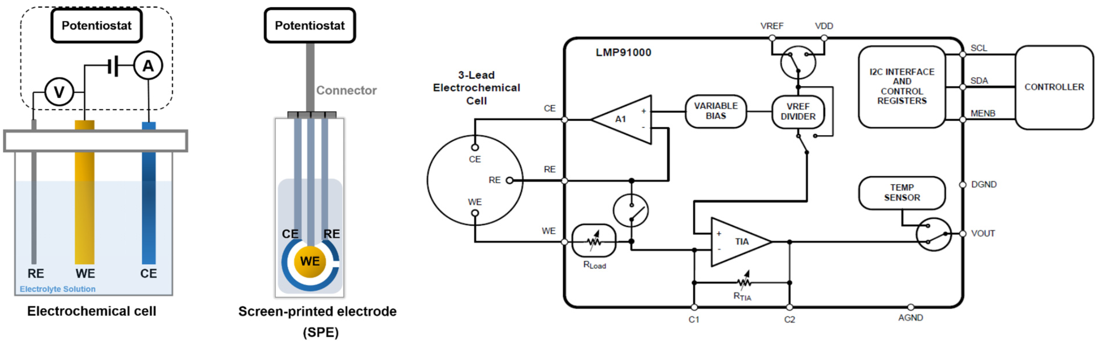
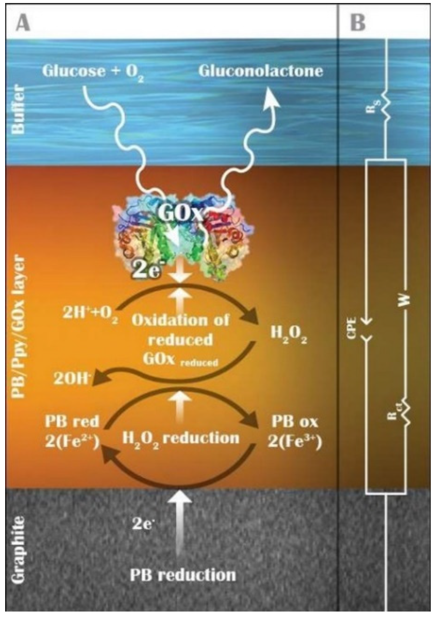
3. Electrochemical Detection of Glucose

In general, electrochemical biosensors are used to detect biological elements by utilizing biological receptors from enzymes or antibodies and transducing the biomolecular effect into an electrical signal in the form of a potential or current across electrodes [36]. Chemical species, such as molecular ions, are called electro-active species if, through the movement of the electrons, they can either be oxidized or reduced at an electrode’s surface [37]. Thus, amperometric sensors, in which a current is measured, are suitable for the detection of electroactive species involved in a biological or chemical recognition process [38].
If the reaction of glucose oxidizing with the glucose analyte is to be detected, then the measured signal is generated by an electrode whose potential is maintained at a constant value relevant to that of a reference electrode. At the same time, the current that results from the electro-active solute contacting the electrode is monitored. The applied potential drives the electron transfer reaction that involves the electroactive species in the solute, thus giving rise to the current representing the rate of the measured reaction. Therefore, the measured current is a direct function of the concentration of glucose changing over time.
The electrochemical method is suitable for compact sensing and wearable applications because of its simplicity, low power requirement, and compatibility with miniaturization [39]. In an electrochemical biosensor, the potentiostat is an essential device because it maintains the electrochemical stability of the biosensor and converts the biosensor’s output into an analog signal [40]. This method suffers from limitations in precision, i.e., the measured current depends on several other factors that are not always easily controlled.
Recently, single-chip potentiostats have been widely developed by researchers to reduce the chip’s size and cost [41,42,43,44,45]. Commercial single-chip potentiostats such as the LMP91000 [46], shown in Figure 2, are available. Some of these commercial single-chip potentiostats are also integrated with a built-in microcontroller, such as the ADuCM355 [47] and AD5940 [48], to facilitate a wide range of programmable functions.

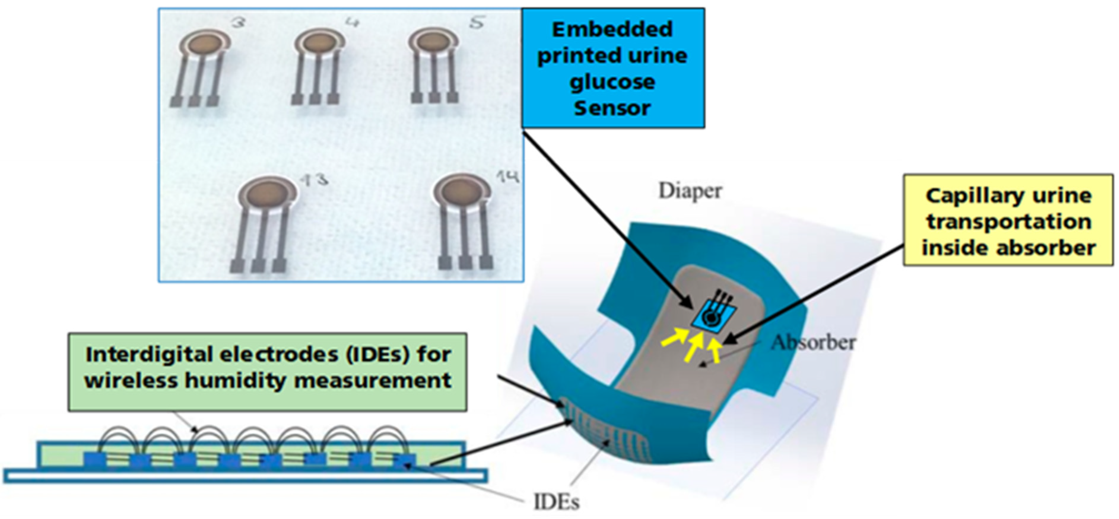
4. Embedded Urine Glucose Sensor

In order to be embedded into a diaper absorber, the sensor strip must be biocompatible and flexible. The microcapillary absorber guides the urine to the detection area with a three-electrode setup, as shown in Figure 3. At the functionalized working electrode, the glucose is enzymatically converted by glucose oxidizing to gluconolactone +H2O2, which oxidizes at the working electrode surface to H2O + O2 + 2e. These two electrons lead to the creation of a current that is directly proportional to the glucose concentration and is measured by the potentiostat in the readout module. This way enables the wetted sensor to measure the amount of glucose in urine and support care workers and physicians by detecting diabetes type II in its early stage and adjusting the correct medication doses of insulin for affected diabetic patients.

5. Readout and Data Acquisition Circuits

To allow medical staff to monitor the glucose level of the patient’s incontinence products in real-time and to understand how the patient’s condition changes, a medical remote monitoring system based on the Internet of Things (IoT) is an advantage. This system provides the possibility of connecting the sensing devices to cloud-based services, allowing extensive information processing for early diagnosis. The IoT medical remote monitoring system operates by establishing a wireless monitoring network in the hospital or patient’s home.
A direct connection between the sensors attached to the adult incontinence products and the read-out circuits can obtain data from the sensors locally without intermediaries. In this case, the user’s mobility and comfort can be considerably held up using wires. Moreover, there is a higher probability of system failure as well as an increase in the risk of medical staff infections due to direct contact with patients. Therefore, a wireless communication system using different protocols is needed to inform the care workers via Cloud Gateway about occurring events in patients and assist in planning the workflow. The goal of these applications is to guarantee continuous monitoring of the physiological status of each patient in real-time through the user management platform while giving them freedom of movement. Hence, medical efficiency will improve while staff will have more time to serve patients and work in a safer environment. Once the urination event occurs, the care workers get a notification for a diaper change, while vital data like glucose values in urine are measured automatically and stored in a cloud database for online diabetes treatment evaluation [50].
The collected physiological signals from the biosensors’ nodes need to be transmitted to central nodes, known as central hubs, and then the accumulative measurements need to be sent to remote medical centers. Due to limited storage and energy resources in the sensor node, the sensory data are regularly sent using short-range transmission to the central nodes [51]. The central nodes usually have large data storage and powerful network connectivity through radio frequency, WiFi, and Bluetooth interfaces, in addition to powerful microcontrollers [52]. Providing reliable data transmission between sensing devices with extremely low-power radios and central nodes in a noisy environment is very challenging. This requires considering different parameters when designing data communication protocols. These parameters include link quality estimation, time synchronization, collision avoidance, and mobility management. The data from the central nodes can be sent directly to the cloud using long-range communication. The cloud servers have massive storage capacity and high computational power that can be provided for such applications. They can also assist with access to common resources in a persistent manner, offering a large number of online on-demand services [53]. A schematic diagram for the integrated system is shown in Figure 4.
Specialists can keep track of the patients’ data through the user management system. In addition, patients who are active outside their homes can transmit information to the care unit after joining the network. When the person under observation moves beyond range, data can be recorded in the sensor’s node local memory and communicated later. GPS can also be integrated into the sensor nodes to help the hospital staff locate remote patients in an emergency [54]. Furthermore, cloud-based systems sustain remote software updates, which enable quick and cost-effective system maintenance. Given its versatility and adaptability, this device concept is expected to provide a leap toward the future of wearable, disposable, easy-to-use, automated, and economical diagnostic tools.

6. Fabrication of Printed Urine Glucose Sensor

For the fabrication of the urine glucose sensor, various printed electronics technologies and advanced materials were applied and combined for the best sensor performance in the prescribed working range.

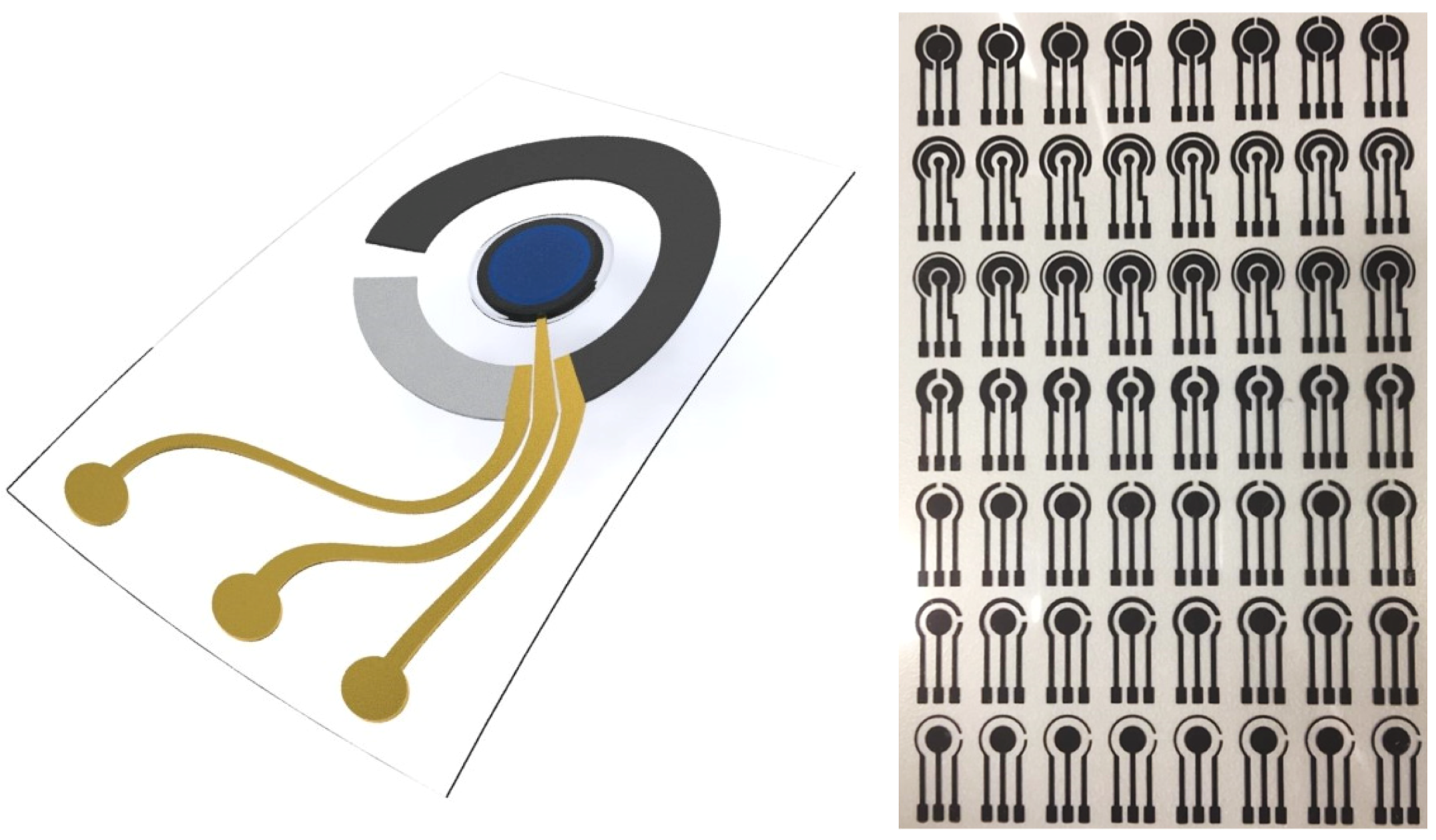
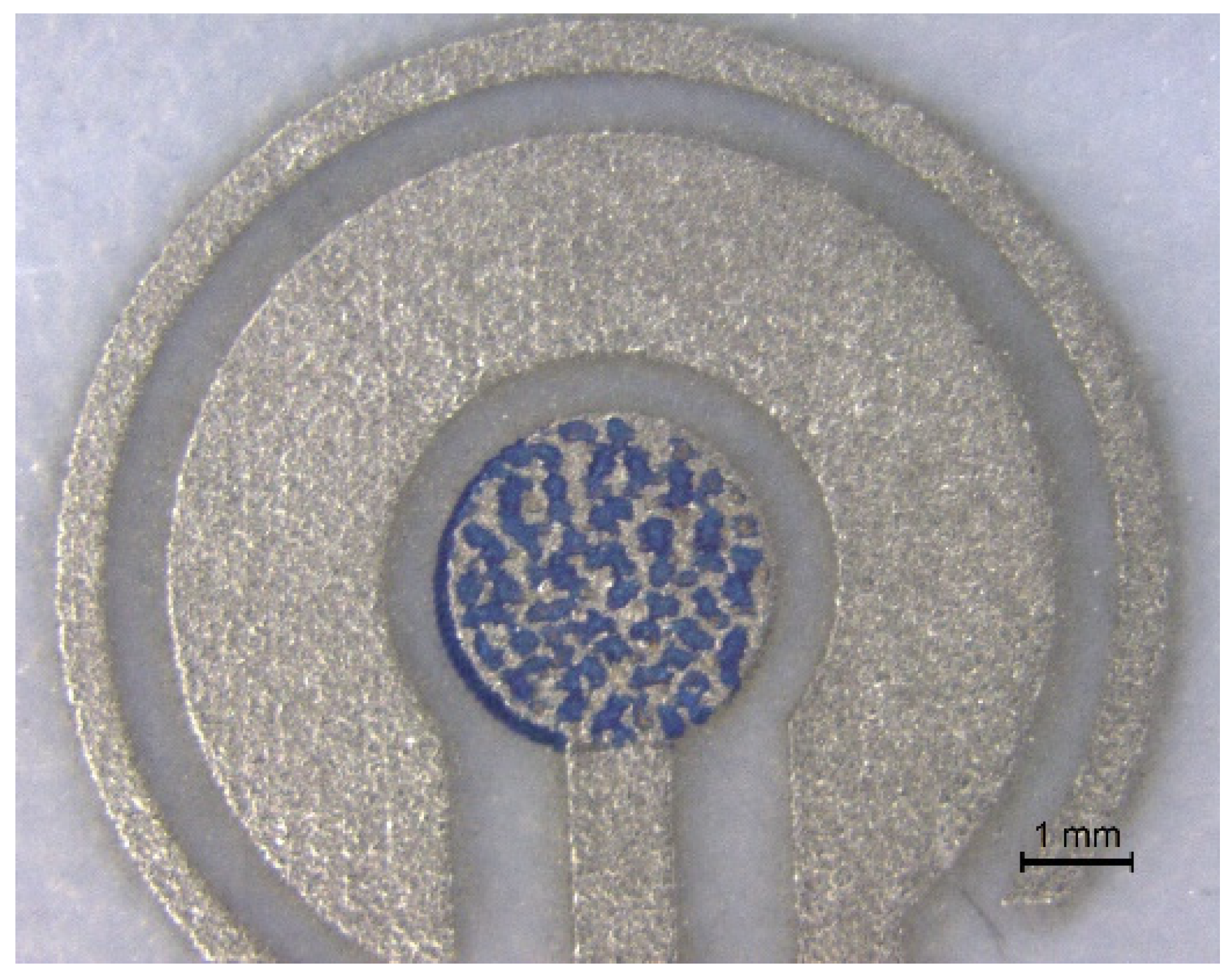
6.1. Screen-Printed Electrodes

First, the three-electrode layer of the electrochemical sensor was screen-printed in various designs using the semi-automated screen printer DEK 248 (from DEK Printing Machines Limited, Dorset, England), as shown in Figure 5. A carbon-graphene paste (C2171023D1 from Sun Chemical Gwent) was optimized for the best electrochemical performance and printed on a PET foil. The nanocoated PET foil Arcophane (TCA 10 2F 125µ STS from Normandy Coating, Arques La Bataille, France) showed the best results regarding the conductivity and adhesion of the screen-printed electrodes after curing for 30 min at 130 °C. The passivation with screen-printed transparent UV curing isolation paste (Bectron DP 84440 from ELANTAS GmbH, Wesel, Germany) is used to insulate and define the active area from the contact pads. The screen-printed electrodes on the flexible substrate showed high conductivity and electrochemical stability and ensured a seamless integration into diapers with the necessary elasticity.

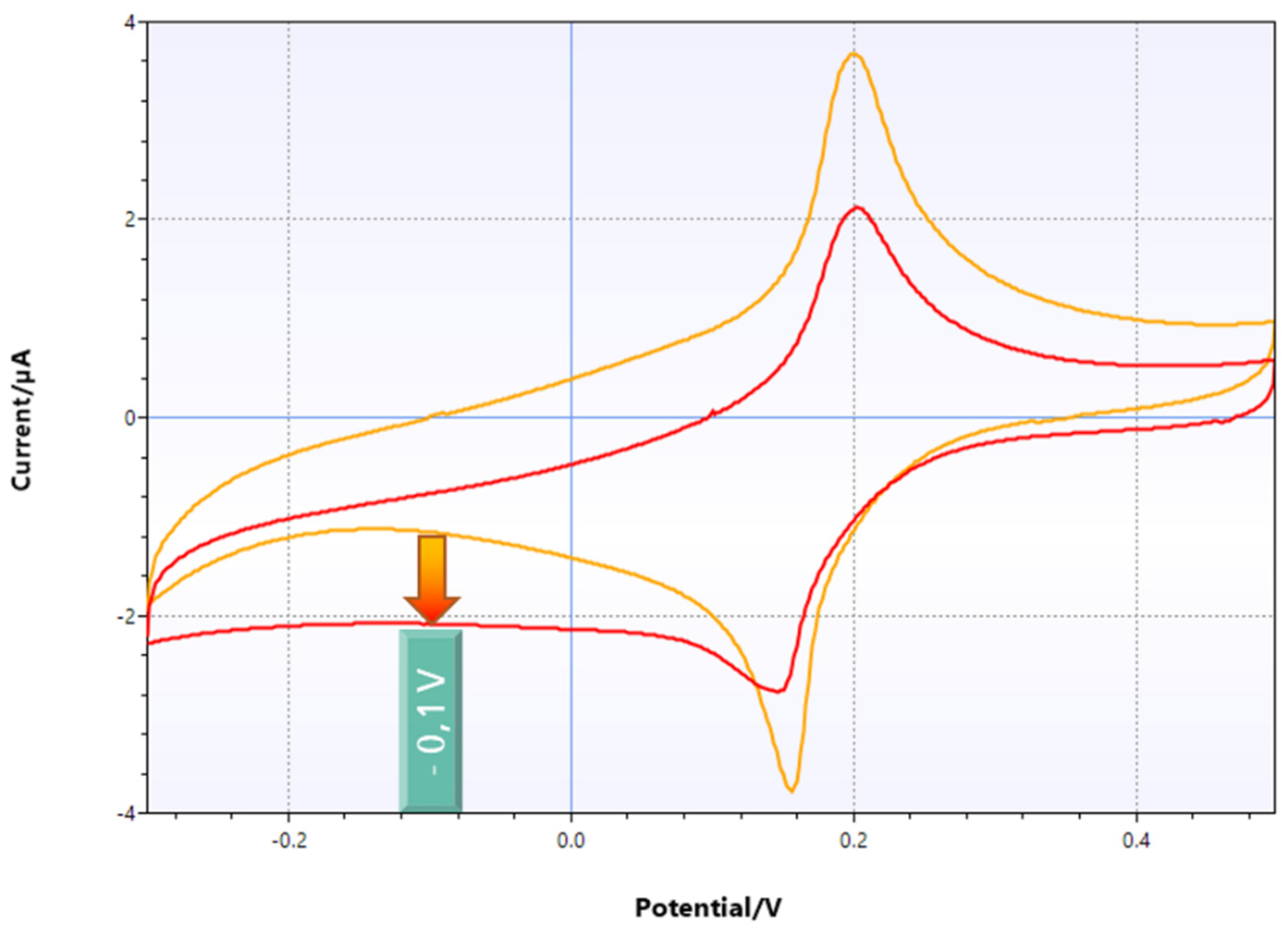
6.2. Enhancement of Sensor Performance Using a Printed Mediator

In order to increase the electron transfer and avoid using expensive materials, such as platin, for printing the electrodes, an additional mediator was added in the layer formation, which built the so-called second-generation glucose sensors. One potential candidate mediator is the inorganic pigment Prussian blue, which is especially used for wearable sensors as it is insoluble in water and therefore practically non-toxic for the human body. The redox mediator offers higher sensitivity at a lower applied working voltage of ~0.05 V, which avoids oxidation of interference substances like uric acid. Prussian blue deposited on the electrode surface acts as a catalyst for the oxidation of the hydrogen peroxide with two electrons per cycle. The oxidized Prussian blue uptakes the two reduction electrons from the working electrode, resulting in a negative current, as shown in Figure 6.
Before functionalizing the working electrode, an electron mediator layer was inkjet-printed. The ink was mixed with a 1% water-based solution of Prussian blue nanoparticles and filled in the inkjet cartridge. The Prussian blue nanoparticles are synthesized from 0.05 M K3Fe(CN)6 and 0.05 M FeCl3 in DI-water [56] and filtered with a 0.2 µm pore filter. Inkjet printing of several layers of Prussian blue on top of the polished working electrode was performed using Dimatix DMP 2850 (from Fujifilm Dimatix, Inc., Lebanon, PA, USA) and dried for 15 min at 90 °C. The thickness of the printed Prussian blue layers was optimized for fast response time, good conductivity, and reproducibility. Due to the hydrophobic nature of the graphite electrode surface upon curing, the Prussian blue nanoparticles form clusters, as shown in Figure 7, which are beneficial for the conductivity of the electrode as the Prussian blue pigment itself is insulating. Five layers of inkjet-printed Prussian blue offered enough mediator particles for the oxidation of H2O2, with a high conductivity and short electron distance between mediator and electrode, and hence a fast response time.

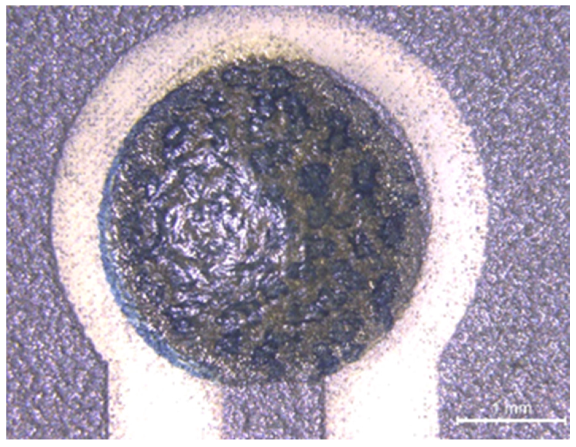
6.3. Biofunctionalization via Drop Casting

A barrier membrane on top of the mediator had to be applied to prevent the mediator from bleeding out and block the interference substances, such as uric acid, from oxidizing at the electrode surface. Chitosan, as a biopolymer and double-functional membrane, is permeable to H2O2 but impermeable to larger molecules such as uric acid from entering or Prussian blue pigment from leaving the electrode surface. An aliquot of 0.2 µL (0.2%) of chitosan in acetic acid was drop-cast on top of the working electrode and naturally dried. It was then washed with DI-water to remove the acid residuals and dried once more [57].
The following step was to mix the enzyme glucose oxidase (from Aspergillus niger) with glutaraldehyde at a ratio of 4:1. Following the instructions for glucose sensing activation solution (ZP1000979 from Zimmer & Peacock AS, Horten, Norway), 2 µL of the solution was dispensed via pipette on top of chitosan before the cross-linker jellified and immobilized the enzyme, as shown in Figure 8. The enzyme-modified electrode could react with glucose at relatively high concentrations of several mMol/L. This reaction causes a change in the current flowing between the working and counter electrodes that is proportional to the concentration of glucose.

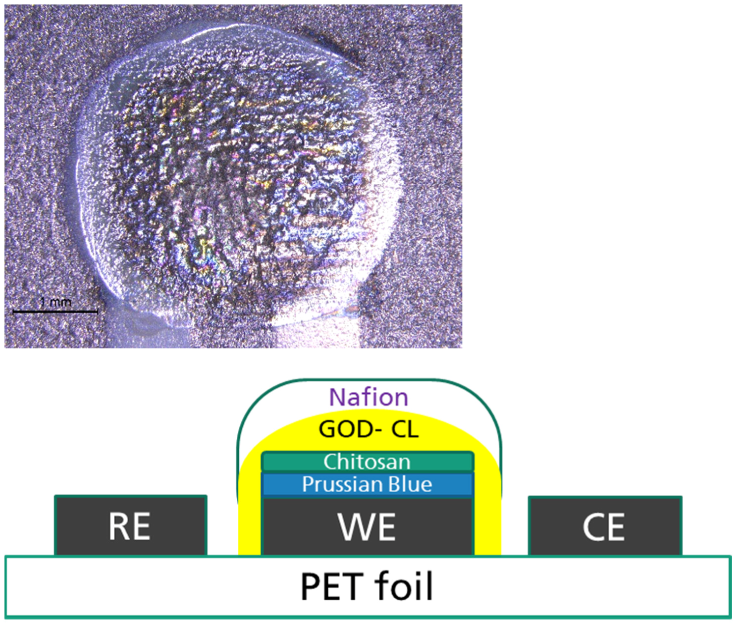
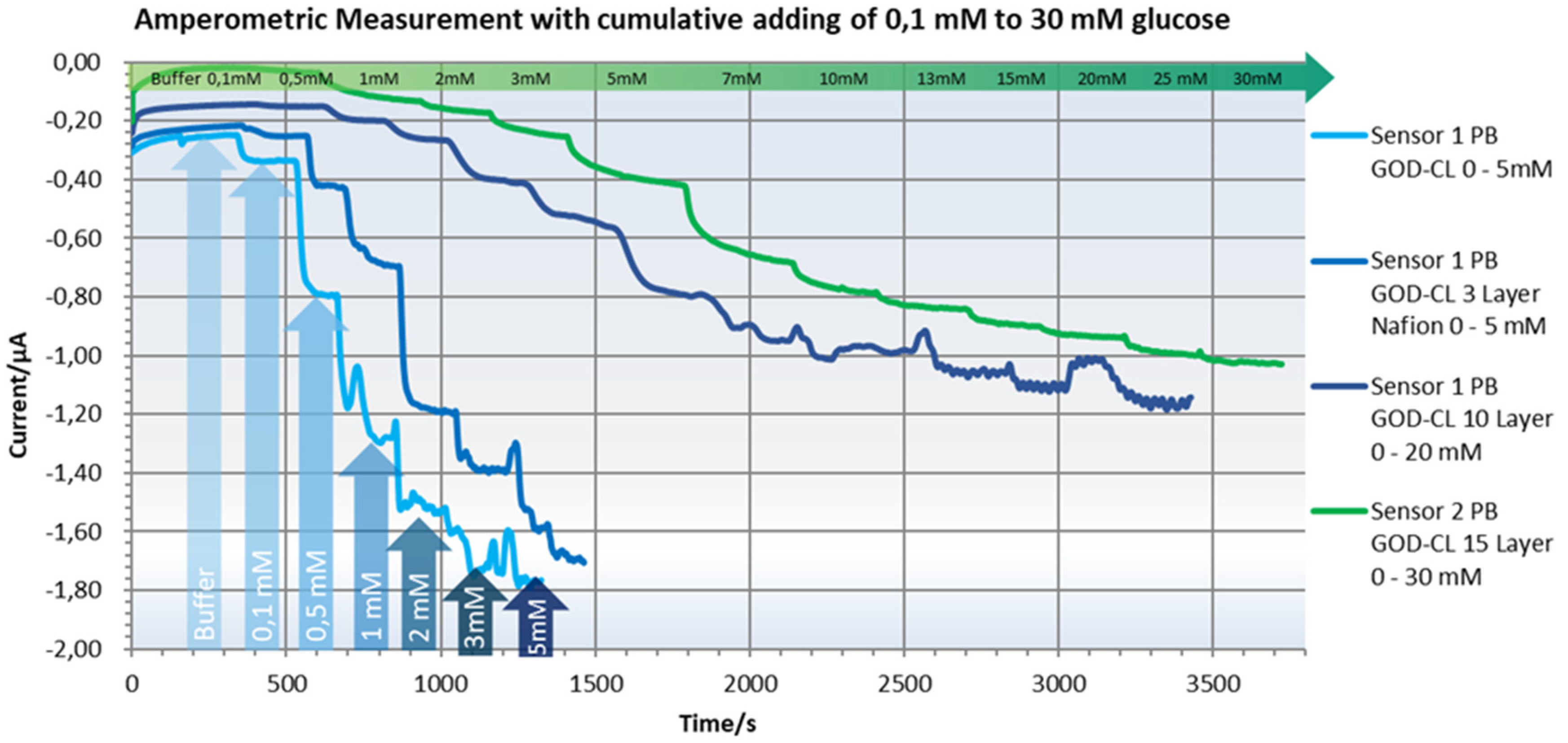
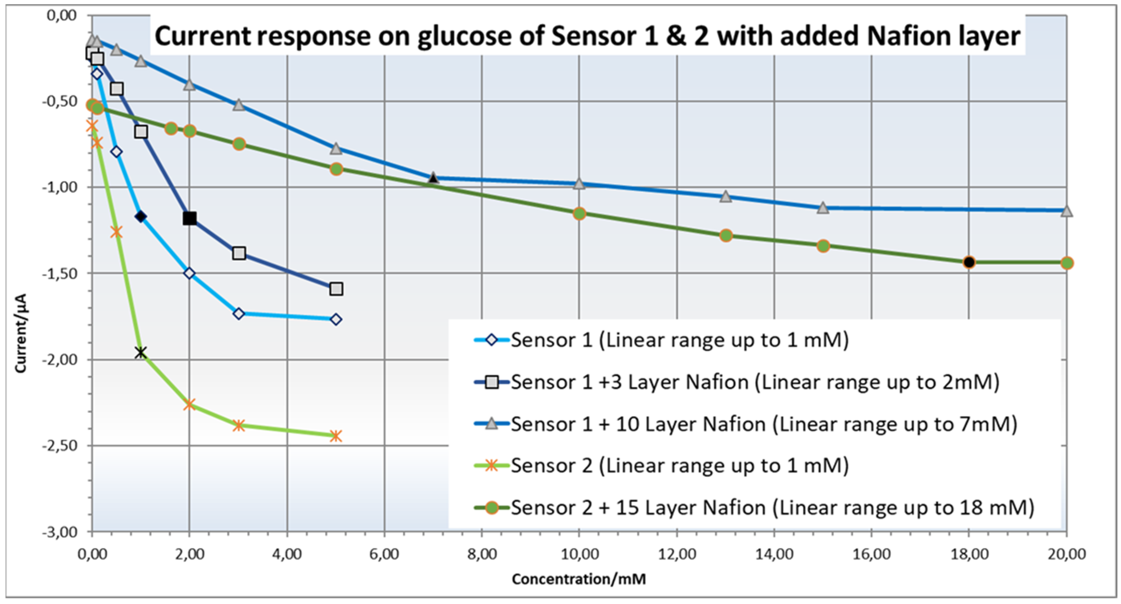
6.4. Printing of a Barrier Membrane against Interfering Substances and Working Range Adjustment

Despite its high sensitivity, glucose detection in urine is subject to severe interferences due to the low concentration of the bioanalyte at 2.2 mM. For example, uric acid and ascorbic acid have oxidation potentials within the operation potential of the glucose sensor at 0.35 V and thus can be oxidized by the enzyme-modified electrode [58]. The blocking of endogenous interference substances in urine as well as enhancing the electron transfer are the major difficulties in the electrochemical detection of glucose, as they cause imprecise results. Interfering species are the largest barriers to shifting biosensors from the laboratory to the field, as real biofluids contain hundreds of different components that influence each other.
In order to overcome the interference, cation exchange membranes are made of materials like chitosan and Nafion, and they typically coat the tops of enzyme-modified electrodes to act as an effective permiselective barrier [59]. This membrane prevents negatively charged species from reaching the electrode surface [60], among which the sensor is most sensitive to uric acid. Eliminating this interference comes at the cost of sensitivity and speed but is crucial for field applications.
As the bare sensor is highly sensitive to glucose in low concentrations (from 0.1 mM to 1 mM with a linear range), its working range also has to be adjusted to measure higher glucose concentrations in urine. Therefore, a Nafion membrane is printed on top of the biosensor to shift and extend the linear working range at the cost of signal strength. This was easily adapted by inkjet-printing multiple layers of Nafion 117 (1% in water; Sigma-Aldrich, Merck KGaA, Darmstadt, Germany) on top of the working electrode. This is shown in Figure 9, along with the final cross section of the manufactured glucose sensor.

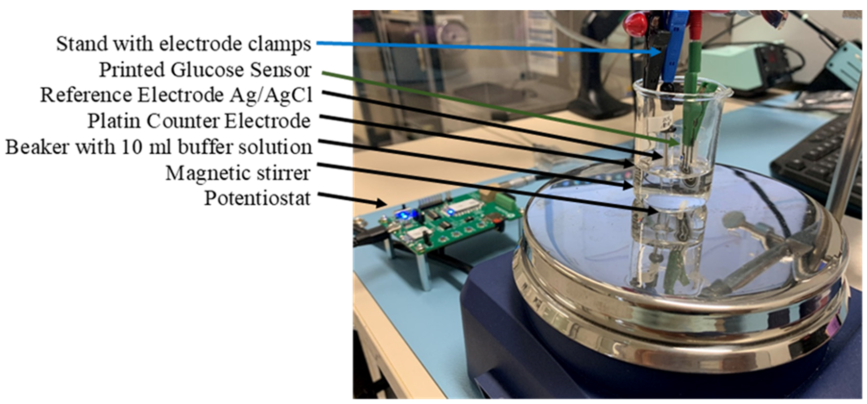
7. Experimental Setup

The following set-up was installed in order to evaluate the printed sensor performance, as shown in Figure 10. As a basis buffer, the acidic phosphate buffer K2HPO4 is used, which has a pH level of 5.5 that is comparable to urine. The pH level of the basis buffer is critical, as buffers with higher pH levels, such as PBS, which has a pH of 7.4, can cause Prussian blue to dissolve and the mediator to bleed out of the working electrode, resulting in a constantly attenuated measured signal.
A 10 mL buffer solution of 0.05 M K2HPO4 and 0.1 M KCl is stirred in a beaker with a magnetic stirrer to ensure a controlled mass transfer measurement and constant current plateaus during amperometric measurements. External electrodes were used to have identical measurement conditions, which avoided measurement errors due to the varying printing and modification results of the internal reference and counter electrodes. Therefore, a platinized wire was used as a counter electrode and an Ag/AgCl Re-1S from ALS Co., Ltd., as a reference electrode. The potentiostat measurements were performed with the EmStat Pico Development Kit with Software PSTrace 5 from PalmSense.

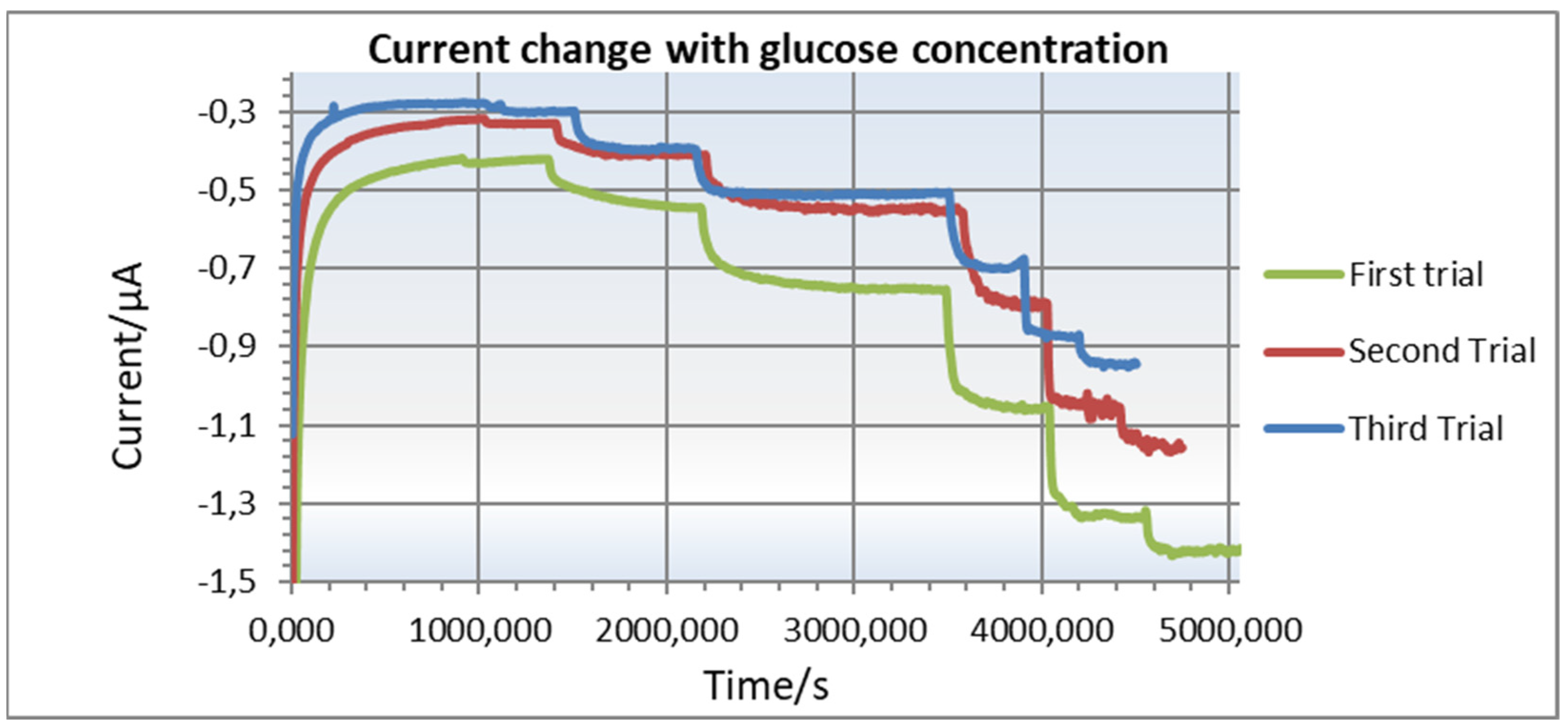
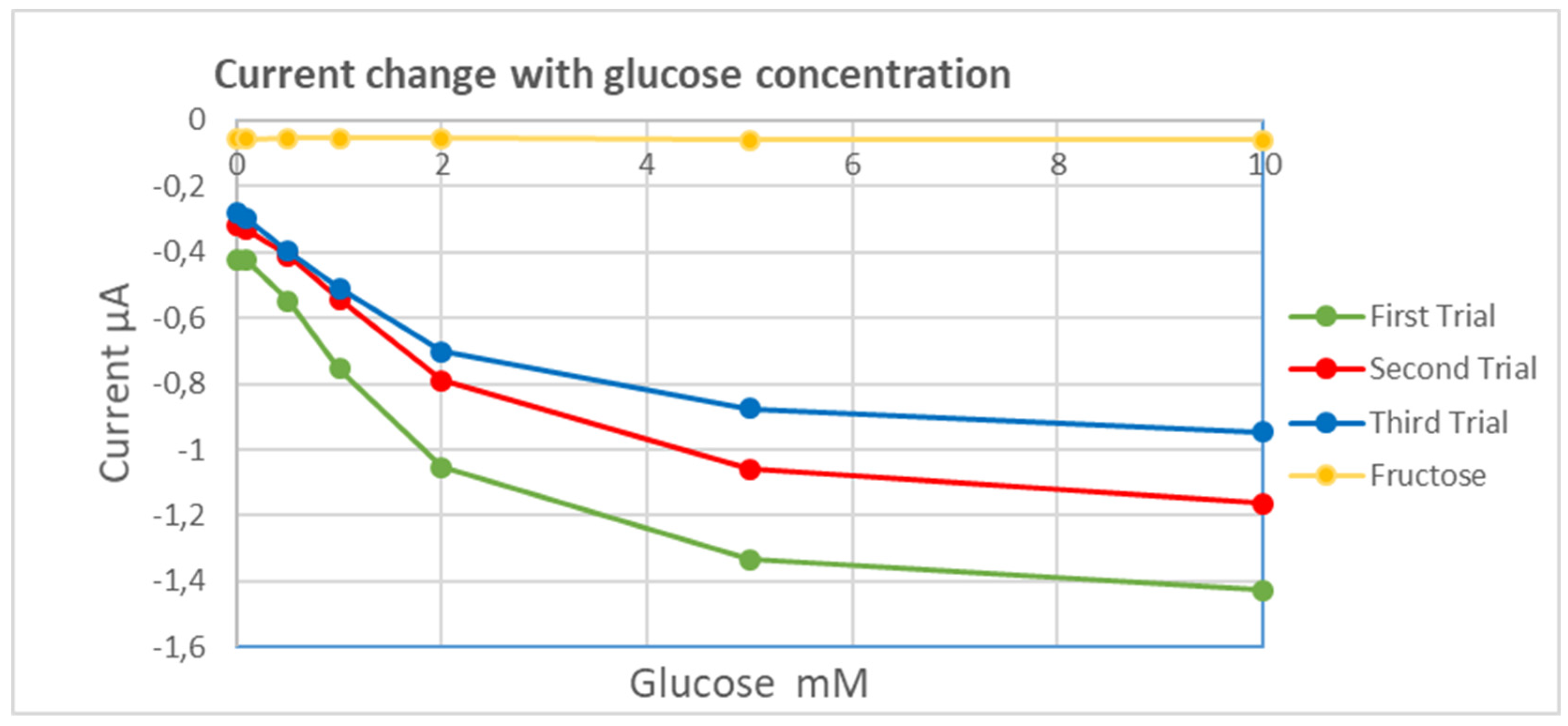
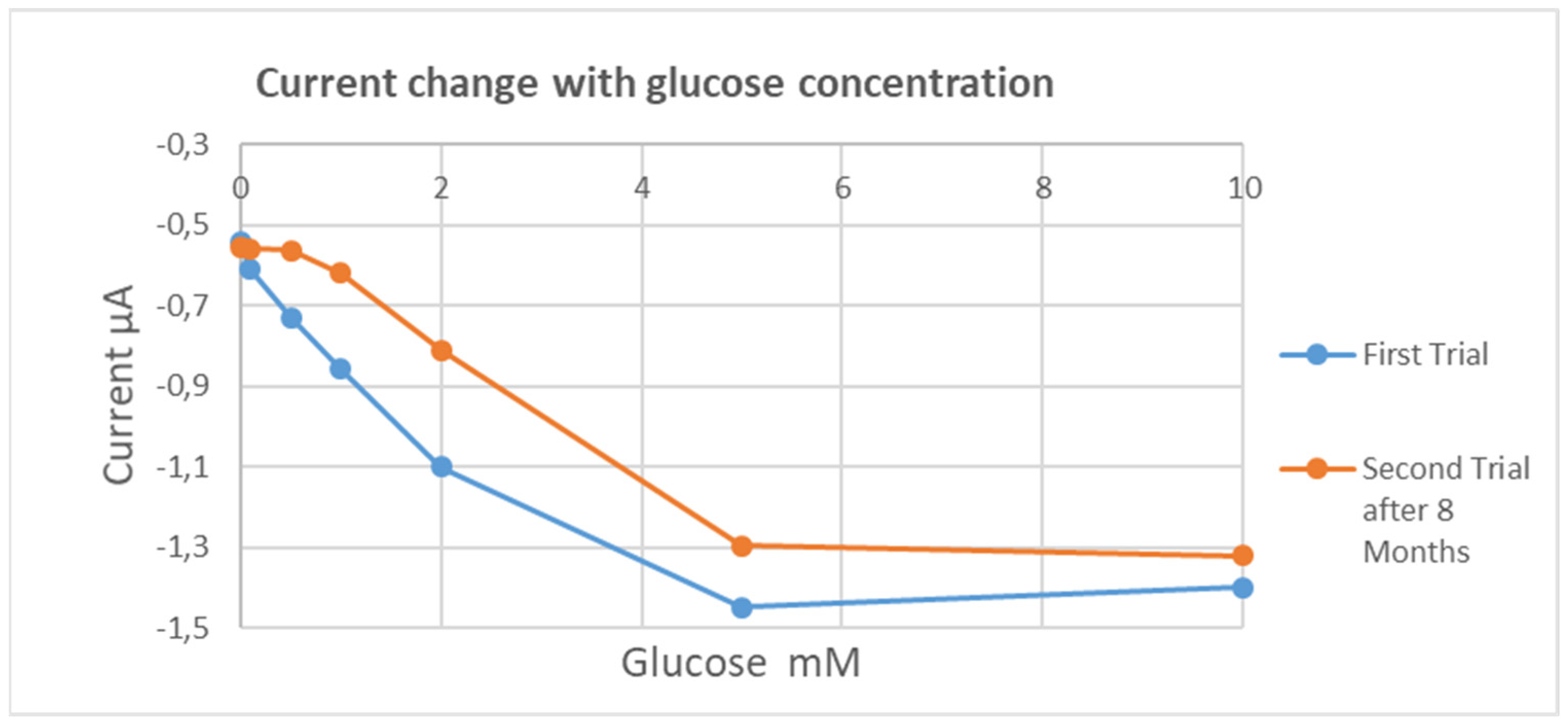
8. Experimental Results

To simulate variations of glucose in physiological urine, different concentrations of glucose were prepared and measured using the printed sensor. The total volume of the mediator in the well around the sensor is 100 μL. Different concentrations of glucose were added to this solution at a 1:20 to 1:5 ratio of the total volume. The glucose concentration varied between 0.1 mM and 30 mM.
Figure 11 shows the cyclic voltammetry (CV) curves of the biofunctionalized printed electrode without a barrier layer, as described in 6.3, from −0.3 V to 0.5 V with a scan rate of 0.01 V/s in buffer (orange) and with 5 mM concentrations of glucose (red). The maximum increase in current at low voltage was measured at −0.1 V, marked as an arrow in the figure, and was therefore chosen as a reference applied voltage for the amperometric measurement.
In order to evaluate the sensor performance, the current changes in real-time were measured using Emstat-pico-development, a poteniostat, and a data acquisition kit [60] (from PalmSens BV, Houten, The Netherlands). This development kit is merely based on the ADuCM355, a cost-effective microcontroller that has a built-in potentiostat and nanoampere current amplifier. Cumulative concentrations of glucose were added to the solution, which resulted in a significant increase in the anodic current, confirming the effective immobilization of GOD and its reaction with glucose. Next, various layers of Nafion were printed, as described in 6.4, to obtain a linear sensor response within the necessary measuring range to detect urine glucose levels from 2 mM up to several dozen mM, as shown in Figure 12.
Fifteen layers of Nafion extend the linear range from 1 mM up to 18 mM. At the same time, the Nafion barrier prevents large interference substances from diffusing into the electrode and distorting the electrical signal.
Plotting the glucose concentration versus current in Figure 13 shows the sensitive but short linear range without barrier membrane and the wide linear range up to 18 mM glucose with sufficient current at a maximum of 1 µA with barrier membrane.
The repeatable sensor performance of 15 layers of Nafion was investigated. For concentrations between 0.1 mM and 2 mM, the current increases almost linearly, and the sensor reaches a plateau after the introduction of 10 mM of glucose, as shown in Figure 14. This agrees well with the results obtained from the commercially available sensors, BVT AC1.GOD [19]. A similar reported printed sensor used to detect glucose in saliva also has similar output current linearity but with less sensitivity [57].
The sensitivity of the sensor towards fructose was also tested, which shows very little change in the sensor’s output current with increasing fructose concentration, as shown in Figure 15.
To evaluate the shelf life of the printed sensors, the performances of devices were stored for different periods of time until fabrications were tested. A sample was kept for 8 months before it was retested. The results of these tests are shown in Figure 16. The shelf life of the devices is usually limited by the immobilization conditions of the enzymes that govern their stability. Once the devices are printed, they can be stored in sealed vacuum bags at room temperature. No significant improvement in shelf life was reported when the sensors were kept in the fridge at 4 °C [57]. Note that the interference effects of the other possible molecules that might be present in urine are not addressed in this study.

9. Conclusions

With the development of the flexible printed glucose sensor by means of printed electronics technology and its adaptation of its working range to the relevant glucose concentrations in urine, it is possible to extend our incontinence sensor system to glucosuria detection. The electrochemical testing of the biosensor shows good responsiveness to pathological urine glucose levels, which can enable noninvasive early detection of elderly-related diabetes type II or control of sufficient insulin medication in incontinent diabetes patients. This second-generation glucose sensor technology is well established and can be manufactured and embedded as a single-use biosensor in a cost-effective way. Together with a wearable readout module of the incontinence sensor system and wireless communication technology, an IoT medical remote monitoring system can be the basis for the digitalization of retirement homes. Finally, these printed sensors can cover a variety of other biorecognition elements by simply changing the enzyme to achieve multifunctionality and target other applications too. Further research to achieve higher sensitivity and selectivity of urinary glucose sensors and to overcome the incurred limitations is still being pursued.

Author Contributions

Conceptualization, H.D.N., M.H. and R.M.A.; methodology, H.D.N., M.H. and R.M.A.; software, R.M.A.; validation, M.H., R.K. and B.W.; formal analysis, M.H.; investigation, H.D.N.; resources, H.D.N.; data curation, M.H. and R.M.A.; writing—original draft preparation, M.H.; writing—review and editing, H.D.N., M.H. and R.M.A.; visualization, M.H. and R.M.A.; supervision, H.D.N.; project administration, H.D.N.; funding acquisition, H.D.N. and R.M.A. All authors have read and agreed to the published version of the manuscript.

Funding

This research was funded by the Institut für angewandte Forschung Berlin (IFAF) and Deutsche Forschungsgesellschaft (DFG) for international cooperation and the Ministry of Education in Saudi Arabia through a Taibah University grant (442/234).

Institutional Review Board Statement

Not applicable.

Informed Consent Statement

Not applicable.

Data Availability Statement

Not applicable.

Conflicts of Interest

The authors declare no conflict of interest.

References

  1. Guariguata, L.; Whiting, D.R.; Hambleton, I.; Beagley, J.; Linnenkamp, U.; Shaw, J.E. Global estimates of diabetes prevalence for 2013 and projections for 2035. Diabetes Res. Clin. Pract. 2014, 103, 137–149. [Google Scholar] [CrossRef]
  2. Tamayo, T.; Brinks, R.; Hoyer, A.; Kuss, O.S.; Rathmann, W. The Prevalence and Incidence of Diabetes in Germany. Dtsch. Arztebl. Int. 2016, 113, 177–182. [Google Scholar] [CrossRef] [PubMed]
  3. Ortiz-Martínez, M.; González-González, M.; Martagón, A.J.; Hlavinka, V.; Willson, R.C.; Rito-Palomares, M. Recent Developments in Biomarkers for Diagnosis and Screening of Type 2 Diabetes Mellitus. Curr. Diab. Rep. 2022, 22, 95–115. [Google Scholar] [CrossRef]
  4. Shitanda, I.; Fujimura, Y.; Nohara, S.; Hoshi, Y.; Itagaki, M.; Tsujimura, S.J. Paper-Based Disk-Type Self-Powered Glucose Biosensor Based on Screen-Printed Biofuel Cell Array. Electrochem. Soc. 2019, 166, B1063–B1068. [Google Scholar] [CrossRef]
  5. Available online: https://www.labce.com/spg506346_clinical_significance_of_glucose_in_the_urine.aspx (accessed on 19 March 2023).
  6. Ngo, H.-D.; Bäuscher, M.; Schiffer, M.; Medinaa, D.M.; Jorcano, J.L.; Acedo, P.; Atta, R.M.H. Development, Simulation and Characterization of a novel Incontinence Sensor System using 2D-Printing Technology with Conductive Polymer PEDOT:PSS. In Printed and Flexible Sensor Technology: Fabrication and Applications; Hg. von Institute of Physics IOP: London, UK, 2021; pp. 1–16. [Google Scholar]
  7. Bäuscher, M.; Wang, B.; Hu, X.; Mackowiak, P.; Merchau, N.; Ehrmann, O.; Schneider-Ramelow, M.; Lang, K.D.; Ngo, H.D. Simulation And Electrical Characterization of A Novel 2D-Printed Incontinence Sensor With Conductive Polymer PEDOT:PSS For Medical Applications. In Proceedings of the 2018 IEEE 20th Electronics Packaging Technology Conference (EPTC), Singapore, 4–7 December 2018; pp. 100–104. [Google Scholar]
  8. Ngo, H.D.; Hoang, T.H.; Baeuscher, M.; Wang, B.; Mackowiak, P.; Grabbert, N.; Weiland, T.; Ehrmann, O.; Lang, K.D.; Schneider-Ramelow, M.; et al. A Novel Low Cost Wireless Incontinence Sensor System (Screen-Printed Flexible Sensor System) for Wireless Urine Detection in Incontinence Materials. Proceedings 2018, 2, 716. [Google Scholar]
  9. Peng, Z.; Xie, X.; Tan, Q.; Kang, H.; Cui, J.; Zhang, X.; Li, W.; Feng, G. Blood glucose sensors and recent advances: A review. J. Innov. Opt. Health Sci. 2022, 15, 2230003. [Google Scholar] [CrossRef]
  10. Villena Gonzales, W.; Mobashsher, A.T.; Abbosh, A. The Progress of Glucose Monitoring—A Review of Invasive to Minimally and Non-Invasive Techniques, Devices and Sensors. Sensors 2019, 19, 800. [Google Scholar] [CrossRef] [PubMed]
  11. Naresh, V.; Lee, N. A Review on Biosensors and Recent Development of Nanostructured Materials-Enabled Biosensors. Sensors 2021, 21, 1109. [Google Scholar] [CrossRef]
  12. Wang, J. Carbon-nanotube based electrochemical biosensors: A review. Electroanalysis 2005, 17, 7–14. [Google Scholar] [CrossRef]
  13. Grieshaber, D.; MacKenzie, R.; Voeroes, J.; Reimhult, E. Electrochemical biosensors-sensor principles and architectures. Sensors 2008, 8, 1400–1458. [Google Scholar] [CrossRef]
  14. Teymourian, H.; Barfidokht, A.; Wang, J. Electrochemical glucose sensors in diabetes management: An updated review (2010–2020). Chem. Soc. Rev. 2020, 49, 7671–7709. [Google Scholar] [CrossRef] [PubMed]
  15. Wu, P.; Shao, Q.; Hu, Y.; Jin, J.; Yin, Y.; Zhang, H.; Cai, C. Direct electrochemistry of glucose oxidase assembled on graphene and application to glucose detection. Electrochim. Acta 2010, 55, 8606–8614. [Google Scholar] [CrossRef]
  16. Gao, W.; Zhou, X.; Heinig, N.F.; Thomas, J.P.; Zhang, L.; Leung, K.T. Nonenzymatic Saliva-Range Glucose Sensing Using Electrodeposited Cuprous Oxide Nanocubes on a Graphene Strip. ACS Appl. Nano Mater. 2021, 4, 4790–4799. [Google Scholar] [CrossRef]
  17. Jalil, A.T.; Ashfaq, S.; Bokov, D.O.; Alanazi, A.M.; Hachem, K.; Suksatan, W.; Sillanpää, M. High-Sensitivity Biosensor Based on Glass Resonance PhC Cavities for Detection of Blood Component and Glucose Concentration in Human Urine. Coatings 2021, 11, 1555. [Google Scholar] [CrossRef]
  18. Reddy, V.S.; Agarwal, B.; Ye, Z.; Zhang, C.; Roy, K.; Chinnappan, A.; Narayan, R.J.; Ramakrishna, S.; Ghosh, R. Recent Advancement in Biofluid-Based Glucose Sensors Using Invasive, Minimally Invasive, and Non-Invasive Technologies: A Review. Nanomaterials 2022, 12, 1082. [Google Scholar] [CrossRef]
  19. Redon, P.; Shahzad, A.; Iqbal, T.; Wijns, W. Development of a New Detection Algorithm to Identify Acute Coronary Syndrome Using Electrochemical Biosensors for Real-World Long-Term Monitoring. Bioengineering 2021, 8, 28. [Google Scholar] [CrossRef]
  20. Available online: https://diagnostics.roche.com/global/en/products/instruments/combur-chemstrip-ins-656.html (accessed on 19 March 2023).
  21. Available online: http://main.diabetes.org/dforg/pdfs/archive/2012-07-anatomy-of-a-test-strip.pdf (accessed on 19 March 2023).
  22. Kim, H.Y.; Jang, K.J.; Veerapandian, M.; Kim, H.C.; Seo, Y.T.; Lee, K.N.; Lee, M.-H. Reusable urine glucose sensor based on functionalized graphene oxide conjugated Au electrode with protective layers. Biotechnol. Rep. 2014, 3, 49–53. [Google Scholar] [CrossRef]
  23. Wang, H.-C.; Lee, A.-R. Recent developments in blood glucose sensors. J. Food Drug Anal. 2015, 2, 191–200. [Google Scholar] [CrossRef]
  24. Abikshyeet, P.; Ramesh, V.; Oza, N. Glucose estimation in the salivary secretion of diabetes mellitus patients. Diabetes Metab. Syndr. Obes. 2009, 5, 149–154. [Google Scholar]
  25. Kumar, S.; Padmashree, S.; Jayalekshmi, R. Correlation of salivary glucose, blood glucose and oral candidal carriage in the saliva of type 2 diabetics: A case–control study. Contemp. Clin. Dent. 2014, 5, 312. [Google Scholar] [CrossRef]
  26. Nayak, M.T.; Gupta, S.; Sunitha, J.; Dawar, G.; Sinha, N.; Rallan, N.S. Correlation of salivary glucose level with blood glucose level in diabetes mellitus. J. Oral Maxillofac. Pathol. 2017, 21, 334–339. [Google Scholar] [CrossRef]
  27. Naing, C.; Mak, J.W. Salivary glucose in monitoring glycaemia in patients with type 1 diabetes mellitus: A systematic review. J. Diabetes Metab. Disord. 2017, 16, 2251–6581. [Google Scholar] [CrossRef]
  28. Go, A.; Kim, H.T.; Park, Y.J.; Park, S.R.; Lee, M.-H. Fabrication of Repeatedly Usable Pt-Electrode Chip Coated with Solidified Glucose Oxidase and Ascorbate Oxidase for the Quantification of Glucose in Urine. IEEE Sens. Lett. 2019, 3, 1500104. [Google Scholar] [CrossRef]
  29. Pezhhan, H.; Akhond, M.; Shamsipur, M. A novel nanoplatform encapsulating glucose oxidase for spectrophotometric biosensing of hydrogen peroxide and glucose. Anal. Methods 2020, 12, 345–357. [Google Scholar] [CrossRef]
  30. Moya, A.; Gabriel, G.; Villa, R.; del Campo, F.J. Inkjet-printed electrochemical sensors. Curr. Opin. Electrochem. 2017, 3, 29–39. [Google Scholar] [CrossRef]
  31. Jeong, G.; Park, S. Review of urinary continence care products using sensor technology to improve effectiveness. Proc. Inst. Mech. Eng. Part H J. Eng. Med. 2019, 233, 91–99. [Google Scholar] [CrossRef]
  32. Yang, C.; Feng, W.; Li, Y.; Tian, X.; Zhou, Z.; Lu, L.; Nie, Y. A promising method for diabetes early diagnosis via sensitive detection of urine glucose by Fe Pd/rGO. Dye. Pigment. 2019, 164, 20–26. [Google Scholar] [CrossRef]
  33. Mohammadifar, M.; Tahernia, M.; Choi, S. An Equipment-Free, Paper-Based Electrochemical Sensor for Visual Monitoring of Glucose Levels in Urine. SLAS Technol. Transl. Life Sci. Innov. 2019, 24, 499–505. [Google Scholar] [CrossRef] [PubMed]
  34. Tekcin, M.; Sayar, E.; Yalcin, M.K.; Bahadir, S.K. Wearable and Flexible Humidity Sensor Integrated to Disposable Diapers for Wetness Monitoring and Urinary Incontinence. Electronics 2022, 11, 1025. [Google Scholar] [CrossRef]
  35. Hassan, M.H.; Vyas, C.; Grieve, B.; Bartolo, P. Recent Advances in Enzymatic and Non-Enzymatic Electrochemical Glucose Sensing. Sensors 2021, 21, 4672. [Google Scholar] [CrossRef]
  36. Angelov, G.V.; Nikolakov, D.P.; Ruskova, I.N.; Gieva, E.E.; Spasova, M.L. Healthcare Sensing and Monitoring. In Enhanced Living Environments; Lecture Notes in Computer, Science; Ganchev, I., Garcia, N., Dobre, C., Mavromoustakis, C., Goleva, R., Eds.; Springer: Cham, Switzerland, 2019; p. 11369. [Google Scholar] [CrossRef]
  37. Barry, R.C.; Lin, Y.H.; Wang, J.; Liu, G.D.; Timchalk, C.A. Nanotechnology-based electrochemical sensors for biomonitoring chemical exposures. J. Expo. Sci. Environ. Epidemiol. 2009, 19, 1–18. [Google Scholar] [CrossRef] [PubMed]
  38. Dey, R.S.; Bera, R.K.; Raj, C.R. Nanomaterial-based functional scaffolds for amperometric sensing of bioanalytes. Anal. Bioanal. Chem. 2013, 405, 3431–3448. [Google Scholar] [CrossRef] [PubMed]
  39. Putzbach, W.; Ronkainen, N.J. Immobilization Techniques in the Fabrication of Nanomaterial-Based Electrochemical Biosensors: A Review. Sensors 2013, 13, 4811–4840. [Google Scholar] [CrossRef] [PubMed]
  40. Levine, P.M.; Gong, P.; Levicky, R.; Shepard, K.L. Active CMOS sensor array for electrochemical biomolecular detection. IEEE J. Solid-State Circuits 2008, 43, 1859–1871. [Google Scholar] [CrossRef]
  41. Turner, R.; Harrison, D.; Baltes, H. A CMOS Potentiostat for Amperometric Chemical Sensors. IEEE J. Solid-State Circuits 1987, 22, 473–478. [Google Scholar] [CrossRef]
  42. Kakerow, R.; Kappert, H.; Spiegel, E.; Manoli, Y. Low Power Single Chip CMOS Potentiostat. In Proceedings of the International Solid-State Sensors and Actuators Conference-TRANSDUCERS’95, Eurosensors IX, Stockholm, Sweden, 25–29 June 1995; Volume 1, pp. 142–145. [Google Scholar]
  43. Bandyopadhyay, A.; Mulliken, G.; Cauwenberghs, G.; Thakor, N. VLSI potentiostat array for distributed electrochemical neural recording. In Proceedings of the 2002 IEEE International Symposium on Circuits and Systems, ISCAS 2002, Scottsdale, AZ, USA, 26–29 May 2002; Volume 2, pp. 740–743. [Google Scholar]
  44. Huang, C.Y.; O’Hare, D.; Chao, I.J.; Wei, H.W.; Liang, Y.F.; Liu, B.D.; Lee, M.H.; Lin, H.Y. Integrated potentiostat for electrochemical sensing of urinary 3-hydroxyanthranilic acid with molecularly imprinted poly (ethylene-co-vinyl alcohol). Biosens. Bioelectron. 2015, 67, 208–213. [Google Scholar] [CrossRef]
  45. Huang, C.-Y.; Huang, H.-T.; Yuan, R.-T. Design of a portable mini potentiostat for electrochemical biosensors. In Proceedings of the 2017 IEEE 2nd Advanced Information Technology, Electronic and Automation Control Conference (IAEAC), Chongqing, China, 25–26 March 2017; pp. 200–203. [Google Scholar] [CrossRef]
  46. LMP91000 Sensor AFE System: Configurable AFE Potentiostat for Low-Power Chemical-Sensing Applications. Available online: https://www.ti.com/lit/ds/symlink/lmp91000.pdf?ts=1674208714033&ref_url=https%253A%252F%252Fwww.google.com%252F (accessed on 7 August 2020).
  47. Precision Analog Microcontroller with Chemical Sensor Interface. Available online: https://www.analog.com/media/en/technical-documentation/data-sheets/aducm355.pdf (accessed on 7 August 2020).
  48. High Precision, Impedance, and Electrochemical Front End. Available online: https://www.analog.com/media/en/technical-documentation/data-sheets/ad5940-5941.pdf (accessed on 7 August 2020).
  49. Damiati, S.; Schuster, B. Electrochemical Biosensors Based on S-Layer Proteins. Sensors 2020, 20, 1721. [Google Scholar] [CrossRef]
  50. Piyare, R. Internet of things: Ubiquitous home control and monitoring system using android based smart phone. Int. J. Internet Things 2013, 2, 5–11. [Google Scholar]
  51. Nia, A.M.; Mozaffari-Kermani, M.; Sur-Kolay, S.; Raghunathan, A.; Jha, N.K. Energy-efficient long-term continuous personal health monitoring. IEEE Trans. Multi-Scale Comput. Syst. 2015, 1, 85–98. [Google Scholar] [CrossRef]
  52. Alemdar, H.; Ersoy, C. Wireless sensor networks for healthcare: A survey. Comput. Netw. 2010, 54, 2688–2710. [Google Scholar] [CrossRef]
  53. Mosenia, A.; Sur-Kolay, S.; Raghunathan, A.; Jha, N.K. Wearable Medical Sensor-Based System Design: A Survey. IEEE Trans. Multi-Scale Comput. Syst. 2017, 3, 124–138. [Google Scholar] [CrossRef]
  54. Fahim, M.; Fatima, I.; Lee, S.; Lee, Y.K. Daily life activity tracking application for smart homes using android smartphone. In Proceedings of the 14th International Conference on Advances Communication Technology (ICACT), Pyeong Chang, Republic of Korea, 19–22 February 2012. [Google Scholar]
  55. Valiūnienė, A.; Rekertaitė, A.I.; Ramanavičienė, A.; Mikoliūnaitė, L.; Ramanavičius, A. Fast Fourier transformation electrochemical impedance spectroscopy for the investigation of inactivation of glucose biosensor based on graphite electrode modified by Prussian blue, polypyrrole and glucose oxidase. Colloids Surf. A Physicochem. Eng. Asp. 2017, 532, 165–171. [Google Scholar] [CrossRef]
  56. Cinti, S.; Cusenza, R.; Moscone, D.; Arduini, F. Paper-based synthesis of Prussian Blue Nanoparticles for the development of whole blood glucose electrochemical biosensor. Talanta 2018, 187, 59–64. [Google Scholar] [CrossRef] [PubMed]
  57. Zhu, J.; Zhu, Z.; Lai, Z.; Wang, R.; Guo, X.; Wu, X.; Zhang, G.; Zhang, Z.; Wang, Y.; Chen, Z. Planar Amperometric Glucose Sensor Based on Glucose Oxidase Immobilized by Chitosan Film on Prussian Blue Layer. Sensors 2002, 2, 127–136. [Google Scholar] [CrossRef]
  58. Bihar, E.; Wustoni, S.; Pappa, A.M.; Salama, K.N.; Baran, D.; Inal, S. A fully inkjet-printed disposable glucose sensor on paper. NPJ Flex. Electron. 2018, 2, 30. [Google Scholar] [CrossRef]
  59. Rick, J.; Tsai, M.C.; Hwang, B.J. Biosensors Incorporating Bimetallic Nanoparticles. Nanomaterials 2015, 6, 5. [Google Scholar] [CrossRef]
  60. Lee, H.; Song, C.; Hong, Y.S.; Kim, M.; Cho, H.R.; Kang, T.; Shin, K.; Choi, S.H.; Hyeon, T.; Kim, D.H. Wearable/disposable sweat-based glucose monitoring device with multistage transdermal drug delivery module. Sci. Adv. 2017, 3, e1601314. [Google Scholar] [CrossRef]
Figure 1. Conventional, reusable screen-printed thick-film sensor on ceramic from Metrohm DropSens [19] (left) semi-quantitative color changing urine test strips Combur-Test® from Roche Diagnostics [20] (middle) and laminated assembly of one-way blood sugar test strips [21] (right). Adapted and reprinted with permission from The American Diabetes Association. Copyright 2023 by the American Diabetes Association.
Micro 03 00035 g001
Figure 2. The two types of three-electrode setup for electrochemical sensing with reference (RE), working (WE), and counter (CE) connected to a potentiostat [49] (left) and block diagram of the LMP91000 potentiostat [46] (right).
Micro 03 00035 g002
Figure 3. Concept of a smart diaper with wireless humidity measurement as an incontinence sensor and an embedded printed urine glucose sensor.
Micro 03 00035 g003
Figure 4. Schematic diagram of the suggested IoT medical remote monitoring system.
Micro 03 00035 g004
Figure 5. Rendered image of flexible Prussian blue glucose sensor (left) and various screen-printed sensor designs with carbon-graphene paste on flexible foil (right).
Micro 03 00035 g005
Figure 6. (A) Schematic representation of glucose biosensor based on graphite electrode with Prussian Blue catalyzed reaction of H2O2 with its corresponding enzymatic reduction and oxidation scheme. (B) Analogy of the biosensors electrical circuit with Rs–electrolyte solution resistance; CPE–constant phase element; Rct–charge-transfer resistance; W–Warburg impedance [55]. Reprinted from Colloids and Surfaces A: Physicochemical and Engineering Aspect, Volume 532, Valiūnienė, A.; Rekertaitė, A.I.; Ramanavičienė, A.; Mikoliūnaitė, L.; Ramanavičius, A., Fast fourier transformation electrochemical impedance spectroscopy for the investigation of inactivation of glucose biosensor based on graphite electrode modified by Prussian Blue, Polypyrrole and Glucose Oxidase., 165–171. Copyright (2017), with permission from Elsevier https://doi.org/10.1016/j.colsurfa.2017.05.048. (25 April 2023).
Micro 03 00035 g006
Figure 7. Inkjet-printed and dried Prussian blue cluster on a working electrode.
Micro 03 00035 g007
Figure 8. Enzyme glucose oxidase is immobilized within the chitosan matrix.
Micro 03 00035 g008
Figure 9. Manufactured urine glucose biosensor with 15 layers of printed Nafion (above) and cross section of an all-printed glucose sensor on foil with the working electrode (WE), reference electrode (RE), and counter electrode (CE) (below).
Micro 03 00035 g009
Figure 10. Test stand setup for evaluating glucose sensors.
Micro 03 00035 g010
Figure 11. The CV curve of the printed glucose sensor in buffer (orange) and added 5 mM of glucose (red). The arrow indicates the maximum increase in the negative current at a low voltage of −0.1 V.
Micro 03 00035 g011
Figure 12. The effect of printing Nafion as an additional membrane layer on top of sensor 1 (blue) and sensor 2 (green) on extending the working range of the glucose sensor up to a concentration of 30 mM.
Micro 03 00035 g012
Figure 13. The current versus glucose concentration plot, with the maximum linear range highlighted as black dots, shows the extension of the linear range and loss of sensitivity with added Nafion layers.
Micro 03 00035 g013
Figure 14. Loss of sensor signals with repeated tests and washing of the sensor after each test.
Micro 03 00035 g014
Figure 15. Variation of the output current with glucose and fructose concentrations for the printed sensor.
Micro 03 00035 g015
Figure 16. Loss of output current in various glucose concentrations for the stored printed sensor.
Micro 03 00035 g016
Disclaimer/Publisher’s Note: The statements, opinions and data contained in all publications are solely those of the individual author(s) and contributor(s) and not of MDPI and/or the editor(s). MDPI and/or the editor(s) disclaim responsibility for any injury to people or property resulting from any ideas, methods, instructions or products referred to in the content.

