
Properties of Mechanochemically Synthesized Famatinite Cu3SbS4 Nanocrystals
 , , , , and
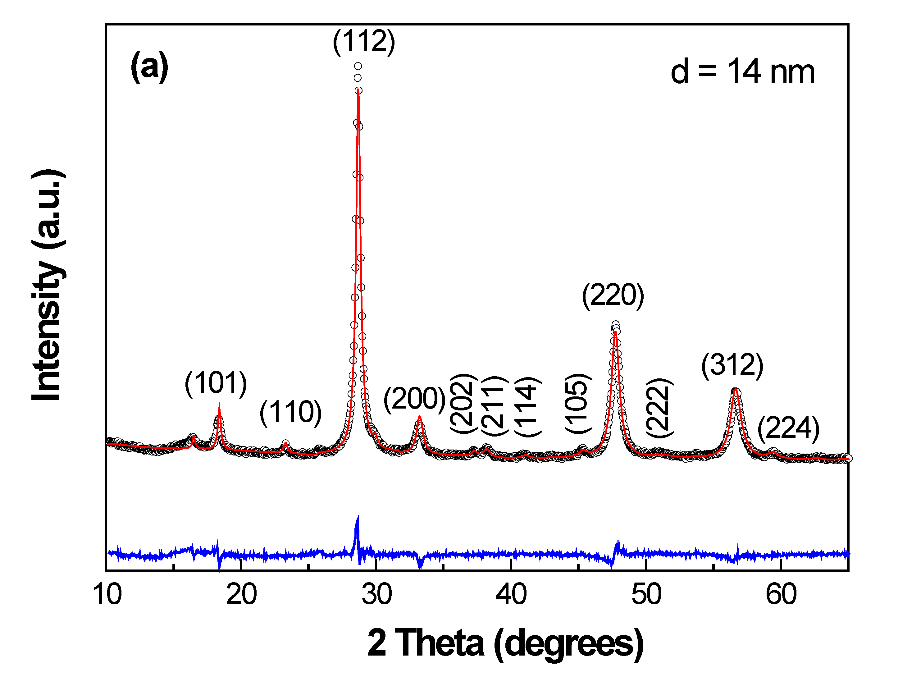
Abstract
:1. Introduction
2. Materials and Methods
3. Results
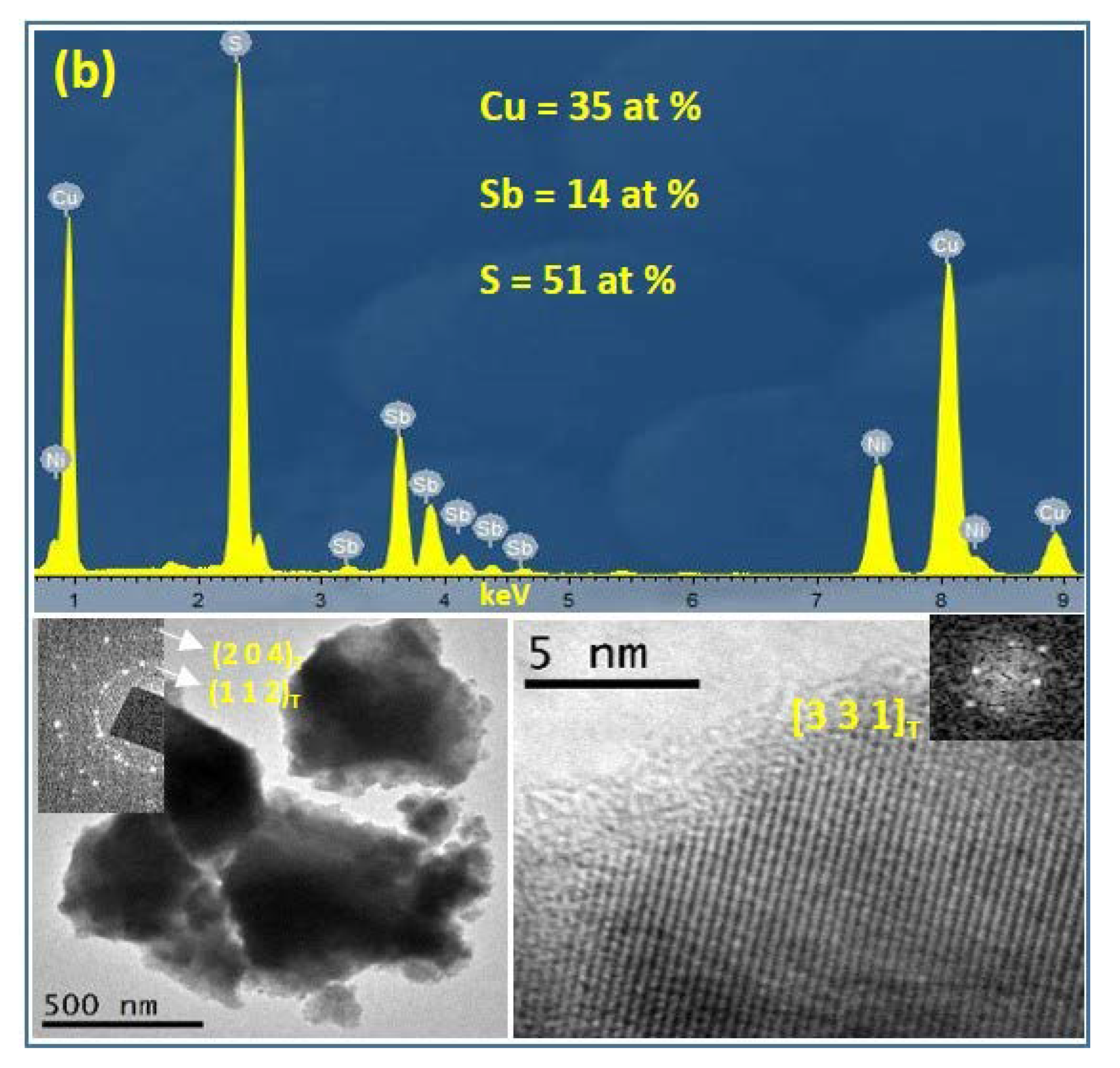
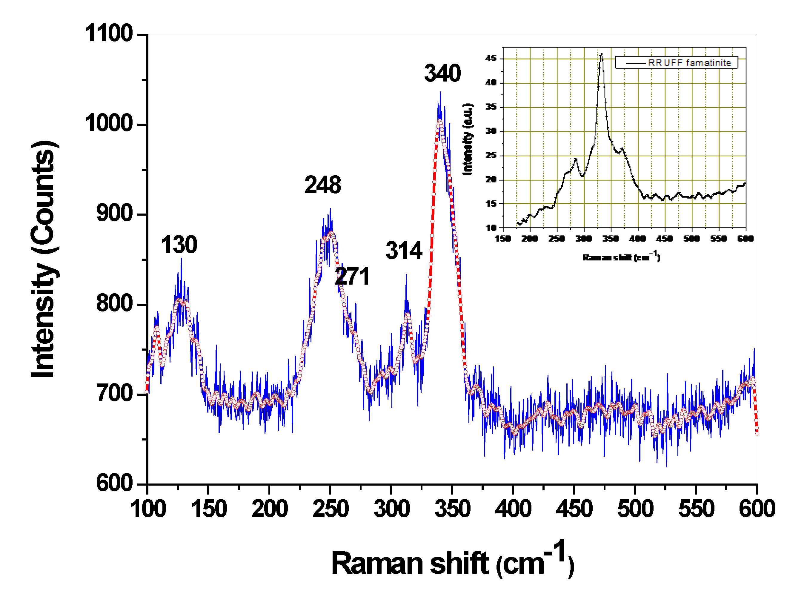
3.1. Structural and Microstructural Characterization
3.2. Surface Properties
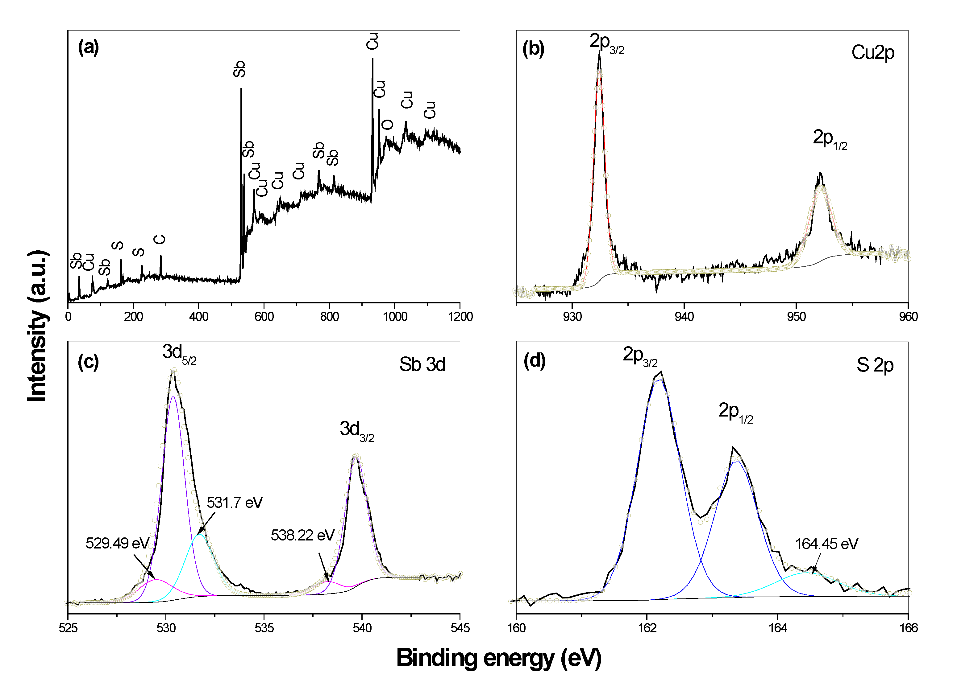
3.2.1. X-ray Photoelectron Spectroscopy (XPS)
3.2.2. Zeta Potential
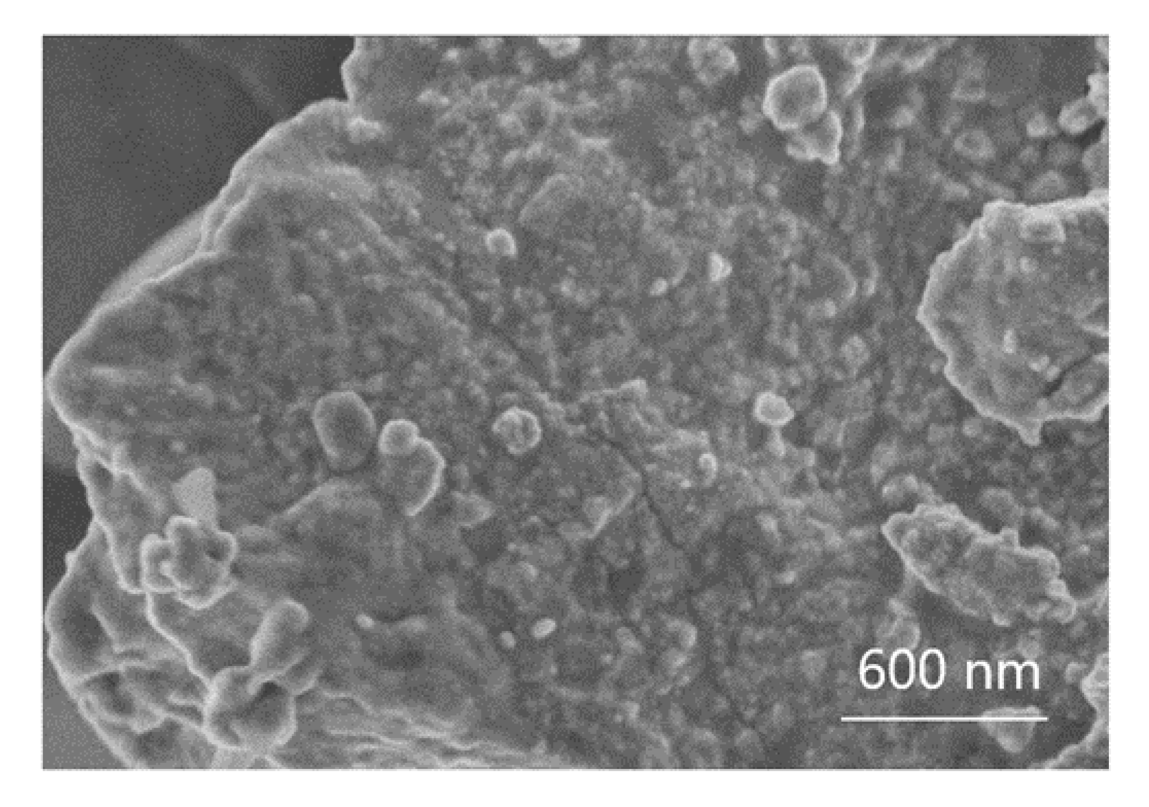
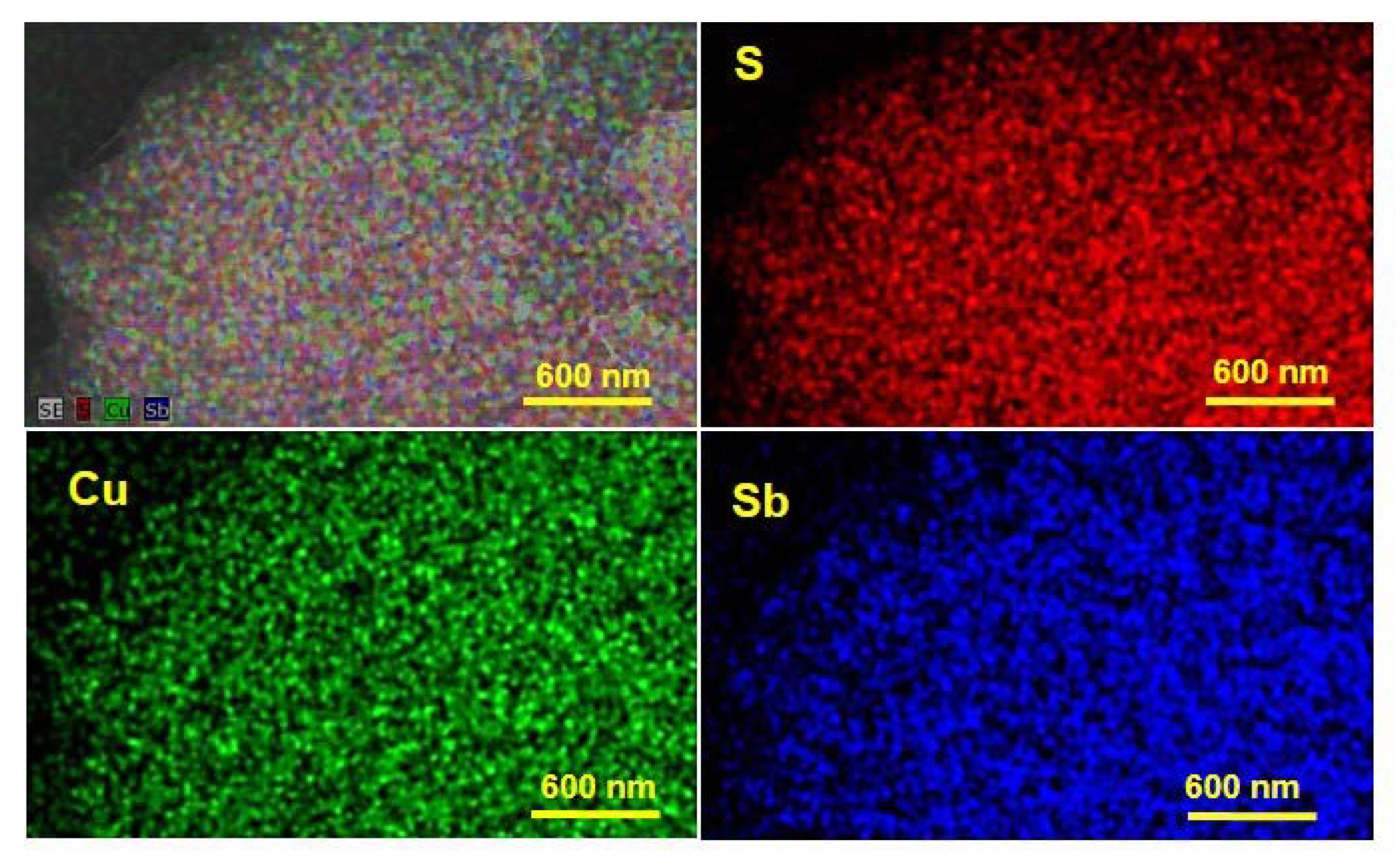
3.2.3. Scanning Electron Microscopy (SEM)
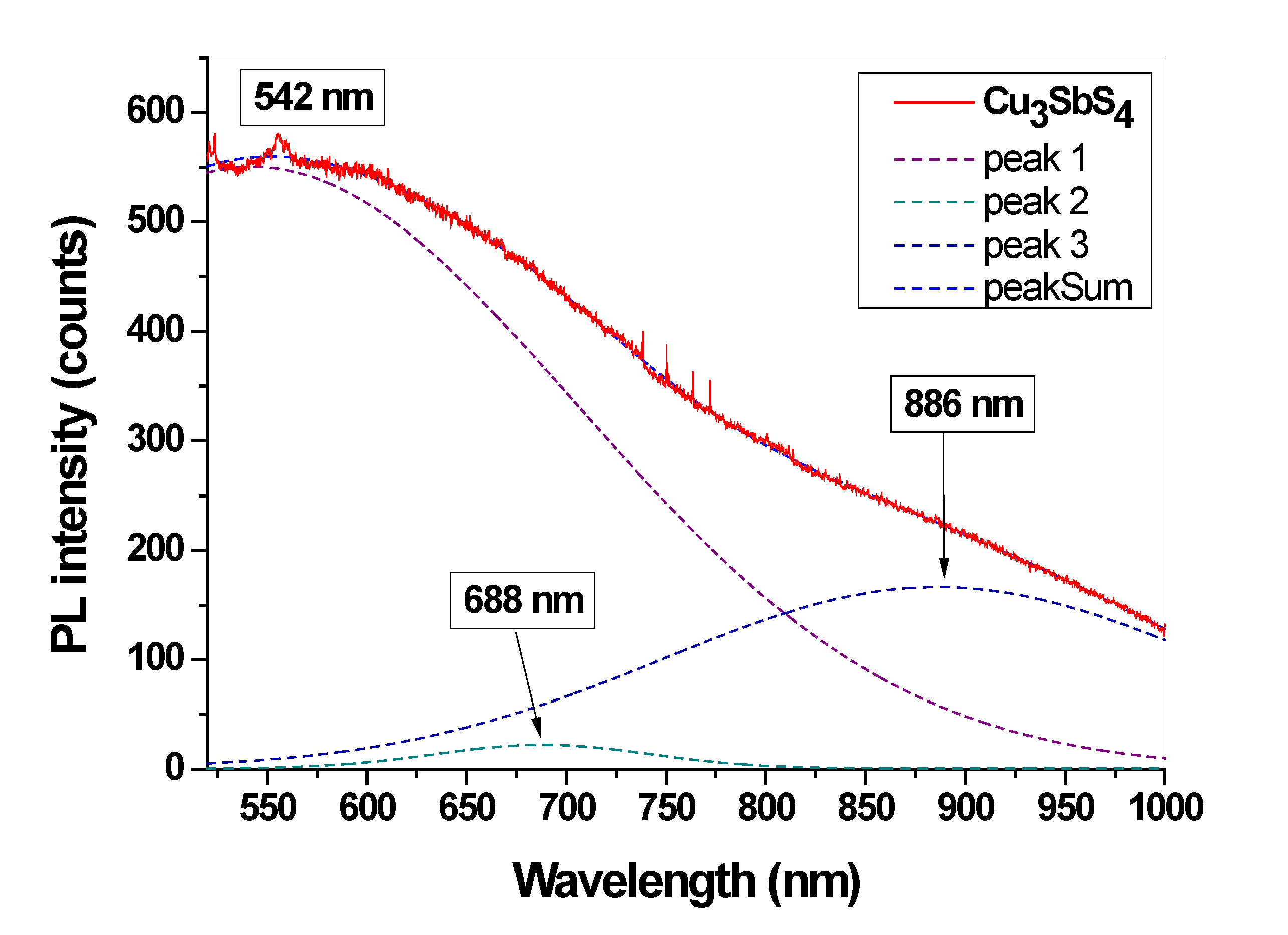
3.3. Micro-Photoluminescence Spectroscopy
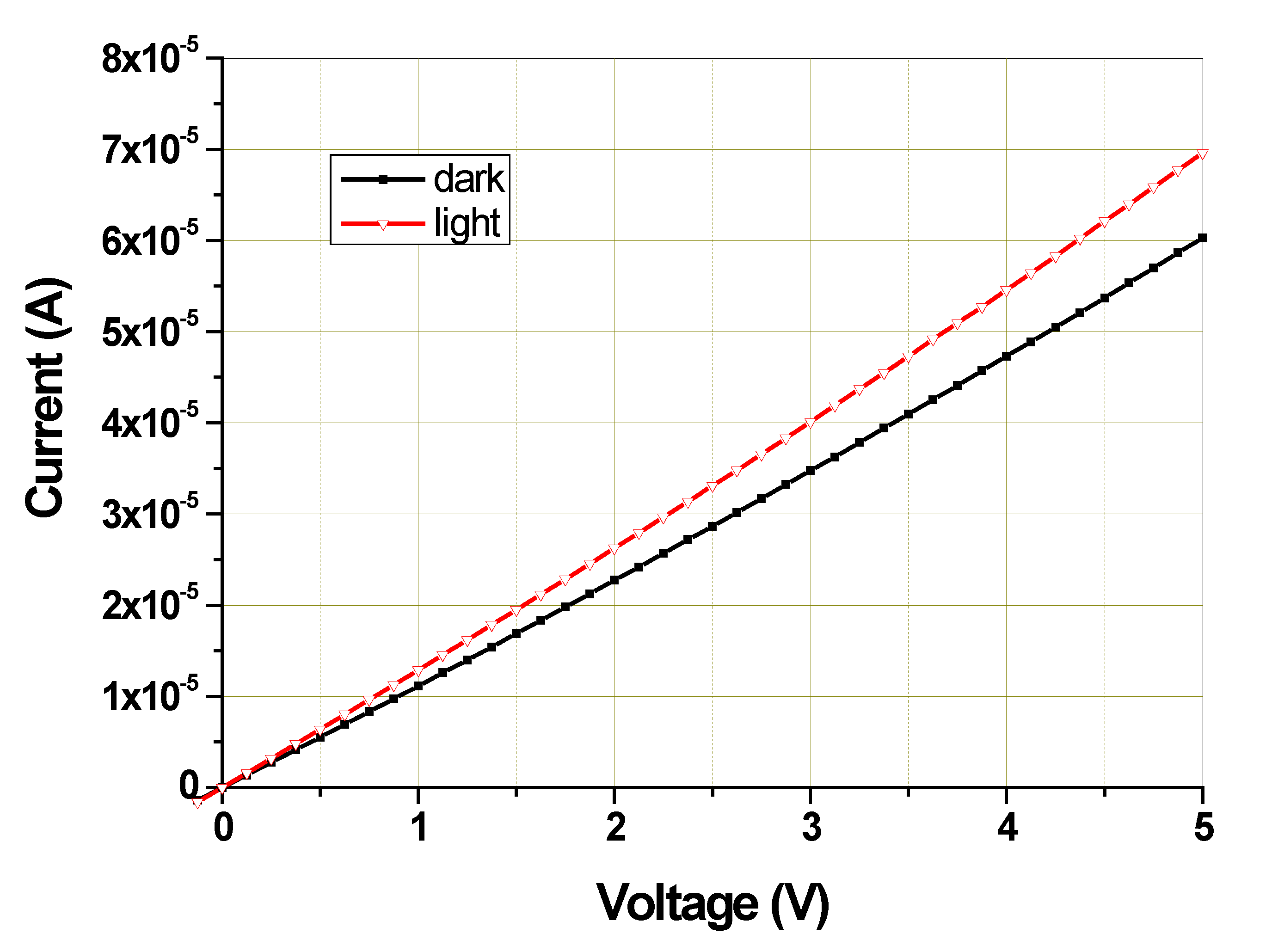
3.4. Optoelectric Measurements
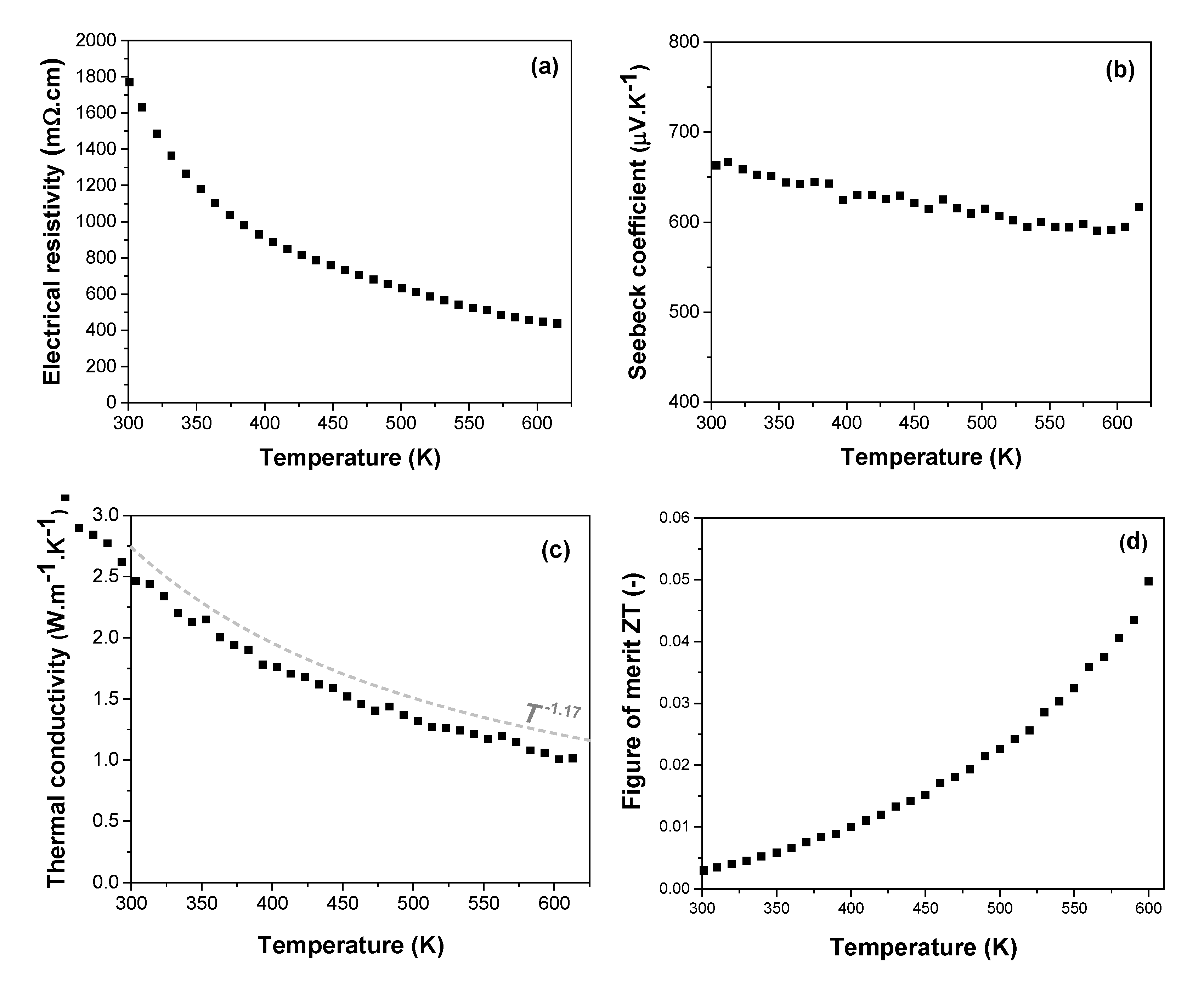
3.5. Thermoelectric Measurements
4. Conclusions
Author Contributions
Funding
Institutional Review Board Statement
Informed Consent Statement
Data Availability Statement
Acknowledgments
Conflicts of Interest
References
- Ghafoor, F.; Ghafoor, B.; Kim, D.; Khan, M.F.; Rehman, M.A. Enhancement in figure of merit in N-type Bi(R)-Te thermoelectric nanomaterials. J. Mater. Res. Technol. 2023, 23, 3617–3625. [Google Scholar] [CrossRef]
- Pan, Y.; Fan, F.R.; Hong, X.C.; He, B.; Le, C.C.; Schnelle, W.; He, Y.K.; Imasato, K.; Borrmann, H.; Hess, C.; et al. Thermoelectric Properties of Novel Semimetals: A Case Study of YbMnSb2. Adv. Mater. 2021, 33, e2003168. [Google Scholar] [CrossRef]
- Tablero, C. Electronic and optical property analysis of the Cu–Sb–S tetrahedrites for high-efficiency absorption devices. J. Phys. Chem. C 2014, 118, 15122–15127. [Google Scholar] [CrossRef]
- Kehoe, A.B.; Temple, D.J.; Watson, G.W.; Scanlon, D.O. Cu(3)MCh(3) (M = Sb, Bi; Ch = S, Se) as candidate solar cell absorbers: Insights from theory. Phys. Chem. Chem. Phys. 2013, 15, 15477–15484. [Google Scholar] [CrossRef]
- Powell, A.V. Recent developments in Earth-abundant copper-sulfide thermoelectric materials. J. Appl. Phys. 2019, 126, 100901. [Google Scholar] [CrossRef] [Green Version]
- Suekuni, K.; Takabatake, T. Research Update: Cu-S based synthetic minerals as efficient thermoelectric materials at medium temperatures. Appl. Mater. 2016, 4, 104503. [Google Scholar] [CrossRef] [Green Version]
- Kavinchan, J.; Saksornchai, E.; Thongtem, S.; Thongtem, T. One-step microwave assisted synthesis of copper antimony sulphide (Cu3SbS4) nanostructures:optical property and formation mechanism study. Chalcogenide Lett. 2018, 15, 599–604. [Google Scholar]
- Ramasamy, K.; Sims, H.; Butler, W.H.; Gupta, A. Selective Nanocrystal Synthesis and Calculated Electronic Structure of All Four Phases of Copper-Antimony-Sulfide. Chem. Mater. 2014, 26, 2891–2899. [Google Scholar] [CrossRef]
- Mariappan, V.K.; Krishnamoorthy, K.; Pazhamalai, P.; Sahoo, S.; Kim, S.J. Layered famatinite nanoplates as an advanced pseudocapacitive electrode material for supercapacitor applications. Electrochim. Acta 2018, 275, 110–118. [Google Scholar] [CrossRef]
- Joel van Embden, Y.T. Synthesis and characterisation of famatinite copper antimony sulfide nanocrystals. J. Mater. Chem. 2012, 22, 11466. [Google Scholar] [CrossRef]
- Chen, G.L.; Wang, W.H.; Zhao, J.F.; Yang, W.Y.; Chen, S.Y.; Huang, Z.G.; Jian, R.K.; Ruan, H.R. Study on the synthesis and formation mechanism of flower-like Cu3SbS4 particles via microwave irradiation. J. Alloys Compd. 2016, 679, 218–224. [Google Scholar] [CrossRef]
- Bella, M.; Rivero, C.; Blayac, S.; Basti, H.; Record, M.C.; Boulet, P. Oleylamine-assisted solvothermal synthesis of copper antimony sulfide nanocrystals: Morphology and phase control. Mater. Res. Bull. 2017, 90, 188–194. [Google Scholar] [CrossRef] [Green Version]
- Lee, G.E.; Pi, J.H.; Kim, I.H. Preparation and Thermoelectric Properties of Famatinite Cu3SbS4. J. Electron. Mater. 2020, 49, 2781–2788. [Google Scholar] [CrossRef]
- Chen, K.; Du, B.L.; Bonini, N.; Weber, C.; Yan, H.X.; Reece, M.J. Theory-Guided Synthesis of an Eco-Friendly and Low-Cost Copper Based Sulfide Thermoelectric Material. J. Phys. Chem. C 2016, 120, 27135–27140. [Google Scholar] [CrossRef] [Green Version]
- Chen, K.; Di Paola, C.; Du, B.L.; Zhang, R.Z.; Laricchia, S.; Bonini, N.; Weber, C.; Abrahams, I.; Yan, H.X.; Reece, M. Enhanced thermoelectric performance of Sn-doped Cu3SbS4. J. Mater. Chem. C 2018, 6, 8546–8552. [Google Scholar] [CrossRef] [Green Version]
- Suzumura, A.; Watanabe, M.; Nagasako, N.; Asahi, R. Improvement in Thermoelectric Properties of Se-Free Cu3SbS4 Compound. J. Electron. Mater. 2014, 43, 2356–2361. [Google Scholar] [CrossRef]
- Chen, K.; Di Paola, C.; Laricchia, S.; Reece, M.J.; Weber, C.; McCabe, E.; Abrahams, I.; Bonini, N. Structural and electronic evolution in the Cu3SbS4-Cu(3)SnS(4)solid solution. J. Mater. Chem. C 2020, 8, 11508–11516. [Google Scholar] [CrossRef]
- Shen, M.J.; Lu, S.Y.; Zhang, Z.F.; Liu, H.Y.; Shen, W.X.; Fang, C.; Wang, Q.Q.; Chen, L.C.; Zhang, Y.W.; Jia, X.P. Bi and Sn Co-doping Enhanced Thermoelectric Properties of Cu3SbS4 Materials with Excellent Thermal Stability. ACS Appl. Mater. Inter. 2020, 12, 8271–8279. [Google Scholar] [CrossRef]
- Baláž, P.; Achimovičová, M.; Baláž, M.; Billik, P.; Cherkezova-Zheleva, Z.; Criado, J.M.; Delogu, F.; Dutková, E.; Gaffet, E.; Gotor, F.J.; et al. Hallmarks of mechanochemistry: From nanoparticles to technology. Chem. Soc. Rev. 2013, 42, 7571–7637. [Google Scholar] [CrossRef] [Green Version]
- James, S.L.; Adams, C.J.; Bolm, C.; Braga, D.; Collier, P.; Friscic, T.; Grepioni, F.; Harris, K.D.M.; Hyett, G.; Jones, W.; et al. Mechanochemistry: Opportunities for new and cleaner synthesis. Chem. Soc. Rev. 2012, 41, 413–447. [Google Scholar] [CrossRef] [Green Version]
- Dutkova, E.; Sayagues, M.J.; Fabian, M.; Balaz, M.; Achimovicova, M. Mechanochemically synthesized ternary chalcogenide Cu3SbS4 powders in a laboratory and an industrial mill. Mater. Lett. 2021, 291, 129566. [Google Scholar] [CrossRef]
- Rodriguez-Carvajal, J. Recent developments of the program FullProf. Commission on powder diffraction (IUCr). Newsletter 2001, 6, 12–19. [Google Scholar]
- Rodriguez-Carvajal, J.; Roisnel, T. Line broadening analysis using FullProf*: Determination of microstructural properties. Eur. Powder Diffr. Epdic 8 2004, 443–444, 123–126. [Google Scholar] [CrossRef]
- Chalapathi, U.; Poornaprakash, B.; Park, S.H. Growth and properties of Cu3SbS4 thin films prepared by a two-stage process for solar cell applications. Ceram. Int. 2017, 43, 5229–5235. [Google Scholar] [CrossRef]
- Rahman, A.A.; Hossian, E.; Vaishnav, H.; Parmar, J.B.; Bhattacharya, A.; Sarma, A. Synthesis and characterization of Cu3SbS4 thin films grown by co-sputtering metal precursors and subsequent sulfurization. Mater. Adv. 2020, 1, 3333–3338. [Google Scholar] [CrossRef]
- Han, G.; Lee, J.W.; Kim, J. Fabrication and Characterization of Cu3SbS4 Solar Cell with Cd-free Buffer. J. Korean Phys. Soc. 2018, 73, 1794–1798. [Google Scholar] [CrossRef]
- Shaji, S.; Vinayakumar, V.; Krishnan, B.; Johny, J.; Kanakkillam, S.S.; Herrera, J.M.F.; Guzman, S.S.; Avellaneda, D.A.; Rodriguez, G.A.C.; Martinez, J.A.A. Copper antimony sulfide nanoparticles by pulsed laser ablation in liquid and their thin film for photovoltaic application. Appl. Surf. Sci. 2019, 476, 94–106. [Google Scholar] [CrossRef]
- Fairthorne, G.; Fornasiero, D.; Ralston, J. Interaction of thionocarbamate and thiourea collectors with sulphide minerals: A flotation and adsorption study. Int. J. Miner. Process. 1997, 50, 227–242. [Google Scholar] [CrossRef]
- Fornasiero, D.; Eijt, V.; Ralston, J. An Electrokinetic Study of Pyrite Oxidation. Coll. Surf. 1992, 62, 63–73. [Google Scholar] [CrossRef]
- Dukhin, S.S.; Derjaguin, B.V. Non-equilibrium Double Layer and Electrokinetic Phenomena. In Surface and Colloid Science; Matijevic, E., Ed.; John Wiley & Sons: New York, NY, USA, 1974; Volume 7, pp. 297–300. [Google Scholar]
- Konkena, B.; Vasudevan, S. Understanding Aqueous Dispersibility of Graphene Oxide and Reduced Graphene Oxide through pK(a) Measurements. J. Phys. Chem. Lett. 2012, 3, 867–872. [Google Scholar] [CrossRef]
- Dutkova, E.; Bujnakova, Z.; Kovac, J.; Skorvanek, I.; Sayagues, M.J.; Zorkovska, A.; Kovac, J.; Balaz, P. Mechanochemical synthesis, structural, magnetic, optical and electrooptical properties of CuFeS2 nanoparticles. Adv. Powder Technol. 2018, 29, 1820–1826. [Google Scholar] [CrossRef]
- Dutkova, E.; Sayagues, M.J.; Fabian, M.; Kovac, J.; Kovac, J.; Balaz, M.; Stahorsky, M. Mechanochemical synthesis of ternary chalcogenide chalcostibite CuSbS2 and its characterization. J. Mater. Sci.-Mater. El. 2021, 32, 22898–22909. [Google Scholar] [CrossRef]
- Mohamadkhani, F.; Heidariramsheh, M.; Javadpour, S.; Ghavaminia, E.; Mahdavi, S.M.; Taghavinia, N. Sb nanocrystals as inorganic hole transporting materials in perovskite solar cells. Sol. Energy 2021, 223, 106–112. [Google Scholar] [CrossRef]
- Shi, L.; Wu, C.Y.; Li, J.J.; Ding, J. Selective synthesis and photoelectric properties of Cu3SbS4 and CuSbS2 nanocrystals. J. Alloys Compd. 2017, 694, 132–135. [Google Scholar] [CrossRef]
- Tanishita, T.; Suekuni, K.; Nishiate, H.; Lee, C.H.; Ohtaki, M. A strategy for boosting the thermoelectric performance of famatinite Cu3SbS4. Phys. Chem. Chem. Phys. 2020, 22, 2081–2086. [Google Scholar] [CrossRef]
- Du, B.L.; Zhang, R.Z.; Chen, K.; Mahajan, A.; Reece, M.J. The impact of lone-pair electrons on the lattice thermal conductivity of the thermoelectric compound CuSbS2. J. Mater. Chem. A 2017, 5, 3249–3259. [Google Scholar] [CrossRef] [Green Version]
- Goto, Y.; Sakai, Y.; Kamihara, Y.; Matoba, M. Effect of Sn-Substitution on Thermoelectric Properties of Copper-Based Sulfide, Famatinite Cu3SbS4. J. Phys. Soc. Jpn. 2015, 84, 044706. [Google Scholar] [CrossRef] [Green Version]
- Balaz, P.; Dutkova, E.; Levinsky, P.; Daneu, N.; Kubickova, L.; Knizek, K.; Balaz, M.; Navratil, J.; Kasparova, J.; Ksenofontov, V.; et al. Enhanced thermoelectric performance of chalcopyrite nanocomposite via co-milling of synthetic and natural minerals. Mater. Lett. 2020, 275, 128107. [Google Scholar] [CrossRef]
- Levinsky, P.; Hejtmanek, J.; Knizek, K.; Pashchenko, M.; Navratil, J.; Masschelein, P.; Dutkova, E.; Balaz, P. Nanograined n- and p-Type Chalcopyrite CuFeS2 Prepared by Mechanochemical Synthesis and Sintered by SPS. Acta Phys. Pol. A 2020, 137, 904–907. [Google Scholar] [CrossRef]
- Wu, J.; Li, F.; Wei, T.R.; Ge, Z.H.; Kang, F.Y.; He, J.Q.; Li, J.F. Mechanical Alloying and Spark Plasma Sintering of BiCuSeO Oxyselenide: Synthesis Process and Thermoelectric Properties. J. Am. Ceram. Soc. 2016, 99, 507–514. [Google Scholar] [CrossRef]
- Balaz, P.; Achimovicova, M.; Balaz, M.; Chen, K.; Dobrozhan, O.; Guilmeau, E.; Hejtmanek, J.; Knizek, K.; Kubickova, L.; Levinsky, P.; et al. Thermoelectric Cu-S-Based Materials Synthesized via a Scalable Mechanochemical Process. ACS Sustain. Chem. Eng. 2021, 9, 2003–2016. [Google Scholar] [CrossRef]


Disclaimer/Publisher’s Note: The statements, opinions and data contained in all publications are solely those of the individual author(s) and contributor(s) and not of MDPI and/or the editor(s). MDPI and/or the editor(s) disclaim responsibility for any injury to people or property resulting from any ideas, methods, instructions or products referred to in the content. |
© 2023 by the authors. Licensee MDPI, Basel, Switzerland. This article is an open access article distributed under the terms and conditions of the Creative Commons Attribution (CC BY) license (https://creativecommons.org/licenses/by/4.0/).
Share and Cite
Dutková, E.; Kováč, J.; Kováč, J., Jr.; Hejtmánek, J.; Levinský, P.; Kashimbetova, A.; Sayagués, M.J.; Fabián, M.; Lukáčová Bujňáková, Z.; Baláž, M.; et al. Properties of Mechanochemically Synthesized Famatinite Cu3SbS4 Nanocrystals. Micro 2023, 3, 458-470. https://doi.org/10.3390/micro3020030
Dutková E, Kováč J, Kováč J Jr., Hejtmánek J, Levinský P, Kashimbetova A, Sayagués MJ, Fabián M, Lukáčová Bujňáková Z, Baláž M, et al. Properties of Mechanochemically Synthesized Famatinite Cu3SbS4 Nanocrystals. Micro. 2023; 3(2):458-470. https://doi.org/10.3390/micro3020030
Chicago/Turabian StyleDutková, Erika, Jaroslav Kováč, Jaroslav Kováč, Jr., Jiří Hejtmánek, Petr Levinský, Adelia Kashimbetova, María Jesús Sayagués, Martin Fabián, Zdenka Lukáčová Bujňáková, Matej Baláž, and et al. 2023. "Properties of Mechanochemically Synthesized Famatinite Cu3SbS4 Nanocrystals" Micro 3, no. 2: 458-470. https://doi.org/10.3390/micro3020030