Article

Structural Consequences of Post-Synthetic Modification of Cu2P3I2

Department of Chemistry, Drexel University, Philadelphia, PA 19104, USA
*
Author to whom correspondence should be addressed.
These authors contributed equally to this work.
Micro 2023, 3(1), 256-263; https://doi.org/10.3390/micro3010018
Submission received: 29 November 2022 / Revised: 1 February 2023 / Accepted: 15 February 2023 / Published: 1 March 2023

Abstract

:
In an attempt to widen the family of Phosphorus Metal Halides (MxPyXz) and enable new applications, post-synthetic modifications to the MxPyXz, Cu2P3I2 have been reported. While such a technique suggests access to an entirely new family of MxPyXz-based materials, we report, in this work, that the ion-exchange process seemingly influences important properties such as the crystallographic pattern and vibrational modes.

1. Introduction

Since the isolation and subsequent vast applications of phosphorene [1], research interest in low-dimensional phosphorus-based materials has increased dramatically. While initial studies focused heavily on phosphorene and its bulk analog, black phosphorus (BP), other allotropic forms have proven successful in various applications and have begun to garner interest. As evidenced by Differential Scanning Calorimetry (DSC) measurements, red phosphorus (RP) may exist in one of five proposed polymorphs denoted by roman numerals from I to V [2]. While types I-III lack full structural elucidation, type I has been characterized as an effective FET [3] and type II has displayed potential in photocatalysis [4]. Type III has only been reportedly observed once by DSC and has not been replicated in literature since [2]. Type IV and V are fully structurally resolved and have been employed in a wide range of applications, including cell imaging and optoelectronic devices [5,6,7,8].
One-dimensional nanorod structures of both type II and IV RP have been reported. However, exposing the nanorods to ambient conditions affords similar oxidation effects to those observed with black phosphorus, limiting their stability and application. Bearing similar rod-like structures to type II and IV RP, Phosphorus Metal Halides (MxPyXz) adduct structures with CuI maintain similar properties, but with enhanced stability [9,10]. Integral to these structures, a phosphorus nanorod is isolated inside a framework of CuI which may be solubilized in aqueous KCN solutions to afford the individual nanorods their own “allotrope” [11]. Owing to the nanorod isolation in the CuI framework, these materials have even been characterized as stable humidity sensors without suffering passivation effects [12]. Since many of these materials have been limited to a surrounding CuI framework, methods that promote different metal halides open a window of opportunity for various applications depending on the constituent metal.
In an attempt to widen the application of such materials by replacing Cu+ with other metal ions, post-synthetic modifications to the MxPyXz, Cu2P3I2 have reportedly facilitated a near 98% ion exchange with silver [13]. While such a technique suggests access to an entirely new family of MxPyXz-based materials, the ion-exchange process seemingly influences important properties such as the crystallographic pattern and vibrational modes. Herein, we report the experimentally obtained Raman spectra for Cu2P3I2 and compare that with the reported post-synthetic Ag2P3I2. Differences in the spectra of Cu2P3I2 and Ag2P3I2 prompted further characterization through powder XRD, cyclic current voltage (CIV), and SEM imaging, which overwhelmingly pointed to the conclusion that the ion exchange of Cu2P3I2 is ineffective in producing pure isotypic Ag2P3I2.

2. Experimental

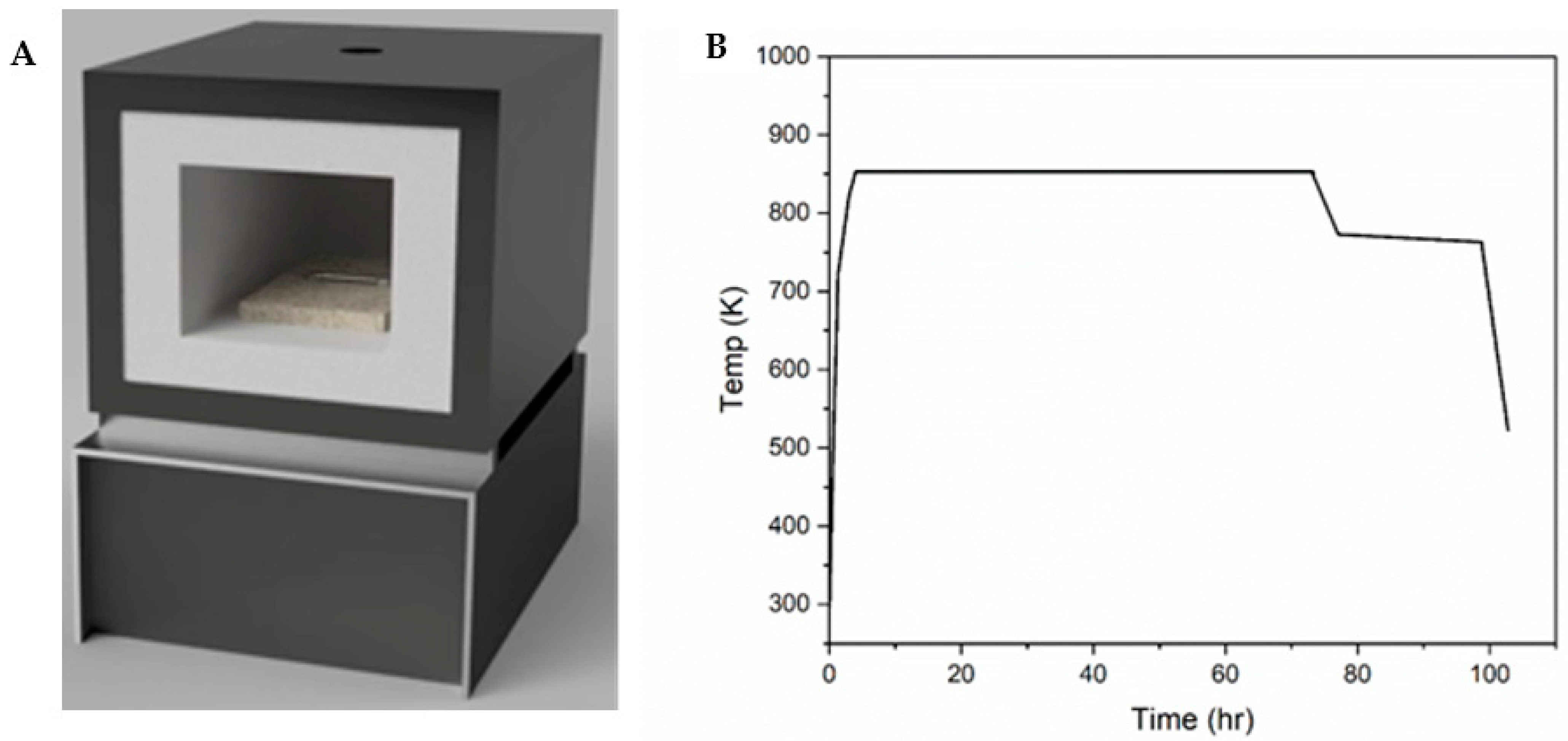
2.1. Cu2P3I2 Synthesis

The Chemical Vapor Transport (CVT) process was performed as previously reported by this lab [9]. In brief, samples were prepared using stoichiometric amounts of CuI and type I, amorphous, red phosphorus (a-RP). For smaller-diameter borosilicate ampoules, a yield of 100 mg was targeted (80.4 mg CuI and 19.6 mg a-RP), and for larger-diameter ampoules, a 250 mg yield was targeted (201.0 mg CuI and 49 mg a-RP). Both scales of reaction yielded similar results. These reagents were added to a borosilicate tube, which was subsequently backfilled with Argon then sealed under vacuum as an ampoule. These ampoules were spaced evenly in the center of a muffle furnace placed orthogonally to the heating element (Figure 1A). The ends of the ampoule were pointed to the heating elements so that they were closer to the heating elements than the center, creating a heating gradient over the length of the ampoule to allow chemical vapor transport. The furnace was heated rapidly to 373 K then heated to 723 K at a rate of 5 K/min. The furnace was then heated at a rate of 1 K/min until it reached 823 K, followed by a rate of 0.5 K/min until it reached 853 K. This temperature was then held for 48 hr. The furnace was then cooled at a rate of 0.33 K/min until it reached 773 K, where it was held for 18 hr (heating/cooling curve shown in Figure 1B). The furnace was then cooled at a rate of 5 K/min until it reached 473 K, at which point the furnace was turned off and the sample was left to equilibrate with room temperature.

2.2. Post-Synthetic Modification

As reported by Möller and Jeitschko [10], the copper cations in Cu2P3I2 nanowires could be displaced with silver cations by adding the wires to a 10% (w/w) aqueous solution of AgNO3 with excess KCN by reaction (1).
Cu2P3I2 + 2[Ag(CN)2] + 4CN → Ag2P3I2 + 2[Cu(CN)4]3−
After a few hours, the solution was centrifuged, and the supernatant liquid was removed. The wires were subsequently washed in methanol after which the suspension was centrifuged, and the supernatant was removed. This allows for formation of post-synthetic Ag2P3I2 by modifying the previously synthesized Cu2P3I2.

2.3. Electrical Characterization

Single-wire devices were prepared on a Si/SiO2 (n-doped) wafer containing 2 gold electrodes separated by a 10 μm channel. When fixed to the electrode surfaces with a graphene-based conductive glue, the single wire spanned the channel; any current resulting from the potential difference between the two electrodes is required to pass through the single wire to reach the ground. Instrument probes were connected to copper wires that were glued to the gold plates with the graphene-based conductive glue. Current v. Potential plots (IV) were generated using a Keithley 2636A Dual-channel System SourceMeter Instrument (Keithley Instruments, Inc. Cleveland, OH, USA). Source and drain probes were attached to the copper wires of the devices and current was measured against a time-varying electric potential. Cyclic IV (CIV) plots were generated using a VersaSTAT 3 Potentiostat Galvanostat (Princeton Applied Research, Oak Ridge, TN, USA). Similar to how the IV plots were generated, source and drain probes were attached to the copper wires of the devices and current was measured against a time-varying electric potential such that the final potential matched the initial. A total of five cycles were run per trial.

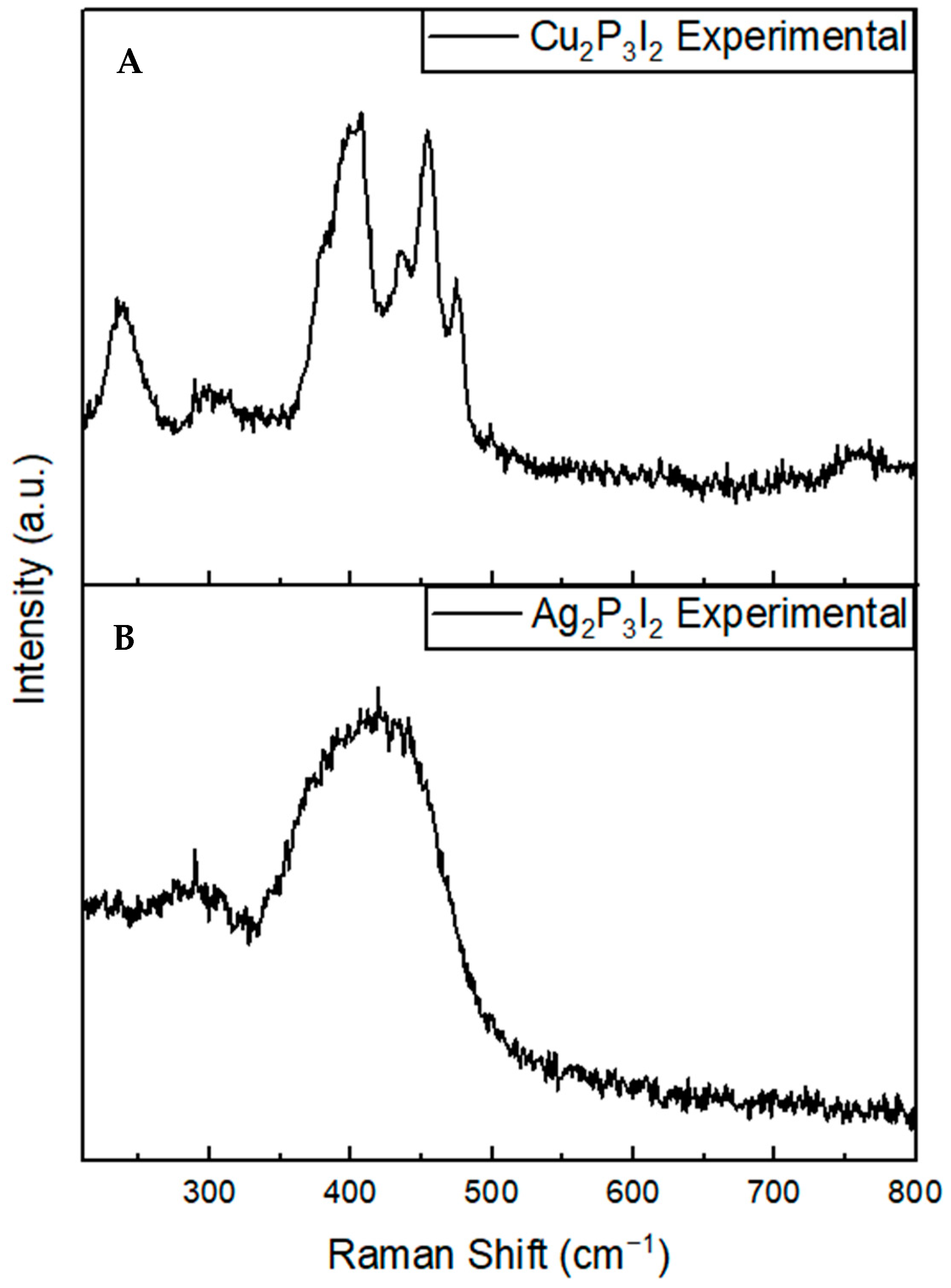
2.4. Raman Spectroscopy

Several analytical methods have been employed to characterize the various phosphorous allotropes, the most common being X-Ray Diffraction (XRD), both powder and single crystal, and Raman Spectroscopy [14,15,16,17,18,19,20]. While XRD patterns provide structural insight, the unique variety of these phosphorus allotropes lead to distinct Raman-active vibrational modes that allow for further differentiation. Using computational data, many have attempted to interpret these spectra to provide specific vibrational modes for each Raman band [14].
Samples of Cu2P3I2 and Ag2P3I2 were loaded onto a glass microscope slide. A Renishaw RM-2000 Vis Raman Spectrometer (Gloucestershire, UK) was used to probe the observed Raman shifts in each sample. Optimal conditions were observed in the absence of ambient light and when samples were exposed to 10% laser strength of a 633 nm beam. Resulting shifts were collected through an 1800 mm diffraction grating. Spectral acquisitions occurred with 10+ accumulations comprised of 10-second exposure times.

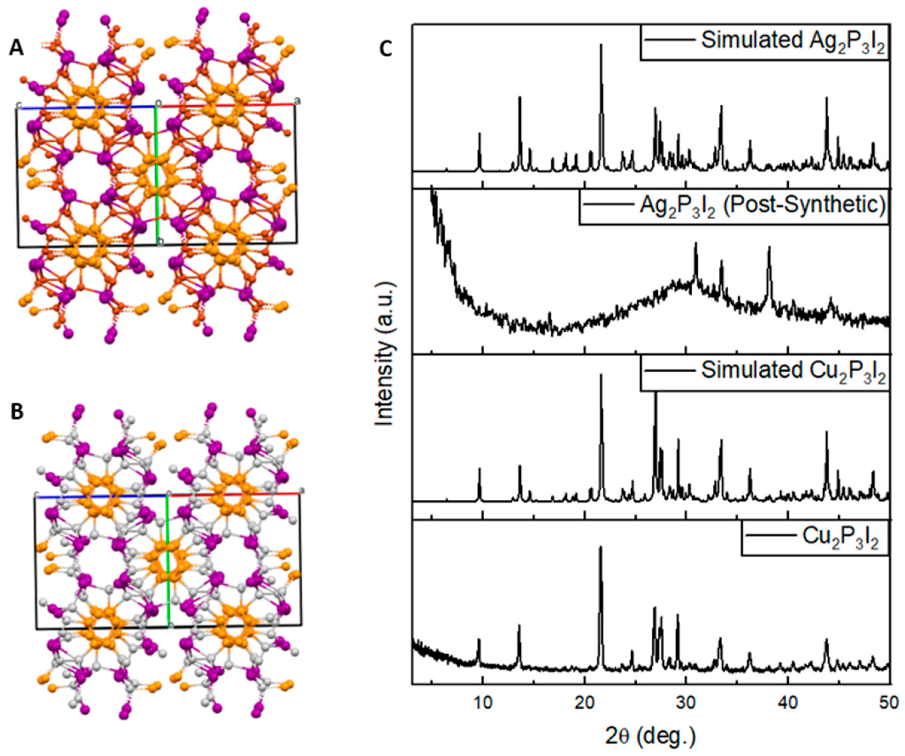
2.5. Powder X-ray Diffraction (P-XRD)

Samples of Cu2P3I2 and Ag2P3I2 were prepared via CVT and post-synthetic modification respectively, as described in the Experimental section. Approximately 100 mg of each sample were ground into fine powders with the use of a mortar and pestle. Powders were placed on glass sample holders and prepared for P-XRD analysis. P-XRD was performed with a Rigaku MiniFlex (Tokyo, Japan) equipped with a Cu Kα radiation source. Diffraction patterns were acquired in the range of 3–50° with a step increase of 0.02° and a dwell time of 1 s. Predicted XRD patterns were generated in Mercury 2022.1.0 (Build 343014; CCDC, Boston, MA, USA).

3. Discussion

The Raman spectrum collected for Cu2P3I2 (Figure 2A) shows unique vibrational modes that are separate and distinguishable from other phosphorus-based materials. Presently, to the authors’ knowledge, there are no literature sources in which the Raman profile of Cu2P3I2 is reported, and therefore, these bands are unassigned. However, the observed Raman activity is consistent with the window in which various P allotropes show activity [16,17]. Conversely, the Ag2P3I2 spectrum (Figure 2B) is highly unresolved and contains only a broad peak in the same window in which individual Cu2P3I2 bands occur. This stark difference suggests that while the Cu2P3I2 is a more highly regular repeating structure, the Ag2P3I2 generated through post-synthetic modification has regions of higher variability. Because this procedure produces a material that is highly irregular, this method is likely ineffective in generating an isotypic silver analog for Cu2P3I2 as originally suggested [10]. The high degree of irregularities is suspected to originate from the fracturing of wires as a result of the larger Ag2P3I2 unit cell and potential solubilizing of the CuI shell when exposed to the KCN solution, both of which will be discussed in greater detail below [10,11,13]. Additional studies may be required to fully determine the Raman modes so that the deformation mechanisms may be validated.
The P-XRD patterns of Cu2P3I2 and Ag2P3I2 (Figure 3C) corroborates what has been suggested by the difference in the Raman profiles—long range periodicity is not maintained after post-synthetic modifications have been made. The experimental pattern for Cu2P3I2 was acquired from a sample prepared via chemical vapor transport as described in the experimental section. The simulated pattern was generated in the crystallographic software Mercury 2022.1.0 (Build 343014), from a CIF previously acquired by our lab [9]. The experimental sample was then converted to Ag2P3I2 by the post-synthetic procedure outlined in the experimental section. Notably, the Ag2P3I2 pattern reveals a few bands that do not match with the Cu2P3I2 pattern, as well as a broad amorphous region. Most importantly, the characteristic peaks of Cu2P3I2 are completely gone, proving long range order is significantly reduced. For further investigation, the Cu2P3I2 CIF file was modified in Mercury 2022.1.0 (Build 343014) to replace the Cu+ ions with Ag+ ions while maintaining isotypic structure (as suggested in the literature), and a resulting simulated pattern for Ag2P3I2 was generated for comparison (Figure 3C, top).
As briefly mentioned previously, one major issue with this synthesis is the potential solubilizing of the CuI framework by the use of KCN. This reagent is necessary, however, for the formation of the complex [Ag(CN)2] which mitigates the amount of surface impurities (compared to just AgNO3 without KCN). Since the pilot study of these MxPyXz materials, it has been observed that the CuI framework can be removed by stirring the solid in a solution of KCN, affording a reddish-brown precipitate composed of P nanorods [11]. As a result, it is reasonable to suspect that the CuI framework is prone to degradation during the ion exchange process which occurs in a KCN solution. To corroborate this, Cu2P3I2 was added to a silver nitrate solution followed by the addition of KCN. The solution immediately developed a reddish-brown color, which is not present if the reagents are added in the correct order. This color change supports prior suggestions that a side reaction between the KCN and the Cu2P3I2 is occurring and is observed in the extraction of the P nanorod from Cu2P3I2.
The other reason for the poor resolution of the Raman is the fracturing of the material. It was found that the post-synthetically modified material was exceptionally brittle and fragile when compared to the precursor (Cu2P3I2); the wires fractured readily under minimal force such as being picked up with forceps. A major contributor to this fragility is likely the deformation seen from intercalating a larger cation into the structure. The unit cell volume of Cu2P3I2 is too small to accommodate the much larger silver cation without structural deformation. Möller and Jeitschko [10] reported that the cell volume increases from 2711 Å3 to 2998 Å3 as a result of the ion exchange, causing a deformation which weakens the material overall. The SEM images of the post-synthetically modified material presented in the Möller paper illustrate that the wires are not one continuous structure; rather, they are fractured into bundles of smaller fibers. As such, not only is the KCN solution suspected to solubilize the metal halide outer layer, but, even after successful ion exchange, the inability to account for the new cell volume forces fracturing.
The post-synthetic modification approach has two further drawbacks separate from the structural issues: it is wasteful, and excess silver is plated onto the wire. To perform post-synthetic modification, a 10% (w/w) solution of silver nitrate must be generated, which comprises a great excess of silver. Much of this solution is wasted, and a new solution must be generated for each synthesis because silver nitrate solutions are UV-active and the silver cations reduce to elemental silver. Furthermore, Cu2P3I2 must first be generated, and all the copper from this pre-cursor becomes waste. A chemical vapor transport reaction would eliminate the need for an expensive reaction solution and the copper intermediate, generating a purer product with less waste. Admittedly, however, such CVT attempts were discussed and found fruitless [10].
As mentioned above, the post-synthetic modification of Cu2P3I2 necessitates the use of [Ag(CN)2] to avoid contamination of the surface with impurities (e.g., CuI, AgI, Ag, etc.). However, by selecting the use of the silver complex, further structural deformations are incurred by the KCN solution and the cell volume of the exchanged product. As evidenced by XRD and Raman, long-range periodicity is sacrificed. In order to assess the implications of such deformations, the electrochemical profile of the material was assessed with a CIV scan from −2 V to +2 V (Figure 4).
In the case of Ag2P3I2, what appears to be redox activity can be observed. The activity is confined to the Ag2P3I2 material and moves depending on the applied voltage (Figures S1 and S2). Furthermore, the appearance of this shifting peak is not wholly consistent; it seems to appear for some batches of Ag2P3I2 but not for others. However, in any case, the peak never occurs for Cu2P3I2. Since there are a significantly large number of variables at play (i.e., surface impurities, KCN etching of the CuI layer, splintering, etc.) it is difficult to assign an explanation. Further studies will be conducted in which the CIV curve will be generated while simultaneously probing other properties of the material such as its Raman profile to identify the formation of any intermediate species that may play a role in the observed redox. Regardless, this observation corroborates the claim that structural deformations are induced by the post-synthetic modification process and illustrates implications this has on potential applications. These results highlight the need to develop Ag2P3I2, and potentially other MxPyXz structures, via chemical vapor transport if the materials’ target application requires a high degree of crystallinity. It is worth noting, however, that the post-synthetic route may still be valid for applications that do not suffer from a lack of long-range crystallinity (i.e., potential antimicrobial activity of post-synthetically modified Ag2P3I2).

4. Conclusions

The post-synthetic modification of Cu2P3I2 presents a potential to expand phosphorus metal halide structures to encompass metal halides besides CuI. While this is a noble effort, the present approach does not seem feasible in producing phase-pure Ag2P3I2 while maintaining isotypic character of its parent material. Herein, we have employed comparative XRD analysis to highlight the reduction in long-range periodicity after post-synthetic modification of Cu2P3I2 to Ag2P3I2 was performed. Additionally, we have corroborated these results with the apparent reduction of defined bands in the Raman profile of the material. This apparent structural deformation has been shown to limit the potential application of these materials by generating inconsistent phenomena in the CIV profile. Therefore, the results of this study highlight the need for synthetic routes that expand the phosphorus metal halide family without compromising its features. However, the post-synthetic route may still successfully provide access to a wider range of phosphorus metal halide materials so long as their target applications do not require crystallinity, such as catalysis.

Supplementary Materials

Supporting Information can be downloaded at: https://www.mdpi.com/article/10.3390/micro3010018/s1; Figure S1: Linear IV curves of Ag2P3I2 single-wire device; Figure S2: Redox peak shifting during CIV at various voltages; Figure S3: SEM images of post-synthetically modified Ag2P3I2.

Author Contributions

G.R.S. and J.T.W. performed the experiments and analyzed the data. H.-F.J. provided the laboratory space and materials with which the experiments were conducted. All the authors have contributed to the design of the experiments, writing, and editing of the paper. All authors have read and agreed to the published version of the manuscript.

Funding

This research received no external funding.

Institutional Review Board Statement

Not Applicable.

Informed Consent Statement

Not Applicable.

Data Availability Statement

Not Applicable.

Conflicts of Interest

The authors declare no conflict of interest.

References

  1. Liu, H.; Neal, A.T.; Zhu, Z.; Luo, Z.; Xu, X.; Tománek, D.; Ye, P.D. Phosphorene: An Unexplored 2D Semiconductor with a High Hole Mobility. ACS Nano 2014, 8, 4033–4041. [Google Scholar] [CrossRef] [PubMed] [Green Version]
  2. Roth, W.L.; DeWitt, T.W.; Smith, A.J. Polymorphism of Red Phosphorus. J. Am. Chem. Soc. 1947, 69, 2881–2885. [Google Scholar] [CrossRef] [PubMed]
  3. Amaral, P.E.M.; Nieman, G.P.; Schwenk, G.R.; Jing, H.; Zhang, R.; Cerkez, E.B.; Strongin, D.; Ji, H. High Electron Mobility of Amorphous Red Phosphorus Thin Films. Angew. Chem. Int. Ed. 2019, 58, 6766–6771. [Google Scholar] [CrossRef] [PubMed]
  4. Zhaojian, S.; Zhang, B.; Yan, Q. Solution Phase Synthesis of the Less-Known Form II Crystalline Red Phosphorus. Inorg. Chem. Front. 2022, 9, 4385–4393. [Google Scholar] [CrossRef]
  5. Smith, J.B.; Hagaman, D.; DiGuiseppi, D.; Schweitzer-Stenner, R.; Ji, H. Ultra-Long Crystalline Red Phosphorus Nanowires from Amorphous Red Phosphorus Thin Films. Angew. Chem. Int. Ed. 2016, 55, 11829–11833. [Google Scholar] [CrossRef] [PubMed]
  6. Amaral, P.E.M.; Hall, D.C.; Pai, R.; Król, J.E.; Kalra, V.; Ehrlich, G.D.; Ji, H.-F. Fibrous Phosphorus Quantum Dots for Cell Imaging. ACS Appl. Nano Mater. 2020, 3, 752–759. [Google Scholar] [CrossRef] [Green Version]
  7. Schusteritsch, G.; Uhrin, M.; Pickard, C.J. Single-Layered Hittorf’s Phosphorus: A Wide-Bandgap High Mobility 2D Material. Nano Lett. 2016, 16, 2975–2980. [Google Scholar] [CrossRef] [PubMed] [Green Version]
  8. Lu, Y.-L.; Dong, S.; He, H.; Li, J.; Wang, X.; Zhao, H.; Wu, P. Enhanced Photocatalytic Activity of Single-Layered Hittorf’s Violet Phosphorene by Isoelectronic Doping and Mechanical Strain: A First-Principles Research. Comput. Mater. Sci. 2019, 163, 209–217. [Google Scholar] [CrossRef]
  9. Amaral, P.E.M.; Ji, H.-F. Stable Copper Phosphorus Iodide (Cu2P3I2) Nano/Microwire Photodetectors. ChemNanoMat 2018, 4, 1083–1087. [Google Scholar] [CrossRef]
  10. Möller, M.H.; Jeitschko, W. Preparation, Properties, and Crystal Structure of the Solid Electrolytes Cu2P3I2 and Ag2P3I2. J. Solid State Chem. 1986, 65, 178–189. [Google Scholar] [CrossRef]
  11. Pfitzner, A.; Bräu, M.F.; Zweck, J.; Brunklaus, G.; Eckert, H. Phosphorus Nanorods—Two Allotropic Modifications of a Long-Known Element. Angew. Chem. Int. Ed. 2004, 43, 4228–4231. [Google Scholar] [CrossRef] [PubMed]
  12. Schwenk, G.R.; Walters, J.T.; Ji, H.-F. Stable Cu2P3I2 and Ag2P3I2 Single-Wire and Thin Film Devices for Humidity Sensing. Micro 2022, 2, 183–190. [Google Scholar] [CrossRef]
  13. Freudenthaler, E. Copper(I) Halide-Phosphorus Adducts: A New Family of Copper(I) Ion Conductors. Solid State Ion. 1997, 101, 1053–1059. [Google Scholar] [CrossRef]
  14. Fasol, G.; Cardona, M.; Hönle, W.; von Schnering, H.G. Lattice Dynamics of Hittorf’s Phosphorus and Identification of Structural Groups and Defects in Amorphous Red Phosphorus. Solid State Commun. 1984, 52, 307–310. [Google Scholar] [CrossRef]
  15. Winchester, R.A.L.; Whitby, M.; Shaffer, M.S.P. Synthesis of Pure Phosphorus Nanostructures. Angew. Chem. 2009, 121, 3670–3675. [Google Scholar] [CrossRef]
  16. Akahama, Y.; Kobayashi, M.; Kawamura, H. Raman Study of Black Phosphorus up to 13 GPa. Solid State Commun. 1997, 104, 311–315. [Google Scholar] [CrossRef]
  17. Kornath, A.; Kaufmann, A.; Torheyden, M. Raman Spectroscopic Studies on Matrix-Isolated Phosphorus Molecules P4 and P2. J. Chem. Phys. 2002, 116, 3323–3326. [Google Scholar] [CrossRef]
  18. Ruan, B.; Wang, J.; Shi, D.; Xu, Y.; Chou, S.; Liu, H.; Wang, J. A Phosphorus/N-Doped Carbon Nanofiber Composite as an Anode Material for Sodium-Ion Batteries. J. Mater. Chem. A 2015, 3, 19011–19017. [Google Scholar] [CrossRef]
  19. Shen, Z.; Hu, Z.; Wang, W.; Lee, S.-F.; Chan, D.K.L.; Li, Y.; Gu, T.; Yu, J.C. Crystalline Phosphorus Fibers: Controllable Synthesis and Visible-Light-Driven Photocatalytic Activity. Nanoscale 2014, 6, 14163–14167. [Google Scholar] [CrossRef] [PubMed]
  20. Nilges, T.; Kersting, M.; Pfeifer, T. A Fast Low-Pressure Transport Route to Large Black Phosphorus Single Crystals. J. Solid State Chem. 2008, 181, 1707–1711. [Google Scholar] [CrossRef]
Figure 1. Reaction ampoules were oriented orthogonally to the heating elements in the walls of the muffle furnace (A) and the typical heating/cooling curve associated with Cu2P3I2 (B).
Micro 03 00018 g001
Figure 2. The Raman spectrum of a sample of Cu2P3I2 produced by chemical vapor transport (A) and Ag2P3I2 produced by post-synthetic modification (B).
Micro 03 00018 g002
Figure 3. Crystal structures of Cu2P3I2 (A) and the modified isotypic Ag2P3I2 (B) as well as their corresponding PXRD predicted and experimental patterns (C).
Micro 03 00018 g003
Figure 4. Cyclic IV curves for Cu2P3I2 produced by chemical vapor transport (top), and Ag2P3I2 produced by post-synthetic modification of Cu2P3I2 (bottom).
Micro 03 00018 g004
Disclaimer/Publisher’s Note: The statements, opinions and data contained in all publications are solely those of the individual author(s) and contributor(s) and not of MDPI and/or the editor(s). MDPI and/or the editor(s) disclaim responsibility for any injury to people or property resulting from any ideas, methods, instructions or products referred to in the content.

