Article

Construction of an Escherichia coli Strain Capable of Utilizing Steamed Rice as the Sole Carbon and Energy Source by Extracellular Expression of Amylase and Its Use for the Production of Putrescine

Division of Applied Microbiology, Kyoto Institute of Technology, Matsugasaki, Sakyo-ku, Kyoto 606-8585, Japan
*
Author to whom correspondence should be addressed.
Appl. Microbiol. 2023, 3(2), 375-387; https://doi.org/10.3390/applmicrobiol3020026
Submission received: 2 April 2023 / Revised: 18 April 2023 / Accepted: 21 April 2023 / Published: 24 April 2023

Abstract

:
The amyE gene encoding α-amylase from Bacillus subtilis 168 was fused to several genes, the products of which are membrane proteins to express AmyE extracellularly. Genes of CapA, a subunit of the capsular poly-γ-glutamate synthetase of Bacillus subtilis subsp. natto; YiaTR232, the first 232 residues of YiaT, an outer membrane protein of Escherichia coli K-12; and PgsA, cytidine 5′-diphosphate-diacylglycerol-glycerol-3-phosphate 3-phosphatidyltransferase of B. subtilis subsp. natto, were used. The Escherichia coli strain harboring the plasmid carrying the pgsA-amyE fusion expressed a very high level of amylase not only on the extracellular surface of the cells but also in the medium and the intracellular space and solubilized steamed rice in two days. The N-terminal amino acid sequence of the amylase purified from the culture medium (Ser-Ala-Glu-Thr-Ala) indicated that it was cleaved at the signal peptide cleavage site of AmyE. The strain SH2204, transformed with pKN11 carrying speA (arginine decarboxylase gene), speB (agmatinase gene) and argAATG Y19C (N-acetylglutamate synthase gene) and pMAN63 carrying pgsA-amyE produced 1.2 mM putrescine from 5 mM arginine by adding 0.5 mM IPTG in eight days. Discarding steamed rice is not only a food loss, but also a waste of renewable, biogenerated resources due to the burning of it with petroleum, increasing carbon dioxide in the atmosphere. The E. coli strain developed in this study can help solve this problem because it can produce an important chemical using steamed rice as the sole carbon and energy source.

1. Introduction

Escherichia coli has been used to produce a variety of useful substances. Recently, attempts have been made to make E. coli produce chemicals that are conventionally produced from petroleum, such as cadaverine [1,2], succinate [3,4], butanol [5,6,7], adipic acid [8,9], propanol [10,11], ethanol [12,13,14] and methane [15]. E. coli requires some carbon sources, such as sugars, to grow. However, E. coli cannot utilize starch because it cannot uptake such a large molecule, nor does it secrete amylase into the extracellular space to break it down into smaller sizes. There have been reports that E. coli, Corynebacterium glutamicum and Lactobacillus casei strains expressing amylase on the extracellular surface of the cells using anchor proteins utilized and grew on soluble starch or corn starch cooked at a low temperature as the sole carbon source [16,17,18,19,20]. Starch is a long-chain compound of glucose linked primarily by α-1,4 glucoside linkage but sometimes branched by α-1,6 glucoside linkage and is insoluble in water. Starch is cleaved and shortened by acid to produce soluble starch. Although the main component of steamed rice is starch, it is not refined and is in the form of rice grains. Unsold boxed lunches on the shelves of convenience stores and supermarkets that are nearing their expiration date, as well as leftover food from restaurants, are discarded and burned using petroleum, even though they are renewable, biogenerated resources, which increases carbon dioxide in the atmosphere. In Japan, steamed rice, the staple food, is served on separate plates or bowls from other dishes in restaurants and packed in a special compartment in lunch boxes. This makes it easy to collect steamed rice separately. Unfortunately, there are no reports of E. coli that can utilize and grow on steamed rice as its sole carbon and energy source. Therefore, we decided to breed E. coli strains that could utilize steamed rice as the sole carbon and energy source. Our first goal of this research is to express high levels of amylase extracellularly using protein fusion of membrane protein and amylase.
We have successfully expressed E. coli SpeA (arginine decarboxylase) using Bacillus natto CapA (a subunit of the capsular poly-γ-glutamate synthetase) and E. coli GGT (γ-glutamyltranspeptidase) using E. coli YiaTR232 (the first 232 residues of an outer membrane protein of YiaT) to produce agmatine [21] and γ-Glu-Val-Gly [22], respectively. In these cases, SpeA and GGT were expressed on the extracellular surface of E. coli cells, and CapA and YiaTR232 served as anchor proteins. As we described in the previous paper [22], there are two pgsA genes in B. subtilis subsp. natto. One encodes a subunit of the capsular poly-γ-glutamate synthetase (which we call capA in this report) and the other encodes cytidine 5′-diphosphate-diacylglycerol-glycerol-3-phosphate 3-phosphatidyltransferase (which we call pgsA in this report). This causes confusion because the same term is used to name the distinct enzyme genes. In fact, we mistakenly started to use the gene coding cytidine 5′-diphosphate-diacylglycerol-glycerol-3-phosphate 3-phosphatidyltransferase instead of a subunit of the capsular poly-γ-glutamate synthetase. In this report, we use the term “pgsA” to indicate the gene encoding cytidine 5′-diphosphatediacylglycerol-glycerol-3-phosphate 3-phosphatidyltransferase according to the KEGG genome database and GenBank. To our surprise, we obtained unexpectedly high amylase activity using PgsA-AmyE protein fusion and report this in this paper.
Petroleum is not only used as a fuel but about 20% of total petroleum consumption is used as a raw material for chemical products (https://oil-info.ieej.or.jp/whats_sekiyu/1-10.html, (accessed on 18 April 2023) in Japanese). Chemical fibers have been mainly produced from petroleum. Nylon 4,6, one of the fiber materials, is a type of polyamide synthesized by polycondensation of adipic acid and putrescine. Due to its excellent properties, such as its heat resistance and solvent resistance, plastics made from it have various applications. As the number of electric vehicles increases, the demand for nylon 4,6 for interiors, motors, controllers and electronic circuit boards is expected to continue to grow. We have developed an E. coli strain KT160 that can produce putrescine [23]. Figure 1 summarizes which parts of the polyamine metabolic pathway of E. coli were modified to obtain strain KT160. SpeA (arginine decarboxylase), SpeB (agmatinase) and ArgAATG Y19C (N-acetylglutamate synthase; the first enzyme of arginine biosynthetic pathway), the initiation codon of which was substituted for the inefficient GTG by ATG and has the arginine-insensitive mutation Y19C overexpressed from the T5 promoter to increase the synthesis of arginine and its conversion to putrescine. ArgR (the repressor of the arginine biosynthetic pathway) was knocked out to release the arginine biosynthetic pathway from repression. The genes of PuuA (γ-glutamylputrescine synthetase), PatA (putrescine aminotransferase), SpeD (S-methionine decarboxylase) and SpeG (spermidine N-acetyltransferase), which metabolize putrescine, were knocked out to stop the consumption of putrescine. To prevent the re-import of excreated putrescine into the cells, the genes encoding the transporters PuuP and PotE were also deleted. In addition, the YifEQ100TAG mutation, the mechanism of which for increasing putrescine yield is still unclear, was also introduced. As a result, strain KT160 synthesized a lot of putrescine but it cannot use steamed rice as its sole carbon and energy source. Our second goal of this research is to demonstrate the feasibility of producing putrescine using E. coli grown on steamed rice as the sole carbon and energy source.

2. Materials and Methods

2.1. Bacterial Strains, Plasmids and Oligonucleotides

The bacterial strains and plasmids and oligonucleotides used in this study are listed in Table 1 and Table 2, respectively. The plasmids were constructed based on pACYC184 and pQE-80L. Strain KT160 [23], which produces a large amount of putrescine outside the cells, was transformed with pMAN63 to obtain SH2287.

2.2. Construction of Plasmids Express α-Amylase in the Extracellular Space of E. coli

Figure 2A (left) shows schematics of the important parts of the plasmids used to express α-amylase. YiaT, the role of which is not known, is an outer membrane protein of E. coli. Han and Lee [16] reported that lipase and amylase were successfully expressed on the extracellular surface of E. coli using the Met1 to Arg232 region of YiaT as an anchor. The Arg232 residue is located in the fifth extracellular loop of YiaT. As described above, CapA is identical to PgsA by Ashiuchi et al. [26], which has been used as an anchor by other researchers [17,18]. The PgsA used in this study is a completely different protein from those that have been used as an anchor protein before by several researchers [17,18]. The pgsA and capA, amyE and yiaT genes were amplified using genomic DNA from B. subtilis subsp. natto, B. subtilis 168 and E. coli K-12 MG1655, respectively, as templates and the primers listed in Table 2 using KOD-plus DNA polymerase (Toyobo, Osaka, Japan). pAN1, pAN2 and pAN7 were obtained using inverse PCR using KOD-plus mutagenesis kit (Toyobo, Osaka, Japan). For pMAN99 and pMAN 100, the amplified DNAs were treated with the In-Fusion HD cloning kit (Takara-Bio, Kyoto, Japan). For pMAN63, pMAN95 and pFT06, the amplified DNAs were digested with restriction endonucleases and then treated with the In-Fusion HD cloning kit. For pAN5, pMAN36 and pMAN39, the amplified DNA was digested with restriction endonucleases and then ligated to the plasmid digested with the same restriction endonucleases.

2.3. Comparison of Amylase Activity by the Iodine-Starch Test

Each plasmid-containing strain was grown in 10 mL LB medium Miller (Becton Dickinson, Sparks, MD, USA) supplemented with 100 μg/mL ampicillin and/or 20 μg/mL chloramphenicol at 37 °C for 16 h with reciprocal shaking at 120 rpm. The culture solution was collected and centrifuged at 5000× g for 5 min at 4 °C. The supernatant was collected in a tube and used as the culture supernatant sample. The cells were washed once with 10 mL of 100 mM Tris-HCl (pH 7.2) and resuspended in 10 mL of the same buffer. The OD600 of the cell suspension was measured. Then, half of the cell suspension was sonicated with an ultrasonic generator (201M, Kubota, Tokyo, Japan) at 200 W for 10 min and centrifuged at 8000× g for 5 min at 4 °C. The supernatant was collected for use as a cell-free extract. Culture supernatants, cell suspensions and cell-free solutions were diluted with 100 mM Tris-HCl (pH 7.2) to correspond to that prepared from a culture with OD600 = 2.0.
Each sample (400 μL) was mixed with 400 μL of 2% (w/v) soluble starch dissolved in 100 mM Tris-HCl (pH 7.2) and incubated at 37 °C for 54 h. An amount of 10 μL of 1 N HCl was added to 100 μL of the reaction mixture and the reaction was terminated by vortexing followed by centrifugation at 8000× g at 4 °C for 10 min. An amount of 90 μL of the supernatant was mixed with 10 μL of 2% iodine-1% potassium iodide solution. An amount of 50 μL of the mixture was transferred to the new tube and photographed.

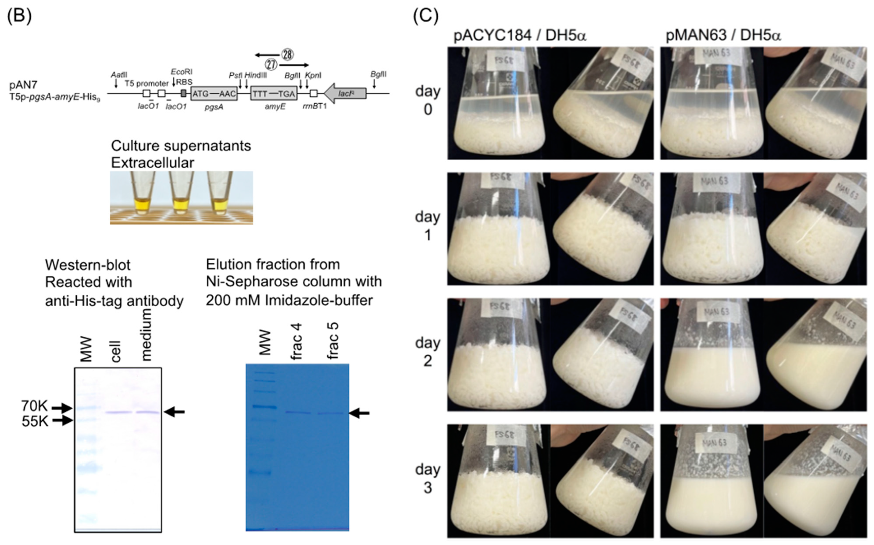
2.4. Western Blot Analysis of α-Amylase Expressed by Strain AN7

Plasmid pAN7, which expresses PgsA-AmyE with 9 additional His residues at the C-terminus, was obtained using inverse PCR using KOD-plus mutagenesis kit with pMAN63 as the template and primers 27 and 28. Strain AN7 (pAN7/DH5α) was grown in 50 mL of LB with 20 μg/mL chloramphenicol at 37 °C with reciprocal shaking at 120 rpm for 90 min. IPTG was added to a final concentration of 0.5 mM and the strain was allowed to grow for 18.5 h. An amount of 1 mL of the culture was centrifuged at 8000× g for 5 min at 4 °C. The collected cells were suspended in 500 μL of 100 mM Tris-HCl (pH 7.4), vortexed and then centrifuged at 8000× g for 5 min at 4 °C. After removing the supernatant, the precipitate was suspended in 100 μL of loading buffer, boiled for 5 min, centrifuged at 8000× g for 5 min and the supernatant was used as the cell sample. The proteins in the culture medium were concentrated as follows. The remaining culture was centrifuged at 5000× g for 5 min at 4oC. Ammonium sulfate was added to the collected supernatant to 80% saturation. The solution was centrifuged at 5000× g for 10 min at 4 °C. The precipitate was dissolved in 1 mL of 100 mM Tris-HCl (pH 7.4) and dialyzed against the same buffer. The dialyzed sample was concentrated to 100 μL using an Amicon Ultra centrifugal filter (Ultracel 30K; Millipore, Billerica, MA, USA). An amount of 10 μL of the concentrated sample was mixed with the same volume of loading buffer and boiled for 5 min to use as the extracellular sample. An amount of 10 μL of the cell and extracellular samples were subjected to SDS-polyacrylamide gel electrophoresis followed by Western blot analysis using rabbit anti-His-tag antibody (Medical & Biological Laboratories, Tokyo, Japan) as the first antibody and goat peroxidase-linked anti-rabbit IgG(H+L) (Jackson Immuno Research Laboratories, West Grove, PA, USA) as the second antibody. POD Immunostain Set (Fujifilm Wako, Osaka, Japan) was used to stain the bands reacted with the second antibody.

2.5. Purification of α-Amylase from the Culture Medium of AN7 and Determination of its N-Terminal Amino Acid Sequence

The remaining concentrated culture supernatant prepared above was subjected to 1.5 mL of Ni-Sepharose 6 Fast Flow (GE Healthcare Bio-Sciences, Uppsala, Sweden) column chromatography. Amylase was eluted with 20 mM Tris-HCl containing 0.5 M NaCl and 200 mM imidazole (pH 7.4), concentrated using an Amicon Ultra centrifugal filter and then was subjected to SDS-polyacrylamide gel electrophoresis followed by blotting onto a PVDF membrane (Immobilon P; Millipore, Billerica, MA, USA), staining with 0.025% Coomassie brilliant blue R-250 in 40% methanol and destaining with 60% methanol. After drying, the stained band was cut off and sent to Hokkaido System Science (Sapporo, Japan) for N-terminal amino acid sequencing by Edman degradation.

2.6. Production of Putrescine in 1 × M9 Steamed Rice Medium

An amount of 45.5 g of polished rice (Akita-komachi brand) and a stirrer bar (5 cm) were placed in a 300-mL conical beaker and washed twice with deionized water. After 54.6 mL of deionized water was poured into the conical beaker, the lid was covered with aluminum foil and the conical beaker was autoclaved at 121 °C for 20 min. After cooling, the rice was mixed with 100 mL of 2 × M9 minimal medium and ampicillin and chloramphenicol were added at the final concentrations of 100 μg/mL and 20 μg/mL, respectively. Strain SH2287 was grown overnight in 50 mL LB medium at 37 °C. Cells were collected by centrifugation at 5000× g for 10 min at 25 °C, washed twice with 1 × M9 buffer and then suspended in 1 mL of 1 × M9. The 1 × M9 steamed rice medium was inoculated with SH2287 prepared as above to an initial OD600 of 0.05 and incubated at 37 °C with stirring at 750 rpm. After 2 days of incubation, arginine and isopropyl-β-D-1-thiogalactopyranoside (IPTG) were added.
At the indicated time, 300 μL of the culture was sampled and the reaction was terminated by mixing with 30 μL 100% trichloroacetic acid. After allowing the sample to stand for 10 min, the sample was centrifuged at 8000× g for 10 min at 4 °C. 4 μL of the supernatant was mixed with 196 μL of HPLC mobile phase. After centrifugation at 8000× g for 5 min at 4 °C, the supernatant was filtrated using the Millex-LH Syringe Driven Filter Unit (Millipore, Billerica, MA, USA). The filtrate was analyzed by HPLC.

2.7. Measurement of Putrescine

An amount of 5 μL of the filtrates prepared as described above was injected into an HPLC (LC-20, Shimadzu, Kyoto, Japan) equipped with a fluorescence detector (RF-10AXL, Shimadzu) and TSKgel Polyaminepak column (4.6 × 50 mm) (Tosoh, Tokyo, Japan). The instrument was used as described previously [27,28]. The means of the putrescine concentrations of three independent cultures are shown in the results with standard deviations calculated by the STDEV function in Excel.

3. Results and Discussion

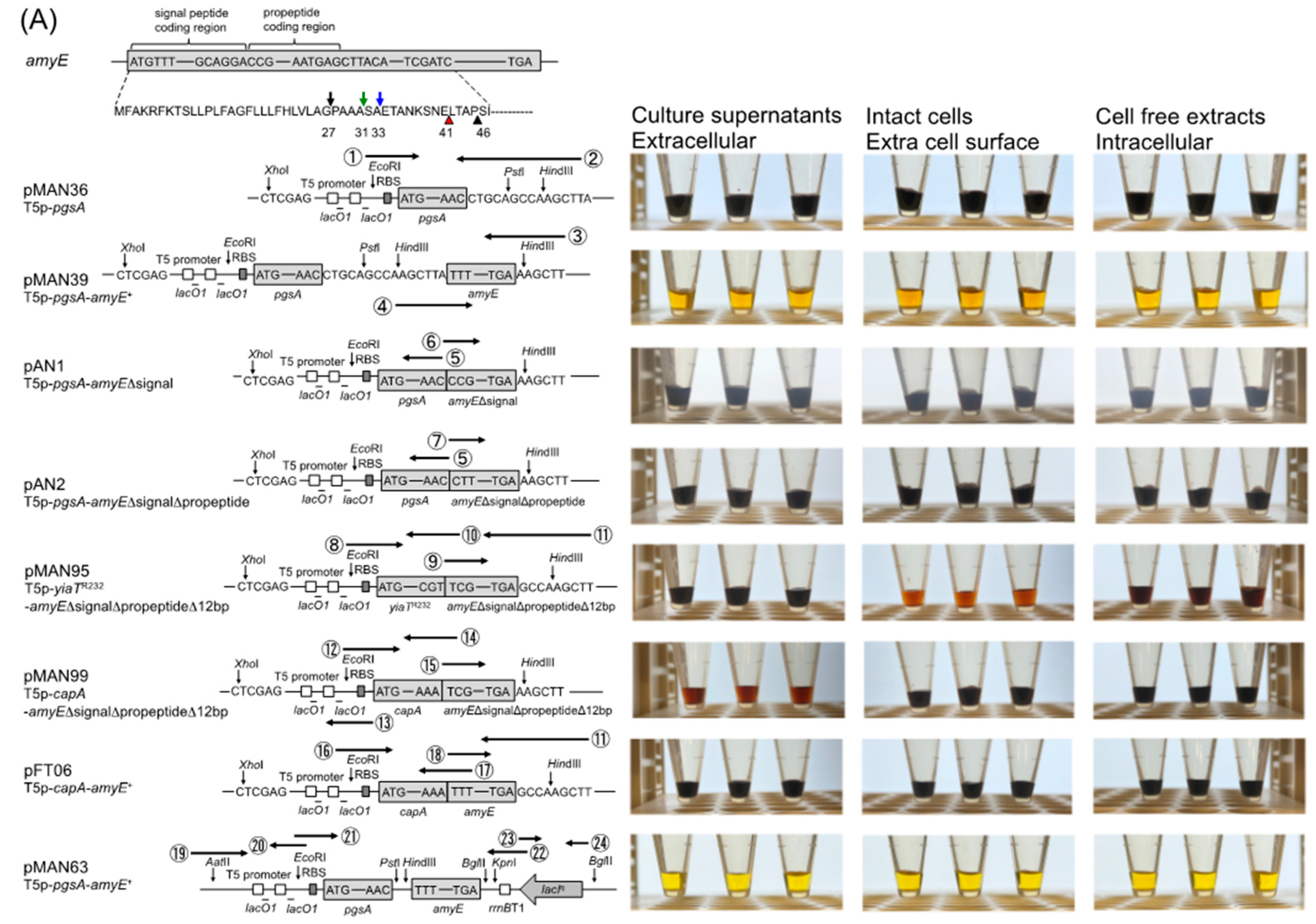
3.1. Comparison of Amylase Activity Expressed from Various Plasmids

There is some controversy as to which amino acid residues make up the signal peptide and the pro-peptide of Bacillus subtilis α-amylase, as shown in the upper left of Figure 2A. Culture supernatants, cell suspensions (intact cells) and cell-free solutions from each strain were mixed with soluble starch. After 54 h of incubation, the samples were subjected to the iodine-starch test and the amount of starch remaining was compared with the color. As starch is digested, the color of the reaction solution with iodine changes from dark bluish purple to brown, to golden, to yellow. As shown in Figure 2A, pMAN95 fused YiaTR232 to Ser46 of AmyE without inserting any additional amino acid residues resulting from the linker. On the other hand, the plasmid of Han and Lee [16] inserted Ser- and Arg-residues in between because of the restriction recognition site. DH5α transformed with pMAN95 expressed amylase on the extracellular surface as reported by Han and Lee [16]. DH5α transformed with pMAN99, which fused CapA to Ser46 of AmyE, expressed amylase in the medium but virtually no activity on the extracellular surface. DH5α transformed with pFT06, which fused CapA to Phe2 of AmyE, did not express amylase. On the other hand, to our surprise, DH5α transformed with pMAN39, which fused PgsA to Phe2 of AmyE, showed very high amylase activity in its culture supernatant, extracellular surface and cell-free solution. In contrast, DH5α transformed with pAN1 plasmid without a signal peptide and DH5α transformed with pAN2 plasmid without a signal peptide and propeptide showed no amylase activity. DH5α was transformed with pMAN36, which contains only pgsA without a stop codon at the regular position but has a stop codon after the addition of Leu-Gln-Pro-Ser-Leu-Ile-Ser. This transformant is a negative control and it had no amylase activity as expected. pMAN63 has the same construct from the T5 promoter-pgsA-amyE region as pMAN39, but the base plasmid is pACYC184 instead of pQE-80L. The color of the reaction mixture used DH5α transformed with pMAN63 is obviously brighter yellow than that of DH5α transformed with pMAN39. Therefore, we decided to use pACYC184-based plasmids.
As DH5α transformed with pMAN39 expressed a large amount of amylase outside, inside and on the extracellular surface of the cells, we wondered how the amylase expressed from this plasmid was processed and excreted into the medium. Plasmid pAN7 was designed to add a His-tag to the C-terminus of amylase (Figure 2B, top). After we confirmed that strain AN7 (pAN7/DH5α) showed very high amylase activity in the culture supernatant (Figure 2B, middle), the total cells and proteins collected from the culture supernatant of AN7 were subjected to SDS-polyacrylamide gel electrophoresis followed by Western blot analysis with anti-His-tag antibody. As shown in Figure 2B (bottom left), both samples showed bands between 70K and 55K. The molecular size of these bands suggested that the signal peptides were cleaved off in these cases. To confirm this, the amylase excreted in the culture supernatant was purified with the Ni-Sepharose column (Figure 2B, bottom right) and subjected to the N-terminal amino acid sequencing. The obtained N-terminal amino acid sequence, Ser-Ala-Glu-Thr-Ala, indicates that the signal peptide is cleaved between Ala31 and Ser32. This signal peptide cleavage site coincides with the site predicted by both SOSUI and SignalP programs.
DH5α transformed with each of pMAN63 and pACYC184 was grown in 1×M9 steamed rice medium to compare rice lysis. As shown in Figure 2C, DH5α transformed with pMAN63 obviously lysed rice on day 2, while the DH5α transformed with pACYC184, which does not express PgsA-AmyE, remained as swollen steamed rice in 1 x M9 steamed rice medium.
These results indicate that our first goal of constructing an E. coli strain that expresses high levels of amylase extracellularly using a membrane protein and grows on steamed rice was achieved.
Figure 2. (A) (Left) Schematics of the important parts of the amyE gene of Bacillus subtilis and translated sequence of the N-terminal part of AmyE (top) and plasmids used in this study. Downward black, green and blue arrows indicate signal peptide cleavage sites predicted by the Bsubcyc website, SOSUI and SignalP programs and Yamazaki et al. [29], respectively. Upward red arrow indicates propeptide cleavage site indicated by Bsubcyc website and Yamazaki et al. [29] and the N-terminal of the purified mature extracellular AmyE was Leu-42, as shown by Mäntsälä and Zalkin [30]. The coding regions for the signal peptide and propeptide shown above the sequence are described based on Bsubcyc. Upward black arrow indicates the position of the N-terminal of AmyE connected to YiaTR232 by Han and Lee [16]. pMAN63 is based on pACYC184 (CamR) and the other plasmids are based on pQE-80L (AmpR). (Right) Comparison of the iodine-starch reaction of the DH5α strains carrying the plasmids shown on the left using culture supernatants, intact cells and cell-free extracts. (B) Schematic of pAN7 (top) and the result of iodine-starch test of AN7 (middle). The result of Western blot analysis of AN7 cells and culture supernatant of AN7 and the result of SDS-PAGE of fractions 4 and 5 from Ni-Sepharose column (bottom). The MW marker used was the clearly stained protein ladder (Takara-Bio; Kyoto, Japan). (C) Comparison of the steamed rice media with pMAN63/DH5α and pACYC184/DH5α on day 0, 1, 2 and 3.
Applmicrobiol 03 00026 g002aApplmicrobiol 03 00026 g002b

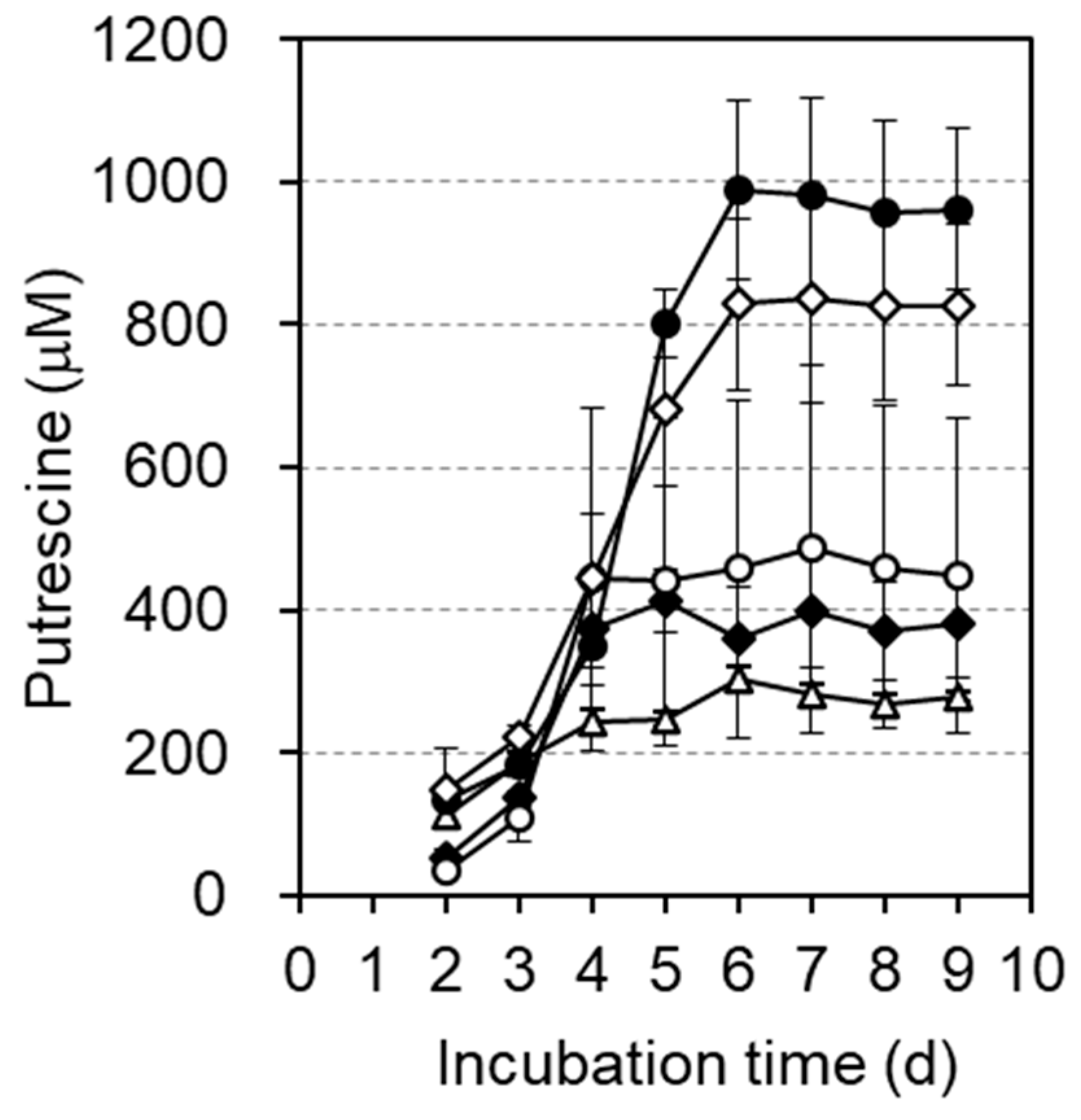
3.2. Comparison of Putrescine Production Expressed from Various Plasmids

Since pMAN63 gave a better result than pMAN39, the tetR gene region of pACYC184 was replaced by the T5 promoter-capA-amyE region of pMAN99 and the T5 promoter-yiaTR232-amyE region of pMAN95 to give pMAN100 and pAN5, respectively. Strain KT160 contains pKN11 in SH2204. Although SH2204 is a ΔargA strain, pKN11 contains argAATG Y19C, which has an efficient initiation codon ATG instead of GTG and a Y19C mutation that results in desensitization to arginine, so that KT160 is not arginine auxotrophic as long as ArgA is expressed from the plasmid. KT160, which produces a large amount of putrescine [23], was transformed with pMAN63, pMAN100 and pAN5, respectively. Putrescine production by these transformants was compared with 1×M9 steamed rice medium with the addition of ampicillin and chloramphenicol, but without the addition of arginine and IPTG as described in Materials and Methods. As shown in Table 3, KT160 transformed with pMAN63 had the highest putrescine production.

3.3. Effect of Arginine Supplementation on Putrescine Production

The effect of the addition of arginine on the 1 × M9 steamed rice medium on putrescine production was investigated.
By adding arginine to the medium, the yield of putrescine increased (Figure 3). The results suggested that the arginine productivity of KT160 transformed with pMAN63 was not sufficient. We did not add IPTG because pMAN63 allowed E. coli to produce a lot of amylase without the addition of IPTG. In addition, we were concerned that too much extracellular expression of amylase could affect the stability of the cell membrane.

3.4. Effect of IPTG Addition on Putrescine Production

Various concentrations of IPTG were added to the medium and the effects were observed in the presence of 5 mM arginine. Up to the addition of 0.5 mM of IPTG, there was a positive effect on putrescine production (Figure 4).
The yield of putrescine was improved to 1.2 mM by the addition of 0.5 mM IPTG. When we used KT160 for putrescine production, we obtained a maximum of about 1 mM putrescine using M9 galactose medium (data not reported). In addition, AN14, which expresses only ArgAATG Y19C from pSH1733 and SpeA and SpeB only from the genome, produced very low levels of putrescine even in the presence of 5 mM arginine. In contrast, SH2287, expressing SpeA and SpeB with ArgAATG Y19C from pKN11, produced significantly more putrescine.

4. Conclusions

We conclude that the supply of carbon and energy from steamed rice works in our developed system. Although there is a need to improve E. coli strains to produce more putrescine, this is the first report of an E. coli strain using steamed rice as the sole carbon and energy source to produce a chemical. Since the addition of IPTG leads to higher costs for industrialization, it is necessary to improve the method of inducing gene expression that does not require IPTG, such as the use of a temperature-sensitive repressor.

Author Contributions

Methodology, H.S.; formal analysis, N.I.; investigation, N.I. and M.N.; resources, M.N.; writing—original draft, H.S.; project administration, H.S. All authors have read and agreed to the published version of the manuscript.

Funding

This research did not receive any specific grant from funding agencies in the public, commercial or not-for-profit sectors.

Institutional Review Board Statement

Not applicable.

Informed Consent Statement

Not applicable.

Data Availability Statement

Some data that support the findings of this study are available from the corresponding author upon reasonable request.

Conflicts of Interest

The authors declare no commercial or financial conflict of interest.

Abbreviations

HPLC: high-performance liquid chromatography; IPTG: isopropyl-β-D-1-thiogalactopyranoside; OD, optical density; Puu: putrescine utilization; PVDF: polyvinylidene difluoride; SDS: sodium dodecyl sulfate.

References

  1. Kwak, D.; Lim, H.; Yang, J.; Seo, S.W.; Jung, G.Y. Synthetic redesign of Escherichia coli for cadaverine production from galactose. Biotechnol. Biofuels 2017, 10, 20. [Google Scholar] [CrossRef]
  2. Ma, W.; Chen, K.; Li, Y.; Hao, N.; Wang, X.; Ouyang, P. Advances in cadaverine bacterial production and its applications. Engineering 2017, 3, 308–317. [Google Scholar] [CrossRef]
  3. Meng, J.; Wang, B.; Liu, D.; Chen, T.; Wang, Z.; Zhao, X. High-yield anaerobic succinate production by strategically regulating multiple metabolic pathways based on stoichiometric maximum in Escherichia coli. Microb. Cell Fact. 2016, 15, 141. [Google Scholar] [CrossRef] [PubMed]
  4. Takahashi, S.; Miyachi, M.; Tamaki, H.; Suzuki, H. The Escherichia coli CitT transporter can be used as a succinate exporter for succinate production. Biosci. Biotechnol. Biochem. 2021, 85, 981–988. [Google Scholar] [CrossRef]
  5. Atsumi, S.; Cann, A.F.; Connor, M.R.; Shen, C.R.; Smith, K.M.; Brynildsen, M.P.; Chou, K.J.Y.; Hanai, T.; Liao, J.C. Metabolic engineering of Escherichia coli for 1-butanol production. Metab. Eng. 2008, 10, 305–311. [Google Scholar] [CrossRef] [PubMed]
  6. Ferreira, S.; Pereira, R.; Wahl, S.A.; Rocha, I. Metabolic engineering strategies for butanol production in Escherichia coli. Biotech. Bioeng. 2020, 117, 2571–2587. [Google Scholar] [CrossRef] [PubMed]
  7. Liu, Y.; Khusnutdinova, S.; Chen, J.; Crisante, D.; Batyrova, K.; Raj, K.; Feigis, M.; Shirzadi, E.; Wang, X.; Dorakhan, R.; et al. Systems engineering of Escherichia coli for n-butane production. Metab. Eng. 2022, 74, 98–107. [Google Scholar] [CrossRef]
  8. Zhao, M.; Huang, D.; Zhang, X.; Koffas, M.A.G.; Zhou, J.; Deng, Y. Metabolic engineering of Escherichia coli for producing adipic acid through the reverse adipate-degradation pathway. Metab. Eng. 2018, 47, 254–262. [Google Scholar] [CrossRef]
  9. Ning, Y.; Liu, H.; Zhang, R.; Jin, Y.; Yu, Y.; Deng, L.; Wang, F. Research progress on the construction of artificial pathways for the biosynthesis of adipic acid by engineered microbes. Fermentation 2022, 8, 393. [Google Scholar] [CrossRef]
  10. Choi, Y.J.; Park, J.H.; Kim, T.Y.; Lee, S.Y. Metabolic engineering of Escherichia coli for the production of 1-propanol. Metab. Eng. 2012, 14, 477–486. [Google Scholar] [CrossRef]
  11. Jain, R.; Sun, X.; Yuan, Q.; Yan, Y. Systematically engineering Escherichia coli for enhanced production of 1,2-propanediol and 1-propanol. ACS Synth. Biol. 2015, 4, 746–756. [Google Scholar] [CrossRef] [PubMed]
  12. Ingram, L.O.; Conway, T.; Clark, D.P.; Sewell, G.W.; Preston, J.F. Genetic engineering of ethanol production in Escherichia coli. Appl. Environ. Microbiol. 1987, 53, 2420–2425. [Google Scholar] [CrossRef] [PubMed]
  13. Fithriani; Suryadarma, P.; Mangunwidjaja, D. Metabolic engineering of Escherichia coli cells for ethanol production under aerobic conditions. Procedia Chem. 2015, 16, 600–607. [Google Scholar] [CrossRef]
  14. Sierra-Ibarra, E.; Alcaraz-Cienfuegos, J.; Vargas-Tah, A.; Rosas-Aburto, A.; Valdivia-López, Á.; Hernández-Luna, M.G.; Vivaldo-Lima, E.; Martinez, A. Ethanol production by Escherichia coli from detoxified lignocellulosic teak wood hydrolysates with high concentration of phenolic compounds. J. Ind. Microbiol. Biotechnol. 2022, 49, kuab077. [Google Scholar] [CrossRef]
  15. Jayalakshmi, S.; Joseph, K.; Sukumaran, V. Methane production from kitchen waste using Escherichia coli. J. Environ. Sci. Eng. 2007, 49, 99–102. [Google Scholar]
  16. Han, M.J.; Lee, S.H. An efficient bacterial surface display system based on a novel outer membrane anchoring element from the Escherichia coli protein YiaT. FEMS Microbiol. Lett. 2015, 362, 1–7. [Google Scholar] [CrossRef] [PubMed]
  17. Narita, J.; Okano, K.; Tateno, T.; Tanino, T.; Sewaki, T.; Sung, M.H.; Fukuda, H.; Kondo, A. Display of active enzymes on the cell surface of Escherichia coli using PgsA anchor protein and their application to bioconversion. Appl. Microbiol. Biotechnol. 2006, 70, 564–572. [Google Scholar] [CrossRef] [PubMed]
  18. Tateno, T.; Fukuda, H.; Kondo, A. Production of L-lysine from starch by Corynebacterium glutamicum displaying α-amylase on its cell surface. Appl. Microbiol. Biotechnol. 2007, 74, 1213–1220. [Google Scholar] [CrossRef]
  19. van Bloois, E.; Winter, R.T.; Kolmar, H.; Fraaije, M.W. Decorating microbes: Surface display of proteins on Escherichia coli. Trends Biotechnol. 2011, 29, 79–86. [Google Scholar] [CrossRef]
  20. Narita, J.; Okano, K.; Kitao, T.; Ishida, S.; Sewaki, T.; Sung, M.H.; Fukuda, H.; Kondo, A. Display of α-amylase on the surface of Lactobacillus casei cells by use of the PgsA anchor protein, and production of lactic acid from starch. Appl. Environ. Microbiol. 2006, 72, 269–275. [Google Scholar] [CrossRef]
  21. Suzuki, H.; Thongbhubate, K.; Muraoka, M.; Sasabu, S. Agmatine production by Escherichia coli cells expressing SpeA on the extracellular surface. Enzyme Microb. Technol. 2023, 162, 110139. [Google Scholar] [CrossRef] [PubMed]
  22. Suzuki, H.; Sasabui, A. First example of the extracellular surface expression of intrinsically periplasmic Escherichia coli γ-glutamyltranspeptidase, a member of the N-terminal nucleophile hydrolase superfamily, and the use of cells as a catalyst for γ-glutamylvalylglycine production. J. Agric. Food Chem. 2023, 71, 1132–1138. [Google Scholar] [PubMed]
  23. Thongbhubate, K.; Irie, K.; Sakai, Y.; Itoh, A.; Suzuki, H. Improvement of putrescine production through the arginine decarboxylase pathway in Escherichia coli K-12. AMB Express 2021, 11, 168. [Google Scholar] [CrossRef]
  24. Chang, A.C.; Cohen, S.N. Construction and characterization of amplifiable multicopy DNA cloning vehicles derived from the P15A cryptic miniplasmid. J. Bacteriol. 1978, 134, 1141–1156. [Google Scholar] [CrossRef]
  25. Ashikaga, S.; Nanamiya, H.; Ohashi, Y.; Kawamura, F. Natural genetic competence in Bacillus subtilis natto OK2. J. Bacteriol. 2000, 182, 2411–2415. [Google Scholar] [CrossRef] [PubMed]
  26. Ashiuchi, M.; Soda, K.; Misono, H. A poly-γ-glutamate synthetic system of Bacillus subtilis IFO3336: Gene cloning and biochemical analysis of poly-γ-glutamate produced by Escherichia coli clone cells. Biochem. Biophys. Res. Commun. 1999, 263, 6–12. [Google Scholar] [CrossRef]
  27. Thongbhubate, K.; Nakafuji, Y.; Matsuoka, R.; Kakegawa, S.; Suzuki, H. Effect of spermidine on biofilm formation in Escherichia coli K-12. J. Bacteriol. 2021, 203, e00652-20. [Google Scholar] [CrossRef]
  28. Kurihara, S.; Oda, S.; Tsuboi, Y.; Kim, H.G.; Oshida, M.; Kumagai, H.; Suzuki, H. γ-Glutamylputrescine synthetase in the putrescine utilization pathway of Escherichia coli K-12. J. Biol. Chem. 2008, 283, 19981–19990. [Google Scholar] [CrossRef]
  29. Yamazaki, H.; Ohmura, K.; Nakayama, A.; Takeichi, Y.; Otozai, K.; Yamasaki, M.; Tamura, G.; Yamane, K. α-Amylase genes (amyR2 and amyE+) from an α-amylase-hyperproducing Bacillus subtilis strain: Molecular cloning and nucleotide sequences. J. Bacteriol. 1983, 156, 327–337. [Google Scholar] [CrossRef]
  30. Mäntsälä, P.; Zalkin, H. Membrane-bound and soluble extracellular alpha-amylase from Bacillus subtilis. J. Biol. Chem. 1979, 254, 8540–8547. [Google Scholar] [CrossRef]
Figure 1. Metabolic pathways of polyamines in E. coli. Mutations introduced into strain KT160 used for putrescine production in [23] are indicated by red symbols and letters. Red arrows pointing upward indicate the overexpressed enzymes, red cross marks indicate the knocked-out genes and red letters on ArgA and YifE indicate the point mutations introduced.
Applmicrobiol 03 00026 g001
Figure 3. Effect of arginine addition on putrescine production by SH2287 (pMAN63/KT160). Arginine solution (pH 7.0) was added to the medium on day 2 at final concentrations of 0 (light triangles), 1 (black diamonds), 2 (light circles), 5 (black circles) and 10 mM (light diamonds). The mean values of three conical beakers are shown with the standard deviation.
Applmicrobiol 03 00026 g003
Figure 4. Putrescine production by AN14 (pSH1733/KT160; closed squares) and effect of IPTG addition on putrescine production by SH2287. IPTG was added to the medium for SH2287 at final concentrations of 0 (light circles), 0.1 (light diamonds), 0.5 (black circles) and 1 mM (black diamonds) with the addition of 5 mM arginine on day 2. The mean values of three conical beakers are shown with the standard deviation. The IPTG 0 mM graph (light circles) is a reuse of data from Figure 3. AN14 (black squares) was grown with the addition of 5 mM arginine and 0.5 mM IPTG as a control.
Applmicrobiol 03 00026 g004
Table 1. Strains and plasmids used in this study.
Strains and PlasmidsGenotypeSource and Reference
(Escherichia coli K-12)
AN7pAN7/DH5αThis study
AN14pMAN63 pSH1733/SH2204This study
DH5αF supE44 ΔlacU169(Φ80 lacZΔM15)
hsdR17 recA1 endA1 gyrA96 thi-1 relA1
KT160pKN11/SH2204[23]
MG1655F rph-1Carol A. Gross
SH2204F rph-1 ΔargR::FRT ΔpatA::FRT[23]
ΔpotE::FRT ΔspeD::FRT ΔspeG::FRT
ΔargA::FRT ΔpuuPA::FRT yifEQ100TAG
zie-296::Tn10
SH2287pMAN63 pKN11/SH2204This study
(plasmids)
pACYC184p15A replicon cat+ tet+Stanley N. Cohen, [24]
pAN1ColE1 replicon bla+ lacIq This study
T5p-pgsA+-amyE(Δsignal)
pAN2 ColE1 replicon bla+ lacIq This study
T5p-pgsA+-amyEΔsignalΔpropeptide
pAN5p15A replicon cat+ This study
T5p-yiaTR232-amyEΔsignalΔpropeptideΔ12bp
pAN7p15A replicon cat+ This study
tetS-T5p-pgsA+-amyE-His9-rrnBT1 lacIq
pFT06ColE1 replicon bla+ lacIq This study
T5p-capA+-amyE+
pKN11ColE1 replicon bla+ lacIq[23]
T5p-speA+-speB+-argAATG Y19C
pMAN36ColE1 replicon bla+ lacIq T5p-pgsA+This study
pMAN39ColE1 replicon bla+ lacIq This study
T5p-pgsA+-amyE+
pMAN63p15A replicon cat+This study
tetS-T5p-pgsA+-amyE+-rrnBT1 lacIq
pMAN95ColE1 replicon bla+ lacIq This study
T5p-yiaTR232-amyEΔsignalΔpropeptideΔ12bp
pMAN99ColE1 replicon bla+ lacIq This study
T5p-capA+-amyEΔsignalΔpropeptideΔ12bp
pMAN100p15A replicon cat+This study
T5p-capA+-amyEΔsignalΔpropeptideΔ12bp
pQE-80LColE1 replicon bla+ lacIq T5p-(His)6Quiagen
pSH1733ColE1 replicon bla+ lacIq T5p-argAATG Y19C[23]
(Bacillus subtilis)
168trpPasteur Institute
subsp. natto OK2bioFujio Kawamura, [25]
Table 2. Primers used to construct plasmids in this study.
No.NameDNA Sequence
1pQE_EcoRI_ATG_pgsA5′-cccgaattcattaaagaggagaaattaactATGTTTAACTTA-
CCGAATAAAATCAC-3′
2PstI_natto_pgsA_Cterm5′-cccctgcagGTTAGATGTTTTTAACGCTTCC-3′
3Bsub168 alpha-amylase_C5′-attaagcttTCAATGGGGAAGAGAACCGC-3′
4Bsub 168 alpha-amylase Hind3_N5′-GCCAAGCTTATTTGCAAAACGATTCAAAAC-3′
5pgsA C-comp5′-GTTAGATGTTTTTAACGCTTCCCAG-3′
6amyE deltasignal_F5′-CCGGCGGCTGCGAGTGCTGAAACGG-3′
7amyE deltasignal_F25′-CTTACAGCACCGTCGATCAAAAGCG-3′
8pQE80L-yiaTR232_infusion-F5′-caatttcacacagaattattaaagaggagaaattaactATGTTA-
ATTAATCGCAATATTG-3′
9amyE(136bp)-F5′-TCGATCAAAAGCGGAACCATTCTTCATGCA-
TGG-3′
10yiaTR232-amyE(136bp)_infuR5′-GGTTCCGCTTTTGATCGAACGATCAATCATC-
GGGCTGTCG-3′
11pQE80L-amyE_infuR.new5′-gctaattaagcttggcTCAATGGGGAAGAGAACCGC-
TTAAGC-3′
12EcoRI_RBS_ATG-ywtBF5′-gaattcattaaagaggagaaattaactATGAAAAAAGAA-
CTGAGCTTTCATGAAAAGCTGC-3′
13RBS_EcoRI-T5pR5′-agttaatttctcctctttaatgaattctgtgtgaaattgttatccgctcac-
aattg-3′
14amyE(136bp)-ywtB_infuR5′-GGTTCCGCTTTTGATCGATTTAGATTTTAGTT-
TGTCACTATGATC-3′
15amyE(136bp)-F5′-TCGATCAAAAGCGGAACCATTCTTCATGCA-
TGG-3′
16infuF_EcoRI-RBS-ywtB(25bp) new5′-gataacaatttcacacagaattcattaaagaggagaaattaact-
ATGAAAAAAGAACTGAGCTTTCATG-3′
17infuR_amyE(15bp)-ywtB(25bp)5′-GAATCGTTTTGCAAATTTAGATTTTAGTTT-
GTCACTATGATC-3′
18amyE-F5′-TTTGCAAAACGATTCAAAACCTCTTTACTG-
CCG-3′
19pACYC184_T5p-F5′-gcgaccacacccgtcctgtggatcgacgtctaagaaaccattattat-
catgacattaacc-3′
20pACYC184_T5p-R5′-ttctcctctttaatgaattctgtgtgaaattgttatccgctcacaattg-3′
21EcoRI_RBS_ATG_iPCR-F5′-gaattcattaaagaggagaaattaactATG-3′
22rrnBT1terminator_lacI-R5′-cccgactggaaagcgggcagtgaggcggatttgtcctactcagga-
gagcg-3′
23lacI-F5′-tcactgcccgctttccagtcgggaaacctgtcg-3′
24pACYC184_lacIqp-R5′-ccgccgccgcaaggaatggtggacaccatcgaatggtgcaaaac-
ctttcgcgg-3′
25BamH1-tamakiN5′-cccggatccgaggccctttcgtcttcacctcgag-3′
26Hind3-amyE-C5′-cccaagcttggcTCAATGGGGAAGAGAACCGCTT-
AAGC-3′
27H5_TGA_BglII_KpnI5′-CATCACCACCATCATTGAagatctgagcatggtaccc-
ttgaggc-3′
28H5_amyE_comp5′-GTGATGATGATGATGGGGAAGAGAACCGC-
TTAAGCCCCAGTC-3′
The numbers of the primers correspond to the numbers of the primers shown in Figure 2 and Table 3. Capital letters indicate the amino acid coding regions and stop codons. Single underlines and double underlines indicate the restriction endonuclease recognition sites and the α-amylase coding regions, respectively.
Table 3. Effect of amylase-expressing plasmid on putrescine yield.
Plasmid Used to Transform KT160Yield of
Putrescine (μM)
SDApplmicrobiol 03 00026 i001
pMAN632923.4
pMAN10022917
pAN52468.3
KT160 (pKN11/SH2204) was transformed with each plasmid. Day 9 yields are compared. SD: standard deviation.
Disclaimer/Publisher’s Note: The statements, opinions and data contained in all publications are solely those of the individual author(s) and contributor(s) and not of MDPI and/or the editor(s). MDPI and/or the editor(s) disclaim responsibility for any injury to people or property resulting from any ideas, methods, instructions or products referred to in the content.

