Article

Whole Genome Sequencing Evidences High Rates of Relapse in Clostridioides difficile Infection Caused by the Epidemic Ribotype 106

Microbiology Department, Hospital Universitari Son Espases, Instituto de Investigación Sanitaria Illes Balears (IdISBa), 07120 Palma de Mallorca, Spain
*
Author to whom correspondence should be addressed.
Appl. Microbiol. 2023, 3(1), 64-75; https://doi.org/10.3390/applmicrobiol3010005
Submission received: 12 November 2022 / Revised: 9 January 2023 / Accepted: 11 January 2023 / Published: 14 January 2023

Abstract

:
An increasing prevalence and spread of Clostridioides difficile infection (CDI) caused by DH/NAP11/106/ST-42 has been observed worldwide, probably fostered by its great capacity to produce spores or by the higher resistance rates observed for some strains. Based on the results of our previous study where RT106 showed higher recurrence rates than other relevant ribotypes, a genetic analysis by whole-genome sequencing (WGS) of primary and recurrent RT106 isolates from ten patients was performed to determine whether the higher rate of recurrence associated with RT106 is due to relapses, caused by the same strain, or reinfections, caused by different strains. MLST profiles, resistance mutations, and phylogenetic relatedness were determined by comparative single nucleotide variant (SNV) analysis. All isolates were classified as ST42, and those belonging to the same patient were isogenic, with one exception; strains belonging to different patients were not with two exceptions, pointing to putative transmission events. Phylogenetic analysis also suggested the presence of similar local epidemic lineages associated with moxifloxacin resistance, except for one patient whose isolates clustered with different nonresistant US strains. Our results show that recurrent CDIs caused by RT06/ST42 are mainly due to relapses caused by the primary strains, showing the higher capacity of RT106/ST42 to persist and cause recurrences as compared to other ribotypes.

1. Introduction

Increasing prevalence and spread of the epidemic DH/NAP11/106 has been reported in the last few years in regions where it was previously rarely found. Although initially identified in the United Kingdom in 1999, RT106 is now one of the predominant strains in many European countries, and is also becoming the most prevalent strain in the United States where RT106 has replaced RT027 as the most predominant ribotype recovered from community-associated C. difficile infections (CDI) and where it has also been reported as the second most prevalent molecular type in acute-care hospitals [1,2,3,4,5].
The last national surveillance study performed in 2013 in Spain suggested ribotypes 078/126, 014, and 001/072 as the most preponderant in this country [6], which was in accordance with our local surveillance data where ribotypes 014 and 078 were described as the most prevalent [7]. However, later studies showed changes in the epidemiology of C. difficile in Spain, such as the 2015–2016 surveillance analysis published by Suárez-Bode et al. [8], revealing RT106 as the main ribotype in both community associated (CA) and health-care facility associated (HCFA) CDIs. High prevalence has also been reported for RT106 in another Spanish hospital where it was also associated with large outbreaks and nosocomial transmission even between non-related patients [9].
The growing incidence and geographic distribution of the epidemic RT106 seems to be favored by different features that some authors have described to be associated with these ribotype, such as high level of sporulation combined with moderate toxin production, and resistance to environmental decontamination, as compared with other ribotypes and reference strains [10,11]. Further, antimicrobial resistance may be an important associated factor, since it is a major contributor to the pathogenesis and global dissemination of epidemic strains, such as BI/NAP1/ 027 [12,13,14]. In this sense, the highest prevalence of resistance has also been related to RT106 strains among adults in Ireland, Scotland, and Spain, especially fluoroquinolone resistance [8,15,16]. Moreover, RT106 also has ten accessory genomic elements (AGEs) with unique sequences that could be related to intestinal mucosal adhesion, biofilm formation, and sporulation, although the function of the proteins transcribed from these sequences need further study [1,17].
It has been speculated that the ability of RT106 to produce high levels of spores may be conducive to causing hospital outbreaks and recurrent disease more effectively than other ribotypes [1,18]. In this sense, different studies have revealed a higher recurrence rate of disease caused by RT106 strains in comparison with other major ribotypes [3,8]. Kociolek et al. identified several genes strongly associated with DH/NAP11/106 that could play a role in providing bacterial competitive advantages and association with recurrent CDI [17].
Different studies have determined whole-genome sequencing (WGS) as the most suitable method for tracking C. difficile due to its high sensitivity in discriminating strain genetic relatedness, as well as to identify putative transmission events [19]. Single nucleotide variant (SNV)-based analysis has also been widely adopted for CDI surveillance and has revealed some new evidence on transmission dynamics and recurrent infections [20]. The main approach of the present study was to analyze by WGS both primary and recurrent RT106 isolates from different patients with recurrent CDI based on the results observed in our previous study [8], where the highest number of patients that presented subsequent recurrences (29%) were associated with RT106, compared with other main ribotypes (6% for RT014, for example). Since RT106 showed a recurrence rate significantly higher than the others (26% vs. 9%, for ribotypes 106 and 014, respectively, p = 0.03), WGS could help us to find out whether the higher rate of recurrence associated with RT106 is, in fact, due to relapses—caused by the same strain—or to reinfections—caused by different strains.

2. Methods

2.1. Study Design and Clostridioides Difficile Isolation

The study was conducted as an observational retrospective analysis performed at the reference Hospital of the Balearic Islands (Spain), a 700-bed tertiary-care university hospital providing medical care for approximately 750,000 inhabitants. All the isolates of C. difficile from a cohort of patients diagnosed of recurrent episodes of CDI caused by RT106 between 2016 and 2018 were selected and included in the study. CDI cases were considered as a recurrence when a positive C. difficile stool specimen between two to eight weeks of the last positive specimen was reported in patients with clinical symptoms and following a previously clinically resolved episode [21]. Moreover, “late recurrences” (more than eight weeks following the previous resolved episode) were also included in this cohort in order to demonstrate whether the recurrence definition time should be extended [8].
All the isolates were obtained from stool specimens from hospital (inpatients), and ambulatory and primary care (outpatients) adult patients and children over two years of age with confirmed diagnosis of CDI. Specimens were cultured on cycloserine–cefoxitin–fructose agar plates (CLO agar; bioMérieux, Marcy l’Etoile, France) in an anaerobic chamber and incubated at 37 °C for 48 h, as previously described [8]. C. difficile isolates were frozen and stored at −80 °C to be processed for WGS at a later stage. Isolates were named as “CD” with the patient number and the letter “a” for initial episodes (first episode), “b” for the first recurrent episode (second episode), and “c” for the second recurrent episode (third episode).
Initial episodes of CDI were classified as health-care facility associated (HCFA-CDI) (hospital or community onset), community associated (CA-CDI), or as an indeterminate disease, according to the established definitions (European Surveillance of Clostridioides (Clostridium) difficile Infections, 2019) [21].

2.2. Molecular Characterization of the Isolates

Selection of isolates from patients with recurrent CDI caused by RT106 was carried out through the previous characterization of isolates by high-resolution capillary gel-based electrophoresis PCR-ribotyping using the protocol previously described by Fawley et al. [22]. Electropherograms were obtained by Gene Mapper (v.4.0.) and analyzed by the Webribo database [23].

2.3. Whole-Genome Sequencing and Analysis

All the isolates included underwent whole-genome sequencing. Genomic DNA was isolated using a commercial extraction kit (High Pure PCR template preparation kit; Roche Diagnostics, Mannheim, Germany). Indexed paired-end libraries were prepared with the Nextera XT DNA library preparation kit (Illumina Inc, San Diego, CA, USA) and then sequenced on an Illumina MiSeq® benchtop sequencer (Illumina Inc, San Diego, CA, USA) with MiSeq reagent kit v3 (Illumina Inc., San Diego, CA, USA), resulting in 300 bp paired-end reads. MLST analysis was performed using the online tool MLST-2.0. (https://cge.cbs.dtu.dk/services/cge/, accessed on 12 June 2020) [24].
To perform pairwise single-nucleotide variant (SNV) analysis, paired-end reads were aligned to the C. difficile DH/NAP11/106/ST-42 genome (GenBank accession: GCA_002234355.1) using Bowtie 2 v2.2.4 (bowtie2 –x <indexed_reference_genome> -q -1 <R1.fastq> -2 <R2.fastq> --phred33, -X 1000 –S <sam_file>) (http://bowtie-bio.sourceforge.net/bowtie2/index.shtml, accessed on 21 December 2015) and, eventually, pileup (sort <bam_file5> <output_directory>, pileup –c –f <reference_genome.fasta> <bam_file6>) and raw (sort <bam_file5> <output_directory>, pileup -vc –f <reference_genome.fasta> <bam_file6>) files were obtained using SAMtools v0.1.16 (https://sourceforge.net/projects/samtools/files/samtools/, accessed on 21 December 2015) and PicardTools v1.140 (https://github.com/broadinstitute/picard, accessed on 21 December 2015) (samtools view –b –S <sam_file> > <bam_file>, SortSaM INPUT=<bam_file> OUTPUT=<bam_file2> SORT_ORDER=coordinate, MarkDuplicates INPUT=<bam_file2> OUTPUT=<bam_file3>, AddOrReplaceReadGroups INPUT=<bam_file3> OUTPUT=<bam_file4>, BuildBamIndex INPUT=<bam_file4>). The Genome Analysis Toolkit (GATK) v3.4-46 (https://www.broadinstitute.org/gatk/, accessed on 21 December 2015) was employed for realignment around InDels (-T RealignerTargetCreator INPUT=<bam_file4> -R <reference-genome.fasta> -o <intervals>, -T IndelRealigner -maxConsensuses 60 --maxReadsForConsensuses 240 --maxReadsForRealignment 6000 –I <bam_file4> --targetintervals <intervals> -o <bam_file5>). SNVs were extracted from the raw files if they met the following criteria: a quality score (Phred-scaled probability of the sample reads being homozygous reference) of at least 50, a root-mean-square (RMS) mapping quality of at least 25, and a coverage depth of at least three reads, excluding all ambiguous variants. MicroInDels were extracted from the total pileup files by applying the following criteria: a quality score of at least 500, an RMS mapping quality of at least 25, and support of at least one-fifth of the covering reads.
No definitive criteria have been established for relatedness definition in C. difficile. Based on previous works, time elapsed between the different episodes and SNVs were considered to define isogenic isolates. Thus, isogenic isolates were considered those collected <124 days apart differing by ≤2 SNVs, or those collected between 124–364 days apart and differing by ≤3 SNVs. While unrelated isolates were considered those differing ≥10 SNVs [19,20]. Isolates differing by 3–10 SNVs were considered “presumably isogenic” or “related” isolates when they belong to the same patients.
SNV calling is also a very useful method for local transmission analysis, therefore plausible transmission events were considered between patients when isolates differed ≤2 SNVs within ≤90 days.
In order to analyze relatedness between isolates, genomes were de novo assembled using SPAdes v3.12.1 (spades.py –o -1 R1.fastq -2 R2.fastq --careful), and a core genome phylogenetic reconstruction was built with Parsnp from the Harvest Suite package v1.2 with default parameters forcing the inclusion of all genomes (parsnp –c –d <genomes_folder> -r <reference_genome.fasta>) and including other available ST42 genomes (ENA Bioproject number: PRJNA340238) [17].
The nucleotide sequences for the twenty-four genomes included in this study have been deposited at DDBJ/ENA/GenBank under the following number project: PRJEB43620.

2.4. Antimicrobial Susceptibility and Mechanisms of Resistance Analysis

Susceptibility to metronidazole, vancomycin, erythromycin, and moxifloxacin using gradient antibiotic strips (Etest®, BioMérieux, Marcy l’Etoile, France) on Brucella agar plates (BD Biosciences, Oxford, UK) was tested for all isolates. The MIC breakpoints used were those established by the European Committee on Antimicrobial Susceptibility Testing (EUCAST) for metronidazole (>2 mg/L) and vancomycin (>2 mg/L) and the ECOFF value for moxifloxacin (4 mg/L) and erythromycin (2 mg/L) (v.8.0; (http://www.eucast.org/clinical_breakpoints/, accessed on 17 June 2018).
Draft genomes were screened to identify acquired genes mediating antimicrobial resistance or chromosomal mutations of resistance for murG, rpoB, gyrA, gyrB, cfr, tetM, catP, and ermB genes, and also by using the online tool Resfinder (https://cge.cbs.dtu.dk/services/ResFinder/, accessed on 20 June 2020) [25,26].

3. Results

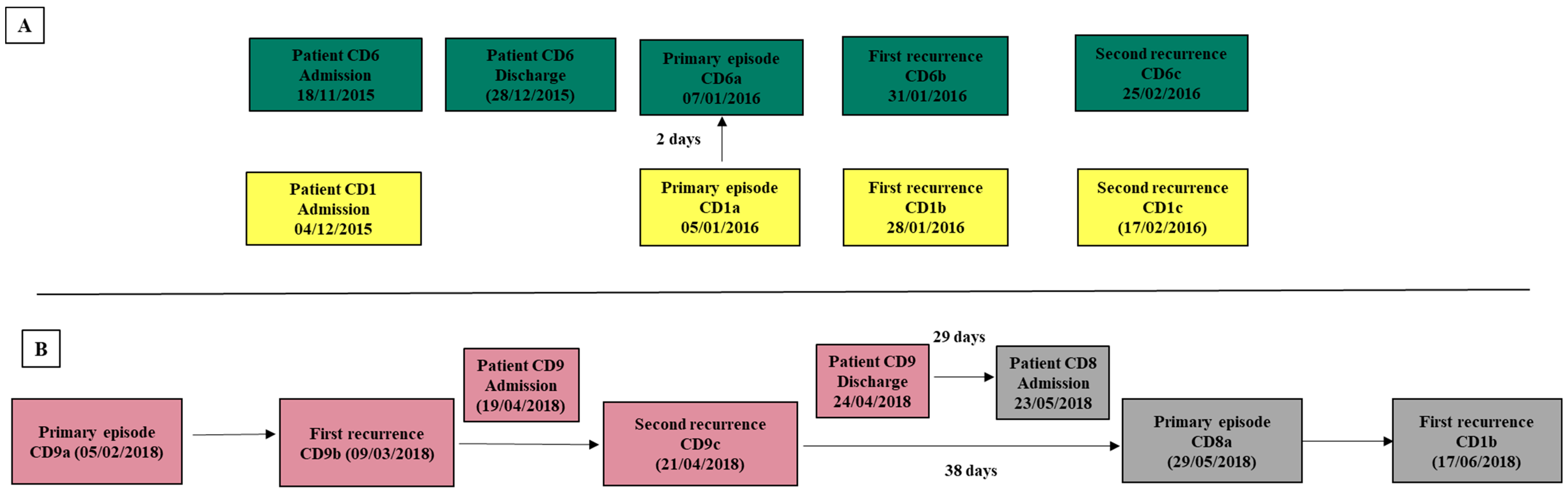
3.1. Patients and Isolates Data Collection

Ten patients with recurrent diagnosis of CDI caused by RT106 were included for the study, assuming a total of twenty-four isolates. Ten isolates were obtained from initial infections and fourteen from recurrent episodes, including more than one recurrence in four patients.
The median age of patients was 64 (2–92) years, and the mean time between different recurrent episodes was 24.5 (19–127) days, including twelve recurrent episodes, according to the accepted definition and two “late recurrences” (78 and 127 days after the previous episode). Primary CDI episodes were considered—three as HCFA-CDI, six as CA-CDI, and one case was classified as an indeterminate disease. Table 1 summarizes the main epidemiological data of patients and their isolates.

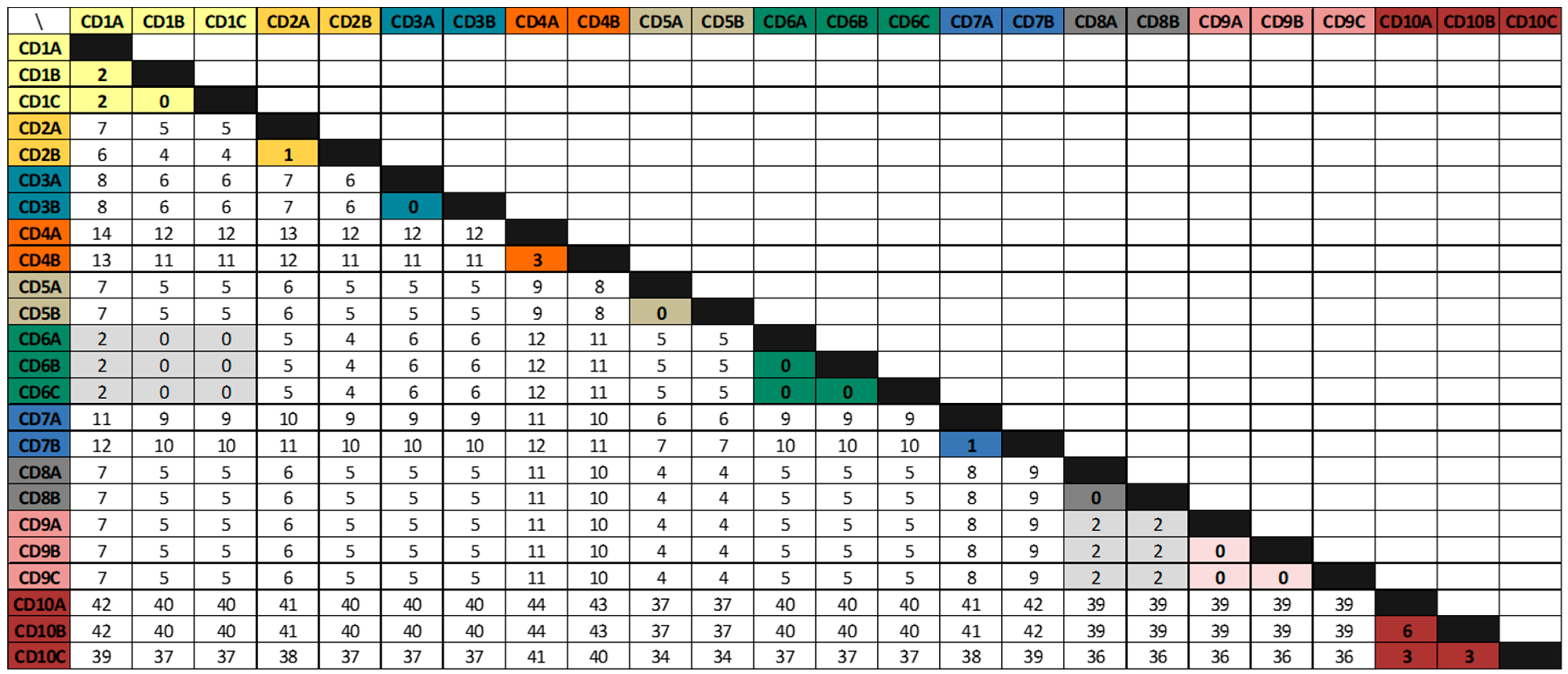
3.2. Whole-Genome Sequencing Analysis

All the isolates included in the study were classified as sequence-type (ST) 42 based on the MLST analysis, hence no other ST belonging to RT106 clade was found in this collection of isolates.
SNV comparative analysis (variant calling) of isolates from the same patient showed that recurrent episodes of patients CD1, CD2, CD3, CD5, CD6, CD7, CD8, and CD9 were caused by isogenic strains, since isolates from the same patient differed by 0 to 2 SNVs and were collected less than 124 days apart. Furthermore, variant calling analysis showed only three SNVs of difference between isolates belonging to patient CD4 and also between isolates CD10a and CD10b and isolate CD10c, all of which had been collected fewer than 124 days apart and, therefore, did not meet the strict definition of isogenic strains. However, these strains were considered “presumably isogenic” isolates. Finally, isolates CD10a and CD10b differed by six SNVs and were, therefore, non-isogenic, but not unrelated strains (Figure 1).
Isolates belonging to different patients differed by 4 to 43 SNVs, with the exception of the isolates of patients CD1 and CD6 on the one hand and isolates of patients CD8 and CD9 on the other, differing by only 2 SNVs, in both cases suggesting plausible transmission events within these patients. Patients CD1 and CD6 had been admitted to the same health-care institution (less than 100 beds) at the same time with CDI cases occurring within ≤90 days. Although the presence of these two patients on the same ward was not confirmed, they likely received care from the same staff. Meanwhile, patients CD8 and CD9 had also been admitted in the same hospital, albeit not during the same period and were admitted to different rooms and even different floors and there is no evidence that they were cared for by the same staff; however, CDI episodes also occurred within ≤90 days (Figure 2).
Comparative analysis between isolates from the same patients showed only two patients with non-synonymous changes between their primary and recurrent isolates (Table 2), one of them (Ala66Val) located at the S-layer protein SlpA. Therefore, no common mutations were found between recurrent isolates either.

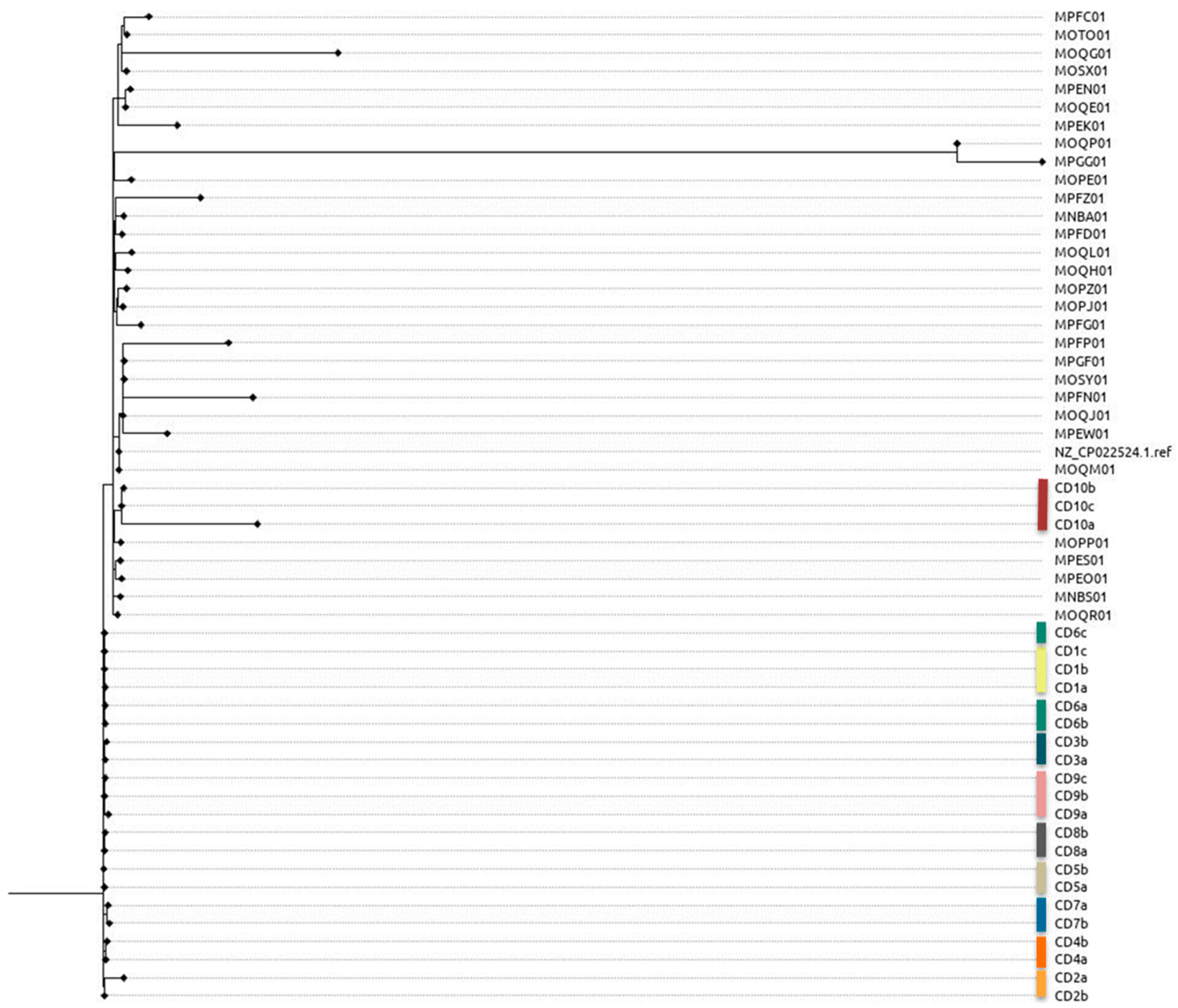
3.3. Phylogenetic Analysis

Core phylogenetic analysis showed three main branches grouping all the isolates in five clusters. Isolates from patient CD10 clustered with some US isolates, included in the phylogenetic tree for comparison, showing greater relatedness with them; and isolates from the rest of patients (CD1–CD9) gathered in four clusters, showing greater similarity between them. Moreover, this analysis supported direct transmission between patients CD1 and CD6 and patients CD8 and CD9, with their respective isolates clustering together in both cases (Figure 3). Synonymous and non-synonymous changes found in the isolates regarding the reference strain also confirm the observed relatedness in the phylogenetic tree, since common changes were found in all the isolates for patients CD1 to CD9, differing from those observed in patient CD10 isolates, confirming the presence of a different lineage in this case. All mutations and nucleotide changes found for all the isolates are summarized in the Supplementary Material (Supplementary Table S1).

3.4. Antimicrobial Susceptibility and Mechanisms of Resistance

All isolates were susceptible to metronidazole and vancomycin and resistant to moxifloxacin with MICs > 32 mg/L, except for isolates from patient CD10 that were also susceptible to moxifloxacin. Only isolate CD3b was considered resistant to erythromycin (MIC 2 mg/L). WGS analysis showed the Thr82Ile mutation in gyrA for moxifloxacin resistant isolates with no other resistance mutations for the screened genes and no other acquired resistant genes were found with the Resfinder analysis.

4. Discussion

Several factors seem to contribute to the emergence and spread of RT106 strains, such as the high level of sporulation demonstrated for this ribotype and the higher rates of antibiotic resistance reported by different studies [8,10,18]. Moreover, some of these advantages appear to be related to the ability of RT106 to produce recurrent disease more effectively than other ribotypes. Kociolek et al. observed that CDI caused by DH/NAP11/106 was more likely to result in multiple CDI relapses (40% vs. 8%; p = 0.05) when they studied the molecular epidemiology by restriction endonuclease analysis (REA) groups among children with multiple CDIs [3]. Based on the higher recurrence rate (26%) that had also been observed for RT106 in our previous study as compared to other major ribotypes (p < 0.05), we sequenced all the isolates from ten different patients with recurrent CDIs caused by this ribotype to find out whether recurrences were caused by the same strain—considering them as relapses—or by different strains of the same ribotype—causing reinfections. Variant calling analysis showed that recurrent episodes were caused mostly by clonal strains, since recurrent isolates were isogenic (≤2 SNVs within isolates) with the strain responsible of the previous episode, apart from one patient where a recurrent isolate was non-isogenic but closely related to the previous strain (only six SNVs within isolates). Based on these findings, it seems that recurrent C. difficile infections caused by RT06/ST-42 are due to relapses caused by a clonal strain, suggesting a higher capacity of these strains to persist and cause a greater number of recurrences than other ribotypes. As other authors have commented previously, we cannot assume that relapses are caused by the persistence of strains in the gut, since spores of C. difficile are capable of persisting for a long time in the environment, hence infections caused by the same strain cannot be ruled out [9]. However, the fact that no isogenic strains were observed among the majority of patients (differing between 4 and 43 SNVs) makes this theory less likely, since more isogenic strains would be expected to be found among the different patients if they had been reinfected with persistent spores of the same strain present in the environment. In this sense, a new important study has recently demonstrated a novel mechanism employed by C. difficile spores to gain entry into the intestinal mucosa via pathways dependent on host fibronectin-α5β1 and vitronectin-αvβ1 where the exosporium protein BclA3, on the spore surface, is required for both entry pathways, thereby contributing to the recurrence of disease [27].
No common mutations were detected in the recurrent isolates to explain adaptation mechanisms or any kind of advantage for these isolates to persist in the human gut; nevertheless, accessory genome elements (AGEs) need further study since some mechanisms related to adaptation or persistence could be related to them. Many of the CDSs identified in the genome of C. difficile are associated with adaptation and proliferation in the gastrointestinal tract (germination, adhesion, and growth) and survival in challenging suboptimal environments (endospore formation), supporting the view that C. difficile lives within a highly dynamic niche and is able to spend a long-time coexisting with its host [28]. In fact, comparative genomic studies found that RT106 had the lowest conservation of the core genes present in the reference strain CD630 of all the ribotypes tested, while showing 100% conservation of the divergent sequences present in the hypervirulent RT027, which may represent genes associated with increased virulence [1], although more studies are needed in order to associate gene sequences with virulent phenotypes and/or epidemic strains. A recent study published by Roxas et al., shows clade-specific properties, including those conferred by genes within the genomic island GI1, present in RT106/ST28/ST42 strains, which could explain the greater emergence of these clones by assessing virulence-associated phenotypes including motility, toxin production, biofilm production, and adhesion to collagen [5]. However, other factors must play a role as well and more studies are needed to confirm and better understand this process.
Further, two plausible transmission events between patients were also documented, since their isolates were isogenic, and episodes took place during the same time period (within ≤90 days). These two events, however, occurred in different situations. In one of the cases, the two patients involved had been admitted to the same health-care center at the same time and they probably received care from the same staff, while in the other case, patients were admitted at the same hospital at different times and with different locations. García-Fernández et al., showed a frequent within-hospital transmission of healthcare-associated ribotypes, including ribotype 106, and they also observed that a significant proportion of transmissions occurred either indirectly, through environmental contamination with C. difficile spores, or from reservoirs outside of CDI patients, such as asymptomatically colonized patients or staff [9].
All the studied isolates were differentiated as ST42, which seems to be the most related sequence type with the increasing dissemination of RT106, although others such as ST28 have also been associated [5,17]. Phylogenetic analysis revealed a close relatedness between isolates from all patients, suggesting the presence of similar local epidemic linages, except in one patient whose isolates clustered with other US isolates. Besides this, all the isolates belonging to the local epidemic lineages were resistant to moxifloxacin, unlike the other US lineage that displayed the previously described Thr82Ile mutation in gyrA. Resistance to fluoroquinolones in DH/NAP11/106 strains has been previously reported in Europe among adults in different countries such as Ireland, Scotland, and Spain [8,15,16], in contrast to isolates reported from North America where resistance to moxifloxacin has not been associated with this ribotype [17].
Our work highlights the importance of WGS to determine relapses—caused by the same strain—or reinfections—caused by different strains. Current definitions are based only on the time elapsed since the previous episode (eight weeks or fewer), but there are “late” recurrences (more than eight weeks) that might be caused by the same strain, highlighting the need to change the standard definition of relapse. Additionally, as other authors have considered, differentiating between relapses and reinfections might be important for controlling CDI, whether through interventions to manage C. difficile transmission, or by implementing treatment policies requiring different handling or even individualized therapeutic strategies [29].
Our study has some limitations insomuch as it was performed in a single center with a limited number of patients; however, it is significant in that all the recurrent isolates except one, very closely related to the previous one (six SNVs), were isogenic with respect to their primary isolates. Moreover, despite it being logical to think that there is a possible limitation in identifying relapses or transmission events using WGS of a single colony, Balaji et al. identified rare within-host genetic diversity of C. difficile in stool of children with CDI or asymptomatic carriage, suggesting that WGS of a single colony from stool will appropriately characterize isolate clonality and putative transmission events in most cases [30]. Although there are different studies that have shown that mixed infections can occur due to different strains, they were not found in our patients [31]; but the persistence of the same strain was demonstrated in these patients.

5. Conclusions

High rates of relapse in CDI caused by RT106/ST42 strains, mainly represented by local epidemic strains associated to fluoroquinolone resistance, have been demonstrated using WGS. However, additional studies are needed to better understand the mechanisms of persistence possessed by these strains to cause recurrent CDI.

Supplementary Materials

The following supporting information can be downloaded at: https://www.mdpi.com/article/10.3390/applmicrobiol3010005/s1, Table S1: Isolates mutations and nucleotide changes.

Author Contributions

Conceptualization, validation, formal analysis, investigation, data curation, writing—original draft preparation, writing—review and editing, visualization and supervision, A.M.; methodology, investigation and data curation, L.S.-B.; conceptualization, validation, formal analysis, data curation and supervision, C.L.-C.; methodology, R.M.A.; conceptualization, visualization and supervision, A.O. All authors have read and agreed to the published version of the manuscript.

Funding

This research received no external funding.

Institutional Review Board Statement

This retrospective study was approved by the Hospital Committee for Clinical Research of the Son Espases University Hospital as a pilot project (INSD/MENA).

Informed Consent Statement

Not applicable.

Data Availability Statement

The nucleotide sequences for the twenty-four genomes included in this study are openly available in DDBJ/ENA/GenBank at https://www.ebi.ac.uk/ena/browser/view/PRJEB43620, reference number PRJEB43620, accessed on 5 August 2021.

Conflicts of Interest

The authors declare no conflict of interest.

References

  1. Carlson, T.J.; Blasingame, D.; Gonzales-Luna, A.J.; Alnezary, F.; Garey, K.W. Clostridioides difficile ribotype 106: A systematic review of the antimicrobial susceptibility, genetics, and clinical outcomes of this common worldwide strain. Anaerobe 2020, 62, 102142. [Google Scholar] [CrossRef] [PubMed]
  2. Wilcox, M.H.; Shetty, N.; Fawley, W.N.; Shemko, M.; Coen, P.; Birtles, A.; Cairns, M.; Curran, M.D.; Dodgson, K.J.; Green, S.M.; et al. Changing epidemiology of Clostridium difficile infection following the introduction of a national ribotyping-based surveillance scheme in England. Clin. Infect. Dis. 2012, 55, 1056–1063. [Google Scholar] [CrossRef] [PubMed] [Green Version]
  3. Kociolek, L.K.; Patel, S.J.; Shulman, S.T.; Gerding, D.N. Molecular epidemiology of Clostridium difficile infections in children: A retrospective cohort study. Infect. Control. Hosp. Epidemiol. 2015, 36, 445–451. [Google Scholar] [CrossRef] [PubMed]
  4. Centers for Disease Control and Prevention. Healthcare-Associated Infections—Community Interface (HAIC). Clostridioides difficile infection (CDI) Tracking. Available online: https://www.cdc.gov/hai/eip/cdiff-tracking.html (accessed on 11 May 2020).
  5. Roxas, B.A.P.; Roxas, J.L.; Claus-Walker, R.; Harishankar, A.; Mansoor, A.; Anwar, F.; Jillella, S.; Williams, A.; Lindsey, J.; Elliott, S.P.; et al. Phylogenomic analysis of Clostridioides difficile ribotype 106 strains reveals novel genetic islands and emergent phenotypes. Sci. Rep. 2020, 10, 22135. [Google Scholar] [CrossRef]
  6. Alcalá, L.; Reigadas, E.; Marín, M.; Martín, A.; Catalán, P.; Bouza, E. Spanish Clostridium difficile Study Group. Impact of clinical awareness and diagnostic tests on the underdiagnosis of Clostridium difficile infection. Eur. J. Clin. Microbiol. Infect. Dis. 2015, 34, 1515–1525. [Google Scholar] [CrossRef]
  7. Weber, I.; Riera, E.; Déniz, C.; Pérez, J.L.; Oliver, A.; Mena, A. Molecular epidemiology and resistance profiles of Clostridium difficile in a tertiary care hospital in Spain. Int. J. Med. Microbiol. 2013, 303, 128–133. [Google Scholar] [CrossRef]
  8. Suárez-Bode, L.; Barrón, R.; Pérez, J.L.; Mena, A. Increasing prevalence of the epidemic ribotype 106 in healthcare facility-associated and community-associated Clostridioides difficile infection. Anaerobe 2019, 55, 124–129. [Google Scholar] [CrossRef]
  9. García-Fernández, S.; Frentrup, M.; Steglich, M.; Gonzaga, A.; Cobo, M.; López-Fresneña, N.; Cobo, J.; Morosini, M.I.; Cantón, R.; Del Campo, R.; et al. Whole-genome sequencing reveals nosocomial Clostridioides difficile transmission and a previously unsuspected epidemic scenario. Sci. Rep. 2019, 9, 6959. [Google Scholar] [CrossRef] [Green Version]
  10. Vohra, P.; Poxton, I.R. Comparison of toxin and spore production in clinically relevant strains of Clostridium difficile. Microbiology (Reading) 2011, 157, 1343–1353. [Google Scholar] [CrossRef] [Green Version]
  11. Vohra, P.; Poxton, I.R. Efficacy of decontaminants and disinfectants against Clostridium difficile. J. Med. Microbiol. 2011, 60, 1218–1224. [Google Scholar] [CrossRef]
  12. Kelly, C.P.; LaMont, J.T. Clostridium difficile—More difficult than ever. N. Engl. J. Med. 2010, 359, 1932–1940, Erratum in N. Engl. J. Med. 2010, 363, 1585. [Google Scholar] [CrossRef] [PubMed]
  13. He, M.; Miyajima, F.; Roberts, P.; Ellison, L.; Pickard, D.J.; Martin, M.J.; Connor, T.R.; Harris, S.R.; Fairley, D.; Bamford, K.B.; et al. Emergence and global spread of epidemic healthcare-associated Clostridium difficile. Nat. Genet. 2013, 45, 109–113. [Google Scholar] [CrossRef] [PubMed]
  14. Dingle, K.E.; Didelot, X.; Quan, T.P.; Eyre, D.W.; Stoesser, N.; Golubchik, T.; Harding, R.M.; Wilson, D.J.; Griffiths, D.; Vaughan, A.; et al. Modernising Medical Microbiology Informatics Group. Effects of control interventions on Clostridium difficile infection in England: An observational study. Lancet Infect. Dis. 2017, 17, 411–421. [Google Scholar] [CrossRef] [PubMed] [Green Version]
  15. Mutlu, E.; Wroe, A.J.; Sanchez-Hurtado, K.; Brazier, J.S.; Poxton, I.R. Molecular characterization and antimicrobial susceptibility patterns of Clostridium difficile strains isolated from hospitals in south-east Scotland. J. Med. Microbiol. 2007, 56, 921–929. [Google Scholar] [CrossRef] [Green Version]
  16. Solomon, K.; Fanning, S.; McDermott, S.; Murray, S.; Scott, L.; Martin, A.; Skally, M.; Burns, K.; Kuijper, E.; Fitzpatrick, F.; et al. PCR ribotype prevalence and molecular basis of macrolide-lincosamide-streptogramin B (MLSB) and fluoroquinolone resistance in Irish clinical Clostridium difficile isolates. J. Antimicrob. Chemother. 2011, 66, 1976–1982. [Google Scholar] [CrossRef]
  17. Kociolek, L.K.; Gerding, D.N.; Hecht, D.W.; Ozer, E.A. Comparative genomics analysis of Clostridium difficile epidemic strain DH/NAP11/106. Microbes Infect. 2018, 20, 245–253. [Google Scholar] [CrossRef]
  18. Baines, S.D.; O’Connor, R.; Saxton, K.; Freeman, J.; Wilcox, M.H. Activity of vancomycin against epidemic Clostridium difficile strains in a human gut model. J. Antimicrob. Chemother. 2009, 63, 520–525. [Google Scholar] [CrossRef] [Green Version]
  19. Eyre, D.W.; Cule, M.L.; Wilson, D.J.; Griffiths, D.; Vaughan, A.; O’Connor, L.; Ip, C.L.; Golubchik, T.; Batty, E.M.; Finney, J.M.; et al. Diverse sources of C. difficile infection identified on whole-genome sequencing. N. Engl. J. Med. 2013, 369, 1195–1205. [Google Scholar] [CrossRef] [Green Version]
  20. Janezic, S.; Rupnik, M. Development and Implementation of Whole Genome Sequencing-Based Typing Schemes for Clostridioides difficile. Front. Public Health 2019, 7, 309. [Google Scholar] [CrossRef]
  21. European Centre for Disease Prevention and Control. European Surveillance of Clostridioides (Clostridium) Difficile Infections. Surveillance Protocol Version 2.4; ECDC: Stockholm, Sweden, 2019.
  22. Fawley, W.N.; Knetsch, C.W.; MacCannell, D.R.; Harmanus, C.; Du, T.; Mulvey, M.R.; Paulick, A.; Anderson, L.; Kuijper, E.J.; Wilcox, M.H. Development and validation of an internationally-standardized, high-resolution capillary gel-based electrophoresis PCR-ribotyping protocol for Clostridium difficile. PLoS ONE 2015, 10, e0118150. [Google Scholar] [CrossRef]
  23. Indra, A.; Huhulescu, S.; Schneeweis, M.; Hasenberger, P.; Kernbichler, S.; Fiedler, A.; Wewalka, G.; Allerberger, F.; Kuijper, E.J. Characterization of Clostridium difficile isolates using capillary gel electrophoresis-based PCR ribotyping. J. Med. Microbiol. 2008, 57, 1377–1382. [Google Scholar] [CrossRef] [PubMed] [Green Version]
  24. Larsen, M.V.; Cosentino, S.; Rasmussen, S.; Friis, C.; Hasman, H.; Marvig, R.L.; Jelsbak, L.; Sicheritz-Pontén, T.; Ussery, D.W.; Aarestrup, F.M.; et al. Multilocus sequence typing of total-genome-sequenced bacteria. J. Clin. Microbiol. 2012, 50, 1355–1361. [Google Scholar] [CrossRef] [PubMed] [Green Version]
  25. Bortolaia, V.; Kaas, R.S.; Ruppe, E.; Roberts, M.C.; Schwarz, S.; Cattoir, V.; Philippon, A.; Allesoe, R.L.; Rebelo, A.R.; Florensa, A.F.; et al. ResFinder 4.0 for predictions of phenotypes from genotypes. J. Antimicrob. Chemother. 2020, 75, 3491–3500. [Google Scholar] [CrossRef]
  26. Zankari, E.; Allesøe, R.; Joensen, K.G.; Cavaco, L.M.; Lund, O.; Aarestrup, F.M. PointFinder: A novel web tool for WGS-based detection of antimicrobial resistance associated with chromosomal point mutations in bacterial pathogens. J. Antimicrob. Chemother. 2017, 72, 2764–2768. [Google Scholar] [CrossRef] [PubMed] [Green Version]
  27. Castro-Córdova, P.; Mora-Uribe, P.; Reyes-Ramírez, R.; Cofré-Araneda, G.; Orozco-Aguilar, J.; Brito-Silva, C.; Mendoza-León, M.J.; Kuehne, S.A.; Minton, N.P.; Pizarro-Guajardo, M.; et al. Entry of spores into intestinal epithelial cells contributes to recurrence of Clostridioides difficile infection. Nat. Commun. 2021, 12, 1140. [Google Scholar] [CrossRef] [PubMed]
  28. Knight, D.R.; Elliott, B.; Chang, B.J.; Perkins, T.T.; Riley, T.V. Diversity and Evolution in the Genome of Clostridium difficile. Clin. Microbiol. Rev. 2015, 28, 721–741. [Google Scholar] [CrossRef] [Green Version]
  29. Cho, J.; Cunningham, S.; Pu, M.; Lennon, R.J.; Dens Higano, J.; Jeraldo, P.; Sampathkumar, P.; Shannon, S.; Kashyap, P.C.; Patel, R. Clostridioides difficile Whole-genome Sequencing Differentiates Relapse With the Same Strain From Reinfection With a New Strain. Clin. Infect. Dis. 2021, 72, 806–813. [Google Scholar] [CrossRef]
  30. Balaji, A.; Ozer, E.A.; Kociolek, L.K. Clostridioides difficile Whole-Genome Sequencing Reveals Limited Within-Host Genetic Diversity in a Pediatric Cohort. J. Clin. Microbiol. 2019, 57, e00559-19. [Google Scholar] [CrossRef] [Green Version]
  31. Behroozian, A.A.; Chludzinski, J.P.; Lo, E.S.; Ewing, S.A.; Waslawski, S.; Newton, D.W.; Young, V.B.; Aronoff, D.M.; Walk, S.T. Detection of mixed populations of Clostridium difficile from symptomatic patients using capillary-based polymerase chain reaction ribotyping. Infect. Control. Hosp. Epidemiol. 2013, 34, 961–966. [Google Scholar] [CrossRef]
Figure 1. SNV comparative analysis within the different patient isolates. Overall, these results showed that all the recurrent episodes were due to isogenic strains with the initial ones, thus all these cases could be classified as relapses, with the exception of the first recurrent episode of patient CD10, where a related yet non-isogenic strain was found, wherefore, it could not be concluded whether a relapse or a reinfection had occurred in that case. Shaded squares point to SNVs within isolates of the same patient and within patients in which putative transmission events occurred.
Applmicrobiol 03 00005 g001
Figure 2. Representation of the two plausible transmission events within patients (each color represents different patients) (A) CD1 and CD6, admitted during a common period; and (B) patients CD8 and CD9, not admitted to hospital at the same time.
Applmicrobiol 03 00005 g002
Figure 3. Maximum-likelihood core genome SNV tree showing representatives of the primary and recurrent isolates from the ten studied patients (CD1–CD10), our reference strain (LC5624; GenBank accession number: CP022524.1), and 30 US available genomes of ST-42 isolates included for comparison (ENA Bioproject number: PRJNA340238) [17].
Applmicrobiol 03 00005 g003
Table 1. Epidemiological data of patients and isolates including type of CDI acquisition, time elapsed between episodes, and pairwise single-nucleotide variant (SNV) analysis.
Patients IDsAgeGenderPrimary EpisodesFirst RecurrenceSecond Recurrence
AcquisitionIsolateIsolateDay aSNVs bIsolateDay aSNVs b
CD183FHCFA-ICDCD1aCD1b 232CD1c200
CD258FHCFA-ICDCD2aCD2b127 *1
CD334MIndeterminateCD3aCD3b78 *0
CD461MCA-ICDCD4aCD4b243
CD592FCA-ICDCD5aCD5b240
CD687FHCFA-ICDCD6aCD6b240CD6c250
CD72MCA-ICDCD7aCD7b241
CD838FCA-ICDCD8aCD8b190
CD969MCA-ICDCD9aCD9b320CD9c430
CD1064MCA-ICDCD10aCD10b436CD10c343
a Days since previous episode; b SNVs within preceding isolate; * Late recurrence.
Table 2. Non-synonymous changes found in recurrent isolates with respect to the primary ones.
Isolate Position Reference Allele Isolate Allele Gene Protein Amino Acid Change
CD7b 3,118,341 G A slpAS-layer protein SlpA Ala166Val
CD10b 1,645,698 G T CGC51_
RS07795
response regulator transcription factor His162Asn
CD10b 1,986,896 G T CGC51_
RS09380
3-dehydroquinate synthase Ala117Ser
CD10b 2,458,725 C A CGC51_
RS11610
cell division protein FtsK Gly420Stop
Disclaimer/Publisher’s Note: The statements, opinions and data contained in all publications are solely those of the individual author(s) and contributor(s) and not of MDPI and/or the editor(s). MDPI and/or the editor(s) disclaim responsibility for any injury to people or property resulting from any ideas, methods, instructions or products referred to in the content.

