Article

Mechanistic Modelling of DNA Damage Repair by the Radiation Adaptive Response Mechanism and Its Significance

by
Łukasz Piotrowski
1,
Julianna Krasowska
1 and
Krzysztof W. Fornalski
1,2,*
1
Faculty of Physics, Warsaw University of Technology (WF PW), 00-662 Warszawa, Poland
2
National Centre for Nuclear Research (NCBJ), 05-400 Otwock-Świerk, Poland
*
Author to whom correspondence should be addressed.
BioMedInformatics 2023, 3(1), 150-163; https://doi.org/10.3390/biomedinformatics3010011
Submission received: 18 December 2022 / Revised: 6 February 2023 / Accepted: 14 February 2023 / Published: 20 February 2023
(This article belongs to the Section Computational Biology and Medicine)

Abstract

:
The radiation adaptive response effect is a biophysical phenomenon responsible for the enhancement of repair processes in irradiated cells. This can be observed in dedicated radiobiological experiments, e.g., where the small priming dose of ionising radiation is given before the high challenging one (the so-called Raper–Yonezawa effect). The situation is more complicated when the whole complex system (the organism) is taken into consideration; many other mechanisms make the adaptive response weaker and—in some cases—practically insignificant. The recently published simplified Monte Carlo model of human lymphocytes irradiation by X-rays allows for the calculation of the level of repair enhancement by the adaptive response when every other cellular biological mechanism is implemented. The qualitative results show that the adaptive response phenomenon, observed with some probability on a basic level, usually blurs among other effects and becomes weaker than expected. Regardless, the radiation adaptive response is still an important biophysical effect which needs to be taken into consideration in low-dose radiobiological studies.

1. Introduction

The very recently published Monte Carlo model [1] describes the influence of radiation on a group of cells with a broad biological background. The model implements the radiation adaptive response mechanism which is responsible for the enhancement of cellular DNA repair and finally reduces the risk of mutation creation. The radiation adaptive response is, however, a rather weak phenomenon which is manifested in dedicated conditions of proper dose and time [1].
The radiation adaptive response phenomenon has been a matter of scientific research for years [2]. The first radiobiological experiments were conducted during the Manhattan Project by Dr. John Raper [3], but the term “adaptive response” in this context was first used a few decades later [4]. The greatest scientific interest in this matter was observed in the 1980s, which resulted in the review collection by UNSCEAR [2]. This was the first comprehensive overview of scientific studies on the radiation adaptive response effect. Later, the number of papers describing the radioadaptation decreased; however, other studies connecting this phenomenon with the so-called radiation hormesis were presented [5]. Indeed, the adaptive response is often given as a basis for many low-dose studies where harmful effects of ionising radiation are not presented [6,7,8].
Today, the radiation adaptive response effect is better understood. Generally, this effect is observed in approx. 50% of expected cases [9], but the reason for this is still unknown. From the experimental point of view, radioadaptation is manifested in two main ways: as a priming dose effect (called the Yonezawa effect), where a small radiation pulse (dose) induces radioadaptive mechanisms, and during constant low-dose-rate irradiation (e.g., in high background radiation areas). The main mechanisms which are responsible for the improved radioprotection are as follows [10]: improved detoxification of free radicals, DNA repair systems, induction of new proteins, enhanced antioxidant production, enhanced immune/inflammatory response, cell cycle regulation, and induction of apoptosis, which relate to the cell cycle, the value of the dose, and individual radiosensitivity.
During the last 20 years, many biophysical and biomathematical models of the adaptive response have been published. The first comprehensive theoretical explanation was proposed by Prof. Ludwig Feinendegen [11,12,13], but in most cases models focus on the priming dose effect (Yonezawa effect)—the special case of the adaptive response where a small priming dose given prior to the high challenging one works as a radioadaptive trigger. The first model describing this phenomenon was created by Smirnova and Yonezawa [14,15] for irradiated mice, where the large set of differential equations correlates dozens of parameters. A similar mathematical approach was proposed by Wodarz et al. [16]. Other models which describe priming dose or constant irradiation approaches were created by scientific groups from France [17,18], Italy [19], Israel [20], and Poland [21,22]. However, they usually focus on the adaptive response as an independent phenomenon without all its surrounding environments which interact with each other.
The presented paper uses the cited Monte Carlo model [1] as a background for our dedicated analysis, which is focused on the significance of the adaptive response within the context of the general induction of DNA damages, their natural repair mechanisms, and the whole tissue environment. We attempt to determine whether the radiation adaptive response is a significant or insignificant factor among all the repair processes of human lymphocytes irradiated by low doses of ionising radiation.

2. Modelling of Radiation’s Influence on Cells

The cited Monte Carlo model [1], which is the basis for our calculations, is a computational iterative algorithm where all cells are arranged within a 3D matrix. During the iteration process, each cell goes through the tree of probabilities which covers all important biological processes (in their general approach), such as cell multiplication, death, or repair. Thus, each cell has one of the following statuses: (a) healthy, (b) damaged, (c) mutated, or (d) cancerous. The damaged cell has U single damages (lesions) which can be repaired in a classical way (with the probability PR) or in an adaptive response scheme (PAR). Obviously, a cell’s status can be changed to healthy when all lesions are repaired (U = 0).
Each cell can be hit by ionising radiation with the probability of [23]
P h i t = 1 e a   D      
where a is a constant related to the type of radiation and cell’s DNA size (a = 1.3 Gy−1 for human lymphocytes irradiated by X-rays) and D is the radiation dose (in grays (Gy)). Next, the radiation hit can cause the creation of a DNA lesion with a probability similar to that of Equation (1): PRDEM = 1 − exp(−a2 D), where a2 = 2.4 Gy−1. Additionally, the lymphocyte DNA can get a single lesion due to natural (metabolic) reasons, unrelated to radiation, with the probability P M = ( 1 τ ) ( 1 e a 3 K n ) + τ , which can be approximated by PM = τ + a3 Kn, where τ = 0.001, a3 = 6.8·10−12 h−3, n = 3, and K is the cell’s age expressed in hours (as simulation takes 1 h as a single time step in the iteration process) [1].
The described Monte Carlo model, which will be used in further investigations, was calibrated on many radiobiological experimental data on human peripheral blood lymphocytes irradiated by X-rays in vitro [1].
The key question is whether the radiation adaptive response phenomenon, given by the probability function PAR, makes an important contribution to repair processes when many cells and their biological interactions are taken into consideration, as a physical complex system [24].

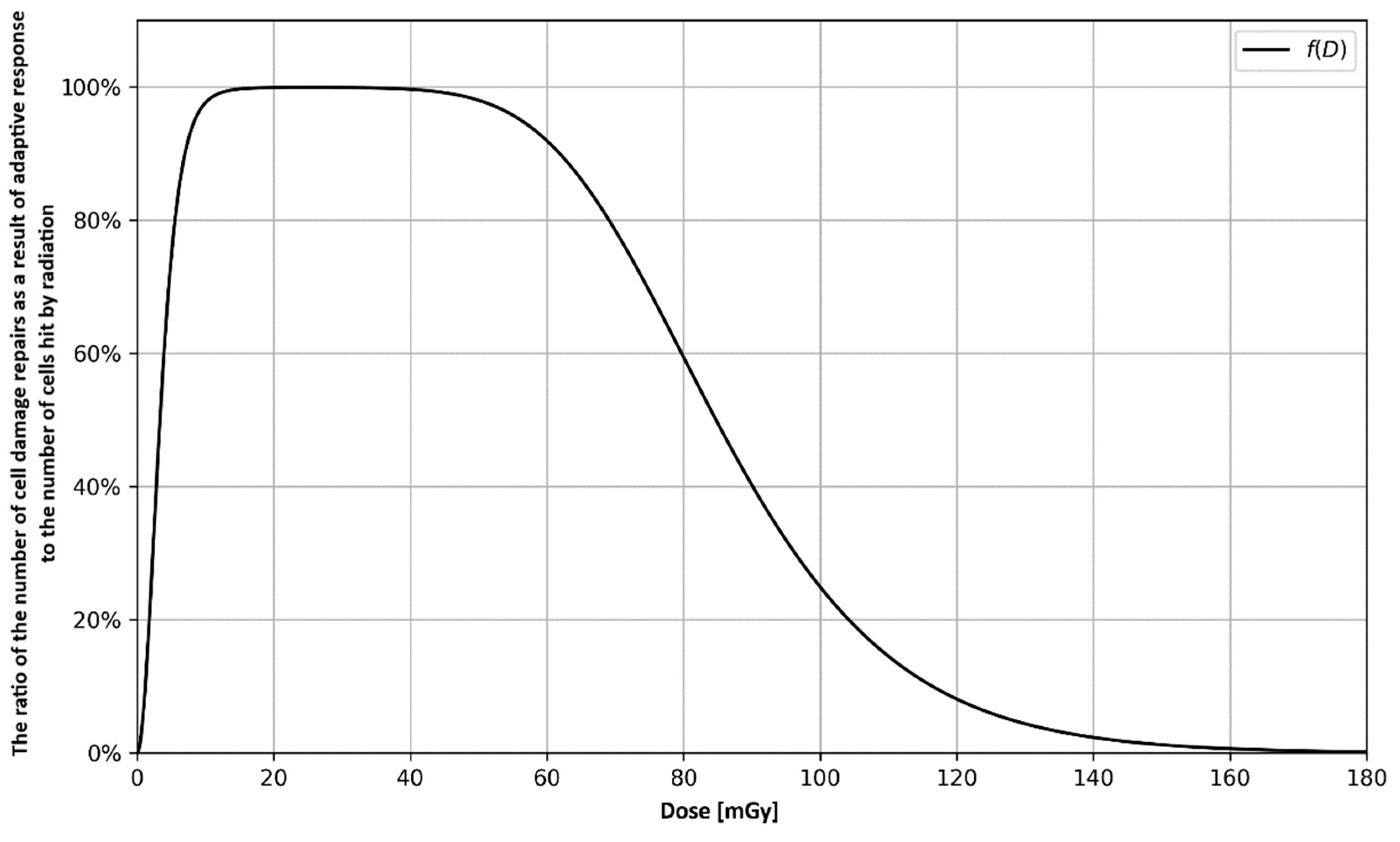
3. Radiation Adaptive Response—Enhancement of the Repair Process

3.1. Analytical Approach

As mentioned in the Introduction section, the radiation adaptive response is a biophysical phenomenon which can enhance the DNA repair processes via the cell’s stimulation by a low dose of ionising radiation [9,10]. In the context of the Monte Carlo model, which was presented in the previous section, the fraction of cells that could be fully ( U = 0 ) or partially ( U > 0 ) repaired is determined by the probability Phit (Equation (1)) after the single-dose pulse given in the zero time step k 0 . Thus, only those cells that have been hit by radiation (with dose D > 0 ) in k 0 can be repaired by radioadaptive mechanisms (it was assumed that D = 0 for time steps k > k 0 ). The probability function of the radiation adaptive response’s appearance in each time step, PAR, is a dose- and time-dependent relationship [1]:
P A R = α 0 D 2 k 2 e α 1 D α 2 k    
where the empirical parameters for human lymphocytes are: α 0 = 22.9   Gy 2   h 3 , α 1 = 79.4   Gy 1 , and α 2 = 0.0832   h 1 .
Now the repair fraction relationship can be calculated. It depends on the dose impulse (D) in the moment of k 0 , as the ratio of the number of all repaired cells (thanks to the adaptive response) to the number of all damaged cells hit by radiation:
f ( D ) = 1 N 0 P h i t ( D ) n = 1 T 0 1 S n ( D )    
where N 0 is the initial number of damaged cells, T 0 is the simulation duration, and S n ( D ) is the recursive sequence that represents the total number of repaired cells in the n-th time step due to the radiation adaptive response phenomenon. The proposed mathematical sequence can be described by
S n ( D ) = { N 0 P h i t ( D ) P A R ( D ,   n )                         if     n = 1 ( N 0 P h i t ( D ) i = 1 n 1 S i ( D ) ) P A R ( D ,   n )       if     n > 1            
In other words, the function proposed by Equation (3) represents the ratio of the total number of repaired cells due to the radiation adaptive response to the number of cells that were hit by the dose impulse at the beginning of the simulation. The purpose of the derived sequence given by Equation (4) is to calculate the number of cells that have been repaired in the n-th time step. The advantage of this sequence is that it is recursive. This allows us to receive the number of cells hit by radiation reduced by the number of cells that have already been repaired in the n − 1 time step and multiply the result by the appropriate probability value P A R . The structure is complex and may be considered in two cases: for n = 1 it returns the number of cells that were repaired in the first time step according to conditional probability, while for n > 1 it uses the previous terms of the sequence.
Equations (3) and (4) were investigated in an analytical way, where N0 = 493,000 cells was tested by different dose pulses in the moment k0; see Figure 1. The cells’ status was checked in the final moment of T = 120 h, where the probability function of the adaptive response is insignificant. Please note that within the range of 10–45 mGy, the ratio of the fraction of cells that are repaired to all cells hit by radiation is ≈100%. This shows the most effective range of the small dose as a trigger for the adaptive response mechanism.
The presented approach can be applied to other irradiation scenarios, such as the Raper–Yonezawa scheme (also called the priming dose effect), where two consecutive doses, small D1 and large D2 with the time interval between them, are taken into consideration [22].
The third special case is connected with the constant dose-rate irradiation. After the integration of Equation (2), one gets
P C = μ 0 D ˙ 2 e μ 1 D ˙ = c o n s t        
which describes the constant probability of the adaptive response’s appearance in a constant dose-rate ( D ˙ ) environment [21,22]. Its empirical parameters were calculated as: μ 0 = 0.0115   year 2   mGy 2 = 882 · 10 3   h 2   mGy 2 and μ 1 = 0.117   year   mGy 1 = 1025   h   mGy 1 for human lymphocytes in vivo among HBRAs’ inhabitants [25], or μ 0 = 4.9 · 10 7   year 2   mGy 2 = 38   h 2   mGy 2 and μ 1 = 0.00131   year   mGy 1 = 11.5   h   mGy 1 for human lymphocytes irradiated by X-rays in vitro [21].

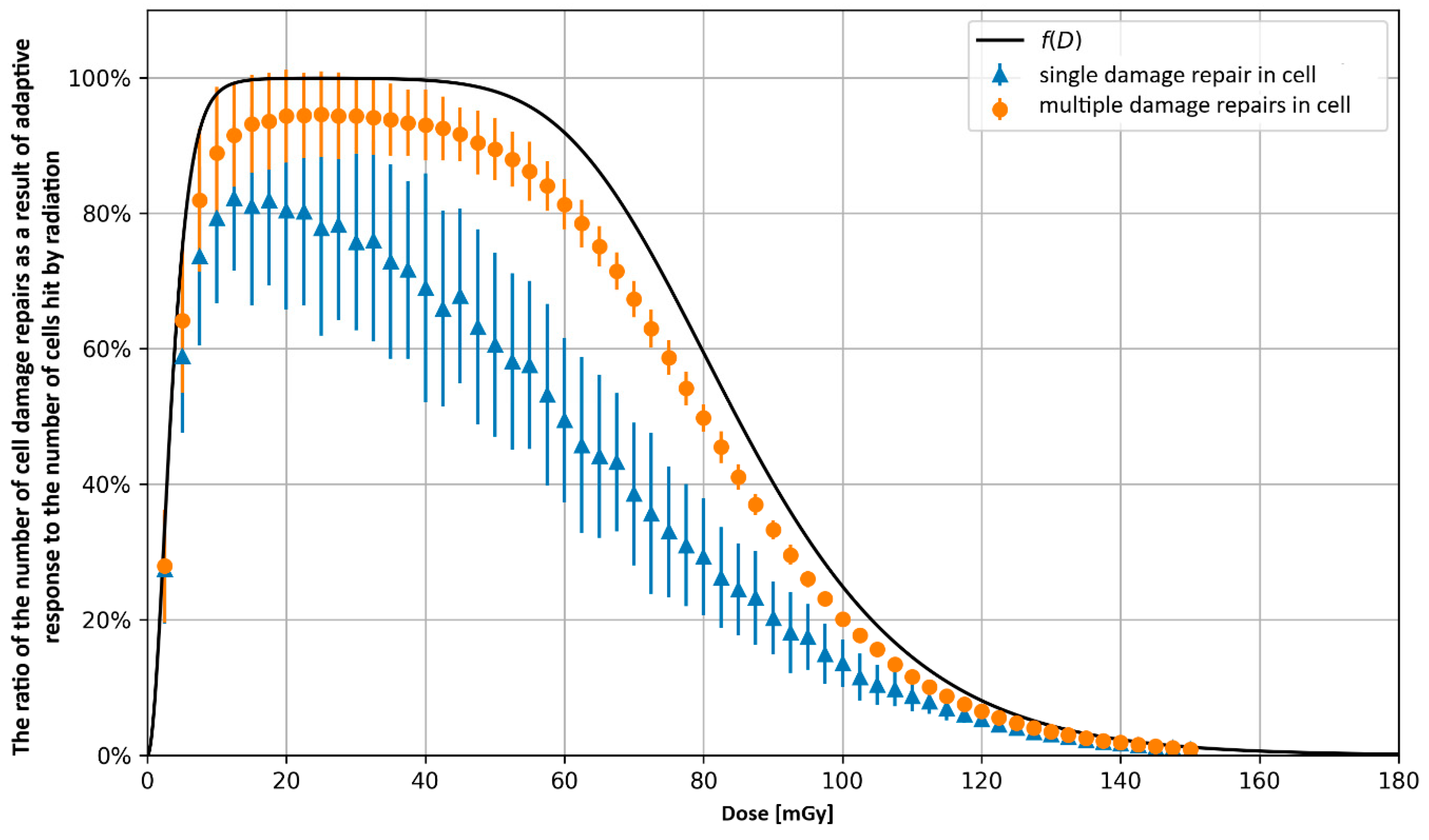
3.2. Monte Carlo Approach—Single Dose

In the previous subchapter, the radioadaptation-related repair mechanism was narrowed down to the one process within the irradiated cells. This means that the presented solution is an ideal one. To make it more realistic, the Monte Carlo approach covers all other biological mechanisms which are presented in the full probability tree [1]. In this context, two simulations were prepared to study the effectiveness of the repair process: (a) full repair of the cell ( U 0 ) and (b) all repair actions in the cell. The first case is comparable to the one presented in the previous subchapter: the repaired cell is taken into consideration one time only. In the second scenario, all radioadaptive-related repair actions are summed even if the same cell was hit many times after the first repair.
The presented approach is a fully stochastic one; thus, many random iterations are needed to get the average results. As with the previous case, N 0 = 493,000 cells was tested by different dose pulses in k0 (there were 150 simulations for one value of dose). The dose range was 2.5–150 mGy, and the next simulation series were carried out every δD = 2.5 mGy. The final result was calculated after T = 120 h, where the probability function of the adaptive response is insignificant (all repair processes are finished). All the results, compared with the previous ideal case, are presented in Figure 2.

3.3. Monte Carlo Approach—Two Doses

Building upon the previous subchapter, the two-dose approach is where the second (challenging) dose is applied some period of time after the first (priming) dose. The induced radioadaptation repair mechanism makes this second dose less detrimental than it would be without the priming dose.
To precisely test it, the adaptive response appearance in time-related relationships was applied in the following irradiation exemplary schemes (the Raper-Yonezawa schemes with priming and challenging doses) using the Monte Carlo model:
  • Scenario no. 1: D1 = 25 mGy, Δt = 24 h, D2 = 1500 mGy
  • Scenario no. 2: D1 = 25 mGy, Δt = 24 h, D2 = 4000 mGy
  • Scenario no. 3: D1 = 25 mGy, Δt = 100 h, D2 = 1500 mGy
  • Scenario no. 4: D1 = 100 mGy, Δt = 24 h, D2 = 1500 mGy
For each irradiation scheme listed above, 64,000 cells were simulated a hundred times—fifty times with the adaptive response included and fifty times without it—and then their average value was determined for subsequent steps in both cases. The graphs show the average number of cells obtained in the simulation, including the adaptive response divided by the average number of cells without the adaptive response (Figure 3, Figure 4, Figure 5 and Figure 6). This ratio value is useful to check whether the adaptive response effect is significant in a specific time range (or not).
Scenario no. 1 is presented in Figure 3, and each subsequent scenario is similarly shown in the consecutively numbered figures. In each scenario the priming dose, D1, was applied 10 h after simulation start, while the challenging dose, D2, was applied Δt after D1.
The radiation adaptive response effects can be observed in the graphs (Figure 3, Figure 4, Figure 5 and Figure 6). It presents itself as an increase in the number of healthy cells and a decrease in the number of damaged cells in the first few tens of hours after the cells receive the priming dose (these are repairs of the spontaneous damages present in the cells) and before receiving the second dose (see Figure 3 and Figure 5).
In Figure 3, the significant adaptive response effect is observed up to 46 h after second dose irradiation; in Figure 4, it is observed up to 30 h. These scenarios correspond to the most optimal irradiation pattern (the second dose is delivered at a time for which the probability of an adaptive response reaches a maximum) to which the model parameters were calibrated [22]. The fading effect of the adaptive response is related to the decrease in the probability of its occurrence combined with the gradual accumulation of naturally occurring damages.
In Figure 4, the effect is very noticeable because, due to the high second dose, most of the cells were killed, and those that survived had a high chance of being damaged, so there were a lot of damages that could be repaired as a result of the adaptive response. In Figure 5, the aforementioned repair of natural damage after receiving the priming dose can be seen, but when the second dose was delivered, the probability of an adaptive response dropped to less than 1%, so it did not have a significant contribution to effects occurring in the cell. Comparing Figure 3 and Figure 6, one can see a decrease in the adaptive response effect due to an increase in the priming dose.
In all cases, the adaptive response had little effect on the results obtained or the effect is noticeable for a short time; even with the most optimal irradiation scenario no. 2, the average difference between the results with and without the adaptive response does not exceed 7%, and the effect fades quickly.

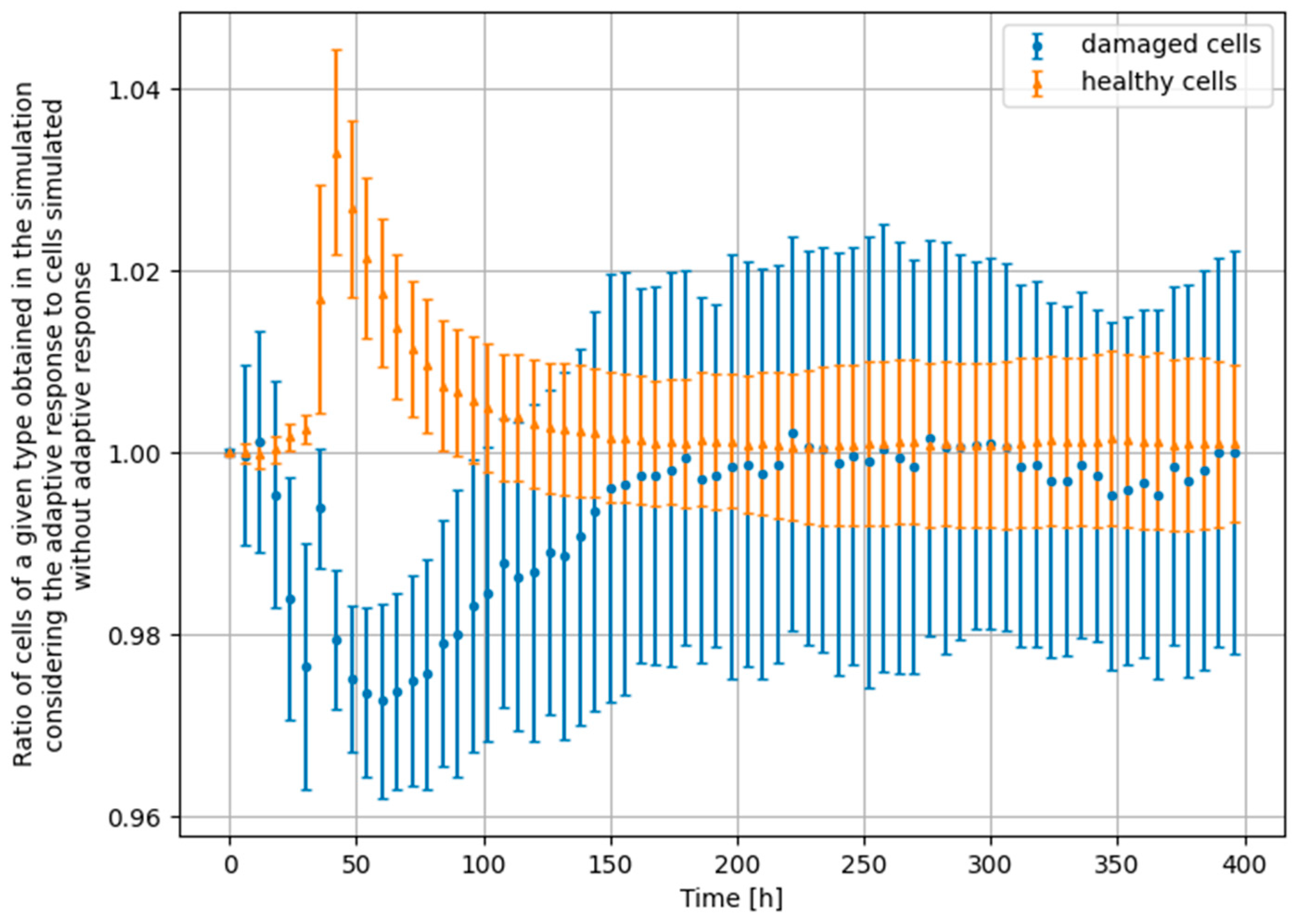
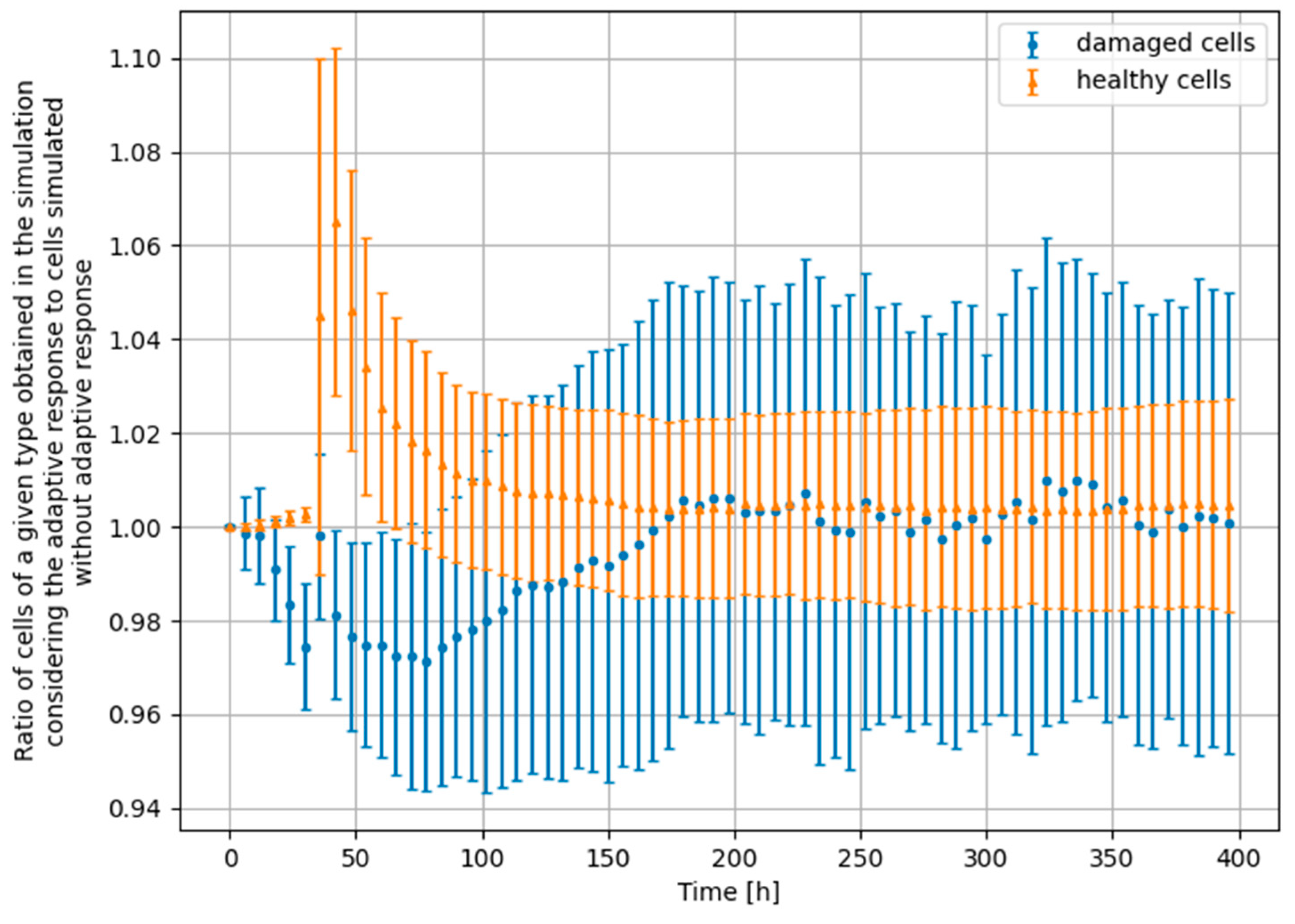
3.4. Monte Carlo Approach—Constant Dose-Rate

The special case of the constant dose-rate means that the irradiation process is constant all the time, which is analogous to the situation found in many high background radiation areas (HBRA) in the world. Thus, let us use Equation (5) as the radiation adaptive response probability, and the parameters described in Section 3.1. Assuming different potential values of dose-rate, the adaptive response was tested in the following irradiation scenarios:
  • Scenario no. 1 (see Figure 7): μ 0 = 882 · 10 3   h 2   mGy 2   ,   μ 1 = 1025   h   mGy 1   , D ˙ = 0.17   mGy   h 1
  • Scenario no. 2 (see Figure 8): μ 0 = 882 · 10 3   h 2   mGy 2   ,   μ 1 = 1025   h   mGy 1 , D ˙ = 0.002   mGy   h 1
  • Scenario no. 3 (see Figure 9): μ 0 = 38   h 2   mGy 2   ,   μ 1 = 11.5   h   mGy 1 , D ˙ = 0.17   mGy   h 1
  • Scenario no. 4 (see Figure 10): μ 0 = 38   h 2   mGy 2   ,   μ 1 = 11.5   h   mGy 1 , D ˙ = 0.002   mGy   h 1
Results for scenario no. 1 are presented in Figure 7, and each subsequent scenario is similarly shown in consecutively numbered figures.
For the dose-rate D ˙ = 0.002 mGy h−1, the probability of a cell being hit by radiation equals P h i t = 2.6 · 10 6 , and that is why in scenarios no. 2 (Figure 8) and no. 4 (Figure 10), the adaptive response phenomenon has not been observed. For the first case (Figure 7), the probability of an adaptive response was close to zero, and again this effect was not observed in the simulation.
For irradiation scenario no. 3, the probability of a cell being hit by radiation was P h i t = 2.2 · 10 4 , and the probability of an adaptive response in constant irradiation was P C = 0.45 (see Figure 9). In this case, more cells were hit at each time step, and an adaptive response had a higher probability to be induced in cells with spontaneous damages. Therefore, Figure 9 shows substantially different behaviour than previous ones.
The simulation at the constant dose-rate was also repeated (64,000 cells simulated three times with the adaptive response included and three times without it) for the adaptive response probability described by Equation (2) (example in Figure 11), and the effect was not observed.

4. Significance of the Radioadaptive-Related Repair

Analysing the results presented in previous chapters (for the single-dose scenario), one can see that in the case of a full repair, the repair efficiency is lower compared to the theoretical curve (Figure 2). However, it still remains at a high level (up to 82%). One should also pay attention to the standard deviation (in Figure 2), which in the case of the second part of the study exceeds 100%. This is due to the fact that in the determined ratio the denominator applies to all cells hit by radiation, while the numerator takes into account the multiple repairs of each cell (one cell can be hit many times).
On the basis of the first part of the study (analytical approach), the ratio of repaired cells to the number of all cells in the initial colony was calculated to check how the effect of the adaptive response occurs in the context of the whole complex living system.
Considering the repair effect of the adaptive response only, it can be concluded that it is clearly visible in the model, and its impact on the strengthening of repair mechanisms is significant. This is evidenced by the results of the first part of the study. However, focusing on the whole system, this phenomenon practically disappears, and it is at the level of 0.126%, which makes it insignificant (see Figure 12). This may explain the fact that the effects associated with the impact of low doses of radiation on the body are visible only in studies dedicated to the adaptive response itself. In contrast, in population studies (individual and epidemiological), these effects are very subtle and, in most cases, disappear among the noise of other effects.
In the two-dose scenario, the adaptive response does occur, but, even with the most optimal irradiation scheme, its effect is small (up to 7% as seen in Figure 4) and it does not last long (up to 80 h as seen in Figure 6). The highest repair effectiveness due to the radioadaptation is observed 20 h to 50 h after the irradiation with the priming dose. Later, the long-lasting effect of a higher number of healthy cells is not a result of new repairs connected to the adaptive response. This effect occurs because a certain amount of time must pass before the ratio between healthy and damaged cells stabilises and goes back to a naturally existing ratio.
At a constant dose-rate, in all but one case the effect was negligible—this is due to the low probability of a cell being hit by radiation for the doses used. The case where the adaptive response occurred at a constant dose-rate corresponds to the situation where the low-dose-rate irradiation induces a repair enhancement for the rest of the cell’s life.
The reasons for the generally low effectivity of repair enhancement due to the adaptive response in our simulation can be complex. The first reason is the model itself, which—as any other model—is just a simplification of the reality. Second, the calibration parameters in the Monte Carlo model are not perfect, because it was based on limited experimental data in vitro [1]. Third, the adaptive response, which is a very subtle effect, spreads over all other effects in irradiated cells, which is quite natural. Thus, the presented results shall be treated more in a qualitative than quantitative way.

5. Discussion and Conclusions

The radiation adaptive response is a fascinating radiobiological and biophysical effect which completely changes the potential negative impact of ionising radiation [10]. This effect, however, is quite weak and difficult to observe—it is estimated that approximately half of dedicated radiobiological experiments show the radioadaptation under narrowed conditions [9]. This means that the value of dose(s) and time interval(s) is of crucial importance—but even then one cannot be sure to observe the effect.
There are, however, many radiobiological experiments which show clear evidence of the adaptive response’s appearance [2,9]. In most cases, the radioadaptation effect is manifested in the priming dose experimental scheme (called the Yonezawa or Raper–Yonezawa scheme), which is the easiest one for experimenters. However, it is still not the case that every fairly well planned and prepared experiment shows the adaptation to low doses of ionising radiation. The key question is: what factor(s) determines the adaptive response’s appearance/disappearance? From what is known today, it is the distribution of individual radiosensitivity [18,21,26].
The presented article shows that these problems are also observed in theoretical simulations. There are many models of adaptive response, but we used our own approach based on the Monte Carlo technique [1], which can result in an analytical solution as well [22]. Of course, ideal modelling of the dedicated observation of the adaptive response gives a high chance for a positive result for different irradiation scenarios. This observation is clearly deduced from the Monte Carlo simulation, which was originally calibrated for human lymphocytes irradiated by X-ray in vitro [1]. However, when the whole organism (or, more precisely, the tissue represented here by a group of cells) is taken into consideration with all its biological effects, the typical radiation adaptive response phenomenon blurs among other effects and becomes weak or sometimes insignificant. This is probably the reason why hormetic effects are rather difficult to observe in epidemiological or case-control studies of low doses’ influence on the human body and its radiation risk [5]. However, the radiation adaptive response is still an important biophysical effect which needs to be taken into consideration in low-dose radiobiological studies.

Author Contributions

Conceptualization, K.W.F., and Ł.P.; methodology, Ł.P., and K.W.F.; software, Ł.P.; validation, J.K. and Ł.P..; formal analysis, Ł.P. and J.K.; investigation, K.W.F.; resources, K.W.F.; data curation, Ł.P.; writing—original draft preparation, K.W.F.; writing—review and editing, Ł.P. and J.K.; visualization, Ł.P., and J.K..; supervision, K.W.F.; project administration, K.W.F. All authors have read and agreed to the published version of the manuscript.

Funding

This research received no external funding.

Informed Consent Statement

Not applicable.

Data Availability Statement

No new data creation.

Acknowledgments

The authors want to thank Ernest Bugała and Rafał Jarmakiewicz (students at the Faculty of Physics, Warsaw University of Technology) for providing adaptive response calibration data from their master and engineer theses.

Conflicts of Interest

The authors declare no conflict of interest.

References

  1. Fornalski, K.W.; Adamowski, Ł.; Bugała, E.; Jarmakiewicz, R.; Kirejczyk, M.; Kopyciński, J.; Krasowska, J.; Kukulski, P.; Piotrowski, Ł.; Ponikowska, J.; et al. Biophysical modeling of the ionizing radiation influence on cells using the stochastic (Monte Carlo) and deterministic (analytical) approaches. Dose-Response, 2022; in-press (published on-line). [Google Scholar] [CrossRef]
  2. UNSCEAR (United Nations Scientific Committee on the Effects of Atomic Radiation) Report. Annex B: Adaptive Responses to Radiation in Cells and Organisms. 1994. Available online: https://www.unscear.org/docs/publications/1994/UNSCEAR_1994_Report.pdf (accessed on 13 February 2023).
  3. Raper, J.R. Effects of total surface beta irradiation. Radiology 1947, 49, 314–324. [Google Scholar] [CrossRef] [PubMed] [Green Version]
  4. Olivieri, G.; Bodycote, J.; Wolff, S. Adaptive response of human lymphocytes to low concentrations of radioactive thymidine. Science 1984, 223, 594–597. [Google Scholar] [CrossRef] [PubMed]
  5. Sanders, C.L. Radiation Hormesis and the Linear-No-Threshold Assumption; Springer: New York, NY, USA, 2010. [Google Scholar]
  6. Wolff, S. The adaptive response in radiobiology: Evolving insights and implications. Environ. Health Perspect. 1998, 106 (Suppl. 1), 277–283. [Google Scholar] [PubMed]
  7. Aurengo, A.; Averbeck, D.; Bonnin, A.; Le Guen, B.; Masse, R.; Monier, R.; Tubiana, M.; Valleron, A.J.; de Vathaire, F. Dose-effect relationships and estimation of the carcinogenic effects of low doses of ionizing radiation. Rep. Fr. Acad. Sci. Natl. Acad. Med. 2005. Available online: https://www.radiochemistry.org/documents/html/033005_rad.html (accessed on 13 February 2023).
  8. Brooks, A.L.; Glines, W.M.; Waltar, A. Proceedings of the Joint American Nuclear Society and Health Physics Society Conference: Applicability of Radiation-Response Models to Low-Dose Protection Standards 1–3 October 2018, Pasco, Washington Introduction—Years in the Making. Health Phys. 2020, 118, 229–231. [Google Scholar] [CrossRef] [PubMed]
  9. Tapio, S.; Jacob, V. Radioadaptive response revisited. Radiat. Environ. Biophys. 2007, 46, 1–12. [Google Scholar] [CrossRef] [PubMed]
  10. Guéguen, Y.; Bontemps, A.; Ebrahimian, T.G. Adaptive responses to low doses of radiation or chemicals: Their cellular and molecular mechanisms. Cell. Mol. Life Sci. 2019, 76, 1255–1273. [Google Scholar] [CrossRef]
  11. Feinendegen, L.E. The role of adaptive responses following exposure to ionizing radiation. Hum. Exp. Toxicol. 1999, 18, 426–432. [Google Scholar] [CrossRef]
  12. Feinendegen, L.E. Low Doses of Ionizing Radiation: Relationship between Biological Benefit and Damage Induction. A Synopsis. World J. Nucl. Med. 2005, 4, 21–34. [Google Scholar]
  13. Feinendegen, L.E. Quantification of adaptive protection following low-dose irradiation. Health Phys. 2016, 110, 276–280. [Google Scholar] [CrossRef]
  14. Smirnova, O.A.; Yonezawa, M. Radioprotection effect of low level preirradiation on mammals: Modeling and experimental investigations. Health Phys. 2003, 85, 150–158. [Google Scholar] [CrossRef] [PubMed]
  15. Smirnova, O.A.; Yonezawa, M. Radioresistance in mammals induced by low-level chronic irradiation: Modeling and experimental investigations. Health Phys. 2004, 87, 366–374. [Google Scholar] [CrossRef]
  16. Wodarz, D.; Sorace, R.; Komarova, N.L. Dynamics of cellular responses to radiation. PLoS Comput. Biol. 2014, 10, e1003513. [Google Scholar] [CrossRef] [PubMed]
  17. Bodgi, L.; Foray, N. The nucleo-shuttling of the ATM protein as a basis for a novel theory of radiation response: Resolution of the linear-quadratic model. Int. J. Radiat. Biol. 2016, 92, 117–131. [Google Scholar] [CrossRef] [PubMed]
  18. Devic, C.; Ferlazzo, M.L.; Foray, N. Influence of individual radiosensitivity on the adaptive response phenomenon: Toward a mechanistic explanation based on the nucleo-shuttling of ATM protein. Dose-Response 2018, 16, 1559325818789836. [Google Scholar] [CrossRef] [PubMed]
  19. Esposito, G.; Campa, A.; Pinto, M.; Simone, G.; Tabocchini, M.A.; Belli, M. Adaptive response: Modelling and experimental studies. Radiat. Prot. Dosim. 2011, 143, 320–324. [Google Scholar] [CrossRef]
  20. Socol, Y.; Shaki, Y.; Dobrzyński, L. Damped-oscillator model of adaptive response and its consequences. Int. J. Low Radiat. 2020, 11, 186–206. [Google Scholar] [CrossRef]
  21. Fornalski, K.W. Radioadaptation and radioresistance during deep space travels. J. Space Saf. Eng. 2022, 9, 385–389. [Google Scholar] [CrossRef]
  22. Fornalski, K.W.; Adamowski, Ł.; Dobrzyński, L.; Jarmakiewicz, R.; Powojska, A.; Reszczyńska, J. The radiation adaptive response and priming dose influence: The quantification of the Raper-Yonezawa effect and its three-parameter model for postradiation DNA lesions and mutations. Radiat. Environ. Biophys. 2022, 61, 221–239. [Google Scholar] [CrossRef]
  23. Dobrzyński, L.; Fornalski, K.W.; Reszczyńska, J.; Janiak, M.K. Modeling cell reactions to ionizing radiation: From a lesion to a cancer. Dose-Response 2019, 17, 1–19. [Google Scholar] [CrossRef] [Green Version]
  24. Piotrowski, Ł. Modelowanie Odpowiedzi Grupy Komórek na Promieniowanie Jonizujące Metodą Monte Carlo. Bachelor’s Thesis, Faculty of Physics, Warsaw University of Technology, Warsaw, Poland, (supervisor: K.W. Fornalski). 2022. Available online: https://www.researchgate.net/publication/368275322_Modelowanie_odpowiedzi_grupy_komorek_na_promieniowanie_jonizujace_metoda_Monte_Carlo (accessed on 13 February 2023).
  25. Bugała, E. Modelowanie Radiacyjnej Odpowiedzi Adaptacyjnej dla Obszarów o Podwyższonym tle Promieniowania Jonizującego (in Polish). Master Thesis, Faculty of Physics, Warsaw University of Technology, Warsaw, Poland, (supervisor: K.W. Fornalski). 2022. Available online: https://www.researchgate.net/publication/368139245_Modelowanie_radiacyjnej_odpowiedzi_adaptacyjnej_dla_obszarow_o_podwyzszonym_tle_promieniowania_jonizujacego (accessed on 13 February 2023).
  26. Fornalski, K.W. Radiation adaptive response and cancer: From the statistical physics point of view. Phys. Rev. E 2019, 99, 22139. [Google Scholar] [CrossRef] [PubMed]
Figure 1. Analytically determined function f ( D ) that represents the cell repair fraction (as a consequence of adaptive response) depending on the dose impulse (D) with simulation time T 0 = 120   h and initial population N 0 = 493,000 .
Biomedinformatics 03 00011 g001
Figure 2. Analytically determined function f ( D ) that represents the cell repair fraction (as a consequence of adaptive response) depending on the dose impulse (D), with parameters as in Figure 1. The simulation results for a single damage repair in cell () and multiple damage repairs in cell () are also included.
Biomedinformatics 03 00011 g002
Figure 3. Results obtained for two-dose approach, irradiation scenario no. 1: D1 = 25 mGy (applied at time t = 10 h), Δt = 24 h, D2 = 1500 mGy (applied at time t = 34 h), see Section 3.3.
Biomedinformatics 03 00011 g003
Figure 4. Results obtained for two-dose approach, irradiation scenario no. 2: D1 = 25 mGy (applied at time t = 10 h), Δt = 24 h, D2 = 4000 mGy (applied at time t = 34 h), see Section 3.3.
Biomedinformatics 03 00011 g004
Figure 5. Results obtained for two-dose approach, irradiation scenario no. 3: D1 = 25 mGy (applied at time t = 100 h), Δt = 24 h, D2 = 1500 mGy (applied at time t = 110 h), see Section 3.3.
Biomedinformatics 03 00011 g005
Figure 6. Results obtained for two-dose approach, irradiation scenario no. 4: D1 = 100 mGy (applied at time t = 10 h), Δt = 24 h, D2 = 1500 mGy (applied at time t = 34 h), see Section 3.3.
Biomedinformatics 03 00011 g006
Figure 7. Results obtained for constant dose-rate approach, irradiation scenario no. 1: parameters μ 0 = 882 · 10 3   h 2   mGy 2 , μ 1 = 1025   h   mGy 1 , and dose rate D ˙ = 0.17   mGy   h 1 applied from t = 10 h.
Biomedinformatics 03 00011 g007
Figure 8. Results obtained for constant dose-rate approach, irradiation scenario no. 2: parameters μ 0 = 882 · 10 3   h 2   mGy 2 , μ 1 = 1025   h   mGy 1 , and dose rate D ˙ = 0.002   mGy   h 1 applied from t = 10 h.
Biomedinformatics 03 00011 g008
Figure 9. Results obtained for constant dose-rate approach, irradiation scenario no. 3: for parameters μ 0 = 38   h 2   mGy 2 , μ 1 = 11.5   h   mGy 1 , and dose rate D ˙ = 0.17   mGy   h 1 applied from t = 10 h. The observed phenomenon is due to both the high probability of an adaptive response and the non-negligible probability of a cell being hit by radiation. The difference in ratio values for healthy and damaged cells is due to their different frequencies in the population.
Biomedinformatics 03 00011 g009
Figure 10. Results obtained for constant dose-rate approach, irradiation scenario no. 4: for parameters μ 0 = 38   h 2   mGy 2 , μ 1 = 11.5   h   mGy 1 , and dose rate D ˙ = 0.002   mGy   h 1 applied from t = 10 h.
Biomedinformatics 03 00011 g010
Figure 11. Results obtained for single-dose approach described by Equation (2) and constant irradiation with D ˙ = 0.17   mGy   h 1 applied from t = 10 h.
Biomedinformatics 03 00011 g011
Figure 12. The cell repair fraction in reference to the global population of the simulation as a function of dose impulse (D). The presented values refer to two variants: the theoretical function f ( D ) and the simulation results of a single damage repair in cell that are presented in Figure 2.
Biomedinformatics 03 00011 g012
Disclaimer/Publisher’s Note: The statements, opinions and data contained in all publications are solely those of the individual author(s) and contributor(s) and not of MDPI and/or the editor(s). MDPI and/or the editor(s) disclaim responsibility for any injury to people or property resulting from any ideas, methods, instructions or products referred to in the content.

