Article

The Antarctic Scallop Adamussium colbecki Is Unable to Transcriptomically Respond to Captivity and Moderate Thermal Stress

by
Samuele Greco
1,†,
Anastasia Serena Gaetano
2,†,
Chiara Manfrin
1,
Francesca Capanni
3,
Gianfranco Santovito
4,
Alberto Pallavicini
1,
Piero Giulio Giulianini
1 and
Marco Gerdol
2,*
1
Department of Life Sciences, University of Trieste, 34127 Trieste, Italy
2
Department of Chemical and Pharmaceutical Sciences, University of Trieste, 34127 Trieste, Italy
3
Department of Physical Sciences, Earth and Environment, University of Siena, 53100 Siena, Italy
4
Department of Biology, University of Padova, 35122 Padova, Italy
*
Author to whom correspondence should be addressed.
These authors contributed equally to this work.
Stresses 2023, 3(2), 475-487; https://doi.org/10.3390/stresses3020034
Submission received: 28 February 2023 / Revised: 16 March 2023 / Accepted: 23 March 2023 / Published: 24 May 2023
(This article belongs to the Section Animal and Human Stresses)

Abstract

:
Adamussium colbecki is a scallop endemic of the Antarctic Ocean, the only modern survivor of the Adamussiini tribe and one of the few bivalves living in polar environments. Compared with other Antarctic animals, very little is known concerning the evolutionary adaptations which allow this species to thrive at sub-zero temperatures. Due to its local abundance and sensitivity to environmental changes, A. colbecki is an interesting model for studying the effects of pollution and climate change in the Antarctic Ocean. Here, we report, for the first time, the application of transcriptomic tools to the study of the effects of a short-to-medium term exposure to a +1.5 °C water temperature increase on three tissues. Although this approach did not highlight any significant change in response to thermal stress, we observed slight alterations in energetic metabolism and nutrient adsorption in the digestive gland, most likely linked with stabling in experimental tanks. The results of our study suggest that A. colbecki may be particularly vulnerable to the effects of climate change due to its complete inability to adapt to temperature increase at the transcriptomic level.

1. Introduction

The scallop Adamussium colbecki (Smith 1902) is widely distributed along the Antarctic coastline in fairly dense populations, at water depths ranging from 0 to 1300 m, reaching exceptional densities (i.e., up to 65/m2) around 70 m, where it can completely cover the sea floor [1,2,3,4]. Nevertheless, its distribution is patchy, likely due to a combination of different biological constraints, such as its limited dispersion ability, its necessity of an extremely stable environment and the presence of predators. A. colbecki is considered to be the lone survivor of a once highly diversified group of pectinid bivalves, i.e., the Adamussiini tribe, which included the extinct genera Antarctipecten, Duplipecten, Lentipecten, Leoclunipecten and Ruthipecten [5,6,7]. Recently, phylotranscriptomic analyses have suggested that the most closely related extant scallops belong to the subfamily Palliolinae [8].
Unlike other Antarctic metazoans, such as notothenioid fish, which display extremely peculiar morphological adaptations, A. colbecki lacks evident modifications compared with its relatives living in temperate waters. The only documented adaptations concern a reduced body mass, which may lower the energy costs associated with swimming activity at low temperatures [9], and a unique shell nanostructural organization, which passively prevents freezing by cryofouling [10]. On the other hand, this species displays a few unique life history traits that allow its survival in the subzero waters of the Antarctic Ocean. These include slower metabolic rates, slower growth and embryonic development, and a longer lifespan compared with non-Antarctic scallops. Altogether, these traits lead to optimized energy budgets during the long polar winter, when food availability is limited [3,11,12]. Adaptations to cold have been studied to a much lesser extent at molecular level, evidencing a particularly high catalase enzymatic activity, which may be interpreted as a protection mechanism against lipid peroxidation in high oxygen solubility conditions [13], and an improved mRNA translational capacity [14]. Moreover, our recent comparative transcriptomics study identified a higher expression of several genes involved in mRNA synthesis and processing in Antarctic scallops, compared to non-Antarctic ones [15]. This suggests that there may still be many more molecular adaptations to cold in these organisms that are yet to be discovered and studied.
Past studies have revealed that A. colbecki is significantly more susceptible to heat stress than other Antarctic mollusks, such as Laternula elliptica and Nacella concinna [16]. As experimentally demonstrated by Bailey and colleagues, A. colbecki shows early signs of distress around 0 °C, progressively becoming unable to respond to stimuli and swim as water temperatures approach 2 °C, suggesting that its upper lethal thermal limit might be close to 3 °C [17,18]. Strikingly, this scallop also displays very low phenotypic plasticity to temperature changes, being unable to modify its swimming behavior, regardless of its acclimation history [17]. Together with current geographical distribution of the species and fossil records of extinct congeneric species in sub-Antarctic islands from the global cooling period of Oligocene [19], the aforementioned studies support the notion that A. colbecki has a very narrow thermal tolerance range.
The few studies carried out so far to investigate this particular aspect in A. colbecki have been only focused on behavior and physiology, with a particular attention on muscle activity [17,18]. Others have explored the combined effects of increased temperature and other pro-oxidant stressors by monitoring different biomarkers [20]. Hence, a complete overview on the molecular responses of this species to heat stress in multiple tissues is still lacking.
The data collected for another Antarctic bivalve species, L. elliptica [21] should not be generalized to A. colbecki, as this species displays a much higher upper lethal thermal limit (i.e., close to 10 °C) [16] and belongs to a phylogenetically distinct bivalve group (the superorder Anomalodesmata). The circum-Antarctic distribution of A. colbecki and its extreme susceptibility to environmental alterations would make it an optimal choice as a sentinel organism to monitor the impact of climate change, mirroring the use that has been previously proposed as a bioindicator of anthropic pollution [22,23]. In fact, according to previous studies, the recent dramatic reduction in Antarctic sea ice cover due to climate change may have a strong impact on the survival of this species [24], with detrimental consequences on Antarctic coastal trophic webs [25].
Predicting the fate of A. colbecki in a global warming scenario would undoubtedly require a multidisciplinary approach, combining existing physiological and biochemical observations to cutting-edge -omic analyses, which hold the potential to provide detailed insights in the molecular responses of different tissues following thermal stress.
We have previously reported a transcriptome assembly for A. colbecki [8], that we used in the present study as a reference. The primary aim of this work consists in evaluating the alterations of gene expression observed throughout 19 days of experimental exposure to moderate thermal stress (i.e., +1.5 °C, compared with environmental conditions) in gills, mantle and digestive gland. The conditions of this heat stress experiment were specifically selected to assess the ability of the Antarctic scallop to withstand a temperature inscrease consistent with the recent forecasts for the Antarctic Ocean in the next few decades, mirroring a realistic scenario [26].
Although the notothenioid fish Trematomus bernacchii displayed significant alterations both due to thermal and stabling stress in a similar experimental setting [27], A. colbecki only showed minimal transcriptomic alterations, which were restricted to stabling stress in the digestive gland, at the latest time point. Given the sensitivity of RNASeq, these results support the hypothesis that this stenotherm bivalve species has an extremely low phenotypic plasticity even to slight warming, leading to alarming considerations concerning the fate of its populations in a climate change scenario.

2. Results

2.1. Transcriptome Assembly and Refinement

The raw assembled transcriptome was highly redundant and counted 702,884 sequences, including exogenous contamination, uncollapsed splicing isoforms and a high number of short low quality contigs. We detected and removed exogenous contamination originated from Trematomus bernacchii and Chionodraco hamatus. Moreover, we also detected and removed contamination from Pleurochrysis pseudoroscoffensis and from an unidentified red alga belonging to the family Rhodomelaceae. Following the removal of low quality contigs and of the aforementioned exogenous contamination, the total number of contigs included in the reference transcriptome was 44,475. These ranged in size from 348 to 39,427 nt, averaging 1329.11 nt (see Table 1 for more assembly stats).
The assembly had a good level of completeness, according to the BUSCO analysis carried out against the OrthoDBv10 Metazoa database. Indeed, 89.9% BUSCOs were present and complete (76.0% single copy and 13.9% duplicated) and just 4.4% and 5.7% orthologs were fragmented and missing, respectively. As previously reported, this non-negligible fraction of missing and fragmented sequences was ascribable to the fact that the three tissues used to generate the assembly were not fully representative of all the totality of transcripts expressed in the tissues of A. colbecki throughout all developmental stages [8].

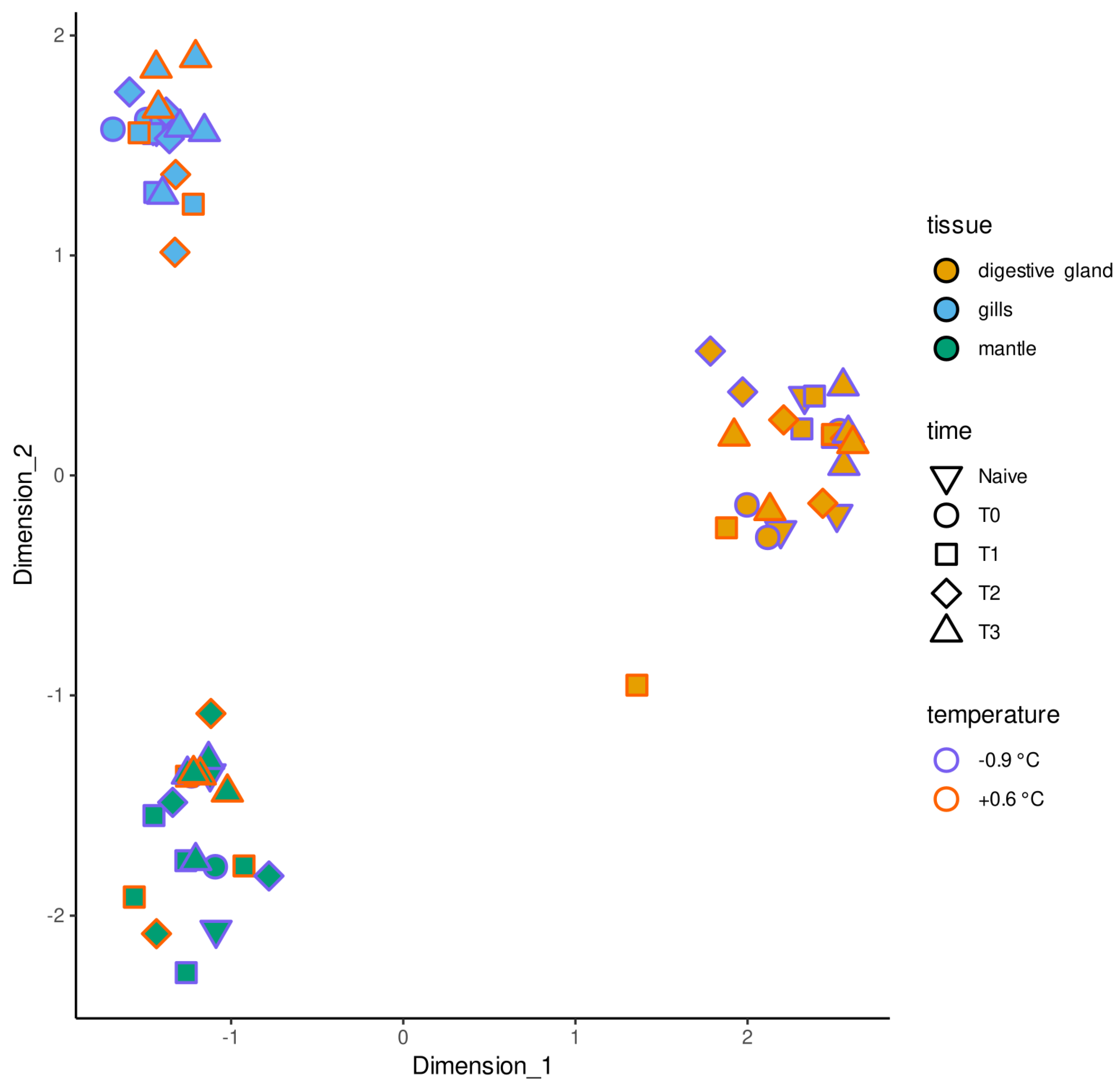
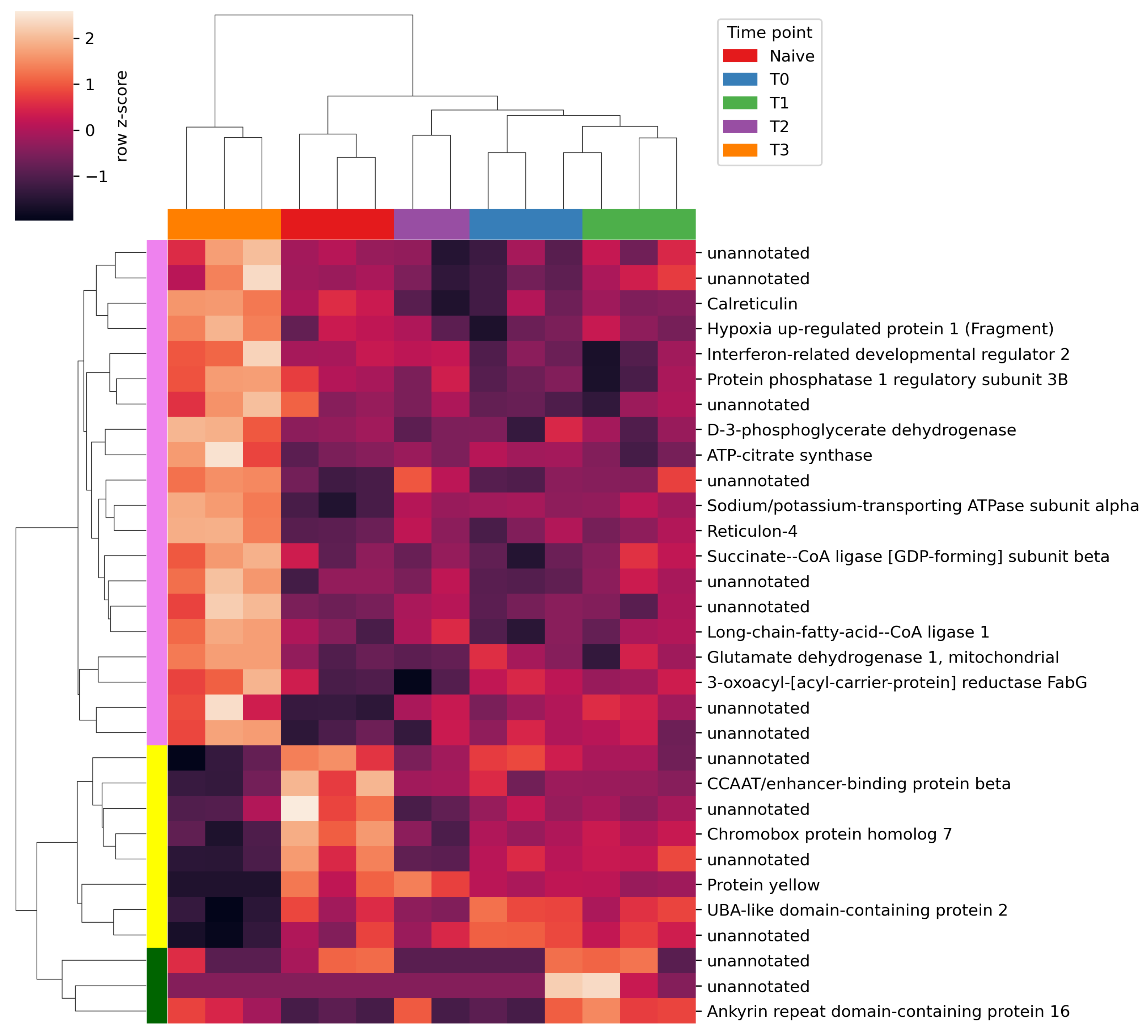
2.2. Differential Gene Expression Analysis

Illumina sequencing provided a total of 490M raw reads, unevenly distributed among the 72 samples, averaging 6.81M reads per sample. The Q30 rate ranged from 83.33% to 93.75% (median = 92.69%). After trimming, the total number of reads was reduced to 415 M, i.e., 5.76 M per sample and all samples retained more than 1 M reads, which we arbitrarily considered as the lower threshold to allow further processing.
The mean mapping rate against the reference transcriptome was 54.21%, with a standard deviation of 10.77%. Samples with low mapping rate (i.e., not reaching the arbitrary threshold of 40%) were marked as potential outliers linked to RNA degradation that occurred during the transportation of samples from Antarctica to Italy, based on the previous experience gathered from a similar experiment carried out on T. bernacchii [27]. The identification of outlier samples was further confirmed by the visual inspection of an MDS plot (based on the preliminary calculation of gene expression values across all samples). This process led to the removal of nine low-quality samples (Table S1). All tissues and experimental time points were in any case represented by at least two independent biological replicates.
As expected, the MDS analysis evidenced a strong clustering of non-outlier samples based on the tissue of origin (Figure 1). On the other hand, no significant clustering of samples obtained from the same tissue was evident, regardless of the time point of sampling and the exposure to higher water temperatures.
After batch effect removal, DGE analysis could not evidence any significant alteration of A. colbecki gene expression profiles in response to thermal stress in any of the three tissues of interest. Stabling stress let to no significant changes in gills and mantle, but was associated with the significant differential regulation of 31 DEGs in the digestive gland, 19 of which had an annotation. In detail, the stabling-specific response of the digestive gland could be divided into three different patterns, highlighted in Figure 2. Among these, those indicated in violet and yellow, respectively, were of greater interest, as they included the genes that were up-regulated and down-regulated at the latest time point, i.e., after a period of 19 days in the experimental tanks. The violet cluster, the largest one, included 20 DEGs and 8 DEGs were placed in the yellow cluster. Three DEGs were placed in an additional cluster (green), characterized by noisy expression levels. Detailed information concerning the 31 DEGs are reported in Table S2.

3. Discussion

We reassembled a high quality transcriptome, with slightly improved metrics compared to the one produced in the original publication [8]. To ensure a reliable calculation of gene expression levels, we carefully evaluated the presence of contamination from exogenous sequences not belonging to the target species. In particular, we detected the presence of contamination from the notothenioids T. bernacchii and C. hamatus which were being studied at the MZS during the XXV PNRA expedition [28], concurrently with the collection and dissection of the biological samples used for the assembly of the transcriptome of A. colbecki. Moreover, traces of P. pseudoroscoffensis, a coccolithophore that was being studied in our laboratory in the same period the reads were prepared for sequencing, were also detected. The presence of sequences from these species is most likely the result of laboratory cross-contamination, originated either during the RNA extraction, or the preparation of sequencing libraries. Finally, since related species have been previously documented in the Southern Ocean (e.g., Picconiella plumosa [29]), we interpret the presence of sequences from the unidentified red alga as the result of the accumulation of algal cells in scallop tissues due to their filter-feeding habits. The removal of all the aforementioned contaminations did not affect the quality and completeness of the transcriptome assembly.
The differential gene expression analysis carried out on this improved reference revaled that the transcriptional landscape of the Antarctic scallop A. colbecki was completely unresponsive to heat stress. Comparatively, the same temperature and timing of exposure led to significant alterations in multiple tissues in the stenotherm notothenioid T. bernacchii [27]. This lack of modulation has profound implications for what concerns the ability of this stenotherm bivalve to cope with the environmental alterations that might affect the Antarctic continent in the years to come.
Although the temperature increase used in this experiment was moderate (i.e., +1.5 °C compared with control tanks and ambient seawater temperature at MZS), A. colbecki was exposed to a temperature that is nearly halfway between optimal living conditions and its upper lethal thermal limit, which is significantly lower than that of other Antarctic mollusks [16]. Indeed, previous experiments have indicated that the swimming activity of A. colbecki starts to be significantly impaired slightly above 0 °C, marking early signatures of stress, and individuals are unable to survive in tanks kept at temperatures higher than 3 °C [17,18]. Consequently, we do not interpret this lack of transcriptional response as an absence of significant organismal stress, as this would be inconsistent with previously collected physiological and behavioral observations, but rather as the inability of A. colbecki to adequately respond to a type of stress that is not normally encountered in its extremely stable natural living environment. The fact that Adamussium populations apparently prefer highly stable shallow coastal environments with permanent or persistent sea-ice coverage [2], thereby not experiencing significant changes in temperature due to solar radiation, would support this interpretation. Moreover, like most other pectinids, adult A. colbecki individuals are generally not attached to the substrate and capable of moving short distances in their natural environment by swimming through valve clapping [9]. As suggested by several authors, the swimming behavior of scallops might have an ecological significance [30] and in the specific case of A. colbecki it may allow the scallops to move to colder areas in response to water warming, thereby not requiring the evolution of alternative sophisticated physiological thermal stress tolerance mechanisms.
It has to be stressed that the data collected in the frame of this study are restricted to three tissues and only provide an overview on the transcriptional changes occurring over a time frame of 19 days. Therefore, the aforementioned considerations should not be extended to other tissues, to other developmental life stages, or considered to be representative of a long-term exposure.
Unlike heat stress, stabling stress led to a measurable, albeit not particularly strong, response, which was restricted to the digestive gland and limited to the latest experimental time point, identifying trends markedly different from those observed in Antarctic fishes adapted to live in the very same extreme and stable environment [27]. The generalized poor response displayed by A. colbecki to stabling stress is strongly consistent with the previous observation that this species is characterized by very little phenotypic plasticity, being fundamentally unable to modify its physiology in response to stress, regardless of its acclimation history [17]. The low number of annotated DEGs hindered the possibility of gaining in-depth functional insights on the biological relevance of such transcriptomic alterations through functional enrichment analyses. Therefore, the discussion will focus on the functions of individual DEGs that might be relevant in the context of stress response in the digestive gland.
As reported in Section 2, DEGs were grouped in two major clusters displaying opposite expression trends at T3, i.e., approximately 25 days post-catch, considering the acclimation period and the duration of the thermal stress experiment. The cluster of DEGs indicated in violet in Figure 2 included several genes involved directly in mitochondrial respiration (i.e., ATP-citrate synthase, D-3-phosphoglycerate dehydrogenase, succinate-CoA ligase), fatty acid metabolism (i.e., long-chain-fatty-acid–CoA ligase 1-like, 3-oxoacyl-[acyl-carrier-protein] reductase FabG-like) and glycogen metabolism (i.e., phosphatase 1 regulatory subunit 3B) [31]. Another up-regulated DEGs was the alpha subunit of the sodium/potassium-transporting ATPase, which acts as the catalytic part of several transport channels. It belongs to the P-Type ATPase family, which provides different functions depending on the cell type and tissue, including absorption of nutrients [32]. Among several other up-regulated uncharacterized DEGs lacking annotation, two relevant genes were reticulon-4 and the interferon-related development regulator (IFRD2). The former is part of the reticulon protein family, mainly involved in shaping endoplasmic reticulum membranes and regulating vesicle trafficking [33]. The latter is an immediate response gene which mediates the activation of several downstream processes, including immune response to infection in invertebrates [34,35]. Although gene expression data did not support a generalized activation of immune response at T3 in the digestive gland, it is plausible that the downstream effects of such the up-regulation of IFRD2 may be observable after a longer stay in experimental tanks.
The down-regulated DEGs belonging to the yellow cluster included the chromobox protein homolog 7 and the CCAAT/enhancer-binding protein beta, two transcription factors that regulate gene expression of multiple targets [36,37,38]. Moreover, the down-regulation of a transcript encoding an ubiquitin A-like protein suggested the ongoing alteration in the ubiquitin-dependent protein degradation mechanisms, even though this protein may also exert different roles, in light of the poor primary sequence homology displayed by this A. colbecki sequence with functionally characterized relatives [39].
Functional information concerning several other DEGs is still lacking and, as previously mentioned, only 60% of all DEGs could be annotated, consistently with the widespread presence of lineage-specific uncharacterized gene families in bivalve genomes [40]. Moreover, the function of the DEGs reported in the previous paragraphs could be only indirectly inferred from studies carried out in phylogenetically distant model organism, most often humans or other vertebrates. Consequently, the biological relevance of the reported alterations should be interpreted with caution. Nevertheless, the gene expression trends we evidenced support the activation of mitochondrial energetic metabolism and alterations in nutrient adsorption, which one of the key physiological roles of the digestive gland in bivalves. Even though the specimens were kept in an open system with fresh seawater directly fed from the ocean, we hypothesize that these DEGs may be linked with the altered diet of the scallops kept under experimental conditions. In fact, due to the low amount of sediment present in the tanks, it is possible that the specimens were unable to actively suspend, by clapping their valves, levels of sediment comparable with those found in the natural environment [41]. Some DEGs, in particular those related with metabolism, may be also connected with the reduced growth rates displayed by A. colbecki in captivity, which were demonstrated to be independent of the availability of nutrients [42].

4. Materials and Methods

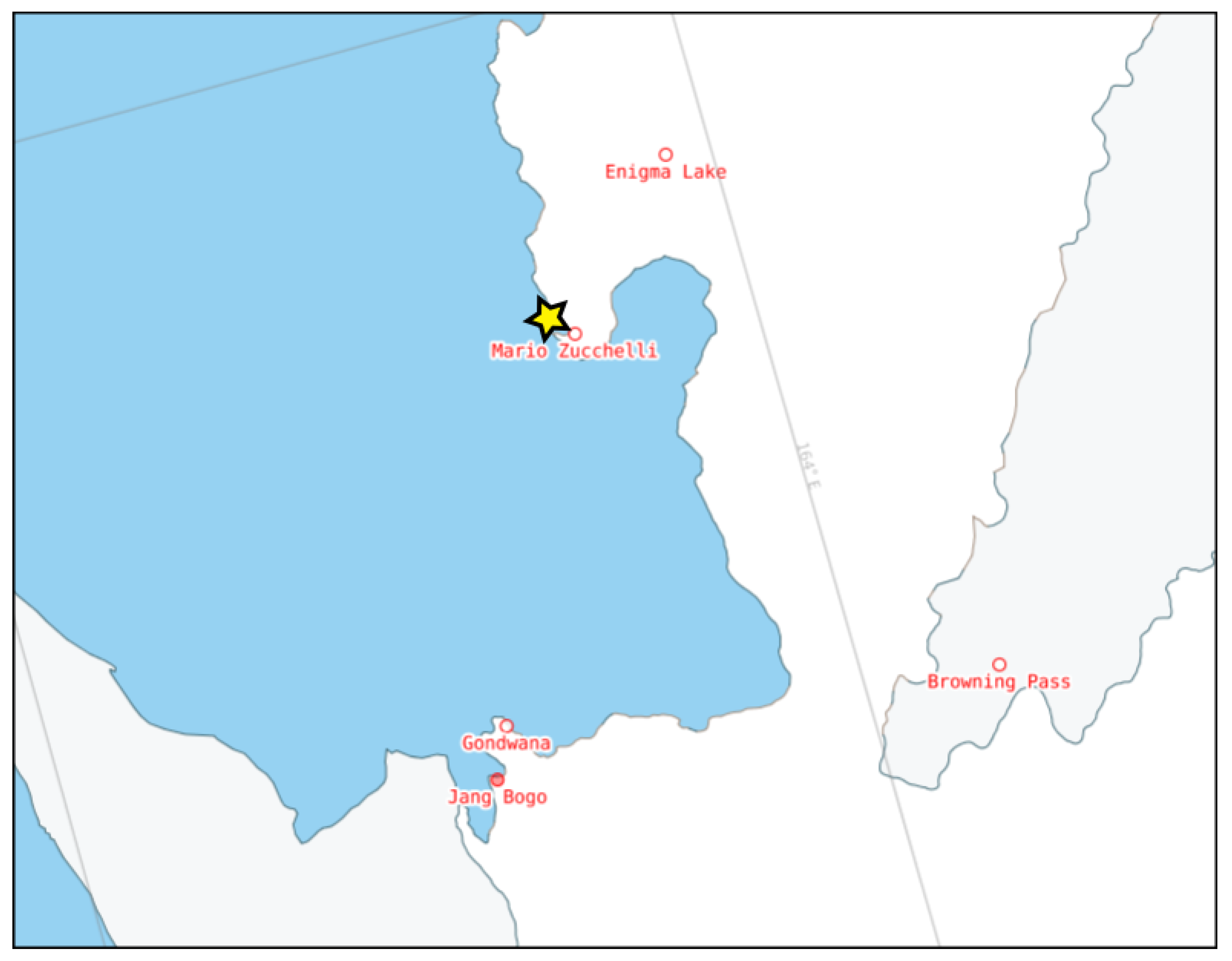
4.1. Sampling and Experimental Setup

A total of 24 specimens of A. colbecki were sampled by scuba divers from the seafloor at the Ross Sea, near the Mario Zucchelli Station (MZS) in Antarctica (Figure 3).
The experimental design was identical to the one previously described in a similar thermal stress experiment carried out in the notothenioid fish T. bernacchii [27]. Briefly, three specimens were immediately sacrificed, dissected, and their tissues (i.e., gills, digestive gland and mantle) were placed in RNAlater (Thermo Fisher Scientific, Waltham, MA, USA), defining the Naive group. The remaining 21 specimens were acclimated at stable conditions for six days in a tank with a direct feed of circulated seawater at −0.9 °C, the ambient surface seawater temperature in the proximity of MZS. Three individuals were sampled at the end of the acclimation period (T0), whereas the remaining individuals were randomly split in two 180 L tanks: one was kept at −0.9 °C (control tank) and the other was warmed to 0.6 °C (Δ = +1.5 °C, treatment tank). Both tanks had seawater continuously circulated directly from the sea, and temperatures were logged with a TinyTag Aquatic data logger. Three individuals were sampled from each tank after 6 h (T1), 7 days (T2) and 19 days (T3). During the sampling process, the gills, the digestive gland and the mantle tissues were collected, stored in 1.5 mL tubes in RNAlater solution at −20 °C and later shipped to Italy in refrigerated tanks, where they were stored at −80 °C until further processing.

4.2. RNA Extraction and Library Preparation

The RNA extraction and library preparation were carried out following the protocol previously described by our group [27]. Briefly, total RNA was isolated from all samples using the Direct-zol™ RNA MiniPrep kit (Zymo Research, Irvine, CA, USA) following the manifacturer’s instructions, and quantified with a NanoDrop™ 2000 spectrophotometer (Thermo Fisher Scientific, Waltham, MA, USA). The total RNA concentration (ng/mL) of the samples was accurately estimated according to the RNA: High Sensitivity Assay on Qubit™ 2.0 fluorometer (Thermo Fisher Scientific, Waltham, MA, USA). The quality of the extracted RNAs was further evaluated using a Bioanalyzer 2100 instrument (Agilent Technologies, Santa Clara, CA, USA), to ensure that the RNA Integrity Number (RIN) was adequate. Libraries compatible with Illumina sequencing were prepared according to the QuantSeq™ 3′ mRNA-Seq Kit forward protocol (Lexogen, Wien, Austria) and their quality was checked with a Bioanalyzer 2100 instrument (Agilent Technologies, Santa Clara, CA, USA). The libraries were pooled according to the Illumina Pooling Calculator (https://support.illumina.com/help/pooling-calculator/pooling-calculator.htm, accessed on 20 August 2019) and sequenced on a NovaSeq 6000 platform (Illumina, San Diego, CA, USA) with a 150 bp single end strategy by CBM—Consorzio per il centro di Biomedicina Molecolare S.C.A.R.L. (Area Science Park, Trieste, Italy).

4.3. Transcriptome Assembly and Refinement

Although we had previously generated a reference transcriptome assembly for A. colbecki [8], this resource was updated with the aim of improving its suitability for the analysis of the sequencing data generated with the approach described in the previous section. Briefly, raw paired-end reads from the previous study (bioproject id PRJNA379393) were quality checked with fastqc v.0.11.9 and muliqc v.1.6 [43], removing leading and trailing homopolymer stretches, adapters, sequences containing unidentified (i.e., N characters) and low quality (PHRED < 30) nucleotides. Resulting reads shorter than the minimum allowed length (i.e., 75 nucleotides) were discarded.
Trimmed reads passing all quality filters were assembled with ORP v2.3.3 [44], and the completeness of the assembly was evaluated with BUSCO v.5.3.2 [45] against the Metazoa OrhoDBv10 database [46].
Contigs with low coverage and/or possibly originating from exogenous contamination were removed as follows: reads were mapped back to the transcriptome with salmon v.1.8.0 [47] and contigs with effective length < 150 nucleotides and Transcripts Per Million (TPM) < 1 were discarded. Exogenous contamination were identified though the detection of corresponding assembled COX1 sequences with tBLASTn. The complete genomes (or, in case of unavailability, transcriptomes) of the putative contaminants were used to filter out contigs not belonging to A. colbecki, defined as those that showed BLASTn matches with e-value < 1 × 10⁻¹⁰.
Different splicing isoforms and allelic variants were clustered into putative genes using the homology-based method proposed by Ono and colleagues [48]. To this end, we used the protein annotations from the genomes of Pecten maximus (xPecMax1.1) [49] and Mizuhopecten yessoensis (ASM211388v2) [50] for the annotation of orthologous genes shared by all Pectinida. This process was also carried out against a non-redundant version of the Uniprot Swissprot database [51], including representative sequences clustered based on 50% reciprocal homology with cd-hit v.4.8.1 [52]. Homology searches were performed with diamond v.0.9.14 [53]. Finally, the transcriptome was functionally annotated with annot.aM, assigning each sequence Gene Ontology terms and conserved Pfam domains [54].

4.4. Gene Expression Quantification and Pre-Processing

Raw reads (deposited in the NCBI SRA database, with BioProject id PRJNA588994) were quality checked and trimmed, as reported above in the case of the paired-end sequences used for transcriptome assembly. Trimmed reads were mapped against the reference transcriptome with salmon v.1.8.0 [47], by setting the –validateMappings flag and providing a transcript-to-gene map obtained with the steps described in the previous subsection. Read mappings from all samples were then merged and saved as read counts to allow Differential Gene Expression (DGE) analysis.
Prior to any analysis, expression values were pre-processed, separately for each tissue, with the filteByExpr function provided by edgeR [55,56,57]. This function allows the removal of lowly expressed genes with automatic parameters, based on the strategy described by Chen and colleagues [56].
The removal of batch effects was performed with empirical controls with RUVseq [58] (RUVg function) and outlier samples (most likely due to a low quality of input RNA) were identified by visual inspection of the MDS plot and removed, ensuring the presence of at least two biological replicates for each experimental time point, condition and tissue. As a final step in data preparation, expression values were normalized with the calcNormFactors function, using the “upperquartile” method.

4.5. Statistical Analysis

As for the preprocessing step, DGE analyses were independently performed for each tissue with the GLM functionality of edgeR. The contrasts used in the analysis were aimed to separately investigate the effects of thermal stress over time (compared with animals kept in control tanks at ambient temperature) and of the time spent in experimental tanks (stabling stress, though the comparison with the naive group). In detail, the effects of thermal stress were evaluated through pairwise comparisons between stressed and control groups at each experimental time point, and between stressed samples at each time point against all control groups, regardless of the time point (only excluding naive samples to account for catch-related stress). DGE was computed with the glmQLFtest function of edgeR. The effect of stabling stress was assessed through the pairwise comparisons between the control samples at each time point and the naive samples (the samples exposed to the heat stress were excluded in this analysis). An anova-like test (searching for difference between any group) was also performed on the full set of naive and control samples. Genes showing FDR adjusted p-value < 0.05 and |logFC| > 1 were selected as DEGs in all tests. The set of DEGs responsive to thermal stress was further refined, removing those whose expression was also significantly altered in response to stabling stress, with the aim to isolate thermal-stress related DEGs, discriminating them from general stress-related DEGs.

5. Conclusions

In this study, we investigated the transcriptomic response of A. colbecki to a moderate temperature increase, compatible with the changes in ocean temperatures expected to occur in several Antarctic coastal areas due to climate change in the next few decades. As the most stenotherm of all Antarctic mollusks, this species was predicted to experience significant stress in response to the 1.5 °C warming applied in the present experiment. Our analyses show that the Antarctic scallop was unable to actuate a detectable molecular response to thermal stress in the mantle, gills and digestive gland. These results support the idea that the stability of the Antarctic Ocean temperatures over millions of years might have led to the loss of the ability to respond to even slight temperature variations in this species. Moreover, the small response displayed by A. colbecki, which was limited to the digestive gland at the latest time point, further supports the near-lack of pheotypic plasticity of this species in response to environmental alterations. Upcoming morphological studies carried out on the same samples will be useful to complement our results and to predict the fate of A. colbecki in a warming ocean scenario.

Supplementary Materials

The following supporting information can be downloaded at: https://www.mdpi.com/article/10.3390/stresses3020034/s1, Table S1: list of samples classified as outliers; Table S2: results of differential expressions analysis of the stabling stress on control and naive samples of the digestive gland.

Author Contributions

Original draft compilation: S.G., A.S.G. and M.G.; proofreading: S.G., A.S.G., C.M., F.C., G.S., A.P., P.G.G. and M.G.; experimental design: C.M., G.S., P.G.G. and M.G.; sampling: P.G.G.; wet lab methods: A.S.G., C.M. and F.C.; bioinformatic analysis: S.G., A.S.G. and M.G.; interpretation of results: S.G., A.S.G., A.P., P.G.G. and M.G. All authors have read and agreed to the published version of the manuscript.

Funding

This work was funded by the Italian Program of Antarctic Research (Grant Nos. PNRA16_00099, PNRA16_00234 and PNRA18_00077).

Institutional Review Board Statement

The sample collection and animal research conducted in this study comply with Italy’s Ministry of Education, University and Research regulations concerning activities and environmental protection in Antarctica and with the Protocol on Environmental Protection to the 137 Antarctic Treaty, Annex II, Art. 3. All the activities on animals performed during the Italian Antarctic Expedition were under the control of a PNRA Ethics Referent, which acts on behalf of the Italian Ministry of Foreign Affairs. In particular, the required data for the project identification code PNRA16_00099 are as follows: Name of the ethics committee or institutional review board: Italian Ministry of Foreign Affairs. Name of PNRA Ethics Referent: Carla Ubaldi, ENEA Antarctica, Technical Unit (UTA).

Informed Consent Statement

Not applicable.

Data Availability Statement

The raw reads generated by RNA-seq have been deposited at the NCBI SRA database under the BioProject ID PRJNA588994.

Acknowledgments

The authors would like to thank Fiorella Florian and Fabrizia Gionechetti for their precious assistance in the wet-lab phase of the experiment. This work was partially funded by The National Research Program in Antarctica PNRA18_00077 AMFEB.

Conflicts of Interest

The authors declare no conflict of interest. The funders had no role in the design of the study; in the collection, analyses, or interpretation of data; in the writing of the manuscript, or in the decision to publish the results.

References

  1. Stockton, W. The Biology and Ecology of the Epifaunal Scallop Adamussium colbecki on the West Side of McMurdo Sound, Antarctica. Mar. Biol. 1984, 78, 171–178. [Google Scholar] [CrossRef]
  2. Schiaparelli, S.; Linse, K. A Reassessment of the Distribution of the Common Antarctic Scallop Adamussium colbecki (Smith, 1902). Deep. Sea Res. Part II Top. Stud. Oceanogr. 2006, 53, 912–920. [Google Scholar] [CrossRef]
  3. Berkman, P.A. The Population Biology of the Antarctic Scallop, Adamussium colbecki (Smith 1902) at New Harbor, Ross Sea. In Antarctic Ecosystems; Kerry, K.R., Hempel, G., Eds.; Springer: Berlin/Heidelberg, Germany, 1990; pp. 281–288. [Google Scholar] [CrossRef]
  4. Chiantore, M.; Cattaneo-Vietti, R.; Povero, P.; Albertelli, G. The Population Structure and Ecology of the Antarctic Scallop Adamussium colbecki in Terra Nova Bay. In Ross Sea Ecology; Springer: Berlin/Heidelberg, Germany, 2000; pp. 563–573. [Google Scholar]
  5. Beu, A.; Taviani, M. Early Miocene Mollusca from McMurdo Sound, Antarctica (ANDRILL 2A Drill Core), with a Review of Antarctic Oligocene and Neogene Pectinidae (Bivalvia). Palaeontology 2014, 57, 299–342. [Google Scholar] [CrossRef]
  6. Quaglio, F.; Warren, L.V.; Anelli, L.E.; Dos Santos, P.R.; Rocha-Campos, A.C.; Gaździcki, A.; Strikis, P.C.; Ghilardi, R.P.; Tiossi, A.B.; Simões, M.G. Shell Beds from the Low Head Member (Polonez Cove Formation, Early Oligocene) at King George Island, West Antarctica: New Insights on Facies Analysis, Taphonomy and Environmental Significance. Antarct. Sci. 2014, 26, 400–412. [Google Scholar] [CrossRef]
  7. Egorova, E.N. Fossil Bivalves in Marine Sediments of Vestfold Hills in Eastern Antarctica. Biol. Bull. 2016, 43, 619–627. [Google Scholar] [CrossRef]
  8. Moro, G.; Buonocore, F.; Barucca, M.; Spazzali, F.; Canapa, A.; Pallavicini, A.; Scapigliati, G.; Gerdol, M. The First Transcriptomic Resource for the Antarctic Scallop Adamussium colbecki. Mar. Genom. 2019, 44, 61–64. [Google Scholar] [CrossRef]
  9. Denny, M.; Miller, L. Jet Propulsion in the Cold: Mechanics of Swimming in the Antarctic Scallop Adamussiumcolbecki. J. Exp. Biol. 2006, 209, 4503–4514. [Google Scholar] [CrossRef] [PubMed]
  10. Wong, W.S.; Hauer, L.; Cziko, P.A.; Meister, K. Cryofouling Avoidance in the Antarctic Scallop Adamussium colbecki. Commun. Biol. 2022, 5, 1–8. [Google Scholar] [CrossRef] [PubMed]
  11. Heilmayer, O.; Brey, T. Saving by Freezing? Metabolic Rates of Adamussium colbecki in a Latitudinal Context. Mar. Biol. 2003, 143, 477–484. [Google Scholar] [CrossRef]
  12. Peck, L.S.; Powell, D.K.; Tyler, P.A. Very Slow Development in Two Antarctic Bivalve Molluscs, the Infaunal Clam Laternula Elliptica and the Scallop Adamussium colbecki. Mar. Biol. 2007, 150, 1191–1197. [Google Scholar] [CrossRef]
  13. Regoli, F.; Nigro, M.; Bertoli, E.; Principato, G.; Orlando, E. Defenses against Oxidative Stress in the Antarctic Scallop Adamussium colbecki and Effects of Acute Exposure to Metals. In Interactions and Adaptation Strategies of Marine Organisms; Springer: Berlin/Heidelberg, Germany, 1997; pp. 139–144. [Google Scholar]
  14. Storch, D.; Heilmayer, O.; Hardewig, I.; Portner, H.O. In Vitro Protein Synthesis Capacities in a Cold Stenothermal and a Temperate Eurythermal Pectinid. J. Comp. Physiol. B Biochem. Syst. Environ. Physiol. 2003, 173, 611–620. [Google Scholar] [CrossRef] [PubMed]
  15. Greco, S.; Gaetano, A.; Voltarel, G.; Pallavicini, A.; Giulianini, P.; Gerdol, M. Comparative Transcriptomic Analysis Reveals Adaptive Traits in Antarctic Scallop Adamussium colbecki. Fishes 2023. under review. [Google Scholar] [CrossRef]
  16. Peck, L.S.; Webb, K.E.; Bailey, D.M. Extreme Sensitivity of Biological Function to Temperature in Antarctic Marine Species. Funct. Ecol. 2004, 18, 625–630. [Google Scholar] [CrossRef]
  17. Bailey, D.M. The Thermal Dependence of Swimming and Muscle Physiology in Temperate and Antarctic Scallops. Ph.D. Thesis, University of St Andrews, St Andrews, UK, 2001. [Google Scholar]
  18. Bailey, D.M.; Peck, L.S.; Bock, C.; Pörtner, H.O. High-Energy Phosphate Metabolism during Exercise and Recovery in Temperate and Antarctic Scallops: An In Vivo 31P-NMR Study. Physiol. Biochem. Zool. 2003, 76, 622–633. [Google Scholar] [CrossRef]
  19. Quaglio, F.; Whittle, R.J.; Gaździcki, A.; Guimarães, S.M. A new fossil Adamussium (Bivalvia: Pectinidae) from Antarctica. Pol. Polar Res. 2010, 31, 291–302. [Google Scholar] [CrossRef]
  20. Benedetti, M.; Lanzoni, I.; Nardi, A.; d’Errico, G.; Di Carlo, M.; Fattorini, D.; Nigro, M.; Regoli, F. Oxidative Responsiveness to Multiple Stressors in the Key Antarctic Species, Adamussium colbecki: Interactions between Temperature, Acidification and Cadmium Exposure. Mar. Environ. Res. 2016, 121, 20–30. [Google Scholar] [CrossRef]
  21. Truebano, M.; Diz, A.P.; Thorne, M.A.; Clark, M.S.; Skibinski, D.O. Proteome Response to Heat Stress in the Antarctic Clam Laternula elliptica. J. Integr. OMICS 2013, 3, 34–43. [Google Scholar] [CrossRef]
  22. Bonacci, S.; Browne, M.A.; Dissanayake, A.; Hagger, J.A.; Corsi, I.; Focardi, S.; Galloway, T.S. Esterase Activities in the Bivalve Mollusc Adamussium colbecki as a Biomarker for Pollution Monitoring in the Antarctic Marine Environment. Mar. Pollut. Bull. 2004, 49, 445–455. [Google Scholar] [CrossRef]
  23. Regoli, F.; Nigro, M.; Chiantore, M.; Winston, G. Seasonal Variations of Susceptibility to Oxidative Stress in Adamussium colbecki, a Key Bioindicator Species for the Antarctic Marine Environment. Sci. Total. Environ. 2002, 289, 205–211. [Google Scholar] [CrossRef]
  24. Cronin, K.E.; Walker, S.E.; Mann, R.; Chute, A.S.; Long, M.C.; Bowser, S.S. Growth and Longevity of the Antarctic Scallop AdamussiumColbecki under Annual and Multiannual Sea Ice. Antarct. Sci. 2020, 32, 466–475. [Google Scholar] [CrossRef]
  25. Heilmayer, O.; Brey, T.; Chiantore, M.; Cattaneo-Vietti, R.; Arntz, W.E. Age and Productivity of the Antarctic Scallop, Adamussium colbecki, in Terra Nova Bay (Ross Sea, Antarctica). J. Exp. Mar. Biol. Ecol. 2003, 288, 239–256. [Google Scholar] [CrossRef]
  26. Masson-Delmotte, V.; Zhai, P.; Pörtner, H.O.; Roberts, D.; Skea, J.; Shukla, P.R.; Pirani, A.; Moufouma-Okia, W.; Péan, C.; Pidcock, R.; et al. Policymakers. In Global Warming of 1.5 C. An IPCC Special Report on the Impacts of Global Warming of 1.5 C above Pre-Industrial Levels and Related Global Greenhouse Gas Emission Pathways, in the Context of Strengthening the Global Response to the Threat of Climate Change, Sustainable Development, and Efforts to Eradicate Poverty; Cambridge University Press: Cambridge, UK; New York, NY, USA, 2018; pp. 3–24. [Google Scholar] [CrossRef]
  27. Greco, S.; Gaetano, A.S.; Furlanis, G.; Capanni, F.; Manfrin, C.; Giulianini, P.G.; Santovito, G.; Edomi, P.; Pallavicini, A.; Gerdol, M. Gene Expression Profiling of Trematomus bernacchii in Response to Thermal and Stabling Stress. Fishes 2022, 7, 387. [Google Scholar] [CrossRef]
  28. Ansaloni, F.; Gerdol, M.; Torboli, V.; Fornaini, N.R.; Greco, S.; Giulianini, P.G.; Coscia, M.R.; Miccoli, A.; Santovito, G.; Buonocore, F.; et al. Cold Adaptation in Antarctic Notothenioids: Comparative Transcriptomics Reveals Novel Insights in the Peculiar Role of Gills and Highlights Signatures of Cobalamin Deficiency. Int. J. Mol. Sci. 2021, 22, 1812. [Google Scholar] [CrossRef] [PubMed]
  29. Zieliński, K. Bottom macroalgae of the Admiralty Bay (King George Island, South Shetlands, Antarctica). Pol. Polar Res. 1990, 11, 95–131. [Google Scholar]
  30. Brand, A.R. Chapter 11—Scallop Ecology: Distributions and Behaviour. In Scallops; Shumway, S.E., Parsons, G.J., Eds.; Elsevier: Amsterdam, The Netherlands, 2016; Volume 40, pp. 469–533. [Google Scholar] [CrossRef]
  31. Mehta, M.B.; Shewale, S.V.; Sequeira, R.N.; Millar, J.S.; Hand, N.J.; Rader, D.J. Hepatic Protein Phosphatase 1 Regulatory Subunit 3B (Ppp1r3b) Promotes Hepatic Glycogen Synthesis and Thereby Regulates Fasting Energy Homeostasis. J. Biol. Chem. 2017, 292, 10444–10454. [Google Scholar] [CrossRef] [PubMed]
  32. Axelsen, K.B.; Palmgren, M.G. Evolution of Substrate Specificities in the P-Type ATPase Superfamily. J. Mol. Evol. 1998, 46, 84–101. [Google Scholar] [CrossRef] [PubMed]
  33. Yang, Y.; Strittmatter, S. The Reticulons: A Family of Proteins with Diverse Functions. Genome Biol. 2007, 8, 234. [Google Scholar] [CrossRef]
  34. Vietor, I.; Huber, L.A. Role of TIS7 Family of Transcriptional Regulators in Differentiation and Regeneration. Differ. Res. Biol. Divers. 2007, 75, 891–897. [Google Scholar] [CrossRef] [PubMed]
  35. Ge, Q.; Li, J.; Wang, J.; Li, J.; Ge, H.; Zhai, Q. Transcriptome Analysis of the Hepatopancreas in Exopalaemon carinicauda Infected with an AHPND-causing Strain of Vibrio parahaemolyticus. Fish Shellfish. Immunol. 2017, 67, 620–633. [Google Scholar] [CrossRef]
  36. Wang, W.; Xia, X.; Mao, L.; Wang, S. The CCAAT/Enhancer-Binding Protein Family: Its Roles in MDSC Expansion and Function. Front. Immunol. 2019, 10, 1804. [Google Scholar] [CrossRef]
  37. Maertens, G.; El, M.A.S.; Racek, T.; Stock, J.; Nicholls, J.; Rodriguez-Niedenführ, M.; Gil, J.; Peters, G. Several Distinct Polycomb Complexes Regulate and Co-Localize on the INK4a Tumor Suppressor Locus. PLoS ONE 2009, 4, e6380. [Google Scholar] [CrossRef] [PubMed]
  38. Li, Q.; Wang, X.; Lu, Z.; Zhang, B.; Guan, Z.; Liu, Z.; Zhong, Q.; Gu, L.; Zhou, J.; Zhu, B.; et al. Polycomb CBX7 Directly Controls Trimethylation of Histone H3 at Lysine 9 at the P16 Locus. PLoS ONE 2010, 5, e13732. [Google Scholar] [CrossRef]
  39. Mueller, T.; Feigon, J. Solution Structures of UBA Domains Reveal a Conserved Hydrophobic Surface for Protein-Protein Interactions. J. Mol. Biol. 2002, 319, 1243–1255. [Google Scholar] [CrossRef]
  40. Gerdol, M.; Moreira, R.; Cruz, F.; Gómez-Garrido, J.; Vlasova, A.; Rosani, U.; Venier, P.; Naranjo-Ortiz, M.; Murgarella, M.; Greco, S.; et al. Massive Gene Presence-Absence Variation Shapes an Open Pan-Genome in the Mediterranean Mussel. Genome Biol. 2020, 21, 275. [Google Scholar] [CrossRef]
  41. Ansell, A.D.; Ansell, A.D.; Cattaneo-Vietti, R.; Chiantore, M. Swimming in the Antarctic Scallop Adamussium colbecki: Analysis of in Situ Video Recordings. Antarct. Sci. 1998, 10, 369–375. [Google Scholar] [CrossRef]
  42. Chiantore, M.; Cattaneo-Vietti, R.; Heilmayer, O. Antarctic Scallop (Adamussium colbecki) Annual Growth Rate at Terra Nova Bay. Polar Biol. 2003, 26, 416–419. [Google Scholar] [CrossRef]
  43. Ewels, P.; Magnusson, M.; Lundin, S.; Käller, M. MultiQC: Summarize Analysis Results for Multiple Tools and Samples in a Single Report. Bioinformatics 2016, 32, 3047–3048. [Google Scholar] [CrossRef]
  44. MacManes, M. The Oyster River Protocol: A Multi-Assembler and Kmer Approach for de Novo Transcriptome Assembly. PeerJ 2018, 6, e5428. [Google Scholar] [CrossRef]
  45. Manni, M.; Berkeley, M.; Seppey, M.; Simão, F.; Zdobnov, E. BUSCO Update: Novel and Streamlined Workflows along with Broader and Deeper Phylogenetic Coverage for Scoring of Eukaryotic, Prokaryotic, and Viral Genomes. Mol. Biol. Evol. 2021, 38, 4647–4654. [Google Scholar] [CrossRef] [PubMed]
  46. Kriventseva, E.; Kuznetsov, D.; Tegenfeldt, F.; Manni, M.; Dias, R.; Simão, F.; Zdobnov, E. OrthoDB V10: Sampling the Diversity of Animal, Plant, Fungal, Protist, Bacterial and Viral Genomes for Evolutionary and Functional Annotations of Orthologs. Nucleic Acids Res. 2019, 47, D807–D811. [Google Scholar] [CrossRef]
  47. Patro, R.; Duggal, G.; Love, M.I.; Irizarry, R.A.; Kingsford, C. Salmon Provides Fast and Bias-Aware Quantification of Transcript Expression. Nat. Methods 2017, 14, 417–419. [Google Scholar] [CrossRef]
  48. Ono, H.; Ishii, K.; Kozaki, T.; Ogiwara, I.; Kanekatsu, M.; Yamada, T. Removal of Redundant Contigs from de Novo RNA-Seq Assemblies via Homology Search Improves Accurate Detection of Differentially Expressed Genes. BMC Genom. 2015, 16, 1031. [Google Scholar] [CrossRef] [PubMed]
  49. Zeng, Q.; Liu, J.; Wang, C.; Wang, H.; Zhang, L.; Hu, J.; Bao, L.; Wang, S. High-Quality Reannotation of the King Scallop Genome Reveals No ‘gene-Rich’ Feature and Evolution of Toxin Resistance. Comput. Struct. Biotechnol. J. 2021, 19, 4954–4960. [Google Scholar] [CrossRef] [PubMed]
  50. Wang, S.; Zhang, J.; Jiao, W.; Li, J.; Xun, X.; Sun, Y.; Guo, X.; Huan, P.; Dong, B.; Zhang, L.; et al. Scallop Genome Provides Insights into Evolution of Bilaterian Karyotype and Development. Nat. Ecol. Evol. 2017, 1, 0120. [Google Scholar] [CrossRef] [PubMed]
  51. The UniProt Consortium. UniProt: The Universal Protein Knowledgebase in 2021. Nucleic Acids Res. 2021, 49, D480–D489. [Google Scholar] [CrossRef]
  52. Li, W.; Jaroszewski, L.; Godzik, A. Clustering of Highly Homologous Sequences to Reduce the Size of Large Protein Databases. Bioinformatics 2001, 17, 282–283. [Google Scholar] [CrossRef]
  53. Buchfink, B.; Xie, C.; Huson, D.H. Fast and Sensitive Protein Alignment Using DIAMOND. Nat. Methods 2015, 12, 59–60. [Google Scholar] [CrossRef]
  54. Greco, S. annot.aM. Available online: https://gitlab.com/54mu/annotaM (accessed on 20 July 2022).
  55. Robinson, M.D.; McCarthy, D.J.; Smyth, G.K. edgeR: A Bioconductor Package for Differential Expression Analysis of Digital Gene Expression Data. Bioinformatics 2010, 26, 139–140. [Google Scholar] [CrossRef] [PubMed]
  56. Chen, Y.; Lun, A.T.L.; Smyth, G.K. From Reads to Genes to Pathways: Differential Expression Analysis of RNA-Seq Experiments Using Rsubread and the edgeR Quasi-Likelihood Pipeline. F1000Research 2016, 5, 1438. [Google Scholar] [CrossRef]
  57. McCarthy, D.J.; Chen, Y.; Smyth, G.K. Differential Expression Analysis of Multifactor RNA-Seq Experiments with Respect to Biological Variation. Nucleic Acids Res. 2012, 40, 4288–4297. [Google Scholar] [CrossRef]
  58. Risso, D.; Ngai, J.; Speed, T.P.; Dudoit, S. Normalization of RNA-seq Data Using Factor Analysis of Control Genes or Samples. Nat. Biotechnol. 2014, 32, 896–902. [Google Scholar] [CrossRef] [PubMed]
Figure 1. Multi Dimensional Scaling scatter plot from all samples. Each marker represents a sample, the filling color of the markers codes for the tissue of origin, the shape codes for the experimental time point and the outline color codes for the group (control and stress temperatures). Outlier samples are not displayed.
Stresses 03 00034 g001
Figure 2. Heatmap of z-scores of log(CPM) of the DEGs identified in the digestive gland in response to stabling stress. Columns represent samples, annotated on top based on the time point. The dendrogram on the left represents hierarchical clustering of genes based on their expression patterns. Only control and naive samples (−0.9 °C) were used in the stabling stress analysis.
Stresses 03 00034 g002
Figure 3. Map of the Antarctica near the Mario Zucchelli Station. The sampling site is highlighted by the icon of a yellow five-pointed star (image source: https://www.add.scar.org/, accessed on 20 March 2023).
Stresses 03 00034 g003
Table 1. Quality and BUSCO metrics of the filtered de novo transcriptome of A. colbecki.
ParameterValue
Total contigs44,475
GC content40.76
Contig N501911
Median contig length886
Mean contig length1329.11
Total assembled bases591,112,155
BUSCO single (%)76
BUSCO duplicated (%)13.9
BUSCO fragmented (%)4.4
BUSCO missing (%)5.7
Disclaimer/Publisher’s Note: The statements, opinions and data contained in all publications are solely those of the individual author(s) and contributor(s) and not of MDPI and/or the editor(s). MDPI and/or the editor(s) disclaim responsibility for any injury to people or property resulting from any ideas, methods, instructions or products referred to in the content.

