Review

Spine Bracing: When to Utilize—A Narrative Review

1
Dr. Kiran C. Patel College of Osteopathic Medicine, Nova Southeastern University, Clearwater, FL 33759, USA
2
College of Medicine, Drexel University, Philadelphia, PA 19129, USA
3
Department of Molecular and Cellular Biology, University of Arizona, Tucson, AZ 85721, USA
4
College of Medicine, University of Florida, Gainesville, FL 32608, USA
5
Department of Neurosurgery, University of Florida, Gainesville, FL 32608, USA
*
Author to whom correspondence should be addressed.
Biomechanics 2023, 3(1), 136-154; https://doi.org/10.3390/biomechanics3010013
Submission received: 26 January 2023 / Revised: 14 February 2023 / Accepted: 15 February 2023 / Published: 10 March 2023

Abstract

:
Spinal bracing is a common non-surgical technique that allows clinicians to prevent and correct malformations or injuries of a patient’s spinal column. This review will explore the current standards of practice on spinal brace utilization. Specifically, it will highlight bracing usage in traumatic injuries, pregnancy, pediatrics, osteoporosis, and hyperkyphosis; address radiological findings concurrent with brace usage; and provide an overview of the braces currently available and advancements in the field. In doing so, we aim to improve clinicians’ understanding and knowledge of bracing in common spinal pathologies to promote their appropriate use and improve patient outcomes.

1. Introduction

Traumatic spine injuries leading to spinal fractures, dislocations, and spinal cord injuries have increased in recent years [1]. The increase has been ascribed to a growing population and rising incidence of traumatic events such as motor vehicle accidents or falls [2]. Further, a uniting factor of these distinct pathologies is the necessity of prompt stabilization to promote expedited recovery [3,4]. Currently, various types of braces are available for spinal injury patients, ranging from soft-cervical collars to skeletal skull traction and halo vests, each with a distinct application [5]. Soft-cervical collars are the most common brace used to limit neck movement and reduce pain [6]. They are typically used in cases of mild to moderate neck pain such as whiplash injury, following a spinal injury, or surgery [6]. Conversely, skeletal skull traction is used to reduce fractures or dislocations of the neck and limit movement of the head and neck [7]. Lastly, halo vests are used to provide stabilization for fractures or dislocations of the cervical spine, and to reduce compression of the spinal cord [8].
Spinal bracing has many indications and applications ranging from prehospital care to hospital care to long-term, chronic care [9,10]. Some radiological findings also identify the need for brace usage [3,11]. Imaging techniques such as radiographs and CT scans can determine the nature of acute trauma pathology such as a fracture or dislocation which may indicate brace usage [3]. Imaging can also describe chronic pathologies such as scoliosis, where bracing can be used as a conservative measure in the initial stage, as an adjunct to surgery, or as a definitive treatment [3].
These are just a few of the many diverse and growing applications of spinal bracing currently in use. Because there is such expansive knowledge and contexts of use for spinal braces, we aim to offer a comprehensive review of literature for clinicians who wish to familiarize and deepen their understanding of bracing techniques and uses available.

2. Methods of Literature Search

An initial query of the PubMed/MEDLINE database was conducted using the terms “Spine”[Mesh] AND “Braces”[Mesh] AND (“Indications” OR “Radiological Findings” OR “Imaging” OR “Innovation” OR “New”). Further references were gathered from the bibliographies of the resulting articles. The query and subsequent search resulted in articles spanning the years 1964 to 2022. All research outputs were evaluated for their relevance to the topic and special consideration was placed on articles dilatating the current standards of practice and indications for spinal orthoses for spinal injury patients. Out of scope, redundant, and nonclinical papers were excluded from review.

3. Spinal Bracing Practices en Route to the ER

Spine bracing is a major staple of emergency medical services (EMS). The primary aim of bracing in EMS is to restrict movement, thereby preventing the progression of an existing spinal injury (SI) or development of a second SI, which can occur while moving an unknowingly damaged spinal column during patient transport and initial treatment [12,13]. Two common instances that warrant emergent spinal bracing include whiplash following a motor vehicle accident (MVA) or suspected SI after a fall [12,14]. In these cases of prehospital trauma, EMTs and paramedics use a long backboard and cervical collar immediately following manual C-spine stabilization [15]. EMS historically prioritizes prehospital bracing [13,14]. However, in recent years, the efficacy of these braces has been questioned due to the lack of evidence-based research on prehospital spinal bracing [16,17]. Further, recent studies found that pre-hospital bracing heightens the risk for pulmonary restriction [18,19,20], decubitus ulceration [21,22], and increased intracranial pressure [14,23,24,25,26]. A randomized control trial (RCT) by Dixon et al. also demonstrated that patient extrication maneuvers with equipment, including cervical collars, resulted in four times more cervical spine movement than self-extrication without bracing equipment [27]. These new findings have been followed by a shift to systematically grade the need for prehospital bracing on a case-by-case basis, partially influenced by the National Emergency X-Radiography Utilization Study Group (NEXUS) and the Canadian C-Spine rules (CCR), which are both used in hospital settings [21,28,29]. Other studies are beginning to compare the prehospital use of soft versus rigid cervical braces [26]. Notably, a systematic review and meta-analysis by Backer et al. published in the European Spine Journal in 2022 suggested that soft cervical collars be the standard as they had fewer complications [26]. The increased efficacy of soft collars is an unprecedented recommendation, given that rigid collars or a combination of semi-rigid collars with bolsters or straps have been the standard since their inception in the 1960s [12,14]. More research is important to elucidate best standards of practice.

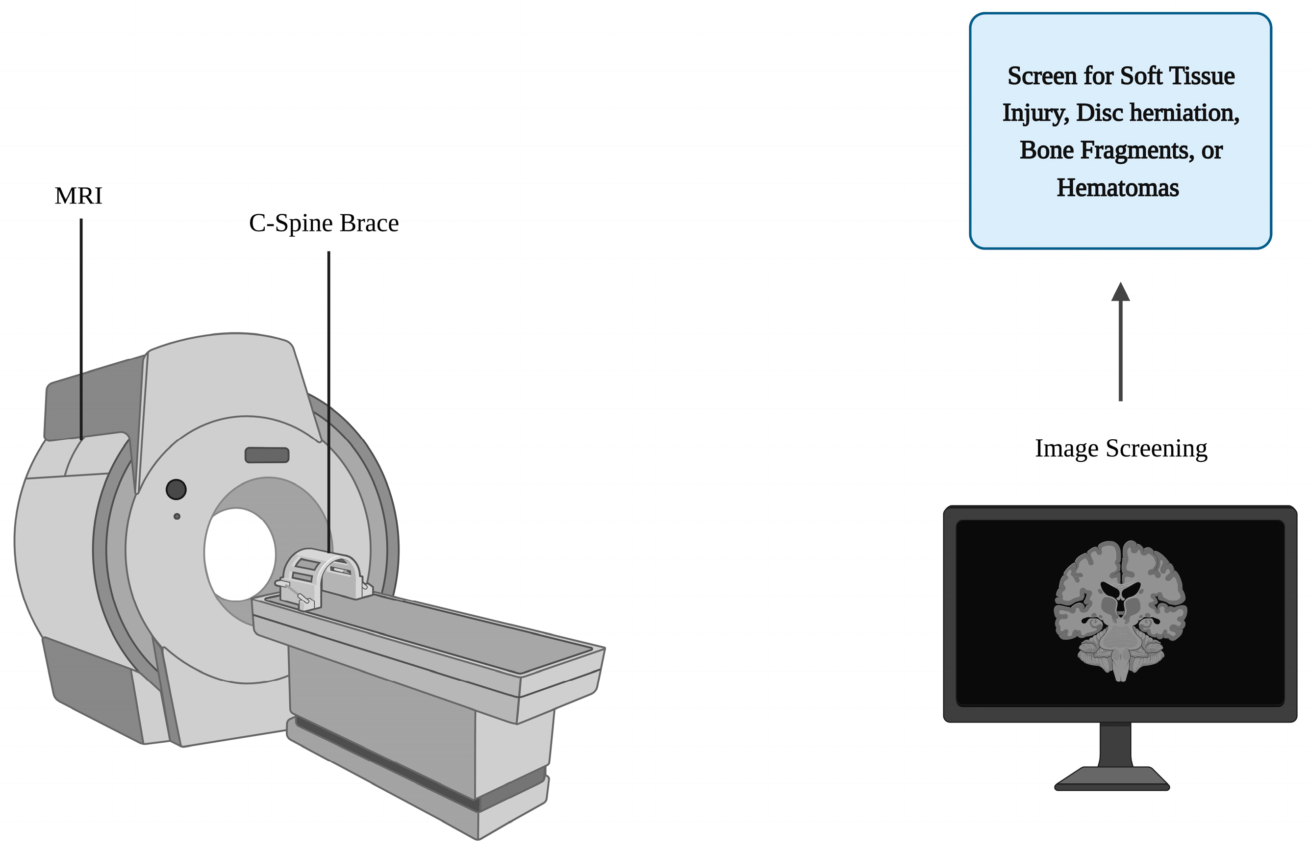
4. Spinal Bracing and Imaging Practices in the Emergency Room

Following a trauma patient’s arrival to the ER, the importance of spine immobilization and imaging persists. Patients with direct cervical, maxillofacial, or head trauma will automatically warrant both cervical restriction and imaging [13]. Stable patients sustaining indirect, penetrating, or blunt trauma are judged on a case-by-case basis [13]. Often, emergency departments anecdotally use the context surrounding the traumatic event to determine the need for cervical spine bracing and imaging. Common circumstances where cervical bracing and imaging will likely be obtained include high-speed MVA (≥35 mph), a death at the scene of the MVA, a fall from a height ≥ 10 feet, significant intracranial injury or skull fracture on head CT, any neurological signs/symptoms, and pelvic fractures. These guidelines are based on the aforementioned NEXUS and CCR, which the American College of Surgeons recommends be used to guide cervical bracing and imaging practices [13]. NEXUS and CCR criteria for cervical spine imaging are summarized in Table 1 and Table 2, respectively. If the patient does not meet any of the NEXUS or CCR criteria for imaging, then they may be cleared and removed from C-spine bracing [13].
NEXUS and CCR had a 99% and 100% sensitivity, respectively, for catching clinically significant cervical spine injuries in their initial studies [11,30]. However, the NEXUS and CCR may not be applicable to all patients, notably elderly and pediatric populations. A 2017 retrospective review found that the NEXUS criteria sensitivity decreases to 94.8% when applied to older adults [31]. Another study found that up to 20% of their older patients with a C-spine fracture reported no pain on initial presentation and denied tenderness to palpation on examination [32]. This is particularly unsettling as the geriatric population is especially vulnerable to musculoskeletal injury due to the age-related decrease in overall bone density [33]. Thus, liberal imaging is recommended over the consideration of NEXUS or CCR criteria in patients older than 55 with suspected SI [31,32,34]. When addressing pediatric patients with suspected SI, the NEXUS and CCR are effective, however, caution is recommended for children younger than 2 years old [35,36,37,38]. Thus, in place of the NEXUS and CCR, providers should consider using a novel scoring system published by the American Association for the Surgery of Trauma (AAST) [39]. They recommend imaging if the pediatric patient is positive for at least three of the following criteria: GCS < 14, GCS eye-opening = 1, involvement in an MVA, and aged 24–36 months old [39]. Concern regarding the radiation of children due to increasing the lifetime risk for cancer is valid, as children undergoing CT scans were found to be at a higher risk of lifetime malignancy than adults undergoing the same imaging [40]. However, the risk-to-benefit ratio of ordering CT imaging leans towards benefit, especially in cases where an SI or traumatic brain injury (TBI) is suspected [40]. Still, out of more than 300,000 children scanned with CT in American EDs annually, fewer than 6000 have TBIs on CT [40]. Thus, there should be continued research examining the impact and specificity of prediction rules, like the AAST criteria, for CT imaging in pediatric patients with either SI or TBI.
When comparing NEXUS and CCR, both criteria have been recommended by the ACS with little justification of one over the other [13]. However, a 2003 prospective cohort study by Stiell et al. found that the CCR outperformed the NEXUS with a higher sensitivity of 99% compared to 90.7% [11]. These findings are supported by a more recent 2021 prospective study by Ghelichkhani et al. who reported that the CCR had a higher discriminatory power than the NEXUS (100% sensitivity vs. 93.4%) [41]. Ghelichkhani et al. also proposed a newer iteration of the CCR: the modified Canadian C-spine rule, which has shown equal sensitivity with increased specificity. The modified CCR excludes the dangerous mechanism and rear-ended MVA from the criteria with the aim of being more suitable in communities where the details of MVAs are not reliably detailed or recorded. The modified CCR also incorporates considerations for intoxication, distracting injury, and focal neurological deficits, which are all part of the NEXUS [41]. In this way, the modified CCR may become the standard criteria for cervical restriction and imaging as it incorporates the best of both the NEXUS and original CCR.
Once the need for imaging is established, a computed tomography (CT) is performed to identify any C-spine fractures [34]. The cervical CT is superior to cervical spine radiographs for patients with suspected blunt cervical SI and is recommended for use by the ACS [34,42]. Following imaging with CT, follow-up imaging with MRI may be warranted, especially if spine surgery is indicated based on CT results [34]. The MRI is most effective at detecting soft tissue injuries, notably contusion or compression of the spinal cord by disc herniation, bone fragments, or hematomas; see Figure 1 [34].
The ACS recommends that providers consider an MRI if their patients have neck pain disproportionately greater than what is initially found on CT imaging [34]. Particular attention should also be given to patients with neurologic symptoms and a GCS less than 15, as these factors were strong predictors of cervical soft tissue injuries [34,43]. If no pathology is found on imaging, the patient may be cleared from C-spinal restriction and their cervical collar may be removed [13].
If a vertebral fracture or soft tissue injury is detected on imaging, the spine surgeon must determine whether to follow operative or nonoperative treatment methods. The determination of treatment depends on spinal column stability, with nonoperative treatments such as bracing used for stable injuries [44]. Spinal instability occurs when the components of the bony spinal column (including joints and ligaments), spinal muscles, or neuronal feedback mechanisms are damaged or disturbed [45]. Determination of spinal stability should be assessed along with a full motor and sensory exam [46,47]. The American Spinal Injury Association (ASIA) score can be used to classify any neurological impairment in a standardized manner [48]. In determining spinal fracture stability, the physician may employ standardized classification systems including the 3-column model, the Thoracolumbar Injury Classification and Severity Score (TLICS), and the Subaxial Cervical Spine Injury Classification System (SLICS) [49,50]. The 3-column model developed by Denis et al. in 1976 was highly revered for its practicality and precision, but it is not without its limitations [51]. The 3-column model assesses the vertebra by dividing it into three coronal cross-sections: anterior, middle, and posterior. This approach notably acknowledges both the mutual inclusivity and independence of mechanical instability and progressive neurologic deterioration [52]. However, over time, the model was criticized for its oversimplicity as clinicians only considered two out of the three columns when determining stability, leading to overlooked injuries and improper treatment [49,51]. The 3-column model was also created before the widespread use of MRIs, and thus does not account for ligamentous injuries with potential progression to instability [49,51]. The Thoracolumbar Injury Classification and Severity Score (TLICS) first conceptualized by the Spine Trauma Group and described by Vaccarro et al. in 2005 makes up for both of these limitations. The TLICS is an algorithmic scoring system that considers the morphology of the injury, the integrity of the posterior ligamentous complex, and the neurologic status of the patient [51]. It gives the physician a reliable, easy-to-use, and objective approach to determining spinal stability and whether or not to operate. The TLICS is points-based and summarized in Table 3. Points are given for certain abnormal findings on imaging or physical exam. Cases with a score of less than 4 are recommended to be managed nonoperatively while cases with a score greater than 4 indicate the need for surgical intervention. Cases with a score of exactly 4 may be operative or nonoperative. The decision to treat surgically must be made by the patient and spinal surgeon together after considering all factors such as medical comorbidities and prior injuries. The Subaxial Cervical Spine Injury Classification System (SLICS) was also developed by Vaccarro et al. and the Spine Trauma Group in 2007 and uses a similar points-based algorithmic approach for the assessment of cervical spine injuries. The SLICS is summarized in Table 4.
If the patient’s SLIC or TLIC score is less than 4 and the criteria for nonoperative treatment are met, a brace may be utilized. Cervical fractures that commonly warrant the use of bracing include isolated C1 fractures, certain C2 Hangman fractures (with less than 3 mm of displacement and no significant angulation), and isolated C2 dens Type I and III fractures [47]. These fractures can be treated with a rigid cervical collar or Halo brace [47]. Thoracolumbar fractures that commonly warrant bracing include anterior wedge compression fractures and fractures of non-load-bearing structures such as the spinous and transverse processes [49]. In these cases, a thoracolumbosacral orthosis (TLSO) may be used to achieve immobilization and facilitate healing. The length of bracing utilization may vary from patient to patient depending on several factors, including but not limited to the severity of the fracture, patient age, smoking history, and certain chronic diseases such as diabetes mellitus [53,54,55]. As such, wear time duration in patients with vertebral fractures may vary but are generally recommended to be between 8 to 12 weeks with radiographs every 4 to 6 weeks to monitor progress [49].

5. Beyond the ER—Spinal Bracing for Spinal Deformities

Outside the emergency room and cases of acute trauma, spinal bracing is primarily utilized in managing and correcting spinal deformities [56]. Common causes of spinal deformity include scoliosis and hyperkyphosis.

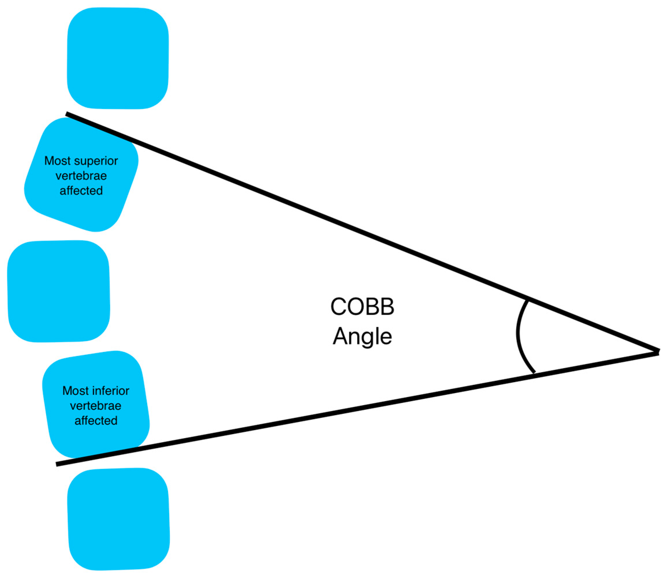
6. Spinal Deformity—Scoliosis

6.1. Diagnosis and Bracing Indications

Scoliosis is a three-dimensional deviation of the spine axis and may present in children, adolescents, or adults [57]. Scoliosis may be primary degenerative, idiopathic, or secondary to disease processes such as osteoporosis or poliomyelitis [58]. Adolescent idiopathic presentation is the most frequent form of scoliosis [59,60]. Treatment for adolescent idiopathic scoliosis can be operative or nonoperative, largely depending on the extent of the deformity [59,60]. The conservative, nonoperative treatment method mainly relies on spinal bracing [60,61]. Scoliosis may be examined clinically using the Adam’s forward bending test or quantified by two measures, angle of trunk rotation (ATR) or Cobb angle. ATR is measured using a scoliometer while the Cobb angle is measured radiographically. The Cobb angle is the angle measured between the most superior and inferior vertebrae effected by the curvature (See Figure 2). Bracing eligibility for the adolescent patient is based on the degree of spinal curvature and rotation, which estimate the risk of scoliosis progression into adulthood [62]. Bracing is indicated if the growing child or adolescent has a Cobb angle or spinal curvature of 25° to 40° or has curves less than 25° along with a documented progression of 5° to 10° in the past six months; measures above 40° typically indicate surgery [59,62,63]. An ATR greater than 7° (BMI < 85th percentile) or 5° (BMI > 85th percentile) is a common clinical predictor of scoliosis and often used as a precursor to Cobb angle measurement [60,64,65,66].
Bracing is contraindicated in children or adolescents who are skeletally mature, overweight, or likely to be non-compliant with wearing the brace [59]. Compliance is essential; the highest benefit from brace usage in idiopathic adolescent scoliosis is seen when it is worn with the prescribed tightness for 18 h or more per day [59,67,68]. An exception to this rule exists in adolescent or pediatric patients with single vertebrae level curvatures who can be treated with night-time bracing alone [69]. Bracing in cases of adolescent idiopathic scoliosis may be discontinued once the patient has reached bone maturity, which can be estimated by either one year after menarche, a six-month halt in height growth, or Risser Sign 5 [59].

6.2. Bracing Perscriptions for Scoliosis

Bracing has been shown to provide little additional benefit after an adolescent patient has reached bone maturity [59]. Thus, early screening and initiation is paramount to bracing’s effectiveness. There is a wide variety of braces to choose from for pediatric or adolescent patients with idiopathic scoliosis. Each type of brace utilizes different correction principles and is used for a specific degree of spinal curvature. The type of brace prescribed is dependent on specific patient preference, cost, and extent of disease. Thoracolumbosacral orthosis (TLSO) is the treatment of choice to halt and reverse scoliosis progression [66,70]. This subset of spinal braces most commonly includes Boston, Charleston, Providence, and SpineCor braces [56,59]. Braces differ in being rigid or soft, depending on what materials are used. Many of the older braces, such as the Boston brace (1972), are made from hard plastic materials or stainless steel and have thus been found to be very uncomfortable and even painful to patients [59,71]. Newer braces, such as the SpineCor brace (the mid-1990s) are made of softer plastics and allow for more flexibility [59]. Using textile fabric materials made of polyester with sandwich mesh and elastic bands can also provide additional comfort and breathability for patients [71]. However, these softer braces may not improve the Cobb angle as much as the rigid braces [71].
Literature on brace usage per day is conflicting, but 18+ h yields consistent results [70,72,73]. The two main factors in successful treatment are time in brace daily and early intervention. As described above, the level of skeletal maturity is a strong indicator of treatment success. Once a patient has reached skeletal maturity or is one year or more post-menarche, bracing may be discontinued or tapered until discontinuation.

6.3. Skeletal Maturity Evaluation in Scoliosis Patients

Skeletal maturity for scoliosis treatment has typically been quantified radiographically using the Risser sign scale of iliac ossification [74]. The Risser scale grades from 0 (no ossification) to 5 (full fusion of the iliac apophysis) in increments of 25% ossification [75]. Risser stages 0–1 have highest risk for spinal curvature progression, but bracing is recommended at grade 4 and below as there may be disease progression until full ossification [74,76,77]. Although the Risser sign is typically used to evaluate skeletal maturity in scoliosis patients specifically, other modalities such as wrist (ages < 18) and clavicle (ages 18–22) imaging may provide more accurate results of bone age [76].

6.4. Adult Scoliosis

Adult patients with scoliosis are likely to present with either primary degenerative scoliosis or progressive idiopathic scoliosis [58]. Primary degenerative scoliosis is the de novo form that occurs in patients with no prior history of scoliosis, whereas the progressive idiopathic type reemerges in patients with an existing history of idiopathic scoliosis [58]. Similar to pediatric and adolescent idiopathic scoliosis, treatment for these adult forms of scoliosis can involve either surgical or nonoperative, conservative treatment such as bracing. However, in adult patients, conservative treatment is much less effective than the surgical alternative [78]. This is largely attributable to an adult’s mature skeletal structure that further stiffens with aging and will not conform, even with tight bracing. Even so, bracing is still used as a nonoperative treatment in adults with scoliosis. While a recent 2020 systematic literature review of 61 studies on adult scoliosis did not find sufficient evidence to suggest that bracing can reliably correct spinal curvature, it did observe that bracing temporarily reduces pain levels and improves function [79]. The brace achieves this improvement as it transfers trunk weight from the ribs to the iliac crests [80]. Bracing also augments lordosis or increased angles in the sagittal plane, which have been found to offset scoliosis or abnormal angles in the coronal plane in some cases, further improving pain levels [80]. As such, cases do exist in which patients with adult scoliosis had improved spinal angles and posture after 8 years of strict adherence to a lumbar brace. A 2016 prospective study with a sample size of 158 patients demonstrated this with 80% of their brace wearers showing unchanged or improved Cobb angles after adhering to bracing for around 8.5 years [80]. However, these results do not represent a statistically significant trend across similar studies [79]. There are plenty of adult scoliosis cases where patients continue to deteriorate despite the use of bracing [79]. Higher-quality studies and specifically randomized control trials (RCTs) are needed to determine bracing’s true utility in treating adult scoliosis, as there are not yet enough reliable findings to suggest their use beyond improving pain levels and function.

7. Spinal Deformity—Hyperkyphosis

Hyperkyphosis is an extreme kyphosis angle or an abnormal curvature of the thoracic spine in the anterior–posterior plane [81]. While the kyphosis angle tends to increase with normal aging, hyperkyphosis is abnormal and notably a consequence of Scheuermann’s Disease or osteoporosis [81,82].
Scheuermann’s Disease is an idiopathic juvenile vertebral hyperkyphosis characterized by wedged vertebrae of at least 5° [83,84]. It is the most common cause of hyperkyphosis of the thoracic or thoracolumbar spine in adolescents [83]. Treatment of Scheuermann’s Disease is predominantly conservative. Bracing is typically recommended in painful cases with mild hyperkyphosis [84]. However, any spinal corrections achieved with bracing usually deteriorate over time, especially with hyperkyphosis angles > 75° [85]. Therefore, other conservative treatments such as physical therapy and exercise programs are commonly recommended [86].
Similar to orthoses for adolescent scoliosis, any bracing prescribed for Scheuermann’s Disease should be utilized for >20 h per day until the patient reaches skeletal maturity [85,87]. Common braces utilized for Scheuermann’s Disease include a TLSO in cases of thoracolumbar Scheuermann’s or a Milwaukee brace (a type of CTLSO) for severe cases [84].
Osteoporosis can also lead to hyperkyphosis and has a very high prevalence ranging from approximately 20% to 40% in older adults [88]. Patients with osteoporosis sustain vertebral compression fractures as a result of the decreased bone density that characterizes the bone disease. Multilevel compression fractures can advance to hyperkyphosis, resulting in the characteristic hunched-over appearance in patients with osteoporosis. The standard treatment of hyperkyphosis in osteoporotic patients with stable compression fractures is bracing for 4 to 12 weeks, with treatment time dependent on patient stability and pain level [89]. A 2020 systematic review by Kweh et al. demonstrated that bracing in the elderly patient with kyphosis and compression fractures resulted in improved biomechanical vertebral stability, reduced kyphotic deformity, enhanced postural stability, greater muscular strength, and superior functional outcomes across four RCTs and three prospective cohort studies [90]. Another systematic review in 2022 by Sánchez-Pinto-Pinto et al. examined 11 RCTs that looked at the efficacy of wearing spinal braces in women with osteoporosis and had similar findings supporting the use of spinal orthoses [91]. In addition to objectively improved kyphosis angles, the brace-wearing patients from these studies reported improvements in pain, muscle strength, pulmonary function, and overall quality of life. Despite these promising findings, the aforementioned reviews cited few studies, thereby reducing the strength of the evidence present. Contrasting findings were documented in another 2020 systematic review by Hofler et al. who examined 16 studies (5 RCTs, 6 nonrandomized prospective cohorts, a retrospective case-control study, and 4 prospective single-arm studies), which overall found that there was limited evidence to suggest that bracing is effective in treating compression fractures in patients with osteoporosis [82]. One randomized control trial went as far as to show that bracing had no significant benefit in improving balance, reducing the dorsal kyphosis angle, or improving the overall quality of life, which is in direct contrast to the benefits reported prior [92]. Aside from not improving health outcomes, bracing has even been reported to worsen outcomes in osteoporotic patients as prolonged spinal restriction can decondition both thoracic and paraspinal muscles, resulting in atrophy, consequently increasing the risk of future falls and fractures by extension [93]. Thus, the consensus on bracing’s efficacy in reliably treating adult hyperkyphosis is still unclear. Like adult scoliosis, more high-quality research is needed to create an informed clinical recommendation for bracing in adult patients with hyperkyphosis.

8. Postoperative Spinal Bracing

Spinal bracing has long been utilized following discectomy, laminectomy, or fusion at both cervical and lumbar levels [94]. However, the use of postoperative bracing is controversial as many recent studies reported that it provides little benefit, despite still being widely prescribed by surgeons [94,95,96,97]. A 2018 systemic review of four RCTs and one prospective cohort study described no significant differences in most measures of disability, pain, quality of life, functional impairment, radiographic outcomes, and safety between patients who utilized bracing after surgery and those who did not [95]. These recent findings are also consistent between both cervical and lumbar surgeries [98,99]. Bracing following cervical procedures can even become harmful as cervical collars have been associated with pain, muscle atrophy, decubitus ulceration, breathing or swallowing discomfort, and difficulty driving [99]. Cervical collars can be expensive, ranging in price from USD25 to USD750, contributing to their questionable use [96,100]. With respect to lumbar procedures, the American Association of Neurological Surgeons (AANS) specifically recommends against the use of bracing postoperatively due to their lower efficacy rating [98,101]. Decrease in the perceived effectiveness of bracing following spine surgery may result from recent improvements in surgical techniques and instruments used in spinal procedures such as anterior cervical discectomy and fusion (ACDF) [98,100]. With improved technology and technique, high levels of internal spinal stabilization are achieved with surgery alone, rendering additional postoperative bracing redundant. However, postoperative bracing may still have utility in patients with poor levels of stabilization or fusion following surgery [100]. One such patient population includes patients who smoke, as cigarettes are detrimental to bone health and healing [102]. Therefore, a combination of surgery followed by a brace to reinforce proper stabilization and fusion may prove more effective than surgery alone. Finally, there may be specific surgical cases where braces may prove to be useful. A study by Duetzmann et al. found that wearing a clavicle brace reduced skin and fascia tension, by extension reducing acute trapezial pain in patients who underwent posterior cervical or cervicothoracic decompressions with or without fusions [103]. Overall, most literature on postoperative orthoses points towards a decreased reliance on postoperative bracing but continued research on the subject is still encouraged, especially in more specific cases such as the clavicle brace.

9. Spinal Bracing for Chronic Lower Back Pain

Similar to the cases of bracing in patients with adult scoliosis and hyperkyphosis, it is unclear to what degree patients with chronic lower back pain (CLBP) can benefit from spinal bracing. A Cochrane review by van Duijvenbode et al. in 2008 examined 15 studies with over 17,000 subjects and could not reliably suggest bracing as a benefit in cases of chronic pain, citing the need for more high-quality research [104]. Since 2008, studies have shown little benefit for treating pain relief when compared to using other nonsurgical options for treatment such as physical therapy [105,106,107]. A RCT from 2017, demonstrated that the combined use of both physical therapy and spinal bracing in patients with CLBP was just as effective in improving posture as in patients receiving physical therapy alone [108]. There was a trend towards decreased pain levels in patients who wore braces, however, the results were not statistically significant [108]. Another advanced prospective RCT from 2021 had similar results, citing no added benefit when spinal orthoses were used in addition to education and exercise to treat CLBP [105]. Despite these findings, bracing may still have some use in providing protection during functional movements. A 2022 RCT by Im et al. showcased that lower back orthoses are beneficial in helping patients with nonspecific lower back pain stand up from sitting [109]. Another study, a questionnaire-based survey of physicians, suggested that lumbar bracing may aid patients while performing labor intensive tasks such as lifting heavy objects [110]. These findings correlate with a common practice in weightlifting gyms, in which athletes wear belts to protect their back while squatting or lifting heavy loads. Interestingly, many of these weightlifting belts are very similar in design to medical lumbar orthoses. These belts protect the wearer by increasing intraabdominal pressure during the lift, which consequently reduces compressive forces on the spine, thereby drastically reducing the risk of spinal injury and pain [111,112]. A psychological component to the weightlifting belt may also be a contributing factor to safety as the tangible contact with the brace serves as a reminder to maintain proper posture while lifting. Medical-grade spinal orthoses may work in a similar fashion in providing support by increasing abdominal pressure during specific movements for the patient with CLBP. This theory is supported by a 2019 RCT that compared the long-term use of lumbosacral orthoses at different levels of brace tightness and pressure [113]. Samani et al. found that while pain levels improved in all groups, the greatest improvement in pain levels and proprioception was recorded by the group who wore the tightest and highest-pressure braces [113]. Thus, while perhaps spinal bracing does not provide any additional pain relief for patients with CLBP, combination with physical therapy may still be useful in instances of physical activities involving lifting or positional back pain.

10. Spinal Bracing for Pregnancy

Over the course of pregnancy, a woman’s body undergoes major physiological changes to accommodate the growing fetus. Overall abdominal size and weight gain secondary to the growing fetus and in part to the increased breast weight and expanded chest cavity, shift the pregnant patient’s center of gravity forward [114]. Changes in size are accompanied by fluctuating hormone levels which increase ligament laxity [114,115]. These hormonal changes along with a forward-shifted center of gravity contribute to spinal misalignment, often seen as kyphosis or lordosis, which may lead to postural compromise, back pain, and gait changes [114]. As such, pregnant women can be prescribed belly wraps and pelvic support bands to reduce discomfort secondary to pregnancy-induced increased body habitus and biomechanical changes [116]. Not only do belly wraps and pelvic bands reduce back pain, but they also help with completing activities of daily living (ADLs) [114,117]. Improving ADLs is an important and often overlooked goal in pregnant patients [117]. A pelvic band would allow women to be more independent and capable both at home and in the workplace. These wraps are also affordable, improving pain and providing increased mobility at a relatively low-cost [117]. The current design for most belly belts revolves around a soft, flexible band that wraps around the pelvis at the level of the anterior superior iliac spine and hugs the bottom of the abdomen [115]. These designs will benefit from expanding the lumbar portion and adding shoulder straps to provide more support for the entire lumbar region and not just the pelvic border. A recent RCT by Heydari et al. showed just this when they compared the effectiveness of their modified belt to current belly belts [115]. Heydari et al. found that the added lumbar support of the modified belt was more effective in reducing pain levels and improving function [115]. Thus, providers should strongly consider prescribing belly belts for their pregnant patients, and more research should examine the utility of expanding pelvic support to include a larger region of the lower back for pregnant patients.

11. Braces Currently Available

Spinal braces or orthopedic braces (orthoses) are designed to serve various areas of the spinal column that each mediate their own set of pathologies. These orthoses are categorized into five categories based on the vertebrae regions the brace supports; see Table 5. Cervical, cervicothoracic, and cervico-thoraco-lumbosacral orthoses support the cervical column and beyond. In comparison, thoraco-lumbar-sacral and lumbosacral orthoses serve the thoracic, lumbar, and sacral regions of the vertebrae.
Cervical orthoses (CO) serve the C1–C6 vertebral regions and can be soft or rigid collars [4]. These collars are commonly prescribed to patients following a whiplash injury. CO allow the patient to conduct activities of daily living (ADLs) as the tissues heal. A short-term 2017 study assessing the range of motion (ROM) of 25 subjects found that both types of CO collars partially immobilize the head and restrict motion [118]. However, soft collars allowed for significantly more mobility compared to rigid collars, with subjects experiencing 31% ROM during flexion compared to 21% with a rigid collar [118]. ADLs typically require 30–50% of neck ROM [118]. Therefore, rigid collars are the best at restricting motion while allowing limited mobility for ADLs [118]. During diagnoses, providers usually prescribe soft cervical orthoses since they are anecdotally stated to result in better prognosis; however, there is no conclusive evidence that these collars lead to a faster recovery time [119,120]. Larger, more rigid braces such as cervicothoracic and cervico-thoraco-lumbosacral orthoses are needed for more serious injuries that affect larger portions of the vertebral column.
Cervicothoracic orthoses (CTO) support the lower-cervical through the mid-thoracic regions—C6–T5 [4,121]. These orthoses are used for cranial and vertebral fractures, after surgical procedures to prevent complications, or for chronic conditions such as scoliosis [66,122]. Devices in this classification include the Minerva brace, the sternal-occipital-mandibular immobilizer (SOMI), and the Halo brace [4]. These orthoses are an enhanced version of a CO with added bracing around the anterior and/or posterior thorax. The oldest of the three, the Minerva brace provides immobilization of the anterior and posterior thorax and cervical region [4,123]. A 1992 study found that the Minerva brace provided the most restriction at C3–C4 and C6–C7 regions [121]. The SOMI brace differs from the Minerva brace in that it lacks posterior support, is easier to put on, and can be used while eating [4,123]. However, the Halo brace is considered the best orthosis to hold the spine firm and is used to treat the upper and lower cervical vertebra [4,121].
In adults, the Halo brace immobilizes the cranium with four pins inserted at a force of 8 in-lb of torque into the outer layer of cranial bone connected to a circular halo structure which is stabilized by a thoracic vest [124]. Since children have a higher risk of complications at 70% compared to adults at 35%, caution is needed when applying a Halo brace [120]. Therefore, modifications such as more pins (8–12) and lower torque (1–5 in-lb) are used to prevent complications such as pin perforation past the outer layers of cranial bone [120]. However, the same modifications are not needed for the elderly population as research has found that the elderly cranium can withstand the standard four-pin arrangement at a torque of 8 in-lb and most complications post-Halo treatment are a result from underlying diseases [124,125].
Cervico-thoracic-lumbosacral orthoses (CTLSO), which serve vertebral regions C2–L5, are less prevalent due to their difficult application and bulky outward appearance [126]. The most notable CTLSO, the Milwaukee device, is used to treat severe spinal abnormalities such as scoliosis [126,127]. However, the brace is losing popularity as patients have reported mental issues with body image while wearing the brace due to its unflattering appearance [127,128]. Due to the plethora of problems with CTLSO, thoraco-lumbosacral orthoses (TLSO) have mostly replaced them.
TLSO devices serve the lower thoracic to lumbar regions and are mostly used to support vertebrae T6-L4 [4]. TLSOs such as the Boston, Charleston, and Providence braces are commonly used to treat adolescent idiopathic scoliosis [66]. Charleston and Providence braces are typical worn overnight for correction of scoliosis [129]. A 2013 study found that pediatric patients with idiopathic scoliosis who wore Boston braces had a 90% success rate of achieving a Cobb angle of <50 degrees [70]. Other less specialized TLSOs include the Jewett brace and the Cruciform Anterior Spinal Hyperextension (CASH) brace may treat various disorders ranging from osteoporotic vertebral fractures (OVF) to Parkinson’s-related camptocormia [130]. There is a high incidence of OVF in the elderly population, which may be successfully treated with Jewett braces [131]. Certain neurodegenerative diseases such as Parkinson’s-related camptocormia may also be treated with TLSOs such as CASH; however, further research is needed [130].
Lumbosacral orthoses (LSO) serve the L3 vertebrae to the lumbosacral junction and may be used to treat lower back pain or post-operatively [130,132]. The LSO works to partially immobilize muscles of the abdominal trunk to reduce muscle use and subsequent pain [133,134]. However, several studies have shown that LSOs do not significantly improve surgery outcomes when worn post-operatively [135,136].

12. The Future of Bracing

The future for spinal bracing lies in enhancing brace comfort and treatment efficacy through personalized bracing. Personalized bracing comes in many forms such as enhancing existing braces through actuators and bands [56]. Actuators are ridged articulations that constantly change the form of the brace while bands hold the brace together with reasonable flexibility; both of which are used to create dynamic braces that morph with the wearer’s body. Actuator-enhanced braces such as the RoSE dynamic exoskeleton, Atlas Japet, ExMS-1, and experimental active-soft brace help the brace adapt in real time to changes in torso stiffness [137]. In essence, the brace is dynamic and changes according to each patient’s injury and treatment progression [137]. The experimental active soft brace is a combination of elastic bands and actuators that claims to correct thoracic cobb angle by 15.96 degrees, producing the desired long-term correction [138]. Band braces aim to be a more comfortable alternative to rigid braces and are more ergonomic on the body by preventing muscle atrophy [56]. Examples of soft-band braces include SpineCor, SpinealiteTM, and the TriaC brace [56]. However, there has been some debate whether these braces are as efficacious as rigid braces in correcting idiopathic scoliosis or aiding in post-operative rehabilitation [139,140,141].
In addition to brace enhancement, recent research has shown that through computer modeling and 3D imaging, physicians can develop orthoses through 3D printing that are specific to the patient’s ailment and more user-friendly than traditional orthoses [142,143]. The historical process of creating custom braces involves plaster molding, which often gets messy and fails to capture the patient’s unique shape [142,144]. Additionally, 3D printing could drive down costs of both plaster and plastic/metal orthoses with in-house operations [144]. Advances in robotics have also introduced motorized exoskeleton technology to treat OVFs and chronic back pain with great promise, but are currently un-tested and expensive [93].

13. Limitations

This review was limited by inherent flaws common to all narrative reviews including an unexhaustive literature review. Further, some of the studies identified in this review failed to stratify outcomes based on interventions, reducing the generalizability of their findings. Despite these limitations, the authors believe this review is an important delineation of uses and standards of practice for spinal orthosis.

14. Conclusions

Spinal bracing has a wide range of applications from preventing further injury after trauma to correcting early-age skeletal malformations. As for applications in trauma, spinal bracing may be lifesaving in instances of serious MVAs or falls. Application of cervical braces by EMS after trauma has been shown to cause more vertebral movement than allowing the patient to brace themselves. Still, the application of braces post-trauma holds great benefits to potential costs. In the instance of serious vertebral injury, the decision between wearing a brace or not could be life-altering. Soft cervical braces are beginning to be recommended over hard braces for non-lifesaving injuries.
The need for bracing is often indicated by multiple sets of criteria from certain governing bodies. The NEXUS and CCR are two sets of criteria commonly used to indicate the necessity for imaging. The CCR has been shown to have the greatest efficacy for detecting spinal injury in mature adults while a new pediatric scoring system published by the AAST is more commonly used for children and adolescents. With younger individuals, the use of radiation imaging should be highly considered under certain circumstances where SI or TBI is likely. If a spinal fracture is identified, vertebral stability should be assessed, and a treatment plan should be established. The Thoracolumbar Injury Classification and Severity Score (TLICS) and the Sub-axial Cervical Spine Injury Classification System (SLICS) are both objective and easy-to-use systems for determining spinal stability.
Bracing for scoliosis treatment is common and has a very high correction rate, but only within a certain developmental timeframe. Children and adolescents are the only demographic that may gain full correction of spinal curvature through bracing alone. Once an individual has reached bone maturity the application of a brace is often unsuccessful in correction, and surgery is indicated. In adults, bracing helps relieve symptoms of scoliosis and prevent further compensation but cannot correct the curvature permanently. Scoliosis screening in children and adolescents is essential for disease prevention and correction and should be routinely completed by pediatric healthcare providers. The use of bracing in adult patients suffering from osteoporotic changes like hyperkyphosis has shown promise in symptom relief but current literature is conflicting.
Post-operative bracing has also come into question and seems to be unnecessary for most spinal surgery patients. Spinal braces should be avoided post-operatively as they restrict natural motion and are often considered redundant as orthopedic hardware stabilizes the spine appropriately, except for patients at risk for weakened stabilization or fusion, such as smokers or diabetics.
Chronic lower back pain (CLBP) rarely indicates the need for bracing and is often unrelieved with bracing techniques. Physical therapy is a much better alternative and has consistently been shown to improve low back pain. Bracing during pregnancy decreases pain and enhances the mother’s ability to perform activities of daily living both at home and in the workplace. Bracing in pregnant patients should be expanded to include the lumbar spine in addition to the pelvis and belly to provide additional support and improved weight distribution.
Current braces and those that will come to market in the future have two main goals in mind: comfort and correction. Braces currently on the market are often bulky, uncomfortable, and visually unappealing, making the wearer less willing to comply with their treatment regimen. Although these braces are very cumbersome, the results are excellent. Newer more flexible braces are more comfortable but less effective within the same timeframe. Testing with new dynamic braces is promising and could help merge the gap between comfort and correction. Patient compliance is a major limiting factor to spinal correction and should be heavily discussed with each patient when comparing bracing options.
The application of spinal braces is essential for multiple pathologies but often overused as a preventative measure. In instances of trauma or young-age scoliosis, the braces are essential in treatment, but in other cases can cause harm or are simply unnecessary. This is not to say that braces should be avoided altogether, as the benefits of spine stabilization could be lifesaving. Overall, spinal bracing is widely used and must be considered as a means of post-traumatic stabilization or spinal correction in many circumstances.

Author Contributions

Conceptualization, B.L.-W. and J.L.C.; writing—original draft preparation, J.L.C., A.N.B., C.P.S. and K.T.R.; writing—review and editing, J.L.C. and B.L.-W.; supervision, B.L.-W. and J.L.C. All authors have read and agreed to the published version of the manuscript.

Funding

This research received no external funding.

Institutional Review Board Statement

Not applicable.

Informed Consent Statement

Not applicable.

Data Availability Statement

Not applicable.

Conflicts of Interest

The authors declare no conflict of interest.

References

  1. Kumar, R.; Lim, J.; Mekary, R.A.; Rattani, A.; Dewan, M.C.; Sharif, S.Y.; Osorio-Fonseca, E.; Park, K.B. Traumatic Spinal Injury: Global Epidemiology and Worldwide Volume. World Neurosurg. 2018, 113, e345–e363. [Google Scholar] [CrossRef]
  2. Devivo, M.J. Epidemiology of traumatic spinal cord injury: Trends and future implications. Spinal Cord 2012, 50, 365–372. [Google Scholar] [CrossRef] [Green Version]
  3. McCarthy, J.; Davis, A. Diagnosis and Management of Vertebral Compression Fractures. Am. Fam. Physician 2016, 94, 44–50. [Google Scholar] [PubMed]
  4. Agabegi, S.S.; Asghar, F.A.; Herkowitz, H.N. Spinal orthoses. J. Am. Acad. Orthop. Surg. 2010, 18, 657–667. [Google Scholar] [CrossRef] [PubMed]
  5. Negrini, S.; Minozzi, S.; Bettany-Saltikov, J.; Chockalingam, N.; Grivas, T.B.; Kotwicki, T.; Maruyama, T.; Romano, M.; Zaina, F. Braces for idiopathic scoliosis in adolescents. Cochrane Database Syst. Rev. 2015, 6, CD006850. [Google Scholar] [CrossRef] [Green Version]
  6. Christensen, S.W.M.; Rasmussen, M.B.; Jespersen, C.L.; Sterling, M.; Skou, S.T. Soft-collar use in rehabilitation of whiplash-associated disorders—A systematic review and meta-analysis. Musculoskelet. Sci. Pract. 2021, 55, 102426. [Google Scholar] [CrossRef] [PubMed]
  7. Jhaveri, S.N.; Zeller, R.; Miller, S.; Lewis, S.J. The effect of intra-operative skeletal (skull femoral) traction on apical vertebral rotation. Eur. Spine J. 2009, 18, 352–356. [Google Scholar] [CrossRef] [Green Version]
  8. Lee, D.; Adeoye, A.L.; Dahdaleh, N.S. Indications and complications of crown halo vest placement: A review. J. Clin. Neurosci. 2017, 40, 27–33. [Google Scholar] [CrossRef] [PubMed]
  9. Sundstrom, T.; Asbjornsen, H.; Habiba, S.; Sunde, G.A.; Wester, K. Prehospital use of cervical collars in trauma patients: A critical review. J. Neurotrauma 2014, 31, 531–540. [Google Scholar] [CrossRef] [Green Version]
  10. Hawary, R.E.; Zaaroor-Regev, D.; Floman, Y.; Lonner, B.S.; Alkhalife, Y.I.; Betz, R.R. Brace treatment in adolescent idiopathic scoliosis: Risk factors for failure-a literature review. Spine J. 2019, 19, 1917–1925. [Google Scholar] [CrossRef] [Green Version]
  11. Stiell, I.G.; Wells, G.A.; Vandemheen, K.L.; Clement, C.M.; Lesiuk, H.; De Maio, V.J.; Laupacis, A.; Schull, M.; McKnight, R.D.; Verbeek, R.; et al. The Canadian C-spine rule for radiography in alert and stable trauma patients. JAMA 2001, 286, 1841–1848. [Google Scholar] [CrossRef] [Green Version]
  12. Zileli, M.; Osorio-Fonseca, E.; Konovalov, N.; Cardenas-Jalabe, C.; Kaprovoy, S.; Mlyavykh, S.; Pogosyan, A. Early Management of Cervical Spine Trauma: WFNS Spine Committee Recommendations. Neurospine 2020, 17, 710–722. [Google Scholar] [CrossRef] [PubMed]
  13. American College of Surgeons. Committee on T: Advanced Trauma Life Support: Student Course Manual; American College of Surgeons: Chicago, IL, USA, 2018. [Google Scholar]
  14. Shank, C.D.; Walters, B.C.; Hadley, M.N. Current Topics in the Management of Acute Traumatic Spinal Cord Injury. Neurocrit. Care 2019, 30, 261–271. [Google Scholar] [CrossRef] [PubMed]
  15. National Association of Emergency Medical Technicians (NAEMT); American College of Surgeons. PHTLS: Prehospital Trauma Life Support; Jones & Bartlett Learning: Burlington, MA, USA, 2020. [Google Scholar]
  16. Hauswald, M.; Ong, G.; Tandberg, D.; Omar, Z. Out-of-hospital spinal immobilization: Its effect on neurologic injury. Acad. Emerg. Med. 1998, 5, 214–219. [Google Scholar] [CrossRef] [PubMed]
  17. Kwan, I.; Bunn, F.; Roberts, I. Spinal immobilisation for trauma patients. Cochrane Database Syst. Rev. 2001, 2001, CD002803. [Google Scholar] [CrossRef] [PubMed]
  18. Bauer, D.; Kowalski, R. Effect of spinal immobilization devices on pulmonary function in the healthy, nonsmoking man. Ann. Emerg. Med. 1988, 17, 915–918. [Google Scholar] [CrossRef] [PubMed]
  19. Totten, V.Y.; Sugarman, D.B. Respiratory effects of spinal immobilization. Prehospital Emerg. Care 1999, 3, 347–352. [Google Scholar] [CrossRef] [PubMed]
  20. Schafermeyer, R.W.; Ribbeck, B.M.; Gaskins, J.; Thomason, S.; Harlan, M.; Attkisson, A. Respiratory effects of spinal immobilization in children. Ann. Emerg. Med. 1991, 20, 1017–1019. [Google Scholar] [CrossRef]
  21. Ham, W.H.; Schoonhoven, L.; Schuurmans, M.J.; Leenen, L.P. Pressure ulcers, indentation marks and pain from cervical spine immobilization with extrication collars and headblocks: An observational study. Injury 2016, 47, 1924–1931. [Google Scholar] [CrossRef]
  22. Barkana, Y.; Stein, M.; Scope, A.; Maor, R.; Abramovich, Y.; Friedman, Z.; Knoller, N. Prehospital stabilization of the cervical spine for penetrating injuries of the neck—Is it necessary? Injury 2000, 31, 305–309. [Google Scholar] [CrossRef]
  23. Nunez-Patino, R.A.; Rubiano, A.M.; Godoy, D.A. Impact of Cervical Collars on Intracranial Pressure Values in Traumatic Brain Injury: A Systematic Review and Meta-Analysis of Prospective Studies. Neurocrit. Care 2020, 32, 469–477. [Google Scholar] [CrossRef]
  24. Ozdogan, S.; Gokcek, O.; Katirci, Y.; Corbacioglu, S.K.; Emektar, E.; Cevik, Y. The effects of spinal immobilization at 20 degrees on intracranial pressure. Am. J. Emerg. Med. 2019, 37, 1327–1330. [Google Scholar] [CrossRef] [PubMed]
  25. Mobbs, R.J.; Stoodley, M.A.; Fuller, J. Effect of cervical hard collar on intracranial pressure after head injury. ANZ J. Surg. 2002, 72, 389–391. [Google Scholar] [CrossRef]
  26. Backer, H.C.; Elias, P.; Braun, K.F.; Johnson, M.A.; Turner, P.; Cunningham, J. Cervical immobilization in trauma patients: Soft collars better than rigid collars? A systematic review and meta-analysis. Eur. Spine J. 2022, 31, 3378–3391. [Google Scholar] [CrossRef]
  27. Dixon, M.; O’Halloran, J.; Cummins, N.M. Biomechanical analysis of spinal immobilisation during prehospital extrication: A proof of concept study. Emerg. Med. J. 2014, 31, 745–749. [Google Scholar] [CrossRef] [Green Version]
  28. Maschmann, C.; Jeppesen, E.; Rubin, M.A.; Barfod, C. New clinical guidelines on the spinal stabilisation of adult trauma patients—Consensus and evidence based. Scand. J. Trauma Resusc. Emerg. Med. 2019, 27, 77. [Google Scholar] [CrossRef] [PubMed]
  29. Vaillancourt, C.; Charette, M.; Kasaboski, A.; Maloney, J.; Wells, G.A.; Stiell, I.G. Evaluation of the safety of C-spine clearance by paramedics: Design and methodology. BMC Emerg. Med. 2011, 11, 1. [Google Scholar] [CrossRef] [Green Version]
  30. Hoffman, J.R.; Mower, W.R.; Wolfson, A.B.; Todd, K.H.; Zucker, M.I. Validity of a set of clinical criteria to rule out injury to the cervical spine in patients with blunt trauma. National Emergency X-Radiography Utilization Study Group. N. Engl. J. Med. 2000, 343, 94–99. [Google Scholar] [CrossRef] [PubMed]
  31. Paykin, G.; O’Reilly, G.; Ackland, H.M.; Mitra, B. The NEXUS criteria are insufficient to exclude cervical spine fractures in older blunt trauma patients. Injury 2017, 48, 1020–1024. [Google Scholar] [CrossRef]
  32. Healey, C.D.; Spilman, S.K.; King, B.D.; Sherrill, J.E., 2nd; Pelaez, C.A. Asymptomatic cervical spine fractures: Current guidelines can fail older patients. J. Trauma Acute Care Surg. 2017, 83, 119–125. [Google Scholar] [CrossRef]
  33. Spaniolas, K.; Cheng, J.D.; Gestring, M.L.; Sangosanya, A.; Stassen, N.A.; Bankey, P.E. Ground level falls are associated with significant mortality in elderly patients. J. Trauma 2010, 69, 821–825. [Google Scholar] [CrossRef] [PubMed]
  34. TQIP ACoSTQIPA: ACS TQIP Best Practices Guidelines in Imaging; American College of Surgeons: Chicago, IL, USA, 2018.
  35. Ehrlich, P.F.; Wee, C.; Drongowski, R.; Rana, A.R. Canadian C-spine Rule and the National Emergency X-Radiography Utilization Low-Risk Criteria for C-spine radiography in young trauma patients. J. Pediatr. Surg. 2009, 44, 987–991. [Google Scholar] [CrossRef]
  36. Viccellio, P.; Simon, H.; Pressman, B.D.; Shah, M.N.; Mower, W.R.; Hoffman, J.R.; for the NEXUS Group. A prospective multicenter study of cervical spine injury in children. Pediatrics 2001, 108, E20. [Google Scholar] [CrossRef] [Green Version]
  37. Garton, H.J.; Hammer, M.R. Detection of pediatric cervical spine injury. Neurosurgery 2008, 62, 700–708. [Google Scholar] [CrossRef] [PubMed]
  38. Lee, S.L.; Sena, M.; Greenholz, S.K.; Fledderman, M. A multidisciplinary approach to the development of a cervical spine clearance protocol: Process, rationale, and initial results. J. Pediatr. Surg. 2003, 38, 358–362. [Google Scholar] [CrossRef] [PubMed]
  39. Pieretti-Vanmarcke, R.; Velmahos, G.C.; Nance, M.L.; Islam, S.; Falcone, R.A.; Wales, P.W.; Brown, R.L.; Gaines, B.A.; McKenna, C.; Moore, F.O.; et al. Clinical clearance of the cervical spine in blunt trauma patients younger than 3 years: A multi-center study of the american association for the surgery of trauma. J. Trauma 2009, 67, 543–549. [Google Scholar] [CrossRef] [Green Version]
  40. Brenner, D.; Elliston, C.; Hall, E.; Berdon, W. Estimated risks of radiation-induced fatal cancer from pediatric CT. AJR Am. J. Roentgenol. 2001, 176, 289–296. [Google Scholar] [CrossRef] [Green Version]
  41. Ghelichkhani, P.; Shahsavarinia, K.; Gharekhani, A.; Taghizadieh, A.; Baratloo, A.; Fattah, F.H.R.; Abbasi, N.; Gubari, M.I.M.; Faridaalee, G.; Dinpanah, H.; et al. Value of Canadian C-spine rule versus the NEXUS criteria in ruling out clinically important cervical spine injuries: Derivation of modified Canadian C-spine rule. Radiol. Med. 2021, 126, 414–420. [Google Scholar] [CrossRef] [PubMed]
  42. Bailitz, J.; Starr, F.; Beecroft, M.; Bankoff, J.; Roberts, R.; Bokhari, F.; Joseph, K.; Wiley, D.; Dennis, A.; Gilkey, S.; et al. CT should replace three-view radiographs as the initial screening test in patients at high, moderate, and low risk for blunt cervical spine injury: A prospective comparison. J. Trauma 2009, 66, 1605–1609. [Google Scholar] [CrossRef] [PubMed]
  43. Duane, T.M.; Young, A.J.; Vanguri, P.; Wolfe, L.G.; Katzen, J.; Han, J.; Mayglothling, J.; Whelan, J.F.; Aboutanos, M.B.; Ivatury, R.R.; et al. Defining the cervical spine clearance algorithm: A single-institution prospective study of more than 9000 patients. J. Trauma Acute Care Surg. 2016, 81, 541–547. [Google Scholar] [CrossRef]
  44. Wood, K.B.; Li, W.; Lebl, D.R.; Ploumis, A. Management of thoracolumbar spine fractures. Spine J. 2014, 14, 145–164. [Google Scholar] [CrossRef] [PubMed]
  45. Panjabi, M.M. The stabilizing system of the spine. Part I. Function, dysfunction, adaptation, and enhancement. J. Spinal Disord. 1992, 5, 383–389. [Google Scholar] [CrossRef]
  46. Fernandez-de Thomas, R.J.; De Jesus, O. Thoracolumbar Spine Fracture; StatPearls: Treasure Island, FL, USA, 2022. [Google Scholar]
  47. McMordie, J.H.; Viswanathan, V.K.; Gillis, C.C. Cervical Spine Fractures Overview; StatPearls: Treasure Island, FL, USA, 2022. [Google Scholar]
  48. Roberts, T.T.; Leonard, G.R.; Cepela, D.J. Classifications In Brief: American Spinal Injury Association (ASIA) Impairment Scale. Clin. Orthop. Relat. Res. 2017, 475, 1499–1504. [Google Scholar] [CrossRef] [PubMed] [Green Version]
  49. Chang, V.; Holly, L.T. Bracing for thoracolumbar fractures. Neurosurg. Focus 2014, 37, E3. [Google Scholar] [CrossRef] [Green Version]
  50. Vaccaro, A.R.; Hulbert, R.J.; Patel, A.A.; Fisher, C.; Dvorak, M.; Lehman, R.A.; Anderson, P.; Harrop, J.; Oner, F.C.; Arnold, P.; et al. The subaxial cervical spine injury classification system: A novel approach to recognize the importance of morphology, neurology, and integrity of the disco-ligamentous complex. Spine 2007, 32, 2365–2374. [Google Scholar] [CrossRef] [PubMed] [Green Version]
  51. Lee, J.Y.; Vaccaro, A.R.; Lim, M.R.; Öner, F.; Hulbert, R.J.; Hedlund, R.; Fehlings, M.G.; Arnold, P.; Harrop, J.; Bono, C.M.; et al. Thoracolumbar injury classification and severity score: A new paradigm for the treatment of thoracolumbar spine trauma. J. Orthop. Sci. 2005, 10, 671–675. [Google Scholar] [CrossRef] [PubMed] [Green Version]
  52. Denis, F. The three column spine and its significance in the classification of acute thoracolumbar spinal injuries. Spine 1983, 8, 817–831. [Google Scholar] [CrossRef]
  53. Saul, D.; Khosla, S. Fracture Healing in the Setting of Endocrine Diseases, Aging, and Cellular Senescence. Endocr. Rev. 2022, 43, 984–1002. [Google Scholar] [CrossRef]
  54. Al-Hadithy, N.; Sewell, M.D.; Bhavikatti, M.; Gikas, P.D. The effect of smoking on fracture healing and on various orthopaedic procedures. Acta Orthop. Belg. 2012, 78, 285–290. [Google Scholar]
  55. Jiao, H.; Xiao, E.; Graves, D.T. Diabetes and Its Effect on Bone and Fracture Healing. Curr. Osteoporos. Rep. 2015, 13, 327–335. [Google Scholar] [CrossRef] [Green Version]
  56. Ali, A.; Fontanari, V.; Fontana, M.; Schmolz, W. Spinal Deformities and Advancement in Corrective Orthoses. Bioengineering 2020, 8, 2. [Google Scholar] [CrossRef] [PubMed]
  57. Trobisch, P.; Suess, O.; Schwab, F. Idiopathic scoliosis. Dtsch. Ärzteblatt Int. 2010, 107, 875–883. [Google Scholar] [CrossRef]
  58. Aebi, M. The adult scoliosis. Eur. Spine J. 2005, 14, 925–948. [Google Scholar] [CrossRef] [PubMed] [Green Version]
  59. Kaelin, A.J. Adolescent idiopathic scoliosis: Indications for bracing and conservative treatments. Ann. Transl. Med. 2020, 8, 28. [Google Scholar] [CrossRef]
  60. Kuznia, A.L.; Hernandez, A.K.; Lee, L.U. Adolescent Idiopathic Scoliosis: Common Questions and Answers. Am. Fam. Physician 2020, 101, 19–23. [Google Scholar] [PubMed]
  61. Rowe, D.E.; Bernstein, S.M.; Riddick, M.F.; Adler, F.; Emans, J.B.; Gardner-Bonneau, D. A meta-analysis of the efficacy of non-operative treatments for idiopathic scoliosis. J. Bone Jt. Surg. Am. 1997, 79, 664–674. [Google Scholar] [CrossRef]
  62. Richards, B.S.; Bernstein, R.M.; D’Amato, C.R.; Thompson, G.H. Standardization of criteria for adolescent idiopathic scoliosis brace studies: SRS Committee on Bracing and Nonoperative Management. Spine 2005, 30, 2068–2075. [Google Scholar] [CrossRef] [PubMed] [Green Version]
  63. Will, R.E.; Stokes, I.A.; Qiu, X.; Walker, M.R.; Sanders, J.O. Cobb angle progression in adolescent scoliosis begins at the intervertebral disc. Spine 2009, 34, 2782–2786. [Google Scholar] [CrossRef]
  64. Bunnell, W.P. An objective criterion for scoliosis screening. J. Bone Jt. Surg. Am. 1984, 66, 1381–1387. [Google Scholar] [CrossRef]
  65. Murrell, G.A.; Coonrad, R.W.; Moorman, C.T., 3rd; Fitch, R.D. An assessment of the reliability of the Scoliometer. Spine 1993, 18, 709–712. [Google Scholar] [CrossRef]
  66. El-Hawary, R.; Chukwunyerenwa, C. Update on evaluation and treatment of scoliosis. Pediatr. Clin. N. Am. 2014, 61, 1223–1241. [Google Scholar] [CrossRef] [PubMed]
  67. Dolan, L.A.; Donzelli, S.; Zaina, F.; Weinstein, S.L.; Negrini, S. Adolescent Idiopathic Scoliosis Bracing Success Is Influenced by Time in Brace: Comparative Effectiveness Analysis of BrAIST and ISICO Cohorts. Spine 2020, 45, 1193–1199. [Google Scholar] [CrossRef]
  68. Karol, L.A.; Virostek, D.; Felton, K.; Jo, C.; Butler, L. The Effect of the Risser Stage on Bracing Outcome in Adolescent Idiopathic Scoliosis. J. Bone Jt. Surg. Am. 2016, 98, 1253–1259. [Google Scholar] [CrossRef] [PubMed] [Green Version]
  69. Katz, D.E.; Richards, B.S.; Browne, R.H.; Herring, J.A. A comparison between the Boston brace and the Charleston bending brace in adolescent idiopathic scoliosis. Spine 1997, 22, 1302–1312. [Google Scholar] [CrossRef] [PubMed]
  70. Weinstein, S.L.; Dolan, L.A.; Wright, J.G.; Dobbs, M.B. Effects of bracing in adolescents with idiopathic scoliosis. N. Engl. J. Med. 2013, 369, 1512–1521. [Google Scholar] [CrossRef] [PubMed] [Green Version]
  71. Hui, C.-L.; Piao, J.; Wong, M.S.; Chen, Z. Study of Textile Fabric Materials used in Spinal Braces for Scoliosis. J. Med. Biol. Eng. 2020, 40, 356–371. [Google Scholar] [CrossRef]
  72. Konieczny, M.R.; Hieronymus, P.; Krauspe, R. Time in brace: Where are the limits and how can we improve compliance and reduce negative psychosocial impact in patients with scoliosis? A retrospective analysis. Spine J. 2017, 17, 1658–1664. [Google Scholar] [CrossRef]
  73. Whitaker, A.T.; Hresko, M.T.; Miller, P.E.; Verhofste, B.P.; Beling, A.; Emans, J.B.; Karlin, L.I.; Hedequist, D.J.; Glotzbecker, M.P. Bracing for juvenile idiopathic scoliosis: Retrospective review from bracing to skeletal maturity. Spine Deform. 2022, 10, 1349–1358. [Google Scholar] [CrossRef]
  74. Costa, L.; Schlosser, T.P.C.; Jimale, H.; Homans, J.F.; Kruyt, M.C.; Castelein, R.M. The Effectiveness of Different Concepts of Bracing in Adolescent Idiopathic Scoliosis (AIS): A Systematic Review and Meta-Analysis. J. Clin. Med. 2021, 10, 2145. [Google Scholar] [CrossRef]
  75. Wittschieber, D.; Schmeling, A.; Schmidt, S.; Heindel, W.; Pfeiffer, H.; Vieth, V. The Risser sign for forensic age estimation in living individuals: A study of 643 pelvic radiographs. Forensic Sci. Med. Pathol. 2013, 9, 36–43. [Google Scholar] [CrossRef]
  76. Subramanian, S.; Viswanathan, V.K. Bone Age; StatPearls: Treasure Island, FL, USA, 2022. [Google Scholar]
  77. Kawasaki, S.; Shigematsu, H.; Tanaka, M.; Suga, Y.; Yamamoto, Y.; Tanaka, Y. Is brace treatment unnecessary for cases of adolescent idiopathic scoliosis above Risser sign 3? J. Orthop. Sci. 2020, 25, 975–979. [Google Scholar] [CrossRef] [PubMed]
  78. Bridwell, K.H.; Glassman, S.; Horton, W.; Shaffrey, C.; Schwab, F.; Zebala, L.P.; Lenke, L.G.; Hilton, J.F.; Shainline, M.; Baldus, C.; et al. Does treatment (nonoperative and operative) improve the two-year quality of life in patients with adult symptomatic lumbar scoliosis: A prospective multicenter evidence-based medicine study. Spine 2009, 34, 2171–2178. [Google Scholar] [CrossRef] [PubMed]
  79. McAviney, J.; Mee, J.; Fazalbhoy, A.; Du Plessis, J.; Brown, B.T. A systematic literature review of spinal brace/orthosis treatment for adults with scoliosis between 1967 and 2018: Clinical outcomes and harms data. BMC Musculoskelet. Disord. 2020, 21, 87. [Google Scholar] [CrossRef] [PubMed] [Green Version]
  80. de Mauroy, J.C.; Lecante, C.; Barral, F.; Pourret, S. Prospective study of 158 adult scoliosis treated by a bivalve polyethylene overlapping brace and reviewed at least 5 years after brace fitting. Scoliosis Spinal Disord. 2016, 11, 28. [Google Scholar] [CrossRef] [Green Version]
  81. Koele, M.C.; Lems, W.F.; Willems, H.C. The Clinical Relevance of Hyperkyphosis: A Narrative Review. Front. Endocrinol. 2020, 11, 5. [Google Scholar] [CrossRef] [Green Version]
  82. Hofler, R.C.; Jones, G.A. Bracing for Acute and Subacute Osteoporotic Compression Fractures: A Systematic Review of the Literature. World Neurosurg. 2020, 141, e453–e460. [Google Scholar] [CrossRef]
  83. Bezalel, T.; Carmeli, E.; Been, E.; Kalichman, L. Scheuermann’s disease: Current diagnosis and treatment approach. J. Back Musculoskelet. Rehabil. 2014, 27, 383–390. [Google Scholar] [CrossRef]
  84. Palazzo, C.; Sailhan, F.; Revel, M. Scheuermann’s disease: An update. Jt. Bone Spine 2014, 81, 209–214. [Google Scholar] [CrossRef]
  85. Sachs, B.; Bradford, D.; Winter, R.; Lonstein, J.; Moe, J.; Willson, S. Scheuermann kyphosis. Follow-up of Milwaukee-brace treatment. J. Bone Jt. Surg. Am. 1987, 69, 50–57. [Google Scholar] [CrossRef]
  86. Weiss, H.R.; Dieckmann, J.; Gerner, H.J. Effect of intensive rehabilitation on pain in patients with Scheuermann’s disease. Stud. Health Technol. Inform. 2002, 88, 254–257. [Google Scholar]
  87. Montgomery, S.P.; Erwin, W.E. Scheuermann’s kyphosis-long-term results of Milwaukee braces treatment. Spine 1981, 6, 5–8. [Google Scholar] [CrossRef]
  88. Katzman, W.B.; Wanek, L.; Shepherd, J.A.; Sellmeyer, D.E. Age-related hyperkyphosis: Its causes, consequences, and management. J. Orthop. Sports Phys. Ther. 2010, 40, 352–360. [Google Scholar] [CrossRef] [PubMed] [Green Version]
  89. Caitriona, C.; Mark, M.G.; Elaine, H.; Claire, G.; Michelle, F.; Persson, U.M.; Sherrington, C.; Blake, C. Management of hospitalised osteoporotic vertebral fractures. Arch. Osteoporos. 2020, 15, 14. [Google Scholar] [CrossRef]
  90. Kweh, B.T.S.; Lee, H.Q.; Tan, T.; Rutges, J.; Marion, T.; Tew, K.S.; Bhalla, V.; Menon, S.; Oner, F.C.; Fisher, C.; et al. The Role of Spinal Orthoses in Osteoporotic Vertebral Fractures of the Elderly Population (Age 60 Years or Older): Systematic Review. Glob. Spine J. 2021, 11, 975–987. [Google Scholar] [CrossRef] [PubMed]
  91. Sanchez-Pinto-Pinto, B.; Romero-Morales, C.; Lopez-Lopez, D.; de-Labra, C.; Garcia-Perez-de-Sevilla, G. Efficacy of Bracing on Thoracic Kyphotic Angle and Functionality in Women with Osteoporosis: A Systematic Review. Medicina 2022, 58, 693. [Google Scholar] [CrossRef] [PubMed]
  92. Di, G.; Di, G.T. Türk Geriatri Dergisi; Geriatri Derneği: Ankara, Turkey, 2006. [Google Scholar]
  93. Mak, S.K.D.; Accoto, D. Review of Current Spinal Robotic Orthoses. Healthcare 2021, 9, 70. [Google Scholar] [CrossRef] [PubMed]
  94. Bible, J.E.; Biswas, D.; Whang, P.G.; Simpson, A.K.; Rechtine, G.R.; Grauer, J.N. Postoperative bracing after spine surgery for degenerative conditions: A questionnaire study. Spine J. 2009, 9, 309–316. [Google Scholar] [CrossRef] [PubMed]
  95. Zhu, M.P.; Tetreault, L.A.; Sorefan-Mangou, F.; Garwood, P.; Wilson, J.R. Efficacy, safety, and economics of bracing after spine surgery: A systematic review of the literature. Spine J. 2018, 18, 1513–1525. [Google Scholar] [CrossRef]
  96. Sinha, S.; Caplan, I.; Schuster, J.; Piazza, M.; Glauser, G.; Sharma, N.; Welch, W.C.; Osiemo, B.; Mcclintock, S.; Ozturk, A.K.; et al. Evaluation of Lumbar Spine Bracing as a Postoperative Adjunct to Single-level Posterior Lumbar Spine Surgery. Asian J. Neurosurg. 2020, 15, 333–337. [Google Scholar] [CrossRef]
  97. Bogaert, L.; Van Wambeke, P.; Thys, T.; Swinnen, T.W.; Dankaerts, W.; Brumagne, S.; Moke, L.; Peers, K.; Depreitere, B.; Janssens, L. Postoperative bracing after lumbar surgery: A survey amongst spinal surgeons in Belgium. Eur. Spine J. 2019, 28, 442–449. [Google Scholar] [CrossRef]
  98. Nasi, D.; Dobran, M.; Pavesi, G. The efficacy of postoperative bracing after spine surgery for lumbar degenerative diseases: A systematic review. Eur. Spine J. 2020, 29, 321–331. [Google Scholar] [CrossRef]
  99. Hasan, S.; Babrowicz, J.; Waheed, M.A.; Piche, J.D.; Patel, R.; Aleem, I. The Utility of Postoperative Bracing on Radiographic and Clinical Outcomes Following Cervical Spine Surgery: A Systematic Review. Glob. Spine J. 2022, 21925682221098361. [Google Scholar] [CrossRef]
  100. Caplan, I.; Sinha, S.; Schuster, J.; Piazza, M.; Glauser, G.; Osiemo, B.; McClintock, S.; Welch, W.; Sharma, N.; Ozturk, A.; et al. The Utility of Cervical Spine Bracing as a Postoperative Adjunct to Single-level Anterior Cervical Spine Surgery. Asian J. Neurosurg. 2019, 14, 461–466. [Google Scholar] [CrossRef] [PubMed]
  101. Dailey, A.T.; Ghogawala, Z.; Choudhri, T.F.; Watters, W.C.; Resnick, D.K.; Sharan, A.; Eck, J.C.; Mummaneni, P.V.; Wang, J.C.; Groff, M.W.; et al. Guideline update for the performance of fusion procedures for degenerative disease of the lumbar spine. Part 14: Brace therapy as an adjunct to or substitute for lumbar fusion. J. Neurosurg. Spine 2014, 21, 91–101. [Google Scholar] [CrossRef] [Green Version]
  102. Yan, C.; Avadhani, N.G.; Iqbal, J. The effects of smoke carcinogens on bone. Curr. Osteoporos. Rep. 2011, 9, 202–209. [Google Scholar] [CrossRef]
  103. Duetzmann, S.; Cole, T.; Senft, C.; Seifert, V.; Ratliff, J.K.; Park, J. Clavicle pain and reduction of incisional and fascial pain after posterior cervical surgery. J. Neurosurg. Spine 2015, 23, 684–689. [Google Scholar] [CrossRef] [PubMed] [Green Version]
  104. van Duijvenbode, I.C.; Jellema, P.; van Poppel, M.N.; van Tulder, M.W. Lumbar supports for prevention and treatment of low back pain. Cochrane Database Syst. Rev. 2008, 2008, CD001823. [Google Scholar] [CrossRef]
  105. Schott, C.; Zirke, S.; Schmelzle, J.M.; Kaiser, C.; Fernandez, L.A.I. Effectiveness of lumbar orthoses in low back pain: Review of the literature and our results. Orthop. Rev. 2018, 10, 7791. [Google Scholar] [CrossRef] [PubMed] [Green Version]
  106. Annaswamy, T.M.M.; Cunniff, K.J.; Kroll, M.M.; Yap, L.; Hasley, M.D.; Lin, C.-K.; Petrasic, J. Lumbar Bracing for Chronic Low Back Pain: A Randomized Controlled Trial. Am. J. Phys. Med. Rehabil. 2021, 100, 742–749. [Google Scholar] [CrossRef]
  107. Mu, X.; Peng, W.; Ou, Y.; Li, P.; Li, Z.; Wei, J. Non-surgical therapy for the treatment of chronic low back pain in patients with Modic changes: A systematic review of the literature. Heliyon 2022, 8, e09658. [Google Scholar] [CrossRef]
  108. Azadinia, F.; Ebrahimi-Takamjani, I.; Kamyab, M.; Parnianpour, M.; Asgari, M. A RCT comparing lumbosacral orthosis to routine physical therapy on postural stability in patients with chronic low back pain. Med. J. Islam. Repub. Iran. 2017, 31, 26. [Google Scholar] [CrossRef] [Green Version]
  109. Im, S.C.; Seo, S.W.; Kang, N.Y.; Jo, H.; Kim, K. The Effect of Lumbar Belts with Different Extensibilities on Kinematic, Kinetic, and Muscle Activity of Sit-to-Stand Motions in Patients with Nonspecific Low Back Pain. J. Pers. Med. 2022, 12, 1678. [Google Scholar] [CrossRef]
  110. Phaner, V.; Fayolle-Minon, I.; Lequang, B.; Valayer-Chaleat, E.; Calmels, P. Are there indications (other than scoliosis) for rigid orthopaedic brace treatment in chronic, mechanical low back pain? Ann. Phys. Rehabil. Med. 2009, 52, 382–393. [Google Scholar] [CrossRef] [Green Version]
  111. Harman, E.A.; Rosenstein, R.M.; Frykman, P.N.; Nigro, G.A. Effects of a belt on intra-abdominal pressure during weight lifting. Med. Sci. Sports Exerc. 1989, 21, 186–190. [Google Scholar] [CrossRef] [Green Version]
  112. Fares, M.Y.; Fares, J.; Salhab, H.A.; Khachfe, H.H.; Bdeir, A.; Fares, Y. Low Back Pain Among Weightlifting Adolescents and Young Adults. Cureus 2020, 12, e9127. [Google Scholar] [CrossRef]
  113. Samani, M.; Shirazi, Z.R.; Hadadi, M.; Sobhani, S. A randomized controlled trial comparing the long-term use of soft lumbosacral orthoses at two different pressures in patients with chronic nonspecific low back pain. Clin. Biomech. 2019, 69, 87–95. [Google Scholar] [CrossRef] [PubMed]
  114. Bin, Y.U. A Review: Biomechanical Substitutions during Pregnancy and Application of Orthoses&quot. J. Univ. Med. Dent. Coll. 2022, 13, 446–452. [Google Scholar] [CrossRef]
  115. Heydari, Z.; Aminian, G.; Biglarian, A.; Shokrpour, M.; Mardani, M.A. Comparison of the Modified Lumbar Pelvic Belt with the Current Belt on Low Back and Pelvic Pain in Pregnant Women. J. Biomed. Phys. Eng. 2022, 12, 309–318. [Google Scholar] [CrossRef] [PubMed]
  116. Borg-Stein, J.; Dugan, S.A.; Gruber, J. Musculoskeletal aspects of pregnancy. Am. J. Phys. Med. Rehabil. 2005, 84, 180–192. [Google Scholar] [CrossRef]
  117. Carr, C.A. Use of a maternity support binder for relief of pregnancy-related back pain. J. Obstet. Gynecol. Neonatal Nurs. 2003, 32, 495–502. [Google Scholar] [CrossRef] [PubMed]
  118. Barati, K.; Arazpour, M.; Vameghi, R.; Abdoli, A.; Farmani, F. The Effect of Soft and Rigid Cervical Collars on Head and Neck Immobilization in Healthy Subjects. Asian Spine J. 2017, 11, 390–395. [Google Scholar] [CrossRef] [PubMed] [Green Version]
  119. Cameron, M.H.; Monroe, L.G. Physical Rehabilitation: Evidence-Based Examination, Evaluation, and Intervention; Saunders/Elsevier: St. Louis, MO, USA, 2007. [Google Scholar]
  120. Muzin, S.; Isaac, Z.; Walker, J.; Abd, O.E.; Baima, J. When should a cervical collar be used to treat neck pain? Curr. Rev. Musculoskelet. Med. 2008, 1, 114–119. [Google Scholar] [CrossRef] [Green Version]
  121. Karimi, M.T.; Kamali, M.; Fatoye, F. Evaluation of the efficiency of cervical orthoses on cervical fracture: A review of literature. J. Craniovertebr. Junction Spine. 2016, 7, 13–19. [Google Scholar] [CrossRef]
  122. Koutsogiannis, P.; Dowling, T.J. Halo Brace; StatPearls: Treasure Island, FL, USA, 2022. [Google Scholar]
  123. Lauweryns, P. Role of conservative treatment of cervical spine injuries. Eur. Spine J. 2010, 19 (Suppl. 1), S23–S26. [Google Scholar] [CrossRef] [PubMed] [Green Version]
  124. Ebraheim, N.A.; Liu, J.; Patil, V.; Sanford, C.G.; Crotty, M.J.; Haman, S.P.; Yeasting, R.A. Evaluation of skull thickness and insertion torque at the halo pin insertion areas in the elderly: A cadaveric study. Spine J. 2007, 7, 689–693. [Google Scholar] [CrossRef]
  125. Glaser, J.A.; Whitehill, R.; Stamp, W.G.; Jane, J.A. Complications associated with the halo-vest. A review of 245 cases. J. Neurosurg. 1986, 65, 762–769. [Google Scholar] [CrossRef] [PubMed]
  126. Kuroki, H. Brace Treatment for Adolescent Idiopathic Scoliosis. J. Clin. Med. 2018, 7, 136. [Google Scholar] [CrossRef] [Green Version]
  127. Misterska, E.; Glowacki, J.; Glowacki, M.; Okret, A. Long-term effects of conservative treatment of Milwaukee brace on body image and mental health of patients with idiopathic scoliosis. PLoS ONE 2018, 13, e0193447. [Google Scholar] [CrossRef] [PubMed] [Green Version]
  128. Fallstrom, K.; Cochran, T.; Nachemson, A. Long-term effects on personality development in patients with adolescent idiopathic scoliosis. Influence of type of treatment. Spine 1986, 11, 756–758. [Google Scholar] [CrossRef]
  129. Ali, A.; Fontanari, V.; Schmoelz, W.; Fontana, M. Actuator and Contact Force Modeling of an Active Soft Brace for Scoliosis. Bioengineering 2022, 9, 303. [Google Scholar] [CrossRef] [PubMed]
  130. Ye, B.K.; Kim, H.S.; Kim, Y.W. Correction of camptocormia using a cruciform anterior spinal hyperextension brace and back extensor strengthening exercise in a patient with Parkinson disease. Ann. Rehabil. Med. 2015, 39, 128–132. [Google Scholar] [CrossRef] [Green Version]
  131. Abe, T.; Shibao, Y.; Takeuchi, Y.; Mataki, Y.; Amano, K.; Hioki, S.; Miura, K.; Noguchi, H.; Funayama, T.; Koda, M.; et al. Initial hospitalization with rigorous bed rest followed by bracing and rehabilitation as an option of conservative treatment for osteoporotic vertebral fractures in elderly patients: A pilot one arm safety and feasibility study. Arch. Osteoporos. 2018, 13, 134. [Google Scholar] [CrossRef] [Green Version]
  132. Jang, S.W.; Yang, H.S.; Kim, Y.B.; Yang, J.C.; Kang, K.B.; Kim, T.W.; Park, K.H.; Jeon, K.S.; Shin, H.D.; Kim, Y.E.; et al. Comparison of the Effectiveness of Three Lumbosacral Orthoses on Early Spine Surgery Patients: A Prospective Cohort Study. Ann. Rehabil. Med. 2021, 45, 24–32. [Google Scholar] [CrossRef]
  133. Azadinia, F.; Ebrahimi, E.T.; Kamyab, M.; Parnianpour, M.; Cholewicki, J.; Maroufi, N. Can lumbosacral orthoses cause trunk muscle weakness? A systematic review of literature. Spine J. 2017, 17, 589–602. [Google Scholar] [CrossRef] [PubMed]
  134. Cholewicki, J.; McGill, K.C.; Shah, K.R.; Lee, A.S. The effects of a three-week use of lumbosacral orthoses on trunk muscle activity and on the muscular response to trunk perturbations. BMC Musculoskelet. Disord. 2010, 11, 154. [Google Scholar] [CrossRef] [Green Version]
  135. Yao, Y.C.; Lin, H.H.; Chang, M.C. Bracing Following Transforaminal Lumbar Interbody Fusion is not Necessary for Patients with Degenerative Lumbar Spine Disease: A Prospective, Randomized Trial. Clin. Spine Surg. 2018, 31, E441–E445. [Google Scholar] [CrossRef] [PubMed]
  136. Yee, A.; Yoo, J.; Marsolais, E.; Carlson, G.; Poe-Kochert, C.; Bohlman, H.; Emery, S. Use of a postoperative lumbar corset after lumbar spinal arthrodesis for degenerative conditions of the spine. A prospective randomized trial. J. Bone Jt. Surg. Am. 2008, 90, 2062–2068. [Google Scholar] [CrossRef] [PubMed]
  137. Park, J.H.; Stegall, P.R.; Roye, D.P.; Agrawal, S.K. Robotic Spine Exoskeleton (RoSE): Characterizing the 3-D Stiffness of the Human Torso in the Treatment of Spine Deformity. IEEE Trans. Neural Syst. Rehabil. Eng. 2018, 26, 1026–1035. [Google Scholar] [CrossRef] [PubMed]
  138. Ali, A.; Fontanari, V.; Schmölz, W.; Agrawal, S.K. Active Soft Brace for Scoliotic Spine: A Finite Element Study to Evaluate in-Brace Correction. Robotics 2022, 11, 37. [Google Scholar] [CrossRef]
  139. Rozek, K.; Jasiewicz, B. Effect of Preoperative SpineCor(R) Treatment on Surgical Outcome in Idiopathic Scoliosis: An Observational Study. Med. Sci. Monit. 2019, 25, 754–759. [Google Scholar] [CrossRef]
  140. Zeh, A.; Planert, M.; Klima, S.; Hein, W.; Wohlrab, D. The flexible Triac-Brace for conservative treatment of idiopathic scoliosis. An alternative treatment option? Acta Orthop. Belg. 2008, 74, 512–521. [Google Scholar] [PubMed]
  141. Guo, J.; Lam, T.P.; Wong, M.; Ng, B.K.W.; Lee, K.M.; Liu, K.L.; Hung, L.H.; Lau, A.H.Y.; Sin, S.W.; Kwok, W.K.; et al. A prospective randomized controlled study on the treatment outcome of SpineCor brace versus rigid brace for adolescent idiopathic scoliosis with follow-up according to the SRS standardized criteria. Eur. Spine J. 2014, 23, 2650–2657. [Google Scholar] [CrossRef] [PubMed]
  142. Norbury, J.W.; Mehta, S.K.; Danison, A.; Felsen, G.S. Braddom’s Physical Medicine and Rehabilitation, 6th ed.; Elsevier: Amsterdam, The Netherlands, 2021. [Google Scholar]
  143. Trauner, K.B. The Emerging Role of 3D Printing in Arthroplasty and Orthopedics. J. Arthroplast. 2018, 33, 2352–2354. [Google Scholar] [CrossRef] [PubMed]
  144. Barrios-Muriel, J.; Romero-Sanchez, F.; Alonso-Sanchez, F.J.; Rodriguez Salgado, D. Advances in Orthotic and Prosthetic Manufacturing: A Technology Review. Materials 2020, 13, 295. [Google Scholar] [CrossRef] [Green Version]
Figure 1. Illustration of MRI machine with cervical spine brace used to screen for soft tissue injuries. Image made with Biorender.com, accessed on 15 November 2022.
Biomechanics 03 00013 g001
Figure 2. Image of Cobb angle measurement.
Biomechanics 03 00013 g002
Table 1. NEXUS criteria.
Imaging of the Cervical Spine Is Recommended with Any of the Following NEXUS Criteria Present
  • Midline spinal tenderness
  • Intoxication
  • Altered level of consciousness
  • Focal neurological deficit
  • Distracting injury
Table 2. CCR criteria.
Imaging of the Cervical Spine Is Recommended with Any of the Following CCR Criteria Present
  • Age > 65 years
  • Fall from > 3 feet or 5 stairs
  • Axial load to the head
  • MVC over 100 km/h
  • Motorized recreational vehicle crash
  • Bicycle collision
  • Paresthesia in extremities
  • Inability to rotate neck < 45°
Table 3. Thoracolumbar Injury Classification System (TLICS).
Mechanism of Injury/Fracture Morphology
  • Compression Fracture:
  • Burst Fracture:
  • Translational/rotational Injury:
  • Distraction Injury:
  • 1 point
  • 2 points
  • 3 points
  • 4 points
Posterior Ligamentous Complex Integrity
  • Intact:
  • Suspected/indeterminate:
  • Injured:
  • 0 points
  • 2 points
  • 3 points
Neurologic Status
  • Intact:
  • Nerve Root Injury:
  • Complete Cord Injury:
  • Incomplete Cord Injury:
  • Cauda Equina Syndrome:
  • 0 points
  • 2 points
  • 2 points
  • 3 points
  • 3 points
Management
  • Nonoperative:
  • Operative:
  • Either:
  • <4 points
  • >4 points
  • =4 points
Table 4. The Subaxial Cervical Spine Injury Classification System (SLICS).
Fracture Morphology
  • No abnormality:
  • Compression endplate disruption/vertebral body fracture:
  • Burst fracture:
  • Distraction Injury:
  • Translational/rotational Injury:
  • 0 points
  • 1 point
  • 2 points
  • 3 points
  • 4 points
Discoligamentous Complex
  • Intact:
  • Indeterminate:
  • Disrupted:
  • 0 points
  • 1 point
  • 2 points
Neurologic Status
  • Intact:
  • Nerve Root Injury:
  • Complete Cord Injury:
  • Incomplete Cord Injury:
  • 0 points
  • 1 point
  • 2 points
  • 3 points
Continuous Cord Compression
  • With neurological deficit:
  • 1 point
Management
  • Nonoperative:
  • Operative:
  • Either:
  • <4 points
  • >4 points
  • =4 points
Table 5. The various types of orthoses, regions of the vertebrae treated, pathologies targeted, and subtypes of each orthosis.
Types of OrthosesVertebral Regions TreatedConditions TargetedSubtypes
Cervical orthosesC1-C6Whiplash related injures Soft collars, rigid collars
Cervicothoracic orthosesC6-T5Cranial, vertebral fractures, post-op care, scoliosisSOMI, Halo, Minerva braces
Cervico-thoracic-lumbar-sacral orthosesC2-L5ScoliosisMilwaukee device
Thoraco-lumbo-sacral orthosesT6-L4Idiopathic scoliosis, OVF, camptocormiaBoston, Charleston, Providence, Jewett, and CASH braces
Lumbosacral orthosesL3-S1Degenerative lumbar vertebral conditions, lower back painSoft braces, rigid braces
Disclaimer/Publisher’s Note: The statements, opinions and data contained in all publications are solely those of the individual author(s) and contributor(s) and not of MDPI and/or the editor(s). MDPI and/or the editor(s) disclaim responsibility for any injury to people or property resulting from any ideas, methods, instructions or products referred to in the content.

