Review

The Impact of Fatigue on Performance and Biomechanical Variables—A Narrative Review with Prospective Methodology

Department of Health and Sport Sciences, Adelphi University, Garden City, NY 11530, USA
*
Author to whom correspondence should be addressed.
Biomechanics 2022, 2(4), 513-524; https://doi.org/10.3390/biomechanics2040040
Submission received: 2 May 2022 / Revised: 22 September 2022 / Accepted: 23 September 2022 / Published: 1 October 2022
(This article belongs to the Topic Human Movement Analysis)

Abstract

:
Landing kinetics and kinematics have historically been correlated with potential injury. A factor that requires more attention associated with its correlation to injury risk includes the impact of physiological fatigue. Fatigue is a multifaceted phenomenon involving central and peripheral factors resulting in a slowing or cessation of motor unit firing and a decrease in maximal force and power. Sports participation rarely results in momentary muscular failure occurring, as many sports consist of intermittent periods of activity that are interspersed with short rest periods that allow for recovery to take place. However, over the course of the competition, fatigue can still accumulate and can result in impaired performance. Current literature on the topic struggles to replicate the peripheral and central metabolic stresses required to induce a state of fatigue that would be equivalent to athletic exposure. Furthermore, the current literature fails to demonstrate consistency regarding the kinetic implications associated with fatigue, which may be secondary to the inconsistencies associated with fatigue protocols utilized. This article focuses on providing an overview of the current literature associated with fatigue’s impact on the kinetics associated with landing from a jump. The article will provide a prospective methodology utilizing repeat bouts of the Wingate Anaerobic Power Test. The proposed protocol may help further our understanding of the relationship between fatigue and lower extremity biomechanics.

1. Introduction

Over a 9-year period, an approximated 6.6 million people presented to emergency departments across the US with knee injuries, half of which were sport related injuries [1]. Historically, ACL injuries have accounted for a majority of the knee injuries that occur during sports participation [1]. The estimated incidence of an ACL injury is between 100,000–200,000 injuries per year in the United States, with other countries showing similar incidence rates [2,3]. Furthermore, there are variations in incidence rates that may depend on age, sex and athletic exposure [4,5]. Mechanisms related to ACL injuries can occur through direct traumatic contact [4]. However, more commonly, these injuries occur through non-contact biomechanical mechanisms [6,7,8,9,10,11]. ACL injuries can also occur with concomitant injuries to collateral ligaments and menisci [12]. Following an ACL tear, reconstructive surgery is the most common and viable treatment option. It is noted that the overall rate of ACL reconstruction has increased by 22% from 2002 to 2014 [12]. This is despite the reported presence of ACL deficient copers and non-copers, which has generated some discussion if reconstructive surgery as the only treatment option [13,14]. An ACL deficient coper is defined as an individual that can resume high-level physical activity secondary to their ability to maintain adequate knee stability [13]. It has been reported that 65% of the patients that undergo reconstructive surgery return to the same level of sport performance [14]. Of those who do return to sports following rehabilitation, they are at an increased risk of re-injury [15,16]. Furthermore, there has been documentation of decreased muscular strength and force attenuation for up to two years after ACL reconstruction [17,18,19,20,21,22,23,24].
There is an abundance of literature indicating the purported risk factors that may contribute to an increased injury risk in athletes. Given the prevalence of non-contact ACL injuries, many of the risk factors that are assessed are often associated with that particular mechanism of injury [6,7,8,9,10,11]. The risk factors associated with non-contact ACL injuries can include both extrinsic factors and intrinsic factors [7]. Extrinsic factors are associated with weather conditions and the playing surface. Intrinsic factors include anatomic deviations, neuromuscular function, biomechanical function, physiological function, and genetics [3,7]. From an intrinsic risk factor perspective, anatomical changes associated with femoral notch width and height and tibial slope have been associated with increased injury risk [7,25,26,27]. Furthermore, increased body weight and BMI have also been identified as potential physiological risk factors associated with injury risk [1,7]. Additionally, several studies have demonstrated the importance of lower extremity musculature co-activation in regard to reducing injury risk [28,29]. Which has led to the development of neuromuscular warm-up programs to help promote optimal contraction times and mitigate injury risk [30]. Regardless of an athlete’s preconceived risk, realistically, trauma to the ACL will occur when an excessive load/force is applied to the ligament beyond the ligaments’ ability to withstand that force. Many times, this will occur as a result of biomechanical factors, but there is not much consistency in terms of which biomechanical changes specifically [3,7,16,31,32,33]. Furthermore, with growing evidence questioning the kinematic associations with injury, perhaps greater reliance needs to be placed on kinetic variations [34,35]. With an understanding of the forces that might cause these non-contact knee injuries, the next step is determining what factors might increase the likelihood of this occurring. There is clear indication that an array of intrinsic and extrinsic factors, as described previously, will impact injury risk [3,7,25,26,27,36]. One factor that has not clearly been identified as a potential risk factor includes fatigue. The physiological and biomechanical implications that fatigue may have on injury risk need to continue to be explored. One of the aims of this article is to provide a perspective on the current literature associated with fatigue. Additionally, the article also aims to provide a research methodology/protocol to expand upon the literature associated with ACL injuries and fatigue.

2. Fatigue Overview

Fatigue is a multifaceted phenomenon involving central and peripheral factors resulting in a slowing or abrupt/temporary cessation of motor unit firing, which may result in decreased maximal force and power. Because of its nature, fatigue begins to accumulate immediately after the initiation of exercise and continues to build until momentary muscular failure (MMF) is reached [37]. Athletic participation, outside of resistive sports, rarely results in MMF. This is likely because many sports consist of intermittent periods of activity that are interspersed with short rest periods. However, over the course of a competition, fatigue can still accumulate and can result in impaired performance [38,39,40,41]. Therefore, it is imperative to understand the various forms of fatigue that one can experience. Typically, fatigue can be explained as having a central and peripheral component [41]. Central and peripheral factors of fatigue result in very different physiological manifestations [41]. Understanding these manifestations is imperative for determining how fatigue may impact the biomechanical aspects associated with injury risk.
Central fatigue is defined as a loss of contractile force or power caused by processes proximal to the neuromuscular junction [37,41,42]. Central fatigue is primarily composed of hormonal changes within the brain, intrinsic drive, and the Central Governor Model (CGM). Furthermore, many studies describe central factors of fatigue to coincide with altered brain neurochemistry along with observed changes in efferent neuron recruitment [43]. Neurochemical changes associated with fatigue are often tied to neurotransmitters including serotonin, dopamine, acetylcholine, angiotensin II, noradrenaline, and nitric oxide [41,43]. These neurotransmitters work intricately with the brain and could often result in manifestations of fatigue, specifically during exercise activity. For example, during exercise, muscles typically utilize branched chain amino acids (BCAA) at an increased rate. As BCAAs are utilized, their concentration in the bloodstream decreases. To enter the brain, BCAAs utilizes the same carrier protein as tryptophan to be transported through the blood–brain barrier. If blood concentrations of BCAAs decrease due to exercise without a corresponding or associated change in tryptophan levels, increased levels of tryptophan will be carried into the brain via the transport proteins. With prolonged exercise, free fatty acids are released into the blood increasing the ratio of free versus bound plasma tryptophan. This, in turn, further increases the amount of tryptophan entering the brain. Greater levels of tryptophan lead to greater levels of 5-hydroxytryptamine, or serotonin. Serotonin is synthesized directly from tryptophan. The final net effect of the serotonergic transmission seems to be an increased level of tiredness, such as the level associated with going to sleep [34,40]. In an effort to counteract these effects, athletes often consume BCAA drinks during prolonged exercise. However, studies have shown no significant improvement while supplementing with these products [37,43].
Intrinsic drive and the CGM work in tandem. Exercise, specifically high intensity exercise, can be uncomfortable due to lactate accumulation. When assessing fatigue, many studies utilize maximum effort protocols, as these protocols will induce fatigue more rapidly compared to submaximal tests [44]. Maximum effort tests, regardless of modality, by definition require the individual to be highly motivated in attempting to apply maximal physical effort. This effort can be expressed in different ways, such as strength, power, or speed, depending on the goal of the specific test. The individual’s motivation and effort are critical to the success of fatigue related research. However, generally many people fail to work at maximum effort due to the associated discomfort that results from working at very high intensities and only with local fatigue protocols could one use an interpolated twitch technique in order to confirm maximal effort [41,43]. If the individual is intrinsically motivated, they are more likely to battle through the sensation of discomfort to achieve their goal. Conversely, if the individuals are extrinsically motivated they will not feel the need or desire to endure the discomfort, which would negatively impact the validity of any maximal effort protocol they are participating in. While maximum effort tests are incredibly useful in small increments, longer tests and tests where the duration is unknown become problematic due to the CGM. The CGM developed by Noakes describes a process that balances ‘the sensory information of exercise’ (feedback information) with ‘the aim of exercise’ (feed-forward information) [42]. Essentially, the brain attempts to pace the individual in expectation of a longer duration. While this is vital for endurance events, it can also skew the data obtained from maximum effort tests [37,43].
Peripheral fatigue is defined as a loss of contraction force or power caused by processes distal to the neuromuscular junction [37,43]. While peripheral fatigue is multifactorial, the most significant factors include the accumulation of metabolites, a lack of energy stores, and compromised blood flow. Current fatigue-based protocols lack the repetitive high intensity efforts that may induce these metabolic changes [44]. Metabolic byproducts, such as hydrogen ions (H⁺), lactate, inorganic phosphate (Pi), adenosine diphosphate (ADP), and more, impair the ability of the muscle to function as they accumulate. Historically, competition at the binding site has been reported [43]. Hydrogen ions and lactate are byproducts formed when pyruvate production exceeds oxidation. Excess pyruvate is then converted to lactic acid which dissociates into lactate and H⁺. The accumulation of H⁺ ions over the course of exercise results in a decreased pH. This potentially interferes with the sarcoplasmic reticulum’s (SR) ability to release calcium ions (Ca2+), troponin C sensitivity to Ca2+ and cross-bridge cycling, leading to impaired muscle force and velocity [45]. ADP and Pi are formed through adenosine triphosphate (ATP) hydrolysis, which is necessary for muscle contraction. In order for cross bridges to form, ATP must bind to the myosin active sites in order to “rotate” the myosin heads and allow for the next round of sliding. ATP is hydrolyzed into ADP and Pi. ADP is essential to create a strong bound state to allow the myosin heads to pull the actin filaments. ADP dissociates, replaced with ATP which then releases the binding and “rotates” the myosin head, starting the cycle all over again. As exercise occurs, ADP accumulates as the breakdown of ATP is greater than its formation. ADP thus rivals ATP for the myosin active sites. As more ADPs occupy these binding sites, less cross bridge formation occurs and the filaments are stuck in the strong bound state. This results in a decrease in velocity but a subsequent increase in force [46]. Inorganic phosphate, or Pi, is a byproduct produced through the anaerobic metabolism of creatine phosphate and the hydrolysis of ATP mentioned previously. The concentration of phosphate can increase rapidly during intense exercise from 5–30 mm [45]. There is a strong correlation between the accumulation of Pi and the loss of force. Currently, the mechanism behind this correlation is still disputed. The prevailing model suggests the Pi interferes with the duty cycle (the state in which actin is strongly bound to myosin) resulting in the detachment of myosin from actin reducing the number of strongly bound cross bridges [46]. Recent studies have come to refute this hypothesis however, instead supporting a model that suggests that elevated Pi levels decrease force through decreasing the energy available from the hydrolysis of ATP. Regardless of the mechanism behind the loss of force production, Pi is also known to impact the sensitivity of Ca2+ limiting muscle activation as well as force production [46]. During exercise, as muscles work, the intramuscular pressure increases which may reduce blood flow into the muscles [45]. With a potential relative decrease in oxygenated blood supply for the working muscles, hypoxemia may occur and metabolic waste accumulates ultimately aiding fatigue [45].
ATP is essential for muscle contractions. ATP fuels the Na+/K+ action potential, the SR Ca2+ release, and the myosin filaments. The activities of these enzymes account for 10%, 30% and 60% of total ATP use, respectively. Glycogen is the primary carbohydrate store necessary for the production of ATP and is stored within skeletal muscles as well as the liver. Muscle glycogen stores are specifically designed to supply muscles with ATP during contraction. As one exercises, the stores are slowly utilized and glycogen becomes limited. Low-muscle glycogen and/or glycolytic-derived energy is associated with impaired SR Ca2+ release, reuptake, and Na+/K+-pump function [45]. With these functions impaired, muscle contraction simply will not occur.
Ultimately, the previously described metabolic factors (H⁺, Pi) culminate, impairing muscle function and resulting in decreased force and power production, which is often associated with the experience of fatigue. While fatigue manifests similarly across all muscles, motor unit composition across working muscles could play a role in fatigability [47]. Muscles are composed of a combination of all three fibers but typically there will be a greater percentage of one fiber depending on the muscle’s role. For example, a study by Staron et al. found that the vastus lateralis of both men and women was, on average, 41% I, 31% IIa, and 20% IIx [48]. The dominant fiber type is type I as the quadriceps utilize endurance tasks daily through walking. Ultimately, every muscle group will have varying ratios of fiber composition. Therefore, from a peripheral fatigue perspective that could result in differences in the rate of fatigue.

3. Impact of Fatigue on Performance and Biomechanical Variables

The degree of muscle fatigue is also dependent upon the intensity of the task and the individual’s perception/motivation. As fibers fatigue, more motor units are recruited in an effort to sustain the desired force. A study by Potvin and Fuglevand assessed the firing rate and force production of a muscle that contained ~120 motor units. At the submaximal load of 20% max force, maximum voluntary excitation progressed from 27.9% to 100% over the course of 511.5 s. Simultaneously, the force capacity of the muscle decreased as fatigue set in. As the target force increased, time till force capacity reached the target force production decreased exponentially. At 50% max force, time to fatigue was 95 s and at 100%, max force was sustained for less than 1 s. Sport is a combination of maximal and submaximal efforts with intermittent rest periods, however, done over an extended period of time, fatigue will set in and performance will suffer [38].
Muscles work in groups to produce the movements seen in sport. The culmination of each muscle’s fatigue may result in decrements to the performance of said movements. This was exemplified by Kennedy and Drake who assessed the acute effects of fatigue on performance in rugby players after partaking in high intensity fatigue protocols that replicated their sport followed by a countermovement jump that was assessed 24 and 48 h after the fatigue protocol. Most variables assessed showed substantial decreases at 24 h post-baseline, with the greatest magnitude noted in mean concentric power, peak velocity, jump height and force at zero velocity. At 48 h post-baseline, substantial increases in eccentric duration, concentric duration and total duration were first observed [39]. In another experiment testing how fatigue impacts performance in female handball athletes, participants were put through varying fatigue protocols that ranged from high intensity to low intensity workouts composed of handball actions in the format of a circuit with a gradual increase in laps around the field. The results showed that the high intensity program displayed a 9.5% decrease in maximum propulsion force and an increase in center of pressure (CoP) area by 224% after the fatigue protocol [40]. The study also demonstrated significant differences in CoP displacement from baseline in both the anteroposteior and mediolateral directions following the fatigue protocol [40].
The impact of fatigue on performance can also be noted through a decrease in power production, directly correlating to a decrease in force production, during a vertical jump test. Cooper et al. also conducted research to find the effect of lower body muscular fatigue on vertical jump performance. In this study, 24 individuals completed the Bosco Protocol to induce fatigue. Following fatigue, participants were instructed to do either a static vertical jump or a countermovement vertical jump. The results showed that the jump height decreased significantly in all participants post fatigue compared to pre-fatigue testing in both conditions. This study also demonstrated decrements in the balance following fatigue [49]. Furthermore, several other studies demonstrate similar findings to the aforementioned conclusion in regard to performance decrements [50,51].
To further highlight the complexity of fatigue and how it may impact performance outcomes, the work of Smith et al., identified the impact of mental fatigue on physical performance in soccer players [52]. Smith found that mental fatigue significantly reduced running distance in the Yo-Yo intermittent recovery test along with reductions in shot speed and accuracy in a group of male soccer players [52]. Mental fatigue has been highlighted as “pyschobiological” impairments that are caused by high mental efforts that require high cognitive and emotional loads ([53], p. 13). This differs from neuromuscular fatigue which is associated with upper motor and lower motor reductions in muscular activation [41]. When discussing athletic competition, it is extremely important to note that physiological fatigue (both central and peripheral) does not occur in isolation [54]. Furthermore, the impact of mental fatigue in conjunction with the physical components of fatigue may have significant implications on performance and movement [52,53].
Fatigue, as described previously, impacts the ability of a muscle to produce force [49]. Theoretically, impaired force production results in alterations to both movement kinetics and kinematics which may lead to performance decrements, as highlighted above [55,56,57]. However, the effect that fatigue has on kinetics and kinematics remains inconclusive [55,56,57,58]. From a kinematic perspective, Quammen et al. observed decreased knee and hip flexion angles across the entirety of the movement when subjects performed a running, stop-jump task (initial contact, peak knee flexion, peak vertical GRF) post fatigue [55]. Additionally, Cortes et al. found progressively decreased knee flexion and increased knee adduction as fatigue accumulated in a crossover single leg hop task following multiple rounds of a fatigue protocol [56]. Lucci et al. found significantly increased knee internal rotation after fatigue and decreased hip flexion, knee flexion and hip internal rotation during unanticipated side-stepping [57]. Conversely, a systematic review of 37 studies regarding lower extremity fatigue and neuromuscular function found that fatigue had no consistent impact on knee flexion angle, with 13 studies reporting increases, 5 studies reporting decreases, and 7 studies reporting no changes regardless of the fatigue protocol [58].
Ground reaction forces are dependent upon how the body impacts the ground, adapts to the impact, and responds accordingly to complete the movement. Similar to the current kinematic data discussed above, the effect of fatigue on kinetics is also inconclusive [55,56,57,58]. Watanabe et al. observed the effects of controlled lower extremity fatigue induced by a short term, high intensity protocol through the changes in ground reaction force from single leg drop jump landings after a fatigue protocol. There was a 10% decrease in the time to peak vertical ground reaction force noted in the fatigued group, as well as a 3.6% reduction in peak vertical ground reaction force, and a 9.4% increase in loading rate [59]. Kellis and Kouvelioti attempted to isolate fatigue to only the knee flexors or the knee extensors using a dynamometer as part of their fatigue protocol. Following fatigue, they observed single leg landing, which demonstrated a decreased peak vertical GRF, but only with fatigue to the knee extensors during a landing task [60]. Conversely, Pappas et al. assessed a bilateral drop jump and found significantly higher peak vertical GRF post fatigue [61]. Brazen et al. assessed the effects of fatigue on landing biomechanics using a single leg drop jump landing. They also found that after the fatigue protocol participants landed with greater peak vertical ground reaction forces when compared with their baseline measurement [62]. Contrary to all the previously mentioned studies, several studies have demonstrated no significant difference in either vertical or posterior GRF when assessing fatigue on a drop landing to a vertical jump or during running-stop-jump activities [55,63,64]. Furthermore, Barber-Westin and Noyes discovered that 14 out of 22 studies in their systematic review reporting data on GRF saw no change in landing forces following fatigue [58].
Given the variability of fatigue protocols utilized in the literature, the final aim of this review is to provide a prospective fatigue protocol with subsequent kinetic analysis. Peak vertical and/or posterior ground reaction forces are incredibly important in regard to observing the stress placed on the knee. As mentioned previously, the knee is a commonly injured joint with athletic exposure [33,65,66,67]. Despite this, only a small section of research has explored the implications of fatigue protocols on multidirectional GRF [68]. It would be hypothesized that fatigue would elicit a significant difference in the magnitude and direction of the GRF vectors during landing, but as previously reported the literature has been inconclusive [68]. Furthermore, there have been various protocols utilized to elicit fatigue, which may contribute to the variability described above [54,58,68].

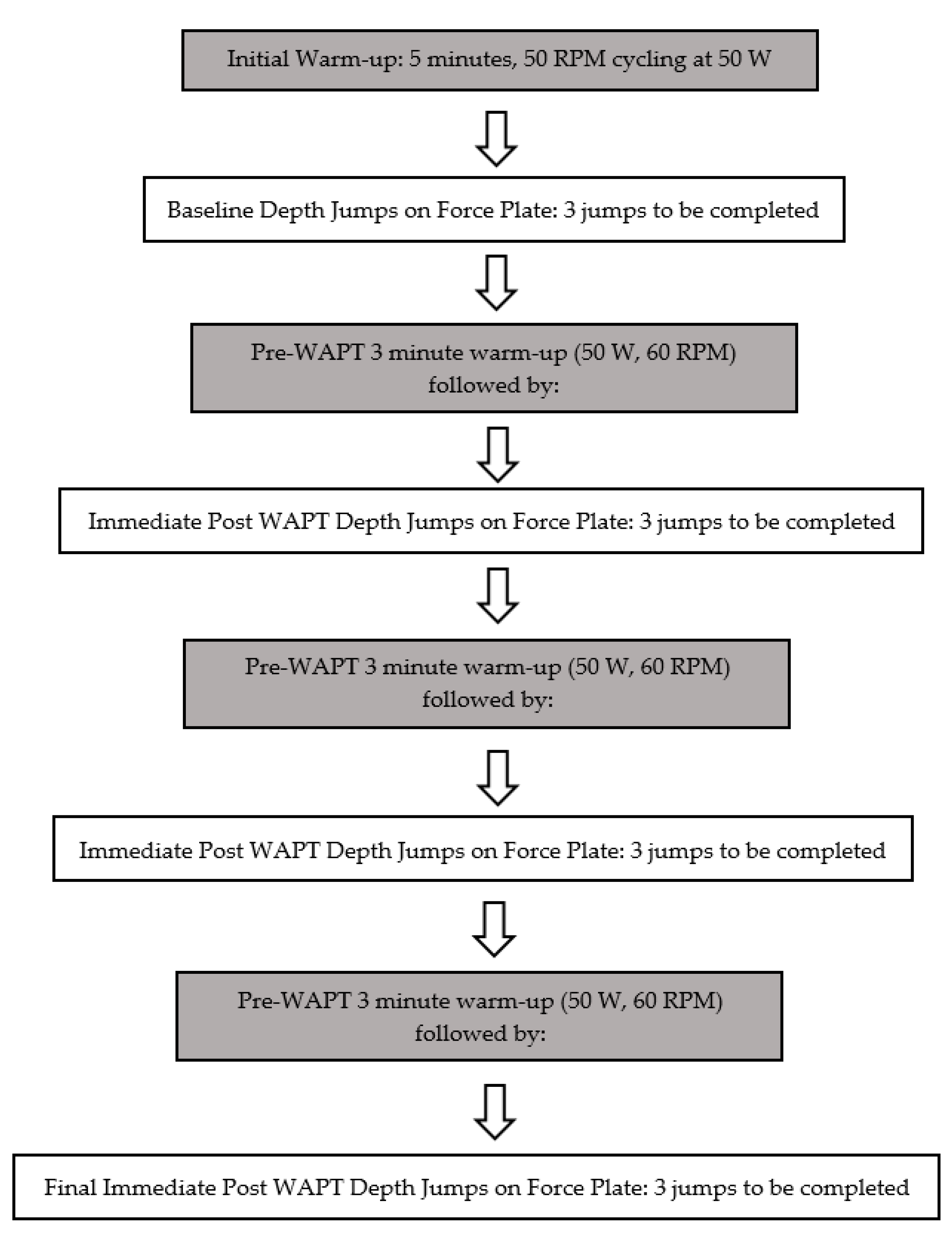
4. Proposed Protocol

The purpose of the proposed protocol is to assess the effects of fatigue on the GRF of a drop jump after repeated bouts of a Wingate Anaerobic Power Test (WAPT). The WAPT is considered the “gold-standard” of anaerobic power measurements [69]. The test consists of maximal effort cycling against a fixed torque factor for 30 s [69]. While the WAPT has historically been used as an assessment of anaerobic power, we feel that it may serve as an ideal test to induce both peripheral and central fatigue when compared to the more commonly utilized task specific protocols which likely only induce peripheral fatigue [68]. According to Benjaminse and colleagues, “General fatigue models appear to have more ecological validity in terms of simulating sports-relevant movement tasks” ([68], p. 578). Recent research has shown that there is no significant difference between lower extremity landing kinematics following task specific protocols [68,70]. With these criteria in mind repeated bouts of the WAPT may be an ideal way to induce general fatigue and more closely simulate metabolic demands associated with team sport [68,71,72]. The general fatigue model that we will propose below, while novel, builds on previous research carried out on the topic {68]. With the lack of homogeneity across protocols used, the proposed protocol may capture components of central and peripheral fatigue and therefore would make an ideal protocol across a broad spectrum of team athletics [68].
Along with the discrepancies in protocols, there continue to be contradictory findings across this topic [68]. Several studies have demonstrated an increase in vertical GRF during fatigued landing [55,61,63], while other studies refute those findings [58,59,60,73]. Therefore, there is little consensus on, how and if, the magnitude of GRF changes with muscle fatigue.
Indeed, one would tend to believe that the discrepancies across the results must be directly correlated with the preceding “fatigue protocol.” The inconsistencies across fatigue protocols may elicit conflicting results [44,68,73]. Given the complexity of fatigue, it is not surprising to see the variability of fatigue protocols utilized in the literature [54,68]. Figure 1, illustrates a proposed protocol that may impact the various components of fatigue. The authors propose a baseline Drop-Jump that is then followed by a series of three Wingate Anaerobic Power Tests (WAPT) separated by 60 s rest between trials. Following the completion of the three WAPT, participants would be asked to complete another Drop-Jump immediately following. The participants will be asked to complete this process three times in total. Therefore, there would be three rounds of three WAPT, which will then be followed by an immediate bilateral drop-jump assessment.
The high-intensity interval bouts on the cycle ergometer in conjunction with the repeated depth jumps would allow for the potential of both central and peripheral fatigue to be achieved [68]. Previous research has made clear that attempts to mimic the kinematic demands of sport participation have proven to be unsuccessful [55,58,59,60,61,63,68]. However, the protocol that we are proposing does not attempt to replicate the kinematic demands of sport participation. Rather, the proposed protocol would attempt to closely align the physiological demands of team sport participation [68,71,72]. Previous research across a variety of team based sports have shown that the physiological requirements for participation are associated with repeated bouts of short duration efforts similar to those that would be utilized in our proposed protocol [74,75,76,77]. Furthermore, we can appreciate that perhaps the movement patterns associated with repeated cycling are not “specific” to all athletes; however, the metabolic requirements could potentially be replicated given the repetitive nature of the protocol [74,75,76,77]. Additionally, the repeated nature of our proposed protocol, while anaerobic in nature, has been shown to tax all three metabolic systems, with significant contribution from the aerobic system, in team sport athletes [72,78,79].
Furthermore, utilizing the WAPT, data can be collected relative to peak power, fatigue index, and rate of peak power development. The data from each WAPT would provide insight into performance output which could subsequently be compared to the series of kinetic measurements from the drop jump assessments. Kinetic variables such as peak vertical GRF, peak posterior GRF, peak anterior GRF, peak medial and lateral GRF, and rate of force development would be collected and compared across the continuum of fatigue [41]. We believe that this may induce a similar complexity of fatigue that may be consistent with an athletic endeavor, unlike what has been noted to be completed in current published laboratory studies, which have tended to utilize more task specific protocols [54,68]. In comparison, the primary benefit of repeated bouts of the WAPT is that it may induce fatigue systemically as opposed to the more task specific protocols that have been previously studied [54,68].
Given that the relationship between fatigue and ACL injury risk is not completely understood, we would hypothesize that perhaps a more vigorous fatigue protocol may be necessary to more closely simulate the fatigue induced by athletic competition [68,71,72]. As mentioned previously, there are various risk factors associated with ACL injury risk [3,7,25,26,27,36]. We hypothesize that the proposed protocol may induce fatigue, which may further impact the kinetics associated with landing. Depth jump assessments have been used to make assumptions regarding ACL injury risk [80,81]. Therefore, we hypothesize that the metabolic requirements associated with the proposed protocol may tax team sport athletes in a way that is more “sport” specific, which may in turn change the biomechanical responses to landing. Finally, as fatigue begins to manifest, the authors would suggest including a perceived fatigue rating scale, to assess perceptual changes in fatigue [54]. However, given that fatigue perception is an individualized experience, that may be a potential confounding variable. Furthermore, we believe that this protocol could be utilized across several athletic populations.

5. Limitations

As previously mentioned, the proposed protocol would attempt to closely align the physiological demands of team sport participation [68,71,72,74,75,76,77]. While the protocol may be suitable for team sport athletes, it may be less suitable for endurance athletes who perform activities at submaximal intensities throughout their respective events. This ultimately would impact how their fatigue is manifested and experienced [82,83]. Furthermore, despite the proposed protocol emphasizing the metabolic specificity associated with team sport, it is accepted that the proposed repeated bouts of cycling are not “specific” to all athletes. Therefore, the task specificity of cycling will certainly impact specific muscular activity [84]. Given the traditional muscular contraction patterns associated with cycling, the protocol may generate fatigue that may not directly apply to individuals that regularly perform activities that involve repetitive high eccentric loading [85].

6. Conclusions

Overall, the multifaceted nature of fatigue likely has a major contribution to discrepancies seen in terms of the biomechanical changes that can occur [55,58,59,60,61,63]. To draw conclusions on the impact of fatigue on ACL injury risk, continued exploration is required [36,68]. Fatigue protocols should attempt to replicate metabolic and peripheral fatigue that would occur within athletic competitions. Furthermore, fatigue protocols must focus on impacting the various components of fatigue with emphasis on promoting a protocol that may even incorporate mental fatigue.

Author Contributions

Conceptualization, M.A., J.P., J.W. and R.M.O.; writing—original draft preparation M.A.; writing—review and editing, J.P., J.W. and R.M.O. All authors have read and agreed to the published version of the manuscript.

Funding

This research received no external funding.

Institutional Review Board Statement

Not applicable.

Informed Consent Statement

Not applicable.

Data Availability Statement

Not applicable.

Conflicts of Interest

The authors declare no conflict of interest.

References

  1. Gage, B.E.; Mcilvain, N.M.; Collins, C.L.; Fields, S.K.; Comstock, R.D. Epidemiology of 6.6 Million Knee Injuries Presenting to United States Emergency Departments from 1999 through 2008. Acad. Emerg. Med. 2012, 19, 378–385. [Google Scholar] [CrossRef] [PubMed]
  2. Vutescu, E.S.; Orman, S.; Garcia-Lopez, E.; Lau, J.; Gage, A.; Cruz, A.I. Psychological and Social Components of Recovery Following Anterior Cruciate Ligament Reconstruction in Young Athletes: A Narrative Review. Int. J. Environ. Res. Public Health 2021, 18, 9267. [Google Scholar] [CrossRef] [PubMed]
  3. Larwa, J.; Stoy, C.; Chafetz, R.S.; Boniello, M.; Franklin, C. Stiff landings, core stability, and dynamic knee valgus: A systematic review on documented anterior cruciate ligament ruptures in male and female athletes. Int. J. Environ. Res. Public Health 2021, 18, 3826. [Google Scholar] [CrossRef] [PubMed]
  4. Montalvo, A.M.; Schneider, D.K.; Webster, K.E.; Yut, L.; Galloway, M.T.; Heidt, R.S., Jr.; Myer, G.D. Anterior cruciate ligament injury risk in sport: A systematic review and meta-analysis of injury incidence by sex and sport classification. J. Athl. Train. 2019, 54, 472–482. [Google Scholar] [CrossRef] [PubMed] [Green Version]
  5. Bram, J.T.; Magee, L.C.; Mehta, N.N.; Patel, N.M.; Ganley, T.J. Anterior Cruciate Ligament Injury Incidence in Adolescent Athletes: A Systematic Review and Meta-analysis. Am. J. Sports Med. 2021, 49, 1962–1972. [Google Scholar] [CrossRef]
  6. Agel, J.; Rockwood, T.; Klossner, D. Collegiate ACL Injury Rates Across 15 Sports: National Collegiate Athletic Association Injury Surveillance System Data Update (2004–2005 Through 2012–2013). Clin. J. Sport Med. 2016, 26, 518–523. [Google Scholar] [CrossRef]
  7. Pfeifer, C.E.; Beattie, P.F.; Sacko, R.S.; Hand, A. Risk Factors Associated with Non-Contact Anterior Cruciate Ligament Injury: A Systematic Review. Int. J. Sports Phys. Ther. 2018, 13, 575–587. [Google Scholar] [CrossRef]
  8. Wetters, N.; Weber, A.E.; Wuerz, T.H.; Schub, D.L.; Mandelbaum, B.R. Mechanism of injury and risk factors for anterior cruciate ligament injury. Oper. Tech. Sports Med. 2016, 24, 2–6. [Google Scholar] [CrossRef]
  9. Bisciotti, G.N.; Chamari, K.; Cena, E.; Bisciotti, A.; Bisciotti, A.; Corsini, A.; Volpi, P. Anterior cruciate ligament injury risk factors in football. J. Sports Med. Phys. Fit. 2019, 59, 1724. [Google Scholar] [CrossRef]
  10. Della Villa, F.; Buckthorpe, M.; Grassi, A.; Nabiuzzi, A.; Tosarelli, F.; Zaffagnini, S.; Della Villa, S. Systematic video analysis of ACL injuries in professional male football (soccer): Injury mechanisms 1738, situational patterns and biomechanics study on 134 consecutive cases. Br. J. Sports Med. 2020, 54, 1423. [Google Scholar] [CrossRef]
  11. Grassi, A.; Smiley, S.P.; Roberti di Sarsina, T.; Signorelli, C.; Marcheggiani Muccioli, G.M.; Bondi, A.; Zaffagnini, S. Mechanisms and situations of anterior cruciate ligament injuries in professional male soccer players: A YouTube-based video analysis. Eur. J. Orthop. Surg. Traumatol. 2017, 27, 967–981. [Google Scholar] [CrossRef] [PubMed]
  12. Herzog, M.M.; Marshall, S.W.; Lund, J.L.; Pate, V.; Mack, C.D.; Spang, J.T. Trends in incidence of ACL reconstruction and concomitant procedures among commercially insured individuals in the United States, 2002–2014. Sports Health 2018, 10, 523–531. [Google Scholar] [CrossRef] [PubMed]
  13. Nematollahi, M.; Razeghi, M.; Tabatabaee, H.; Piroozi, S.; Shirazi, Z.R.; Rafiee, A. A new approach to differentiate those with anterior cruciate ligament deficient knees into copers and non-copers. Physiother. Pract. Res. 2016, 37, 73–78. [Google Scholar] [CrossRef]
  14. Rodriguez, K.; Soni, M.; Joshi, P.K.; Patel, S.C.; Shreya, D.; Zamora, D.I.; Patel, G.S.; Grossmann, I.; Sange, I. Anterior Cruciate Ligament Injury: Conservative Versus Surgical Treatment. Cureus 2021, 13, e2. [Google Scholar] [CrossRef]
  15. King, E.; Richter, C.; Daniels, K.; Franklyn-Miller, A.; Falvey, E.; Myer, G.D.; Jackson, M.; Moran, R.; Strike, S. Biomechanical but Not Strength or Performance Measures Differentiate Male Athletes Who Experience ACL Reinjury on Return to Level 1 Sports. Am. J. Sports Med. 2021, 49, 918–927. [Google Scholar] [CrossRef]
  16. Barber-Westin, S.; Noyes, F.R. One in 5 Athletes Sustain Reinjury upon Return to High-Risk Sports after ACL Reconstruction: A Systematic Review in 1239 Athletes Younger Than 20 Years. Sports Health 2020, 12, 587–597. [Google Scholar] [CrossRef]
  17. Johnston, P.T.; McClelland, J.A.; Feller, J.A.; Webster, K.E. Knee muscle strength after quadriceps tendon autograft anterior cruciate ligament reconstruction: Systematic review and meta-analysis. Knee Surg. Sports Traumatol. Arthrosc. 2021, 29, 2918. [Google Scholar] [CrossRef]
  18. Barfod, K.W.; Feller, J.A.; Hartwig, T.; Devitt, B.M.; Webster, K.E. Knee extensor strength and hop test performance following anterior cruciate ligament reconstruction. Knee 2019, 26, 149–154. [Google Scholar] [CrossRef] [Green Version]
  19. Aune, A.K.; Holm, I.; Risberg, M.A.; Jensen, H.K.; Steen, H. Four-strand hamstring tendon autograft compared with patellar tendon-bone autograft for anterior cruciate ligament reconstruction: A randomized study with two-year follow-up. Am. J. Sports Med. 2001, 29, 722–728. [Google Scholar] [CrossRef]
  20. Xergia, S.A.; McClelland, J.A.; Kvist, J.; Vasiliadis, H.S.; Georgoulis, A.D. The influence of graft choice on isokinetic muscle strength 4–24 months after anterior cruciate ligament reconstruction. Knee Surg. Sports Traumatol. Arthrosc. 2011, 19, 768–780. [Google Scholar] [CrossRef] [Green Version]
  21. Bizzini, M.; Gorelick, M.; Munzinger, U.; Drobny, T. Joint laxity and isokinetic thigh muscle strength characteristics after anterior cruciate ligament reconstruction: Bone patellar tendon bone versus quadrupled hamstring autografts. Clin. J. Sport Med. 2006, 16, 4–9. [Google Scholar] [CrossRef] [PubMed]
  22. Gobbi, A.; Mahajan, S.; Zanazzo, M.; Tuy, B. Patellar tendon versus quadrupled bone-semitendinosus anterior cruciate ligament reconstruction: A prospective clinical investigation in athletes. Arthrosc. J. Arthrosc. Relat. Surg. 2003, 19, 592–601. [Google Scholar] [CrossRef]
  23. Mattacola, C.G.; Perrin, D.H.; Gansneder, B.M.; Gieck, J.H.; Saliba, E.N.; McCue III, F.C. Strength, functional outcome, and postural stability after anterior cruciate ligament reconstruction. J. Athl. Train. 2002, 37, 262. [Google Scholar] [PubMed]
  24. Jansson, K.A.; Linko, E.; Sandelin, J.; Harilainen, A. A prospective randomized study of patellar versus hamstring tendon autografts for anterior cruciate ligament reconstruction. Am. J. Sports Med. 2003, 31, 12–18. [Google Scholar] [CrossRef] [PubMed]
  25. Beynnon, B.D.; Hall, J.S.; Sturnick, D.R.; DeSarno, M.J.; Gardner-Morse, M.; Tourville, T.W.; Vacek, P.M. Increased slope of the lateral tibial plateau subchondral bone is associated with greater risk of noncontact ACL injury in females but not in males: A prospective cohort study with a nested, matched case-control analysis. Am. J. Sports Med. 2014, 42, 1039–1048. [Google Scholar] [CrossRef]
  26. Bisson, L.J.; Gurske-DePerio, J. Axial and sagittal knee geometry as a risk factor for noncontact anterior cruciate ligament tear: A case-control study. Arthrosc. J. Arthrosc. Relat. Surg. 2010, 26, 901–906. [Google Scholar] [CrossRef]
  27. Dare, D.M.; Fabricant, P.D.; McCarthy, M.M.; Rebolledo, B.J.; Green, D.W.; Cordasco, F.A.; Jones, K.J. Increased lateral tibial slope is a risk factor for pediatric anterior cruciate ligament injury: An MRI-based case-control study of 152 patients. Am. J. Sports Med. 2015, 43, 1632–1639. [Google Scholar] [CrossRef]
  28. de Sire, A.; Demeco, A.; Marotta, N.; Spano, R.; Curci, C.; Fari, G.; Ammendolia, A. Neuromuscular impairment of knee stabilizers muscles in a COVID-19 cluster of female volleyball players: Which role for rehabilitation in the post-COVID-19 return-to-play? Appl. Sci. 2022, 12, 557. [Google Scholar] [CrossRef]
  29. de Sire, A.; Marotta, A.; Demeco, A.; Moggio, L.; Paola, P.; Marotta, N.; Ammendolia, A. Electromyographic assessment of anterior cruciate ligament injury risk in male tennis players: Which role for visual input? A proof-of-concept study. Diagnostics 2021, 11, 997. [Google Scholar] [CrossRef]
  30. de Sire, A.; Demeco, A.; Marrota, N.; Moggio, L.; Palumbo, A.; Iona, T.; Ammendolia, A. Anterior cruciate ligament injury prevention exercises: Could a neuromuscular warm-up improve muscle pre-activation before a soccer game? A proof-of-principle study on professional football players. Appl. Sci. 2021, 11, 4958. [Google Scholar] [CrossRef]
  31. Leppänen, M.; Pasanen, K.; Kujala, U.M.; Vasankari, T.; Kannus, P.; Äyrämö, S.; Parkkari, J. Stiff landings are associated with increased ACL injury risk in young female basketball and floorball players. Am. J. Sports Med. 2017, 45, 386–393. [Google Scholar] [CrossRef]
  32. Leppänen, M.; Pasanen, K.; Krosshaug, T.; Kannus, P.; Vasankari, T.; Kujala, U.M.; Parkkari, J. Sagittal plane hip, knee, and ankle biomechanics and the risk of anterior cruciate ligament injury: A prospective study. Orthop. J. Sports Med. 2017, 5, 2325967117745487. [Google Scholar] [CrossRef] [PubMed] [Green Version]
  33. Yu, B.; Garrett, W.E. Mechanisms of non-contact ACL injuries. Br. J. Sports Med. 2007, 41 (Suppl. 1), I47–I51. [Google Scholar] [CrossRef] [Green Version]
  34. Romero-Franco, N.; Ortego-Mate, M.; Molina-Mula, J. Knee Kinematics During Landing: Is It Really a Predictor of Acute Noncontact Knee Injuries in Athletes? A Systematic Review and Meta-analysis. Orthop. J. Sports Med. 2020, 8, 232596712096. [Google Scholar] [CrossRef] [PubMed]
  35. Nilstad, A.; Petushek, E.; Mok, K.M.; Bahr, R.; Krosshaug, T. Kiss goodbye to the ‘kissing knees’: No association between frontal plane inward knee motion and risk of future non-contact ACL injury in elite female athletes. Sports Biomech. 2021, 1–15. [Google Scholar] [CrossRef] [PubMed]
  36. Bittencourt, N.F.; Meeuwisse, W.H.; Mendonça, L.D.; Nettel-Aguirre, A.; Ocarino, J.M.; Fonseca, S.T. Complex systems approach for sports injuries: Moving from risk factor identification to injury pattern recognition—narrative review and new concept. Br. J. Sports Med. 2016, 50, 1309–1314. [Google Scholar] [CrossRef] [Green Version]
  37. Ament, W.; Verkerke, G.J. Exercise and Fatigue. Sports Med. 2009, 39, 382–422. [Google Scholar] [CrossRef]
  38. Potvin, J.R.; Fuglevand, A.J. A motor unit-based model of muscle fatigue. PLoS Comput. Biol. 2017, 13, e1005581. [Google Scholar] [CrossRef] [Green Version]
  39. Kennedy, R.A.; Drake, D. The effect of acute fatigue on countermovement jump performance in rugby union players during preseason. J. Sports Med. Phys. Fit. 2017, 57, 1261. [Google Scholar] [CrossRef]
  40. Bedo, B.L.; Pereira, D.R.; Moraes, R.; Kalva-Filho, C.A.; Will-De-Lemos, T.; Santiago, P.R. The rapid recovery of vertical force propulsion production and postural sway after a specific fatigue protocol in female handball athletes. Gait Posture 2020, 77, 52–58. [Google Scholar] [CrossRef]
  41. Dotan, R.; Woods, S.; Contessa, P. On the reliability and validity of central fatigue determination. Eur. J. Appl. Physiol. 2021, 121, 2393. [Google Scholar] [CrossRef] [PubMed]
  42. Ghorbani, M.; Clark, C.C. Brain function during central fatigue induced by intermittent high-intensity cycling. Neurol. Sci. 2021, 2411, 1–7. [Google Scholar] [CrossRef] [PubMed]
  43. Cordeiro, L.M.S.; Rabelo, P.C.R.; Moraes, M.M.; Teixeira-Coelho, F.; Coimbra, C.C.; Wanner, S.P.; Soares, D.D. Physical exercise-induced fatigue: The role of serotonergic and dopaminergic systems. Braz. J. Med. Biol. Res. 2017, 50, e6432. [Google Scholar] [CrossRef]
  44. Jayalath, J.L.R.; de Noronha, M.; Weerakkody, N.; Bini, R. Effects of fatigue on ankle biomechanics during jumps: A systematic review. J. Electromyogr. Kinesiol. 2018, 42, 81–91. [Google Scholar] [CrossRef]
  45. Wan, J.; Qin, Z.; Wang, P.; Sun, Y.; Liu, X. Muscle fatigue: General understanding and treatment. Exp. Mol. Med. 2017, 49, e384. [Google Scholar] [CrossRef] [PubMed] [Green Version]
  46. Debold, E.P. Recent Insights into Muscle Fatigue at the Cross-Bridge Level. Front. Physiol. 2012, 3, 151. [Google Scholar] [CrossRef] [Green Version]
  47. Scott, W.; Stevens, J.; Binder–Macleod, S.A. Human Skeletal Muscle Fiber Type Classifications. Phys. Ther. 2001, 81, 1810. [Google Scholar] [CrossRef]
  48. Staron, R.S.; Hagerman, F.C.; Hikida, R.S.; Murray, T.F.; Hostler, D.P.; Crill, M.T.; Toma, K. Fiber Type Composition of the Vastus Lateralis Muscle of Young Men and Women. J. Histochem. Cytochem. 2000, 48, 623–629. [Google Scholar] [CrossRef] [Green Version]
  49. Cooper, C.N.; Dabbs, N.C.; Davis, J.; Sauls, N.M. Effects of Lower-Body Muscular Fatigue on Vertical Jump and Balance Performance. J. Strength Cond. Res. 2020, 34, 2903. [Google Scholar] [CrossRef]
  50. Harato, K.; Morishige, Y.; Niki, Y.; Kobayashi, S.; Nagura, T. Fatigue and recovery have different effects on knee biomechanics of drop vertical jump between female collegiate and recreational athletes. J. Orthop. Surg. Res. 2021, 16, 1–7. [Google Scholar]
  51. Bobbert, M.F.; Van Der Krogt, M.M.; Van Doorn, H.; De Ruiter, C.J. Effects of fatigue of plantarflexors on control and performance in vertical jumping. Med. Sci. Sports Exerc. 2011, 43, 673–684. [Google Scholar] [CrossRef] [PubMed]
  52. Smith, M.R.; Coutts, A.J.; Merlini, M.; Deprez, D.; Lenoir, M.; Marcora, S.M. Mental fatigue impairs soccer-specific physical and technical performance. Med. Sci. Sports Exerc. 2016, 48, 267–276. [Google Scholar] [CrossRef] [PubMed]
  53. Demeco, A.; de Sire, A.; Marotta, N.; Spano, R.; Lippi, L.; Palumbo, A.; Ammendolia, A. Match analysis, physical training, risk of injury, and rehabilitation in padel: Overview of the literature. Int. J. Environ. Res. Public Health 2022, 19, 4153. [Google Scholar] [CrossRef] [PubMed]
  54. Twomey, R.; Aboodarda, S.J.; Kruger, R.; Culos-Reed, S.N.; Temesi, J.; Millet, G.Y. Neuromuscular fatigue during exercise: Methodological considerations, etiology and potential role in chronic fatigue. Neurophysiol. Clin. 2017, 47, 95–110. [Google Scholar] [CrossRef]
  55. Quammen, D.; Cortes, N.; Van Lunen, B.L.; Lucci, S.; Ringleb, S.I.; Onate, J. Two Different Fatigue Protocols and Lower Extremity Motion. J. Athl. Train. 2012, 47, 32–41. [Google Scholar] [CrossRef] [Green Version]
  56. Cortes, N.; Greska, E.; Ambegaonkar, J.P.; Kollock, R.O.; Caswell, S.V.; Onate, J.A. Knee kinematics is altered post-fatigue while performing a crossover task. Knee Surg. Sports Traumatol. Arthrosc. 2013, 22, 2202. [Google Scholar] [CrossRef] [Green Version]
  57. Lucci, S.; Cortes, N.; Lunen, B.V.; Ringleb, S.; Onate, J. Knee and hip sagittal and transverse plane changes after two fatigue protocols. J. Sci. Med. Sport 2011, 14, 453–459. [Google Scholar] [CrossRef] [Green Version]
  58. Barber-Westin, S.D.; Noyes, F.R. Effect of Fatigue Protocols on Lower Limb Neuromuscular Function and Implications for Anterior Cruciate Ligament Injury Prevention Training: A Systematic Review. Am. J. Sports Med. 2017, 45, 3388. [Google Scholar] [CrossRef]
  59. Watanabe, S.; Aizawa, J.; Shimoda, M.; Enomoto, M.; Nakamura, T.; Okawa, A.; Yagishita, K. Effect of short-term fatigue 3396, induced by high-intensity exercise, on the profile of the ground reaction force during single-leg anterior drop-jumps. J. Phys. Ther. Sci. 2016, 28, 3371. [Google Scholar] [CrossRef] [Green Version]
  60. Kellis, E.; Kouvelioti, V. Agonist versus antagonist muscle fatigue effects on thigh muscle activity and vertical ground reaction during drop landing. J. Electromyogr. Kinesiol. 2009, 19, 55–64. [Google Scholar] [CrossRef]
  61. Pappas, E.; Hagins, M.; Sheikhzadeh, A.; Nordin, M.; Rose, D. Biomechanical Differences between Unilateral and Bilateral Landings from a Jump: Gender Differences. Clinical J. Sport Med. 2007, 17, 263–268. [Google Scholar] [CrossRef] [PubMed]
  62. Brazen, D.M.; Todd, M.K.; Ambegaonkar, J.P.; Wunderlich, R.; Peterson, C. The Effect of Fatigue on Landing Biomechanics in Single-Leg Drop Landings. Clin. J. Sport Med. 2010, 20, 286–292. [Google Scholar] [CrossRef] [PubMed]
  63. Wong, T.L.; Huang, C.F.; Chen, P.C. Effects of Lower Extremity Muscle Fatigue on Knee Loading During a Forward Drop Jump to a Vertical Jump in Female Athletes. J. Hum. Kinet. 2020, 72, 5–13. [Google Scholar] [CrossRef] [Green Version]
  64. Zhang, X.; Xia, R.; Dai, B.; Sun, X.; Fu, W. Effects of Exercise-Induced Fatigue on Lower Extremity Joint Mechanics, Stiffness, and Energy Absorption during Landings. J. Sports Sci. Med. 2018, 17, 640–649. [Google Scholar]
  65. Ueno, R.; Navacchia, A.; DiCesare, C.A.; Ford, K.R.; Myer, G.D.; Ishida, T.; Hewett, T.E. Knee abduction moment is predicted by lower gluteus medius force and larger vertical and lateral ground reaction forces during drop vertical jump in female athletes. J. Biomech. 2020, 103, 109669. [Google Scholar] [CrossRef]
  66. Evans-Pickett, A.; Davis-Wilson, H.C.; Luc-Harkey, B.A.; Blackburn, J.T.; Franz, J.R.; Padua, D.A.; Pietrosimone, B. Biomechanical effects of manipulating peak vertical ground reaction force throughout gait in individuals 6–12 months after anterior cruciate ligament reconstruction. Clin. Biomech. 2020, 76, 105014. [Google Scholar] [CrossRef]
  67. Heebner, N.R.; Rafferty, D.M.; Wohleber, M.F.; Simonson, A.J.; Lovalekar, M.; Reinert, A.; Sell, T.C. Landing kinematics and kinetics at the knee during different landing tasks. J. Athl. Train. 2017, 52, 1101–1108. [Google Scholar] [CrossRef] [Green Version]
  68. Benjaminse, A.; Webster, K.E.; Kimp, A.; Meijer, M.; Gokeler, A. Revised approach to the role of fatigue in anterior cruciate ligament injury prevention: A systematic review with meta-analyses. Sports Med. 2019, 49, 565–586. [Google Scholar] [CrossRef] [Green Version]
  69. Lunn, W.R.; Axtell, R.S. Validity and reliability of the lode excalibur sport cycle ergometer for the wingate anaerobic test. J. Strength Cond. Res. 2021, 35, 2894–2901. [Google Scholar]
  70. Borotikar, B.S.; Newcomer, R.; Koppes, R.; McLean, S.G. Combined effects of fatigue and decision making on female lower limb landing postures: Central and peripheral contributions to ACL injury risk. Clin Biomech. 2008, 23, 81–92. [Google Scholar] [CrossRef]
  71. Degens, H.; Stasiulis, A.; Skurvydas, A.; Statkeviciene, B.; Venckunas, T. Physiological comparison between non-athletes, endurance, power and team athletes. Eur. J. Appl. Physiol. 2019, 119, 1377–1386. [Google Scholar] [CrossRef] [PubMed] [Green Version]
  72. Beneke, R.; Pollmann, C.; Bleif, I.; Leithäuser, R.M.; Hütler, M. How anaerobic is the Wingate Anaerobic Test for humans? Eur. J. Appl. Physiol. 2002, 87, 388–392. [Google Scholar] [CrossRef] [PubMed]
  73. Zadpoor, A.A.; Nikooyan, A.A. The effects of lower-extremity muscle fatigue on the vertical ground reaction force: A meta-analysis. Proceedings of the Institution of Mechanical Engineers Part H. J. Eng. Med. 2012, 226, 579–588. [Google Scholar] [PubMed]
  74. Ben Abdelkrim, N.; Castagna, C.; Jabri, I.; Battikh, T.; El Fazaa, S.; El Ati, J. Activity profile and physiological requirements of junior elite basketball players in relation to aerobic-anaerobic fitness. J. Strength Cond. Res. 2010, 24, 2330. [Google Scholar] [CrossRef] [PubMed]
  75. Vigne, G.; Gaudino, C.; Rogowski, I.; Alloatti, G.; Hautier, C. Activity profile in elite Italian soccer team. Int. J. Sports Med. 2010, 31, 304–310. [Google Scholar] [CrossRef]
  76. Sunderland, C.D.; Edwards, P.L. Activity Profile and Between-Match Variation in Elite Male Field Hockey. J. Strength Cond. Res. 2017, 31, 758–764. [Google Scholar] [CrossRef] [Green Version]
  77. Vigh-Larsen, J.F.; Haverinen, M.T.; Panduro, J.; Ermidis, G.; Andersen, T.B.; Overgaard, K.; Krustrup, P.; Parkkari, J.; Avela, J.; Kyröläinen, H.; et al. On-Ice and Off-Ice Fitness Profiles of Elite and U20 Male Ice Hockey Players of Two Different National Standards. J. Strength Cond. Res. 2020, 34, 3369. [Google Scholar] [CrossRef]
  78. Gharbi, Z.; Dardouri, W.; Haj-Sassi, R.; Chamari, K.; Souissi, N. Aerobic and anaerobic determinants of repeated sprint ability in team sports athletes. Biol Sport. 2015, 32, 207–212. [Google Scholar] [CrossRef]
  79. Yamagishi, T.; Babraj, J. Influence of recovery intensity on oxygen demand and repeated sprint performance. J. Sports Med. Phys. Fit. 2016, 56, 1103–1112. [Google Scholar]
  80. Orishimo, K.F.; Kremenic, I.J.; Pappas, E.; Hagins, M.; Liederbach, M. Comparison of landing biomechanics between male and female professional dancers. Am. J. Sports Med. 2009, 37, 2187. [Google Scholar] [CrossRef]
  81. Liederbach, M.; Kremenic, I.J.; Orishimo, K.F.; Pappas, E.; Hagins, M. Comparison of landing biomechanics between male and female dancers and athletes, part 2: Influence of fatigue and implications for anterior cruciate ligament injury. Am. J. Sports Med. 2014, 42, 1089. [Google Scholar] [CrossRef] [PubMed]
  82. Jurasz, M.; Boraczyński, M.; Wójcik, Z.; Gronek, P. Neuromuscular Fatigue Responses of Endurance- and Strength-Trained Athletes during Incremental Cycling Exercise. Int. J. Environ. Res. Public Health 2022, 1095, 19. [Google Scholar] [CrossRef] [PubMed]
  83. Jeukendrup, A.E. Nutrition for endurance sports: Marathon 8839, triathlon, and road cycling. J. Sports Sci. 2011, 29 (Suppl. 1), S91–S99. [Google Scholar] [CrossRef] [PubMed]
  84. Park, S.; Caldwell, G.E. Muscular activity patterns in 1-legged vs. 2-legged pedaling. J. Sport Health Sci. 2021, 10, 99–106. [Google Scholar] [CrossRef] [PubMed]
  85. Clos, P.; Lepers, R. Leg Muscle Activity and Perception of Effort before and after Four Short Sessions of Submaximal Eccentric Cycling. Int. J. Environ. Res. Public Health 2020, 17, 7702. [Google Scholar] [CrossRef]
Figure 1. This figure represents a flowchart of the proposed fatigue protocol. The protocol is designed to allow analysis of rate of fatigue from the WAPT, while tracking kinetic variables across the continuum of fatigue. WAPT: Wingate Anaerobic Power Test; W: Watts RPM: Revolutions per Minute.
Biomechanics 02 00040 g001
Publisher’s Note: MDPI stays neutral with regard to jurisdictional claims in published maps and institutional affiliations.

