Review

Recent Progress on Natural Rubber-Based Materials Containing Metallic and Metal Oxide Nanoparticles: State of the Art and Biomedical Applications

by
Nayrim B. Guerra
1,
Jordana Bortoluz
1,
Andressa R. Bystronski
1,
Ana Elisa D. Maddalozzo
1,
Danielle Restelatto
1,
Mariana Roesch-Ely
2,
Declan M. Devine
3,
Marcelo Giovanela
1,* and
Janaina S. Crespo
1,3,*
1
Área do Conhecimento de Ciências Exatas e Engenharias, Universidade de Caxias do Sul, Rua Francisco Getúlio Vargas 1130, Caxias do Sul 95070-560, RS, Brazil
2
Área do Conhecimento de Ciências da Vida, Instituto de Biotecnologia, Universidade de Caxias do Sul, Rua Francisco Getúlio Vargas 1130, Caxias do Sul 95070-560, RS, Brazil
3
Materials Research Institute, Technological University of the Shannon: Midlands Midwest, Athlone, N37HD68 Co. Westmeath, Ireland
*
Authors to whom correspondence should be addressed.
Compounds 2023, 3(2), 310-333; https://doi.org/10.3390/compounds3020023
Submission received: 3 January 2023 / Revised: 17 February 2023 / Accepted: 23 March 2023 / Published: 28 March 2023

Abstract

:
Diseases caused by infections are becoming harder to treat as the antibiotics used become less effective. A combination of strategies to develop active biomaterials that enhance antibacterial effects are desirable, especially ones that cause fewer side effects and promote healing properties. The combination of nanotechnology with substances that have intrinsic antibacterial activity can result in the advance of innovative biomedical materials. In this sense, the goal of this work is to provide a summary of natural rubber latex materials obtained from the Hevea brasiliensis tree loaded with metallic and metal oxide nanoparticles. These nanoparticles have unique size-dependent chemical and physical characteristic that make them appropriate for use in pharmaceutical and medical devices, while natural rubber latex is a natural and biocompatible polymer with an intrinsic antibacterial effect. Moreover, we outline here the origin, extraction methods, and composition of natural rubber latex and different techniques for the synthesis of nanoparticles, including physical, chemical, and biological approaches. Finally, we summarize, for the first time, the state of the art in obtaining natural rubber-based materials with metallic and metallic oxide nanoparticles for biomedical applications.

Graphical Abstract

1. Introduction

The demand for the manufacture of active biomaterials with antimicrobial properties is increasing. In recent years, the healthcare industry has been offering antimicrobial solutions for an extensive variety of applications, from medical devices to everyday materials. In this context, the rubber industry benefits from the implementation of biomedical technologies capable of reducing microorganisms, increasing the time of use of existing products. Many antimicrobial substances have been used to confer these properties on latex materials [1].
Natural rubber latex (NRL) extracted from rubber trees is one of the economic pillars of several Southeast Asian countries [2]. From this natural polymer, different products can be prepared, and many of them are used in the health industry. Natural rubber latex foam (NRLF), for example, is a porous, low-density type of rubber that is mainly used to make mattresses and pillows [3]. NRLF is easily attacked by ultraviolet (UV) light, and a prolonged exposure to sunlight favors rapid bacterial growth [4]. NRL is also the most commonly used raw material for the manufacture of gloves, essential personal protective equipment in the healthcare sector, as they provide a protective barrier against infectious organisms necessary for the safety of patients and medical staff [5]. Enhancing the biomedical properties of NRL materials will provide NRL with antibacterial and antiviral properties without altering its physical properties or causing hypersensitivity.
Nanoparticles (NPs), specifically metallic and metal oxide NPs, have been widely used in biomedical engineering due to their specific size-dependent physical and chemical properties [6]. According to the World Health Organization (WHO), in addition to their reduced dimensions and selectivity for bacteria, metallic NPs have also proved to be efficient against pathogens registered as a priority [7]. Several studies have demonstrated the strong antimicrobial effects of metallic and metal oxide NPs against multiple species of bacteria [8,9,10], and currently, many biomedical devices contain metallic NPs as they help to prevent and reduce the rate of the spread of infectious diseases [11]. Because of the well-detailed antimicrobial activity against Gram-negative bacteria, such as Escherichia coli and Salmonella, and Gram-positive bacteria, such as Staphylococcus aureus and Listeria monocytogenes, these particles can be used as a coadjuvant or replacement for common antibiotics to combat bacteria resistance, especially regarding the group of Gram-negative bacteria, once they are more pathogenic in their susceptibility to antibiotics [12]. NPs use different routes compared to traditional treatments, being active against bacteria that commonly develop antibiotic resistance and targeting some biomolecules that compromise the progress of a resistance strain [13]. The antimicrobial mechanism of action of NPs can be understood by models such as oxidative stress induction [14], metal ion release [15], or non-oxidative mechanisms [16]; it is important to note that these models can occur separately or simultaneously.
A literature review was performed employing the Scopus and Web of Science databases and indicated that, over the last decade, research based on NRL incorporated with NPs has been continually increasing, with a rise in the number of manuscripts published each year. However, studies using NPs incorporated into NRL for biomedical applications are still scarce (Figure 1). Recently, many review articles on metallic and metal oxide NPs have been published which focused on their antimicrobial activities. The vast majority of published works on metallic and metallic oxide NPs impregnated in NRL matrices were cited, and these will be discussed in this review. We emphasize that so far, in the biomedical area, there is no review article that compiles and helps readers to better develop this theme.

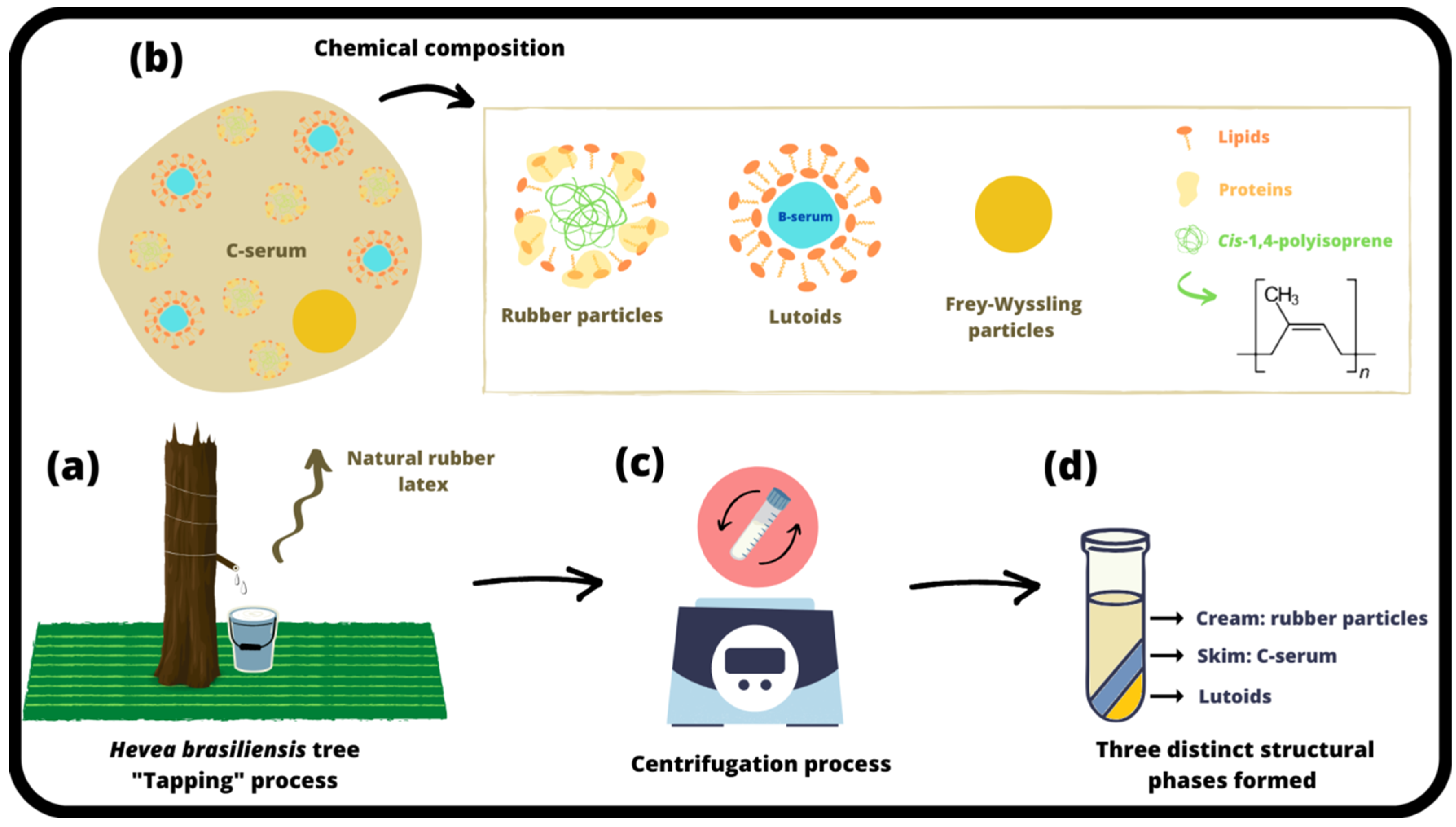
2. Natural Rubber Latex: Source, Composition, and Antimicrobial Properties

It is known that about 20,000 different tree species are able to produce latex. However, Hevea brasiliensis alone supplied about 13.9 million tons of natural rubber in 2018 [17,18]. The NRL produced by this tree is of great importance in the world due to its excellent unique properties that can be used for several industrial applications, such as in the manufacturing of gloves, condoms, rubber bands, balloons, foam mattresses, and elastic thread, among others [19,20].
Hevea brasiliensis is a very tall tree made of softwood and it has a Brazilian origin [17,21], although it has also been extensively cultivated in Southeast Asia [22]. The latex of this tree is considered to be the cytoplasm of cells called laticifers [23]. This milk liquid can also be described as a natural polymer with a molecular weight of 104 to 105 kDa [24,25].

2.1. Obtaining and Composition of NRL

To obtain NRL from the tree, the vessels present in its bark are opened through a process known as “tapping”, in which a thin shaving of bark (about 1.0 mm thick) is removed to a depth very close to the cambium. The tapping cut (Figure 2a) is made at an angle of approximately 30° to the horizontal direction from high left to low right. NRL is in the vessels at a high hydrostatic pressure (approximately 1.0 to 1.9 MPa), which is called turgor. Turgor is at its maximum before sunrise, so tapping needs to be started at dawn to obtain a better NRL yield [19,26]. This milky liquid will flow for several hours until the vessels form a clot. If it is necessary to reopen the next day, the latex flows again [27].
After collection, the NRL must be processed immediately; otherwise, additives to prevent spontaneous coagulation of the rubber are necessary. Thus, the collected NRL is stabilized by the addition of about 0.01% ammonia, 0.05% sodium sulfite, or 0.02% formaldehyde [27]. Among them, the most widely used preservative agent of NRL is ammonia, since it increases the pH of the NRL and consequently prevents microbial activity for a long time [19,28].
NRL from Hevea brasiliensis is known to be a colloidal dispersion, with different particle sizes (from nanometers to microns) walled by a thin layer of proteins, lipids, and chains of fatty acids. These characteristics provide a negative charge to the particles and make the colloidal medium stable. NRL is composed of rubber particles (37 to 67% w/w fresh latex), lutoids (12 to 22% w/w fresh latex), and Frey-Wyssling particles (2 to 3% w/w fresh latex), all of them disseminated in the cytoplasmic serum (C-serum) (18 to 29% w/w fresh latex) (Figure 2b) [18].
The rubber particles are the most important NRL constituents and they are made with a monolayer of lipids and proteins surrounding a hydrophilic core of poly(cis-1,4-isoprene) chains [18]. In 1991, Tanaka [29] showed that these polymer chains are made up of an initiating group (called ω), two poly(trans-1,4-isoprene) units, successive poly(cis-1,4-isoprene) units, and a final group (called α). The lipids and proteins, in turn, have an essential function to ensure that the latex particles are stable [26].
The second most abundant constituent is the C-serum, which concentrates everything that makes up the NRL. The lutoid particles are constituted of a bilayer of lipids molecules surrounding a hydrophilic core made of B-serum, which is a destabilizer of rubber hydrocarbon [18,26]. The B-serum is rich in several enzymes, and the largest protein found in lutoid particles is the hevein [18]. Frey-Wyssling particles, the lesser constituent, are yellowish and have a complex structure, which performs some important biochemical activities, with possible rubber biosynthesis sites [30].
When NRL undergoes the centrifugation process (Figure 2c), three phases formed by these structural constituents are evident (Figure 2d). They are usually called cream (large rubber particles), skim (small rubber particles and Frey-Wyssling particles spread in C-serum), and lutoids [18,30]. The centrifugation process is essential, especially when NRL is used as a biomaterial. Phase separation allows the removal of some high-molecular-weight proteins that are considered allergenic [31,32,33].
Besides the structural aspects, NRL is also subdivided for its different chemical compounds. Before undergoing any chemical modification, this material can be made of 35.0% poly(cis-1,4-isoprene), 1.5% proteins, 1.3% lipids, 1.5% carbohydrates, 0.5% minerals, 0.5% organic solutes, and 59.7% water [18]. Some authors emphasize that the NRL composition also depends on the season of the year, the geographic origin of the tree, and its age [18,21,26,34,35].

2.2. Antimicrobial Properties of NRL

All the chemical compounds present in NRL can influence its biological activities and have some importance in terms of its properties. It has already been proven that several of the components present in the structure of NRL stimulate angiogenesis, cell adhesion, and the formation of an extracellular matrix, promoting tissue replacement and regeneration. Thus, some NRL-based materials have been widely applied and marketed in over 60 countries as dressings for the treatment of diabetic patients who suffer from difficult curable wounds, which is why it is considered an excellent biomaterial [31,32].
In addition to these properties, it is known that, to resist the frequent tapping that rubber trees undergo daily, they need to have a self-defense mechanism [36]. For this reason, some authors have also explored the antimicrobial properties exhibited by the chemical compounds present in NRL. This characteristic was shown in the study by Boonrasri et al. [37], who investigated the antibacterial activity of NRL-based films with different concentrations of chitosan. The biocomposite films showed a high ability to prevent the growth of Staphylococcus aureus. Furthermore, it was also found that the greater the amount of chitosan present in the film, the greater the inhibition effect. These authors concluded that these films can be used for the preparation of gloves with antibacterial activity or in catheters. In the same way, Arakkal et al. [38] evaluated the antibacterial activity of NRL films containing polyelectrolyte derivatives of chitosan for antibacterial applications. In this study, they also demonstrated a significant reduction in bacterial colony formation when they came into contact with NRL polymeric composites.
Some studies have revealed that NRL has an important role regarding the antimicrobial characteristics of this material. It is estimated that about 1499 types of different proteins are part of the composition of NRL [17]. Among them, the main ones are chitinase, β-1,3-glucanase, hevamines, hevein, glucosidase, β-galactosidase, N-acetyl-β-glucosaminidase, and polyphenol oxidase, a protease inhibitor [22].
The antimicrobial activity of hevein, a protein of a small size and the major component of B-serum, was verified by Van Parijs et al. [39]. To evaluate this property, they used various pathogenic microorganisms, such as Pyrenophora tritici-repentis, Botrytis cinerea, Septoria nodorum, Fusarium culmorum, Trichoderma hamatum, Fusarium oxysporum, Pyricularia oryzae, and Phycomyces blakesleeanus. The authors concluded that this protein has effective antimicrobial properties, and B-serum is an important active fraction of the NRL.
Some years later, Kanokwiroon et al. [22] also studied hevein and demonstrated excellent results for different microorganisms, such as Candida krusei, Candida albicans, and Candida tropicalis. The two first ones, in turn, showed improved activities. The minimal inhibitory concentration (MIC) value was 190 and 95 µg mL−1, respectively. Moreover, the hevein also induced a growth inhibition of those fungi in disk diffusion systems.
Two β-1,3-glucanase isozymes (GI and GII) from the lutoid particles were purified and evaluated by Churngchow et al. [40]. The study revealed an interesting result since it was observed that the more frequent the tapping process, the greater the amount of GI and GII in the NRL. In addition, they showed that the GII protein presented major antifungal activity and played an important role in protecting the tree against microorganisms.
The activity of the C-serum phase as an antimicrobial agent of NRL was also evaluated. In the work by Daruliza et al. [41], its anti-fungal properties were demonstrated. Experiments using the disk diffusion method revealed that the NRL C-serum presents an effective antifungal property for Aspergillus niger, while the results related to Candida albicans were not satisfactory. Furthermore, the authors emphasize that this study confirms that the biological activity of Hevea brasiliensis serum can contribute to the development of several products in the polymer industry and in the pharmaceutical and healthcare sectors.

3. Metallic and Metal Oxide NPs: Synthesis and Biological Properties

The development of new antimicrobial products is necessary to avoid the transmission of infection in healthcare environments [42,43]. In this sense, metallic and metal oxide NPs have been gaining attention due to their unique size-dependent physical and chemical properties [44]. The best known examples of this category are the NPs of elements such as silver, copper, gold, palladium, and platinum, which are used in varied areas of application (catalytic, biomedical, and electronic) as their properties are distinguished from those presented by the bulk [45]. The studies show that these NPs are especially effective against Gram-negative and Gram-positive bacteria, such as Staphylococcus aureus, Pseudomonas aeruginosa, and Escherichia coli [13]. Detailed information on these metallic and metal oxide NPs will be presented and discussed in the next subsections.

3.1. Silver Nanoparticles

Traditionally, silver is known as a substance with antibacterial properties. The concentration of AgNPs added is proportionally responsible for the antibacterial action, as well as the size and shape of the NPs. Studies show that the minimum inhibitory concentration (MIC) of AgNPs against Gram-negative and Gram-positive bacterial strains depend strongly on the factors already mentioned, but these are approximately 1.0 and 1.5–2.0 μg mL−1, respectively [46]. Therefore, its NPs are currently being used in a wide variety of commercial products [47]. Silver nanoparticles (AgNPs) are used in the form of wound dressings, the coating of prostheses, and surgical utensils due to their antimicrobial properties against bacteria and protozoa, in addition to being effective in eliminating some fungi and viruses [48]. AgNPs are used in the coating of medical devices, the water and wastewater treatment industry, smart fabrics, wound dressings, and the food industry. They also have electrochemical and bioluminescent properties that enable their application in optical and biological nanosensors [49].
However, NPs can present cytotoxicity in addition to antibacterial activity; this is an effect that depends on factors such as the size, dose, and exposure time of NPs. The continued use of these NPs can lead to the development of diseases and their leaching into aquatic environments, polluting groundwater. In contact with water, NPs oxidize and form Ag(I) species, ions that are toxic to marine organisms and can return to the human body due to the food chain. It is not known exactly what effect of AgNPs will be generated by a prolonged exposure over time, but a series of in vitro tests observed toxicity in keratinocyte cells, liver cells, and colon cells, among others. In vivo tests with rodents have shown that AgNPs accumulate in the lung, spleen, and liver [46].

3.2. Copper Nanoparticles

Due to their high oxidative ability in contact with air, the use of copper nanoparticles (CuNPs) presents some challenges. The formation of copper oxide results in a loss in intensity of the antibacterial activity, so CuNPs must have high stabilization in the substrate to which they are added. To that end, chelates promote the stability of NPs, which will be surrounded by organic molecules. Chelates with amino acids showed antibacterial activity ten times higher than that presented by CuNPs. The use of different materials in these compounds will modify the antimicrobial activity, which may be superior to some bacteria in relation to others. In addition to the oxidation factor, CuNPs also suffer from agglomeration, which must be minimized. In this way, NPs will be more dispersed, offering a larger zone of interaction between particles and micro-organisms, leading to an increase in their toxicity [49].
In addition to antibacterial activity, it has been reported in the literature that CuNPs also have anticancer and cytotoxic properties. Cell death or the damage of cancer cells is caused by the generation of reactive oxygen species (ROS), which cause cellular lipid peroxidation, DNA damage, and protein oxidation [50].

3.3. Gold Nanoparticles

Gold nanoparticles (AuNPs) have a high biocompatibility and can therefore be used in different medical applications [51]. They can have different sizes, formats, and aggregation capabilities [52]. The size and shape of the NPs influence their physical–chemical, electrical, and optical properties [53]. The AuNPs have an average particle size smaller than AgNPs and they are less rough. A high roughness is important for a high antibacterial efficacy, as bacteria are attracted by irregular metal particles and are thus eliminated [54].
AuNPs have been the subject of many studies due to their applications in the development of new agents and because they are of stable, nontoxic in nature, inert, and controllable sizes [55]. In addition, they absorb light through the surface plasma resonance phenomenon, affecting the production of intracellular ROS [56]. However, AuNPs, despite their wide range of applications in electronics, biomedicine, and nanotechnology, have a feeble antibacterial property compared to other metallic NPs [57]. To increase the antibacterial capacity of AuNPs, several studies involving the synthesis of composite materials with other antibacterial agents have been carried out [54,58,59].

3.4. Metal Oxide Nanoparticles

Nanometer-sized metallic oxides have also attracted the attention of scientists, mainly due to the existence of a negative surface charge, or one that favors their functionalization with different molecules [60]. Metal oxides NPs have a high stability, the possibility of synthesis in different sizes, and ease of preparation and incorporation into hydrophobic and hydrophilic matrices [61]. They can be synthesized from metals or metal oxides, such as silver, gold, titanium, zinc, copper, magnesium, calcium, and iron [62]. In contrast, metallic oxide NPs tend to be less stable than metallic nanomaterials (composed of a single metallic element), and these are more susceptible to dissolution and the release of ions when inserted into a biological medium [63]. In this regard, a great deal of research is being carried out to assess the possible toxic effects, as well as the computational methods, to predict their toxicity [64,65].
Zinc oxide nanoparticles (ZnONPs), for example, have several applications in the engineering and biomedical fields. They offer new possibilities for biomedical applications, ranging from diagnosis to treatment [66]. Furthermore, they are responsible for speeding up the rate of wound healing, since zinc is an important trace element found in the muscles, bones, and skin [67]. In the rubber industry, ZnONPs are widely applied as they offer wear resistance of the rubber compound, improve its toughness, and prevent aging, amongst other functions [68]. At present, ZnONPs are being investigated as associates of antimicrobial agents, which is one of the most important reasons for their use [69]. Compared with other metal oxide NPs, ZnONPs are inexpensive and have a low toxicity [70].
Other metal oxide NPs used in the biomedical field are titanium oxide (TiO2NPs), which have high photocatalytic activity and thermo-stability, which makes them applicable for use in biomedical applications, such as in the treatment of cancer using phototherapy and for use against different bacterial contaminations [71,72]. Nanostructured TiO2 has a great potential for application due to its good biocompatibility, intrinsic properties, and different manufacturing techniques [73]. Furthermore, TiO2NPs have also presented antifungal activity against diverse species of fungus [74]. In these NPs, it is important to point out that mixed polymorphs of TiO2 (anatase and rutile) are more effective for biomedical applications than using only one crystalline phase [75].
Iron oxide magnetic nanoparticles (Fe3O4NPs) are also used in the biomedical field, specifically as a component of biosensors, in magnetic resonance studies and cancer cell treatment. However, their most discussed application is in targeted drug delivery, because of their unique magnetic properties, biodegradability, low toxicity, and excellent biocompatibility [76]. Despite the fact that these NPs have a highly reactive surface, there is only one piece of research that has used NRL as a stabilizing agent in the synthesis of magnetic Fe3O4NPs for biomedical applications [77].

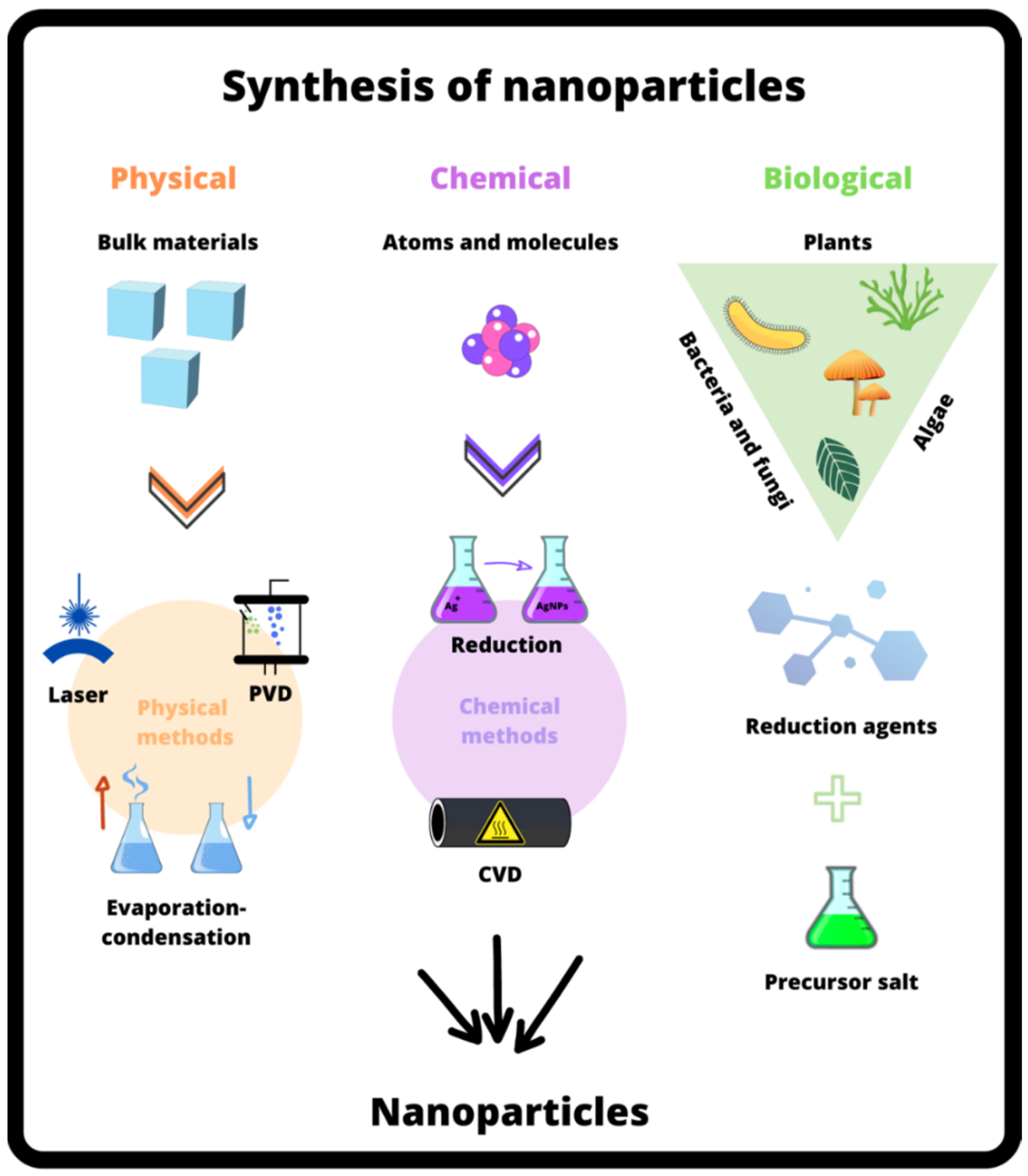
3.5. Synthesis of NPs

The properties of NPs depend on the synthesis method used; therefore, different methods of synthesis of antimicrobial NPs are used depending on the final applications. The synthesis methods can be grouped into biological or green, chemical, and physical methods (Figure 3) [78].
In general, physical methods have a high energy consumption and high costs, whereas chemical synthesis methods produce a great number of NPs in short periods of time; however, there are several studies that indicate that in the use of toxic chemicals, the generated by-products are a disadvantage. For this reason, “green synthesis” methods have received special attention in recent years [79].

3.5.1. Physical Approach

The technique of physical vapor deposition (PVD) magnetron sputtering can be utilized in the manufacture of metallic NPs. The process is environmentally friendly and takes place inside a vacuum chamber, avoiding the occurrence of contaminants in the coating. The deposition occurs through a metallic target, which will be bombarded by ions resulting from the plasma with an inert gas, such as argon. A momentum transfer resulting from this collision will lead to the ejection of atoms from the target, which will be deposited on the sample surface, forming the NPs [80]. In this regard, Garcia et al. [81] demonstrated how AgNPs synthesized by sputtering led a latex substrate which demonstrated bacteriostatic activity against Staphylococcus aureus to be employed in the production of dressings for people with a healing deficiency, such as diabetics. These authors concluded that NPs at a concentration of 2.87 μg cm−2 did not show toxicity to human cells and presented a hydrophilic character to the polymeric matrix, a factor that can prevent bacterial growth [81].
Another widely used physical method is evaporation-condensation, which takes place in a tube furnace and allows the formation of NPs of varying sizes [82]. Inside the tube, there is the source of the metal to be synthesized, which is vaporized into the carrier gas [83]. However, this technique has some disadvantages, such as high energy consumption, a large occupied space, and the time it takes to acquire thermal stabilization [84].
A more modern method uses laser ablation to produce NPs by removing them from a solid target in a medium [82]. The control of the characteristics of the NPs depends on the period of radiation, laser fluency, wavelength, the nature of the material to be synthesized, and the medium used. Since there is no use of chemical reagents, the NPs produced by this technique are considered pure and/or without contamination [83]. The disadvantage is when there is the formation of many NPs in the medium, which will prevent the laser from passing through and will absorb its energy, instead of being absorbed by the target [85].
Metal oxide NPs have also been synthesized using distinct physical methods. Gunnarsson et al. [86] obtained TiO2NPs by hollow sputtering in an argon/oxygen atmosphere using high-power pulsation. In turn, Dreesen et al. [87] synthesized TiO2NPs with a high surface coverage using magnetron reactive direct current (DC) sputtering. This same technique was also employed by Kwoka et al. [88] to obtain ZnO nanostructures using a zinc target at an 80 W DC power.

3.5.2. Chemical Approach

One of the chemical methods for the generation of metallic and metal oxide NPs is the reduction of metallic salts by using a stabilizing agent to prevent their aggregation. The characteristics of the NPs generated depend on the type of stabilizer and reducing agent. Some of the chemical agents used in this process are hydrazine, amino-boranes, polyols, hydrochloric acid, oleylamine, citrate, and sodium borohydride (NaBH4) [77,83]. However, this technique has some restrictions, such as the toxicity of the reducing agent, its high cost, and the presence of impurities [85]. The Turkevich protocol, introduced in 1951, is the most popular chemical method, and it uses sodium citrate as a stabilizing and reducing agent [89].
In addition to the reduction method, another chemical approach technique is chemical vapor deposition (CVD), where the target material chemically reacts with gaseous molecules, leading to their release in the form of volatile molecules. The surface morphology is well controlled, but the precursor gases can be toxic, corrosive, and explosive [85]. Additionally, this method allows a satisfactory dispersion of NPs over the substrate, on both flat and 3D samples (as nanotubes) [90].
In this perspective, Danna et al. [91] projected research in which AgNPs were added into the NRL matrix. The purpose of the research was to create a biocompatible composite. To produce this material, NRL membranes were first generated by the casting method. Afterward, the films were submerged in silver nitrate (AgNO3) at a concentration of 3.0 × 10⁻⁵ M, which was kept in a sand bath at 80 °C for varying times. The scanning electron microscopy (SEM) analysis exhibited that NPs were dispersed through the polymeric matrix, and the existence of AgNPs was effectively confirmed by plasmon absorption and elemental analysis. The cell viability assay proved that the AgNPs caused no toxicity to CHO-K1 cells that were in contact with the extracts obtained from the samples for 24 h. With the increase in the reduction time, the intensity of the peak on the UV-Vis analysis was proportional to the AgNPs concentration and also likely to increase the size of the AgNPs in the polymer matrix.
In order to develop an antibacterial material capable of support in guided bone regeneration, Marques et al. [92] combined the properties of NRL and AgNPs in a composite. To produce the NPs, a chemical reduction method was used with AgNO3 (2.0 mM) and NaBH4 (4.0 mM). The solution was stirred for 12 h and was later added to the latex, reaching the final concentration of AgNPs of 0.4%. The final solution was located in a Petri dish (casting method) at 40 °C until it reached complete polymerization. The assays were promising, showing that the AgNPs were not cytotoxic to CDLH1 lineage cells and that they aided in a faster calcification, as seen in in vivo tests with Wistar rats.
Arsalani et al. [77] synthesized magnetic Fe3O4NPs using a co-precipitation method at 90 °C. The process was described as being fast, economical, and environmentally friendly. The NPs were synthesized from ferrous chloride tetrahydrate (FeCl2·4H2O) and ferric chloride hexahydrate (FeCl3·6H2O), dissolved in aqueous solution of hydrochloric acid (5.45 M) and added, under stirring, to a solution of ammonium hydroxide previously heated to 90 °C for 10 min. Afterwards, the solution was kept in an ultrasonic bath for 1 h and then separated by magnetic precipitation, rinsed with Milli-Q water, and dried in an oven at 32 °C. Fe3O4NPs of a spherical shape with a size of 12 nm were obtained. The results demonstrated that the NRL stabilized the magnetic NPs, and these are considered useful for biomedical applications.

3.5.3. Biological Approach

Natural products play an important role in the formation and transformation of substances at the nanoscale. These natural processes can be used to minimize the environmental impact of other methods, so biological techniques such as green synthesis have been increasingly used for the synthesis of metallic and metal oxide NPs. In this method of synthesis, chemicals derived from living organisms are used in place of the reducing agent and stabilizer that were previously used in the chemical approach, such as fungi, bacteria, algae, and plants [82]. This method is cost-effective, can be easily reproduced on a large scale, and is eco-friendly. Furthermore, there is no use of high pressure, temperature, energy, and toxic chemicals [85].
Plant extracts can be used as reducing agents and, as they are abundant in nature, they have the lowest cost. Several parts of plants can be applied as substrates, such as leaves, fruits, seeds, and roots. Plants that have been studied in the synthesis of NPs include magnolia, plane tree, banana, and peppermint [82]. NPs that can be produced with this technology are gold, silver, copper, and zinc [85]. Green synthesis using plants is considered to be of a great advantage for antimicrobial methods as they have an extensive spectrum of biological activity because of the presence of phytocompounds. In addition, they present a great cost benefit, quick synthesis, and also good stability [52].
The main plant compounds used are phenolic acids, flavonoids, terpenoids, and alkaloids, which have the function of reducing the metallic ion and leading to the formation of metallic and metal oxide NPs. These primary and secondary metabolites are involved in redox reactions, so they are used as reducing agents. Prior to NPs synthesis, the bio-reducing agent must be purified and then placed in contact with the aqueous solution containing the precursor metal. Spontaneous reactions will take place at room temperature, producing NPs [85]. In addition to plants, fungi can be utilized to synthesize metallic and metal oxide NPs, such as Humicula sp., Cryphonectria sp., Verticillium, Aspergillus flavus, and Fusarium oxysporum [82,93]. The enzymes and proteins of these fungi are the compounds used as reducing agents.
For some authors, the “green” syntheses of NPs mean those that use organic molecules, which interact with the particles and grant them stability against agglomeration and oxidation. In this scenario, polymeric molecules have been used due to the fact that their long chains offer many binding sites that can stabilize NPs. Among these biomolecules, the natural latex of Hevea brasiliensis, a tree native to the Amazon rainforest, stands out. The research developed by Guidelli et al. [94], for example, introduced the green synthesis of colloidal AgNPs using Hevea brasiliensis latex. The technique in question was simple, cheap, and ecologically correct. The resulting composite can be applied as a wound dressing that will aid the healing process due to latex angiogenesis and the antibacterial activity of AgNPs. In this work, a solution of latex and Milli-Q™ water was prepared, and later, AgNO3 was added. The final solution was heated at 100 °C for 1 h and the results showed that there was a greater formation of AgNPs as the amount of NRL and AgNO3 increased, and that the particle size was also dependent on these concentrations. The Fourier transform infrared spectroscopy results indicate that the amine groups present in ammonia were responsible for the Ag(I) reduction, and that the poly(cis-1,4-isoprene) molecules of the NRL act as a capping agent that prevents the agglomeration of NPs. According to the research, certain latex proteins are essential for the development and passivation of NPs. In this case, the carbonyl groups from the amino acid residues and the peptides of proteins have a robust affinity for metals, thus avoiding their agglomeration.
Using a similar procedure, Bakar et al. [95] synthesized AgNPs in the NRL matrix. For this, NRL was added to deionized water and 2.94 × 10⁻⁵ mol of AgNO3. The resulting mixture contained about 0.03% (v/v) silver based on dry rubber and was placed in an oven at 50 °C to complete the polymerization process. The results suggested that the proteins present in NRL play an important role in the stabilization and growth of NPs. The interface regions between rubber particles, where protein mixing occurs, contained AgNPs in significant concentrations.
In another study, Rathnayake et al. [96] synthesized AgNPs using green synthesis, where the NPs were reduced in situ by NRL. The NPs were reduced from AgNO3 (0.1 M) added directly to centrifuged latex, being mixed for 8 h at 60 °C. Next, the reaction was brought to room temperature and stirred slowly for another 24 h. Then, the mixture was stored in a closed amber glass and the modified NRLF with AgNPs was produced, which was yellowish in color. This method produced NPs smaller than 100 nm. AgNPs were reduced by substances present in the serum fraction of the centrifuged NRL, which have not been described in the present study, but it is believed that the proteins present in the NRL are of great importance for silver reduction. Moreover, the NRLF modified with AgNPs was tested for Escherichia coli, Staphylococcus epidermidis, and Staphylococcus aureus, obstructing bacterial growth [96].
In the studies conducted by Phinyocheep et al. [97], AgNPs were synthesized from AgNO3 by incorporating it into NRL, without the use of a reducing agent. AgNO3 was added to the NRL and kept at room temperature. After that, the mixture was heated to 60 °C and stirred for 8 h. With this green method, spherical-shaped NPs of sizes between 5 and 30 nm were obtained. In this work, the authors also concluded that the existing proteins in the NRL were responsible for the reduction, in situ, of silver. AgNPs were responsible for the antibacterial effect on the compound, which was tested for Escherichia coli and Staphylococcus aureus [97].
Another study performed by Guidelli et al. [98] presented the chemical reduction of AgNPs added to NRL to release the silver present in the dressings. The NPs were synthesized from AgNO3 using NaBH4. The reaction was stirred for 12 h and then added to NRL, which was dried at 40 °C. Spherical-shaped NPs were obtained with a size of 30 nm. The results demonstrated that NRL membranes provide an effective matrix for AgNPs release, promoting angiogenesis and providing the dressing with antimicrobial characteristics [98].
In the same work previously reported by Arsalani et al. [77], Fe3O4NPs were obtained using the green method. For this, different volumes of NRL (100 to 800 μL) were added to the aqueous ammonium hydroxide solution and stirred for 3 min. Then, a mixed solution of ferrous and ferric chloride was added to the NRL solutions, and the formation of a black precipitate was observed. The size distribution and average size of Fe3O4NPs was influenced by the amount of NRL (13 ± 2.8, 10.3 ± 2.2, and 7.9 ± 1.5 nm for 100 μL, 400 μL, and 800 μL of NRL, respectively). The authors concluded that this behavior occurs with an increase in the concentration of NRL, in which more NRL molecules were bound to the surface of the NPs, preventing agglomeration and decreasing their growth [77]. Table 1 summarizes the different routes of synthesis of metallic and metal oxide NPs reported in the literature.

3.6. Mechanism of Bactericidal Activity of NPs

There is still no consensus on the exact mechanism by which metallic and metal oxide NPs exert their antibacterial and cytotoxic functions. Among the possibilities to explain this phenomenon, the first one is related to the electrostatic attraction between the negatively charged microbial cells and the positive ions released by the NPs. The NPs form a complex with electron donors, such as oxygen, nitrogen, phosphorus, or sulfur, which are in the proteins present in the cell walls [99]. The ions will then bind to the cell surface, causing damage to the bacteria walls, inactivating proteins and enzymes. When incorporated by the cell, the ions will harm the mitochondria [100], leading to the formation of unwanted ROS and the interruption of the release of adenosine triphosphate (ATP). The ROS in question are, in turn, responsible for altering the DNA, and the ions may also be responsible for the malfunctioning of protein synthesis, denaturizing ribosomal cytoplasmic components [44]. It is necessary to emphasize that the characteristics of each classification of bacteria influence the activity of NPs. Gram-positive bacteria have a dense layer of peptidoglycan on their cell wall, whereas Gram-negative bacteria have a thin layer of peptidoglycan with an outer lipopolysaccharide membrane. For this reason, Gram-positive bacteria are more resistant to the bactericidal effect arising from NPs [99].
The second possibility to explain the antibacterial effect is related to the metallic and metal oxide NPs configuration, which are able to denature cell membranes by themselves. Furthermore, due to their reduced size, NPs have an ability to permeate the cell membrane and modify its arrangement. As a result of this activity, damage to organelles and cell lysis can occur [44]. NPs may also be involved in bacterial signal transduction, which is directly affected by the phosphorylation of protein substrates. Moreover, they can dephosphorylate tyrosine residues from peptide substrates. The interruption of transduction leads to cell apoptosis and to the end of the organism’s multiplication [101]. Despite representing a promising solution to the contamination by microorganisms and biofilm formation, metallic NPs have some disadvantages, which are outlined in Table 2.

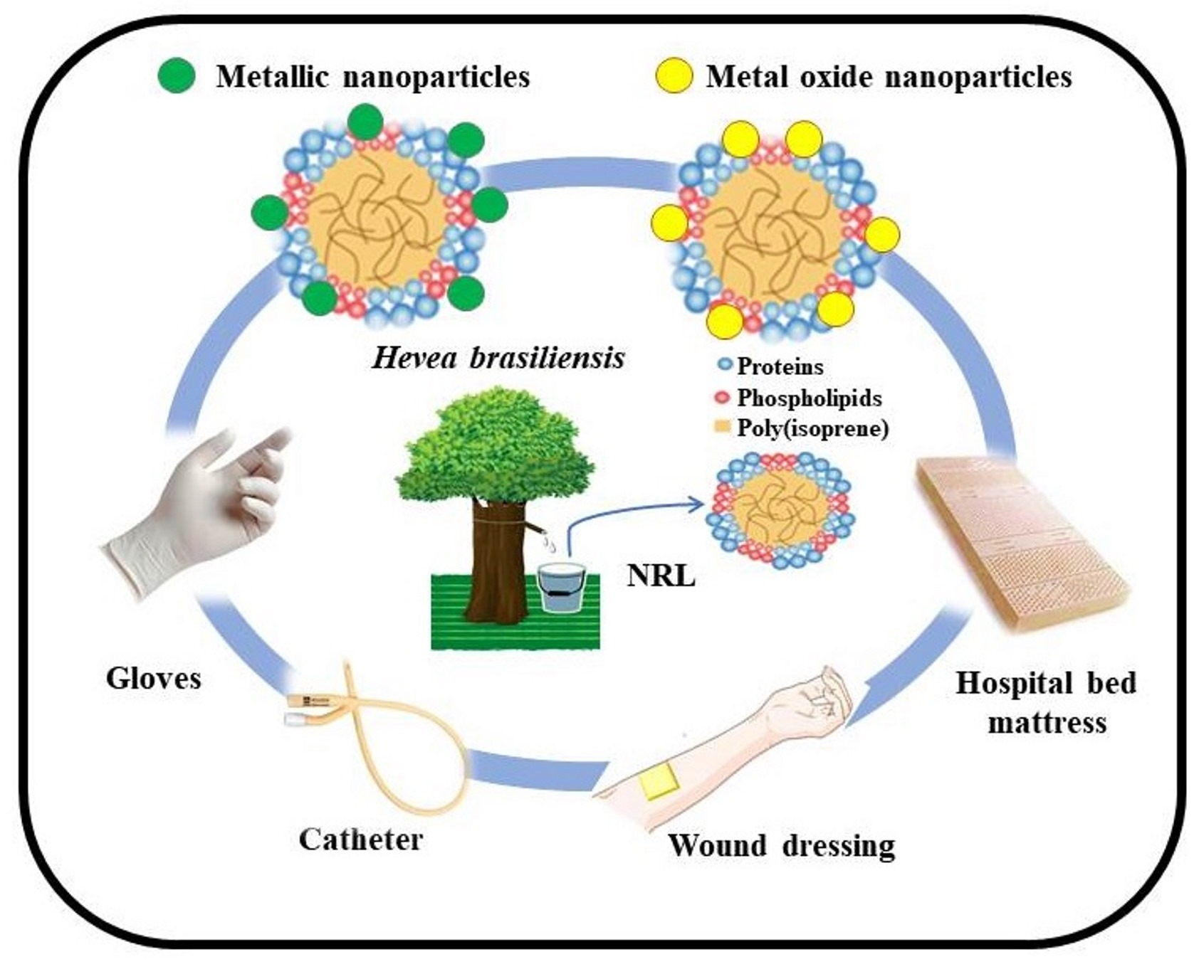
4. Natural Rubber-Based Materials Containing Metallic and Oxide NPs

Latex foams are often used in hospital materials, such as pillows and mattresses, which require antimicrobial properties [114]. NRL has several high-molecular-weight proteins that can cause allergies in humans. There are some studies that show that if latex is not deproteinized before its use, some proteins can persist in rubber products and attract bacteria that exist in the environment, or that it can be a problem in the future medical application of latex [115,116,117]. In this context, incorporating NPs with antimicrobial activity has become an option to try to solve this problem. In this section, we summarize and discuss some research studies involving natural rubber-based materials containing metallic and metal oxide NPs for biomedical applications (Table 3). Despite the wide variety of existing NPs, we note that AgNPs are currently the most used as fillers in NR matrices. We also observed that, depending on the purpose of the mixture, different strategies of mixtures between the NRL and the NPs are applied, such as mixing and agitation, the in situ synthesis of NPs in NRL without reducing agent, NRL membranes immersed in NPs solution, NPs deposited by the magnetron sputtering technique, etc.
Mam et al. [118] studied NRLF with an antibacterial property against Escherichia coli and Staphylococcus aureus by mechanically dispersing AgNPs in the polymeric matrix. Disks 6 mm in diameter and 1.5 mm in thickness, containing latex and latex with 0.2 parts per hundred (phr) of AgNPs, were placed in Petri dishes with a concentration of bacteria of 106 colony per milliliter (CFU/mL) and kept at 37 °C for 24 h. Samples without AgNPs showed a small diffusion halo caused by the presence of ZnONPs in the polymeric matrix, [119] whereas samples with 0.2 phr of AgNPs provided larger inhibitory zones. The authors concluded that only 0.2 phr of AgNPs increased the antibacterial capacity of NRLF by 43.8% against Escherichia coli and 25% against Staphylococcus aureus. The results also demonstrate that AgNPs effectively inhibited bacterial growth against Escherichia coli compared to Staphylococcus aureus, which was explained by the electrostatic attraction among the cell membranes of negatively charged bacteria and positively charged AgNPs [118].
Rathnayake et al. [96] synthesized AgNPs into the centrifuged NRL by the in situ reduction of AgNO3, without the addition of any reducing or stabilizing agent. The agar diffusion technique was used to estimate the antimicrobial activity against Staphylococcus epidermidis, Staphylococcus aureus, and Escherichia coli strains. Bacterial concentrations were quantified by the optical density (OD) at 600 nm. NRL samples with AgNPs showed an evident reduction in the microbial population. After 3 h, more than 95% of the OD of the bacterial culture medium was reduced, confirming strong antimicrobial activities. Therefore, the green method produced AgNPs incorporating NRL materials with an antimicrobial activity very similar to the NPs synthesized with trisodium citrate as the reducing agent. The authors concluded that the green approach method is an alternative new way to prepare AgNPs incorporated into the NRL matrix with extraordinary antimicrobial activities [96].
Silver nanocolloids obtained by the chemical reduction method of AgNO3 by trisodium citrate in an aqueous medium and subsequently incorporated into NRL foam were investigated by Rathnayake et al. [120]. Antibacterial activities were tested against Gram-positive and Gram-negative bacteria, while antifungal activities were tested against Aspergillus niger. They verified the rise in the antibacterial activity of the compounds that were absent in the NRL samples, as well as their antifungal properties against Aspergillus niger.
AgNPs synthesized from Magnolia kobus leaf extract were used to coat the outer zone of latex foam materials using two different methods: ultrasonic treatment and dip coating [114]. The resulting AgNP-coated foams using both methods were oven-dried for 24 h at 50 °C. The antibacterial properties were estimated by calculating viable Escherichia coli cells after 24 h of growth in shake flask cultures. Smaller AgNPs showed greater antibacterial activity due to their higher specific surface area. Ultrasonic treatment showed a greater adsorption and less desorption of AgNPs compared to dip coating, resulting in greater antibacterial activity [114].
Phinyocheep et al. [97] prepared composites of AgNPs and NRL with antimicrobial action. AgNPs were synthesized in situ in NRL, using AgNO3 without a reducing agent. Diffusion disk testing against Escherichia coli and Staphylococcus aureus bacteria showed a clear zone of inhibition in the composites, indicating that AgNPs are responsible for the antibacterial characteristic of the rubber latex mixture.
Suwatthanarak et al. [121] used only AgNO3 as a source of Ag(I) ions to synthesize AgNPs in skim NRL. They worked on the hypothesis that latex proteins might control particle growth, acting as a stabilizing agent. The antimicrobial activity of the composite against Staphylococcus aureus and Escherichia coli was analyzed using the antimicrobial disk sensitivity test. After 24 h, the composites showed that they can inhibit the growth of Escherichia coli and Staphylococcus aureus with inhibition zones of up to 8 mm [121].
Rathnayake et al. [1] modified the surface of the latex foam matrix by adsorption to nanometric AgNPs synthesized in situ. First, the NRL formulation was prepared by the Dunlop process, and different amounts of AgNO3 and trisodium citrate were added under stirring. Qualitative and quantitative determinations of antimicrobial activity were carried out on foam rubber pieces with and without AgNPs with a 20 × 20 mm format. As a result, they obtained inhibition zones approximately 5 mm thick around the foam rubber with AgNPs incorporated, indicating antimicrobial activity against Staphylococcus aureus and Escherichia coli. The bacteria population of Staphylococcus aureus was progressively reduced with the NRL time modified with AgNPs. A proposed bacterial inhibition mechanism by composites was designed [1].
Table 3. Reported methodologies of the preparation of materials based on NRL and metallic and metal oxide NPs.
Rubber TypeNPs TypeSynthesis MethodComposite ProductionComposite ApplicationReference
NRLFAgNPs colloid
(200,000 ppm)
Chemical methodMixing and stirring of the silver colloid with the NRLAntibacterial activity against E. coli and S. aureusMam et al. [118]
Centrifuged NRLAgNPsGreen synthesisSynthesized AgNPs into the centrifuged NRL in situAntimicrobial properties against E. coli, S. aureus, and
S. epidermidis
Rathnayake et al. [96]
NRLFSilver nanocolloidsChemical methodSilver nanocolloid incorporated into NRLF formulationAntibacterial and antifungal propertiesRathnayake et al. [120]
Latex foamsAgNPsBiological methods (Magnolia kobus leaf extract)Dip coating and ultrasonic treatmentAntibacterial activity against E. coliSong et al. [114]
NRLAgNPsGreen synthesisIn situ synthesis of AgNPs in NRL without reducing agentAntimicrobial properties against
E. coli and S. aureus
Phinyocheep et al. [97]
Skim NRL (with 0.05 wt% dry rubber content)AgNPsChemical methodSilver nitrate solution mixed with diluted skim NRLAntimicrobial properties against
E. coli and S. aureus
Suwatthanarak et al. [121]
NRLFAgNPsChemical methodAgNPs solution incorporated into the foam matrix by stirringAntibacterial activity against
S. aureus
Rathnayake et al. [1]
NRL concentratedAgNPsChemical method with microwave using PVPMixing and casting methodAntimicrobial properties against E. coli and S. aureusPrasanseang et al. [122]
NRLFAg-doped TiO2NPsChemical methodMixing and stirring of Ag-TiO2NPs with the NRLAntimicrobial activity against
S. epidermidis, methicillin-resistant S. aureus, and E. coli strains
Rathnayake et al. [123]
NRLAgNPsPhysical methodNRL-propolis membranes with AgNPs deposited by magnetron sputtering techniqueDressings with bactericide propertiesGarcia et al. [81]
NRLAgNPsChemical methodCasting methodDressing for the treatment of infectious processesMiranda et al. [124]
NRLAuNPsPurchased from
Sigma-Aldrich
NRL membranes immersed in the AuNPs solutionDressings for the treatment of the Leishmaniasis parasiteBarboza-Filho et al. [125]
NRLAuNPsChemical methodDifferent concentrations of NRL in water and mixed with HAuCl4 suspensionCell imaging and anticancer treatmentSantos et al. [126]
NRLFZnONPsPurchased from
Sigma-Aldrich
ZnONPs were incorporated into the NRL matrix and cured in oven for 2 h at 100 °CAntimicrobial activity against
S. aureus and E. coli
Rathnayake et al. [127]
NRZnONPs with CaCO3Supplied by Global Chemical Co., Ltd.Latex mixing techniqueDevelopment of gloves, condoms, and clothesKrainoi et al. [128]
In 2006, Noh [129] presented a patent on the production of an antibacterial latex foam containing AgNPs. The composite was made through a series of steps by mixing an aqueous suspension containing silver powder with a latex solution, and then aging the mixture to obtain antibacterial latex foam. In this invention, latex foam can be applied in various ways in products that are intended to prevent bacterial growth [129].
Concentrated natural rubber Ag-latex hybrid sheets (Ag-NRL) were prepared using an easy and economical method by Prasanseang et al. [122]. Initially, AgNPs were synthesized using microwaves and polyvinylpyrrolidone (PVP). Then, the Ag-NRL hybrid sheet samples were prepared by the mixing and casting method. The antibacterial properties of Ag-NRL sheets were tested by the agar disk diffusion method with Staphylococcus aureus and Escherichia coli. The results displayed that the hybrid sheets had optimal antibacterial properties against these bacteria, and the inhibition zones were dependent on the volume of AgNPs [122].
Metronidazole and AgNPs were mixed with NRL membranes and assessed as a controlled release system. The in vitro release tests showed that AgNPs increased the efficiency and specificity of metronidazole, reducing the side-effects, toxicity, and drug amount. The compounds released by the membranes did not cause hemolytic damage. These results indicated that the NRL membrane could be used as a dressing with the simultaneous release of drugs and drugs functionalized with AgNPs [124].
Rathnayake et al. [123] synthesized AgNPs-doped TiO2 nanopowder and later prepared composites with NRLF, which showed antimicrobial activity against Gram-positive bacteria species Staphylococcus epidermis, Gram-positive bacteria resistant to methicillin Staphylococcus aureus, and Gram-negative Escherichia coli.
AuNPs incorporated in NRL membranes were developed to be used as dressings in the treatment of lesions by avoiding the growth of germ in the wounds [125]. They observed that the increase in AuNPs decreases the promastigotes number in the culture medium. This behavior was accredited to the electrostatic interaction among the AuNPs and the germ due to the organic complexes formed on the surface of the NRL [125]. On the other hand, Garcia et al. [81] also developed NRL dressings enriched with red propolis, but with AgNPs. The cytotoxicity was assessed through 3-[4,5-dimethylthiazol-2-yl]-2,5-diphenyltetrazolium bromide (MTT) and agar diffusion assays in secondary and stem cells, indicating that the adipose-derived stem cell associated with NRL films can be considered promising for an application in the healing of occlusive wounds.
Santos et al. [126] synthesized colloidal AuNPs using latex from Hevea brasiliensis as a reducing–stabilizing agent and subsequently evaluated there in vitro genotoxicity and cytotoxicity. During the synthesis, experimental parameters such as the pH, temperature, time, and concentrations of NRL and gold salt were evaluated. The toxicological results indicated that for the 5.0 × 10⁹ AuNPs/mL, the cytotoxicity and genotoxicity were minimal, suggesting biocompatibility for normal cells [126].
As discussed previously, ZnONPs are very prominent due to their antimicrobial properties. In this sense, ZnONPs were added as a crystallizing agent to the NRL, while a control sample was completed without the addition of ZnO particles. Quantitatively, the determination of the antimicrobial activity was performed according to the method of Miles and Misra [127]. From the results obtained, the authors concluded that ZnONPs were responsible for the antimicrobial activity of NRLF against Staphylococcus aureus and Escherichia coli. It was proved that the antibacterial activities of NRLF with ZnNPs were higher than with microsized ZnO [127]. Krainoi et al. [128] prepared NR films filled with ZnONPs previously coated with calcium carbonate (CaCO3) in proportions of 90:10 and 60:40. The use of modified ZnONPs provided the effective killing of Escherichia coli and Staphylococcus aureus bacteria, through the ROS formation and Zn(II) ions that were transferred through the NR molecules through electrostatic forces.
Metallic and metal oxide NPs have shown satisfactory results when impregnated in NRL matrices. In all the works evaluated here, it could be seen that the addition of NPs provided a substantial improvement in the antimicrobial activity of this matrix. However, when the mixtures were carried out by the in situ synthesis of NPs in NRL (green synthesis), we noticed that the composites presented more attractive antimicrobial activities. Considering that NRL is the most used raw material for the manufacture of products in the health sector such as gloves, clothing, and condoms, among others, the results presented here are promising for the improvement of the rubber industry to meet the existing high demand for materials with active biological properties.

5. Conclusions

The use of NRL incorporated with NPs is a recurring theme, since more than 200 manuscripts have been published in the last decade, evidencing the scientific interest in relation to the advantages of the application of nanotechnologies. The research indicates that the intrinsic antimicrobial properties of NRL can be increased through the incorporation of metallic or metal oxide NPs, which makes this material an efficient and low-cost option for several biomedical applications.
The consulted literature focused on metallic NPs, AgNPs, CuNPs, and AuNPs, and metal oxides NPs: TiO2NPs, ZnONPs, and Fe3O4NPs. The choice of these NPs can be attributed to their efficiency and wide application. It is interesting to note that diverse physical, biological, and chemical methods for synthesizing metallic and metal oxides NPs were mentioned; however, green synthesis stood out and was shown to be a sustainable alternative since it does not generate by-products, uses nontoxic reagents, and requires less energy. However, until today, one of the main challenges is to overcome the agglomeration of NPs due to the interactions that exist between them.
Another relevant point observed was that the reduction of metallic salts, which is quite common in green synthesis, can occur in situ in the NRL matrix. The proteins present in the composition of NRL play a central role in the reduction stage, and the poly(cis-1,4-isoprene) molecules of NRL behave as coating agents, preventing the aggregation of NPs. In this sense, it was possible to develop a new field of application for NRL, representing an enormous potential to be used on an industrial scale in the near future.
In general, studies dealing with biomedical applications of NRL matrices and NPs are still scarce, and challenges in processing and dispersion are strongly needed. Among all the works evaluated, the incorporation of AgNPs in NRL matrices for biomedical applications has been expanded. However, it is interesting to note that there is a deficiency in publications using other metallic, as well as metal oxide NPs that also exhibit excellent antimicrobial properties. Finally, we observed a lack of consensus regarding the stages of the antibacterial activity of NPs. Due to the considerable mobility of NPs in a biological environment, this is a critical issue to address before the final applications of the materials. Finally, recent advances in biomedical applications of NRL and NPs have demonstrated the multidisciplinary required in future research that includes different NPs and increases the possibility of using these composites.

Author Contributions

Conceptualization, N.B.G., J.B., M.G. and J.S.C.; methodology, N.B.G., J.B., M.G. and J.S.C.; writing–original draft preparation, N.B.G., J.B., A.R.B., A.E.D.M., D.R., M.R.-E., D.M.D., M.G. and J.S.C.; writing—review and editing, M.G. and J.S.C.; funding acquisition, J.S.C. All authors have read and agreed to the published version of the manuscript.

Funding

The authors would like to thank Coordenação de Aperfeiçoamento de Pessoal de Nível Superior (CAPES, Brazil)—Finance Code 001, and Conselho Nacional de Desenvolvimento Científico e Tecnológico (CNPq, Brazil) for their financial support.

Institutional Review Board Statement

Not applicable.

Informed Consent Statement

Not applicable.

Data Availability Statement

Data is contained within the article.

Conflicts of Interest

The authors declare no conflict of interest.

References

  1. Rathnayake, I.; Ismail, H.; Bin Azahari, B.; Darsanasiri, N.D.; Rajapakse, S. Synthesis and Characterization of Nano-Silver Incorporated Natural Rubber Latex Foam. Polym. Technol. Eng. 2012, 51, 605–611. [Google Scholar] [CrossRef]
  2. Fong, Y.C.; Khin, A.A.; Lim, C.S. Conceptual Review and the Production, Consumption and Price Models of the Natural Rubber Industry in Selected ASEAN Countries and World Market. Asian J. Econ. Model. 2018, 6, 403–418. [Google Scholar] [CrossRef] [Green Version]
  3. Suksup, R.; Sun, Y.; Sukatta, U.; Smitthipong, W. Foam rubber from centrifuged and creamed latex. J. Polym. Eng. 2019, 39, 336–342. [Google Scholar] [CrossRef]
  4. Bode, H.B.; Kerkhoff, K.; Jendrossek, D. Bacterial Degradation of Natural and Synthetic Rubber. Biomacromolecules 2001, 2, 295–303. [Google Scholar] [CrossRef]
  5. Yip, E.; Cacioli, P. The manufacture of gloves from natural rubber latex. J. Allergy Clin. Immunol. 2002, 110, S3–S14. [Google Scholar] [CrossRef]
  6. Khan, I.; Saeed, K.; Khan, I. Nanoparticles: Properties, applications and toxicities. Arab. J. Chem. 2019, 12, 908–931. [Google Scholar] [CrossRef]
  7. Sánchez-López, E.; Gomes, D.; Esteruelas, G.; Bonilla, L.; Lopez-Machado, A.L.; Galindo, R.; Cano, A.; Espina, M.; Ettcheto, M.; Camins, A.; et al. Metal-Based Nanoparticles as Antimicrobial Agents: An Overview. Nanomaterials 2020, 10, 292. [Google Scholar] [CrossRef] [PubMed] [Green Version]
  8. Garza-Cervantes, J.A.; Chávez-Reyes, A.; Castillo, E.C.; García-Rivas, G.; Ortega-Rivera, O.A.; Salinas, E.; Ortiz-Martínez, M.; Gómez-Flores, S.L.; Peña-Martínez, J.A.; Pepi-Molina, A.; et al. Synergistic Antimicrobial Effects of Silver/Transition-metal Combinatorial Treatments. Sci. Rep. 2017, 7, 903–920. [Google Scholar] [CrossRef] [Green Version]
  9. Prabhu, S.; Poulose, E.K. Silver nanoparticles: Mechanism of antimicrobial action, synthesis, medical applications, and toxicity effects. Int. Nano Lett. 2012, 2, 32–42. [Google Scholar] [CrossRef] [Green Version]
  10. Grigore, M.E.; Biscu, E.R.; Holban, A.M.; Gestal, M.C.; Grumezescu, A.M. Methods of Synthesis, Properties and Biomedical Applications of CuO Nanoparticles. Pharmaceuticals 2016, 9, 75. [Google Scholar] [CrossRef]
  11. Bankier, C.; Matharu, R.K.; Cheong, Y.K.; Ren, G.G.; Cloutman-Green, E.; Ciric, L. Synergistic Antibacterial Effects of Metallic Nanoparticle Combinations. Sci. Rep. 2019, 9, 16074–16082. [Google Scholar] [CrossRef] [PubMed] [Green Version]
  12. Eren, T.; Baysal, G.; Doğan, F. Biocidal Activity of Bone Cements Containing Curcumin and Pegylated Quaternary Polyethylenimine. J. Polym. Environ. 2020, 28, 2469–2480. [Google Scholar] [CrossRef]
  13. Wang, L.; Hu, C.; Shao, L. The antimicrobial activity of nanoparticles: Present situation and prospects for the future. Int. J. Nanomed. 2017, 12, 1227–1249. [Google Scholar] [CrossRef] [PubMed] [Green Version]
  14. Gurunathan, S.; Han, J.W.; Dayem, A.A.; Eppakayala, V.; Kim, J.-H. Oxidative stress-mediated antibacterial activity of graphene oxide and reduced graphene oxide in Pseudomonas aeruginosa. Int. J. Nanomed. 2012, 7, 5901–5914. [Google Scholar] [CrossRef] [Green Version]
  15. Nagy, A.; Harrison, A.; Sabbani, S.; Munson, R.S., Jr.; Dutta, P.K.; Waldman, W.J. Silver nanoparticles embedded in zeolite membranes: Release of silver ions and mechanism of antibacterial action. Int. J. Nanomed. 2011, 6, 1833–1852. [Google Scholar] [CrossRef] [Green Version]
  16. Leung, Y.H.; Ng, A.M.C.; Xu, X.; Shen, Z.; Gethings, L.A.; Wong, M.T.; Chan, C.M.N.; Guo, M.Y.; Ng, Y.H.; Djurišić, A.B.; et al. Mechanisms of Antibacterial Activity of MgO: Non-ROS Mediated Toxicity of MgO Nanoparticles towards Escherichia coli. Small 2013, 10, 1171–1183. [Google Scholar] [CrossRef]
  17. Berthelot, K.; Peruch, F.; Lecomte, S. Highlights on Hevea brasiliensis (pro)hevein proteins. Biochimie 2016, 127, 258–270. [Google Scholar] [CrossRef]
  18. Bottier, C. Biochemical Composition of Hevea Brasiliensis Latex: A Focus on the Protein, Lipid, Carbohydrate and Mineral Contents. In Advances in Botanical Research; Elsevier: Amsterdam, The Netherlands, 2020; pp. 201–237. [Google Scholar] [CrossRef]
  19. Kurian, T.; Mathew, N.M. Natural Rubber: Production, Properties and Applications. In Biopolymers; John Wiley & Sons, Inc.: Hoboken, NJ, USA, 2011; pp. 403–436. [Google Scholar] [CrossRef]
  20. Pinto-Salamanca, C.-E.; Rigail-Cedeño, A.F.; Oliveros, M.E.M. Synthesis and characterization of natural rubber/clay nanocomposite to develop electrical safety gloves. Mater. Today Proc. 2020, 33, 1949–1953. [Google Scholar] [CrossRef]
  21. Miranda, M.C.R.; Borges, F.A.; Barros, N.R.; Filho, N.A.S.; Mendonça, R.J.; Herculano, R.D.; Cilli, E.M. Evaluation of peptides release using a natural rubber latex biomembrane as a carrier. Amino Acids 2018, 50, 503–511. [Google Scholar] [CrossRef] [Green Version]
  22. Kanokwiroon, K.; Teanpaisan, R.; Wititsuwannakul, D.; Hooper, A.B.; Wititsuwannakul, R. Antimicrobial activity of a protein purified from the latex of Hevea brasiliensis on oral microorganisms. Mycoses 2008, 51, 301–307. [Google Scholar] [CrossRef]
  23. Martin, M.N. The Latex of Hevea brasiliensis Contains High Levels of Both Chitinases and Chitinases/Lysozymes. Plant Physiol. 1991, 95, 469–476. [Google Scholar] [CrossRef] [PubMed] [Green Version]
  24. Joseph, J.E.; Krishnaswamy, V.G. Isolation of natural rubber latex degrading bacterial consortium from rubber plantation area. Eur. J. Exp. Biol. 2020, 10, 1–9. [Google Scholar]
  25. Button, D.W. Building a natural rubber latex compound. J. Chem. Educ. 1957, 34, 255–260. [Google Scholar] [CrossRef]
  26. Nair, K.P. Tree Crops; Springer International Publishing: Cham, Switzerland, 2021. [Google Scholar] [CrossRef]
  27. Semegen, S.T. Rubber, Natural. In Encyclopedia of Physical Science and Technology, 3rd ed.; Elsevier: Amsterdam, The Netherlands, 2003; pp. 381–394. [Google Scholar] [CrossRef]
  28. Nascimento, R.M.D.; de Paula, A.J.; Oliveira, N.C.; Alves, A.C.; Aquino, Y.M.L.D.O.; Filho, A.G.S.; Rodrigues, J.E.F.S.; Hernandes, A.C. Towards the production of natural rubber-calcium phosphate hybrid for applications as bioactive coatings. Mater. Sci. Eng. C 2019, 94, 417–425. [Google Scholar] [CrossRef]
  29. Tanaka, Y. Recent Advances in Structural Characterization of Elastomers. Rubber Chem. Technol. 1991, 64, 325–385. [Google Scholar] [CrossRef]
  30. Guerra, N.B.; Pegorin, G.S.; Boratto, M.H.; de Barros, N.R.; Graeff, C.F.D.O.; Herculano, R.D. Biomedical applications of natural rubber latex from the rubber tree Hevea brasiliensis. Mater. Sci. Eng. C 2021, 126, 112126–112144. [Google Scholar] [CrossRef]
  31. Herculano, R.D.; Silva, C.P.; Ereno, C.; Guimaraes, S.A.C.; Kinoshita, A.; de Oliveira Graeff Graeff, C.F. Natural rubber latex used as drug delivery system in guided bone regeneration (GBR). Mater. Res. 2009, 12, 253–256. [Google Scholar] [CrossRef] [Green Version]
  32. Herculano, R.D.; Tzu, L.C.; Silva, C.P.; Brunello, C.A.; de Queiroz, A.A.; Kinoshita, A.; Graeff, C.F.D.O. Nitric oxide release using natural rubber latex as matrix. Mater. Res. 2011, 14, 355–359. [Google Scholar] [CrossRef] [Green Version]
  33. de Barros, N.R.; Miranda, M.C.R.; Borges, F.A.; de Mendonça, R.J.; Cilli, E.M.; Herculano, R.D. Oxytocin Sustained Release Using Natural Rubber Latex Membranes. Int. J. Pept. Res. Ther. 2016, 22, 435–444. [Google Scholar] [CrossRef] [Green Version]
  34. Ferreira, M.; Mendonca, R.; Coutinho-Netto, J.; Mulato, M. Angiogenic properties of natural rubber latex biomembranes and the serum fraction of Hevea brasiliensis. Braz. J. Phys. 2009, 39, 564–569. [Google Scholar] [CrossRef]
  35. Brüning, K. Natural Rubber. In Encyclopedia of Polymeric Nanomaterials; Springer: Berlin/Heidelberg, Germany, 2015; pp. 1377–1382. [Google Scholar] [CrossRef]
  36. Giordani, R.; Gachon, C.; Buc, J.; Regli, P.; Jacob, J.L. Antifungal action of Hevea brasiliensis latex. Its effect in combination with fluconazole on Candida albicans growth. Mycoses 1999, 42, 465–474. [Google Scholar] [CrossRef]
  37. Boonrasri, S.; Sae–Oui, P.; Rachtanapun, P. Chitosan and Natural Rubber Latex Biocomposite Prepared by Incorporating Negatively Charged Chitosan Dispersion. Molecules 2020, 25, 2777. [Google Scholar] [CrossRef] [PubMed]
  38. Arakkal, A.; Aazem, I.; Honey, G.; Vengellur, A.; Bhat, S.G.; Sailaja, G.C.S. Antibacterial Polyelectrolytic chitosan derivatives conjugated natural rubber latex films with minimized bacterial adhesion. J. Appl. Polym. Sci. 2020, 138, 49608. [Google Scholar] [CrossRef]
  39. Van Parijs, J.; Broekaert, W.F.; Goldstein, I.J.; Peumans, W.J. Hevein: An antifungal protein from rubber-tree (Hevea brasiliensis) latex. Planta 1991, 183, 258–264. [Google Scholar] [CrossRef] [PubMed] [Green Version]
  40. Churngchow, N.; Suntaro, A.; Witttsuwannnakul, R. β-1,3-glucanase isozymes from the latex of Hevea brasiliensis. Phytochemistry 1995, 39, 505–509. [Google Scholar] [CrossRef]
  41. A Daruliza, K.M.; Lam, K.L.; Yang, K.L.; Priscilla, J.T.; Sunderasan, E.; Ong, M.T. Anti-fungal effect of Hevea brasiliensis latex C-serum on Aspergillus niger. Eur. Rev. Med. Pharmacol. Sci. 2011, 15, 1027–1033. [Google Scholar]
  42. Singh, A.; Gautam, P.K.; Verma, A.; Singh, V.; Shivapriya, P.M.; Shivalkar, S.; Sahoo, A.K.; Samanta, S.K. Green synthesis of metallic nanoparticles as effective alternatives to treat antibiotics resistant bacterial infections: A review. Biotechnol. Rep. 2020, 25, e00427–e00440. [Google Scholar] [CrossRef]
  43. Wilson, D.N.; Hauryliuk, V.; Atkinson, G.C.; O’Neill, A.J. Target protection as a key antibiotic resistance mechanism. Nat. Rev. Microbiol. 2020, 18, 637–648. [Google Scholar] [CrossRef]
  44. Ahmad, S.A.; Das, S.S.; Khatoon, A.; Ansari, M.T.; Afzal, M.; Hasnain, S.; Nayak, A.K. Bactericidal activity of silver nanoparticles: A mechanistic review. Mater. Sci. Energy Technol. 2020, 3, 756–769. [Google Scholar] [CrossRef]
  45. Medina, J.; Garcia-Perez, V.I.; Zanella, R. Metallic composites based on Ag, Cu, Au and Ag-Cu nanoparticles with distinctive bactericidal effect on varied species. Mater. Today Commun. 2021, 26, 102182–102192. [Google Scholar] [CrossRef]
  46. Liao, C.; Li, Y.; Tjong, S.C. Bactericidal and Cytotoxic Properties of Silver Nanoparticles. Int. J. Mol. Sci. 2019, 20, 449. [Google Scholar] [CrossRef] [PubMed] [Green Version]
  47. Siddiqi, K.S.; Husen, A.; Rao, R.A.K. A review on biosynthesis of silver nanoparticles and their biocidal properties. J. Nanobiotechnol. 2018, 16, 14. [Google Scholar] [CrossRef] [PubMed]
  48. Islam, A.; Jacob, M.V.; Antunes, E. A critical review on silver nanoparticles: From synthesis and applications to its mitigation through low-cost adsorption by biochar. J. Environ. Manag. 2021, 281, 111918. [Google Scholar] [CrossRef] [PubMed]
  49. Fernando, S.; Gunasekara, T.; Holton, J. Antimicrobial Nanoparticles: Applications and mechanisms of action. Sri Lankan J. Infect. Dis. 2018, 8, 2–11. [Google Scholar] [CrossRef]
  50. Yaqub, A.; Malkani, N.; Shabbir, A.; Ditta, S.A.; Tanvir, F.; Ali, S.; Naz, M.; Kazmi, S.A.R.; Ullah, R. Novel Biosynthesis of Copper Nanoparticles Using Zingiber and Allium sp. with Synergic Effect of Doxycycline for Anticancer and Bactericidal Activity. Curr. Microbiol. 2020, 77, 2287–2299. [Google Scholar] [CrossRef] [PubMed]
  51. Khademi-Azandehi, P.; Moghaddam, J. Green synthesis, characterization and physiological stability of gold nanoparticles from Stachys lavandulifolia Vahl extract. Particuology 2015, 19, 22–26. [Google Scholar] [CrossRef]
  52. Vijayakumar, S.; Vaseeharan, B.; Malaikozhundan, B.; Gopi, N.; Ekambaram, P.; Pachaiappan, R.; Velusamy, P.; Murugan, K.; Benelli, G.; Kumar, R.S.; et al. Therapeutic effects of gold nanoparticles synthesized using Musa paradisiaca peel extract against multiple antibiotic resistant Enterococcus faecalis biofilms and human lung cancer cells (A549). Microb. Pathog. 2017, 102, 173–183. [Google Scholar] [CrossRef]
  53. Gopinath, K.; Kumaraguru, S.; Bhakyaraj, K.; Mohan, S.; Venkatesh, K.S.; Esakkirajan, M.; Kaleeswarran, P.; Alharbi, N.S.; Kadaikunnan, S.; Govindarajan, M.; et al. Green synthesis of silver, gold and silver/gold bimetallic nanoparticles using the Gloriosa superba leaf extract and their antibacterial and antibiofilm activities. Microb. Pathog. 2016, 101, 1–11. [Google Scholar] [CrossRef]
  54. Guerra, R.; Lima, E.; Guzmán, A. Antimicrobial supported nanoparticles: Gold versus silver for the cases of Escherichia coli and Salmonella typhi. Microporous Mesoporous Mater. 2012, 170, 62–66. [Google Scholar] [CrossRef]
  55. Katas, H.; Lim, C.S.; Azlan, A.Y.H.N.; Buang, F.; Busra, M.F.M. Antibacterial activity of biosynthesized gold nanoparticles using biomolecules from Lignosus rhinocerotis and chitosan. Saudi Pharm. J. 2019, 27, 283–292. [Google Scholar] [CrossRef]
  56. Khan, F.U.; Chen, Y.; Ahmad, A.; Tahir, K.; Khan, Z.U.; Khan, A.U.; Khan, S.U.; Raza, M.; Wan, P. Visible light inactivation of E. coli, Cytotoxicity and ROS determination of biochemically capped gold nanoparticles. Microb. Pathog. 2017, 107, 419–424. [Google Scholar] [CrossRef]
  57. Hussein, M.A.M.; Grinholc, M.; Dena, A.S.A.; El-Sherbiny, I.M.; Megahed, M. Boosting the antibacterial activity of chitosan–gold nanoparticles against antibiotic–resistant bacteria by Punicagranatum L. extract. Carbohydr. Polym. 2021, 256, 117498. [Google Scholar] [CrossRef] [PubMed]
  58. Xie, Y.; Yang, J.; Zhang, J.; Zheng, W.; Jiang, X. Activating the Antibacterial Effect of 4,6-Diamino-2-pyrimidinethiol-Modified Gold Nanoparticles by Reducing their Sizes. Angew. Chem. Int. Ed. 2020, 59, 23471–23475. [Google Scholar] [CrossRef] [PubMed]
  59. Yang, X.; Yang, J.; Wang, L.; Ran, B.; Jia, Y.; Zhang, L.; Yang, G.; Shao, H.; Jiang, X. Pharmaceutical Intermediate-Modified Gold Nanoparticles: Against Multidrug-Resistant Bacteria and Wound-Healing Application via an Electrospun Scaffold. ACS Nano 2017, 11, 5737–5745. [Google Scholar] [CrossRef]
  60. Krishnan, B.; Mahalingam, S. Improved surface morphology of silver/copper oxide/bentonite nanocomposite using aliphatic ammonium based ionic liquid for enhanced biological activities. J. Mol. Liq. 2017, 241, 1044–1058. [Google Scholar] [CrossRef]
  61. Nikolova, M.P.; Chavali, M.S. Metal Oxide Nanoparticles as Biomedical Materials. Biomimetics 2020, 5, 27. [Google Scholar] [CrossRef]
  62. Niño-Martínez, N.; Salas Orozco, M.F.; Martínez-Castañón, G.-A.; Torres Méndez, F.; Ruiz, F. Molecular Mechanisms of Bacterial Resistance to Metal and Metal Oxide Nanoparticles. Int. J. Mol. Sci. 2019, 20, 2808. [Google Scholar] [CrossRef] [Green Version]
  63. Manuja, A.; Kumar, B.; Kumar, R.; Chhabra, D.; Ghosh, M.; Manuja, M.; Brar, B.; Pal, Y.; Tripathi, B.; Prasad, M. Metal/metal oxide nanoparticles: Toxicity concerns associated with their physical state and remediation for biomedical applications. Toxicol. Rep. 2021, 8, 1970–1978. [Google Scholar] [CrossRef] [PubMed]
  64. Kovalishyn, V.; Abramenko, N.; Kopernyk, I.; Charochkina, L.; Metelytsia, L.; Tetko, I.V.; Peijnenburg, W.; Kustov, L. Modelling the toxicity of a large set of metal and metal oxide nanoparticles using the OCHEM platform. Food Chem. Toxicol. 2018, 112, 507–517. [Google Scholar] [CrossRef] [Green Version]
  65. Winkler, D.A. Role of Artificial Intelligence and Machine Learning in Nanosafety. Small 2020, 16, 2001883–2001889. [Google Scholar] [CrossRef]
  66. Kalpana, V.N.; Rajeswari, V.D. A Review on Green Synthesis, Biomedical Applications, and Toxicity Studies of ZnO NPs. Bioinorg. Chem. Appl. 2018, 2018, 3569758. [Google Scholar] [CrossRef] [PubMed]
  67. Gudkov, S.V.; Burmistrov, D.E.; Serov, D.A.; Rebezov, M.B.; Semenova, A.A.; Lisitsyn, A.B. A Mini Review of Antibacterial Properties of ZnO Nanoparticles. Front. Phys. 2021, 9, 641481. [Google Scholar] [CrossRef]
  68. Sahoo, S.; Maiti, M.; Ganguly, A.; George, J.J.; Bhowmick, A.K. Effect of zinc oxide nanoparticles as cure activator on the properties of natural rubber and nitrile rubber. J. Appl. Polym. Sci. 2007, 105, 2407–2415. [Google Scholar] [CrossRef]
  69. Pushpalatha, C.; Suresh, J.; Gayathri, V.; Sowmya, S.; Augustine, D.; Alamoudi, A.; Zidane, B.; Albar, N.H.M.; Patil, S. Zinc Oxide Nanoparticles: A Review on Its Applications in Dentistry. Front. Bioeng. Biotechnol. 2022, 10, 917990. [Google Scholar] [CrossRef] [PubMed]
  70. Jiang, J.; Pi, J.; Cai, J. The Advancing of Zinc Oxide Nanoparticles for Biomedical Applications. Bioinorg. Chem. Appl. 2018, 2018, 1062562. [Google Scholar] [CrossRef] [PubMed]
  71. Çeşmeli, S.; Avci, C.B. Application of titanium dioxide (TiO2) nanoparticles in cancer therapies. J. Drug Target. 2019, 27, 762–766. [Google Scholar] [CrossRef]
  72. Ilyas, M.; Waris, A.; Khan, A.U.; Zamel, D.; Yar, L.; Baset, A.; Muhaymin, A.; Khan, S.; Ali, A.; Ahmad, A. Biological synthesis of titanium dioxide nanoparticles from plants and microorganisms and their potential biomedical applications. Inorg. Chem. Commun. 2021, 133, 108968. [Google Scholar] [CrossRef]
  73. Naseri, N.; Janfaza, S.; Irani, R. Visible light switchable bR/TiO 2 nanostructured photoanodes for bio-inspired solar energy conversion. RSC Adv. 2015, 5, 18642–18646. [Google Scholar] [CrossRef]
  74. De Filpo, G.; Palermo, A.M.; Rachiele, F.; Nicoletta, F.P. Preventing fungal growth in wood by titanium dioxide nanoparticles. Int. Biodeterior. Biodegrad. 2013, 85, 217–222. [Google Scholar] [CrossRef]
  75. Jafari, S.; Mahyad, B.; Hashemzadeh, H.; Janfaza, S.; Gholikhani, T.; Tayebi, L. Biomedical Applications of TiO2 Nanostructures: Recent Advances. Int. J. Nanomed. 2020, 15, 3447–3470. [Google Scholar] [CrossRef]
  76. Shen, L.; Li, B.; Qiao, Y. Fe3O4 Nanoparticles in Targeted Drug/Gene Delivery Systems. Materials 2018, 11, 324. [Google Scholar] [CrossRef] [PubMed] [Green Version]
  77. Arsalani, S.; Guidelli, E.J.; Araujo, J.; Bruno, A.C.; Baffa, O. Green Synthesis and Surface Modification of Iron Oxide Nanoparticles with Enhanced Magnetization Using Natural Rubber Latex. ACS Sustain. Chem. Eng. 2018, 6, 13756–13765. [Google Scholar] [CrossRef]
  78. Yaqoob, A.A.; Umar, K.; Ibrahim, M.N.M. Silver nanoparticles: Various methods of synthesis, size affecting factors and their potential applications—A review. Appl. Nanosci. 2020, 10, 1369–1378. [Google Scholar] [CrossRef]
  79. Correa, M.G.; Martínez, F.B.; Vidal, C.P.; Streitt, C.; Escrig, J.; de Dicastillo, C.L. Antimicrobial metal-based nanoparticles: A review on their synthesis, types and antimicrobial action. Beilstein J. Nanotechnol. 2020, 11, 1450–1469. [Google Scholar] [CrossRef]
  80. Giovanela, C.S.C.G.; Crespo, M.J.S.; Roesch-Ely, M.; Henriques, J.A.P.; Aguzzoli, C.; Maddalozzo, A.E.D. Filmes, Processos de Obtenção dos Filmes e Uso dos Filmes. Patent BR1020190107154 2019. [Google Scholar]
  81. Garcia, C.S.C.; Maddalozzo, A.E.D.; Garcia, P.M.C.; Fontoura, C.P.; Rodrigues, M.M.; Giovanela, M.; Henriques, J.A.P.; Aguzzoli, C.; Crespo, J.D.S.; Roesch-Ely, M. Natural Rubber Films Incorporated with Red Propolis and Silver Nanoparticles Aimed for Occlusive Dressing Application. Mater. Res. 2021, 24, 1–16. [Google Scholar] [CrossRef]
  82. Syafiuddin, A.; Salmiati; Salim, M.R.; Kueh, A.B.H.; Hadibarata, T.; Nur, H. A Review of Silver Nanoparticles: Research Trends, Global Consumption, Synthesis, Properties, and Future Challenges. J. Chin. Chem. Soc. 2017, 64, 732–756. [Google Scholar] [CrossRef]
  83. Chugh, H.; Sood, D.; Chandra, I.; Tomar, V.; Dhawan, G.; Chandra, R. Role of gold and silver nanoparticles in cancer nano-medicine. Artif. Cells Nanomed. Biotechnol. 2018, 46, 1210–1220. [Google Scholar] [CrossRef]
  84. Natsuki, J.; Natsuki, T.; Hashimoto, Y. A Review of Silver Nanoparticles: Synthesis Methods, Properties and Applications. Int. J. Mater. Sci. Appl. 2015, 4, 325. [Google Scholar] [CrossRef] [Green Version]
  85. Jamkhande, P.G.; Ghule, N.W.; Bamer, A.H.; Kalaskar, M.G. Metal nanoparticles synthesis: An overview on methods of preparation, advantages and disadvantages, and applications. J. Drug Deliv. Sci. Technol. 2019, 53, 101174. [Google Scholar] [CrossRef]
  86. Gunnarsson, R.; Helmersson, U.; Pilch, I. Synthesis of titanium-oxide nanoparticles with size and stoichiometry control. J. Nanopart. Res. 2015, 17, 353–358. [Google Scholar] [CrossRef] [Green Version]
  87. Dreesen, L.; Colomer, J.-F.; Limage, H.; Giguère, A.; Lucas, S. Synthesis of titanium dioxide nanoparticles by reactive DC magnetron sputtering. Thin Solid Films 2009, 518, 112–115. [Google Scholar] [CrossRef]
  88. Kwoka, M.; Lyson-Sypien, B.; Kulis, A.; Maslyk, M.; Borysiewicz, M.A.; Kaminska, E.; Szuber, J. Surface Properties of Nanostructured, Porous ZnO Thin Films Prepared by Direct Current Reactive Magnetron Sputtering. Materials 2018, 11, 131. [Google Scholar] [CrossRef] [PubMed] [Green Version]
  89. Shi, L.; Buhler, E.; Boué, F.; Carn, F. How does the size of gold nanoparticles depend on citrate to gold ratio in Turkevich synthesis? Final answer to a debated question. J. Colloid Interface Sci. 2017, 492, 191–198. [Google Scholar] [CrossRef] [PubMed]
  90. Piszczek, P.; Lewandowska, A.; Radtke, A.; Jędrzejewski, T.; Kozak, W.; Sadowska, B.; Szubka, M.; Talik, E.; Fiori, F. Biocompatibility of Titania Nanotube Coatings Enriched with Silver Nanograins by Chemical Vapor Deposition. Nanomaterials 2017, 7, 274. [Google Scholar] [CrossRef] [PubMed] [Green Version]
  91. Danna, C.S.; Cavalcante, D.G.S.M.; Gomes, A.S.; Kerche-Silva, L.E.; Yoshihara, E.; Osorio-Román, I.O.; Salmazo, L.O.; Rodríguez-Pérez, M.A.; Aroca, R.F.; Job, A.E. Silver Nanoparticles Embedded in Natural Rubber Films: Synthesis, Characterization, and Evaluation of In Vitro Toxicity. J. Nanomater. 2016, 2016, 2368630. [Google Scholar] [CrossRef] [Green Version]
  92. Marques, L.; Martinez, G.; Guidelli, J.; Tamashiro, J.; Segato, R.; Payão, S.L.M.; Baffa, O.; Kinoshita, A. Performance on Bone Regeneration of a Silver Nanoparticle Delivery System Based on Natural Rubber Membrane NRL-AgNP. Coatings 2020, 10, 323. [Google Scholar] [CrossRef] [Green Version]
  93. Singh, J.; Dutta, T.; Kim, K.-H.; Rawat, M.; Samddar, P.; Kumar, P. ‘Green’ synthesis of metals and their oxide nanoparticles: Applications for environmental remediation. J. Nanobiotechnol. 2018, 16, 84–89. [Google Scholar] [CrossRef]
  94. Guidelli, E.J.; Ramos, A.P.; Zaniquelli, M.E.D.; Baffa, O. Green synthesis of colloidal silver nanoparticles using natural rubber latex extracted from Hevea brasiliensis. Spectrochim. Acta Part A Mol. Biomol. Spectrosc. 2011, 82, 140–145. [Google Scholar] [CrossRef]
  95. Bakar, N.H.H.; Ismail, J.; Bakar, M. Synthesis and characterization of silver nanoparticles in natural rubber. Mater. Chem. Phys. 2007, 104, 276–283. [Google Scholar] [CrossRef]
  96. Rathnayake, I.; Ismail, H.; Azahari, B.; De Silva, C.; Darsanasiri, N. Imparting antimicrobial properties to natural rubber latex foam via green synthesized silver nanoparticles. J. Appl. Polym. Sci. 2013, 131, 1–10. [Google Scholar] [CrossRef]
  97. Phinyocheep, P. In-situ green synthesis of silver nanoparticles in natural rubber latex for fabricating rubber composite with antimicrobial property. Int. J. Sci. Innov. Technol. 2021, 4, 11–20. [Google Scholar]
  98. Guidelli, É.J.; Kinoshita, A.; Ramos, A.P.; Baffa, O. Silver nanoparticles delivery system based on natural rubber latex membranes. J. Nanopart. Res. 2013, 15, 1536. [Google Scholar] [CrossRef]
  99. Fanoro, O.T.; Oluwafemi, O.S. Bactericidal Antibacterial Mechanism of Plant Synthesized Silver, Gold and Bimetallic Nanoparticles. Pharmaceutics 2020, 12, 1044. [Google Scholar] [CrossRef] [PubMed]
  100. Khorrami, S.; Zarrabi, A.; Khaleghi, M.; Danaei, M.; Mozafari, M.R. Selective cytotoxicity of green synthesized silver nanoparticles against the MCF-7 tumor cell line and their enhanced antioxidant and antimicrobial properties. Int. J. Nanomed. 2018, 13, 8013–8024. [Google Scholar] [CrossRef] [PubMed] [Green Version]
  101. Yin, I.X.; Zhang, J.; Zhao, I.S.; Mei, M.L.; Li, Q.; Chu, C.H. The Antibacterial Mechanism of Silver Nanoparticles and Its Application in Dentistry. Int. J. Nanomed. 2020, 15, 2555–2562. [Google Scholar] [CrossRef] [PubMed] [Green Version]
  102. Souza, T.A.; Franchi, L.P.; Rosa, L.R.; Veiga, M.; Takahashi, C.S. Cytotoxicity and genotoxicity of silver nanoparticles of different sizes in CHO-K1 and CHO-XRS5 cell lines. Mutat. Res. Toxicol. Environ. Mutagen. 2016, 795, 70–83. [Google Scholar] [CrossRef] [PubMed]
  103. Tan, H.-L.; Teow, S.-Y.; Pushpamalar, J. Application of Metal Nanoparticle–Hydrogel Composites in Tissue Regeneration. Bioengineering 2019, 6, 17. [Google Scholar] [CrossRef] [PubMed] [Green Version]
  104. Jia, Y.-P.; Ma, B.-Y.; Wei, X.-W.; Qian, Z.-Y. The in vitro and in vivo toxicity of gold nanoparticles. Chin. Chem. Lett. 2017, 28, 691–702. [Google Scholar] [CrossRef]
  105. Tentor, F.R.; de Oliveira, J.H.; Scariot, D.B.; Lazarin-Bidóia, D.; Bonafé, E.G.; Nakamura, C.V.; Venter, S.A.; Monteiro, J.P.; Muniz, E.C.; Martins, A.F. Scaffolds based on chitosan/pectin thermosensitive hydrogels containing gold nanoparticles. Int. J. Biol. Macromol. 2017, 102, 1186–1194. [Google Scholar] [CrossRef]
  106. Boonkaew, B.; Kempf, M.; Kimble, R.; Cuttle, L. Cytotoxicity testing of silver-containing burn treatments using primary and immortal skin cells. Burns 2014, 40, 1562–1569. [Google Scholar] [CrossRef] [PubMed] [Green Version]
  107. Söderstjerna, E.; Bauer, P.; Cedervall, T.; Abdshill, H.; Johansson, F.; Johansson, U.E. Silver and Gold Nanoparticles Exposure to In Vitro Cultured Retina—Studies on Nanoparticle Internalization, Apoptosis, Oxidative Stress, Glial- and Microglial Activity. PLoS ONE 2014, 9, e105359. [Google Scholar] [CrossRef] [PubMed] [Green Version]
  108. Kostić, D.D.; Malagurski, I.S.; Obradović, B.M. Transport of silver nanoparticles from nanocomposite Ag/alginate hydrogels under conditions mimicking tissue implantation. Chem. Ind. 2017, 71, 383–394. [Google Scholar] [CrossRef]
  109. Gunawan, C.; Teoh, W.Y.; Marquis, C.P.; Amal, R. Induced Adaptation of Bacillus sp. to Antimicrobial Nanosilver. Small 2013, 9, 3554–3560. [Google Scholar] [CrossRef]
  110. Panáček, A.; Kvítek, L.; Smékalová, M.; Večeřová, R.; Kolář, M.; Röderová, M.; Dyčka, F.; Šebela, M.; Prucek, R.; Tomanec, O.; et al. Bacterial resistance to silver nanoparticles and how to overcome it. Nat. Nanotechnol. 2017, 13, 65–71. [Google Scholar] [CrossRef]
  111. Guo, Z.; Chen, Y.; Wang, Y.-H.; Jiang, H.; Wang, X. Advances and challenges in metallic nanomaterial synthesis and antibacterial applications. J. Mater. Chem. B 2020, 8, 4764–4777. [Google Scholar] [CrossRef]
  112. Avirdi, E.; Hooshmand, S.E.; Sepahvand, H.; Vishwanathan, V.; Bahadur, I.; Katata-Seru, L.M.; Varma, R.S. Ionic liquids-assisted greener preparation of silver nanoparticles. Curr. Opin. Green Sustain. Chem. 2021, 33, 100581–100593. [Google Scholar] [CrossRef]
  113. Długosz, O.; Szostak, K.; Staroń, A.; Pulit-Prociak, J.; Banach, M. Methods for Reducing the Toxicity of Metal and Metal Oxide NPs as Biomedicine. Materials 2020, 13, 279. [Google Scholar] [CrossRef] [Green Version]
  114. Song, J.Y.; Kwon, E.-Y.; Kim, B.S. Antibacterial latex foams coated with biologically synthesized silver nanoparticles using Magnolia kobus leaf extract. Korean J. Chem. Eng. 2012, 29, 1771–1775. [Google Scholar] [CrossRef]
  115. Kahn, S.L.; Podjasek, J.O.; Dimitropoulos, V.A.; Brown, C.W. Natural rubber latex allergy. Dis.-Mon. 2016, 62, 5–17. [Google Scholar] [CrossRef]
  116. Katrancha, E.D.; Harshberger, L.A. Nursing students with latex allergy. Nurse Educ. Pract. 2012, 12, 328–332. [Google Scholar] [CrossRef] [PubMed]
  117. Charous, B.; Tarlo, S.; Charous, M.; Kelly, K. Natural rubber latex allergy in the occupational setting. Methods 2002, 27, 15–21. [Google Scholar] [CrossRef] [PubMed]
  118. Mam, K.; Dangtungee, R. Effects of silver nanoparticles on physical and antibacterial properties of natural rubber latex foam. Mater. Today Proc. 2019, 17, 1914–1920. [Google Scholar] [CrossRef]
  119. Zhang, L.; Jiang, Y.; Ding, Y.; Povey, M.; York, D. Investigation into the antibacterial behaviour of suspensions of ZnO nanoparticles (ZnO nanofluids). J. Nanopart. Res. 2007, 9, 479–489. [Google Scholar] [CrossRef]
  120. Rathnayake, W.; Ismail, H.; Baharin, A.; Darsanasiri, A.; Rajapakse, S. Synthesis and characterization of nano silver based natural rubber latex foam for imparting antibacterial and anti-fungal properties. Polym. Test. 2012, 31, 586–592. [Google Scholar] [CrossRef]
  121. Suwatthanarak, T.; Than-Ardna, B.; Danwanichakul, D.; Danwanichakul, P. Synthesis of silver nanoparticles in skim natural rubber latex at room temperature. Mater. Lett. 2016, 168, 31–35. [Google Scholar] [CrossRef]
  122. Prasanseang, W.; Sriwong, C.; Choojun, K. Effect of Synthesized Ag Nanoparticles by Using the Different Amounts of Polyvinylpyrrolidone for Ag-Natural Rubber Hybrid Sheets and their Antibacterial Properties. Key Eng. Mater. 2017, 751, 270–276. [Google Scholar] [CrossRef]
  123. Rathnayake, I.U.; Ismail, H.; De Silva, C.R.; Darsanasiri, N.D.; Bose, I. Antibacterial effect of Ag-doped TiO2 nanoparticles incorporated natural rubber latex foam under visible light conditions. Iran. Polym. J. 2015, 24, 1057–1068. [Google Scholar] [CrossRef]
  124. Miranda, M.C.R.; Sato, N.C.; Brasil, G.S.P.; Piazza, R.D.; Jafelicci, M.; de Barros, N.R.; Borges, F.A.; Batagin-Neto, A.; Silva, W.D.M.; Herculano, R.D.; et al. Silver nanoparticles effect on drug release of metronidazole in natural rubber latex dressing. Polym. Bull. 2021, 79, 9957–9973. [Google Scholar] [CrossRef]
  125. Barboza-Filho, C.G.; Cabrera, F.C.; Dos Santos, R.J.; Saez, J.A.D.S.; Job, A.E. The influence of natural rubber/Au nanoparticle membranes on the physiology of Leishmania brasiliensis. Exp. Parasitol. 2012, 130, 152–158. [Google Scholar] [CrossRef]
  126. Santos, N.M.; Gomes, A.S.; Cavalcante, D.G.; Santos, L.F.; Teixeira, S.R.; Cabrera, F.C.; Job, A.E. Green synthesis of colloidal gold nanoparticles using latex from Hevea brasiliensis and evaluation of their in vitro cytotoxicity and genotoxicity. IET Nanobiotechnol. 2019, 13, 307–315. [Google Scholar] [CrossRef] [PubMed]
  127. Rathnayake, W.G.I.U.; Ismail, H.; Baharin, A.; Bandara, I.M.C.C.D.; Rajapakse, S. Enhancement of the antibacterial activity of natural rubber latex foam by the incorporation of zinc oxide nanoparticles. J. Appl. Polym. Sci. 2013, 131, 1–8. [Google Scholar] [CrossRef]
  128. Krainoi, A.; Poomputsa, K.; Kalkornsurapranee, E.; Johns, J.; Songtipya, L.; Nip, R.L.; Nakaramontri, Y. Disinfectant natural rubber films filled with modified zinc oxide nanoparticles: Synergetic effect of mechanical and antibacterial properties. Express Polym. Lett. 2021, 15, 1081–1100. [Google Scholar] [CrossRef]
  129. Seon-Woo, N. Antibacterial latex foam containing nano-silver particles and method of producing the same. KR100495530B1, 16 June 2006. [Google Scholar]
Figure 1. Number of articles indexed in Scopus and Web of Science databases from January 2013 to April 2022 based on the following keywords: “NRL”, “nanoparticles”, and “biomedical applications” and their combinations.
Compounds 03 00023 g001
Figure 2. Schematic representation of the (a) tapping process; (b) composition of the NRL: rubber particles, lutoids, and Frey-Wyssling particles spread in the C-serum phase; (c) NRL centrifugation process; (d) NRL and its three distinct structural phases formed after the centrifugation process.
Compounds 03 00023 g002
Figure 3. Biological and physical–chemical methods of NPs synthesis.
Compounds 03 00023 g003
Table 1. Reported methodologies of the metallic and metal oxide NPs synthesis.
NanoparticleSynthesis MethodPrecursor AgentReaction Time (min)Reaction Temperature (°C)Average Size
(nm)
ShapeObservationReference
AgNPsChemical methodAgNO330 to 1208065 to 85--Danna et al. [91]
AgNPsChemical methodAgNO3/NaBH4--10 to 20SphereNPs concentration
in NRL 0.4%
Marques et al. [92]
AgNPsPhysical methodAgNO3/NaBH420-4 to 10Sphere and aggregatesUV power
light 250 W
Bakar et al. [95]
AgNPsGreen synthesisAgNO3601002 to 100Sphere and aggregates50 to 400 μL of NRL were testedGuidelli et al. [94]
AgNPsChemical methodAgNO3/NaBH454030Sphere and aggregates-Guidelli et al. [98]
Fe3O4NPsChemical methodFeCl3·6 H2O
FeCl2·4 H2O
609012Sphere-Arsalani et al. [77]
Fe3O4NPsGreen synthesisFeCl3·6 H2O
FeCl2·4 H2O
70907.9 to 13Sphere100 to 800 μL of NRL were testedArsalani et al. [77]
Table 2. Summary of some disadvantages of metallic NPs.
DisadvantagesConsequence/Examples
CytotoxicityThere are concerns about the cytotoxic effect of metallic NPs, as the mechanism of interaction of NPs with cells is still not fully understood [102]. This may occur because there is a large variation in the parameters in relation to NPs, such as their size, shape, and surface charge [103,104]. Extended exposure to AgNPs through oral and inhalation can lead to Argyria or Argyrosis, i.e., chronic disorders of skin microvessels and eyes in humans. In vitro cell culture studies have indicated the toxic effects of AgNPs in immortal human skin keratinocytes, human erythrocytes, human neuroblastoma cells, human embryonic kidney cells, human liver cells, and human colon cells. In vivo animal studies have revealed the toxic effects of AgNPs in rodents by accumulating in their liver, spleen, and lung [46].
Interactions for different cell lineagesDifferent cell lineages exhibit distinct cytotoxic responses. Vero cells (African green monkey renal epithelial cells), for example, have been shown to be more susceptible to chitosan/pectin/AuNPs hydrogel than LLCMK2 cells (Macaca mulatta renal epithelial cells) [105]. We can also mention the case of the hydrogel dressing containing AgNPs, which exhibited a different level of toxicity in relation to immortal keratinocytes and primary keratinocytes [106]. Therefore, there is a certain challenge when choosing a cell line that is more suitable for biocompatibility testing in the study of a material containing NPs [103].
Migration of NPs to undesirable sitesNPs cannot only be directly absorbed by the cells of exposed organs, but they can also be translocated to other organs, causing unwanted toxicity or other adverse effects [107]. The NPs leaching depends on the hydrodynamic conditions at the implantation site [108]. The material safety assessment should be extensively conducted in adjacent tissues and organs, and not limited to the site where the artifact will be implanted [103].
Resistance to NPs by bacteriaBacteria such as Bacillus subtilis have the ability to adapt to cellular oxidative stress produced by Ag(I) [109]. Bacteria can develop a resistance to AgNPs after a repeated exposure, and resistance evolves without any genetic change. Only phenotypic change is needed to reduce the stability of NPs and thus eliminate the antibacterial activity of AgNPs [110,111].
Nanoparticle aggregation processNPs tend to aggregate or flocculate and are not stable in aqueous solutions [112]. Aggregation affects the stability of NPs and limits their use as drug carriers. Particle collisions due to Brownian motion leads to aggregation and precipitation. Therefore, it is vital to obtain NPs that are well dispersed and stable in the solution phase (mainly in phosphate-buffered saline). A possible solution is to increase the repulsion between NPs, which increases their colloidal dispersion. However, in the case of NPs used in biomedical therapies, chemical stability in the biological environment is hard to obtain. For example, acidic conditions found in cancer cells can cause the aggregation of many NPs [113].
Pollution of riverbedsThe extensive application and production of AgNPs can increase their release in aquatic environments such as rivers and lakes. For example, AgNPs can be released from antimicrobial fabrics into water during washing, thereby polluting groundwater. Once AgNPs enter the freshwater environment, they generally oxidize to Ag(I) ions that are toxic to aquatic organisms. Furthermore, ionic silver can stabilize into sparingly soluble salts. By accumulating in aquatic organisms, AgNPs can enter the human body through the food chain [46].
Disclaimer/Publisher’s Note: The statements, opinions and data contained in all publications are solely those of the individual author(s) and contributor(s) and not of MDPI and/or the editor(s). MDPI and/or the editor(s) disclaim responsibility for any injury to people or property resulting from any ideas, methods, instructions or products referred to in the content.

