Article

Molecular Detection of Cryptosporidium Species in Wildlife and Humans at the Wildlife-Human Interface around Queen Elizabeth National Park, Uganda

by
Claire Mack Mugasa
1,*,
Bernadette Basuta Mirembe
2,
Sylvester Ochwo
3,
Joseph Nkamwesiga
3,4,
Christian Ndekezi
3,5,
Tobias Tusabe
6,
Abubakar Musoba
3 and
Clovice Kankya
2
1
Department of Biotechnical and Diagnostic Sciences, College of Veterinary Medicine, Animal Resources and Biosecurity, Makerere University, Kampala P.O. Box 7062, Uganda
2
Department of Biosecurity, Ecosystems and Public Health, College of Veterinary Medicine, Animal Resources and Biosecurity, Makerere University, Kampala P.O. Box 7062, Uganda
3
Molecular Biology Laboratory, College of Veterinary Medicine, Animal Resources and Biosecurity, Makerere University, Kampala P.O. Box 7062, Uganda
4
International Livestock Research Institute, Nairobi P.O. Box 30709-00100, Kenya
5
MRC/UVRI and LSHTM Uganda Research Unit, Plot 51-59 Nakiwogo Road, Entebbe P.O. Box 49, Uganda
6
Department of Pathology, Faculty of Medicine, Mbarara University of Science and Technology, Mbarara P.O. Box 1410, Uganda
*
Author to whom correspondence should be addressed.
Parasitologia 2023, 3(2), 181-193; https://doi.org/10.3390/parasitologia3020019
Submission received: 28 February 2023 / Revised: 6 April 2023 / Accepted: 3 May 2023 / Published: 12 May 2023

Abstract

:
To date, information on Cryptosporidium spp. infection status among people and wild animals living at the wildlife-human interface such as Queen Elizabeth National Park (QENP) is scarce. The aim of this study is to document the molecular detection of Cryptosporidium spp. in wild animals, and people, around QENP in the Kasese District. A total of 308 patients from four health centres and 252 wildlife animals from six species across 13 sampling areas were analysed microscopically and with PCR for Cryptosporidium spp. detection. The parasitological and molecular prevalence of Cryptosporidium spp. in humans was 40% and 53%, respectively; Kasenyi Health Centre recorded the highest percentage of positive stool samples for both tests. Wildlife species had an overall molecular percentage positivity of 30.16%; however, considering individual animal species that were sampled, the Waterbucks had the highest positivity rate, that is, 54.54%. All the samples were confirmed as genus Cryptosporidium with less species discrimination as our PCR target was a short fragment. There is a need to investigate the risk factors that predispose to high Cryptosporidium infection in the study area, especially in Kasenyi. In-depth investigation of the genetic diversity of Cryptosporidium spp. circulating at the human, livestock, and wildlife interface is imperative in devising disease management strategies.

1. Introduction

Cryptosporidium is a Gregarine protozoan parasite of the order Eucoccidiida, suborder Eimeriina, and family Cryptosporidiidae [1]. Worldwide, Cryptosporidium is largely recognised as one of the most common diarrhoea-causing parasites [2,3]. It is an emerging zoonotic enteric pathogen, and its known zoonotic potential makes it a threat to public health worldwide [4]. The global importance of cryptosporidiosis as a diarrhoeal disease earned it a place on the World Health Organization’s Neglected Diseases Initiative along with other infectious diseases [5]. In the last few decades, cryptosporidiosis has emerged as the second major cause of diarrhoea and death among infants in developing countries [6,7]. Environmental risk of Cryptosporidium spp. Infection, as for some other diarrhoeal diseases, has been linked to poor sanitation and hygiene and inadequate access to safe water with faecal contamination of food and water occurring in the environment [8,9]. The most common clinical manifestation of cryptosporidiosis is diarrhoea which is characteristically profuse and watery and often contains mucus but rarely bloody or leucocytes [10,11]. Bowel motions may be abundant and as frequent as 10 or more per day, thereby contributing to rapid weight loss among patients [12,13]. Whereas Cryptosporidium infection usually manifests in acute self-limiting gastroenteritis, it can develop into a chronic and life-threatening diarrhoeal illness in immunocompromised individuals such as in HIV/AIDS [14].
The burden of cryptosporidiosis globally is measured as 8.4 million disability-adjusted life years (DALYs) and 99,800 deaths [15], whereas for children under 5 in Uganda, the total DALYs have been estimated to be 371,080 in 2016 [16]. Despite this burden, there are only a few studies about Cryptosporidium spp. infection and its burden in Uganda. Previous studies on Cryptosporidium spp. in Uganda have concentrated on areas with non-human primates [17,18,19], as well as in urban areas [20]. leaving a huge gap on Cryptosporidium infection among people living in more rural settings of Uganda as well as the infection status of people living at the wildlife interface, such as Queen Elizabeth National Park (QENP, https://doi.org/10.1101/2023.02.27.23286549, accessed on 30 January 2023), with more diverse herbivorous animals has been rarely reported [21]. Inquiry into the status of Cryptosporidium infection in rural areas close to livestock and wildlife will add valuable information such as the extent and prevalence of circulating species and probable management and control measures among others. The aim of this study, therefore, is to document the percentage positivity of Cryptosporidium infection among people attending selected health centres in the Kasese District, western Uganda as well as in selected wild animals in the same locality. The health centres act as sentinel sites for a catchment area of people living in the human-wildlife interface, bordering QENP.

2. Materials and Methods
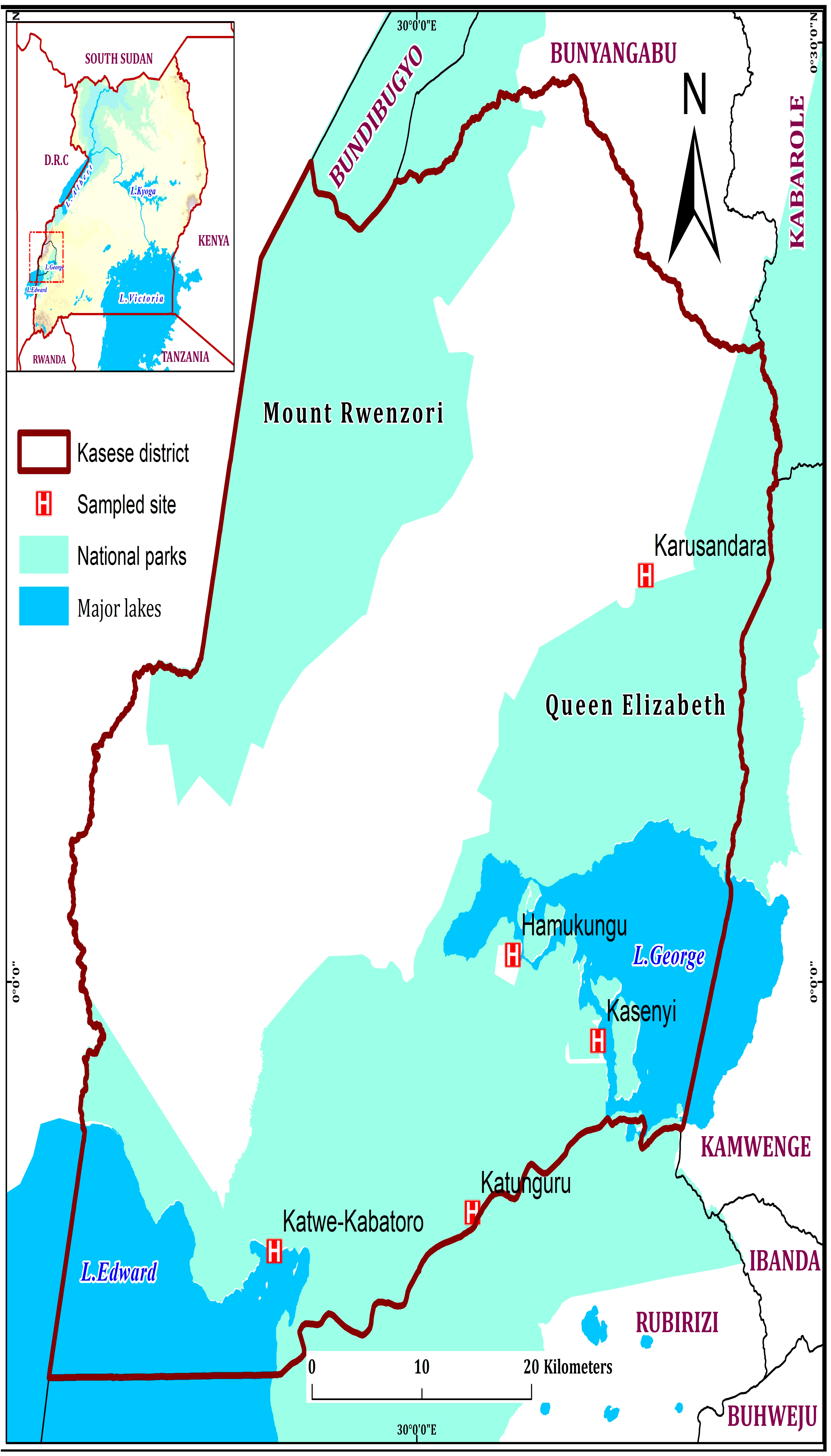
2.1. Study Area

The study was carried out in the Kasese District that is located in western Uganda (0°12′ S and 0°26′ N; 29°42′ E and 30°18′ E), has an area of 3389.8 km2, 86% is dry land, and 63% of the latter is for nature and wildlife conservation including Queen Elizabeth National Park (QENP), leaving limited land available for inhabitation and agriculture; 2% of the land area is occupied by permanent swamp and wetland [22,23]. Seventeen percent (17%) of QENP land is in the Kasese district and houses a number of fishing villages including Hamukungu, Kasenyi, Katunguru, and Katwe-Kabatoro (all in public enclaves), Kahendero, Katunguru, Kazinga, Kisenyi, Rwenshama, and Kayanja (park enclaves) (Figure 1). The study area thus commonly has wildlife, livestock and humans closely sharing this habitat as well as the natural resources such as water (Figure 2).

2.2. Study Population

The study participants who visited a health centre in the Kasese District with at least a trained nurse or laboratory technician (Katwe-Kabatoro HC, Karusandara, Kasenyi, Hamukungu, and Katunguru (Figure 1) and wild animals in selected areas within QENP (i,e., Kasenyi Koblek, Katunguru gate, Channel track, Janet track, Nyamunuka stretch, Camp site, Lake Katwe near institution, Research track, Katwe fishing village, Research koblek, Kaguta track, Mweya jet, and Mweya peninsula).
The community included in this study consisted of self-reporting patients at five health centre IIs in the Kasese District with diarrhoea and/or enteric complaints. Patient demographic characteristics including age, sex, HIV status, and village of origin were recorded.

2.3. Study Design and Sample Calculation

This was a cross-sectional study in which all consenting patients attending the five selected health centres with diarrhoea and/or enteric complaints and herbivorous wild animals around QENP. The samples were collected between June and October 2017. A total of 560 samples were collected, including 308 samples from symptomatic individuals in the community and 252 wildlife samples.

Exclusion Criteria

Patients that were moribund or those that did not consent.

2.4. Sample Collection, Processing, Transportation, and Storage

Approximately two teaspoonsful of stool samples were collected from patients attending health centre IIs in the study area. Individual patient’s stool sample was collected into a screw-capped stool collecting container to which three parts of 75% ethanol was later added and mixed thoroughly. The containers were then further sealed with parafilm and coded before they were loaded into leak-proof biohazard bags and placed into sample carrier cool boxes ready for transportation to the Molecular Biology Laboratory (MOBILA), College of Veterinary Medicine, Animal Resources and Biosecurity (COVAB), Makerere University Kampala for laboratory analysis.
Fresh faecal samples of wild animals were collected by tracking selected wild herbivorous animals near and around the human settlements at the different landing sites around Queen Elizabeth National Park, Kasese District, Uganda. Briefly, herds of different target species were followed and forced to move to a safe distance to enable the researchers to access their fresh droppings. The faecal sample was similarly collected and treated as described in the human stool sample collection.

2.5. Microscopic Detection of Cryptosporidium Oocysts in Stool

Glass slides with air-dried thick faecal smears were placed on a staining rack, flooded with Auramine-phenol stain for 10 min, and then rinsed with running water. The smears were immersed briefly (2 min) in acid alcohol (0.5 concentrated HCL, 0.5 g NaCl, 75 mL of absolute ethanol, and 25 mL of distilled water), rinsed with running water, flooded with 0.1% of potassium permanganate for 30 s, rinsed with running water, and air dried. Slides were examined using fluorescence microscopy at low magnification (×40). [24] Cryptosporidium oocysts appear as brightly fluorescent discs against a dark background (Figure 3).
Positive specimens were re-examined at high power magnification (×100) with oil immersion to ensure accurate identification of the oocysts. Three laboratory technologists performed the microscopic examination one being the tie-breaker.

2.6. DNA Extraction and PCR Amplification

DNA was extracted from all human samples and 100 wildlife samples randomly selected from the pool. Briefly, DNA was extracted from the stool samples using Quick-DNA Fecal/Soil Microbe Kits (Zymo Research, Tustin, CA, USA) with slight, in-house, modifications. All centrifugation steps were done at 10,000× g. Briefly, 900 µL of in-house prepared STE buffer (0.1 M Nacl, 10 mM Tris-Cl was added to 500 µL of faecal sludge in a 2000 µL (pH 8.0), 1% SDS, 1 mM NaEDTA (pH 8.0), 0.6 M β-Mercaptoethanol) microcentrifuge tube and incubated at 55 °C for 3 h with intermittent vortexing every 15 min. The microcentrifuge tubes were then centrifuged for 1 min, and 1000 µL of the supernatant was placed into a 2.5 mL collection tube to which 1000 µL of ice-cold absolute ethanol was added. The mixture (800 µL) was then transferred into a Zymo column™ (Zymo Research, Tustin, CA, USA) and centrifuged for 1 min until the entire mixture was passed through the column. DNA PreWash buffer™ (300 µL) was added to the column and centrifuged before washing with 500 µL of DNA wash buffer™. DNA was eluted in 100 µL of DNA elution buffer and stored at −20 °C until required.
PCR amplification: Molecular confirmation of Cryptosporidium spp. was carried out using a nested PCR targeting 18s rDNA as previously described by Kuzehkanan et al., (2011) [25], to yield an amplicon of 285 bp (Figure 4).

2.7. PCR Amplicon Sequencing

The PCR amplicons were purified using Qiagen PCR purification Kit (Qiagen, Germantown, MD, USA). We prepared 100 µL of the amplicons, 10 µL of the purified product was used to prepare representative gels, and the remainder was shipped to INQABA BIOTEC (Pretoria, South Africa) for sequencing. Purified PCR products were sequenced using the ABI 3500XL Genetic Analyzer, POP7™, BrilliantDye™ Terminator v3.1 (Thermo Fisher Scientific, Waltham, MA, USA). The nucleotide sequences of eight representative samples were deposited in the GenBank under accession numbers MK301300.1, MK301301.1, MK301302.1 MK301303.1, MK301304.1, MK301305.1, MK301306.1, and MK301307.1.

2.8. Data Analysis

The raw nucleotide sequences (ab1 files) were viewed and edited with BioEdit software version 7.0.0 [26], to remove any ambiguous sequences. Multiple sequence alignments and phylogenetic trees were constructed using MEGA X software (version 10) [27]. The representative reference sequences were downloaded from the web-based GenBank housed at the NCBI following a BLASTn search. The microscopy and PCR screening data were analyzed using STATA version 14 software to reveal the diagnostic accuracies of the two tests. Sensitivity was calculated by dividing the proportion of samples that tested positive divided by the total number of the true positives, whereas the specificity was calculated as true negative divided by the total number of samples that tested negative. Logistic regression analysis of various patient characteristics was completed using modified Poisson regression with robust standard errors to elucidate the possible associations. We computed the percentage prevalence of Cryptosporidium infection for both wildlife and human samples by dividing the number positive by the number tested. However, for wildlife samples, we did not have enough samples to categorically refer to the resulting proportion as “prevalence” since it may not be representative of the wildlife population in the entire national park.

2.9. Ethical Considerations

The study was reviewed by the School of Medicine, Research and Ethics Committee (SOM-REC) under the reference (#REC REF 2017-142) and approved by Uganda National Council for Science and Technology (UNCST) under review number HS 2333. The study was explained to each individual patient or guardian so as to attain informed consent. Following sample collection by the nurse or laboratory technician on call, the patients were treated for diarrhoea following the treatment regimen at the given health centres. No follow-up in the community was done because of budgetary constraints. Patients that met the inclusion criteria had their stool samples collected into sterile screw-capped stool collection containers.

3. Results

A total of 308 human faecal samples were collected from the five health centres. We analysed 282 samples in the laboratory since 8% (26/308) of the samples were either inaccurately collected or had their labels erased, thus excluded from the analysis. Most of the stool samples analysed were from Kasenyi HCII 28% (79/282), whereas the least were from Hamukungu HCII 13.5% (38/282). More than half of the faecal samples 54% (151/282) were collected from female patients. The majority of the patients, 31.6% (89/282), were between the ages of 5–18 years followed closely by children below five years of age, that is, 30.9% (87/282); the least number of samples (n = 6) were from patients over 65 years of age. The average age was 17 years and ranged from 3 months to 96 years of age. Watery stool samples accounted for 36.9% (104/282) of all samples analysed followed by solid hard stool, whereas soft solid stool accounted for the least proportion. (Table 1).

3.1. Prevalence of Cryptosporidium Species

Cryptosporidium parvum was found to be the predominant circulating species in sampled patients. The overall prevalence of Cryptosporidium species in the community around QENP was 53% (95% CI 47–58) and 40% (95% CI 34–46) using microscopy thick smear and 18S rDNA nested PCR, respectively. Patients attending Kasenyi HCII recorded arguably the highest positivity (27.8% and 24% detected using microscopy and PCR respectively), whereas those attending Hamukungu HCII recorded the lowest microscopy and PCR positivity (13.5% and 8%, respectively) (Table 1). The watery stool showed the highest percentage positivity using either detection method; the soft solid stool showed the lowest positivity results when tested using microscopy, whereas the watery stool with rice pellets yielded the lowest percentage positivity upon testing with PCR.
The overall percentage positivity of Cryptosporidium among wild animals was 78% with warthogs showing the highest proportion of 86%. Sampling points with the highest positivity rates (100%) were Camp site 2 (n = 5), Channel track (n = 9), and Kasenyi Koblek n = 1), whereas Katwe fishing village had the lowest positivity rate of 40%. (2/5) (Table 2).
A total of 252 wild samples were analysed from different herbivorous animals within QENP, these include (Waterbucks, Uganda Kobs, Elephants, Hippopotamus, Buffalos, Warthogs). The majority of the samples were collected from buffalo (57 samples, 22.6%), whereas the least were collected from Hippopotamus (23 samples, 9.1%) (Table 2).
A blast search of the sequenced 18S rDNA showed that the query sequences (sequence from this study) were similar to Cryptosporidium parvum and Cryptosporidium hominis.

3.2. Microscopy Thick Smear Sensitivity and Specificity on Stool Samples

Microscopy thick smear sensitivity observed was 41.2% (95% CI 33.2–49.6), whereas its specificity was 61.2% (95% CI 52.4–69.5). A positive predictive value of 54.0% (95% CI 46.8–60.9) and a negative predictive value of 48.5% (95% CI 43.8–53.3) were observed while investigating human stool samples (Table 3).

4. Discussion

In Uganda, Cryptosporidium infection has been shown to be an important risk factor for malnutrition including stunting in children, thus diagnosis and treatment of the infection may reduce the prospect of prolonged dehydration and the risk of wasting and probably loss of life [20]. The overall molecular prevalence of Cryptosporidium spp. in this study was found to be higher than what was previously reported (21%) within the vicinity of free-range gorillas in Bwindi Impenetrable National Park, Uganda [27] and among humans living around Kibale National Park also in western Uganda where prevalence was 32.4% [19]. This difference is probably associated with the current study area interfaces which are predominantly habituated by herbivorous wild game which are in close contact with humans. Proximity with livestock is known to be a risk factor for Cryptosporidium infection in humans [28,29]. In addition, the health centres sampled during our study are located close to open water bodies (Lake George and Lake Edward), thus patients live in communities in close contact with the water bodies. Proximity and fetching water from open water sources has been regarded as an epidemiological factor that increases the probability of Cryptosporidium infection [19,30,31] and may be one of the reasons for the high prevalence noted in this study.
The rural nature of the study area with limited health care coverage may also be a reason for the high prevalence of Cryptosporidium infection; this has also been reported in previous studies as a risk factor for infection [19]. Yet another study that was carried out in Gombe National Park, Kigoma District located in northwestern Tanzania revealed a much lower prevalence of 4.3% [32]. However, the population that was investigated in Kigoma was in close proximity with domestic animals specifically dog, goat, and/or sheep. Such a low prevalence may be explained by the varying potential of Cryptosporidium spp. to infect their hosts [33]. Elsewhere in Africa, the prevalence of Cryptosporidium has been reported in studies in various provinces in Egypt among children attending public hospitals; prevalence as high as 33.3% in the Ismailia province [34] and as low as 3.6% in Demietta province [35] have been recorded. Similarly, in South America, children younger than 2 years of age are frequently infected in developing countries like Peru [36] and Guatemala [37]. In the current study, children under five years had a molecular prevalence of 35%, slightly higher than the children aged 5–8 years (31%), although the number of stool samples was not sufficient to determine whether or not the difference was statistically significant.
A prevalence of 19.5%, much lower than that in the current study was reported in diarrhoeal patients of all age groups attending the outpatient clinics in Qaluibia province, Egypt [38]. The high prevalence registered in the current study could also be due to the use of water from sources contaminated by Cryptosporidium oocysts from faeces of livestock and wildlife. Cryptosporidium has been found to account for 60.3% of waterborne protozoan parasitic outbreaks globally from 2004 to 2010 [10]. The highest proportion of positive stool samples was recorded among patients attending Kasenyi Health Centre that is located in a busy Kasenyi fishing village and landing site. Such areas are characterised by a high population of people, limited toilet coverage, limited access to potable water, and poor general hygiene which are risk factors for Cryptosporidium infections and cryptosporidiosis [39,40,41,42]. It is thus crucial that genotyping is done in such areas to determine the level of genetic diversity of infecting Cryptosporidium spp., the source of contamination, and the route through which they are transmitted.
There are a number of molecular tools that have been employed in genotyping and subtyping; one of the frequently used tools is a gene that encodes a 60-kilodalton (KDa) precursor glycoproteins (GP60). Akiyoshi and others in 2006 utilised the GP60 gene to evaluate the genetic diversity of Cryptosporidium infection among children admitted to Mulago hospital, in Uganda. The data showed extensive genetic heterogeneity with three (3) new C. parvum alleles and suggested a high likelihood that the route of transmission is anthroponotic [43,44].
In the current study, sequencing of positive samples revealed that the infecting species were majorly C. parvum, and only one isolate was closer to C. hominis. The size of the 18s rDNA PCR amplicon (285 bp) generated in this study, however, was not big enough to reveal major peculiarities among these sequenced isolates considering that the 18S gene is highly conserved among Cryptosporidium spp. The BLASTn search however revealed very high similarities with other species in the GenBank and thus formed the basis for their clustering and classification. Another target that has previously been employed in genotyping is Cryptosporidium oocyst wall protein (COWP) which was used by Nizeyi and colleagues in 2002 [28], who aimed to identify the genotype of C. parvum causing infections in humans sharing habitats with free-ranging mountain gorillas in the Bwindi Impenetrable National Park, Uganda [17]. Results from that study demonstrated C. parvum Genotype 2 infections in members of the local community living in the vicinity of the Bwindi Impenetrable National Park who frequently enter habitats of mountain gorillas and interact with them; the same genotype had been previously isolated from mountain gorillas [18] and particularly prevalent in cattle that grazed within the park [43], and elsewhere [45]. In previous studies elsewhere in Africa, C. hominis has been reported to be more dominant among infected humans unlike what is presented in the current study. In a study in the Ismailia province of Egypt, Helmy and colleagues in 2013 [46] reported that C. hominis accounted for 60.5% of human cryptosporidiosis compared to 38.2% of C. parvum. The prevalence was much higher than in the current study, probably because the previous study investigated samples from diarrhoeic children under 10 years old from farming households and in close proximity to animals, whereas the current study tested both children and adult patients. Another study in Nigeria by Ukwah et al., 2017 [47], reported that C. hominis was the most predominant species accounting for 48.0% of human cryptosporidiosis followed by C. parvum which accounted for 44.0%. These studies suggest that the transmission is anthroponotic. However, another study done in Ethiopia by Adamu et al., in 2010 [48] reported only one isolate as C. hominis compared to 39 out of 41 that were characterized as C. parvum; this is similar to the findings in our study where only one isolate was characterized as C. hominis, suggesting zoonotic transmission. In our study, the analysed faecal matter of wild animals revealed infection among all sampled animal species and culminating in a very high positivity rate of Cryptosporidium infection of up to 78%. This scenario increases the likelihood of Cryptosporidium spp. transmission to humans and their livestock through faecal contamination of water sources and vegetation by wildlife faecal contamination [30]. A total of 28 faecal samples of warthog were collected; the highest number (20/28) with a 90% positivity rate was collected from Mweya Peninsula, an area that is populated by tourists as well as local communities and especially employees of the various tourism establishments in QENP. Other notable wild animal species are buffalo (n = 27) and elephant (n = 18) which had appreciable positivity rates of 85% and 83%; in both cases, the positivity rates are much higher compared to that reported by Abu et al., 2011 in Kruger National Park (KNP), South Africa, which was 4.2% and 1.4%, respectively [49].
In the current study, microscopy was, in some instances, reported to detect more samples with Cryptosporidium infection than PCR. In our view, this may be attributed to the complex composition of faeces that may contain PCR inhibitors such as bile salts and polysaccharides. It has been reported that the presence of inhibitors can dramatically reduce the sensitivity and amplification efficiency of PCR [50]. Moreover, the use of auramine Phenol for staining the oocysts of Cryptosporidium in faecal samples prior to microscopy has been considered routine because it is inexpensive and fast. Cryptosporidium oocysts stained with auramine phenol are easily detected because they appear as very characteristic brightly fluorescent discs against a dark background using a fluorescent microscope. In their study to evaluate various tests in detecting intestinal cryptosporidiosis, auramine phenol was found to be simple fluorescent staining, highly sensitive, specific, cost-effective, and less time-consuming that can be relied upon for diagnosis [51].
Primarily, C. parvum is considered the zoonotic species of human cryptosporidiosis; infection in most cases is due to water contaminated from animal faeces and/or due to contact with animals [52]. There is, however, an indication that there is no true host specificity of some species since they have been detected in human cryptosporidiosis, these species include C. baileyi, C. canis, C. felis, C. meleagridis, C. bovis, C. suis, C. aAnderson, and C. muris [47]. The zoonotic potential of these species has to be considered lower, although not in immunosuppressed persons [52].
There are reports in previous studies that human infection with Cryptosporidium has been by occupational exposure to infected animals (mainly calves) as well as companion animals, specifically dogs and cats; the latter have been implicated as probable sources of Cryptosporidium infection because they are sometimes infected with Cryptosporidium parvum [23].
This study shows for the first time that Cryptosporidium is circulating in populations in the QENP. The study also reveals a high prevalence of Cryptosporidium infection among patients attending health centres around QENP in the Kasese District as well as the high positivity rate among various herbivorous wild animals. However, in order to determine the zoonotic potential of Cryptosporidium spp. in QENP at the human/livestock/wildlife interface, prevalence and molecular characterisation of Cryptosporidium strains in patients and animals in communities neighbouring QENP should be carried out.
The study had limitations due to limited funding; genotyping the C. parvum-positives samples using markers such as gp60 that would have added value in relation to infection dynamics and genetic diversity of infection in the study populations was not done. The 18S rDNA amplicon that was sequenced in the current study was not long enough to generate differences usable in genotyping. In addition, the number of faecal samples as well as sites of sampling used may have been increased to better estimate the prevalence of infection in humans and animals.
There is a need to investigate the risk factors that predispose to such high Cryptosporidium infection in the study area, especially in Kasenyi where the prevalence was significantly higher than the rest of the areas studied. It is imperative to study the genetic diversity of Cryptosporidium spps in humans but also in livestock and wild animals using a longer PCR amplicon so as to generate more information on the species circulating and mode of transmission at the interface, thus devise strategies to control and management Cryptosporidium infection in the population.

Author Contributions

Conceptualization, C.M.M. and S.O.; Methodology. C.M.M., S.O., T.T., J.N., C.N., A.M. and B.B.M.; Software, C.N., J.N. and S.O.; Formal analysis, C.N., J.N. and S.O.; Investigation, C.M.M., B.B.M., S.O., T.T. and J.N.; Resources. C.M.M. and C.K.; data curation, C.M.M., C.N., J.N. and S.O.; writing—original draft preparation, C.M.M., S.O., C.K., J.N., C.N., A.M. and B.B.M.; writing—review and editing, C.M.M., S.O., T.T., C.K., J.N., C.N., A.M. and B.B.M.; visualization, C.M.M., J.N. and A.M.; supervision, C.M.M. and S.O.; project administration, C.K.; funding acquisition, C.K. All authors have read and agreed to the published version of the manuscript.

Funding

This research was funded by The Norwegian Programme for Capacity Development in Higher Education and Research for Development (NORHED), under the Project for Capacity Building in Zoonotic Disease Management Using Integrated Ecosystems Health Approach at the Human-Livestock-Wildlife Interface in the Eastern and Southern Africa (CAPAZOMANINTECO) project number UGA/0031/NORHED 1.

Institutional Review Board Statement

The study was conducted in accordance with the Declaration of Helsinki, and approved by the School of Medicine, Research and Ethics Committee (SOM-REC) of the College of Health Sciences, Makerere University under the reference (no. REC REF 2017-142 and approved by Uganda National Council for Science and Technology (UNCST) under review no. HS 2333.

Informed Consent Statement

Informed consent was obtained from all subjects involved in the study.

Data Availability Statement

Nucleotide sequences of eight representative samples were deposited in the GenBank.

Acknowledgments

The authors thank the health personnel at the study health centres in the study for enabling the collection of the stool samples, and Makerere University for facilitating the study.

Conflicts of Interest

The authors declare no conflict of interest.

References

  1. Carmena, D. Waterborne transmission of Cryptosporidium and Giardia: Detection, surveillance and implications for public health. In Current Research, Technology and Education Topics in Applied Microbiology and Microbial Biotechnology; Formatex Research Center: Badajoz, Spain, 2010; Volume 20, pp. 3–4. [Google Scholar]
  2. Efstratiou, A.; Ongerth, J.E.; Karanis, P. Waterborne transmission of protozoan parasites: Review of worldwide outbreaks—An update 2011–2016. Water Res. 2017, 114, 14–22. [Google Scholar] [CrossRef] [PubMed]
  3. Ryan, U.; Hijjawi, N.; Xiao, L. Foodborne cryptosporidiosis. Int. J. Parasitol. 2018, 48, 1–12. [Google Scholar] [CrossRef] [PubMed]
  4. Thompson, R.C.; Palmer, C.S.; O’Handley, R. The public health and clinical significance of Giardia and Cryptosporidium in domestic animals. Vet. J. 2008, 177, 18–25. [Google Scholar] [CrossRef]
  5. Savioli, L.; Smith, H.; Thompson, A. Giardia and Cryptosporidium join the ’Neglected Diseases Initiative. Trends Parasitol. 2006, 22, 203–208. [Google Scholar] [CrossRef] [PubMed]
  6. Kotloff, K.L.; Nataro, J.P.; Blackwelder, W.C.; Nasrin, D.; Farag, T.H.; Panchalingam, S.; Wu, Y.; Sow, S.O.; Sur, D.; Breiman, R.F.; et al. Burden and aetiology of diarrhoeal disease in infants and young children in developing countries (the Global Enteric Multicenter Study, GEMS): A prospective, case-control study. Lancet 2013, 382, 209–222. [Google Scholar] [CrossRef]
  7. Striepen, B. Parasitic infections: Time to tackle cryptosporidiosis. Nature 2013, 503, 189–191. [Google Scholar] [CrossRef]
  8. Baldursson, S.; Karanis, P. Waterborne transmission of protozoan parasites: 2004–2010. Water Res. 2011, 45, 6603–6614. [Google Scholar] [CrossRef]
  9. Aldeyarbi, H.M.; Abu El-Ezz, N.M.T.; Karanis, P. Cryptosporidium and cryptosporidiosis: The African perspective. Environ. Sci. Pollut. Res. Int. 2016, 23, 13811–13821. [Google Scholar] [CrossRef]
  10. Squire, S.A.; Ryan, U. Cryptosporidium and Giardia in Africa: Current and future challenges. Parasites Vectors 2017, 10, 195–227. [Google Scholar] [CrossRef]
  11. Fayer, R.; Ungar, B.L. Cryptosporidium spp. and cryptosporidiosis. Microbiol. Rev. 1986, 50, 458–483. [Google Scholar] [CrossRef]
  12. Cabada, M.M.; White, A.C. Treatment of cryptosporidiosis: Do we know what we think we know? Curr. Opin. Infect. Dis. 2010, 23, 494–499. [Google Scholar] [CrossRef] [PubMed]
  13. Wang, R.J.; Li, J.Q.; Chen, Y.C.; Zhang, L.X.; Xiao, L.H. Widespread occurrence of Cryptosporidium infections in patients with HIV/AIDS: Epidemiology, clinical feature, diagnosis, and therapy. Acta Trop. 2018, 187, 257–263. [Google Scholar] [CrossRef] [PubMed]
  14. O’Connor, R.M.; Shaffie, R.; Kang, G.; Ward, H.D. Cryptosporidiosis in patients with HIV/AIDS. Aids 2011, 25, 549–560. [Google Scholar] [CrossRef] [PubMed]
  15. Hotez, P.J. Could Nitazoxanide be added to other essential medicines for integrated neglected tropical disease control and elimination? PLoS Negl. Trop. Dis. 2014, 8, e2758. [Google Scholar] [CrossRef]
  16. Khalil, I.A.; Troeger, C.; Rao, P.C.; Blacker, B.F.; Brown, A.; Brewer, T.G.; Colombara, D.V.; De Hostos, E.L.; Engmann, C.; Guerrant, R.L.; et al. Morbidity, mortality, and long-term consequences associated with diarrhoea from Cryptosporidium infection in children younger than 5 years: A meta-analyses study. Lancet Glob. Health 2018, 6, e758–e768. [Google Scholar] [CrossRef]
  17. Nizeyi, J.B.; Mwebe, R.; Nanteza, A.; Cranfield, M.R.; Kalema, G.R.N.N.; Graczyk, T.K. Cryptosporidium sp. and Giardia sp. infections in mountain gorillas (Gorilla gorilla beringei) of the Bwindi Impenetrable National Park, Uganda. J. Parasitol. 1999, 85, 1084–1088. [Google Scholar] [CrossRef]
  18. Graczyk, T.K.; DaSilva, A.J.; Cranfield, M.R.; Nizeyi, J.B.; Kalema, G.R.; Pieniazek, N.J. Cryptosporidium parvum genotype 2 infections in free-ranging mountain gorillas (Gorilla gorilla beringei) of the Bwindi Impenetrable National Park, Uganda. Parasitol. Res. 2001, 87, 368–370. [Google Scholar] [CrossRef]
  19. Salyer, S.J.; Gillespie, T.R.; Rwego, I.B.; Chapman, C.A.; Goldberg, T.L. Epidemiology and molecular relationships of Cryptosporidium spp. in people, primates, and livestock from Western Uganda. PLoS Negl. Trop. Dis. 2012, 6, e1597. [Google Scholar] [CrossRef]
  20. Tumwine, J.K.; Kekitiinwa, A.; Nabukeera, N.; Akiyoshi, D.E.; Rich, S.M.; Widmer, G.; Feng, X.; Tzipori, S. Cryptosporidium parvum in children with diarrhoea in Mulago Hospital, Kampala, Uganda. Am. J. Trop. Med. Hyg. 2003, 68, 710–715. [Google Scholar] [CrossRef]
  21. Kankya, C.; Okello, J.; Baguma, J.N.; Wambi, R.; Ninsiima, L.R.; Tubihemukama, M.; Kulabako, C.T.; Munyeme, M.; Hartmack, S.; Okello, W. Prevalence and factors associated with cryptosporidiosis among livestock and dogs in Kasese District, Uganda: A cross-sectional study. medRxiv 2023. [Google Scholar] [CrossRef]
  22. Available online: www.kasese.go.ugKaseseDistrict (accessed on 30 January 2023).
  23. NECOC. Kasese Hazard, Risk and Vulnerability Profiles—Western Uganda Region. Available online: opm.go.ug (accessed on 30 January 2023).
  24. Kuzehkanan, B.A.; Rezaeian, M.; Zeraati, H.; Mohebali, M.; Meamar, A.R.; Babaei, Z.; Kashi, L.; Heydarnezhadi, M.; Rezaie, S. A sensitive and specific PCR based method for identification of Cryptosporidium Sp. using new primrs from 18S Ribosomal RNA. Iran J. Parasitol. 2011, 6, 1–7. [Google Scholar]
  25. Casemore, D.P.; Armstrong, M.; Sands, R.L. Laboratory diagnosis of cryptosporidiosis. J. Clin. Path. 1985, 38, 1337–1341. [Google Scholar] [CrossRef] [PubMed]
  26. Hall, T. BioEdit: An important software for molecular biology. GERF Bull. Biosci. 2011, 2, 60–61. [Google Scholar]
  27. Kumar, S.; Stecher, G.; Li, M.; Knyaz, C.; Tamura, K. MEGA X: Molecular Evolutionary Genetics Analysis across Computing Platforms. Mol, Biol. Evol. 2018, 35, 1547–1549. [Google Scholar] [CrossRef]
  28. Nizeyi, J.B.; Sebunya, D.; Dasilva, A.J.; Cranfield, M.R.; Pieniazek, N.J.; Graczyk, T.K. Cryptosporidiosis in people sharing habitats with free-ranging mountain gorillas (Gorilla gorilla beringei), Uganda. Am. J. Trop. Med. Hyg. 2002, 66, 442–444. [Google Scholar] [CrossRef]
  29. Casemore, D.P.; Wright, S.E.; Coop, R.L. Cryptosporidiosis–Human and animal epidemiology. In Cryptosporidium and Cryptosporidiosis; Fayer, R., Ed.; CRC Press: Boca Raton, FL, USA, 1997; pp. 65–92. [Google Scholar]
  30. Karanis, P.; Kourenti, C.; Smith, H. Waterborne transmission of protozoan parasites: A worldwide review of outbreaks and lessons learnt. J. Water Health 2007, 5, 1–38. [Google Scholar] [CrossRef]
  31. Alexander, K.A.; Carzolio, M.; Goodin, D.; Vance, E. Climate change is likely to worsen the public health threat of diarrheal disease in Botswana. Int. J. Environ. Res. Public Health 2013, 10, 1202–1230. [Google Scholar] [CrossRef]
  32. Parsons, M.B.; Travis, D.; Lonsdorf, E.V.; Lipende, I.; Roellig, D.M.A.; Kamenya, S.; Zhang, H.; Xiao, L.; Gillespie, T.R. Epidemiology and molecular characterization of Cryptosporidium spp. in humans, wild primates, and domesticated animals in the greater Gombe ecosystem, Tanzania. PLoS Negl. Trop. Dis. 2015, 10, e0003529. [Google Scholar] [CrossRef]
  33. Xiao, L.H.; Feng, Y.Y. Zoonotic cryptosporidiosis. FEMS Immunol. Med. Microbiol. 2008, 52, 309–323. [Google Scholar] [CrossRef]
  34. Shoukry, N.M.; Dawoud, H.A.; Haridy, F.M. Studies on zoonotic cryptosporidiosis parvum in Ismailia Governorate, Egypt. J Egypt Soc. Parasitol. 2009, 39, 479–488. [Google Scholar]
  35. Samn, K.; Samn, A.; Abou El-Nour, M. A survey of Giardia and Cryptosporidium spp. in Rural and Urban community in North Delta, Egypt. N. Y. Sci. J. 2012, 5, 49–54. [Google Scholar]
  36. Cama, V.A.; Bern, C.; Roberts, J.; Cabrera, L.; Sterling, C.R.; Ortega, Y.; Gilman, R.H.; Xiao, L. Cryptosporidium species and subtypes and clinical manifestations in children, Peru. Emerg. Infect. Dis. 2008, 14, 1567–1574. [Google Scholar] [CrossRef] [PubMed]
  37. Bern, C.; Hernandez, B.; Lopez, M.B.; Arrowood, M.J.; De Merida, A.M.; Klein, R.E. The contrasting epidemiology of Cyclospora and Cryptosporidium among outpatients in Guatemala. Am. J. Trop. Med. Hyg. 2000, 63, 231–235. [Google Scholar] [CrossRef] [PubMed]
  38. Abdel-Maboud, A.I.; Rossignol, J.F.; Mostafa, M.S. Cryptosporidiosis in Benha, study of some modalities in diagnosis and treatment. J. Egypt. Soc. Parasitol. 2000, 30, 717–725. [Google Scholar]
  39. Sinyangwe, N.N.; Siwila, J.; Muma, J.B.; Chola, M.; Michelo, C. Factors associated with Cryptosporidium infection among adult HIV positive population in contact with livestock in Namwala District, Zambia. Front. Public Health 2020, 8, 74. [Google Scholar] [CrossRef]
  40. Yongsi, H.B.N. Suffering for water, suffering from water: Access to drinking water and associated health risks in Cameroon. J. Health Popul. Nutr. 2010, 28, 424–435. [Google Scholar] [CrossRef]
  41. Ayalew, D.; Boelee, E.; Endeshaw, T.; Petros, B. Cryptosporidium and Giardia infection and drinking water sources among children in Lege Dini, Ethiopia. Trop. Med. Int. Health 2008, 13, 472–475. [Google Scholar] [CrossRef]
  42. Raccurt, C.P.; Brasseur, P.; Verdier, R.I.; Li, X.; Eyma, E.; Stockman, C.P.; Agnamey, P.; Guyot, K.; Totet, A.; Liautaud, B.; et al. Human cryptosporidiosis and Cryptosporidium spp. in Haiti. Trop. Med. Int. Health 2006, 11, 929–934. [Google Scholar] [CrossRef]
  43. Akiyoshi, D.E.; Tumwine, J.K.; Bakeera-Kitaka, S.; Tzipori, S. Subtype analysis of Cryptosporidium isolates from children in Uganda. J. Parasitol. 2006, 92, 1097–1100. [Google Scholar] [CrossRef]
  44. Graczyk, T.K.; Fayer, R.; Cranfield, M.R. Zoonotic potential of cross-transmission of Cryptosporidium parvum: Implications for waterborne cryptosporidiosis. Parasitol. Today 1997, 13, 348–351. [Google Scholar] [CrossRef]
  45. Fayer, R.; Morgan, U.; Upton, S.J. Epidemiology of Cryptosporidium: Transmission, detection and identification. Int. J. Parasitol. 2000, 30, 1305–1322. [Google Scholar] [CrossRef] [PubMed]
  46. Helmy, Y.A.; Krücken, J.; Nöckler, K.; von Samson-Himmelstjerna, G.; Zessin, K.-H. Molecular epidemiology of Cryptosporidium in livestock animals and humans in the Ismailia province of Egypt. Vet. Parasitol. 2013, 193, 15–24. [Google Scholar] [CrossRef] [PubMed]
  47. Ukwah, B.N.; Ezeonu, I.M.; Ezeonu, C.T.; Roellig, D.; Xiao, L. Cryptosporidium species and subtypes in diarrheal children and HIV-infected persons in Ebonyi and Nsukka, Nigeria. J. Infect. Dev. Ctries 2017, 11, 173–179. [Google Scholar] [CrossRef] [PubMed]
  48. Adamu, H.; Petros, B.; Hailu, A.; Petry, F. Molecular characterization of Cryptosporidium isolates from humans in Ethiopia. Acta Trop. 2010, 115, 77–83. [Google Scholar] [CrossRef] [PubMed]
  49. Samra, N.A.; Jori, F.; Samie, A.; Thompson, P. The prevalence of Cryptosporidium spp. oocyst in wild mammals in the Kruger National Park, South Africa. Vet. Parasitol. 2011, 175, 155–159. [Google Scholar] [CrossRef]
  50. Ramakers, C.; Ruijter, J.M.; Deprez, R.H.L.; Moorman, A.F.M. Assumption-free analysis of quantitative real-time polymerase chain reaction (PCR) data. Neurosci. Lett. 2003, 339, 62–66. [Google Scholar] [CrossRef]
  51. Khurana, S.; Sharma, P.; Sharma, A.; Malla, N. Evaluation of Ziehl-Neelsen staining, auramine phenol staining, antigen detection enzyme linked immunosorbent assay and polymerase chain reaction, for the diagnosis of intestinal cryptosporidiosis. Trop. Parasitol. 2012, 2, 20–23. [Google Scholar] [CrossRef]
  52. Lendner, M.; Etzold, M.; Daugschies, A. Kryptosporidiose--ein Update [Cryptosporidiosis--an update]. Berl. Munch Tierarztl. Wochenschr. 2011, 124, 473–484. [Google Scholar]
Figure 1. Study area showing the location of health centres (H) where study samples were collected.
Parasitologia 03 00019 g001
Figure 2. (A,B): Warthogs scavenging around homesteads in Kasenyi signifying a human-wildlife interface that is typical in the study area where faecal sampling was done.
Parasitologia 03 00019 g002
Figure 3. Cryptosporidium oocysts appear as fluorescent dots after staining with auramine-phenol.
Parasitologia 03 00019 g003
Figure 4. A selection of positive DNA samples after nested PCR; the arrow indicates the target band of 285 bp on the 100 bp molecular marker (M). N-negative control (Millipore water), P-positive control (previously confirmed with a COWP target: KY586963), 1–14 selected stool samples.
Parasitologia 03 00019 g004
Table 1. Distribution of Cryptosporidium infection in stool samples from human patients around Queen Elizabeth National Park.
MicroscopyPCR
CharacteristicNumber AnalyzedNumber Positive% Positivity (95% CI)Number Positive% Positivity (95% CI)
Location
Kasenyi HCII792227.8 (18.3–39.1)1924.1 (15.1–35)
Karusandara HCIII711825.4 (15.8–37.1)1318.3 (10.1–29.3)
Katwe-Kabatooro HCII49816.3 (7.3–29.7)1224.5 (13.3–38.9)
Katunguru HCII45715.6 (6.5–29.5)1124.4 (12.9–39.5)
Hamukungu HCII38513.2 (4.4–28.1)37.9 (1.7–21.4)
Age group (Years)
<5872731 (21.5–41.9)3034.5 (24.6–45.4)
5–18892831.5 (22–42.2)2831.5 (22–42.2)
19–35701724.3 (14.8–36)1927.1 (17.2–39.1)
36–6528310.7 (2.3–28.2)13.6 (0.1–18.3)
>65600 (0–45.9)00 (0–45.9)
Not indicated200 (0–84.2)00 (0–84.2)
Gender
Female1518153.6 (45.4–61.8)8657 (48.7–65)
Male1295945.7 (36.9–54.7)5542.6 (34–51.6)
Not indicated200 (0–84.2)00 (0–84.2)
Stool consistency
Watery1043836.5 (27.3–46.6)3836.5 (27.3–46.6)
Watery with rice pellets35411.4 (3.2–26.7)411.4 (3.2–26.7)
Solid hard772228.6 (18.8–40)2228.6 (18.8–40)
Mucoid39512.8 (4.3–27.4)512.8 (4.3–27.4)
Solid soft2428.3 (1–27)312.5 (2.7–32.4)
Not indicated300 (0–70.8)00 (0–70.8)
Table 2. Percentage positivity of Cryptosporidium spp. in wild herbivorous animals in QENP.
Sampling SiteWildlife Species Sampled (No. Positive/ No. Sampled)Overall
BuffaloElephantHippopotamusUganda KobWarthogsWater Buck% Positivity
Camp site 20100% (5/5)0000100% (5/5)
Channel track0100% (5/5)100% (4/4)000100% 9/9)
Janet track78% (7/9) 0000078% (7/9)
Kaguta track0000100% (1/1)45% (5/11)50% (6/12)
Kasenyi Koblek000100% (1/1)00100% (1/1)
Katunguru gate000% (0/1)00% (0/1)100% (2/2)50% (2/4)
Katwe fishing village050% (1/2)0% (0/1)0050% (1/2)40% (2/5)
Lake Katwe near institution067% (2/3)000067% (2/3)
Mweya Jet67% (2/3)00075% (3/4)071% (5/7)
Mweya peninsula000100% (1/1)91% (20/22)091% (21/23)
Nyamunuka stretch067% (2/3)000067% (2/3)
Research Koblek93% (14/15)0000093% (14/15)
Researchers track00050% (2/4)0050% (2/4)
Overall85% (23/27)83% (15/18)67% (4/6)67% (4/6)86% (24/28)53% (8/15)78% (78/100)
Table 3. Comparison between of PCR and microscopy for the detection of Cryptosporidium in stool samples.
Number of Samples with the Following Results Microscopy
PositiveNegativeTotal
PCRPositive6152113
Negative8782169
Total148134282
Disclaimer/Publisher’s Note: The statements, opinions and data contained in all publications are solely those of the individual author(s) and contributor(s) and not of MDPI and/or the editor(s). MDPI and/or the editor(s) disclaim responsibility for any injury to people or property resulting from any ideas, methods, instructions or products referred to in the content.

