Article

Molecular Identification of Cryptosporidium spp., and Giardia duodenalis in Dromedary Camels (Camelus dromedarius) from the Algerian Sahara

1
Laboratory of Molecular and Evolutionary Parasitology, RAPID Group, School of Biosciences, University of Kent, Canterbury, Kent CT2 7NJ, UK
2
Laboratory of Sciences and Living Techniques, Institute of Agronomic and Veterinary Sciences, University of Souk Ahras, Annaba Road, Souk Ahras 41000, Algeria
3
Faculty of Nature and Life Sciences, University of Djelfa, Moudjbara Road, BP 3117, Djelfa 17000, Algeria
4
Faculty of Sciences, University of Mohamed Boudiaf, BP 166, M’sila 28000, Algeria
5
Regional Veterinary Laboratory of El Oued, National Institute of Veterinary Medicine, BP 250, Ouargla 30000, Algeria
6
School of Science and Gut Microbiome Research Group, Mae Fah Luang University, Chiang Rai 57100, Thailand
*
Author to whom correspondence should be addressed.
Parasitologia 2023, 3(2), 151-159; https://doi.org/10.3390/parasitologia3020016
Submission received: 14 March 2023 / Revised: 31 March 2023 / Accepted: 11 April 2023 / Published: 14 April 2023

Abstract

:
(1) Intestinal microbial parasites are major contributors to the global burden of gastrointestinal disease. Such infections are mainly caused by Cryptosporidium, Giardia duodenalis, and Entamoeba histolytica. These parasites are transmitted either directly or indirectly through oral–fecal routes. Previous reports suggested that camels could play a role in the zoonotic transmission of various clinically and veterinary important intestinal parasites, however, limited data are available on intestinal infections of camels, particularly on a molecular level. We aimed to explore the occurrence of these three parasites in camels (Camelus dromedarius) in Algeria. (2) A total of 68 samples—63 stool samples from camels and five from the environment—were collected from two desert regions in Algeria and analyzed using PCR and qPCR methods. (3) Overall, 7% of the camels tested positive for zoonotic subtypes of Cryptosporidium spp., while 16% of the camels tested positive for G. duodenalis. Two environmental samples also tested positive for G. duodenalis. None of the samples were positive for Entamoeba histolytica. (4) Our results provide one of the first molecular-based identification of these gut parasites in dromedary camels in Algeria. The presence of G. duodenalis in the host and the environment unveils, in part, the circulation route of this parasite. Our results will spearhead further investigations into the prevalence and epidemiology of gut parasites in hoofed animals and raise questions concerning their role in health and disease in the area.

1. Introduction

Historically, the dromedary camel (Camelus dromedarius) is a characteristic animal of economic, social, and cultural importance in the African and Asian desert regions. This animal is also a staple food source for the people of the Sahara [1]. Dromedary camels can be infected by many parasitic species, including protozoa, helminths, and arthropods. These infections cause considerable economic losses due to reduced milk and meat production, as well as stunted growth and mortality cases. Among these parasitic species, Cryptosporidium spp., Giardia duodenalis (syn. Giardia intestinalis or Giardia lamblia), and Entamoeba histolytica are of zoonotic importance worldwide. These protozoan pathogens infect the digestive tract of a broad range of vertebrates [2,3], causing cryptosporidiosis, giardiasis, and amoebiasis, respectively, the former two of which are associated with human foodborne and waterborne outbreaks [4,5].
Limited data are available on the epidemiology of these diseases in dromedary camels compared to other livestock. Most studies are solely microscopy-based. Therefore, knowledge on the epidemiology of Cryptosporidium spp., G. duodenalis and Entamoeba spp. from camel-raising areas remains fragmentary. Prevalence rates of Cryptosporidium spp. in the dromedary camel range from 0% to 88%. In Africa, the parasite has been reported in Tunisia, Egypt, and Algeria [6,7,8], and in Iraq and Iran [9,10] in Asia. In this host, Cryptosporidium parvum, C. andersoni, Cryptosporidium rat genotype IV and Cryptosporidium camel genotype were detected using molecular approaches [8,11,12,13]. In Bactrian camel (Camelus bactrianus), C. parvum, C. muris, C. andersoni, C. bovis, C. hominis, C. ubiquitum, and C. occultus have been found [14,15,16,17,18,19].
The occurrence of G. duodenalis in dromedary, Bactrian, and other camelids has been poorly documented [20,21,22], with prevalence ranging from 7% to 47% [9,23,24,25]. To the authors’ knowledge, no data are available on genotyping and subtyping of G. duodenalis in dromedary camels. The prevalence rates of Entamoeba spp. in dromedary camels range from 2% to 20% [9,25]. Among the Entamoeba species, E. histolytica is the most virulent. Human infections with E. histolytica are a global health problem worldwide and are of zoonotic concern. The role livestock plays as potential reservoirs for this species is ambiguous [26].
All three genera display a high degree of genetic diversity and have members that infect livestock. Within Cryptosporidium, at least 46 valid species and more than 120 genotypes are recognized [27,28,29]. Of these, C. parvum is the most common zoonotic species infecting animals [30]. Giardia duodenalis is considered a complex comprising eight genetically distinct assemblages (genotypes; A-H) [3,29]. Of those, assemblages A and B are the most reported zoonotic genotypes in both animals and humans [3,31], while assemblage E has been frequently found in hoofed animals [31]. Similarly, a high degree of genetic diversity is also noted in Entamoeba, which is found in a broad range of hosts [32,33]. To the authors’ knowledge, there are no molecular data in the presence of E. histolytica and, furthermore, limited molecular data is available on Cryptosporidium spp. in dromedary camels.
The present study aimed to investigate occurrence and molecularly characterize Cryptosporidium spp., G. duodenalis, and E. histolytica in dromedary camels (C. dromedarius) from two regions located in the Algerian Sahara.

2. Results

2.1. Occurrence of Cryptosporidium Species and Subtypes

Cryptosporidium spp. was present in 7% (5/63) of the dromedary camels (Table 1 and Table 2). Two infected camels (a male and a female) were aged below three years, two were adults (a male and a female) aged above six years, while data were not available for one. Four samples were identified as C. parvum and one as C. bovis (Table 1). The C. parvum isolates had 99.53% identity with the reference sequence MN914085. The C. bovis isolate shared 99.19% identity with the reference sequence EF514234. The C. parvum gp60 sequences were subtyped as IIaA15G2R1, IIaA17G2R1, IIaA18G2R1, and IIdA19G1. The IIaA15G2R1 subtype was detected in two different camels, while the remaining subtypes were detected in three different camels. One camel showed mixed infection with IIaA15G2R1 and IIaA18G2R1. The C. bovis sample was not amplified at the gp60 locus since we would require a different set of primers than the ones utilized in this study. Environmental samples were negative for Cryptosporidium spp.
Four Cryptosporidium-positive camels from El-Oued region were of Sahraoui breed. These camels were infected with C. parvum IIaA15G2R1, IIaA17G2R1, IIaA18G2R1, and C. bovis. Additionally, one camel infected with C. parvum IIdA19G1 from Tindouf region was of Azaouat breed (Table 2).

2.2. Genotyping of Giardia duodenalis

Giardia duodenalis was detected in 17.6% (12/68) of all the samples, which consisted of 10 stool samples from 63 dromedary camels (Table 1 and Table 2). Five positive camels (four males and one female) were aged below three years, two (a male and a female) were aged between four and five years, and for three there were no available data. All ten infected camels were of Sahraoui breed from El-Oued region (Table 2). Among the five examined environmental collections, G. duodenalis was detected in one soil and one water sample. The sample obtained from soil was successfully genotyped as an assemblage based on sequence analysis of the tpi gene. The sequence shared 100% identity with the reference sequence EF654686 of G. duodenalis assemblage E. The remaining samples could not be genotyped due to the low quantity of the target nucleic acid and/or chimera formation.

2.3. Occurrence of Entamoeba histolytica

No sample tested positive for E. histolytica based on the probe-specific quantitative real-time PCR.

3. Discussion

The epidemiology of cryptosporidiosis and giardiasis in dromedary camels from camel-raising areas is poorly known as compared to data available in ruminants. Distinctly variable prevalence rates of Cryptosporidium spp. and G. duodenalis have been documented in different studies. In Algeria, the prevalence of Cryptosporidium spp., ranging from 1.8% to 10%, has been reported, and the study herein is in agreement [8,34,35,36]. The overall prevalence of G. duodenalis in dromedaries was 16%. Infection rates ranging between 7% to 39% have been reported in very few studies from the same hosts in Iraq and Saudi Arabia [9,23,25]. Different factors such as study design, diagnostic methods, and sample size could account for the variation of infection rates across studies.
We identified C. bovis and the zoonotic C. parvum subtypes IIaA15G2R1, IIaA18G2R1, IIaA17G2R1, and IIdA19G1. A previous molecular study in Algeria revealed a divergent C. parvum subtype (If-like-A22G2), which was genetically related to the C. hominis subtype family in two dromedary camels [8]. In addition to C. parvum and C. bovis, dromedary camels have previously been found to be infected with C. andersoni, Cryptosporidium rat genotype IV, and Cryptosporidium camel genotype in China and Egypt [11,13]. Three identified C. parvum subtypes belong to the subtype family IIa, which is mostly found in cattle, while one subtype belongs to the IId subtype family, which is mostly found in small ruminants [37,38]. The C. parvum subtypes IIaA15G2R1 and IIaA18G2R1 were detected for the first time in the dromedary camel in Algeria, while IIaA17G2R1 and IIdA19G1 were detected previously in the same host from Australia and Egypt, respectively [12,13]. The IIaA15G2R1 is a major hyper transmissible zoonotic subtype, is widely distributed, and is responsible for numerous outbreaks of human cryptosporidiosis [37,38]. The remaining identified subtypes have also been detected in humans worldwide, indicating their zoonotic potential [39,40,41,42,43]. Human infections with these subtypes, particularly with IIaA15G2R1, have been reported in different studies from North African and Middle East countries [44]. Consequently, the findings of the present study showed that dromedary camels could play an important role as reservoirs of zoonotic C. parvum subtypes. Future investigations should focus on not only camels, but also their handlers and their families.
We identified G. duodenalis in dromedary camels, soil, and water. This is the first time that this species was molecularly identified in this host. All ten animals infected were from the same region (El-Oued), while none of the Tindouf hosts were positive. This indicates a possible circulation of G. duodenalis amongst the camels residing in the El-Oued region. Notably, however, the animals were not diarrheic at the time of collection. Previously, only assemblages E and A were detected in camelids in China [24,45]. While we were unable to perform genotyping, our results are of significance regarding the transmission dynamics of G. duodenalis. This prompted us to investigate the presence of this parasite in the environment. One water and one soil sample were indeed positive for the parasite. This strongly points towards circulation of G. duodenalis between hosts and the environment. The finding of G. duodenalis in such an extreme environment is of note and should be investigated further.
It was worth noting that the occurrence of both parasites was higher in the breed Sahraoui. This predominance was likely attributed to the higher sample size of camels for this breed; all animals examined in El-Oued region were of the Sahraoui breed. Moreover, the difference in the infection rates between the two regions may also be attributed to sample size. Dromedary camels from El-Oued region live in close proximity to sheep and goats. In the Tindouf region, the presence of dogs was observed nearby the camels. This proximity with small ruminants may be one of reasons for the difference in the infection frequency observed between the two regions, specifically for G. duodenalis. A larger study focusing on collecting samples and metadata from all these animals would provide a clearer picture on the role of the hosts/breeds in the transmission dynamics of these parasites.

4. Materials and Methods

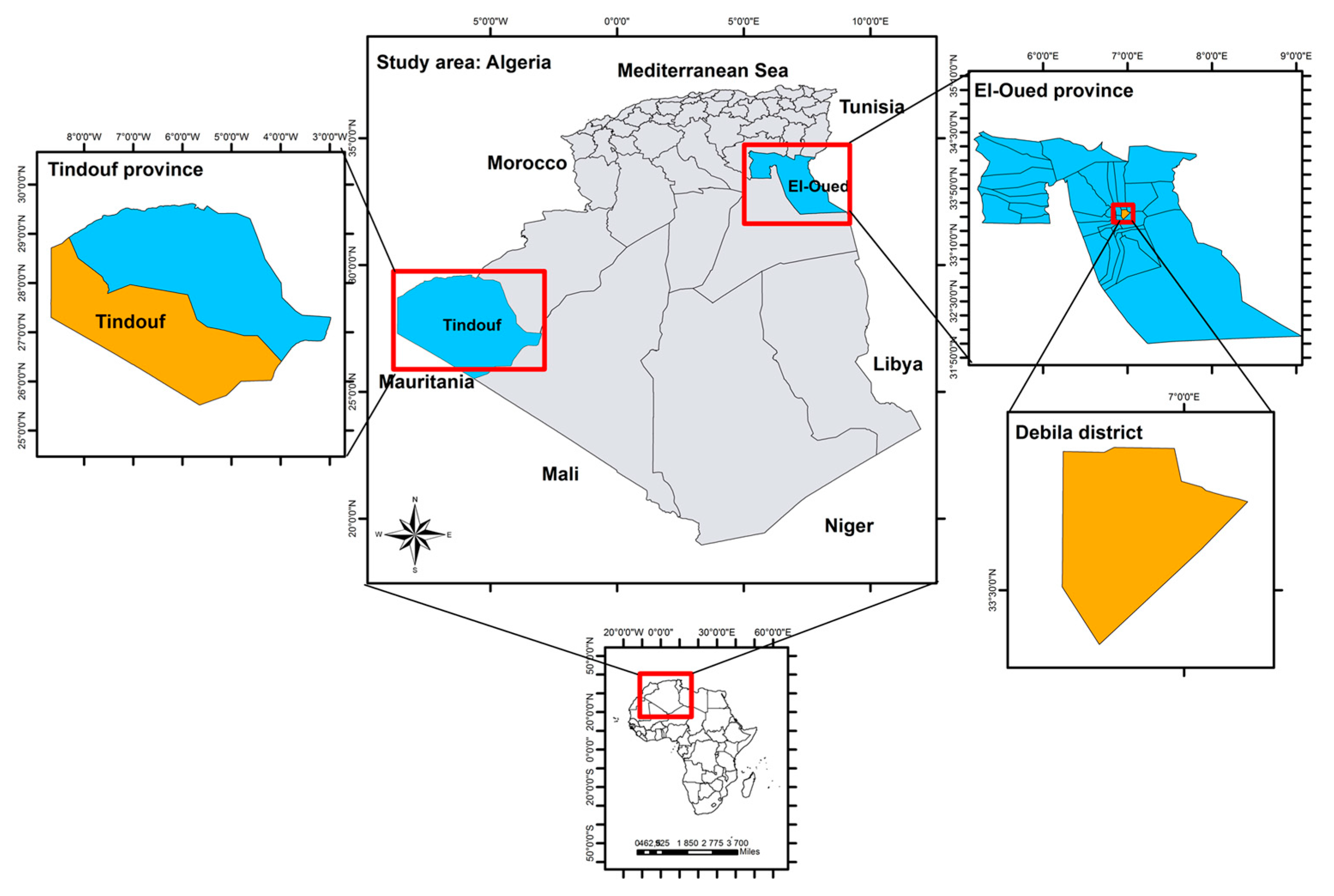
4.1. Study Area and Sample Collection

The present study was conducted between April 2017 and September 2021 in El-Oued and Tindouf provinces of the Algerian Sahara (Figure 1). El-Oued is situated in the north-east of the desert close to the Tunisian border. Tindouf is located in the extreme west of southern Algeria, close to the borders of Morocco and Mauritania.
A total of 63 fecal samples from seven different farms of dromedary camels (C. dromedarius) were collected (Table 2). Twenty-six samples were from four farms in the Tindouf region, while 37 came from three farms in the El-Oued area. Samples were collected immediately after defecation. None of the hosts showed any signs of diarrhea at the time of sampling. Gender, age, breed, and breeding system of the camels were recorded at the time of sampling. Fecal samples were stored in potassium dichromate 2.5% until molecular analyses. Additionally, five environmental samples were collected from El-Oued region. These consisted of four soil samples of 20 g of sand each and they were collected from the field surrounding the camel farms. The one water sample of 40 mL was collected from a tap connected to a ground drinking water source located near the farms.

4.2. DNA Extraction, PCRs Analysis, and Sequencing

The genomic DNA (gDNA) from stool and soil samples was extracted using a Pure LinkTM Microbiome DNA Purification Kit (Invitrogen, Carlsbad, CA, USA) according to the manufacturer’s protocol. DNA from the water sample was extracted using E.Z.N.A. Water DNA Kit (OMEGA, Stamford, CT, USA) according to the manufacturer’s protocol. The extracted DNA was stored at −20 °C until molecular analysis.
Species determination of Cryptosporidium spp. and genotyping were performed using a nested PCR approach to amplify fragments of two genes encoding the small subunit rRNA (SSU, product size: ∼631 bp) and a 60 kDa glycoprotein (gp60, product size; ~850 bp) as previously described [30,46,47,48]. For each SSU-positive sample, three separate gp60 PCR amplifications were performed to ensure the detection of mixed infection. All nested PCR products were purified using the Thermo Scientific GeneJET Gel Extraction Kit (Thermo Fisher Scientific, CA, USA) according to the manufacturer’s protocol and sequenced bidirectionally using internal PCR primers at Eurofins (Cologne, Germany).
The detection of G. duodenalis was performed by probe-based quantitative real-time PCR targeting a fragment of 62 bp of the SSU gene, as previously described [49,50]. Each reaction consisted of a total volume of 20 µL containing 10 µL 1X Luna Universal Probe qPCR Master Mix (New England Biolabs, MA, USA), 0.8 µL of 0.4 µM G. duodenalis-specific primers Giardia-80F (5′-GACGGCTCAGGACAACGGTT-3′) and Giardia-127R (5′-TTGCCAGCGGTGTCCG-3′), 0.4 µL of 0.2 µM G. duodenalis specific probe (FAM-5′-CCCGCGGCGGTCCCTGCTAG-3′-black hole quencher), 1 µL of 0.5 µg/µL bovine serum Albumin (BSA) (Promega Madison, WI, USA), 5 µL of nuclease-free H2O (Promega Madison, WI, USA), and 2 µL of extracted DNA. PCR conditions were as follows: initial denaturation step at 95 °C for 2 min, followed by 50 cycles of 95 °C for 15 s, annealing temperature at 57 °C for 30 s, and a final elongation step at 72 °C for 30 s. The qPCR positive samples were furthermore genotyped through nested amplification of the glutamate dehydrogenase (gdh) [51] and triosephosphate isomerase (tpi) [52] gene, and through a semi-nested PCR targeting β-giardin (bg) [53]. A sample positive with G. duodenalis was included as a positive control in each PCR run and a water sample as negative control. Subsequently, positive PCR products were purified using Thermo Scientific GeneJET Gel Extraction Kit (Thermo Fisher Scientific, CA, USA) according to the manufacturer’s protocol. Sequencing was performed at Eurofins (Cologne, Germany) in both directions using the internal primers; tpiR2 (5′-GTGGCCACCACICCCGTGCC-3′) and tpiF2 (5′-CCCTTCATCGGIGGTAACTT-3′) if sample(s) tested positive for tpi; gdh3 (5′-GTGGCGCARGGCATGATGCA-3′) and gdh4 (5′- ATGACYGAGCTYCAGAGGCACGT-3′) if sample(s) tested positive for gdh; and bg3 (5′-CATAACGACGCCATCGCGGCTCTCAGGAA-3′) and bg4 (5′ GAGGCCGCCCTGGATCTTCGAGACGAC-3′) if sample(s) tested positive for bg.
Cryptosporidium and G. duodenalis samples with unclear chromatograms were cloned using the pGEM-T easy vector system I (Promega, Madison, WI, USA) as previously described [54].
The presence of E. histolytica was screened by probe-specific quantitative real-time PCR targeting a fragment of 172 bp of the SSU rRNA gene, as previously described [50]. Each reaction consisted of a total volume of 20 µL comprising of 10 µL 1X Luna Universal Probe qPCR Master Mix (New England Biolabs, MA, USA), 0.8 µL of 0.4 µM primers Ehd-239F (5′-ATTGTCGTGGCATCCTAACTCA-3′) and Ehd-88R (5′-GCGGACGGCTCATTATAACA-3′), 0.4 µL of 0.2 µM E. histolytica specific probe (VIC-5′-TCATTGAATGAATTGGCCATTT-3′-NFQ), 1 µL of 0.5 µg/µL BSA (Promega Madison, WI, USA), 5 µL nuclease-free H2O (Promega Madison, WI, USA), and 2µL of extracted DNA. PCR conditions consisted of an initial denaturation step of 95 °C for 2 min, followed by 50 cycles of 95 °C for 15 s, annealing temperature at 54 °C for 30 s, and final elongation step at 72 °C for 30 s. Positive DNA of an E. histolytica isolate was included as a positive control for each PCR run, as well as PCR water as negative control.

4.3. Sequence Analyses

Obtained sequences were visualized and edited manually with the software SnapGeneViewer v.6.0.2, and Chromas pro v.2.1.10. Subsequently, the newly acquired sequences were used as queries to perform BLAST against the GenBank database. Generated nucleotide sequences in the present study were deposited in GenBank under accession numbers OQ613344-OQ613345 for Cryptosporidium spp., OQ622075-OQ622079 for C. parvum subtypes and OQ622080 for G. duodenalis.

5. Conclusions

The present study provides molecular data on the occurrence of zoonotic C. parvum subtypes and G. duodenalis in dromedary camels from two regions of the Algerian Sahara. Further studies with a larger sample size in the different regions of the Sahara are needed to better understand the epidemiology of cryptosporidiosis, giardiasis, and amoebiasis in these animals, particularly regarding prevalence, host-adapted species/genotypes, and public health significance of the respective responsible pathogens. Sample collections from the herders and their families, along with the surrounding environment, are urgently needed to disentangle the role of camels as sources and/or reservoirs of intestinal parasites.

Author Contributions

S.M.; Conceptualization, methodology, software, validation, formal analysis, investigation, resources, data curation, writing—original draft preparation, writing—review and editing, visualization. N.R. (Nassiba Reghaissiaand); Methodology, resources, writing—review and editing. A.L.; writing—review and editing. H.S.; Methodology. N.R. (Nacira Remdani); methodology. E.G., writing—review and editing, supervision. A.D.T.; Conceptualization, writing—review and editing, supervision. project administration funding acquisition. All authors have read and agreed to the published version of the manuscript.

Funding

SM was supported by a Ph.D. studentship from the Global Challenges Doctoral Centre at the University of Kent.

Institutional Review Board Statement

The animal study protocol was approved by the Institutional Review Board (or Ethics Committee) of Research Center in Agropastoralism of Ziane Achour University, Djelfa Algeria in accordance Ministry of Higher Education and Scientific Research, for studies involving animals.

Informed Consent Statement

Not applicable.

Data Availability Statement

All data have been submitted to GenBank under accession numbers: OQ613344-OQ613345 for Cryptosporidium spp., OQ622075-OQ622079 for C. parvum subtypes and OQ622080 for G. duodenalis.

Acknowledgments

We thank members of the Tsaousis laboratory and the camels’ keepers for assisting with sample collection and accommodating us during our visitations.

Conflicts of Interest

The authors declare no conflict of interest. The funders had no role in the design of the study; in the collection, analyses, or interpretation of data; in the writing of the manuscript; or in the decision to publish the results.

References

  1. FAO. World Food and Agriculture—Statistical Yearbook 2021; FAO: Rome, Italy, 2021. [Google Scholar] [CrossRef]
  2. Fayer, R. Taxonomy and species delimitation in Cryptosporidium. Exp. Parasitol. 2010, 124, 90–97. [Google Scholar] [CrossRef]
  3. Feng, Y.; Xiao, L. Zoonotic potential and molecular epidemiology of Giardia species and giardiasis. Clin. Microbiol. Rev. 2011, 24, 110–140. [Google Scholar] [CrossRef] [PubMed] [Green Version]
  4. Ryan, U.; Hijjawi, N.; Feng, Y.; Xiao, L. Giardia: An under-reported foodborne parasite. Int. J. Parasitol. 2019, 49, 1–11. [Google Scholar] [CrossRef] [PubMed]
  5. Ryan, U.; Hijjawi, N.; Xiao, L. Foodborne cryptosporidiosis. Int. J. Parasitol. 2018, 48, 1–12. [Google Scholar] [CrossRef] [PubMed] [Green Version]
  6. Soltane, R.; Guyot, K.; Dei-Cas, E.; Ayadi, A. Prevalence of Cryptosporidium spp. (Eucoccidiorida: Cryptosporiidae) in seven species of farm animals in Tunisia. Parasite 2007, 14, 335–338. [Google Scholar] [CrossRef] [PubMed] [Green Version]
  7. Abdel-Wahab, A.; Abdel-Maogood, S. Identification of Cryptosporidium Species Infecting Camels (Camelus dromedarius) in Egypt. J. Am. Sci. 2011, 7, 609–612. [Google Scholar]
  8. Baroudi, D.; Zhang, H.; Amer, S.; Khelef, D.; Roellig, D.M.; Wang, Y.; Feng, Y.; Xiao, L. Divergent Cryptosporidium parvum subtype and Enterocytozoon bieneusi genotypes in dromedary camels in Algeria. Parasitol. Res. 2018, 117, 905–910. [Google Scholar] [CrossRef]
  9. Hussin, A.G.; Khalaf, J.M.; Ali, H.M. Detection of intestinal protozoa in camels and their breeders in Najef, Iraq. Res. J. Vet. Pract. 2015, 3, 53–57. [Google Scholar] [CrossRef] [Green Version]
  10. Sazmand, A.; Rasooli, A.; Nouri, M.; Hamidinejat, H.; Hekmatimoghaddam, S. Prevalence of Cryptosporidium spp. in camels and involved people in Yazd province, Iran. Iran. J. Parasitol. 2012, 7, 80–84. Available online: https://ijpa.tums.ac.ir/index.php/ijpa/article/view/224 (accessed on 12 April 2023).
  11. Gu, Y.; Wang, X.; Zhou, C.; Li, P.; Xu, Q.; Zhao, C.; Liu, W.; Xu, W. Investigation on Cryptosporidium infections in wild animals in a zoo in Anhui province. J. Zoo. Wildl. Med. 2016, 47, 846–854. [Google Scholar] [CrossRef] [PubMed]
  12. Zahedi, A.; Lee, G.K.C.; Greay, T.L.; Walsh, A.L.; Blignaut, D.J.C.; Ryan, U.M. First report of Cryptosporidium parvum in a dromedary camel calf from Western Australia. Acta Parasitol. 2018, 63, 422–427. [Google Scholar] [CrossRef] [PubMed]
  13. El-Alfy, E.S.; Abu-Elwafa, S.; Abbas, I.; Al-Araby, M.; Al-Kappany, Y.; Umeda, K.; Nishikawa, Y. Molecular screening approach to identify protozoan and trichostrongylid parasites infecting one-humped camels (Camelus dromedarius). Acta Trop. 2019, 197, 105060. [Google Scholar] [CrossRef] [PubMed]
  14. Kváč, M.; Sak, B.; Květoňová, D.; Ditrich, O.; Hofmannová, L.; Modrý, D.; Vítovec, J.; Xiao, L. Infectivity, pathogenicity, and genetic characteristics of mammalian gastric Cryptosporidium spp. in domestic ruminants. Vet. Parasitol. 2008, 153, 363–367. [Google Scholar] [CrossRef] [PubMed]
  15. Wang, R.; Zhang, L.; Ning, C.; Feng, Y.; Jian, F.; Xiao, L.; Lu, B.; Ai, W.; Dong, H. Multilocus phylogenetic analysis of Cryptosporidium andersoni (Apicomplexa) isolated from a bactrian camel (Camelus bactrianus) in China. Parasitol. Res. 2008, 102, 915–920. [Google Scholar] [CrossRef] [PubMed]
  16. Liu, X.; Zhou, X.; Zhong, Z.; Deng, J.; Chen, W.; Cao, S.; Fu, H.; Zuo, Z.; Hu, Y.; Peng, G. Multilocus genotype and subtype analysis of Cryptosporidium andersoni derived from a Bactrian camel (Camelus bactrianus) in China. Parasitol. Res. 2014, 113, 2129–2136. [Google Scholar] [CrossRef] [PubMed]
  17. Cao, Y.; Cui, Z.; Zhou, Q.; Jing, B.; Xu, C.; Wang, T.; Qi, M.; Zhang, L. Genetic diversity of Cryptosporidium in bactrian camels (Camelus bactrianus) in Xinjiang, Northwestern China. Pathogens 2020, 9, 946. [Google Scholar] [CrossRef]
  18. Wang, X.; Zhang, Z.; Yin, W.; Zhang, Q.; Wang, R.; Duan, Z. Interactions between Cryptosporidium, Enterocytozoon, Giardia and intestinal microbiota in bactrian camels on Qinghai-Tibet Plateau, China. Appl. Sci. 2021, 11, 3595. [Google Scholar] [CrossRef]
  19. Morgan, U.M.; Xiao, L.; Monis, P.; Sulaiman, I.; Pavlasek, I.; Blagburn, B.; Olson, M.; Upton, S.J.; Khramtsov, N.V.; Lal, A.; et al. Molecular and phylogenetic analysis of Cryptosporidium muris from various hosts. Parasitology 2000, 120 Pt 5, 457–464. [Google Scholar] [CrossRef] [PubMed] [Green Version]
  20. Kiorpes, A.L.; Kirkpatrick, C.E.; Bowman, D.D. Isolation of Giardia from a Llama and from Sheep. Can. J. Vet. Res 1987, 51, 277–280. [Google Scholar] [PubMed]
  21. Al-Jabr, O.A.; Mohammed, G.E.; Al-Hamdan, B.A. Giardiosis in camels (Camelus dromedarius). Vet. Rec. 2005, 157, 350–352. [Google Scholar] [CrossRef] [PubMed]
  22. Locklear, T.R.; Videla, R.; Breuer, R.M.; Mulon, P.-Y.; Passmore, M.; Mochel, J.P.; Gerhold, R.; Schaefer, J.J.; Smith, J.S. Presentation, clinical pathology abnormalities, and identification of gastrointestinal parasites in camels (Camelus bactrianus and Camelus dromedarius) presenting to two North American veterinary teaching hospitals. A Retrospective Study: 1980–2020. Front. Vet. Sci. 2021, 8, 1672. [Google Scholar] [CrossRef] [PubMed]
  23. Jawad, H.H.; Jasim, G.A. Molecular study of Cryptosporidium spp. and Giardia lamblia which cause diarrhea in camels (Camillus dromedaries) in Al-Diwaniyah and Al-Najaf provinces/Iraq. AL-Qadisiyah J. Vet. Med. Sci. 2016, 15, 70–75. [Google Scholar]
  24. Zhao, S.-S.; Li, Y.-H.; Zhang, Y.; Zhou, Q.; Jing, B.; Xu, C.-Y.; Zhang, L.-X.; Song, J.-K.; Zhao, G.-H. Multilocus genotyping of Giardia duodenalis in Bactrian camels (Camelus bactrianus) in China. Parasitol. Res. 2020, 119, 3873–3880. [Google Scholar] [CrossRef]
  25. Alfatah, M.E.A. Prevalence of gastrointestinal parasitic infestations with hematobiochemical disorders in dromedary camel. Egypt. Acad. J. Biol. Sci. E. Med. Entomol. Parasitol. 2021, 13, 1–9. [Google Scholar] [CrossRef]
  26. Ai, S.; Zhang, Z.; Wang, X.; Zhang, Q.; Yin, W.; Duan, Z. The first survey and molecular identification of Entamoeba spp. in farm animals on Qinghai-Tibetan Plateau of China. Comp. Immunol. Microbiol. Infect. Dis. 2021, 75, 101607. [Google Scholar] [CrossRef] [PubMed]
  27. Zahedi, A.; Ryan, U. Cryptosporidium—An update with an emphasis on foodborne and waterborne transmission. Res. Vet. Sci. 2020, 132, 500–512. [Google Scholar] [CrossRef]
  28. Prediger, J.; Ježková, J.; Holubová, N.; Sak, B.; Konečný, R.; Rost, M.; McEvoy, J.; Rajský, D.; Kváč, M. Cryptosporidium sciurinum n. sp. (apicomplexa: Cryptosporidiidae) in eurasian red squirrels (Sciurus vulgaris). Microorganisms 2021, 9, 2050. [Google Scholar] [CrossRef]
  29. Ryan, U.M.; Feng, Y.; Fayer, R.; Xiao, L. Taxonomy and molecular epidemiology of Cryptosporidium and Giardia—A 50 year perspective (1971–2021). Int. J. Parasitol. 2021, 51, 1099–1119. [Google Scholar] [CrossRef]
  30. Pinto, P.; Ribeiro, C.A.; Hoque, S.; Hammouma, O.; Leruste, H.; Détriché, S.; Canniere, E.; Daandels, Y.; Dellevoet, M.; Roemen, J.; et al. Cross-Border investigations on the prevalence and transmission dynamics of Cryptosporidium species in dairy cattle farms in Western Mainland Europe. Microorganisms 2021, 9, 2394. [Google Scholar] [CrossRef]
  31. Cacciò, S.M.; Lalle, M.; Svärd, S.G. Host specificity in the Giardia duodenalis species complex. Infect. Genet. Evol. 2018, 66, 335–345. [Google Scholar] [CrossRef]
  32. Jinatham, V.; Popluechai, S.; Clark, C.G.; Gentekaki, E. Entamoeba chiangraiensis n. sp. (Amoebozoa: Entamoebidae) isolated from the gut of Asian swamp eel (Monopterus albus) in northern Thailand. Parasitology 2019, 146, 1719–1724. [Google Scholar] [CrossRef] [PubMed] [Green Version]
  33. Jacob, A.S.; Busby, E.J.; Levy, A.D.; Komm, N.; Clark, C.G. Expanding the Entamoeba Universe: New Hosts Yield Novel Ribosomal Lineages. J. Eukaryot. Microbiol. 2016, 63, 69–78. [Google Scholar] [CrossRef] [PubMed]
  34. Bouragba, M.; Laatamna, A.E.; Cheddad, F.E.; Baroudi, D.; Houali, K.; Hakem, A. Gastrointestinal parasites of dromedary camel (Camelus dromedarius) in Algeria. Vet. World 2020, 13, 1635–1640. [Google Scholar] [CrossRef] [PubMed]
  35. Laatamna, A.K.; Belkessa, S.; Khalil, A.; Afidi, A.; Benmahdjouba, K.; Belalmi, R.; Benkrour, M.; Ghazel, Z.; Hakem, A.; Aissi, M. Prevalence of Cryptosporidium spp. in farmed animals from steppe and high plateau regions in Algeria. Trop. Biomed. 2018, 35, 724–735. [Google Scholar] [PubMed]
  36. Ouchene, N.; Khelifi-Touhami, N.A. Prevalence of Cryptosporidium spp. in humans and dromedaries (Camelus dromedarius) in Algeria. Vet. Stanica 2023, 54, 185–191. [Google Scholar] [CrossRef]
  37. Xiao, L.; Feng, Y. Molecular epidemiologic tools for waterborne pathogens Cryptosporidium spp. and Giardia duodenalis. Food Waterborne Parasitol. 2017, 8–9, 14–32. [Google Scholar] [CrossRef]
  38. Feng, Y.; Ryan, U.M.; Xiao, L. Genetic diversity and population structure of Cryptosporidium. Trends Parasitol. 2018, 34, 997–1011. [Google Scholar] [CrossRef]
  39. Roelfsema, J.H.; Sprong, H.; Caccio, S.M.; Takumi, K.; Kroes, M.; van Pelt, W.; Kortbeek, L.M.; van der Giessen, J.W.B. Molecular characterization of human Cryptosporidium spp. isolates after an unusual increase in late summer 2012. Parasites Vectors 2016, 9, 138. [Google Scholar] [CrossRef] [Green Version]
  40. Insulander, M.; Silverlås, C.; Lebbad, M.; Karlsson, L.; Mattsson, J.G.; Svenungsson, B. Molecular epidemiology and clinical manifestations of human cryptosporidiosis in Sweden. Epidemiol. Infect. 2013, 141, 1009. [Google Scholar] [CrossRef] [Green Version]
  41. Waldron, L.S.; Dimeski, B.; Beggs, P.J.; Ferrari, B.C.; Power, M.L. Molecular epidemiology, spatiotemporal analysis, and ecology of sporadic human cryptosporidiosis in Australia. Appl. Environ. Microbiol. 2011, 77, 7757–7765. [Google Scholar] [CrossRef] [Green Version]
  42. Chalmers, R.M.; Robinson, G.; Elwin, K.; Elson, R. Analysis of the Cryptosporidium spp. and gp60 subtypes linked to human outbreaks of cryptosporidiosis in England and Wales, 2009 to 2017. Parasites Vectors 2019, 12, 95. [Google Scholar] [CrossRef] [PubMed]
  43. Alves, M.; Xiao, L.; Antunes, F.; Matos, O. Distribution of Cryptosporidium subtypes in humans and domestic and wild ruminants in Portugal. Parasitol. Res. 2006, 99, 287–292. [Google Scholar] [CrossRef] [PubMed]
  44. Hijjawi, N.; Zahedi, A.; Al-Falah, M.; Ryan, U. A review of the molecular epidemiology of Cryptosporidium spp. and Giardia duodenalis in the Middle East and North Africa (MENA) region. Infect. Genet. Evol. 2022, 98, 105212. [Google Scholar] [CrossRef] [PubMed]
  45. Zhang, Q.; Zhang, Z.; Ai, S.; Wang, X.; Zhang, R.; Duan, Z. Cryptosporidium spp., Enterocytozoon bieneusi, and Giardia duodenalis from animal sources in the Qinghai-Tibetan Plateau Area (QTPA) in China. Comp. Immunol. Microbiol. Infect. Dis. 2019, 67, 101346. [Google Scholar] [CrossRef] [PubMed]
  46. Ziegler, P.E.; Nydam, D.; Wade, S.; Schaaf, S.L. Evaluation of polymerase chain reaction diagnosis of Cryptosporidium spp. in dairy cattle and wildlife. Vet. Ther. 2007, 8, 148–159. Available online: https://www.researchgate.net/publication/6220860 (accessed on 12 March 2023).
  47. Alves, M.; Xiao, L.; Sulaiman, I.; Lal, A.A.; Matos, O.; Antunes, F. Subgenotype analysis of Cryptosporidium isolates from humans, cattle, and zoo ruminants in Portugal. J. Clin. Microbiol. 2003, 41, 2744–2747. [Google Scholar] [CrossRef] [Green Version]
  48. Reghaissia, N.; Maxamhud, S.; Laatamna, A.; Samari, H.; Dahmane, A.; Berima, R.; Abdelli, A.; Hakem, A.; Baroudi, D.; Tsaousis, A.D. First epidemiological report on the prevalence and associated risk factors of Cryptosporidium spp. in farmed marine and wild freshwater fish in central and eastern of Algeria. Acta Parasitol. 2022, 67, 1152–1161. [Google Scholar] [CrossRef]
  49. Hove, R.T.; Schuurman, T.; Kooistra, M.; Möller, L.; Van Lieshout, L.; Verweij, J.J. Detection of diarrhoea-causing protozoa in general practice patients in The Netherlands by multiplex real-time PCR. Clin. Microbiol. Infect. 2007, 13, 1001–1007. [Google Scholar] [CrossRef] [Green Version]
  50. Verweij, J.J.; Blange, R.A.; Templeton, K.; Schinkel, J.; Brienen, E.A.T.; van Rooyen, M.A.A.; van Lieshout, L.; Poldermann, A.M. Simultaneous Detection of Entamoeba histolytica, Giardia lamblia, and Cryptosporidium parvum in Fecal Samples by Using Multiplex Real-Time PCR. J. Clin. Microbiol. 2004, 42, 1220–1223. [Google Scholar] [CrossRef] [Green Version]
  51. Cacciò, S.M.; Beck, R.; Lalle, M.; Marinculic, A.; Pozio, E. Multilocus genotyping of Giardia duodenalis reveals striking differences between assemblages A and B. Int. J. Parasitol. 2008, 38, 1523–1531. [Google Scholar] [CrossRef]
  52. Sulaiman, I.M.; Fayer, R.; Bern, C.; Gilman, R.H.; Trout, J.M.; Schantz, P.M.; Das, P.; Lal, A.A.; Xiao, L. Triosephosphate Isomerase Gene Characterization and Potential Zoonotic Transmission of Giardia duodenalis. Emerg. Infect. Dis. 2003, 9, 1444–1452. [Google Scholar] [CrossRef] [PubMed]
  53. Cacciò, S.M.; De Giacomo, M.; Pozio, E. Sequence analysis of the b-giardin gene and development of a polymerase chain reaction-restriction fragment length polymorphism assay to genotype Giardia duodenalis cysts from human faecal samples 2002. Int. J. Parasitol. 2002, 32, 1023–1030. [Google Scholar] [CrossRef] [PubMed]
  54. Betts, E.L.; Gentekaki, E.; Thomasz, A.; Breakell, V.; Carpenter, A.I.; Tsaousis, A.D. Genetic diversity of Blastocystis in non-primate animals. Parasitology 2018, 145, 1228–1234. [Google Scholar] [CrossRef] [PubMed] [Green Version]
Figure 1. A map illustration of the study sites used for sample collection throughout this study.
Parasitologia 03 00016 g001
Table 1. Occurrence of Cryptosporidium species/subtypes and G. duodenalis in fecal samples from examined dromedary camels.
RegionN° of Examined Fecal SamplesCryptosporidium spp.G. duodenalis
No. of positive
samples (camels)
Species identitySubtypesNo. of positive
samples (camels)
El-Oued3743 × C. parvumIIaA15G2R1 a10 c
IIaA17G2R1
IIaA18G2R1 b
1 × C. bovis-
Tindouf 2611 × C. parvumIIdA19G10
a This subtype was detected in two samples from two different dromedary camels. b This subtype was detected in one dromedary camel infected with another subtype IIaA15G2R1 (mixed infection). c Genotyping (assemblage determination) was not obtained for G. duodenalis positive samples.
Table 2. Overall information on the epidemiological data on the overall information as well as the samples infected with Cryptosporidium spp. and Giardia duodenalis.
CharacterCategoryNo. of SampleNo. of Positive Cryptosporidium spp.No. of Positive G. duodenalis
RegionTindouf2610
El-Oued42412
Total68512
SourceStool63510
Soil401
Water101
Total68512
GenderMale2925
Female2722
Unknown713
Total63510
Age<5 years2627
5–10 years2020
>10 years1000
Unknown713
Total63510
BreedAzaouat1010
Hamra200
Tergui200
Reguibi1100
Mauritanian100
Sahraoui37410
Total63510
Breeding systemExtensive5741
Semi-intensive619
Total63510
Disclaimer/Publisher’s Note: The statements, opinions and data contained in all publications are solely those of the individual author(s) and contributor(s) and not of MDPI and/or the editor(s). MDPI and/or the editor(s) disclaim responsibility for any injury to people or property resulting from any ideas, methods, instructions or products referred to in the content.

