Article

Exploring Adult Age-at-Death Research in Anthropology: Bibliometric Mapping and Content Analysis

by
Vanessa Campanacho
1,* and
Francisca Alves-Cardoso
2
1
Bioanthropologist, New York, NY 11803, USA
2
LABOH—Laboratory of Biological Anthropology and Human Osteology, CRIA—Centro em Rede de Investigação em Antropologia, Faculdade de Ciências Sociais e Humanas, Universidade NOVA de Lisboa, 1069-061 Lisboa, Portugal
*
Author to whom correspondence should be addressed.
Forensic Sci. 2023, 3(1), 125-148; https://doi.org/10.3390/forensicsci3010011
Submission received: 30 January 2023 / Revised: 1 March 2023 / Accepted: 6 March 2023 / Published: 8 March 2023
(This article belongs to the Special Issue Estimating Age in Forensic Anthropology)

Abstract

:
Although there are known limited skeletal traits that can be used to estimate age-at-death, an increasing body of literature is addressing this topic. This is particularly true in journals dedicated to forensic anthropology and past population studies. Research has focused mostly on methodological developments, aiming to update and validate age-at-death methods’ accuracy, with recurrent formulation, reformulation, testing, and re-testing of classical methodological approaches in multiple populational datasets and using novel statistical approaches. This paper explores aging research in adults published over the last century, aiming to portray major research agendas and highlight main institutions and co-authorship networks. A comprehensive dataset of bibliometric data from 1225 publications on age-at-death estimation, published between 1890 and October 2022, was used in the analysis. Major results showed that since the 1990s there has been continuous growth in aging research, predominantly by institutions in the United States. However, in the last 2 decades, research contributions from institutions with a wider geographical location were observed. Moreover, the research terms associated with aging are not limited to bone changes. Rather, dental-related changes are major contributors to aging research. Temporal trends suggested changes in research agendas related to terms and institutional co-authorships which may bring more inclusive and accurate-related method developments.

1. Introduction

A fundamental step in creating a person’s biological profile while examining their skeletal remains is estimating their age-at-death, since it will help identify that person [1]. Adult age estimation typically focuses on the degenerescence of skeletal anatomy over time. Examples include the metamorphosis of the pelvic joints and sternal rib ends [2,3,4,5,6,7,8], synostosis of the cranial sutures [9,10], or dental changes [11,12,13,14]. Unfortunately, adult age-at-death existing methodologies lack the necessary accuracy, and reliability, to offer precise age-at-death [15]. Hence, methodological developments aiming to update and validate the accuracy of the aging methods have, for more than a century, led to the formulation, reformulation, testing, and re-testing of methods in multiple populational datasets on a global scale. More recently, this field advancement includes the incorporation of novel statistical approaches in line with artificial intelligence and machine learning [16,17,18].
A brief historical overview leads us to examine cranial suture synostosis in the late 19th century as the first evidence linking bone changes with age in adults [19]. Later, research on age-related changes in other bone and dental traits was expanded. Todd’s works [2,20] exemplify early age estimation methods developed by directly crossing trait expressions with age, by establishing range values of pubic bone changes, expressed as phases and related to individuals’ reported ages. In the 1970s and 1980s, linear regression models with classical and inverted calibration emerged [21,22,23,24], based on the false premise that traits degenerate linearly with age [25]. Linear regression methods incurred in a systematic error, misclassifying older individuals as younger, and younger individuals as older, rendering the method unreliable [17,26,27,28,29]. Age mimicry bias is also a result of linear regression methods since the estimated age mimics the age distribution of the dataset used for the method development [26,30,31]. Consequently, classic linear analysis has been largely abandoned in favor of more complex statistical approaches, such as Bayesian analysis. As Bayesian analysis has grown in popularity, it has provided a new statistical standpoint on the aging estimation field. Based on a prior probability chosen by the researcher, It estimates the maximum likelihood of probable age ranges [32,33,34,35]. Transition Analysis is a multifactorial aging method based on the Bayes theorem which generates a maximum likelihood estimate of the age-of-transition from a younger to an older stage [32]. However, Transition Analysis testing showed age inaccuracy predictions, for older individuals, as reported for most conventional aging methods [36]. Other statistical techniques include artificial neural networks [16], Sugeno Fuzzy integral [37], decision trees, nearest neighbors, computational intelligence methods, and a group of adaptive models’ evolution methods [17]. All these have benefited from the development of artificial intelligence. Furthermore, with technological advancements, the age-at-death assessment was taken to the virtual plane through the analysis of three-dimensional (3D) replicas of bone elements [15,38,39,40,41].
Over the years, research on age-at-death estimation has produced a large body of scientific literature that has yet to be fully explored. A few manuscripts have provided systematic reviews: for instance, Bass [42,43] offered summaries of anthropological research published between 1953 and 1978, which included a few studies on age-at-death methods; Falys and Lewis [29] examined the age-at-death estimation methods in 200 papers published in three major anthropological and archaeological journals between 2004 and 2009; and Alves-Cardoso and Campanacho [44] conducted a bibliometric analysis of 376 anthropological papers on documented skeletal collections, many of which highlighting age-at-death assessment. These studies, however, have been limited to short time periods, publication sources, or sample sources. With this in mind, the goal of this study was to explore age-related anthropological research published over the last century. The aim was to better understand the trends in age-related research, highlighting the institutional structures associated with the research, and co-authorship genealogies.

2. Materials and Methodological Approach

The methodological approach will focus on content analysis and bibliometric mapping. Content analysis was carried out through text mining, aiming to gain insights into the structure and development of a specific subject of study [45]—which, in this research, is an aging estimation based on adult human remains. The bibliometric mapping contributed further revealing the most commonly used age-related skeletal elements and methods. Combined, text data and bibliometric analysis offer a glimpse into future research directions, which are presently emerging.
Firstly, a comprehensive dataset of bibliometric data from publications on age-at-death estimation was compiled using the free Dimensions database interface (https://www.dimensions.ai/ (accessed on 17 September 2022)). Data extraction was conducted in two phases: in Phase I (17 September 2022), titles and abstracts publications targeted at Dimensions followed a Boolean query with the keywords: (“Age at death” AND “Skelet*”) OR (“Age assessment” AND “Skelet*”) OR (“Aging” AND “Pelvis”) OR (“Age” AND “Skeleton”) OR (“age at death” AND “Human remains”) OR (“Age estimation” AND “Pelvis”) OR (“Age” AND “Pubic symphysis”) OR (“Age” AND “Auricular surface”) OR (“Age” AND “Acetabulum”) OR (“Age” AND “Sternal rib”) OR (“Age” AND “Cranial sutures”) OR (“Age” AND “Medial clavicle”) OR (“Age” AND “Bone degeneration”) OR (“Age” AND “Bone metamorphosis”) OR (“Age” AND “Entheseal Changes”) OR (“Age” AND “teeth”) OR (“Age” AND “dental”) OR (“Age” AND “Documented osteological collection”) OR (“Age” AND “Identified skeleton collection”) OR (“Age” AND “Os Coxae”) OR (“Age” AND “Pelvic”) OR (“Age” AND “Osteoarthritis”) OR (“Age changes”) OR (“Age” AND “Machine learning”) OR (“Age-at-death” AND “Automated”). Phase I search was restricted to articles and book chapters in the following academic fields: anthropology; evolutionary biology; biological sciences; history and archaeology; archaeology; studies in human society. The Dimensions’ research fields were classified using the Australian and New Zealand Standard Research Classification [46]. The search keywords used were broad in order to maximize the aggregation of relevant outputs. The bibliometric dataset was then preprocessed to ensure that it only contained publications addressing adult age-at-death estimation. This allowed exclusion of false positives, such as publications addressing age-at-death estimation in subadults. In Phase II (24 September to 16 October 2022), a second Dimensions search was conducted to include publications not identified in Phase I due to, for example, not matching the queries. This Phase II comprised the reviewing of references and citations of the titles obtained in Phase I. Secondly, visualization of bibliometric networks and text mining was carried out using the VOSviewer software, version 1.6.18. The frequency of words/terms associated with aging research and found in titles and abstracts was computed through a word cloud processed by Word Art (https://wordart.com/create (accessed on 21 December 2022).
The results presentation considered the following dataset analysis: (1) overall bibliometric trends in aging research, with data on publications per decade; (2) institutional and organizational networking relationships based on co-authorships; and (3) title and abstract words/terms content analysis based on co-occurrence and most cited papers. These lines of analysis were used to identify the main bibliometric trends in aging research, and the main institutional and organizational networking relationships. Time trends were also explored to assess changes and emerging patterns and research specificities, not only related to content (e.g., new methodological development, anatomical biases, and dataset biases), but also related to institutional and co-authorship profiles. In a second approach, the bibliometric analysis focused on the four journals with the highest number of publications designated TOP4 journals. This allowed determining whether the TOP4 journals mirrored the overall bibliometric analysis, and/or if patterns per each journal existed.

3. Results and Discussion

3.1. Overall Bibliometric Trends in Aging Research

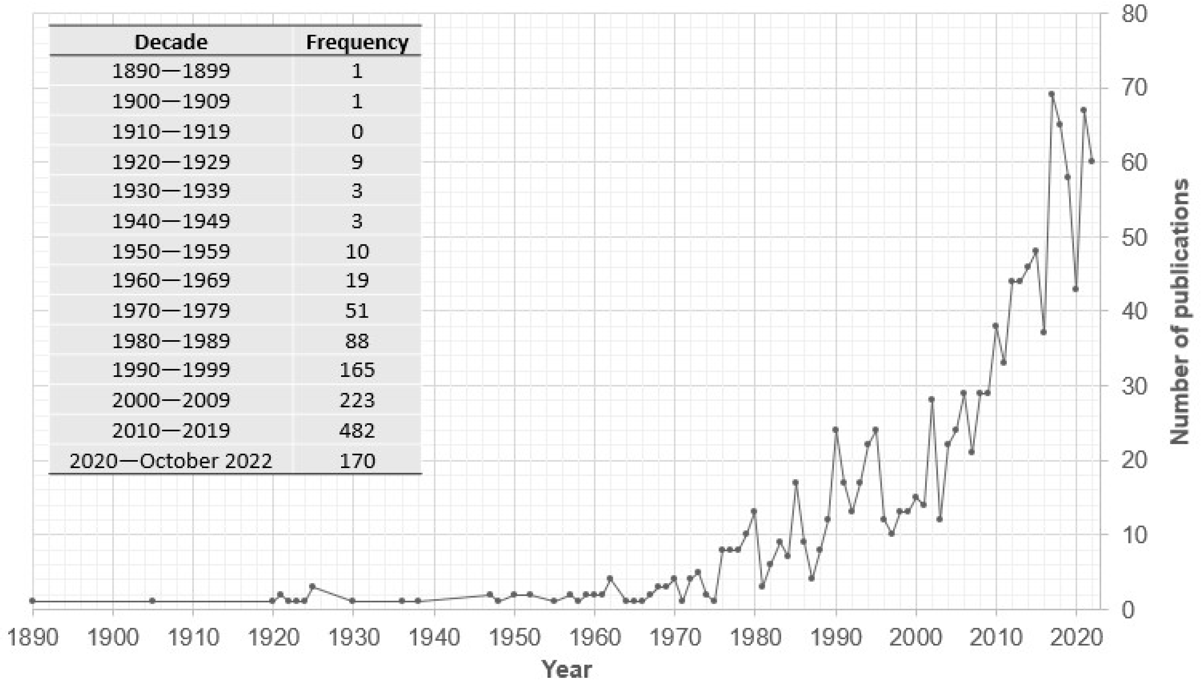
3.1.1. Age-at-Death Research Per Decades

Title and abstract search on Dimensions yielded a total of 1225 publications, with publication dates ranging from 1890 to October 2022. Publications were in English, French, Polish, Spanish, Portuguese, German, Czech, Hungarian, and Japanese. Some abstracts were translated from their original languages into English; hence, it was possible to access the content. Most were published as articles, in peer review journals (n = 1123), with the remaining being allocated to book chapters (n = 73), encyclopedia entries (n = 7), preprints (n = 2), and 13 conference proceedings. There was no information about the type of source for seven titles, but these were included in the content analysis.
Although 1225 publications were analyzed, those might not fully reflect the corpus of work in age-at-death research. Nevertheless, it still enables exploring research trends, institutional collaborative networks, and their impact on aging research. We may argue that the number of publications on aging estimation is in line with the difficulty of predicting, with a high degree of certainty, the exact age-of-death in adults. This is especially true in forensic anthropology, where accuracy toward a positive identification is key [47,48]. Furthermore, as the discipline becomes more present in academia and research agendas [44], the number of publications on individual profiling also grew. This will be further supported when exploring the data per publication source (discussed below: 3.2. Bibliometric mapping of the TOP 4 journals).
The analysis of the publication per decade, and year breakdown (Figure 1), shows that despite some fluctuations, there is a positive trend in publications, namely, from 1980 and 1990 onwards with a peak of publication in 2017 (n = 69; Figure 1), with publications decreasing sharply between 2019 (n = 58) and 2020 (n = 43). Again, the interest in this research topic can be explained by the increase in the number of professionals and students dedicated, worldwide, to human remains assessment, whereas the sharp decrease in publications in 2019 and 2020 could be related to the COVID-19 pandemic: global government shutdowns restricting access to osteological collections and equipment for data collection and analysis. In 2021, the number of publications quickly recovered (n = 67), which may be associated with the easing of restrictions imposed during the COVID-19 pandemic.
Despite fluctuations in the number of publications, the last decade (2010–2019) accounts for the most publications number (n = 482) to date, testifying to the increased interest in aging research. The ability to estimate a person’s age-at-death as accurately as possible is paramount in forensic anthropology [47,48], and desirable in studies related to past populations [49], therefore justifying its interest. However, accurate age-at-death estimation continues to be challenging.
Between 2020 and October 2022, 170 titles were published; when compared to the last decade, this represents 35.27% of all publications from the previous decade. Hence, if the pace of aging research papers continues to increase at the current rate, it is possible that in four years, in 2028, there will be close to 500 publications more on aging research. We are, therefore, driven to ask—what has been published on aging research over the past century? Is there still more to explore? The focus of future aging research may still be on reformulating and testing, and improving current methods in various osteological collections, particularly documented skeletal collections. However, with the recent emergence of artificial intelligence-based age analysis (e.g., [16,17,18]), we may see the use of more automated methods based on prior knowledge—including errors and biases—in skeletal aging methods. This is especially true given the rapid development of artificial intelligence technology and its increased application in science [50]. Hence, future research needs to consider not only new methodological and statistical approaches to aging methods, but also try to understand what other biological variables affect bone and dental aging in adult individuals, as only a few studies on the topic have emerged [15,51,52,53,54,55,56,57].

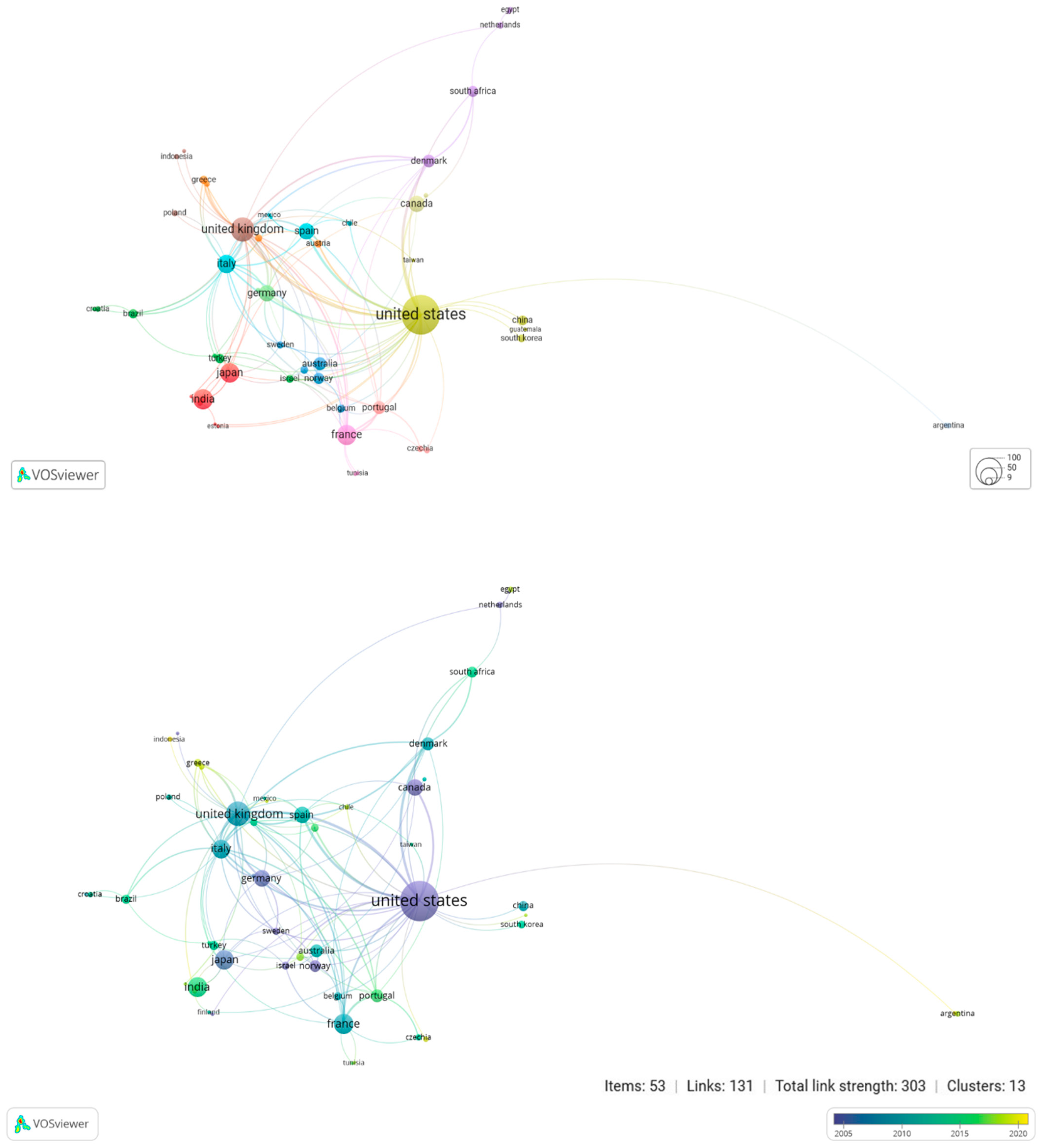
3.1.2. Institutional Co-Authorship Network

All co-authored documents, with at least one collaborative paper among institutions, were included in this analysis. This resulted in 35 clusters, as shown in Figure 2. The institutions with the highest co-authorship network included the University of Coimbra, the National Museum of Natural History, Case Western Reserve University, the University of Toronto, the University of Tennessee, the University of Oslo, the University of Milan, and the University of Edinburgh: all located in Europe and North America. The collaboration was not direct in all cases, but intermediated with other academic institutions with lesser publication profiles. Moreover, smaller collaborative networks occurred within and between non-Western institutions, primarily with Brazilian and Asian institutions.
It was interesting to note that the majority of the collaborations emerged from 2010 onwards (Figure 2), with the University of Coimbra and the University of Edinburgh aggregating the most collaborative profiles. In the past, such roles were attributed to the National Museum of Natural History, Case Western Reserve University, University of Toronto, and the University of Tennessee. It was also interesting to note that most collaborative networks for non-Western institutions took place after the 2010s, suggesting that aging research had not been a major focus of interest in the past. The visualization of the institutional co-authorship network over the years clearly shows that there has been a global expansion, and interest, in age estimation research, which has led to newer collaborations, as well as the development of more localized, smaller, collaborative networks. Such countries are primarily located in the global south and Asia. A similar pattern was found by Alves-Cardoso and Campanacho [44] when reviewing publications linking forensic anthropology with research undertaken using documented skeletal collections. As argued by Alves-Cardoso and Campanacho [44], the emergence of newer institutions contributing to aging research, which takes place mostly after 2014, is most probably related to the creation of new human-documented skeletal collections. These collections are the bases of major hypothesis-driven research on biological profiling since they allow control for age-at-death and sex when developing new methods of research [44].
The viewing of the institutional co-authorship network per country highlighted the United States had a major focus on collaborative networks (n = 288; Figure 3). However, these collaborations related mostly to publications prior to 2010. Other visual collaboration clusters were linked to the United Kingdom, France, India, Japan, Italy, Spain, and Germany, though not as prominently as the United States, and typically with more recent papers (after the 2010s).
In 2017, the number of international collaborations increased, as evidenced by the emergence of partnerships with Egypt, Malaysia, Chile, Saudi Arabia, Romania, Argentina, Greece, Cyprus, Tunisia, Guatemala, Thailand, Mexico, and Colombia (Figure 3). This could explain the increase in the number of publications on age estimation from 2016 to 2017 (37 to 69 publications, respectively) seen in Figure 1, as collaborations and publications grow on a global scale. The number of publications from newer and emerging countries, i.e., which have only recently become engaged in aging research, is likely to increase in the coming years.

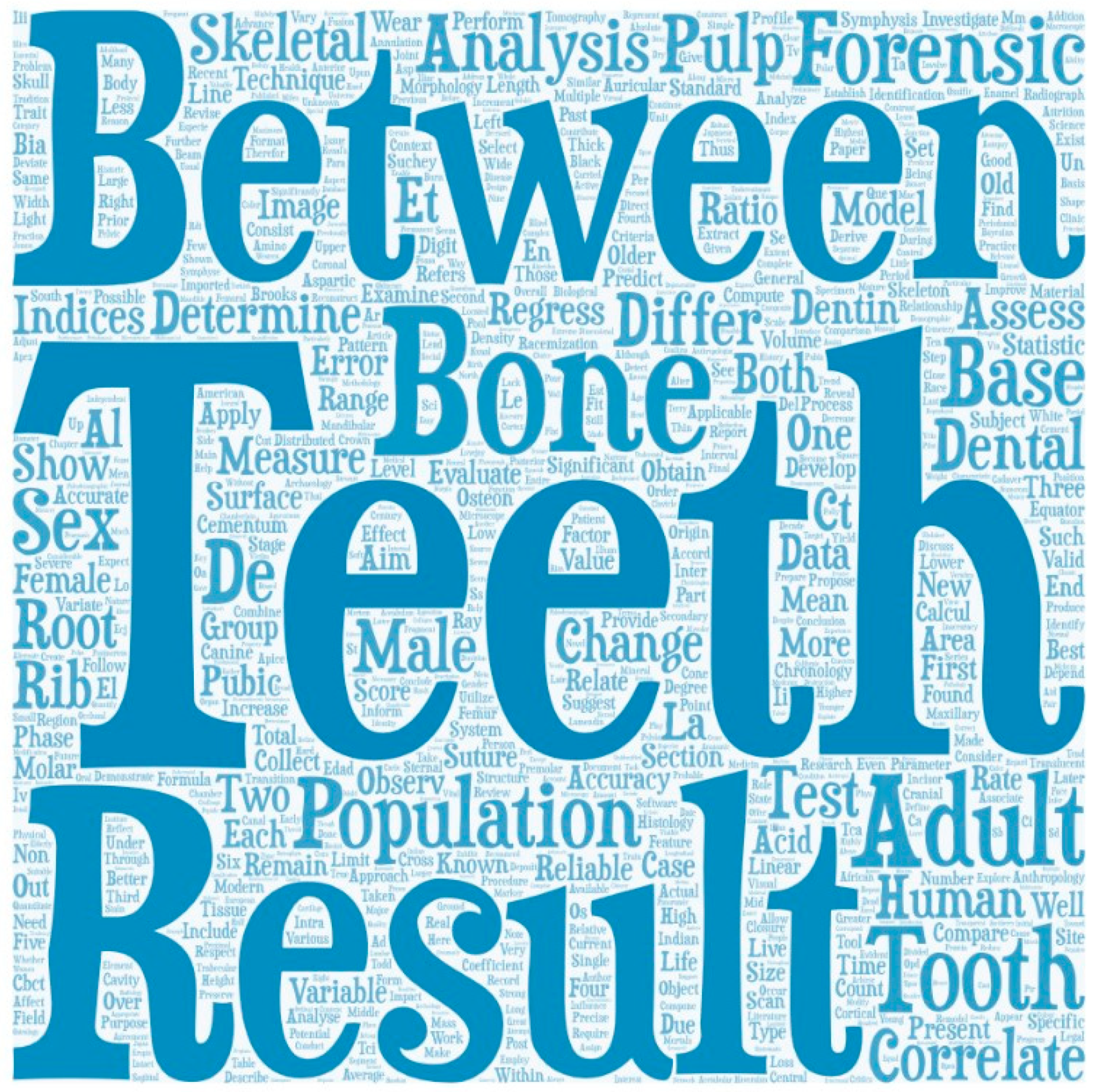
3.1.3. Words/Terms Content Analysis

Vague terms whose frequencies were in the thousands, such as ‘Age’, cluttered the analysis making it difficult to visualize which topics received more attention from the scientific community in age estimation studies. Therefore, some of the most frequent terms found in an initial content analysis (i.e., age (n = 7719); estimation (n = 3404); method (n = 2606); use (n = 2296); study (1297); death (1142); year (1123); sample (1082); and individual (n = 1053) were eliminated as they were found to be redundant. No vague terms, such as ‘Number’ (n = 200), were eliminated because they occurred less frequently and did not clutter the analysis. A similar approach was followed for terms analysis for the TOP4 journals (see Section 3.2.3). Figure 4 displays the most frequently used terms. It also revealed an emphasis on dental age analysis as the most frequent words were ‘Teeth’ (n = 942), ‘Tooth’ (n = 763), ‘Dental’ (n = 587), ‘Pulp’ (n = 531), ‘Dentin’ (n = 506), and ‘Root’ (n = 462). Even though ‘Bone’ (n = 810) appears almost as frequently as ‘Teeth’, terms associated with bone analysis were less common: those frequently used were ‘Rib’ (n = 401), ‘Suture’ (n = 353), ‘Pubic’ (n = 383), and ‘Auricular’ (n = 220). Among the terms associated with bone analysis, the least mentioned was ‘Acetabulum’ (n = 72). This difference may reflect newer research topics—hence, less visible in publications—as opposed to others. For example, interest in age-at-death studies based on the acetabulum may be considered fairly recent: the first mentions were made by Rougé-Maillart et al. [58] and Rissech et al. [59], thus contributing to a lower frequency of the term ‘Acetabulum’. While İşcan et al. [5,6,7], and Lovejoy et al. [60] studies on degenerescence of the sternal end of the fourth rib, and auricular surface (respectively) were published in the 1980s, being seminal research on age estimation. Hence, their earlier publication date and influential contribution to age estimation research may account for the highest frequency of the terms ‘Rib’ and ‘Auricular’ in the content analysis. The term ‘Rib’ may further stand out, when compared to ‘Auricular’, because: age estimation based on ribs is linked to macroscopic and histological methods of analysis, whereas auricular surface analysis is mostly performed by macroscopic observation; and, due to the fact that sternal end of ribs has been studied in deceased individuals, during autopsies and in osteological collections, whereas auricular surface studies have primarily been conducted using osteological collections.
Alongside the use of terms related to anatomy, it is also worth noting the frequency of the terms ‘Forensic’ (n = 761) and ‘Population’ (n = 790). The former is associated with papers on biological profiling, which includes age-at-death estimation methods in the forensic sciences [44]. Moreover, given that the aging process differs between individuals and populations [15,25,54], it is not surprising that the term ‘Population’ appears frequently. This awareness has led to the development and, most importantly, the testing of aging methods in samples drawn from different populations [61,62].
The content analysis, based on the co-occurrence of terms, revealed four major clusters related to dental-related age estimation (see Figure 5), demonstrating once more a greater prominence of tooth analysis in age estimation research. A cluster of terms related to macroscopic bone analysis is reported in red, opposite to the clusters related to dental-related age estimation. The pubic symphysis, sternal rib end, auricular surface, and cranial sutures synostosis are viewed as the most often used age estimation methods [63]; therefore, it is surprising their lower co-occurrence compared to terms associated with dental aging. The emphasis on dental-related age estimation is also contrary to an electronic survey undertaken by members of the American Association of Forensic Sciences [64]. The survey revealed respondents preferred to use traditional methods developed on bone, such as pubic symphysis, the sternal rib ends, the auricular surface, and the cranial sutures (ranked in that order), regardless of the participants’ experience, rather than teeth [64]. The mapping of the co-occurrence of terms per year of publications highlighted the fact that dental-related age research gained a profile mostly after 2006.
In a more central position of the co-occurrence network, we find a cluster related to histological methods (yellow cluster: Figure 5). Although primarily associated with bone analysis, with the term ‘Section’ connecting most clusters, it also connects with dental-related age estimation. The yellow cluster is less dense, with fewer publications, despite histological methods being used since Kerley’s [65] age estimation paper. The reasons for this are probably because histological methods involve a specific set of skills, not always taught in anthropological degrees, as well as necessary equipment for sample preparation, and bone destruction, which is not always permissible or ethical [1,66].
Figure 5 also shows that, although analysis of the metamorphosis of the pubic symphysis and the auricular surface first appeared in the 1920s and 1985, respectively, it continues to be a topic of research in more recent years. This exemplifies, to perfection, the continuous assessment and adaptation of more classical age-at-death methods. The same observation is true for the term ‘Suchey Brooks method’, which has, since its publication in 1990, continued to influence age estimation research, and has been revisited by subsequent research, having benefited from technological development [67]. After 2015, in yellow also, the co-occurrence of terms is biased towards the applicability of technological development, and newer statistical approaches to aging research. These are exemplified by terms such as ‘Transition analysis’, ‘Tomography’, ‘Tomography image’, ‘CT scan’, ‘Cone beam’, ‘Pulp’, Pulp volume’ [41,67,68,69].

3.1.4. TOP5 Most Cited Articles

Three of the five most cited articles (TOP5) were published in the American Journal of Physical Anthropology, now the American Journal of Biological Anthropology (which we will use in this paper), and the other two in human evolution journals (Table 1). The articles were published between 1980 and 2002. Three papers—Lovejoy et al. [60], Brooks and Suchey [3], and Buckberry and Chamberlain [4]—discuss age-related degenerescence of the pelvic joints; a fourth paper discusses the fusion of the ectocranial suture closure with age [10]; and a fifth paper is a review article with recommendations for biological profiling of human remains [70]. Given that the terms connected to teeth age-related changes were the most frequent, surprisingly, the articles that feature in the TOP5 do not focus on dental analysis—this shows that term co-occurrence and publications frequency are not necessarily related to citation indexes. This TOP5 shows that bone-related research stands as the formative literature for those teaching and learning age estimation methods. For example, macroscopic aging techniques such as those used by Lovejoy et al. [60], Brooks and Suchey [3], Buckberry and Chamberlain [4], and Meindl and Lovejoy [10] are traditionally taught, and tested, in anthropological and forensic courses, contributing to a higher citation rate by students and professionals. Dental methodologies that involve radiological and/or histological analysis may not be as accessible if an institution lacks the necessary equipment, and skilled human resources to develop and teach such approaches to age assessment. Which, consequently, will diminish research publications.

3.2. Bibliometric Mapping of the TOP 4 Journals

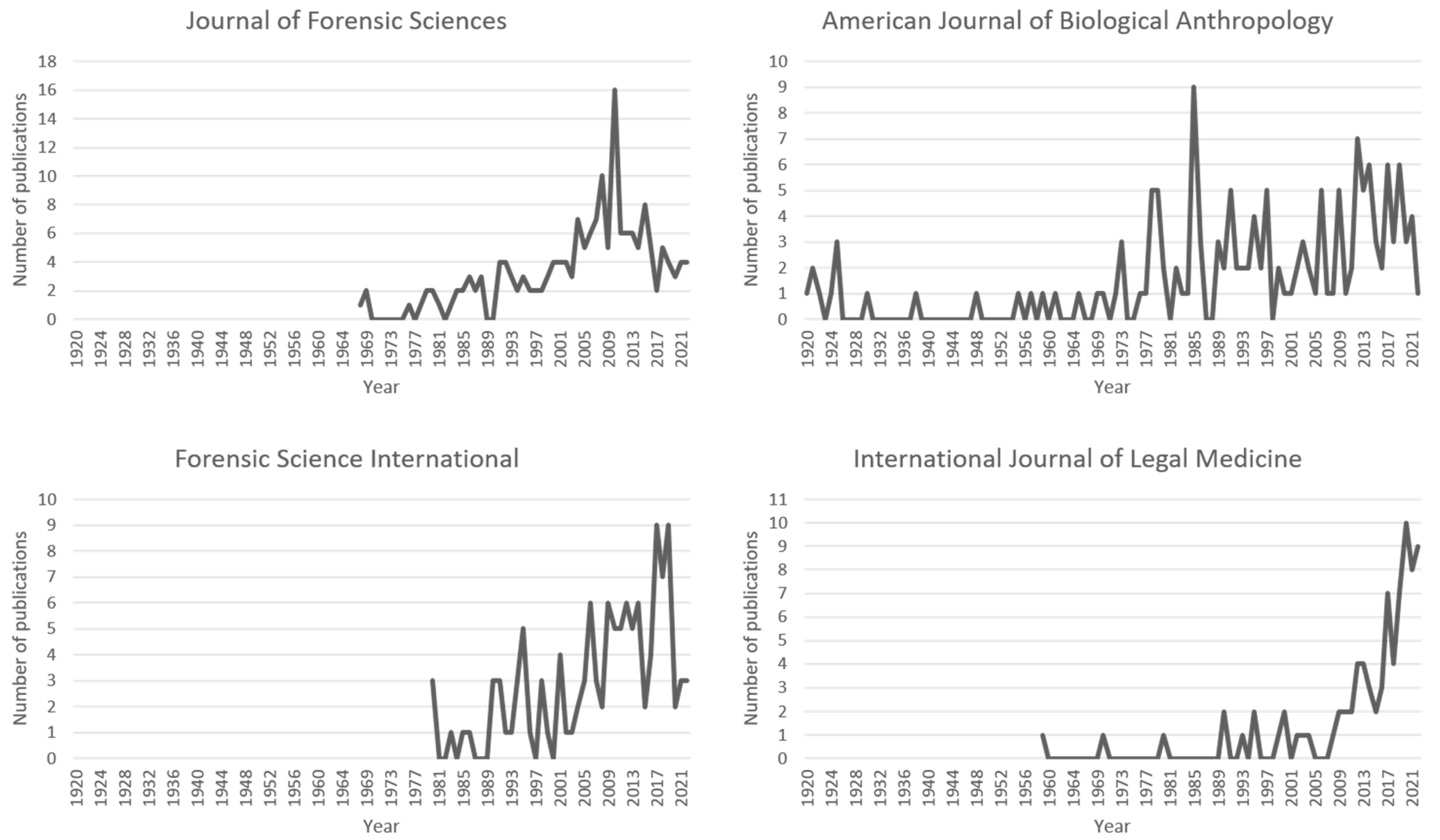
3.2.1. Number of Publications Per Journal in Aging Research

Forensic research features heavily in the TOP4 journals as shown in Table 2, indicating a growing interest in the research of biological profiling in forensic sciences. The American Journal of Biological Anthropology, which contains some of the most cited titles (Table 1), is also one of the TOP4 journals with the second highest number of aging publications. The remaining papers are distributed in various journals, in which forensics continues to be well present. However, there is a significant body of literature (34.91%) in journals with fewer than ten papers on aging research. This may be due to the exponential growth in the number of journals, giving authors a wider range of journal options in which to publish.
The breakdown of the number of articles published per TOP4 journals shows the longevity of the journals, alongside their contribution to the topic (Figure 6). For example, while the American Journal of Biological Anthropology has been publishing since the 1920s, forensic journals began publishing from 1959s onwards, contributing significantly towards aging research dissemination. Despite considerable fluctuations in the number of papers published each year, journals showed a consistent positive growth over the years, although three of the TOP4 journals showed a decrease in publications recently. The International Journal of Legal Medicine is the only one with continued growth, as detailed in Figure 6. This could indicate a broader range of options for publishing age research, especially given the rapid emergence of new journals; and/or a higher rate of desk rejection of age papers in the TOP4 journals.

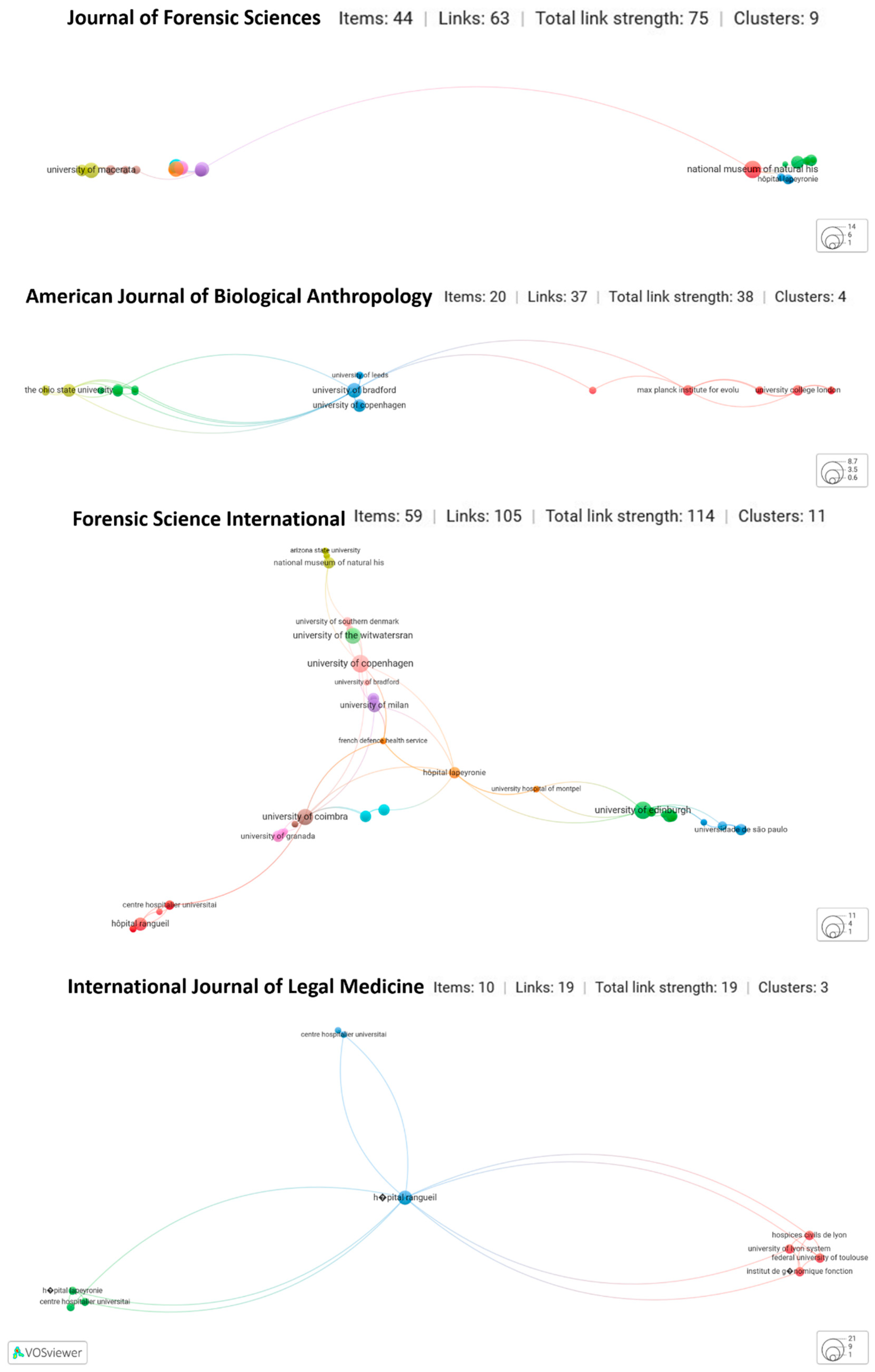
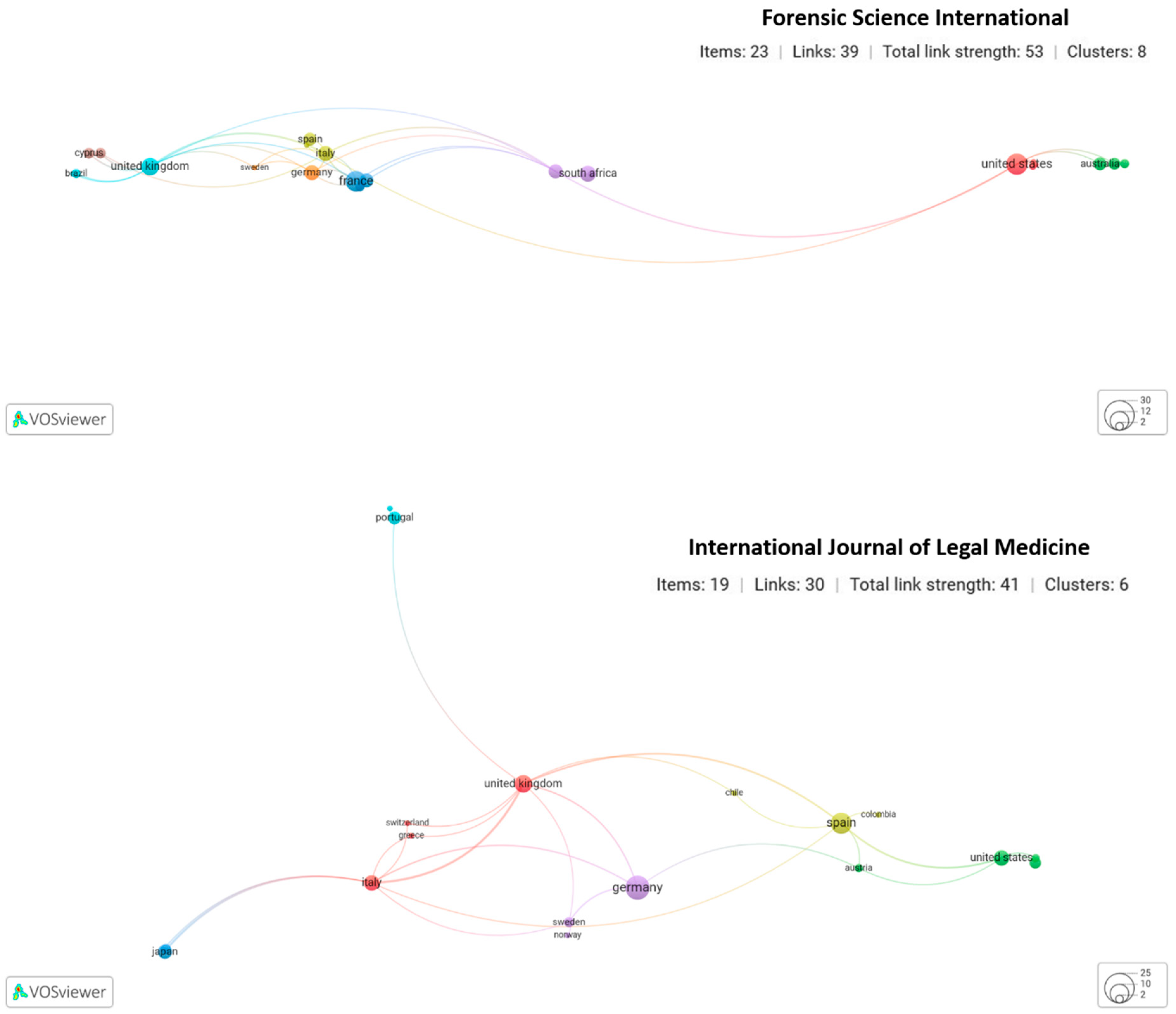
3.2.2. Co-Authorship among Institutions and Institutional Countries for the TOP4 Journals

When compared to the overall analysis (Section 3.1.2; Figure 2), co-authorship by institutions occurred on a smaller scale (Figure 7). Collaborations were primarily with and within North American and European institutions (Figure 7, Figure 8 and Figure 9), demonstrating a weaker global collaboration than seen for the overall trend for age publications (Figure 3). This could indicate a global preference for other journal outlets, or systemic limitations by newer and emerging countries in this line of publication to publish in the TOP4 journals.

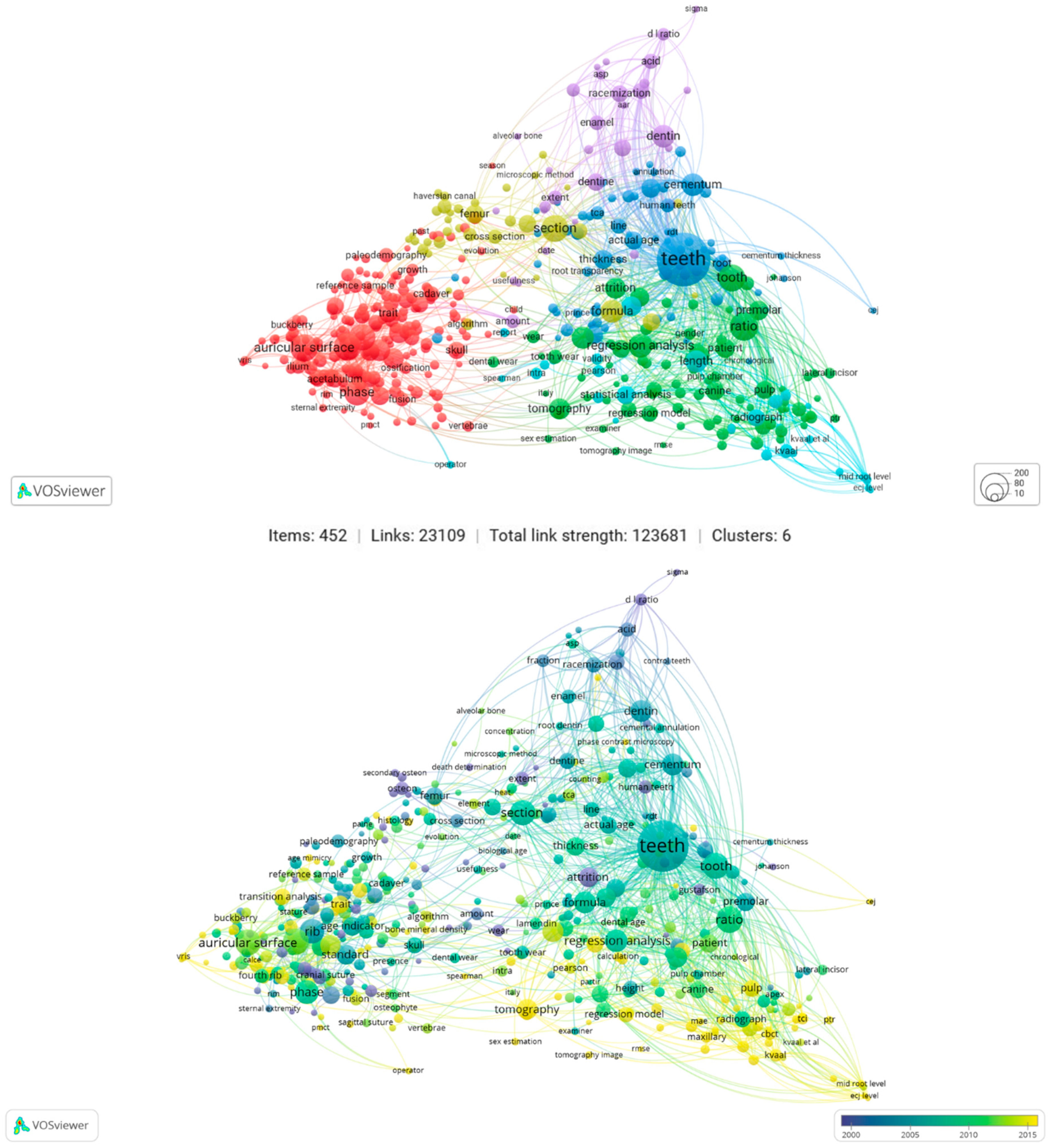
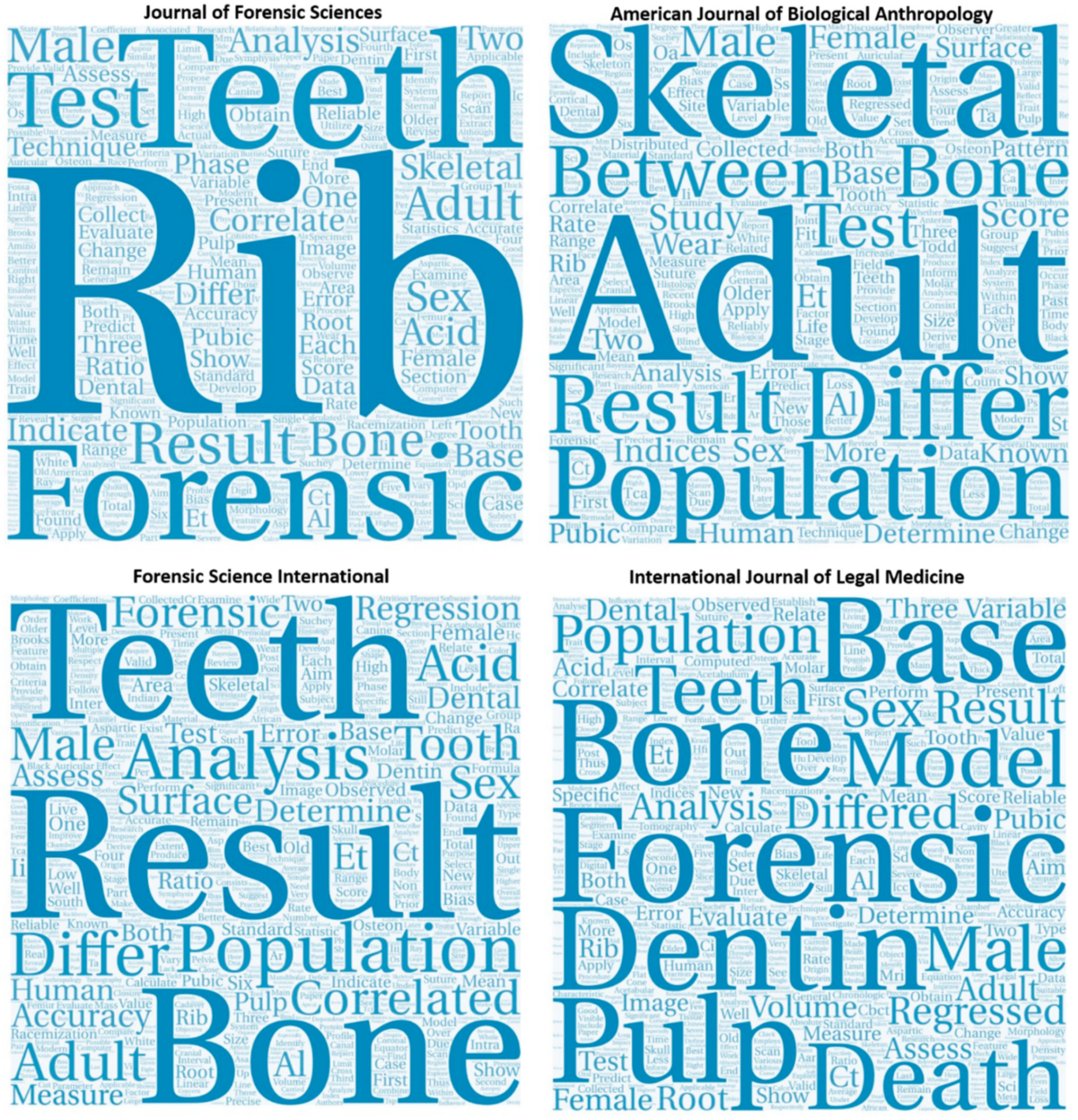
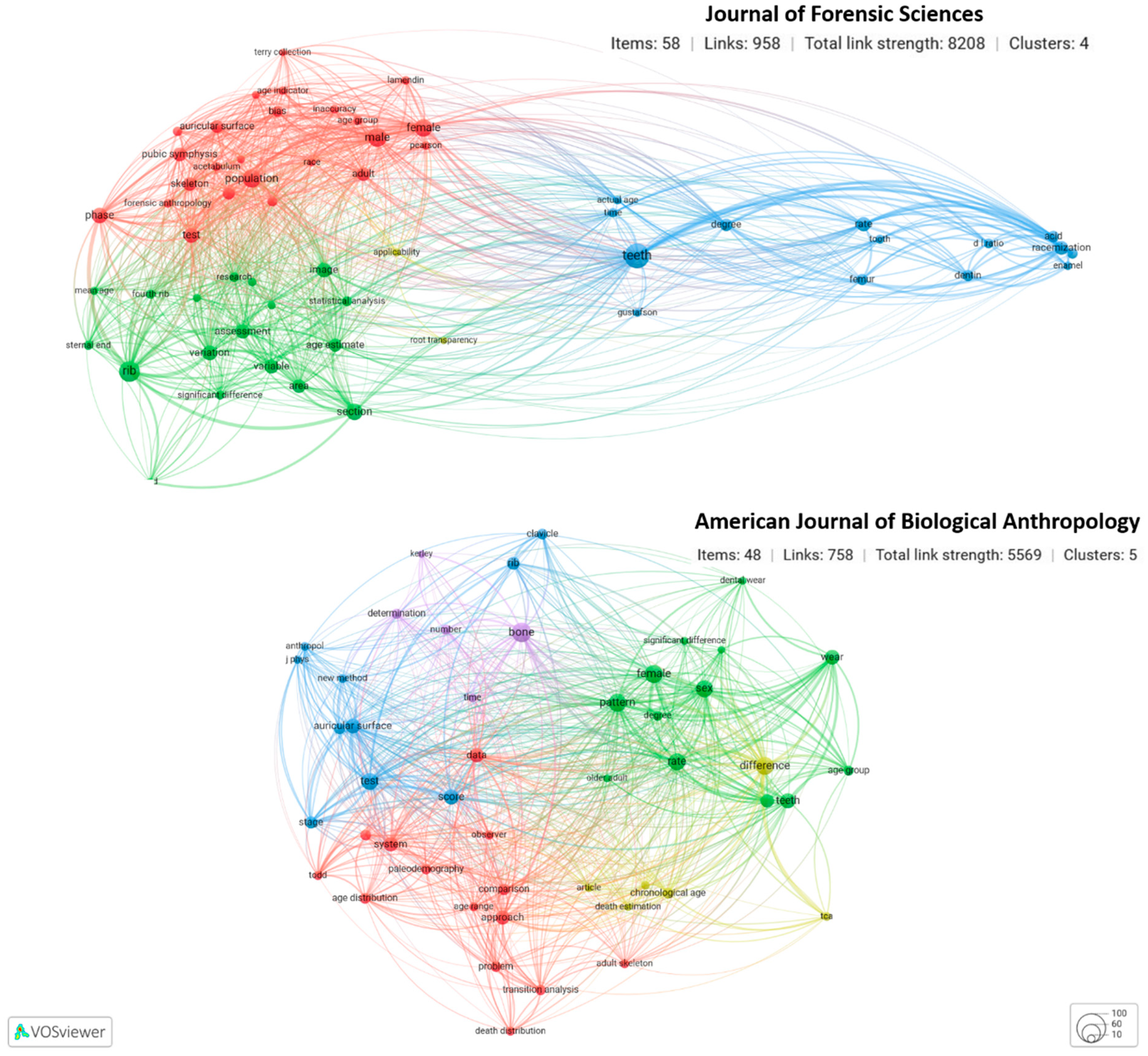
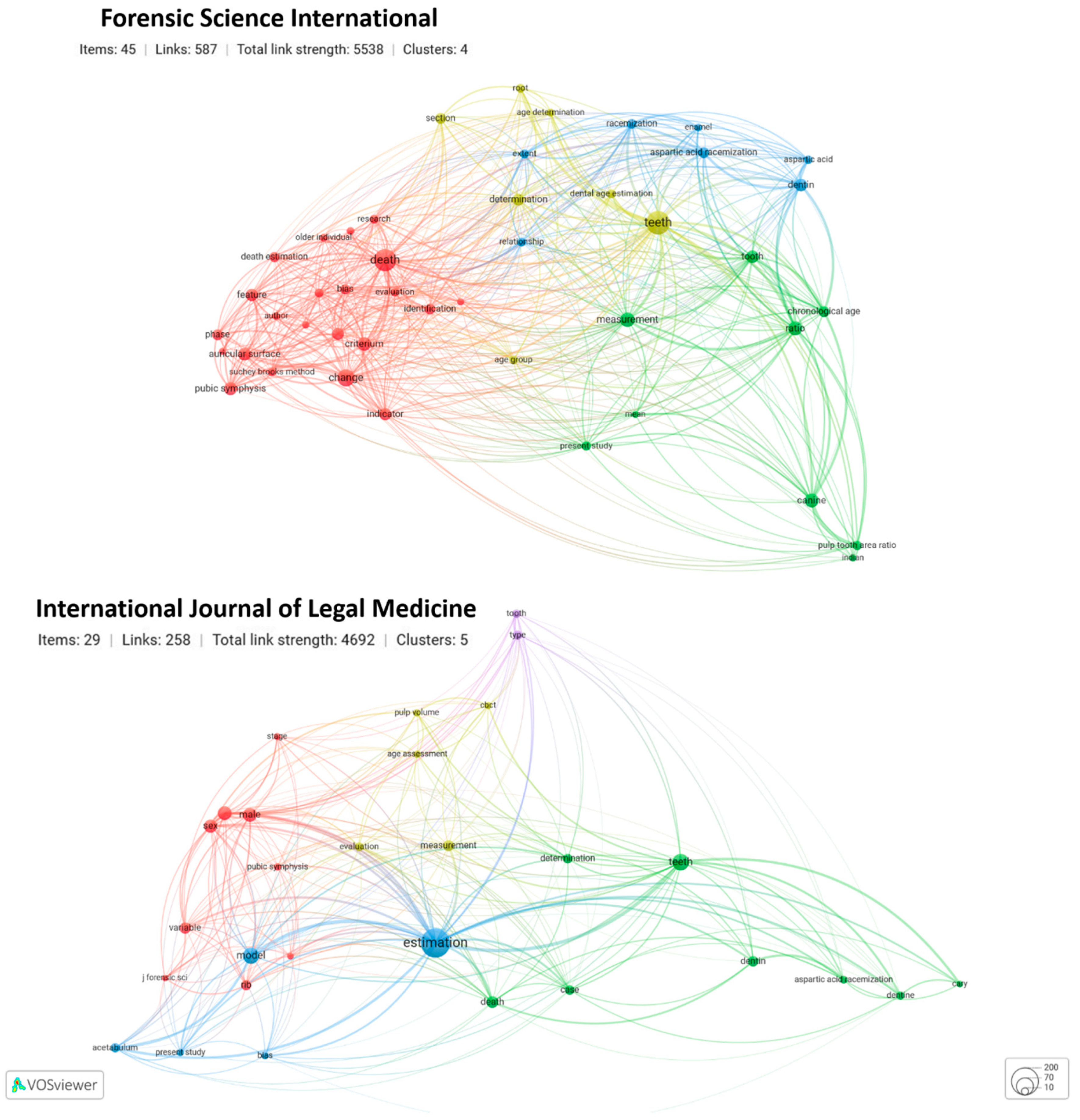
3.2.3. Frequency and Co-Occurrences of Terms for the TOP4 Journals

Figure 10 illustrates the most frequent terms for the TOP4 journals. Data showed that the Forensic Science International and the International Journal of Legal Medicine both had a similar frequency for ‘Bone’ and dental-related terms (Figure 4). While the term ‘Rib’ appeared more frequently in the Journal of Forensic Sciences. For the American Journal of Biological Anthropology, terms associated with bone analysis and the term ‘Population’ appeared more frequently. This suggests that the Journal of Forensic Sciences and the American Journal of Biological Anthropology placed a greater emphasis on publishing aging methods that examined age-related bone degenerescence than the other two TOP4 journals.
The visualization of the co-occurrence of terms in the Journal of Forensic Sciences and the American Journal of Biological Anthropology showed different patterns (Figure 11). The Journal of Forensic Sciences showed three major connecting poles, of which terms associated with teeth are singled out (blue color) (Figure 5). The co-occurrence of terms associated with bone analysis is represented by the red and green clusters, and although they also gather individually, there is a stronger connection between them. The red cluster is associated mostly with terms related to anatomy, collection, and biological profiling, while the green cluster has a stronger focus on statistical analysis. There is a third cluster, the yellow cluster is smaller in size, and connectivity, with only two terms co-occurring (i.e., ‘Root transparency’ and ‘Applicability’), which is linked to the other three clusters further suggesting that although grouped differently, they are not exclusive. The American Journal of Biological Anthropology provided a completely different pattern of clusters. Five major clusters were identified, but the level of connectivity between them is high. This suggests that the papers published were more inclusive in the terminology used, most probably aggregating multi-methodological and multi-sampled research including teeth and bone. The investigation of dental wear and age was the primary focus of the co-occurrence of terms relating to teeth.
The co-occurrence of terms for Forensic Science International and the International Journal of Legal Medicine can be found in Figure 12, with both journals showing distinctive patterns of term connectivity. From the TOP4 journals, Forensic Science International cluster pattern is closest to the one observed for the overall trend (Figure 5). Teeth-associated terms stand out in the Forensic Science International, establishing a network with the remaining clusters, including the cluster ‘Death’ which connects with bone analyses (as opposed to teeth). The International Journal of Legal Medicine co-occurrence network is less complex, with fewer clusters, and more sparse connectivity between them. However, as observed for the other journal, teeth-related terms continue to stand out, but for this journal, the term ‘Estimation’ is the one supporting cluster connectivity, highlighting the degree of quantitative and qualitative assessment of age-at-death estimation methods.

3.2.4. Citations for the TOP4 Journals

Table 3 shows the TOP5 most cited articles from the TOP4 journals. The profile of the articles per journal varies, as does the number of citations. For example, the two most cited articles published by the Journal of Forensic Sciences were those of İşcan et al. [5] and İşcan et al. [7] on age estimation by observing the sternal extremity of the fourth rib. While the most cited article published by the American Journal of Biological Anthropology was that of Lovejoy et al. [60] on the metamorphosis of the auricular surface of the ilium. The Forensic Science International’s most cited paper is a review article on aging by Cunha et al. [71], which evaluates and makes recommendations for age estimation based on various factors such as the level of preservation of the body, elements present, and sex. Finally, The paper by Ritz-Timme et al. [72] on assessed aging methodologies and made recommendations is the most cited paper published by the International Journal of Legal Medicine. We have two papers on methods and two reviews as major research articles as the most cited in these highly referenced journals.

4. Conclusions

Estimating age-at-death in human remains of adult individuals is a challenging exercise. It is not only dependent on the experience of the researchers undertaking the task but also intrinsically linked to the methods used. The last century has seen a flourishing engagement on this research topic. Methods have diversified the skeletal traits (in bone and teeth) used for age assessment. Technological advances offer novel possibilities for exploring how bone and teeth biology reflect a person’s aging process: not only recurring to new statistical approaches (some benefiting from artificial intelligence), but also by diversifying what is observed—solely macroscopic observation has given rise to micro and virtual observation. Moreover, more samples have become available for hypothesis testing and methodological developments, allowing control for individual and population variability. Furthermore, with the growing number of human-documented collections available worldwide, it has become possible to account for social, economic, cultural, and historical variables when designing age estimation research. Furthermore, with the added scenario of virtuality, many collections are no longer limited to real dry bone analysis, but also virtual facsimiles. Finally, it is also necessary to consider forensic anthropology’s impact to develop the most accurate possible aging method. The relevance of forensic anthropology in this research topic is well supported by the number of publications featured in forensic journals and the growing number of journals dedicated to forensic sciences.
All of the above have accounted for the results found when mapping bibliometric data and undertaking the content analysis: there has been continuous positive growth on this research agenda; methods and samples have diversified, although some constancy exists as to the sample core—either bone or teeth, with the latter leading research agendas; many methods are revisitation and reformulation of classical approaches—primarily due to new technological access. The complexity of all these possibilities is well illustrated by institutional co-authorship networking maintained over the years, with Western universities leading publications ranking. However, it was also observed that in recent years there has been a diversification of institutional contributors no longer limited to past geographic patterns, namely, North America and Europe.
Ultimately, age estimation methods will continue to be a research agenda in the coming years. As a result, we may expect many more publications. Based on the temporal visualization trends highlighted in this paper, we anticipate that novel technologies applied in dental-age estimation research will find their way to bone base research. Furthermore, research undertaken in geographically dispersed institutions will make this research more inclusive and hopefully bring novelty. In all, a higher level of accuracy in results is necessary; hence, having passed a century of publications on aging research, we need to question if mere repetition, replication, reformulation, and sample changes are sufficient to advance the field. Nonetheless, testing new and old age estimation methodologies will always be necessary, particularly in the legal context. In the United States, for example, Daubert v. Merrell Dow Pharmaceuticals, Inc., 509 U.S. 579 (1993) requires experts to use methods that have been tested and their error rates established in order to be admissible in court.

Author Contributions

Conceptualization, V.C. and F.A.-C.; methodology, V.C. and F.A.-C.; formal analysis, V.C.; writing—original draft preparation, V.C. and F.A.-C.; writing—review and editing final version V.C. and F.A.-C.; funding acquisition, F.A.-C. All authors have read and agreed to the published version of the manuscript.

Funding

Francisca Alves Cardoso (FAC) research is funded by the research project Life After Death: Rethinking Human Remains and Human Osteological Collections as Cultural Heritage and Biobanks (ref: 2020.01014.CEECIND/CP1634/CT0002) funded by the Portuguese Fundação para a Ciência e Tecnologia (FCT). FAC is further supported by CRIA (ref: UID/04038/2020) funded by FCT.

Institutional Review Board Statement

Not applicable.

Informed Consent Statement

Not applicable.

Data Availability Statement

Data are currently available at Zenodo.org (DOI:10.5281/zenodo.7592809).

Acknowledgments

We would like to acknowledge the contributions made by all individuals whose study of remains, and/or visual data, has allowed for the development of the age-at-death estimation field. We thank the two anonymous reviewers whose comments helped improve the manuscript.

Conflicts of Interest

The authors declare no conflict of interest. The funders had no role in the design of the study; in the collection, analyses, or interpretation of data; in the writing of the manuscript; or in the decision to publish the results.

References

  1. Ubelaker, D.H.; Khosrowshahi, H. Estimation of Age in Forensic Anthropology: Historical Perspective and Recent Methodological Advances. Forensic Sci. Res. 2019, 4, 1–9. [Google Scholar] [CrossRef] [PubMed] [Green Version]
  2. Todd, T.W. Age Changes in the Pubic Bone. I. The Male White Pubis. Am. J. Phys. Anthropol. 1920, 3, 285–334. [Google Scholar] [CrossRef]
  3. Brooks, S.; Suchey, J.M. Skeletal Age Determination Based on the Os Pubis: A Comparison of the Acsádi-Nemeskéri and Suchey-Brooks Methods. Hum. Evol. 1990, 5, 227–238. [Google Scholar] [CrossRef]
  4. Buckberry, J.L.; Chamberlain, A.T. Age Estimation from the Auricular Surface of the Ilium: A Revised Method. Am. J. Phys. Anthropol. 2002, 119, 231–239. [Google Scholar] [CrossRef] [PubMed]
  5. İşcan, M.Y.; Loth, S.R.; Wright, R.K. Age Estimation from the Rib by Phase Analysis: White Males. J. Forensic Sci. 1984, 29, 11776J. [Google Scholar] [CrossRef]
  6. İşcan, M.Y.; Loth, S.R.; Wright, R.K. Metamorphosis at the Sternal Rib End: A New Method to Estimate Age at Death in White Males. Am. J. Phys. Anthropol. 1984, 65, 147–156. [Google Scholar] [CrossRef] [PubMed]
  7. İşcan, M.Y.; Loth, S.R.; Wright, R.K. Age Estimation from the Rib by Phase Analysis: White Females. J. Forensic Sci. 1985, 30, 11018J. [Google Scholar] [CrossRef]
  8. Calce, S.E. A New Method to Estimate Adult Age-at-Death Using the Acetabulum. Am. J. Phys. Anthropol. 2012, 148, 11–23. [Google Scholar] [CrossRef]
  9. Todd, T.W.; Lyon, D.W. Endocranial Suture Closure. Its Progress and Age Relationship. Part I.—Adult Males of White Stock. Am. J. Phys. Anthropol. 1924, 7, 325–384. [Google Scholar] [CrossRef]
  10. Meindl, R.S.; Lovejoy, C.O. Ectocranial Suture Closure: A Revised Method for the Determination of Skeletal Age at Death Based on the Lateral-Anterior Sutures. Am. J. Phys. Anthropol. 1985, 68, 57–66. [Google Scholar] [CrossRef]
  11. Gustafson, G. Microscopic Examination of Teeth as a Means of Identification in Forensic Medicine. J. Am. Dent. Assoc. 1947, 35, 720–724. [Google Scholar] [CrossRef] [PubMed]
  12. Lamendin, H. Radicular dentin and estimation of age. Inf. Dent. 1972, 54, 1647–1662. [Google Scholar] [PubMed]
  13. Lamendin, H.; Baccino, E.; Humbert, J.F.; Tavernier, J.C.; Nossintchouk, R.M.; Zerilli, A. A Simple Technique for Age Estimation in Adult Corpses: The Two Criteria Dental Method. J. Forensic Sci. 1992, 37, 13327J. [Google Scholar] [CrossRef]
  14. Bosmans, N.; Ann, P.; Aly, M.; Willems, G. The Application of Kvaal’s Dental Age Calculation Technique on Panoramic Dental Radiographs. Forensic Sci. Int. 2005, 153, 208–212. [Google Scholar] [CrossRef]
  15. Campanacho, V. The Influence of Skeletal Size on Age-Related Criteria from the Pelvic Joints in Portuguese and North American Samples. Ph.D. Thesis, University of Sheffield, Sheffield, UK, 2016. [Google Scholar]
  16. Corsini, M.-M.; Schmitt, A.; Bruzek, J. Aging Process Variability on the Human Skeleton: Artificial Network as an Appropriate Tool for Age at Death Assessment. Forensic Sci. Int. 2005, 148, 163–167. [Google Scholar] [CrossRef]
  17. Buk, Z.; Kordik, P.; Bruzek, J.; Schmitt, A.; Snorek, M. The Age at Death Assessment in a Multi-Ethnic Sample of Pelvic Bones Using Nature-Inspired Data Mining Methods. Forensic Sci. Int. 2012, 220, 294.e1–294.e9. [Google Scholar] [CrossRef] [PubMed]
  18. Navega, D.; Costa, E.; Cunha, E. Adult Skeletal Age-at-Death Estimation through Deep Random Neural Networks: A New Method and Its Computational Analysis. Biology 2022, 11, 532. [Google Scholar] [CrossRef]
  19. Dwight, T. The Closure of the Cranial Sutures as a Sign of Age. Boston Med. Surg. J. 1890, 122, 389–392. [Google Scholar] [CrossRef] [Green Version]
  20. Todd, T.W. Age Changes in the Pubic Bone: Age Changes in the Pubic Bone. Am. J. Phys. Anthropol. 1921, 4, 1–70. [Google Scholar] [CrossRef] [Green Version]
  21. Hanihara, K.; Suzuki, T. Estimation of Age from the Pubic Symphysis by Means of Multiple Regression Analysis. Am. J. Phys. Anthropol. 1978, 48, 233–239. [Google Scholar] [CrossRef]
  22. Snow, C.C. Equations for Estimating Age at Death from the Pubic Symphysis: A Modification of the McKern-Stewart Method. J. Forensic Sci. 1983, 28, 864–870. [Google Scholar] [CrossRef] [PubMed]
  23. Katz, D.; Suchey, J.M. Age Determination of the Male Os Pubis. Am. J. Phys. Anthropol. 1986, 69, 427–435. [Google Scholar] [CrossRef] [PubMed]
  24. Katz, D.; Suchey, J.M. Race Differences in Pubic Symphyseal Aging Patterns in the Male. Am. J. Phys. Anthropol. 1989, 80, 167–172. [Google Scholar] [CrossRef]
  25. Schmitt, A.; Murail, P.; Cunha, E.; Rougé, D. Variability of the Pattern of Aging on the Human Skeleton: Evidence from Bone Indicators and Implications on Age at Death Estimation. J. Forensic Sci. 2002, 47, 1203–1209. [Google Scholar] [CrossRef]
  26. Bocquet-Appel, J.-P.; Masset, C. Farewell to Paleodemography. J. Hum. Evol. 1982, 11, 321–333. [Google Scholar] [CrossRef]
  27. Aykroyd, R.G.; Lucy, D.; Pollard, A.M.; Solheim, T. Technical Note: Regression Analysis in Adult Age Estimation. Am. J. Phys. Anthropol. 1997, 104, 259–265. [Google Scholar] [CrossRef]
  28. Aykroyd, R.G.; Lucy, D.; Pollard, A.M.; Roberts, C.A. Nasty, Brutish, but Not Necessarily Short: A Reconsideration of the Statistical Methods Used to Calculate Age at Death from Adult Human Skeletal and Dental Age Indicators. Am. Antiq. 1999, 64, 55–70. [Google Scholar] [CrossRef]
  29. Falys, C.G.; Lewis, M.E. Proposing a Way Forward: A Review of Standardisation in the Use of Age Categories and Ageing Techniques in Osteological Analysis (2004–2009): Proposing a Way Forward. Int. J. Osteoarchaeol. 2011, 21, 704–716. [Google Scholar] [CrossRef]
  30. Bocquet-Appel, J.P.; Masset, C. Paleodemography: Expectancy and False Hope. Am. J. Phys. Anthropol. 1996, 99, 571–583. [Google Scholar] [CrossRef]
  31. Meindl, R.S.; Russell, K.F. Recent Advances in Method and Theory in Paleodemography. Annu. Rev. Anthropol. 1998, 27, 375–399. [Google Scholar] [CrossRef]
  32. Boldsen, J.; Milner, G.R.; Konigsberg, L.M.; Wood, J.W. Transition Analysis: A New Method for Estimating Age from the Skeleton. In Paleodemography: Age Distributions from Skeletal Samples; Hoppa, R.D., Vaupel, J.W., Eds.; Cambridge University Press: Cambridge, UK, 2002; pp. 73–106. [Google Scholar]
  33. Chamberlain, A.T. Demography in Archaeology, 1st ed.; Cambridge University Press: Cambridge, UK, 2006; ISBN 978-0-521-59651-0. [Google Scholar]
  34. Godde, K.; Hens, S.M. Age-at-Death Estimation in an Italian Historical Sample: A Test of the Suchey-Brooks and Transition Analysis Methods. Am. J. Phys. Anthropol. 2012, 149, 259–265. [Google Scholar] [CrossRef] [PubMed]
  35. Jackes, M. Representativeness and Bias in Archaeological Skeletal Samples. In Social Bioarchaeology; Agarwal, S.C., Glencross, B.A., Eds.; Wiley-Blackwell: Oxford, UK, 2011; pp. 107–146. ISBN 978-1-4443-9053-7. [Google Scholar]
  36. Milner, G.R.; Boldsen, J.L. Transition Analysis: A Validation Study with Known-Age Modern American Skeletons. Am. J. Phys. Anthropol. 2012, 148, 98–110. [Google Scholar] [CrossRef] [PubMed]
  37. Anderson, M.F.; Anderson, D.T.; Wescott, D.J. Estimation of Adult Skeletal Age-at-Death Using the Sugeno Fuzzy Integral. Am. J. Phys. Anthropol. 2009, 142, 30–41. [Google Scholar] [CrossRef] [Green Version]
  38. Ferrant, O.; Rougé-Maillart, C.; Guittet, L.; Papin, F.; Clin, B.; Fau, G.; Telmon, N. Age at Death Estimation of Adult Males Using Coxal Bone and CT Scan: A Preliminary Study. Forensic Sci. Int. 2009, 186, 14–21. [Google Scholar] [CrossRef] [PubMed]
  39. Stoyanova, D.; Algee-Hewitt, B.F.B.; Slice, D.E. An Enhanced Computational Method for Age-at-Death Estimation Based on the Pubic Symphysis Using 3D Laser Scans and Thin Plate Splines: Age-At-Death Estimation Using TPS. Am. J. Phys. Anthropol. 2015, 158, 431–440. [Google Scholar] [CrossRef] [PubMed]
  40. Stoyanova, D.K.; Algee-Hewitt, B.F.B.; Kim, J.; Slice, D.E. A Computational Framework for Age-at-Death Estimation from the Skeleton: Surface and Outline Analysis of 3D Laser Scans of the Adult Pubic Symphysis. J. Forensic Sci. 2017, 62, 1434–1444. [Google Scholar] [CrossRef]
  41. Kotěrová, A.; Štepanovský, M.; Buk, Z.; Brůžek, J.; Techataweewan, N.; Velemínská, J. The Computational Age-at-death Estimation from 3D Surface Models of the Adult Pubic Symphysis Using Data Mining Methods. Sci. Rep. 2022, 12, 10324. [Google Scholar] [CrossRef]
  42. Bass, W.M. Recent Developments in the Identification of Human Skeletal Material. Am. J. Phys. Anthropol. 1969, 30, 459–461. [Google Scholar] [CrossRef]
  43. Bass, W.M. Developments in the Identification of Human Skeletal Material (1968–1978). Am. J. Phys. Anthropol. 1979, 51, 555–562. [Google Scholar] [CrossRef]
  44. Alves-Cardoso, F.; Campanacho, V. The Scientific Profiles of Documented Collections via Publication Data: Past, Present, and Future Directions in Forensic Anthropology. Forensic Sci. 2022, 2, 37–56. [Google Scholar] [CrossRef]
  45. Chen, X.; Chen, J.; Wu, D.; Xie, Y.; Li, J. Mapping the Research Trends by Co-Word Analysis Based on Keywords from Funded Project. Procedia Comput. Sci. 2016, 91, 547–555. [Google Scholar] [CrossRef] [Green Version]
  46. Dimensions Dimensions Analytics: The Basics. 2022. Available online: https://www.dimensions.ai/resources/product-guide-dimensions-analytics/ (accessed on 1 November 2022).
  47. Baccino, E.; Schmitt, A. Determination of Adult Age at Death in the Forensic Context. In Forensic Anthropology and Medicine; Schmitt, A., Cunha, E., Pinheiro, J., Eds.; Humana Press: Totowa, NJ, USA, 2006; pp. 259–280. ISBN 978-1-58829-824-9. [Google Scholar]
  48. Cunha, E. Aging the Death: The Importance of Having Better Methods for Age at Death Estimation of Old Individuals. Ann. Med. 2021, 53, S1. [Google Scholar] [CrossRef] [PubMed]
  49. Paleodemography: Age Distributions from Skeletal Samples; Cambridge Studies in Biological and Evolutionary Anthropology; Hoppa, R.D.; Vaupel, J.W. (Eds.) Cambridge University Press: Cambridge, UK, 2008; ISBN 978-0-521-08916-6. [Google Scholar]
  50. Xu, Y.; Liu, X.; Cao, X.; Huang, C.; Liu, E.; Qian, S.; Liu, X.; Wu, Y.; Dong, F.; Qiu, C.-W.; et al. Artificial Intelligence: A Powerful Paradigm for Scientific Research. Innovation 2021, 2, 100179. [Google Scholar] [CrossRef]
  51. Hoppa, R.D. Population Variation in Osteological Aging Criteria: An Example from the Pubic Symphysis. Am. J. Phys. Anthropol. 2000, 111, 185–191. [Google Scholar] [CrossRef]
  52. Taylor, K.M. The Effects of Alcohol and Drug Abuse on the Sternal End of the Fourth Rib. Ph.D. Thesis, University of Arizona, Tucson, AZ, USA, 2000. [Google Scholar]
  53. Mays, S. An Investigation of Age-Related Changes at the Acetabulum in 18th-19th Century Ad Adult Skeletons from Christ Church Spitalfields, London. Am. J. Phys. Anthropol. 2012, 149, 485–492. [Google Scholar] [CrossRef] [PubMed]
  54. Mays, S. The Effect of Factors Other than Age upon Skeletal Age Indicators in the Adult. Ann. Hum. Biol. 2015, 42, 332–341. [Google Scholar] [CrossRef]
  55. Campanacho, V.; Santos, A.L.; Cardoso, H.F.V. Assessing the Influence of Occupational and Physical Activity on the Rate of Degenerative Change of the Pubic Symphysis in Portuguese Males from the 19th to 20th Century. Am. J. Phys. Anthropol. 2012, 148, 371–378. [Google Scholar] [CrossRef] [PubMed]
  56. Merritt, C.E. The Influence of Body Size on Adult Skeletal Age Estimation Methods: Influence of Body Size on Age Estimation. Am. J. Phys. Anthropol. 2015, 156, 35–57. [Google Scholar] [CrossRef] [Green Version]
  57. Wescott, D.J.; Drew, J.L. Effect of Obesity on the Reliability of Age-at-Death Indicators of the Pelvis: Effects of Obesity on Age-at-death. Am. J. Phys. Anthropol. 2015, 156, 595–605. [Google Scholar] [CrossRef]
  58. Rougé-Maillart, C.; Telmon, N.; Rissech, C.; Malgosa, A.; Rougé, D. The Determination of Male Adult Age at Death by Central and Posterior Coxal Analysis—A Preliminary Study. J. Forensic Sci. 2004, 49, 208–214. [Google Scholar] [CrossRef]
  59. Rissech, C.; Estabrook, G.F.; Cunha, E.; Malgosa, A. Using the Acetabulum to Estimate Age at Death of Adult Males. J. Forensic Sci. 2006, 51, 213–229. [Google Scholar] [CrossRef] [PubMed]
  60. Lovejoy, C.O.; Meindl, R.S.; Pryzbeck, T.R.; Mensforth, R.P. Chronological Metamorphosis of the Auricular Surface of the Ilium: A New Method for the Determination of Adult Skeletal Age at Death. Am. J. Phys. Anthropol. 1985, 68, 15–28. [Google Scholar] [CrossRef] [PubMed]
  61. Gocha, T.P.; Ingvoldstad, M.E.; Kolatorowicz, A.; Cosgriff-Hernandez, M.-T.J.; Sciulli, P.W. Testing the Applicability of Six Macroscopic Skeletal Aging Techniques on a Modern Southeast Asian Sample. Forensic Sci. Int. 2015, 249, 318.e1–318.e7. [Google Scholar] [CrossRef] [PubMed] [Green Version]
  62. Du, H.; Li, G.; Zheng, Q.; Yang, J. Population-Specific Age Estimation in Black Americans and Chinese People Based on Pulp Chamber Volume of First Molars from Cone Beam Computed Tomography. Int. J. Leg. Med. 2022, 136, 811–819. [Google Scholar] [CrossRef]
  63. Garvin, H.M.; Passalacqua, N.V.; Uhl, N.M.; Gipson, D.R.; Overbury, R.S.; Cabo, L.L. Developments in Forensic Anthropology: Age-at-Death Estimation. In A Companion to Forensic Anthropology; Dirkmaat, D.C., Ed.; Wiley-Blackwell: West Sussex, UK, 2012; pp. 202–223. ISBN 978-1-4051-9123-4. [Google Scholar]
  64. Garvin, H.M.; Passalacqua, N.V. Current Practices by Forensic Anthropologists in Adult Skeletal Age Estimation: Age Estimation Practices. J. Forensic Sci. 2012, 57, 427–433. [Google Scholar] [CrossRef]
  65. Kerley, E.R. The Microscopic Determination of Age in Human Bone. Am. J. Phys. Anthropol. 1965, 23, 149–163. [Google Scholar] [CrossRef]
  66. Squires, K.; García-Mancuso, R. Desafíos Éticos Asociados al Estudio y Tratamiento de Restos Humanos En Las Ciencias Antropológicas En El Siglo XXI. Rev. Argent. Antrop. Biol. 2021, 23, 34. [Google Scholar] [CrossRef]
  67. Telmon, N.; Gaston, A.; Chemla, P.; Blanc, A.; Joffre, F.; Rougé, D. Application of the Suchey-Brooks Method to Three-Dimensional Imaging of the Pubic Symphysis. J. Forensic Sci. 2005, 50, 507–512. [Google Scholar] [CrossRef]
  68. Warrier, V.; Kanchan, T.; Garg, P.K.; Dixit, S.G.; Krishan, K.; Shedge, R. CT-Based Evaluation of the Acetabulum for Age Estimation in an Indian Population. Int. J. Leg. Med. 2022, 136, 785–795. [Google Scholar] [CrossRef]
  69. Warrier, V.; Shedge, R.; Garg, P.K.; Dixit, S.G.; Krishan, K.; Kanchan, T. Computed Tomographic Evaluation of the Acetabulum for Age Estimation in an Indian Population Using Principal Component Analysis and Regression Models. Int. J. Leg. Med. 2022, 136, 1637–1653. [Google Scholar] [CrossRef]
  70. Ferembach, D.; Schwindezky, I.; Stoukal, M. Recommendations for Age and Sex Diagnoses of Skeletons. J. Hum. Evol. 1980, 9, 517–549. [Google Scholar] [CrossRef]
  71. Cunha, E.; Baccino, E.; Martrille, L.; Ramsthaler, F.; Prieto, J.; Schuliar, Y.; Lynnerup, N.; Cattaneo, C. The Problem of Aging Human Remains and Living Individuals: A Review. Forensic Sci. Int. 2009, 193, 1–13. [Google Scholar] [CrossRef]
  72. Ritz-Timme, S.; Cattaneo, C.; Collins, M.J.; Waite, E.R.; Schütz, H.W.; Kaatsch, H.-J.; Borrman, H.I.M. Age Estimation: The State of the Art in Relation to the Specific Demands of Forensic Practise. Int. J. Leg. Med. 2000, 113, 129–136. [Google Scholar] [CrossRef]
  73. Cameriere, R.; Ferrante, L.; Cingolani, M. Variations in Pulp/Tooth Area Ratio as an Indicator of Age: A Preliminary Study. J. Forensic Sci. 2004, 49, 1–3. [Google Scholar] [CrossRef]
  74. Konigsberg, L.W.; Herrmann, N.P.; Wescott, D.J.; Kimmerle, E.H. Estimation and Evidence in Forensic Anthropology: Age-at-Death. J. Forensic Sci. 2008, 53, 541–557. [Google Scholar] [CrossRef]
  75. Lovejoy, C.O. Dental Wear in the Libben Population: Its Functional Pattern and Role in the Determination of Adult Skeletal Age at Death. Am. J. Phys. Anthropol. 1985, 68, 47–56. [Google Scholar] [CrossRef] [PubMed]
  76. Kvaal, S.I.; Kolltveit, K.M.; Thomsen, I.O.; Solheim, T. Age Estimation of Adults from Dental Radiographs. Forensic Sci. Int. 1995, 74, 175–185. [Google Scholar] [CrossRef]
  77. Ogino, T.; Ogino, H.; Nagy, B. Application of Aspartic Acid Racemization to Forensic Odontology: Post Mortem Designation of Age at Death. Forensic Sci. Int. 1985, 29, 259–267. [Google Scholar] [CrossRef]
  78. Solheim, T. A New Method for Dental Age Estimation in Adults. Forensic Sci. Int. 1993, 59, 137–147. [Google Scholar] [CrossRef]
  79. Paewinsky, E.; Pfeiffer, H.; Brinkmann, B. Quantification of Secondary Dentine Formation from Orthopantomograms?A Contribution to Forensic Age Estimation Methods in Adults. Int. J. Leg. Med. 2005, 119, 27–30. [Google Scholar] [CrossRef]
  80. Ritz, S.; Schütz, H.W.; Peper, C. Postmortem Estimation of Age at Death Based on Aspartic Acid Racemization in Dentin: Its Applicability for Root Dentin. Int. J. Leg. Med. 1993, 105, 289–293. [Google Scholar] [CrossRef] [PubMed]
  81. Ferrante, L.; Cameriere, R. Statistical Methods to Assess the Reliability of Measurements in the Procedures for Forensic Age Estimation. Int. J. Leg. Med. 2009, 123, 277–283. [Google Scholar] [CrossRef] [PubMed]
  82. Landa, M.I.; Garamendi, P.M.; Botella, M.C.; Alemán, I. Application of the Method of Kvaal et al. to Digital Orthopantomograms. Int. J. Leg. Med. 2009, 123, 123–128. [Google Scholar] [CrossRef] [PubMed]
Figure 1. Number of publications per year. Table: frequency of publications per decade.
Forensicsci 03 00011 g001
Figure 2. The upper image illustrates the network of publications co-authorship by institutions, while the lower image displays the same co-authorship network per year. The interactive map is accessible at https://app.vosviewer.com/?json=https://drive.google.com/uc?id=1wqzWo6Q8Xhl1VbaI5oWI18P2bXESjXcO (accessed on 15 January 2023).
Forensicsci 03 00011 g002
Figure 3. The upper image shows the network visualization co-authorship of institutional countries, and the lower image replicated the data per year. All publications were included regardless of co-authorship count—23 clusters were identified. Bibliometric mapping is interactable at https://app.vosviewer.com/?json=https://drive.google.com/uc?id=1JQISmtge-GFubEc6qieJjb4iCyeymqrV (accessed on 15 January 2023).
Forensicsci 03 00011 g003
Figure 4. Word cloud for terms used on the title and abstracts for age research.
Forensicsci 03 00011 g004
Figure 5. Upper image: network visualization co-occurrence of terms. Lower image: overlay visualization co-occurrence of terms per year. Co-occurrence of terms were extracted from the title and abstract using a full counting method. The minimum number of terms was set at 10; only 753 of the 17,172 terms met the requirement. VOSviewer then assigned a relevance score to each of the 753 terms and chose 60% of the most relevant terms. The analysis ultimately highlighted 452 terms and six clusters of term co-occurrences. Visible and interactable at https://app.vosviewer.com/?json=https://drive.google.com/uc?id=1GEq4bVmSNCUczngsCtdvpZ59EVudK_iu (Accessed on 15 January 2023).
Forensicsci 03 00011 g005
Figure 6. Distribution of the number of publications per year for the TOP4 journals.
Forensicsci 03 00011 g006
Figure 7. Network visualization of co-authorship by institutions for publications in the TOP4 journals. The interactable maps are accessible at: Journal of Forensic Sciences https://app.vosviewer.com/?json=https://drive.google.com/uc?id=1KwDJ4Ps_jf7yCejj1yfVZtkLub5TA2Ch; American Journal of Biological Anthropology https://app.vosviewer.com/?json=https://drive.google.com/uc?id=133lAqjDSo1FarDSFYY8K1i4-haXZ-ydc; Forensic Science International https://app.vosviewer.com/?json=https://drive.google.com/uc?id=1I4o_h7VP2vnHJcYp7pHr-4j9oP2NTdo2; International Journal of Legal Medicine https://app.vosviewer.com/?json=https://drive.google.com/uc?id=14aWpyQ_Ru27pjkdCNukSdgA_48Lqhb7H (Accessed on 16 January 2023).
Forensicsci 03 00011 g007
Figure 8. Network visualization of co-authorship by institutional countries for publications in the Journal of Forensic Sciences and the American Journal of Biological anthropology. The interactable maps are accessible at: Journal of Forensic Sciences https://app.vosviewer.com/?json=https://drive.google.com/uc?id=1v0tbG8dZP4-bXM5GMTIugKEH9FB9hzMh; American Journal of Biological Anthropology https://app.vosviewer.com/?json=https://drive.google.com/uc?id=1FLvcCMZYKpb0GZgxcG8HQ9jU0RjNeyI9 (Accessed on 16 January 2023).
Forensicsci 03 00011 g008
Figure 9. Network visualization of co-authorship by institutional countries for publications of the Forensic Science International and the International Journal of Legal Medicine. The interactable maps are accessible at: Forensic Science International https://app.vosviewer.com/?json=https://drive.google.com/uc?id=1dy7RVu39t4y7UyjpAvtIhnVQsKLIAp1_; International Journal of Legal Medicine https://app.vosviewer.com/?json=https://drive.google.com/uc?id=1glHeo3fqa56IEGXq9aq586JRmgmjtojU (Accessed on 16 January 2023).
Forensicsci 03 00011 g009
Figure 10. Word Clouds for frequent terms used in title and abstracts for the TOP4 journals.
Forensicsci 03 00011 g010
Figure 11. Network visualization for the co-occurrence of terms for the Journal of Forensic Sciences and the American Journal of Biological Anthropology of the TOP4 journals. The interactable maps are accessible at: Journal of Forensic Sciences https://app.vosviewer.com/?json=https://drive.google.com/uc?id=16pEsDcuttezxVFMqVbfvmqtddQaANKR6; American Journal of Biological Anthropology https://app.vosviewer.com/?json=https://drive.google.com/uc?id=10LVG4GlLiRc3WewE98doQ3AWmo8QO8-e (Accessed on 16 January 2023).
Forensicsci 03 00011 g011
Figure 12. Network visualization for the co-occurrence of terms for Forensic Science International and International Journal of Legal Medicine and the American Journal of Biological Anthropology of the TOP4 journals. The interactable maps are accessible at: Forensic Science International https://app.vosviewer.com/?json=https://drive.google.com/uc?id=1Hax3Qkt7os5JqKl61TEeYRrDzW7Sk4hS; International Journal of Legal Medicine https://app.vosviewer.com/?json=https://drive.google.com/uc?id=1KOSNyp6sb6y18c18n_IPaRoQEfUjHQ-R (Accessed on 16 January 2023).
Forensicsci 03 00011 g012
Table 1. TOP5 Titles most cited.
TitleJournalYearAuthorsTimes Cited
Skeletal age determination based on the os pubis: A comparison of the Acsádi-Nemeskéri and Suchey-Brooks methodsHuman Evolution1990Brooks and Suchey [3]1262
Chronological metamorphosis of the auricular surface of the ilium: A new method for the determination of adult skeletal age at deathAmerican Journal of Biological Anthropology1985Lovejoy et al. [60]1154
Ectocranial suture closure: A revised method for the determination of skeletal age at death based on the lateral-anterior suturesAmerican Journal of Biological Anthropology1985Meindl and Lovejoy [10]891
Recommendations for age and sex diagnoses of skeletonsJournal of Human Evolution1980Ferembach et al. [70] 624
Age estimation from the auricular surface of the ilium: A revised methodAmerican Journal of Biological Anthropology2002Buckberry and Chamberlain [4]475
Table 2. Frequency and percentage of publications per journal in aging.
Source TitleFrequencyPercent
Journal of Forensic Sciences17715.76
American Journal of Biological Anthropology15213.54
Forensic Science International12110.77
International Journal of Legal Medicine827.30
Journal of Forensic Dental Sciences252.23
International Journal of Osteoarchaeology242.14
Journal of Forensic Odonto-Stomatology201.78
Legal Medicine191.69
Anthropologischer Anzeiger181.60
Journal of Forensic and Legal Medicine151.34
Australian Journal of Forensic Sciences151.34
Forensic Imaging151.34
Journal of Archaeological Science141.25
Bulletins et Mémoires de la Société d’Anthropologie de Paris121.07
Homo121.07
Japanese Journal of Legal Medicine100.89
[Remaining journal titles—9 to 1 publications]39234.91
Table 3. TOP5 titles most cited for the TOP4 journals.
TOP4 JournalsTitleYearAuthorsTimes Cited
Journal of Forensic SciencesAge estimation from the rib by phase analysis: white males.1984İşcan et al. [5]387
Age estimation from the rib by phase analysis: white females.1985İşcan et al. [7]345
A simple technique for age estimation in adult corpses: the two criteria dental method.1992Lamendin et al. [13]251
Variations in pulp/tooth area ratio as an indicator of age: a preliminary study.2004Cameriere et al. [73]157
Estimation and Evidence in Forensic Anthropology: Age-at-Death2008Konigsberg et al. [74]149
American Journal of Biological AnthropologyChronological metamorphosis of the auricular surface of the ilium: A new method for the determination of adult skeletal age at death1985Lovejoy et al. [60]1154
Ectocranial suture closure: A revised method for the determination of skeletal age at death based on the lateral-anterior sutures1985Meindl and Lovejoy [10]891
Age estimation from the auricular surface of the ilium: A revised method2002Buckberry and Chamberlain [4]475
Age changes in the pubic bone. I. The male white pubis1920Todd [2]448
Dental wear in the Libben population: Its functional pattern and role in the determination of adult skeletal age at death1985Lovejoy [75]429
Forensic Science InternationalThe problem of aging human remains and living individuals: A review2009Cunha et al. [71]403
Age estimation of adults from dental radiographs1995Kvaal et al. [76]353
Application of aspartic acid racemization to forensic odontology: Post mortem designation of age at death1985Ogino et al. [77]108
A new method for dental age estimation in adults1993Solheim [78]105
The application of Kvaal’s dental age calculation technique on panoramic dental radiographs2004Bosmans et al. [14]99
International Journal of Legal MedicineAge estimation: The state of the art in relation to the specific demands of forensic practise2000Ritz-Timme et al. [72]356
Quantification of secondary dentine formation from orthopantomograms a contribution to forensic age estimation methods in adults2004Paewinsky et al. [79]165
Postmortem estimation of age at death based on aspartic acid racemization in dentin: Its applicability for root dentin1993Ritz et al. [80]90
Statistical methods to assess the reliability of measurements in the procedures for forensic age estimation2009Ferrante and Cameriere [81]68
Application of the method of Kvaal et al. to digital orthopantomograms2008Landa et al. [82]62
Disclaimer/Publisher’s Note: The statements, opinions and data contained in all publications are solely those of the individual author(s) and contributor(s) and not of MDPI and/or the editor(s). MDPI and/or the editor(s) disclaim responsibility for any injury to people or property resulting from any ideas, methods, instructions or products referred to in the content.

