Article

Description of Two Fungal Endophytes Isolated from Fragaria chiloensis subsp. chiloensis f. patagonica: Coniochaeta fragariicola sp. nov. and a New Record of Coniochaeta hansenii

by
Carolina Campos-Quiroz
1,
Jean Franco Castro
1,*,
Cecilia Santelices
1,
Jorge Carrasco-Fernández
1,
Matías Guerra
1,
Diego Cares-Gatica
1,
Javiera Ortiz-Campos
1,
Yocelyn Ocares
1,
Lorena Barra-Bucarei
1 and
Bart Theelen
2
1
Instituto de Investigaciones Agropecuarias, INIA Quilamapu. Av. Vicente Méndez 515, Chillán 3800062, Ñuble, Chile
2
Westerdijk Fungal Biodiversity Institute, 3584 CT Utrecht, The Netherlands
*
Author to whom correspondence should be addressed.
Taxonomy 2023, 3(2), 183-203; https://doi.org/10.3390/taxonomy3020014
Submission received: 16 December 2022 / Revised: 24 March 2023 / Accepted: 27 March 2023 / Published: 3 April 2023

Abstract

:
Prospection of the endosphere of the native plant Fragaria chiloensis subsp. chiloensis f. patagonica from the foothills of the Chilean Andes led to the isolation of two strains of the genus Coniochaeta. We addressed the taxonomic placement of these strains based on DNA sequencing data using the ITS and LSU genetic markers, morphological features, and biochemical traits. One of these strains was identified as Coniochaeta hansenii, for which the anamorph and teleomorph states were described. The second strain did not seem to match any of the currently described species of this genus; therefore, we propose the name Coniochaeta fragariicola sp. nov.

1. Introduction

The genus Coniochaeta (Sacc.) Cooke [1] (Coniochaetaceae, Coniochaetales, Ascomycota) is characterized by strongly melanized, cleistothecial or perithecial, usually setose ascomata with cylindrical, unitunicate asci, and one-celled ascospores with a germ slit. Anamorphs are predominantly phialidic and often produce intercalary conidiogenous cells with lateral necks, giving rise to one-celled conidia in slimy masses, with some members developing microcyclic conidiation [2,3]. According to Index Fungorum (www.indexfungorum.org, (accessed on 12 December 2022)), the genus includes 117 species, many of which have only been described based on their morphological characteristics, with no information concerning sequenced DNA regions for phylogenetic analysis. This group of fungi occurs in both terrestrial and aquatic environments and has been reported on bark, dung, lichens, soil, wood, polluted water, submerged plant material, and other substrates [4,5,6].
Previous studies have revealed that members of Coniochaeta may have either beneficial or harmful effects on plants. Many of its members occur as endophytes of phylogenetically diverse hosts, and some species produce antimicrobial secondary metabolites that aid in the protection against plant pathogenic species of Colletotrichum, Verticillium, and other genera [7,8]. On the other hand, certain Coniochaeta species have the ability to infect woody plants, such as apricot and peach [9], while others are lignicolous, humicolous, and coprohilus [10]. However, they are generally regarded as low-virulent, opportunistic pathogens that proliferate on wounded, senescent, or infected plant tissues [4]. Genome mining and transcriptome analyses have discovered that some members of Coniochaeta have exceptional lignocellulolytic machinery to deconstruct lignin [11]. Several Coniochaeta species, mostly known as anamorphs, have been implicated in superficial and deep-seated, life-threatening forms of phaeohyphomycosis in humans and dogs [12,13,14]. Other species are known for their teleomorphs, such as coprohilous Coniochaeta with poly-spored asci [15].
The taxonomic history of Coniochaeta is complex, and early phylogenetic studies found that, under its traditional circumscription, this genus was polyphyletic [16]. Nevertheless, in a molecular study conducted by García et al. [2], a redefinition of the genus was proposed, which included the synonymization of three morphologically similar genera, i.e., Coniochaetidium, Ephemeroascus, and Poroconiochaeta with Coniochaeta. Further taxonomic improvements were made with the transfer of several species originally described in Rosellinia and in the asexually typified genus Lecythophora to Coniochaeta, based on molecular and phenotypic data [12,17]. Despite all these advancements towards a more stable taxonomy of this genus, no DNA sequence data is currently available for many of its members and many species most likely remain undiscovered, especially in poorly studied regions.
During taxonomic studies on the microfungi associated with the native Chilean wild strawberry Fragaria chiloensis (Rosaceae, Rosales), two strains of the genus Coniochaeta were isolated. One of these strains did not seem to match any of the currently described species of this genus. In this paper, we addressed the taxonomic placement of these strains based on the ITS and LSU genetic markers, morphological features, and biochemical traits.

2. Materials and Methods

2.1. Sampling and Fungal Isolation

In 2020, 12 plants of Fragaria chiloensis subsp. chiloensis f. patagonica were collected from a forested area located in Pinto, Ñuble region, Chile (36.818367 S, 71.622217 W; altitude: 697 m.a.s.l.). All 12 plants were considered as one sample and were placed inside a sterile plastic bag and transported to the laboratory in a dark container. The sample was dissected into three sections: (i) roots, (ii) crown, and (iii) the phyllosphere containing the petioles and leaves. Each section was individually surface sterilized by submerging the tissues for 1 min in 70% ethanol, 3 min in 1.5% NaClO, and 1 min in 96% ethanol; finally, they were rinsed three times in sterile distilled water [18,19]. Nine tissues 5 mm in diameter were cut with a sterile scalpel and distributed on 24-well sterile microplates containing noble agar (NA). The plates were incubated at 25 °C for 2 wk and checked under a microscope every day for the emergence of endophytic fungi from the tissues. Upon emergence, fungal tips were removed using a sterile needle and inoculated on NA plates and incubated for 7 d at 25 °C. Two yeast-like strains were recovered and deposited in the Chilean Collection of Microbial Genetic Resources (CChRGM) of the Institute of Agricultural Research, Chile, and in the Culture Collection of Fungi and Yeasts (CBS) of the Westerdijk Fungal Biodiversity Institute, The Netherlands, under the codes RGM 3301 and RGM 3311, and CBS 149284 and CBS 149292, respectively.

2.2. DNA Sequence Analyses

Genomic DNA isolation was carried out with the Wizard Genomic DNA Purification Kit (Promega), following the manufacturer’s instructions with the following modifications: 30–60 mg of fungal biomass from a 7 to 14 d old culture, grown in 10 mL of potato dextrose broth, was used as the initial material. The fungal biomass was placed in sterile 1.5 mL microcentrifuge tubes and, after adding a lysis solution, it was ground with a micropestle to disrupt the mycelial cell walls. Samples were then incubated in a water bath at 65 °C for 30 min, and 10 μL of 3 M sodium acetate was added for DNA precipitation. DNA concentration and purity were spectrophotometrically measured using a Take3 microplate reader (Epoch, Biotek Inc., Winooski, VT, USA) and the Gen5 software version 2.09 (Biotek Inc., Winooski, VT, USA).
The nuclear ribosomal internal transcribed spacer (ITS) region and a portion of the nuclear ribosomal large subunit RNA (LSU) gene were PCR amplified using the primers ITS1 (5′-TCCGTAGGTGAACCTGCGG-3′), ITS4 (5′-TCCTCCGCTTATTGATATGC-3′) [20] and LR0R (5′-ACCCGCTGAACTTAAGC-3′) [21], LR6 (5′-CGCCAGTTCTGCTTACC-3′) [22], respectively. The PCR program used for amplifying both genetic markers was the following: 95 °C for 5 min (initial denaturation), 35 cycles of 94 °C for 30 s (denaturation), 52 °C for 30 s (annealing), 72 °C for 1 min (extension), and 72 °C for 8 min (final extension). Each amplification was verified in 1% agarose gels stained with GelRed (Maestrogen). Amplicons were sent to Macrogen (Republic of Korea) for Sanger sequencing with both forward and reverse primers. The ITS of Coniochaeta hansenii CBS 885.68 and Coniochaeta leucoplaca CBS 486.73 were amplified and sequenced at the Westerdijk Fungal Biodiversity Institute, Utrecht, The Netherlands, using the primers V9G (5′-TTACGTCCCTGCCCTTTGTA-3′) [23] and LS266 (5′-GCATTCCCAAACAACTCGACTC-3′) [24]. Nucleotide sequences were analyzed, trimmed, and assembled using Sequencher version 5.4.6 (Gene Codes, Ann Arbor, MI, USA). Basic Local Alignment Search Tool (BLAST) searches were carried out against the Nucleotide Collection (nr/nt) database using the ITS nucleotide sequences of strains RGM 3301 and RGM 3311. Nucleotide sequences for ITS and LSU of Coniochaeta reference strains were retrieved from previous studies [6,9,17,25,26,27] and downloaded from GenBank [28] (Table 1). Local BLAST searches were performed with SequenceServer [29], using a custom database. For phylogenetic purposes, multiple sequence alignments were sequentially conducted for a dataset of ITS and LSU nucleotide sequences using the T-Coffee [30] webserver with default parameters. Multiple sequence alignments were analyzed through the Transitive Consistency Score (TCS) webserver and manually edited [31]. A phylogenetic tree was estimated using the maximum likelihood tree-making algorithm on the IQ-TREE webserver with a 1000 SH-aLRT test and ultrafast bootstrap (UFboot) repetitions [32]. The best nucleotide substitution models for ITS1, 5.8S, ITS2, and LSU were TIM3ef+G, TIM3ef+I+G, K80+G, and TIM1+I+G, respectively; these were estimated using the Akaike Information Criterion (AIC) in jModelTest version 2.1.10 [33,34]. Each node of the phylogenetic tree displays the SH-aLRT [35] and UFBoot [36] support values, where SH-aLRT ≥ 80% and UFboot ≥ 95% indicate the reliability of the node. Phylogenetic data can be found at Dryad (https://datadryad.org/stash, accessed on 12 December 2022) [37]. All sequences obtained in this study were deposited in GenBank, and the accession numbers are listed in Table 1.

2.3. Macroscopic and Microscopic Characterization

The macromorphological characteristics of the two isolates were described in three different media: potato dextrose agar (PDA, DifcoTM), oatmeal agar (OA), and malt-extract agar (MEA, DifcoTM). Five millimeter-sized discs were excised with a cork borer from the periphery of an actively growing fungal colony of each strain, previously grown on PDA at 20 °C for 7 d, and placed in the center of Petri dishes. The plates were incubated at 25 °C in the dark for 21 d.
The micromorphological characteristics of the strain RGM 3311 were described in OA, rabbit dung agar (RDA), potato carrot agar (PCA), and Leonian’s agar. This strain was incubated at 25 °C in the dark for a period of 60 to 75 d. In the case of RGM 3301, the micromorphological characteristics were evaluated in the following media: MEA, V8 agar, OA, corn flour agar, malt agar, Sabouraud (DifcoTM)—glucose agar, synthetic poor nutrient agar (SNA) with sterile filter paper, well-water agar (20%), modified Leonian’s agar, PCA, RDA, and PDA after incubation at 20 and 25 °C in the dark at 20 °C for 12h/12h light and dark intervals, for up to 60 d. The rabbit dung agar was prepared as follows: 20 g of rabbit dung was sterilized at 121 °C for two 15 min sessions; the dung was then ground and resuspended in 1 L of distilled water and supplemented with 20 g of agar; and RDA was sterilized at 121 °C for 15 min. Other media were prepared according to Crous et al. [60].
Microscopic structures were observed using a Nikon Eclipse 80i microscope and the software NIS-Elements version 2.2 (Nikon Instruments Inc., Tokyo, Japan). Samples were placed in 60% lactic acid and stained with 5% cotton blue. Micromorphometric measurements were reported with a 95% confidence interval.

2.4. Phenotypic Characterization

The isolates were examined for phenotypic properties using GEN III microplates (Biolog Inc., Hayward, CA, USA) to assess their ability to oxidize a broad range of carbon sources and resist inhibitory agents [61,62]. Strains RGM 3301 and RGM 3311 were inoculated in PDA and incubated at 25 °C for 10 d for biomass production. The GEN III microplates were inoculated with a conidial suspension of RGM 3301 in the viscous inoculating fluid A (IF-A) at a cell density of 0.025 (600 nm), while RGM 3311 was inoculated in IF-A with a ground mycelium at a cell density of 0.070 (600 nm). The inoculated plates were incubated at 25 °C for 11 d in the dark. Data from microplates were analyzed in an Epoch microplate spectrophotometer (Biotek Inc., Winooski, VT, USA) at 550 nm.

2.5. Data and Image Processing

Microscopic measurements and data from phenotypic characterization were processed in Google Spreadsheet. Images were edited using GIMP version 2.10.32 and Inkscape version 1.1.

3. Results and Discussion

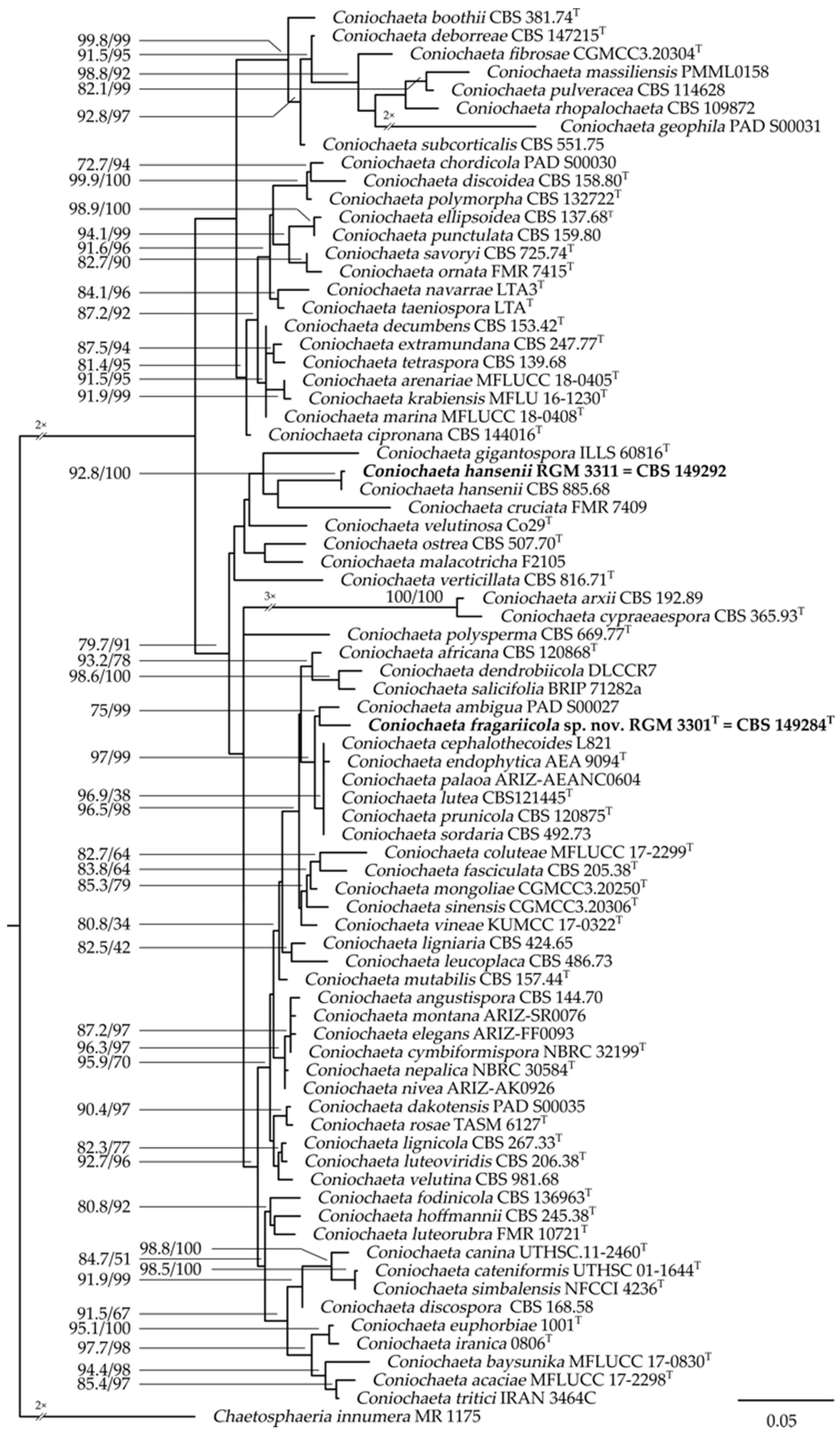
3.1. Taxonomic Position of the Isolates

Both endophyte strains isolated from Fragaria chiloensis subsp. chiloensis f. patagonica were affiliated with the genus Coniochaeta, based on a maximum likelihood (ML) analysis of the concatenated datasets of ITS and LSU (Figure 1).
BLAST searches using the ITS sequence of RGM 3301 showed identities of 95.77% with Coniochaeta sp. DSM 101981 (GenBank accession: KX096678), Sordariomycetes sp. A45 (KX611033), and Sordariomycetes sp. ARIZ:SR0072 (KP991848) as the best hits. In turn, the best hits when using the ITS sequence of RGM 3311 were fungal sp. 2735 YZ-2011 (HM439584) with a 99.14% identity, followed by Sordariomycetes sp. A45 (KX611033) and Coniochaeta ligniaria B121 (KX090317), both with an identity of 91.01%.
A BLAST search using the individual ITS and LSU nucleotide sequences against a local database of Coniochaeta spp. revealed identical sequences between RGM 3311 and C. hansenii CBS 885.68 (ITS identities: 183/183 (100%), no gaps, LSU identities: 456/456 (100%), no gaps), while differences in these nucleotide sequence alignments were found when comparing with the second-best hits, Coniochaeta montana ARIZ-AZ0093 (ITS identities: 346/374 (90.1%), 4 gaps) and Coniochaeta discospora CBS 168.58T (LSU identities: 864/879 (98.3%), no gaps). Moreover, the strain RGM 3311 formed a single clade with Coniochaeta hansenii CBS 885.68 with a high branch support (92.8/100, SH-aLRT/UFBoot) and a similar branch length, suggesting that both strains represent one and the same species.
Coniochaeta cruciata FMR 7409 and Coniochaeta gigantospora ILLS 60816T, the closest neighbors to RGM 3311 in the ML tree, showed branch support values below the reliability threshold. In addition, similarities between the ITS and LSU nucleotide sequences of strain RGM 3311 with the respective genetic marker of C. cruciata FMR 7409 (no ITS sequence was available; LSU: 438/458 (96%), 2 gaps) and C. gigantospora ILLS 60816T (ITS identities: 495/548 (86.68%), 10 gaps; no LSU sequence was available) lead us to conclude that RGM 3311 is closely related to C. hansenii CBS 885.68 and distant to its closest neighbors. Therefore, it is proposed that strain RGM 3311 corresponds to C. hansenii.
On the other hand, the strain RGM 3301 was placed in the ML phylogenetic tree in a separate clade from RGM 3311. The strain RGM 3301 was grouped in a single clade with Coniochaeta ambigua PAD S00027T, displaying a node support value that suggests they are related to one another (75/99, SH-aLRT/UFBoot). The nucleotide sequence alignment of the ITS1 and ITS2 of C. ambigua PAD S00027T compared to the respective sequences of Coniochaeta sp. RGM 3301 indicated low similarity values (ITS1 identities: 65/76 (86%), 3 gaps; ITS2 identities: 153/162 (94%), 6 gaps), suggesting that these two species are not closely associated.
According to a BLAST search against a Coniochaeta spp. local database, the best matches using the ITS nucleotide sequence of RGM 3301 were Coniochaeta cephalothecoides L821 (identities: 543/568 (94.54%), 5 gaps) and Coniochaeta endophytica AEA 9094T (identities: 536/562 (94.31%), 5 gaps), while when using the LSU nucleotide sequence the best matches were C. endophytica AEA 9094T (identities: 1006/1007 (99.21%), 2 gaps) and Coniochaeta simbalensis NFCCI 4236T (identities: 893/914 (97.7%), 21 gaps). The species C. cephalothecoides L821 and C. endophytica AEA 9094T were located in a separated clade, but next to that of the strain Coniochaeta sp. RGM 3301 (Figure 1). Altogether, these two sister clades showed a strong branch support value (97/99, SH-aLRT/UFBoot), indicating a close relationship among all these Coniochaeta species (Figure 1). It seems sensible to consider Coniochaeta sp. RGM 3301 is closely related to C. ambigua PAD S00027T, C. cephalothecoides L821, C. endophytica AEA 9094T, and C. simbalensis NFCCI 4236T, but is, nonetheless, a separate species. Therefore, we propose the name Coniochaeta fragariicola sp. nov. for the strain RGM 3301T.

3.2. Macromorphological Characterization of the Isolates

Coniochaeta fragariicola sp. nov. RGM 3301T and C. hansenii RGM 3311 colonies displayed yeast-like growth with entire margins, a circular shape, and sparse aerial mycelium in the three media tested: PDA, OA, and MEA (Figure 2 and Table 2). Coniochaeta fragariicola sp. nov. RGM 3301T showed different morphologies depending on the media tested. For instance, in PDA, the colony was rugose, with a pale-yellow pigmentation that changed to a brown-orange in the center after 15 d and yellow towards the margin after 21 d (Figure 2a). In OA and MEA, the colony had a glabrous appearance with a predominate pale orange color in the center and yellow-white towards the margin (Figure 2b,c). Moreover, in the MEA, the margin was more filamentous than in the other media (Figure 2c).
Coniochaeta hansenii RGM 3311 showed similar morphological characteristics in PDA (Figure 2d) and MEA (Figure 2f). For instance, colonies were shiny, with a moist appearance, radial grooves, and an undulating margin; they were orange in the center and yellow-gray towards the margin. In OA, the colony was drier, with an entire margin (Figure 2e). From the bottom, colonies showed similar characteristics to those observed in the respective media tested.

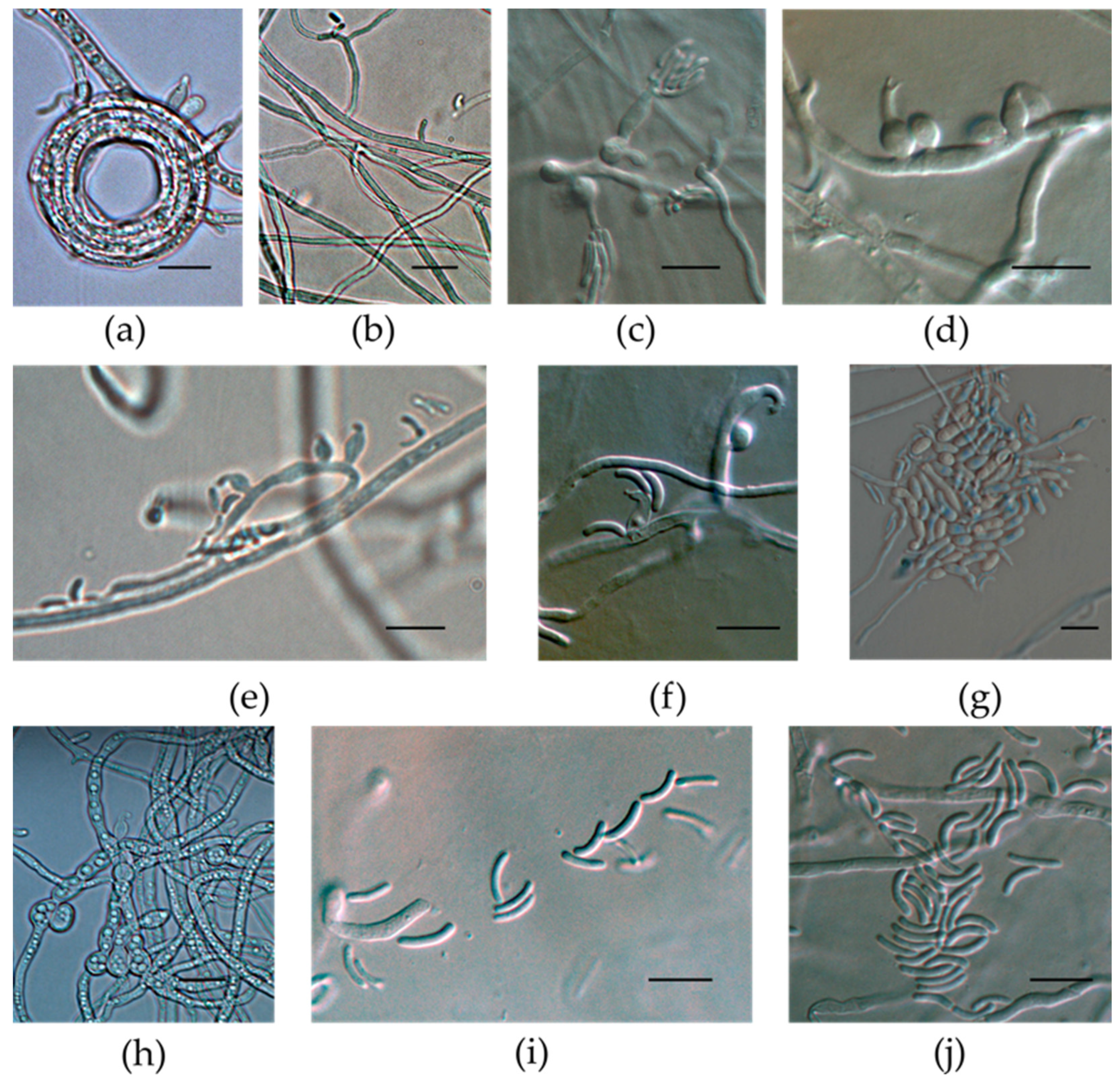
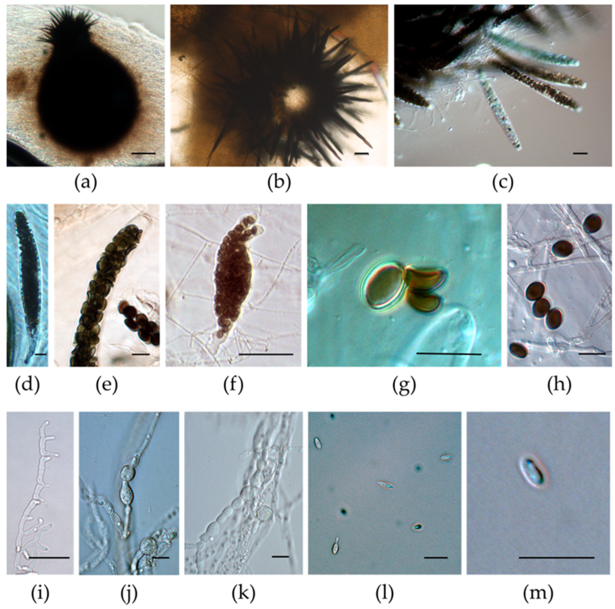
3.3. Micromorphological Characterization of the Isolates

Micromorphological characteristics of Coniochaeta fragariicola sp. nov. RGM 3301T = CBS 149284T were evaluated in several media. The anamorph of C. fragariicola sp. nov. RGM 3301T was obtained in PDA, OA, MEA, and PCA, whereas, for the teleomorph, only immature perithecia were produced in PCA and no further development was observed even though they were tested in all of the aforementioned media and conditions.
In PDA, hyphae were narrow, hyaline, multi-guttulate, and non-septate, with smooth walls, and frequently found in hyphal whorls (1.01–) 1.68–2.08 (–3.01) µm wide (Figure 3a,b). The conidiogenous cell was hyaline, smooth-walled, mostly ampulliform to lageniform, occasionally globose to subglobose, narrower at the base and wider in the middle, with a curved apex, (3.64–) 5.02–6.03 (–9.58) × (2.06–) 2.56–2.76 (–3.32) µm (Figure 3c–f), while in MEA and PCA the conidiogenous cell was subglobose to cylindrical and the apex was curved and also acute, with or without the formation of a collarette, showing microcyclic conidiation in MEA, after 8 d, at 25 °C (Figure 3g). Chlamydospores were hyaline, globose to subglobose, and they were produced in chains or groups of (2.69–) 3.94–4.47 (–5.57) µm, and were similar across all tested media (Figure 3h). Conidia were hyaline, smooth, and non-septate; allantoid demonstrated rounded ends and were generally bi-guttulate of (3.64–) 5.03–5.99 (9.58) µm × <1 µm (Figure 3i,j). Conidia with <1 µm were also observed in OA.
Comparisons of C. fragariicola sp. nov. RGM 3301T were carried out with reference strains whose anamorph had been previously described, including the conidiogenous cell, conidia, microcyclic conidiation, and in some cases, the presence or absence of chlamydospores. Only the teleomorph of Coniochaeta ambigua PAD S00027T had been described; therefore, it was not possible to compare it with other strains. The conidiogenous cell of C. endophytica AEA 9094T [52] was characterized by reduced cylindrical protrusions from the hyphae or as discrete phialides; phialidic conidiogenous cells ampulliform were (5.2–10.3 × 2.3–2.9 μm wide), similar to C. fragariicola sp. nov. RGM 3301T. However, the conidia of C. endophytica AEA 9094T differed from those recorded for C. fragariicola sp. nov. RGM 3301T, the former being shorter and wider in diameter ((2.5–) 3.1–3.4 (–4.4) μm × (1.3) 1.6–1.8 (2.4) μm) and ellipsoidal to fusiform in shape, while the conidia of C. fragariicola sp. nov. RGM 3301T were allantoid. Coniochaeta endophytica, unlike C. fragariicola sp. nov. RGM 3301T, did not develop chlamydospores or microcyclic conidiation.
Coniochaeta cephalotheicoides L821 developed conidiogenous cells similar to those observed in RGM 3301T, in that they were phialidic, terminal or lateral, borne on branched hyphae, hyaline, mostly ampulliform and sometimes ovoid or cylindrical (5–15 μm × 2.5–3.5 μm), with a distinct collarette. However, the conidia described for C. cephalotheicoides L821 were smaller, wider (2.5–5 μm × 1–2 μm), and more ellipsoidal than those observed in C. fragariicola sp. nov. RGM 3301T [63]. Additionally, the formation of mucous heads in the conidia of C. cephalotheicoides L821 was not observed in C. fragariicola sp. nov. RGM 3301T. Coniochaeta palaoa ARIZ-AEANC0604T and C. fragariicola sp. nov. RGM 3301T presented similarities in their phialide shape, but the conidial size was shorter and wider (3.6–4.7 μm × 1.5–1.9 μm) than the former [52]. In addition, C. palaoa ARIZ-AEANC0604T did not produce chlamydospores, unlike C. fragariicola sp. nov. RGM 3301T which did produce chlamydospores. Similarly, Coniochaeta lutea CBS 121445T developed conidia that were shorter and wider ((3.1) 3.7–4.1 (5.4) μm × (1.0) 1.3–1.6 (1.8) μm) than C. fragariicola sp. nov. RGM 3301T. The shape of the conidia in C. lutea CBS 121445T was bacilliform to allantoid [52], whereas in C. fragariicola sp. nov. RGM 3301T the conidia were allantoid. Coniochaeta prunicola displayed a similar conidia size ((2.5–) 3.5–6 (–8) μm × 1–2 (–3.0) μm) and shape, but microcyclic conidiation was not observed. Coniochaeta sordaria, another species close to C. fragariicola sp. nov. RGM 3301T in the phylogenetic tree, did not develop any anamorph stage; therefore, it was not possible to carry out morphological comparisons [64].
The conidiogenous cells of Coniochaeta nivea were usually originated from cylindrical protuberances of densely packed hyphae and only occasionally from phialides, which were unusual in C. fragariicola sp. nov. RGM 3301T [26]. Similarly, the conidiogenous cells in Coniochaeta elegans were mostly reduced to cylindrical protrusions from the hyphae, with collarettes, a condition that was not common in C. fragariicola sp. nov. RGM 3301T, which presented mostly ampulliform conidiogenous cells with curved apices, regardless of whether or not they formed a collarette. Conidiation in C. fragariicola sp. nov. RGM 3301T occurs through the conidiogenous cell and by microcyclic conidiation, whereas conidiation in Coniochaeta elegans, as well as in C. nivea, occurs only through the conidiogenous cell [26]. Moreover, the conidia in C. elegans and C. nivea were shorter and wider when compared with C. fragariicola sp. nov. RGM 3301T (3.0–4.5 × 1.1–1.8 µm in C. elegans; 3.7–4.1 × 1.3–1.6 µm in C. nivea).
The conidia shape and length of Coniochaeta angustispora CBS 144.70 and Coniochaeta dumosa BF 42889 were similar to those recorded in the strain RGM 3301, but wider in C. angustispora and C. dumosa BF 42889 (1.25–1.75 µm and 1.5–2.5 µm, respectively); also, C. angustispora CBS 144.70 and C. dumosa BF 42889 displayed a slimy mass of conidia, which was not seen in C. fragariicola sp. nov. RGM 3301T [39,63]. The conidia of C. fragariicola sp. nov. RGM 3301T differed from those reported for Coniochaeta coluteae MFLUCC 17–2299T, which were oblong, ellipsoidal to allantoid, and slightly shorter ((3.6) 4.5 ± 0.5 (6.3) μm) and wider ((0.7) 1.6 ± 0.3 (2.4) μm)); additionally, the conidiogenous cell was similar in morphology, but shorter ((3.3) 4.3 ± 0.5 (5.2) µm × (2.1) 3.1 ± 0.4 (3.8) µm) than that of C. fragariicola sp. nov. RGM 3301T [38]. Likewise, the conidia of Coniochaeta dendrobiicola MCC 1811T were mostly cylindrical to allantoid and non-guttulate, unlike those of C. fragariicola sp. nov. RGM 3301T, which were more homogeneous in size and shape (allantoid and guttulate). The sizes of the conidia of C. dendrobiicola MCC 1811T were significantly longer and wider (4.35–11.28 µm × 1.2–2.3 µm) than those observed in C. fragariicola sp. nov. RGM 3301T [46], while the conidia of Coniochaeta montana ARIZ-SR0076T were smaller and wider (3.4–4.5 µm × 1.9–2.1 µm) than those of C. fragariicola sp. nov. RGM 3301T. C. montana ARIZ-SR0076T did not present microcyclic conidiation or chlamydospores [26], as observed in C. fragariicola sp. nov. RGM 3301T.
To the best of our knowledge, C. hansenii RGM 3311 = CBS 149292 corresponds to the first report of this species in Chile. In OA, C. hansenii RGM 3311 displayed pyriform, black ascomata with thick setae at the apex and finer setae (bristly hair) on the rest of the perithecium with a size of (195.46–) 348.41–412.03 (–618.55) µm × (193.52–) 270.37–311.07 (–387.51) µm; one ostiole was observed (Figure 4a,b). Setae were dark brown and straight, with pointed ends of (48.08–) 67.88–79.00 (–109.98) µm × (4.00–) 5.4–6.04 (–7.51) µm. Asci were clavate and slightly flattened at the apex, with the presence of an apical ring, enlarged subapical part, sometimes ascus with bifurcate apex and fusiform inflated, (85.96–) 105.42–116.98 (–142.11) µm × (13.47–) 16.88–18.27 (–21.92) µm (Figure 4c–f). Ascospores, nearly 128 in number, were brown, concave, smooth, not septate, discoidal, and (6.25–) 7.16–7.68 (–9.82) µm long × (5.43–) 5.85–6.07 (6.59) µm wide × (1.67–) 2.19–2.45 (–3.02) µm thick; they were ellipsoidal when viewed from the side and reddish-brown when old (Figure 4g,h). Hyphae hyaline were septate, smooth-walled, and (1.11–) 1.71–2.155 (–3.00) µm wide (Figure 4i). Chlamydospore hyaline were globose, clavate, doliiform, multi-guttulate, and (5.37–) 9.19–10.59 (–14.1) µm × (5.69–) 7.42–8.30 (–10.28) µm (Figure 4j,k). The conidia were only observed in Leonian’s agar and were sparse, hyaline, non-septate, cylindrical to ellipsoidal, occasionally fusiform, occasionally guttulate, and (4.1–) 4.79–5.23 (–6.67) µm × (0.93–) 1.37–1.52 (–2.31) µm wide (Figure 4l,m). Microcyclic conidiation was also observed.
The teleomorph of C. hansenii RGM 3311 was observed in OA, PDA, and RDA, while the anamorph was scarcely produced in Leonian’s agar. In accordance with the molecular analysis, Coniochaeta hansenii RGM 3311 shared similar micromorphometric characteristics with C. hansenii (≡ Sordaria hansenii Oudem. 1882), as described by Oudemans [65]. The perithecia in this description were subglobose, measuring 350 µm in diameter, and featuring a neck with a short-conical shape, similar to those observed in C. hansenii RGM 3311. The setae in the original description were 23 µm long, which were about three-fold shorter than those observed in C. hansenii RGM 3311, though both terminated in bristly hairs. A report of C. hansenii in Italy [15] also corroborates the characteristics of the perithecium of C. hansenii RGM 3311, where both were brown to dark brown to black, pyriform, setose above the perithecium; however, it was bigger in the report from Italy (480–680 μm × 350–430 μm) [15].
The asci in C. hansenii RGM 3311 were shorter than those reported in both of the original descriptions and in the Italian strain (150 μm and 170–230 μm, respectively), though they shared the same shape overall [15,65]. In turn, the ascus width in RGM 3311 was similar to the original description (12 μm), but smaller when compared to the Italian report (20–27 μm). The key of Coniochaeta species with poly-spored asci, developed by Doveri [15] indicates that the number of spores per ascus is a distinctive feature for C. hansenii species. According to this key, the asci of C. hansenii species are clavate, rarely cylindric-clavate, measuring 125–230 μm × 17–32 μm, and containing 64 to 128 spores of a discoidal shape, measuring 6–10 μm × 4–9 μm × 3.5–7 μm. This general description matched that described here for C. hansenii RGM 3311. In addition, the Italian C. hansenii strain and Coniochaeta polyspora [66] contained possibly 128 spores per ascus, similar to C. hansenii RGM 3311, whereas, other Coniochaeta species, such as Coniochaeta philocoproides [67] and Coniochaeta polymegasperma [68] had 32-spored ascus, and Coniochaeta polysperma [69] 512-spored ascus. Most Coniochaeta species whose teleomorph had been reported contained 8 spores per ascus [15].

3.4. Biochemical Features of the Isolates

Both of the isolates C. fragariicola sp. nov. RGM 3301T and C. hansenii RGM 3311 oxidized the monosaccharides D-fructose-6-PO4, L-fucose, α-D-glucose, D-mannose, methyl pyruvate, β-methyl-D-glucoside, and L-rhamnose, as well as the disaccharides D-cellobiose, gentiobiose, β-D-lactose, D-maltose, D-melibiose, and D-trehalose, sugars that were also used by Coniochaeta massiliensis [54]. Sucrose and stachyose oxidation were negative in C. massiliensis, while C. fragariicola sp. nov. RGM 3301T and C. hansenii RGM 3311 oxidized these disaccharides. Both strains oxidized polymers such as dextrin and pectin, as well as the amino-sugar N-acetyl-D-glucosamin; however, only the strain Coniochaeta hansenii RGM 3311 utilized N-acetyl-D-galactosamine, N-acetyl-β-D-mannosamine, and N-acetyl neuraminic acid. In relation to these sugars, enzymes such as glycoside hydrolases, glycosyl transferases, and polysaccharide lyases have been identified in Coniochaeta spp. [11,70]. Both isolates also oxidized sugar alcohols such as D-arabitol, glycerol, and D-salicin, while D-sorbitol, D-mannitol, and myo-inositol were only oxidized by C. fragariicola sp. nov. RGM 3301T, which suggests that this strain presented an active mannitol cycle (positive for D-mannitol, D-fructose-6-PO4, and D-fructose) [71]. Our isolates utilized organic acids acetic acid, γ-amino-butryric acid, citric acid, formic acid, L-galacturonic acid lactone, D-galacturonic acid, D-gluconic acid, D-glucuronic acid, α-hydroxy-butyric acid, β-hydroxy-D,L-butyric acid, α-keto-butyric acid, α-keto-glutaric acid, L-lactic acid, D-lactic acid methyl ester, D-malic acid, L-malic acid, mucic acid, propionic acid, and quinic acid. Coniochaeta fragariicola sp. nov. RGM 3301T and C. hansenii RGM 3311 oxidized amino acids such as L-alanine, L-glutamic acid, glycyl-L-proline, and L-pyroglutamic, while only C. hansenii RGM 3311 used D-amino acids such as D-aspartic and D-serine. Both strains were inhibited by 4% and 8% NaCl, guanidine HCl, niaproof 4, and lithium chloride, but not by 1% NaCl. These strains were not inhibited by antibiotics such as aztreonam, fusidic acid, lincomycin, minocycline, nalidixic acid, rifamycin SV, troleandomycin, and vancomycin, and they grew in the presence of potassium tellurite, tetrazolium salts, D-serine, sodium bromate, and 1% sodium lactate; only sodium butyrate inhibited C. hansenii RGM 3311, while it did not inhibit C. fragariicola sp. nov. RGM 3301T.

3.5. Taxonomy of the Two Endophytes of Fragaria chiloensis subsp. chiloensis f. patagonica

Coniochaeta fragariicola Campos-Quiroz and Castro sp. nov.—Mycobank 846930; Figure 3.
Etymology. “fragari-”, reflects the host from which the fungus was isolated, Fragaria chiloensis subsp. chiloensis f. patagonica. “-icola”, dweller.
In PDA. Hyphae hyaline, multi-guttulate, smooth-walled, sometimes helicoid in shape, non-septate, narrow, and (1.01–) 1.68–2.08 (–3.01) µm wide. Conidiogenous cell hyaline, smooth, mostly ampulliform to lageniform, occasionally globose to subglobose, narrower at the base and wider in the middle, with a curved apex, and (3.64–) 5.02–6.03 (–9.58) µm × (2.06–) 2.56–2.76 (–3.32) µm. On the contrary, in MEA and PC, the conidiogenous cell was subglobose to cylindrical, with a curved apex, sometimes a collarette was also formed. Microcyclic conidiation in MEA, after 8 d, at 25 °C. Chlamydospores hyaline, globose to subglobose, and produced in chains or groups, (2.69–) 3.94–4.47 (–5.57) µm. Conidia hyaline, smooth, non-septate, allantoid, with rounded ends that were generally bi-guttulate, and (3.64–) 5.03–5.99 (9.58) µm × <1 µm, L/W ratio = 4.9 ± 1.3.
Material examined: CHILE, Pinto, Ñuble, 36.818367 S, 71.622217 W; altitude: 697 m.a.s.l.; endophyte of Fragaria chiloensis subsp. chiloensis f. patagonica, 13 July 2020. Collected by J.F. Castro and M. Guerra, isolated by Cecilia Santelices. The holotype was deposited in the Chilean Collection of Microbial Genetic Resources (CChRGM) under the code RGM 3301 (the isotype was deposited in the CBS-KNAW culture collection under the code CBS 149284).
Notes: Coniochaeta fragariicola sp. nov. grew at a pH of 5 and 6 and in the presence of 1% NaCl. Coniochaeta fragariicola sp. nov. oxidized acetic acid, N-acetyl-D-glucosamine, L-alanine, γ-amino-butryric acid, D-arabitol, L-arginine, L-aspartic acid, L-butyric acid, citric acid, D-cellobiose, dextrin, formic acid, D-fructose, D-fructose-6-PO4 (weakly), L-glucose, L-galactonic acid lactone, D-galactose, D-galacturonic acid, gentiobiose, D-gluconic acid, α-D-glucose, D-glucuronic acid, L-glutamic acid, glycerol, glycyl-L-proline, α-hydroxy-butyric acid, β-hydroxy-D,L-butyric acid, α-keto-butyric acid, α-keto-glutaric acid, L-lactic acid, D-lactic acid methyl ester, β-D-lactose, D-malic acid, L-malic acid, D-maltose, D-mannitol (weakly), D-mannose, D-melibiose, β-methyl- D-glucoside, methyl pyruvate, mucic acid (weakly), myo-inositol, L-pyroglutamic acid, pectin, propionic acid, quinic acid, D-raffinose (weakly), L-rhamnose, D-salicin, L-serine, D-sorbitol, D-trehalose, D-turanose, and Tween 40, though it did not oxidize sucrose, stachyose, N-acetyl-β-D-mannosamine, N-acetyl-D-galactosamine, N-acetyl neuraminic acid, 3-methyl glucose, D-fucose, inosine, D-glucose-6-PO4, D-aspartic acid, gelatin, L-histidine, glucuronamide, D-saccharic acid, p-hydroxy-phenylacetic acid, bromo-succinic acid, and acetoacetic acid. It was sensitive to 4% NaCl, 8% NaCl, guanidine HCl, niaproof 4, and lithium chloride, but not to aztreonam, fusidic acid, lincomycin, minocycline, nalidixic acid, rifamycin SV, troleandomycin, vancomycin, D-serine, sodium bromate, sodium butyrate, 1% sodium lactate, potassium tellurite, tetrazolium blue, or tetrazolium violet.
Coniochaeta hansenii (Oudem.) Cain, Studies of Coprophilous Spaeriales in Ontario: 63 (1934) (Figure 4).
Sordaria hansenii Oudem., Hedwigia, 21: 123 (1882) (basionym).
Philocopra hansenii (Oudem.) Oudem., Hedwigia, 21: 160 (1882).
In OA. Ascomata pyriform, black, with thick setae at the apex and finer setae (bristly hairs) on the rest of the perithecium; they were (195.46–) 348.41–412.03 (–618.55) µm × (193.52–) 270.37–311.07 (–387.51) µm, and one ostiole was observed. Setae dark brown, straight, with pointed ends, and (48.08–) 67.88–79.00 (–109.98) µm × (4.00–) 5.4–6.04 (–7.51) µm. Asci clavate, and slightly flattened at the apex, with the presence of an apical ring, enlarged subapical part, sometimes ascus showed a bifurcate apex and fusiform, and were (85.96–) 105.42–116.98 (–142.11) µm × (13.47–) 16.88–18.27 (–21.92) µm. Ascospores totaled nearly 128, and were brown, concave, smooth, not septate, discoidal from the top, and (6.25–) 7.16–7.68 (–9.82) µm long × (5.43–) 5.85–6.07 (6.59) µm wide × (1.67–) 2.19–2.45 (–3.02) µm thick; they were ellipsoidal from the side, reddish-brown when age, and with an L/W ratio = 1.2 ± 0.1. Hyphae hyaline, septate, smooth-walled, and (1.11–) 1.71–2.16 (–3.00) µm wide. Chlamydospores in PDA were hyaline, globose, clavate, doliiform, multi-guttulate, and were (5.37–) 9.19–10.59 (–14.1) µm × (5.69–) 7.42–8.30 (–10.28) µm, with an L/W ratio = 1.3 ± 0.3. Conidia in Leonian’s agar were sparse, hyaline, non-septate, cylindrical to ellipsoidal, occasionally fusiform, occasionally guttulate, and (4.1–) 4.79–5.23 (–6.67) µm × (0.93–) 1.37–1.52 (–2.31) µm wide, with an L/W ratio = 3.5 ± 0.7; microcyclic conidiation was also observed.
Material examined: CHILE, Pinto, Ñuble, 36.818367 S, 71.622217 W; altitude: 697 m.a.s.l. endophyte strain of Fragaria chiloensis subsp. chiloensis f. patagonica, 13 July 2020. Collected by J.F. Castro and M. Guerra, isolated by Cecilia Santelices. Deposited in the Chilean Collection of Microbial Genetic Resources (CChRGM) with the code RGM 3311 and in the CBS under the code CBS 149292.
Notes: Coniochaeta hansenii RGM 3311 shared identical ITS and LSU sequences with C. hansenii CBS 885.68 (ITS identities: 183/183 (100%), no gaps, LSU identities: 456/456 (100%), no gaps), followed by C. montana ARIZ-AZ0093 (ITS identities: 346/374 (90.1%), 4 gaps). In the ML phylogenetic tree, the strain was grouped with C. hansenii CBS 885.68 with high bootstrap support. The ascospores in C. hansenii RGM 3311 were discoidal and nearly 128-spored, which matches the description for the species C. hansenii, according to the taxonomic key of Coniochaeta species developed by Doveri [15]. In this work, we described the anamorph of C. hansenii. The strain C. hansenii RGM 3311 grew at a pH of 5 and 6 and in the presence of 1% NaCl. This strain oxidized the following Biolog GEN III substrates: acetic acid, N-acetyl-D-galactosamine, N-acetyl-β-D-mannosamine (weakly), N-acetyl-D-glucosamine, N-acetyl neuraminic acid, L-alanine, γ-amino-butryric acid, D-arabitol (weakly), D-aspartic acid, bromo-succinic acid, D-cellobiose, citric acid, dextrin, formic acid, D-fructose-6-PO4, L-fucose, L-galactonic acid lactone, D-galacturonic acid, gelatin, gentiobiose, D-gluconic acid, α-D-glucose, D-glucose-6-PO4, glucuronamide (weakly), D-glucuronic acid, L-glutamic acid, glycerol, glycyl-L-proline, L-histidine, α-hydroxy-butyric acid, β-hydroxy-D,L-butyric acid (weakly), inosine, α-keto-butyric acid, α-keto-glutaric acid, D-lactic acid methyl ester, L-lactic acid, β-D-lactose, D-malic acid, L-malic acid, D-maltose, D-mannose, D-melibiose, methyl pyruvate, β-methyl- D-glucoside, mucic acid, pectin, propionic acid, L-pyroglutamic acid, quinic acid, L-rhamnose, D-saccharic acid, D-salicin, D-serine, D-trehalose, D-turanose, and tween 40, but it does not oxidize sucrose, stachyose, D-raffinose, D-fructose, D-galactose, 3-methyl glucose, D-fucose, D-sorbitol, D-mannitol, myo-inositol, L-arginine, L-aspartic acid, L-serine, p-hydroxy-phenylacetic acid, and acetoacetic acid. It was sensitive to 4% NaCl, 8% NaCl, guanidine HCl, niaproof 4, lithium chloride, sodium butyrate, but not to aztreonam, fusidic acid, lincomycin, minocycline, nalidixic acid, rifamycin SV, troleandomycin, or vancomycin, nor to D-serine, sodium bromate, 1% sodium lactate, potassium tellurite, tetrazolium blue, or tetrazolium violet.

4. Conclusions

This work contributes to expanding our current knowledge regarding the geographic extent and host source of species of the genus Coniochatea. Here, we report the description of two endophytic Coniochaeta strains, isolated from Fragaria chiloensis subsp. chiloensis f. patagonica collected from the foothills of the Chilean Andes.
Phylogenetic studies and macro- and micro-morphological analyses confirmed that strain RGM 3311 corresponded to C. hansenii, which to the best of our knowledge corresponds to the first report of this strain in Chile. Additionally, we described the previously unknown anamorph of this species. The second isolate corresponded to a novel species of Coniochaeta, named C. fragariicola sp. nov. RGM 3301T = CBS 149284T that was closely related to C. ambigua PAD S00027T, C. cephalothecoides L821, C. endophytica AEA 9094T, and C. simbalensis NFCCI 4236T. The conidial width of C. fragariicola sp. nov. RGM 3301T was a distinctive feature of this novel species.
It is worth mentioning that some reference species of the genus Coniochaeta have only been described in morphological terms, and the genetic barcoding scheme of other species of this genus is incomplete, making it difficult to assign names to new isolates. We suggest a revision of the genus Coniochaeta, including the sequencing of genetic markers from reference specimens with their incomplete genetic barcoding as well as the sequencing of other molecular markers. We also suggest including phenotypic analyses to account for another layer of information when analyzing the taxonomic position of new isolates. Further research is needed to understand the interaction between these endophyte species and the Chilean wild strawberry plant.

Author Contributions

Conceptualization, J.F.C. and C.S.; methodology, J.F.C., C.C.-Q., M.G., J.C.-F., Y.O., B.T. and J.O.-C.; software, J.F.C., C.C.-Q., M.G. and J.C.-F.; validation, J.F.C. and C.S.; investigation, D.C.-G., C.C.-Q., J.C.-F. and M.G.; resources, J.F.C. and L.B.-B.; data curation, J.F.C., C.C.-Q. and J.C.-F.; writing—original draft preparation, J.F.C.; writing—review and editing, J.F.C., C.S., B.T. and C.C.-Q.; visualization, J.F.C., C.C.-Q. and C.S.; supervision, J.F.C.; project administration, J.F.C.; funding acquisition, J.F.C. All authors have read and agreed to the published version of the manuscript.

Funding

This research was funded by the FONDECYT INICIACIÓN (Grant: 11191074) from the Chilean National Agency for Research and Development (ANID) and the FONDEQUIP Program of ANID (Grant: EQM200205).

Data Availability Statement

GenBank accession numbers submitted in this work are OP962067-70. Phylogenetic data are available from Dryad: https://datadryad.org/stash/share/QP9NZ96mvlEabPd4M7i2If8bW7oXx7JZnhgstznMIh4, accessed on 25 March 2023.

Conflicts of Interest

The authors declare no conflict of interest.

References

  1. Cooke, M.C. Coniochaeta, Sacc. Grevillea 1887, 16, 16. [Google Scholar]
  2. García, D.; Stchigel, A.M.; Cano, J.; Calduch, M.; Hawksworth, D.L.; Guarro, J. Molecular phylogeny of Coniochaetales. Mycol. Res. 2006, 110, 1271–1289. [Google Scholar] [CrossRef] [PubMed]
  3. Guarro, J.; Gené, J.; Stchigel, A.M.; Figueras, M.J. Atlas of Soil Ascomycetes; CBS-KNAW Fungal Biodiversity Centre: Utrecht, The Netherlands, 2012. [Google Scholar]
  4. Mahoney, D.P.; LaFavre, J.S. Coniochaeta Extramundana, with a synopsis of other Coniochaeta species. Mycologia 1981, 73, 931–952. [Google Scholar] [CrossRef]
  5. Dayarathne, M.C.; Jones, E.B.G.; Maharachchikumbura, S.S.N.; Devadatha, B.; Sarma, V.V.; Khongphinitbunjong, K.; Chomnunti, P.; Hyde, K.D. Morpho-molecular characterization of microfungi associated with marine based habitats. Mycosphere 2020, 11, 1–188. [Google Scholar] [CrossRef]
  6. Si, H.L.; Su, Y.M.; Zheng, X.X.; Ding, M.Y.; Bose, T.; Chang, R.L. Phylogenetic and morphological analyses of Coniochaeta isolates recovered from Inner Mongolia and Yunnan revealed three new endolichenic fungal species. MycoKeys 2021, 83, 105–121. [Google Scholar] [CrossRef]
  7. Rosa, L.H.; Queiroz, S.C.N.; Moraes, R.M.; Wang, X.; Techen, N.; Pan, Z.; Cantrell, C.L.; Wedge, D.E. Coniochaeta ligniaria: Antifungal activity of the cryptic endophytic fungus associated with autotrophic tissue cultures of the medicinal plant Smallanthus sonchifolius (Asteraceae). Symbiosis 2013, 60, 133–142. [Google Scholar] [CrossRef]
  8. Xie, J.; Strobel, G.A.; Feng, T.; Ren, H.; Mends, M.T.; Zhou, Z.; Geary, B. An endophytic Coniochaeta velutina producing broad spectrum antimycotics. J. Microbiol. 2015, 53, 390–397. [Google Scholar] [CrossRef] [Green Version]
  9. Damm, U.; Fourie, P.H.; Crous, P.W. Coniochaeta (Lecythophora), Collophora gen. nov. and Phaeomoniella species associated with wood necroses of Prunus trees. Persoonia 2010, 24, 60–80. [Google Scholar] [CrossRef] [Green Version]
  10. Checa, J.; Barrasa, J.; Moreno, G.; Fort, F.; Guarro, J. The genus Coniochaeta (Sacc.) Cooke (Coniochaetaceae, Ascomycotina) in Spain. Cryptog. Mycol 1988, 9, 1–34. [Google Scholar]
  11. Jiménez, D.J.; Hector, R.E.; Riley, R.; Lipzen, A.; Kuo, R.C.; Amirebrahimi, M.; Barry, K.W.; Grigoriev, I.V.; van Elsas, J.D.; Nichols, N.N. Draft genome sequence of Coniochaeta ligniaria NRRL 30616, a lignocellulolytic fungus for bioabatement of inhibitors in plant biomass hydrolysates. Genome Announc. 2017, 5, e01476-16. [Google Scholar] [CrossRef] [Green Version]
  12. Khan, Z.; Gené, J.; Ahmad, S.; Cano, J.; Al-Sweih, N.; Joseph, L.; Chandy, R.; Guarro, J. Coniochaeta polymorpha, a new species from endotracheal aspirate of a preterm neonate, and transfer of Lecythophora species to Coniochaeta. Antonie Leeuwenhoek 2013, 104, 243–252. [Google Scholar] [CrossRef] [PubMed]
  13. Troy, G.C.; Panciera, D.L.; Pickett, J.P.; Sutton, D.A.; Gene, J.; Cano, J.F.; Guarro, J.; Thompson, E.H.; Wickes, B.L. Mixed infection caused by Lecythophora canina sp. nov. and Plectosphaerella cucumerina in a German shepherd dog. Med. Mycol. 2013, 51, 455–460. [Google Scholar] [CrossRef] [Green Version]
  14. de Hoog, S.; Guarro, J.; Gené, J.; Ahmed, S.; Al-Hatmi, A.; Figueras, M.; Vitale, R. Atlas of Clinical Fungi, 2nd ed.; Centraalbureau voor Schimmelcultures: Utrecht, The Netherlands; Universitat Rovira i Virgili Reus: Catalonia, Spain, 2020. [Google Scholar]
  15. Doveri, F. Description of Chaetomium aureum, Corynascus sepedonium and Coniochaeta hansenii newly recorded from Italy, and a key to coprophilous Chaetomiaceae and Coniochaetaceae. Ascomycete Org. 2016, 8, 7–24. [Google Scholar]
  16. Friebes, G.; Jaklitsch, W.M.; García, S.; Voglmayr, H. Lopadostoma taeniosporum revisited and a new species of Coniochaeta. Sydowia 2016, 68, 87–97. [Google Scholar]
  17. Forin, N.; Vizzini, A.; Fainelli, F.; Ercole, E.; Baldan, B. Taxonomic re-examination of nine Rosellinia types (Ascomycota, Xylariales) stored in the Saccardo Mycological Collection. Microorganisms 2021, 9, 666. [Google Scholar] [CrossRef]
  18. Carrasco-Fernández, J.; Guerra, M.; Castro, J.F.; Bustamante, L.; Barra-Bucarei, L.; Ceballos, R.; Fernández, N.; Edgington, S.; France, A. Plant growth promoting rhizobacteria from Juan Fernández archipelago improve germination rate of endangered plant Solanum fernandezianum Phil. Chil. J. Agric. Res. 2020, 80, 41–49. [Google Scholar] [CrossRef]
  19. Castro, J.F.; Guerra, M.; Carrasco-Fernández, J.; Ortiz-Campos, J.; Cares-Gatica, D.; Campos-Quiroz, C.; Correa, F.; Beltrán, M.F.; Sagredo, B.; Valdés, J.H. Draft genome sequence of Pseudomonas sp. strain RGM 3321, a phyllosphere endophyte from Fragaria chiloensis subsp. chiloensis f. patagonica. Microbiol. Resour. Announc. 2022, 11, e0033522. [Google Scholar] [CrossRef]
  20. White, T.; Bruns, T.; Lee, S.; Taylor, J.; Innis, M.; Gelfand, D.; Sninsky, J. Amplification and direct sequencing of fungal ribosomal RNA genes for phylogenetics. In PCR—Protocols and Applications—A Laboratory Manual; Innis, M.A., Gelfand, D.H., Sninsky, J.J., White, T.J., Eds.; Academic Press: New York, NY, USA, 1990; Volume 31, pp. 315–322. [Google Scholar]
  21. Rehner, S.A.; Samuels, G.J. Molecular systematics of the Hypocreales: A teleomorph gene phylogeny and the status of their anamorphs. Can. J. Bot. 1995, 73, 816–823. [Google Scholar] [CrossRef]
  22. Vilgalys, R.; Hester, M. Rapid genetic identification and mapping of enzymatically amplified ribosomal DNA from several Cryptococcus species. J. Bacteriol. 1990, 172, 4238–4246. [Google Scholar] [CrossRef] [Green Version]
  23. de Hoog, G.S.; van den Ende, A.H.G.G. Molecular diagnostics of clinical strains of filamentous Basidiomycetes. Mycoses 1998, 41, 183–189. [Google Scholar] [CrossRef]
  24. Van den Ende, A.; De Hoog, G. Variability and molecular diagnostics of the neurotropic species Cladophialophora bantiana. Stud. Mycol. 1999, 43, 151–162. [Google Scholar]
  25. Nasr, S.; Bien, S.; Soudi, M.R.; Alimadadi, N.; Shahzadeh Fazeli, S.A.; Damm, U. Novel Collophorina and Coniochaeta species from Euphorbia polycaulis, an endemic plant in Iran. Mycol. Prog. 2018, 17, 755–771. [Google Scholar] [CrossRef]
  26. Arnold, A.E.; Harrington, A.H.; Huang, Y.-L.; Ren, J.M.; Massimo, N.C.; Knight-Connoni, V.; Inderbitzin, P. Coniochaeta elegans sp. nov., Coniochaeta montana sp. nov. and Coniochaeta nivea sp. nov., three new species of endophytes with distinctive morphology and functional traits. Int. J. Syst. Evol. Microbiol. 2021, 71, 005003. [Google Scholar] [CrossRef] [PubMed]
  27. Vu, D.; Groenewald, M.; de Vries, M.; Gehrmann, T.; Stielow, B.; Eberhardt, U.; Al-Hatmi, A.; Groenewald, J.Z.; Cardinali, G.; Houbraken, J.; et al. Large-scale generation and analysis of filamentous fungal DNA barcodes boosts coverage for kingdom fungi and reveals thresholds for fungal species and higher taxon delimitation. Stud. Mycol. 2019, 92, 135–154. [Google Scholar] [CrossRef] [PubMed]
  28. Sayers, E.W.; Cavanaugh, M.; Clark, K.; Pruitt, K.D.; Schoch, C.L.; Sherry, S.T.; Karsch-Mizrachi, I. GenBank. Nucleic Acids Res. 2021, 49, D92–D96. [Google Scholar] [CrossRef]
  29. Priyam, A.; Woodcroft, B.J.; Rai, V.; Moghul, I.; Munagala, A.; Ter, F.; Chowdhary, H.; Pieniak, I.; Maynard, L.J.; Gibbins, M.A.; et al. Sequenceserver: A modern graphical user interface for custom BLAST databases. Mol. Biol. Evol. 2019, 36, 2922–2924. [Google Scholar] [CrossRef]
  30. Notredame, C.; Higgins, D.G.; Heringa, J. T-Coffee: A novel method for fast and accurate multiple sequence alignment. J. Mol. Biol. 2000, 302, 205–217. [Google Scholar] [CrossRef] [Green Version]
  31. Chang, J.M.; Di Tommaso, P.; Lefort, V.; Gascuel, O.; Notredame, C. TCS: A web server for multiple sequence alignment evaluation and phylogenetic reconstruction. Nucleic Acids Res. 2015, 43, W3–W6. [Google Scholar] [CrossRef] [Green Version]
  32. Nguyen, L.T.; Schmidt, H.A.; von Haeseler, A.; Minh, B.Q. IQ-TREE: A fast and effective stochastic algorithm for estimating maximum-likelihood phylogenies. Mol. Biol. Evol. 2015, 32, 268–274. [Google Scholar] [CrossRef]
  33. Darriba, D.; Taboada, G.L.; Doallo, R.; Posada, D. jModelTest 2: More models, new heuristics and parallel computing. Nat. Methods 2012, 9, 772. [Google Scholar] [CrossRef] [Green Version]
  34. Guindon, S.; Gascuel, O. A simple, fast, and accurate algorithm to estimate large phylogenies by maximum likelihood. Syst. Biol. 2003, 52, 696–704. [Google Scholar] [CrossRef] [PubMed] [Green Version]
  35. Guindon, S.; Dufayard, J.-F.; Lefort, V.; Anisimova, M.; Hordijk, W.; Gascuel, O. New algorithms and methods to estimate maximum-likelihood phylogenies: Assessing the performance of PhyML 3.0. Syst. Biol. 2010, 59, 307–321. [Google Scholar] [CrossRef] [PubMed] [Green Version]
  36. Minh, B.Q.; Nguyen, M.A.T.; von Haeseler, A. Ultrafast approximation for phylogenetic bootstrap. Mol. Biol. Evol. 2013, 30, 1188–1195. [Google Scholar] [CrossRef] [Green Version]
  37. Castro, J.F.C.; Campos-Quiroz, C.; Carrasco-Fernández, J.; Guerra, M.; Santelices, C.; Cares-Gatica, D.; Ortiz-Campos, J.; Ocares, Y.; Barra-Bucarei, L.; Theelen, B. Description of two fungal endophytes isolated from Fragaria chiloensis subsp. chiloensis f. patagonica: Coniochaeta fragariicola sp. nov. and a new record of Coniochaeta hansenii, Dryad, Dataset. 2023. [Google Scholar]
  38. Samarakoon, M.C.; Gafforov, Y.; Liu, N.; Maharachchikumbura, S.S.; Bhat, J.D.; Liu, J.-K.; Promputtha, I.; Hyde, K.D. Combined multi-gene backbone tree for the genus Coniochaeta with two new species from Uzbekistan. Phytotaxa 2018, 336, 43–58. [Google Scholar] [CrossRef]
  39. Hawksworth, D.; Yip, H. Coniochaeta angustispora sp. nov. from roots in Australia, with a key to the species known in culture. Aust. J. Bot. 1981, 29, 377–384. [Google Scholar] [CrossRef]
  40. Wanasinghe, D.N.; Phukhamsakda, C.; Hyde, K.D.; Jeewon, R.; Lee, H.B.; Gareth Jones, E.B.; Tibpromma, S.; Tennakoon, D.S.; Dissanayake, A.J.; Jayasiri, S.C.; et al. Fungal diversity notes 709–839: Taxonomic and phylogenetic contributions to fungal taxa with an emphasis on fungi on Rosaceae. Fungal Divers. 2018, 89, 1–236. [Google Scholar] [CrossRef]
  41. Perdomo, H.; García, D.; Gené, J.; Cano, J.; Sutton, D.A.; Summerbell, R.; Guarro, J. Phialemoniopsis, a new genus of Sordariomycetes, and new species of Phialemonium and Lecythophora. Mycologia 2013, 105, 398–421. [Google Scholar] [CrossRef]
  42. Han, J.; Liu, C.; Li, L.; Zhou, H.; Liu, L.; Bao, L.; Chen, Q.; Song, F.; Zhang, L.; Li, E.; et al. Decalin-containing tetramic acids and 4-hydroxy-2-pyridones with antimicrobial and cytotoxic activity from the fungus Coniochaeta cephalothecoides collected in Tibetan Plateau (Medog). J. Org. Chem. 2017, 82, 11474–11486. [Google Scholar] [CrossRef]
  43. Coronado-Ruiz, C.; Avendaño, R.; Escudero-Leyva, E.; Conejo-Barboza, G.; Chaverri, P.; Chavarría, M. Two new cellulolytic fungal species isolated from a 19th-century art collection. Sci. Rep. 2018, 8, 7492. [Google Scholar] [CrossRef] [Green Version]
  44. Van Der Linde, E.J. Coniochaeta cypraeaspora sp. nov. with a Paecilomyces conidial state. Mycol. Res. 1991, 95, 510–512. [Google Scholar] [CrossRef]
  45. Crous, P.W.; Hernández-Restrepo, M.; Schumacher, R.K.; Cowan, D.A.; Maggs-Kölling, G.; Marais, E.; Wingfield, M.J.; Yilmaz, N.; Adan, O.C.G.; Akulov, A.; et al. New and interesting fungi. 4. Fungal Syst. Evol. 2021, 7, 255–343. [Google Scholar] [CrossRef] [PubMed]
  46. Crous, P.W.; Carnegie, A.J.; Wingfield, M.J.; Sharma, R.; Mughini, G.; Noordeloos, M.E.; Santini, A.; Shouche, Y.S.; Bezerra, J.D.P.; Dima, B.; et al. Fungal Planet description sheets: 868–950. Persoonia 2019, 42, 291–473. [Google Scholar] [CrossRef] [PubMed]
  47. Harrington, A.H.; del Olmo-Ruiz, M.; U’Ren, J.M.; Garcia, K.; Pignatta, D.; Wespe, N.; Sandberg, D.C.; Huang, Y.-L.; Hoffman, M.T.; Arnold, A.E. Coniochaeta endophytica sp. nov., a foliar endophyte associated with healthy photosynthetic tissue of Platycladus orientalis (Cupressaceae). Plant Fungal Syst. 2019, 64, 65–79. [Google Scholar] [CrossRef] [Green Version]
  48. Vázquez-Campos, X.; Kinsela, A.S.; Waite, T.D.; Collins, R.N.; Neilan, B.A. Fodinomyces uranophilus gen. nov. sp. nov. and Coniochaeta fodinicola sp. nov., two uranium mine-inhabiting Ascomycota fungi from northern Australia. Mycologia 2014, 106, 1073–1089. [Google Scholar] [CrossRef]
  49. Raja, H.A.; Shearer, C.A.; Raja, H.A.; Fournier, J.; Miller, A.N. Freshwater ascomycetes: Coniochaeta gigantospora sp. nov. based on morphological and molecular data. Mycoscience 2012, 53, 373–380. [Google Scholar] [CrossRef]
  50. Weber, E.; Gorke, C.; Begerow, D. The Lecythophora-Coniochaeta complex. II. Molecular studies based on sequences of the large subunit of ribosomal DNA. Nova Hedwig. 2002, 74, 187–200. [Google Scholar] [CrossRef]
  51. Weber, E. The Lecythophora-Coniochaeta complex: I. Morphological studies on Lecythophora species isolated from Picea abies. Nova Hedwig. 2002, 74, 159–185. [Google Scholar] [CrossRef]
  52. Arnold, A.E.; Harrington, A.H.; U’Ren, J.M.; Oita, S.; Inderbitzin, P. Two new endophytic species enrich the Coniochaeta endophytica/C. prunicola clade: Coniochaeta lutea sp. nov. and C. palaoa sp. nov. Plant Fungal Syst. 2021, 66, 66–78. [Google Scholar] [CrossRef]
  53. Jones, E.B.G.; Devadatha, B.; Abdel-Wahab, M.A.; Dayarathne, M.C.; Zhang, S.-N.; Hyde, K.D.; Liu, J.-K.; Bahkali, A.H.; Sarma, V.V.; Tibell, S.; et al. Phylogeny of new marine Dothideomycetes and Sordariomycetes from mangroves and deep-sea sediments. Bot. Mar. 2020, 63, 155–181. [Google Scholar] [CrossRef] [Green Version]
  54. Kabtani, J.; Militello, M.; Ranque, S. Coniochaeta massiliensis sp. nov. isolated from a clinical Sampl28. J. Fungi 2022, 8, 999. [Google Scholar] [CrossRef]
  55. van Heerden, A.; van Zyl, W.H.; Cruywagen, C.W.; Mouton, M.; Botha, A. The lignicolous fungus Coniochaeta pulveracea and its interactions with syntrophic yeasts from the woody phylloplane. Microb. Ecol. 2011, 62, 609–619. [Google Scholar] [CrossRef] [PubMed]
  56. Romero, A.I.; Carmaran, C.C.; Lorenzo, L.E. A new species of Coniochaeta with a key to the species known in Argentina. Mycol. Res. 1999, 103, 689–695. [Google Scholar] [CrossRef]
  57. Zare, R.; Asgari, B.; Gams, W. The species of Coniolariella. Mycologia 2010, 102, 1383–1388. [Google Scholar] [CrossRef] [PubMed]
  58. Hyde, K.D.; Dong, Y.; Phookamsak, R.; Jeewon, R.; Bhat, D.J.; Jones, E.B.G.; Liu, N.-G.; Abeywickrama, P.D.; Mapook, A.; Wei, D.; et al. Fungal diversity notes 1151–1276: Taxonomic and phylogenetic contributions on genera and species of fungal taxa. Fungal Divers. 2020, 100, 5–277. [Google Scholar] [CrossRef] [Green Version]
  59. Réblová, M.; Winka, K. Phylogeny of Chaetosphaeria and its anamorphs based on morphological and molecular data. Mycologia 2000, 92, 939–954. [Google Scholar] [CrossRef]
  60. Crous, P.W.; Verkley, G.J.M.; Groenewald, J.Z.; Samson, R.A. Fungal Biodiversity, 2nd ed.; CBS-KNAW Fungal Biodiversity Centre: Utrecht, The Netherlands, 2009. [Google Scholar]
  61. Castro, J.F.; Nouioui, I.; Sangal, V.; Trujillo, M.E.; Montero-Calasanz, M.D.C.; Rahmani, T.; Bull, A.T.; Asenjo, J.A.; Andrews, B.A.; Goodfellow, M. Geodermatophilus chilensis sp. nov., from soil of the Yungay core-region of the Atacama Desert, Chile. Syst. Appl. Microbiol. 2018, 41, 427–436. [Google Scholar] [CrossRef]
  62. Castro, J.F.; Nouioui, I.; Sangal, V.; Choi, S.; Yang, S.J.; Kim, B.Y.; Trujillo, M.E.; Riesco, R.; Montero-Calasanz, M.D.C.; Rahmani, T.P.D.; et al. Blastococcus atacamensis sp. nov., a novel strain adapted to life in the Yungay core region of the Atacama Desert. Int. J. Syst. Evol. Microbiol. 2018, 68, 2712–2721. [Google Scholar] [CrossRef]
  63. Kamiya, S.; Uchiyama, S.; Udagawa, S.-I. Two new species of Coniochaeta with a cephaiothecoid peridium wall. Mycoscience 1995, 36, 377–383. [Google Scholar] [CrossRef]
  64. Petrak, F. Coniochaeta sordaria. Sydowia 1953, 7, 1–4. [Google Scholar]
  65. Oudemans, C.A.J.A. Sordariae novae. Hedwigia 1882, 21, 123–124. [Google Scholar]
  66. Phillips, W.; Plowr. Sordaria polyspora. Grevillea 1881, 10, 73. [Google Scholar]
  67. Cain, R.F. Coniochaeta philocoproides (Griffiths). Univ. Toronto Stud. Biol. Ser. 1934, 38, 65. [Google Scholar]
  68. Richardson, M. Coniochaeta polymegasperma and Delitschia trichodelitschioides, two new coprophilous ascomycetes. Mycol. Res. 1998, 102, 1038–1040. [Google Scholar] [CrossRef]
  69. Furuya, K.; Udawaga, S.-i. Coprophilous Pyrenomycetes from Japan IV. Trans. Mycol. Soc. Jpn. 1976, 17, 248–261. [Google Scholar]
  70. Jiménez, D.J.; Wang, Y.; Chaib de Mares, M.; Cortes-Tolalpa, L.; Mertens, J.A.; Hector, R.E.; Lin, J.; Johnson, J.; Lipzen, A.; Barry, K.; et al. Defining the eco-enzymological role of the fungal strain Coniochaeta sp. 2T2.1 in a tripartite lignocellulolytic microbial consortium. FEMS Microbiol. Ecol. 2020, 96, fiz186. [Google Scholar] [CrossRef]
  71. Wyatt, T.T.; van Leeuwen, M.R.; Wösten, H.A.B.; Dijksterhuis, J. Mannitol is essential for the development of stress-resistant ascospores in Neosartorya fischeri (Aspergillus fischeri). Fungal Genet. Biol. 2014, 64, 11–24. [Google Scholar] [CrossRef] [Green Version]
Figure 1. Maximum likelihood phylogenetic tree of Coniochaeta species inferred from W-IQ-TREE analysis of concatenated ITS and LSU data sets. Support values are represented by SH-aLRT/UFboot as numbers above the nodes. SH-aLRT ≥ 80% and UFboot ≥ 95% from 1000 bootstrap replicates are considered reliable branch support values. Support values at a node were not shown when both values were below the reliability threshold. Newly reported strains are shown in bold. Ex-type species are indicated with a “T”. Chaetosphaeria innumera MR 1175 was used as an outgroup, and the tree was rooted to the outgroup.
Taxonomy 03 00014 g001
Figure 2. Cultural characteristics of Coniochaeta spp. isolated from Fragaria chiloensis subsp. chiloensis f. patagonica. Coniochaeta fragariicola sp. nov. RGM 3301T (top, (ac)) and Coniochaeta hansenii RGM 3311 (bottom, (df)) growing on (a,d) potato dextrose agar (PDA), (b,e) oatmeal agar (OA), (c) malt-extract agar (MEA) at 25 °C for 15 d (half-left of each plate) and 21 d (half-right of each plate).
Taxonomy 03 00014 g002
Figure 3. Coniochaeta fragariicola sp. nov. RGM 3301T. Hyphae (a,b), conidiogenous cell (cf); collarette observed in (d), microcyclic conidiation (g), chlamydospores (h), and conidia (i,j). The structures were recorded in potato dextrose agar, except for (g), which was recorded in MEA. The bar scale was 10 µm.
Taxonomy 03 00014 g003
Figure 4. Coniochaeta hansenii RGM 3311. Perithecium, on a bar scale of 100 µm (a), perithecium ostiole, on a bar scale of 20 µm (b), asci, on a bar scale of 20 µm (c), individual ascus (df), ascus with bifurcate apex (f), bar scales: 10 µm (d), 20 µm (e), 50 µm (f); ascospores, bar scale of 10 µm (g,h); hyphae, bar scale of 10 µm (i); chlamydospores, bar scale of 10 µm (j,k); conidia (l,m). The structures were recorded in oatmeal agar, except for (f) which was observed in rabbit dung agar, (ik) potato dextrose agar, and (l,m) Leonian’s agar. The structures were recorded in oatmeal agar.
Taxonomy 03 00014 g004
Table 1. GenBank accession number, collection number, host/source, and country of origin of Coniochaeta spp. used for the phylogenetic analyses.
GenBank Accession
Fungal SpeciesCollection NumberHost/SourceCountryITSLSUReference
Coniochaeta acaciaeMFLUCC 17-2298TAcacia sp.UzbekistanMG062735MG062737[38]
Coniochaeta africanaCBS 120868TPrunus salicinaSouth AfricaGQ154539GQ154601[9]
Coniochaeta ambiguaPAD S00027TSambucus racemosaItalyITS1: MW626895, ITS2:
MW626903
-[17]
Coniochaeta angustisporaCBS 144.70Trio compostNetherlandsMH859528MH871308[27,39]
Coniochaeta arenariaeMFLUCC 18-0405TAmmophila arenariaUnited Kingdom-MN017893[5]
Coniochaeta arxiiCBS 192.89SoilJapanMH862164-[17,27]
Coniochaeta baysunikaMFLUCC 17-0830TRosa sp.UzbekistanMG828880MG828996[40]
Coniochaeta boothiiCBS 381.74TSoil, mudIndiaNR_159776AJ875226[2]
Coniochaeta caninaUTHSC 11-2460T R-4810TOsteolytic lesion in a German shepherd dogUSAJX481775JX481774[12,13]
Coniochaeta cateniformisUTHSC 01-1644TCanine bone marrowUSAHE610331HE610329[41]
Coniochaeta chordicolaPAD S00030TOn a ropeItalyITS1: MW626898, ITS2: MW626905-[17]
Coniochaeta cephalothecoidesL821Fruiting bodies of Trametes sp.Tibet, ChinaKY064029KY064030[42]
Coniochaeta cipronanaCBS 144016TArt lithographCosta RicaNR_157478-[43]
Coniochaeta coluteaeMFLUCC 17-2299TColutea paulseniiUzbekistanMG137251MG137252[38]
Coniochaeta cruciataFMR 7409SoilNigeria-AJ875222[2]
Coniochaeta cymbiformisporaNBRC 32199TSwamp soilJapanLC146726LC146726Ban et al., unpublished
Coniochaeta cypraeaesporaCBS 365.93TDead grass from termite moundSouth AfricaMH862420MH874073[27,44]
Coniochaeta dakotensisPAD S00035TCrataegus sp.USAITS1: MW626902, ITS2: MW626910-[17]
Coniochaeta deborreaeCBS 147215TSoilBelgiumMW883413MW883808[45]
Coniochaeta decumbensCBS 153.42TStrawberry fruit, Fragaria sp.The NetherlandsNR_144912NG_067257[41]
Coniochaeta dendrobiicolaMCC 1811TRoots of Dendriobium lognicornuNepalMK225602MK225603[46]
Coniochaeta discoideaCBS 158.80TPaddy soilJapanNR_159779NG_064120[2]
Coniochaeta discosporaCBS 168.58THorse dungCanadaMH857740MH869278[27]
Coniochaeta elegansARIZ-FF0093TJuniperus deppeanaUSAMZ262389-[26]
Coniochaeta ellipsoideaCBS 137.68TSoilJapanMH859091MH870804[27]
Coniochaeta endophyticaAEA 9094TEndophyte of Platycladus orientalisUSAEF420005EF420069[47]
Coniochaeta euphorbiaeCBS 139768THealthy plant of Euphorbia polycaulisIranKP941076KP941075[25]
Coniochaeta extramundanaCBS 247.77TSoilUSAMH861057MH872828[27]
Coniochaeta fasciculataCBS 205.38TButterSwitzerlandHE610336AF353598[41]
Coniochaeta fibrosaeCGMCC3.20304TMedulla of the lichen Candelaria fibrosaChinaMW750760MW750758[6]
Coniochaeta fodinicolaCBS 136963TUranium mine raffinateAustraliaJQ904603KF857172[48]
Coniochaeta fragariicola sp. nov.RGM 3301T = CBS 149284TFragaria chiloensis subsp. chiloensis f. patagonicaChileOP962067 *OP962069 *This work
Coniochaeta geophilaPAD S00031TSandy ground among mossesBelgiumITS2: MW626906-[17]
Coniochaeta gigantosporaILLS 60816TSubmerged wood of Fraxinus excelsiorFranceJN684909JN684909[49]
Coniochaeta hanseniiCBS 885.68Dung of rabbitThe NetherlandsOP962072 *AJ875223[2]
Coniochaeta hanseniiRGM 3311 = CBS 149292Fragaria chiloensis subsp. chiloensis f. patagonicaChileOP962068 *OP962070 *This work
Coniochaeta hoffmanniiCBS 245.38TButterSwitzerlandHE610332AF353599[41,50]
Coniochaeta iranicaCBS 139767THealthy plant of Euphorbia polycaulisIranKP941078KP941077[25]
Coniochaeta krabiensisMFLU 16-1230TSubmerged marine woodThailand-MN017892[5]
Coniochaeta leucoplacaCBS 486.73Agricultural soilThe NetherlandsOP962071 *MH872465[27]
Coniochaeta ligniariaCBS 110467Picea abiesGermany-AF353583[41,50]
Coniochaeta ligniariaCBS 424.65Decaying wood in herbariumUSAMH858650MH870292[51]
Coniochaeta lignicolaCBS 267.33TUnknownSwedenNR_111520NG_067344[41,50]
Coniochaeta luteaCBS 121445TNecrotic wood of Prunus salicinaSouth AfricaGQ154541GQ154603[52]
Coniochaeta luteorubraFMR 10721TLeg woundUSAHE610330HE610328[41]
Coniochaeta luteoviridisCBS 206.38TButterSwitzerlandHE610333AF353603[41,50]
Coniochaeta malacotrichaF2105Orthotomicus laricisGermany-AF353588[50]
Coniochaeta marinaMFLUCC 18-0408TSwedenPiece of driftwood retrieved from the seaMK458764MK458765[53]
Coniochaeta massiliensisPMML0158THuman body (abscess on hand)FranceOM366153OM366268[54]
Coniochaeta mongoliaeCGMCC3.20250TMedulla of the lichen Ramalina sinensisChinaMW077645MW077646[6]
Coniochaeta montanaARIZ-SR0076TJuniperus deppeanaUSAMZ262414-[26]
Coniochaeta mutabilisCBS 157.44TRiver waterGermanyHE610334AF353604[41,50]
Coniochaeta navarraeCBS 141016TBark of Ulmus sp.SpainKU762326KU762326[16]
Coniochaeta nepalicaNBRC 30584TSoilNepalLC146727LC146727Ban S. et al., unpublished data
Coniochaeta niveaARIZ-AK0926TOphioparma ventosaUSAMZ262367-[26]
Coniochaeta ornataFMR 7415TSoilRussia-AJ875228[2]
Coniochaeta ostreaCBS 507.70TLarrea sp. twigsUSANR_159772NG_064080[2,27]
Coniochaeta palaoaARIZ-AEANC0604TEndophytic in healthy Hypnum sp.USAMZ241149-[52]
Coniochaeta polymorphaCBS 132722TEndotracheal aspirate of a preterm neonateKuwaitHE863327HE863327[12]
Coniochaeta polyspermaCBS 669.77TDung of hareJapanMH861109MH872868[27]
Coniochaeta prunicolaCBS 120875TPrunus armeniacaSouth AfricaGQ154540GQ154602[9]
Coniochaeta pulveraceaCBS 114628Rinsing machine, in soft drink factoryTurkeyMW883414GQ351560[45,55]
Coniochaeta punctulataCBS 159.80River sludgeJapanMH861254MH873024[27]
Coniochaeta rhopalochaetaCBS 109872Bulnesia retamas, decorticated woodArgentinaNR_172554GQ351561[56]
Coniochaeta rosaeTASM 6127TBranches of Rosa hissaricaUzbekistanNR_157509NG_066204[40]
Coniochaeta savoryiCBS 725.74TWood of Juniperus scopulorumUnited KingdomMH860890MH872627[2]
Coniochaeta simbalensisNFCCI 4236TSoilIndiaNR_164024MG917738Rana and Singh, unpublished
Coniochaeta sinensisCGMCC3.20306TMedulla of the lichen Ramalina sinensisChinaMW422269MW422265[6]
Coniochaeta sordariaCBS 492.73-Germany-MH878380[6]
Coniochaeta subcorticalisCBS 551.75Pinus sylvestrisNorwayMW883416AF353593[45,50]
Coniochaeta taeniosporaCBS 141014TQuercus petraeaAustriaKU762324KU762324[16]
Coniochaeta tetrasporaCBS 139.68SoilJapanMH859093MH870806[27]
Coniochaeta velutinaCBS 981.68Waste stabilization pondUSAMH859264MH870991[27]
Coniochaeta velutinosaCo29TLeaf of Hordeum vulgareIranGU553327GU553330[57]
Coniochaeta verticillataCBS 816.71TAgricultural soilThe NetherlandsNR_159774AJ875232[2]
Coniochaeta vineaeKUMCC 17-0322TDead vineChinaNR_168225 [58]
Chaetosphaeria innumeraMR 1175Fagus sylvaticaCzech RepublicAF178551-[59]
T = ex-type isolates; * sequences obtained in this work; strains described in this work are in boldface; Abbreviations: AEM: A. Elizabeth Arnold; ARIZ: University of Arizona’s Robert L. Gilbertson Mycological Herbarium, Tucson, AZ, USA; CBS: Centraalbureau voor Schimmelcultures, Utrecht, The Netherlands; CGMCC: China General Microbiological Culture Collection Center, Beijing, China; FMR: Facultad de Medicina, Reus, Tarragona, Spain; ILLS: Illinois Natural History Survey Fungarium, Champaign, IL, USA; KUMCC: Kunming Institute of Botany Culture Collection, Yunnan, China; MCC: Microbial Culture Collection (MCC) of the National Centre for Cell Science (NCCS), University of Pune Campus, Pune, India; MFLUCC: Mae Fah Luang University Culture Collection, Chiang Rai, Thailand; MFLU: Mae Fah Luang University, Chiang Rai, Thailand; MR: Martina Réblová; NBRC: NITE Biological Resource Center, Department of Biotechnology, National Institute of Technology and Evaluation, Kisarazu, Chiba, Japan; NFCCI: National Fungal Culture Collection of India, Agharkar Research Institute, Pune, India; PAD: Herbarium of the Padova Botanical Garden, Padua, Italy; PMML: Institut Hospitalo-Universitaire Méditerranée Infection, Marseille, France; RGM: Chilean Collection of Microbial Genetic Resources (CChRGM), Chillán, Chile; TASM: Tashkent Mycological Herbarium, Tashkent, Uzbekistan; UTHSC: Fungus Testing Laboratory, Department of Pathology, University of Texas Health Science Center at San Antonio, TX, USA.
Table 2. Colony features of Coniochaeta spp. isolated from Fragaria chiloensis subsp. chiloensis f. patagonica in different media.
Culture Medium 1
StrainPDAOAMEA
Coniochaeta fragariicola RGM 3301Orange-brown in the center and faded-yellow towards the margin, flat colony with entire margin; from the bottom, the same characteristics. A growth diameter of 50 mm. Grows between 15 and 35 °C, optimal between 25 and 30 °C. Pale orange-pink colony in the center and grayish-white towards the margin, flat with entire margin; from the bottom, the same characteristics. A growth diameter of 86 mm.Bright pale orange-pink in the center and yellowish-gray towards the margin, flat colony with entire margin; from the bottom, the same color. A growth diameter of 45 mm.
Coniochaeta hansenii RGM 3311Orange with a slightly irregular margin, radial grooves from the center to the edge of the colony; from the bottom, the same coloration but paler.
A growth diameter of 37 mm. Grows between 10 and 25 °C, optimal 25 °C.
Pale orange, flat with entire margin; from the bottom, the same characteristic.
A growth diameter of 37 mm.
Pale orange, flat colony with entire margin, underside of the same coloration; from the bottom the same characteristics.
A growth diameter of 23 mm.
1 Growth conditions: 25 °C for 21 d.
Disclaimer/Publisher’s Note: The statements, opinions and data contained in all publications are solely those of the individual author(s) and contributor(s) and not of MDPI and/or the editor(s). MDPI and/or the editor(s) disclaim responsibility for any injury to people or property resulting from any ideas, methods, instructions or products referred to in the content.

