Review

Bifunctional Role of Fe(II)/2OG-Dependent TET Family 5-Methylcytosine Dioxygenases and ALKBH2,3 in Modified Cytosine Demethylation

by
Aninda Sundar Dey
Division of Pharmacology & Pharmaceutical Sciences, University of Missouri-Kansas City, Kansas City, MO 64108, USA
BioChem 2022, 2(3), 171-181; https://doi.org/10.3390/biochem2030012
Submission received: 10 March 2022 / Revised: 6 May 2022 / Accepted: 17 June 2022 / Published: 4 July 2022

Abstract

:
Three forms of methylated cytosines are present in the eukaryotic genome: 3-methylcytosine, 4-methylcytosine and 5-methylcytosine. 3-methylcytosines create methyl lesions, which impair local DNA function and flexibility, resulting in replication and transcription error. On the other hand, 5-methylcytosine is usually present at the gene promoter which blocks transcription and translation. Fe(II)/2OG-dependent nucleic acid-modifying enzymes are the class of enzymes responsible for the demethylation of these modified cytosines. ALKBH2 and 3 remove 3-methylcytosine via a one-step direct demethylation process. On the other hand, active demethylation of 5mC is initiated by Ten-Eleven Translocation (TET)-family dioxygenases. Via oxidative demethylation, TET1-3 converts 5mC into 5-hydroxymethylcytosine, 5-formylcytosine and 5-carboxylcytosine. Remarkably, recent findings demonstrate that ALKBH2,3 possess oxidative demethylation properties, along with direct demethylation. On the other hand, the TET family of enzymes possess direct demethylation properties along with oxidative demethylation. Here we review the importance of methylated cytosines in human DNA, their origin, function and removal. In addition, we discuss the recent findings of extraordinary flexibility of Fe(II)/2OG-dependent nucleic acid-modifying enzymes ALKBH2,3 and TET family of enzymes in cytosine demethylation, as well as their impact on epigenetics.

1. Introduction

The four nitrogen bases present in deoxyribonucleic acid (DNA) are adenine, guanine, cytosine and thymine. Together with the four nitrogenous bases, many modified bases are also found in eukaryotic DNA such as methyladenine, methylcytosine, etc. [1]. These modified bases play an important role in DNA structure and DNA-protein interactions, which in turn regulate gene expression [2]. Among all the modified bases present in DNA, methylcytosine is most studied. In eukaryotic DNA, the methyl group in cytosine can be attached to either nitrogen at position 3 or amino group at position 4 or carbon at position 5 (Figure 1) which are represented as 3-methylcytosine, 4-methylcytosine and 5-methylcytosine respectively [3,4,5].
During the DNA damage, 3-methylcytosines (3mC) are generated. These 3mC bases create methyl lesions which impair local DNA function [6]. In normal cells, these lesions are removed by DNA repair pathways, but they may persist in cancer cells, leading to malignancy. Furthermore, when 3mC is encountered, high fidelity polymerases are inhibited, affecting transcription as well [7]. So, the removal of 3mC is crucial for the prevention of cancer. On the other hand, in 5-methylcytosine (5mC), the methyl group is present at the C5 position of the cytosine molecule. In mammalian cells, 5mC in the CpG dinucleotide context plays critical roles in X-chromosome activation, nuclear reprogramming, genomic imprinting and gene expression [8,9,10]. The dynamic epigenetic modulation of 5mC is also important during different phases of pluripotency, differentiation and development. [11]. In addition, these modified bases are generally found in gene promoter, which inhibits gene expression. So, the removal of 5mC is crucial for gene expression and transcription [12].
2-oxoglutarate (2OG)-dependent dioxygenases are a type of non-heme iron protein that catalyzes a variety of reactions. Hydroxylation, ring expansions, ring closures, demethylation and desaturations are examples of these reactions. [13]. The Fe(II)/2-Oxoglutarate (2OG)-dependent dioxygenase superfamily of enzymes are extremely diverse and involved in modifying nucleic acids and proteins, lipid-related metabolism, plant metabolite biosynthesis, antibiotic biosynthesis, etc. [14]. One significant subgroup of this dioxygenase superfamily is nucleic acid-modifying enzymes. To date, only two classes of enzymes have been identified that can catalyze nucleic acid base modifications [15]. ALKBH and TET are the two classes of enzymes that can remove methyl group (DNA demethylation) from modified cytosines of DNA (3mC and 5mC respectively), which is physiologically important in cancer prevention. The ALKBH family uses direct demethylation whereas oxidative demethylation is the key process of the TET family of enzymes [16,17].
There are nine different types of ALKBH present in humans which are ALKBH 1–8 and FTO [18]. Among these ALKBH1, 2 and 3 are involved in the demethylation of 3mC in DNA. These enzymes use a direct demethylation process to remove the methyl group from cytosine in a single step (Figure 2A) [16]. On the other hand, the TET dioxygenases are responsible for demethylation of 5mC in DNA. TET1–3 are the three types of TET enzymes present in humans. The process by which TET enzymes remove the 5mC is oxidative demethylation [17]. In this process the TET enzymes iteratively oxidize the 5-mC to 5-hydroxymethylcytosine (5hmC), 5-formylcytosine (5fC) and 5-carboxylcytosine (5caC) (Figure 2B). Ultimately, 5fC and 5caC are excised by thymine DNA glycosylase [19].
Surprisingly a recent study has demonstrated that, in addition to direct demethylation, ALKBH2 and 3 are also capable of oxidative demethylation [20]. Furthermore, in another investigation, the TET family of enzymes demonstrated both direct and oxidative demethylation capabilities [21]. This remarkable flexibility of Fe(II)/2OG-dependent nucleic acid-modifying dioxygenases suggests a broader impact in epigenetics.

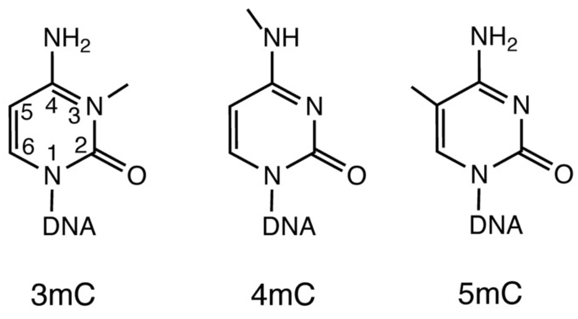
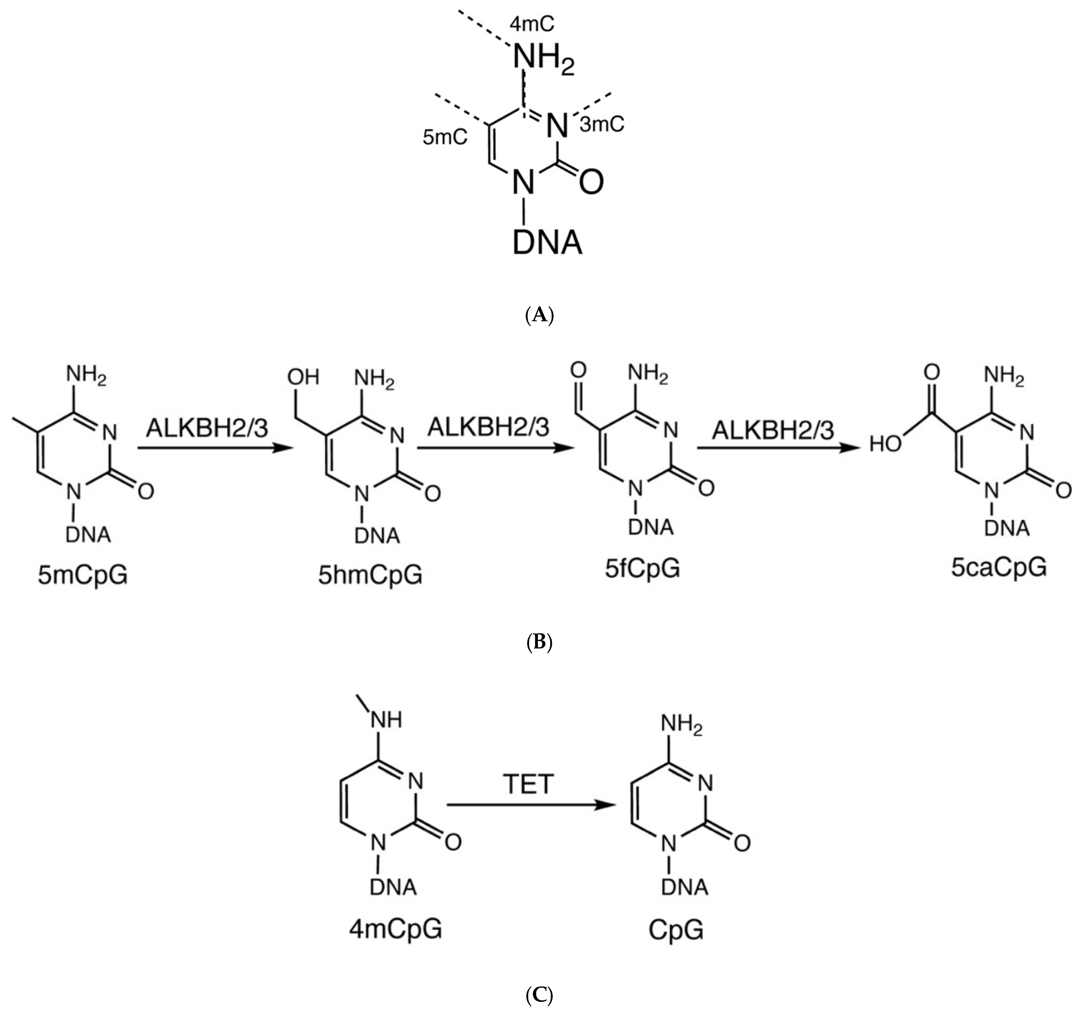
2. Origin and Function of Modified Methylcytosines in DNA

Although 5mC is the most common methylated cytosine, 3-methylcytosines are also present in human DNA [6]. This modified cytosine is generated during DNA damage by non-enzymatic DNA methylation. It can be catalyzed enzymatically and chemically in ribonucleic acid (RNA), but in the case of DNA, it is always non-enzymatic [22]. During these non-enzymatic processes, direct methyl group transfer helps in the formation of 3mC. Direct transfer can take place from endogenous S-adenosyl methionine (SAM) or by an exogenous chemical methylating compound such as methyl methanesulfonate (MMS) [23]. 3mC produced by DNA damage creates methyl lesions in DNA. These methyl lesions have a huge impact on local DNA structure and stability, which in turn can affect replication and transcription [6].
Despite significant studies on 5mC and 3mC that have been carried out, work on 4mC is still in the rudimentary phase [24]. 4mC is primarily present in prokaryotes however, current high-sensitivity methods can detect it in eukaryotes also, such as single-molecule real-time sequencing, [25]. In prokaryotes, 4mC is used in conjunction with a restriction–modification system, which helps in differentiating from exogenous pathogenic DNA and inhibits degrading host DNA from restriction enzymes [26]. In addition, it also regulates and corrects mistakes in DNA replication. The 4mC DNA methyltransferases methylate the amino group present at the C4 position of cytosine [27]. These enzymes detect a certain DNA sequence and methylate a cytosine inside it.
In DNA, the C5 position of cytosine (5mC) has mostly been methylated. 5mC is highly enriched in CpG dinucleotide context (5mCpG; p = Phosphate group between the two nucleotides) [28]. In addition to CpG dinucleotides, several recent studies have found widespread C5 methylation in non-CpG contexts (5mCpH, where H = A, T or C) [29,30,31]. DNA methyltransferase enzymes (DNMT) carry out the methylation at the C5 position in DNA. Using S-adenosyl methionine (SAM) as a cofactor. DNMTs transfer one methyl group to the C5 position of the cytosine. The key enzyme responsible for CpG methylation is DNMT1. In mammalian cells it is the most abundant DNA methyltransferase and is also maintenance methyltransferase. In maintenance methylation, DNMT1 adds a methyl group to DNA when one strand is already methylated [32]. On the other hand, recent studies have demonstrated that DNMT3A and DNMT3B can methylate cytosine residues in the CpH context, with a preference for CA >> CT > CC [33,34]. DNMT3A and DNMT3B are the key de novo methyltransferases [35]. In de novo methylation, these methyltransferases add methylation in newly synthesized DNA without the presence of any cytosine methylation in the opposite strand. 5mC plays an important function in tissue-specific gene expression, human development and as a biomarker of aging [36]. Aside from that, when it is found in the gene promoter, it inhibits gene expression. As 3mC and 5mC have been detected in the human genome, we will concentrate on these two modified cytosines further.

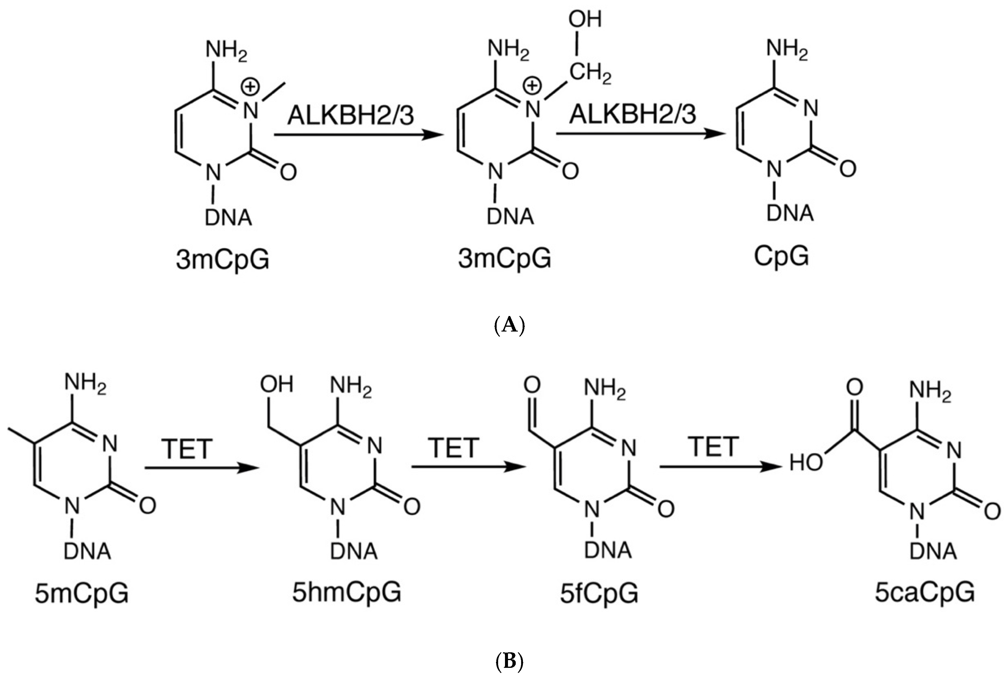
3. Removal of Modified Cytosines by Oxidative and Direct Demethylation

As the presence of 3mC affects the local flexibility and functioning of DNA, removal of this modified cytosine is crucial for the proper functioning of DNA. In humans, the active removal of 3mC marks is initiated by ALKBH2 and 3. These Fe(II)- and 2-OG-dependent dioxygenases convert 3mC into unmodified cytosine via direct demethylation [18]. In this process, the methyl group is being removed from cytosine in a single step (Figure 2A). The reaction takes place in the presence of Fe(II), 2OG and oxygen. In this reaction, 2OG and oxygen are converted to succinate, CO2 and H2O [14]. Functional domains of ALKBH2 and 3 and amino acid residue interacting with the methylcytosine during the enzymatic action are shown in Figure 3A,B. On the other hand, the presence of 5-methylcytosine in the CpG island of a promoter region inhibits gene expression. In addition to that, it also renders the chromatin more tightly packed and inaccessible for the transcriptional machinery [37]. Demethylation of this modified cytosine involves the removal of the methyl group and initiates transcriptional activation and gene expression [37]. In humans, the active removal of 5mC marks is initiated by TET family 5methylcytosine dioxygenases. These Fe(II)- and 2-OG-dependent dioxygenases convert 5mC into 5hmC, 5fC and 5caC by sequential oxidation steps [17,38] (Figure 2B). The reaction takes place in the presence of Fe(II), 2OG and oxygen. 2OG and oxygen is converted to succinate, CO2 and H2O in every step of the sequential oxidation. This mechanism is called oxidative demethylation. Finally, thymine-DNA glycosylase uses the base–excision repair route to convert 5fC and 5caC to unmodified cytosine residues [19]. Functional domains of TET 1–3 and amino acid residue interacting with the methylcytosine during the enzymatic action are shown in Figure 4A,B.

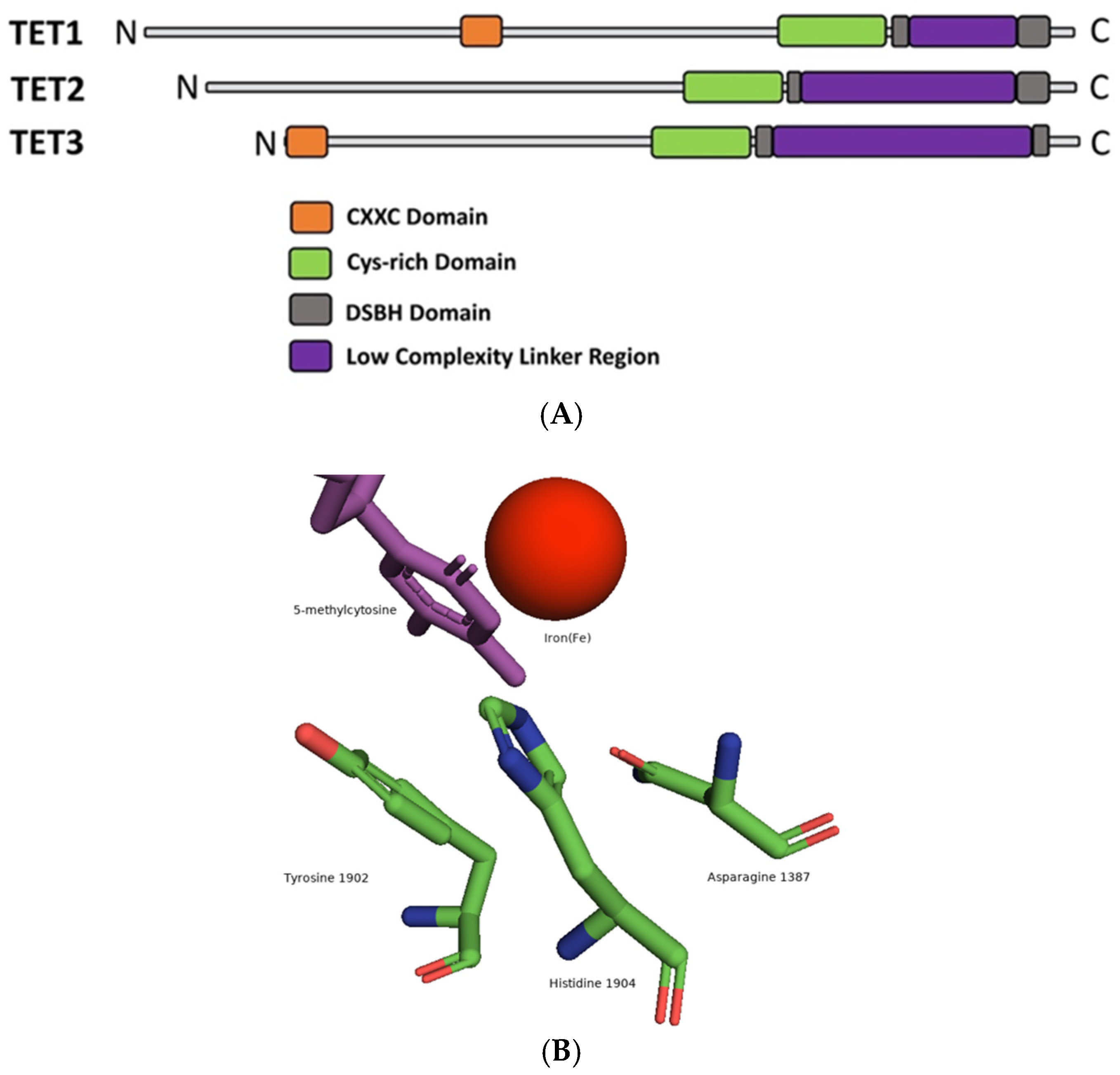
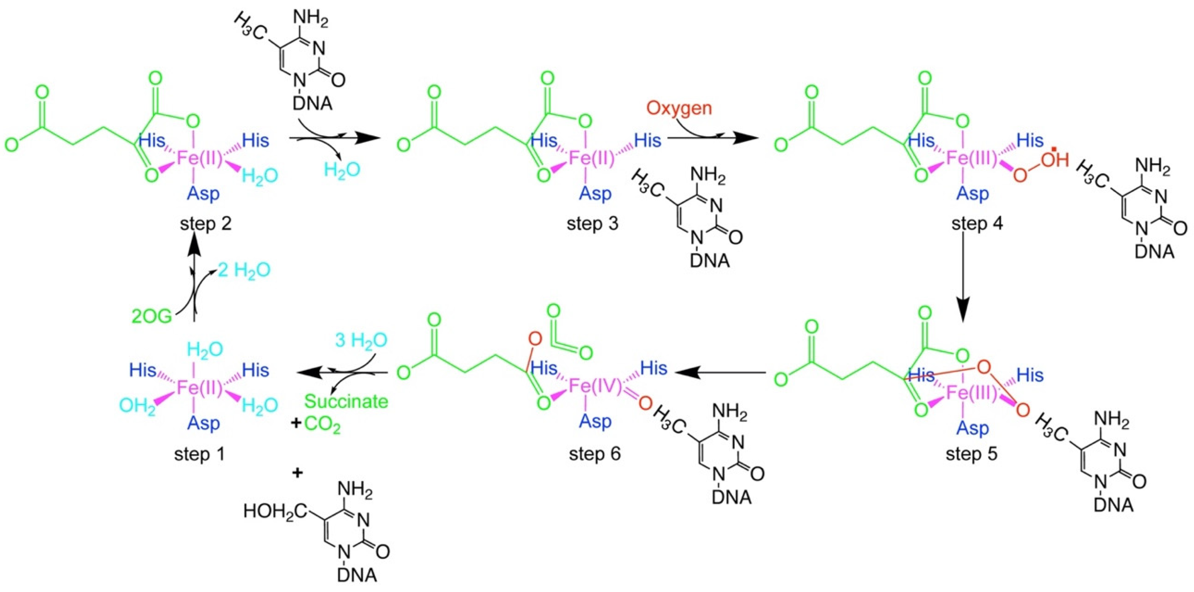
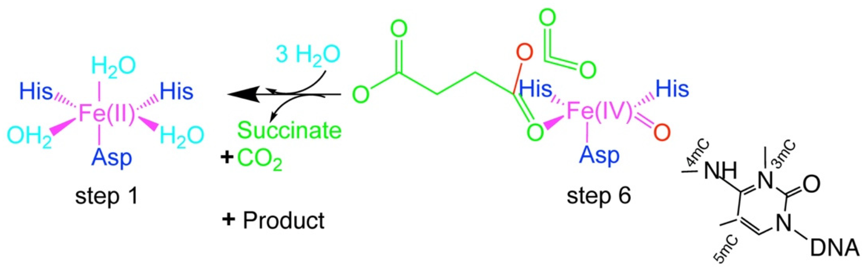
4. Fe(II)/2OG-Dependent Nucleic Acid-Modifying Dioxygenase

Fe(II)/2OG-dependent dioxygenases are the superfamily of enzymes involved in modifying nucleic acids and proteins, lipid-related metabolism, plant metabolite biosynthesis, antibiotic biosynthesis, etc [15]. Under the dioxygenase superfamily, one major subfamily is nucleic acid-modifying enzymes. To date, only two families of enzymes under the Fe(II)/2OG-dependent dioxygenase superfamily that catalyze nucleic acid base modifications has been identified [14]. These are the Fe(II)/2OG-dependent ALKBH family and TET family of enzymes. The ALKBH family uses direct demethylation for nucleic acid modifications, whereas oxidative demethylation is the key process for the TET family of enzymes.
All Fe(II)/2OG-dependent dioxygenase members have a distorted double-stranded β-helix core (DSBH) fold with eight strands (I–VIII) forming two sheets and a well-protected 2OG and iron (Fe) binding pocket [39]. Catalysis and oxidation of the substrate, along with oxidation of the co-substrate 2OG, require Fe(II) for all the 2OG-dependent oxygenases that have been reported to date. [14]. 2OG-dependent dioxygenases also use oxygen. During this process, the destiny of one oxygen atom in the dioxygen molecule is determined by the kind of oxidation reaction catalyzed, whereas the other oxygen atom is integrated into the carboxylic acid group of succinates. Since this oxygenase incorporates both the atoms of oxygen into products, they are also known as dioxygenases [14].
The important steps involved in Fe(II)/2OG-dependent dioxygenase reaction (Figure 5) are as follows. Step 1: Bidentate coordination of 2OG to the Fe(II) center, which the protein coordinates facially by one carboxylate (Aspartate or Glutamate), two histidine ligands and three water molecules. Step 2: Generation of an open coordination site on the Fe(II) upon binding of the 2OG. Step 3: Substrate binding (5mC) to the iron center. Step 4: Oxidative addition of oxygen to the Fe(II) center to yield an Fe(III) superoxide intermediate. Step 5 and Step 6: Attack of the distal oxygen atom by oxygen on C2 of 2OG to initiate O–O cleavage, decarboxylation and formation of a ferryl intermediate. Step 1: Formation of the hydroxylated product 5-hydroxymethylcytosine (5CH2OH) and a coordinatively unsaturated Fe(II) site by formal recombination of R and the coordinated hydroxyl radical. After the formation of 5-hydroxymethylcytosine, it follows the same cycle (sequential oxidation) to produce 5-formylcytosine (5CHO) and 5-carboxylcytosine (5COOH) (Figure 2B). In the case of 3mC, 3CH2OH will form at the end of step 6. It exits the cytosine to form unmethylated cytosine because it is unstable at the N3 position (Figure 2A).

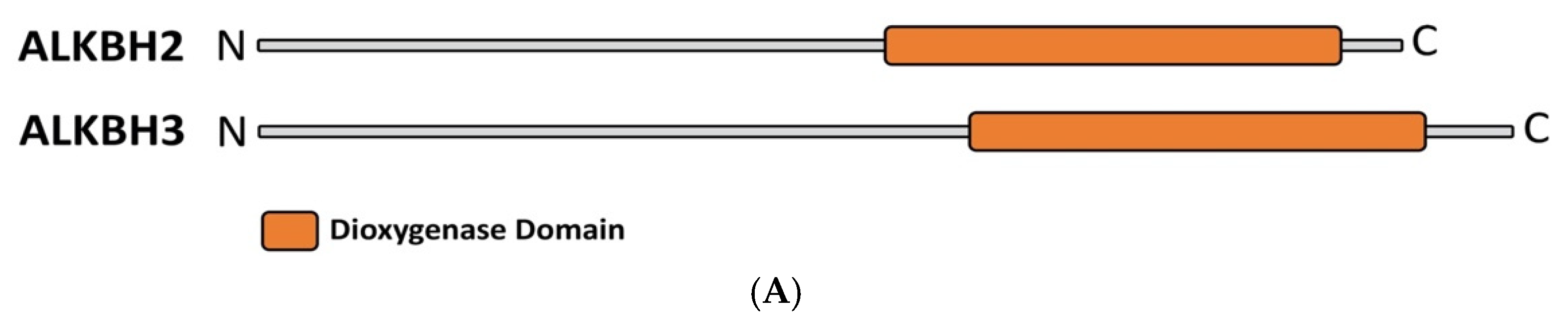
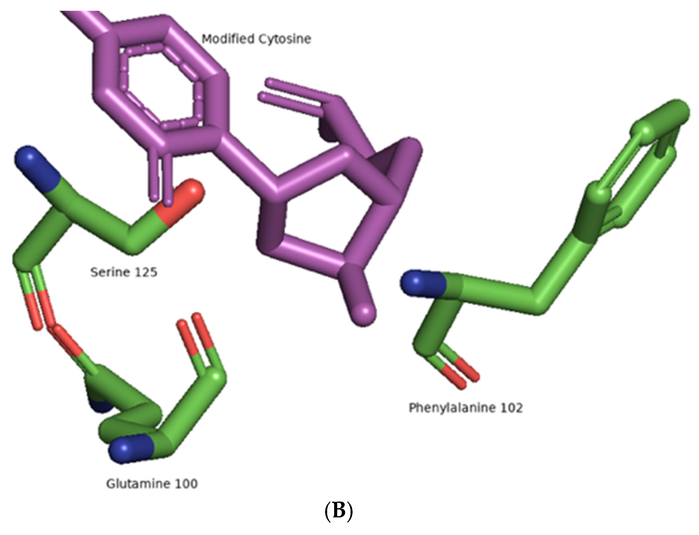
5. The Flexibility of ALKBH 2/3 and TET Family of Enzymes:

The proximity of cytosine methylation has been demonstrated in Figure 6A. 3-methylcytosine and 5-methylcytosine are in the opposing position (Syn-Anti), whereas 4-methylcytosine is present between 3mC and 5mC. In recent years, the theoretical calculations of Bian et al. indicated that ALKBH2 and 3 can bind to 5mC in syn conformation, which is comparable to 3-methylcytosine, the common substrate of these enzymes. The group also demonstrated that ALKBH2 and 3 can oxidize 5mC into 5hmC, 5fC and 5caC by sequential oxidation. This finding demonstrated that ALKBH2 and 3 possess oxidative demethylation properties along with direct demethylation (Figure 6B) [20]. In addition, Ghanty et al. discovered that TET enzymes are efficient in direct demethylation of methylated cytosine (4mC). They also demonstrated the similar proficiency of TET enzymes in either oxidation of 5mC or demethylation of 4mC. This suggests that the TET family of enzymes also possess direct demethylation properties along with oxidative demethylation (Figure 6C) [21].
In many chemical reactions, enzymes require high specificity to distinguish between closely similar substrates. On the other hand, enzymes like cytochrome P450 exhibit wide substrate tolerance [40]. The extraordinary results of ALKBH and TET enzymes suggest that Fe(II)/2OG-dependent nucleic acid-modifying dioxygenases are likely to be found in a middle territory of substrate tolerance. Any well-placed C-H bond (3mC, 4mC, 5mC) is likely to be targeted to complete the oxidative cycle following the formation of a highly reactive Fe IV-oxo intermediate (Figure 7).
In the removal of 3mC and 4mC, direct demethylation plays a crucial role because it is the carbon–nitrogen bond that is easier to break in a single step compared to a carbon–carbon bond. On the other hand, with the removal of 5mC, the conversion takes place via oxidative demethylation. The difficulty of breaking non-activated carbon–carbon bonds inhibits one-step conversion of 5-methylcytosine (5mC) to cytosine (C) (Table 1). Although further research is required to resolve questions about direct demethylation of 4mC by ALKBH2,3 and direct demethylation of 3mC by TET enzymes.

6. Significance & Future Research

6.1. Significance of Functional Flexibility on DNA

In addition to four normal bases, oxidative demethylation of 5mC produces 5hmC, 5fC and 5caC. Due to its importance in gene regulation 5mC is known as the 5th base. In addition, 5hmC is often termed the sixth DNA base due to its role in transcription regulation. A recent study has also shown that it is a stable epigenetic marker [41]. Like 5mC, 5hmC is also recognized by unique reader proteins such as Ring Finger Domains 2, which controls gene expression [42]. For a long time, 5fC and 5caC were thought to be demethylation intermediates found in mammalian DNA. However, recent research found 5fC to be a stable DNA modification, indicating that 5fC plays a functional role in epigenetics [43]. Furthermore, it appears that 5fC influences the rate of nucleotide incorporation as well as the specificity of RNA polymerase II (RNAPII) [44]. Kellinger et al. also suggest that 5caCs, like 5fC, have comparable effects on RNAPII. Yang et al. found that the transcription factor TCF4 predominantly binds to DNA with 5caC markers [45]. Taken together, these observations clearly imply that cytosine modifications (5mC, 5hmC, 5fC and 5caC) are playing essential roles in transcription regulation.
Since the formation of 3mC is associated with DNA damage and ALKBH2,3 eliminates this modified cytosine, these enzymes are known as DNA repair enzymes. The recent discovery of the oxidative demethylation properties of DNA repair enzymes (ALKBH2,3) can open a new window in generating epigenetic cytosine modifications. Vice-versa, the direct demethylation property also suggests the DNA repair mechanism of the TET family of enzymes. These newly observed molecular mechanisms suggest a possible connection between DNA repair and gene regulation.

6.2. Significance of Functional Flexibility on RNA

Although the functional flexibility of these enzymes has been demonstrated on DNA (Table 1), it is not still clear how it will affect RNA. The mechanism and significance of oxidative demethylation of RNA are still in their rudimentary stages. Cytosine methylation at the C5 position (5mrC) is a prevalent modification in highly abundant RNAs, such as mRNA, tRNA and rRNA [46]. 5mrC has recently gained attention as a key posttranscriptional modification of RNA. Increasingly, evidence suggests that 5mrC modification is involved in a variety of cellular functions such as mRNA stability, gene transcription and tRNA translation [46,47,48]. In addition, 5mrC modification in RNA is linked with human diseases such as tumorigenesis [46]. Recent studies have also detected modified higher order oxidative products of 5mrC in mammalian RNA, e.g., 5hmrC (5-hydroxymethylcytosine), 5frC (5-formylcytosine) and 5carC (5-carboxylcytosine) [49,50,51,52]. A growing number of studies have found evidence that these modified cytosines may also have roles in embryogenesis, neuronal differentiation and translation [48,49].
However, oxidative demethylation of 5mrC and the generation of oxidative products is not well understood. A recent study demonstrated that TET2 can oxidize 5mrC to primarily 5hmrC [53]. TET1 can act on 5frC to convert it to only 5carC [54]. Another study demonstrated that Fe(II)/2OG-dependent dioxygenase ALKBH1 can only oxidize 5mrC to 5frC. While 5hmrC was not identified as an intermediate modification [55]. In all three cases, RNA was not a preferred substrate for oxidative demethylation by TET1 and 2 or ALKBH1. Given the role of RNA oxidation in fundamental biological processes and diseases, there is a critical need to elucidate the function and mechanism of 5mC oxidation of RNA in human cells.
Although the functional flexibility of ALKBH3 was established in DNA, no work has been done on RNA. After the very first crystallization of TET dioxygenase, using the “Dali” search all known protein structures in the Protein Data Bank (PDB) were examined and it was observed that TET2 is structurally very similar to AlkB family members (more specifically human ALKBH3) [56]. In addition, previous substrate specificity studies had demonstrated that ALKBH3 has a higher preference for single-stranded nucleic acid: RNA and single-stranded DNA. The reason for this is that ALKBH3 lacks the RKK motif [57]. This motif is used by other ALKBH enzymes (e.g., ALKBH2) to interact with the complementary strand of double-stranded DNA. As a result, in recent work, it has also been shown that ALKBH3 has almost 6–8 fold higher activity in single-strand DNA compared to double-strand DNA [20]. This may explain the flexibility of ALKBH3 in RNA. Finally, in addition to the role of ALKBH2 and 3, the remarkable bifunctional ability of TET dioxygenases also has importance in RNA biology. N4-methylation is a physiologically important RNA alteration which might serve as a TET dioxygenase substrate and may have a significant role.

Funding

The funds for publication were provided by the UMKC School of Pharmacy, Division of Pharmacology and Pharmaceutical Sciences.

Institutional Review Board Statement

Not applicable.

Informed Consent Statement

Not applicable.

Data Availability Statement

Not applicable.

Conflicts of Interest

The author declares no conflict of interest.

References

  1. Bilyard, M.K.; Becker, S.; Balasubramanian, S. Natural, modified DNA bases. Curr. Opin. Chem. Biol. 2020, 57, 1–7. [Google Scholar] [CrossRef] [PubMed]
  2. Kumar, S.; Chinnusamy, V.; Mohapatra, T. Epigenetics of Modified DNA Bases: 5-Methylcytosine and Beyond. Front. Genet. 2018, 9, 640. [Google Scholar] [CrossRef] [Green Version]
  3. Kawasaki, H.; Ninomiya, S.; Yuki, H. High-performance liquid chromatographic determination of 3-methylcytosine in deoxyribonucleic acid treated with carcinogenic methylating agents in vitro and in vivo. Chem. Pharm. Bull. 1985, 33, 1170–1174. [Google Scholar] [CrossRef] [PubMed] [Green Version]
  4. Yu, M.; Ji, L.; Neumann, D.A.; Chung, D.H.; Groom, J.; Westpheling, J.; He, C.; Schmitz, R.J. Base-resolution detection of N4-methylcytosine in genomic DNA using 4mC-Tet-assisted-bisulfite- sequencing. Nucleic Acids Res. 2015, 43, e148. [Google Scholar] [CrossRef] [Green Version]
  5. Lister, R.; Pelizzola, M.; Dowen, R.H.; Hawkins, R.D.; Hon, G.; Tonti-Filippini, J.; Nery, J.R.; Lee, L.; Ye, Z.; Ngo, Q.M.; et al. Human DNA methylomes at base resolution show widespread epigenomic differences. Nature 2009, 462, 315–322. [Google Scholar] [CrossRef] [Green Version]
  6. Pataillot-Meakin, T.; Pillay, N.; Beck, S. 3-methylcytosine in cancer: An underappreciated methyl lesion? Epigenomics 2016, 8, 451–454. [Google Scholar] [CrossRef] [Green Version]
  7. Singer, B.; Kusmierek, J.T. Chemical mutagenesis. Annu. Rev. Biochem. 1982, 51, 655–693. [Google Scholar] [CrossRef]
  8. Schultz, M.D.; He, Y.; Whitaker, J.W.; Hariharan, M.; Mukamel, E.A.; Leung, D.; Rajagopal, N.; Nery, J.R.; Urich, M.A.; Chen, H.; et al. Human body epigenome maps reveal noncanonical DNA methylation variation. Nature 2015, 523, 212–216. [Google Scholar] [CrossRef]
  9. Bonasio, R.; Tu, S.; Reinberg, D. Molecular signals of epigenetic states. Science 2010, 330, 612–616. [Google Scholar] [CrossRef] [Green Version]
  10. Feng, S.; Jacobsen, S.E.; Reik, W. Epigenetic reprogramming in plant and animal development. Science 2010, 330, 622–627. [Google Scholar] [CrossRef] [Green Version]
  11. Boland, M.J.; Nazor, K.L.; Loring, J.F. Epigenetic regulation of pluripotency and differentiation. Circ. Res. 2014, 115, 311–324. [Google Scholar] [CrossRef] [PubMed] [Green Version]
  12. Handy, D.E.; Castro, R.; Loscalzo, J. Epigenetic modifications: Basic mechanisms and role in cardiovascular disease. Circulation 2011, 123, 2145–2156. [Google Scholar] [CrossRef] [PubMed] [Green Version]
  13. Flashman, E.; Schofield, C.J. The most versatile of all reactive intermediates? Nat. Chem. Biol. 2007, 3, 86–87. [Google Scholar] [CrossRef] [PubMed]
  14. Islam, M.S.; Leissing, T.M.; Chowdhury, R.; Hopkinson, R.J.; Schofield, C.J. 2-Oxoglutarate-Dependent Oxygenases. Annu. Rev. Biochem. 2018, 87, 585–620. [Google Scholar] [CrossRef] [PubMed]
  15. Herr, C.Q.; Hausinger, R.P. Amazing Diversity in Biochemical Roles of Fe(II)/2-Oxoglutarate Oxygenases. Trends Biochem. Sci. 2018, 43, 517–532. [Google Scholar] [CrossRef]
  16. Waheed, S.O.; Ramanan, R.; Chaturvedi, S.S.; Lehnert, N.; Schofield, C.J.; Christov, C.Z.; Karabencheva-Christova, T.G. Role of Structural Dynamics in Selectivity and Mechanism of Non-heme Fe(II) and 2-Oxoglutarate-Dependent Oxygenases Involved in DNA Repair. ACS Cent. Sci. 2020, 6, 795–814. [Google Scholar] [CrossRef]
  17. Ito, S.; Shen, L.; Dai, Q.; Wu, S.C.; Collins, L.B.; Swenberg, J.A.; He, C.; Zhang, Y. Tet proteins can convert 5-methylcytosine to 5-formylcytosine and 5-carboxylcytosine. Science 2011, 333, 1300–1303. [Google Scholar] [CrossRef] [Green Version]
  18. Martinez, S.; Hausinger, R.P. Catalytic Mechanisms of Fe(II)- and 2-Oxoglutarate-dependent Oxygenases. J. Biol. Chem. 2015, 290, 20702–20711. [Google Scholar] [CrossRef] [Green Version]
  19. Zhang, L.; Lu, X.; Lu, J.; Liang, H.; Dai, Q.; Xu, G.L.; Luo, C.; Jiang, H.; He, C. Thymine DNA glycosylase specifically recognizes 5-carboxylcytosine-modified DNA. Nat. Chem. Biol. 2012, 8, 328–330. [Google Scholar] [CrossRef] [Green Version]
  20. Bian, K.; Lenz, S.A.P.; Tang, Q.; Chen, F.; Qi, R.; Jost, M.; Drennan, C.L.; Essigmann, J.M.; Wetmore, S.D.; Li, D. DNA repair enzymes ALKBH2, ALKBH3, and AlkB oxidize 5-methylcytosine to 5-hydroxymethylcytosine, 5-formylcytosine and 5-carboxylcytosine in vitro. Nucleic Acids Res. 2019, 47, 5522–5529. [Google Scholar] [CrossRef] [Green Version]
  21. Ghanty, U.; Wang, T.; Kohli, R.M. Nucleobase Modifiers Identify TET Enzymes as Bifunctional DNA Dioxygenases Capable of Direct N-Demethylation. Angew. Chem. Int. Ed. Engl. 2020, 59, 11312–11315. [Google Scholar] [CrossRef] [PubMed]
  22. Friedman, S. In vitro methylation of rat liver tRNA-synthesis of 3-methylcytosine and 1-methylhypoxantheine. Biochem. Biophys. Res. Commun. 1977, 74, 1505–1510. [Google Scholar] [CrossRef]
  23. Rydberg, B.; Lindahl, T. Nonenzymatic methylation of DNA by the intracellular methyl group donor S-adenosyl-L-methionine is a potentially mutagenic reaction. EMBO J. 1982, 1, 211–216. [Google Scholar] [CrossRef] [PubMed]
  24. Zhao, Z.; Zhang, X.; Chen, F.; Fang, L.; Li, J. Accurate prediction of DNA N(4)-methylcytosine sites via boost-learning various types of sequence features. BMC Genom. 2020, 21, 627. [Google Scholar] [CrossRef] [PubMed]
  25. Davis, B.M.; Chao, M.C.; Waldor, M.K. Entering the era of bacterial epigenomics with single molecule real time DNA sequencing. Curr. Opin. Microbiol. 2013, 16, 192–198. [Google Scholar] [CrossRef] [PubMed] [Green Version]
  26. Zeng, F.; Fang, G.; Yao, L. A Deep Neural Network for Identifying DNA N4-Methylcytosine Sites. Front. Genet. 2020, 11, 209. [Google Scholar] [CrossRef] [PubMed] [Green Version]
  27. Rathi, P.; Maurer, S.; Summerer, D. Selective recognition of N4-methylcytosine in DNA by engineered transcription-activator-like effectors. Philos. Trans. R. Soc. Lond. B Biol. Sci. 2018, 373, 20170078. [Google Scholar] [CrossRef] [Green Version]
  28. Luo, C.; Hajkova, P.; Ecker, J.R. Dynamic DNA methylation: In the right place at the right time. Science 2018, 361, 1336–1340. [Google Scholar] [CrossRef] [Green Version]
  29. Pinney, S.E. Mammalian Non-CpG Methylation: Stem Cells and Beyond. Biology 2014, 3, 739–751. [Google Scholar] [CrossRef] [Green Version]
  30. Jang, H.S.; Shin, W.J.; Lee, J.E.; Do, J.T. CpG and Non-CpG Methylation in Epigenetic Gene Regulation and Brain Function. Genes 2017, 8, 148. [Google Scholar] [CrossRef] [Green Version]
  31. He, Y.; Ecker, J.R. Non-CG Methylation in the Human Genome. Annu. Rev. Genom. Hum. Genet. 2015, 16, 55–77. [Google Scholar] [CrossRef] [Green Version]
  32. Edwards, J.R.; Yarychkivska, O.; Boulard, M.; Bestor, T.H. DNA methylation and DNA methyltransferases. Epigenet. Chromatin 2017, 10, 23. [Google Scholar] [CrossRef] [PubMed] [Green Version]
  33. Dukatz, M.; Adam, S.; Biswal, M.; Song, J.; Bashtrykov, P.; Jeltsch, A. Complex DNA sequence readout mechanisms of the DNMT3B DNA methyltransferase. Nucleic Acids Res. 2020, 48, 11495–11509. [Google Scholar] [CrossRef] [PubMed]
  34. Gowher, H.; Jeltsch, A. Enzymatic properties of recombinant Dnmt3a DNA methyltransferase from mouse: The enzyme modifies DNA in a non-processive manner and also methylates non-CpG [correction of non-CpA] sites. J. Mol. Biol. 2001, 309, 1201–1208. [Google Scholar] [CrossRef] [PubMed]
  35. Okano, M.; Bell, D.W.; Haber, D.A.; Li, E. DNA methyltransferases Dnmt3a and Dnmt3b are essential for de novo methylation and mammalian development. Cell 1999, 99, 247–257. [Google Scholar] [CrossRef] [Green Version]
  36. Salameh, Y.; Bejaoui, Y.; El Hajj, N. DNA Methylation Biomarkers in Aging and Age-Related Diseases. Front. Genet. 2020, 11, 171. [Google Scholar] [CrossRef]
  37. Wu, H.; Zhang, Y. Reversing DNA methylation: Mechanisms, genomics, and biological functions. Cell 2014, 156, 45–68. [Google Scholar] [CrossRef] [Green Version]
  38. Tahiliani, M.; Koh, K.P.; Shen, Y.; Pastor, W.A.; Bandukwala, H.; Brudno, Y.; Agarwal, S.; Iyer, L.M.; Liu, D.R.; Aravind, L.; et al. Conversion of 5-methylcytosine to 5-hydroxymethylcytosine in mammalian DNA by MLL partner TET1. Science 2009, 324, 930–935. [Google Scholar] [CrossRef] [Green Version]
  39. Ponnaluri, V.K.; Maciejewski, J.P.; Mukherji, M. A mechanistic overview of TET-mediated 5-methylcytosine oxidation. Biochem. Biophys. Res. Commun. 2013, 436, 115–120. [Google Scholar] [CrossRef]
  40. Jung, S.T.; Lauchli, R.; Arnold, F.H. Cytochrome P450: Taming a wild type enzyme. Curr. Opin. Biotechnol. 2011, 22, 809–817. [Google Scholar] [CrossRef] [Green Version]
  41. Bachman, M.; Uribe-Lewis, S.; Yang, X.; Williams, M.; Murrell, A.; Balasubramanian, S. 5-Hydroxymethylcytosine is a predominantly stable DNA modification. Nat. Chem. 2014, 6, 1049–1055. [Google Scholar] [CrossRef] [PubMed] [Green Version]
  42. Chen, W.; Wu, M.; Hang, T.; Wang, C.; Zhang, X.; Zang, J. Structure insights into the molecular mechanism of the interaction between UHRF2 and PCNA. Biochem. Biophys. Res. Commun. 2017, 494, 575–580. [Google Scholar] [CrossRef] [PubMed]
  43. Bachman, M.; Uribe-Lewis, S.; Yang, X.; Burgess, H.E.; Iurlaro, M.; Reik, W.; Murrell, A.; Balasubramanian, S. 5-Formylcytosine can be a stable DNA modification in mammals. Nat. Chem. Biol. 2015, 11, 555–557. [Google Scholar] [CrossRef] [PubMed] [Green Version]
  44. Kellinger, M.W.; Song, C.X.; Chong, J.; Lu, X.Y.; He, C.; Wang, D. 5-formylcytosine and 5-carboxylcytosine reduce the rate and substrate specificity of RNA polymerase II transcription. Nat. Struct. Mol. Biol. 2012, 19, 831–833. [Google Scholar] [CrossRef] [PubMed] [Green Version]
  45. Yang, J.; Horton, J.R.; Li, J.; Huang, Y.; Zhang, X.; Blumenthal, R.M.; Cheng, X. Structural basis for preferential binding of human TCF4 to DNA containing 5-carboxylcytosine. Nucleic Acids Res. 2019, 47, 8375–8387. [Google Scholar] [CrossRef] [PubMed]
  46. Xue, C.; Zhao, Y.; Li, L. Advances in RNA cytosine-5 methylation: Detection, regulatory mechanisms, biological functions and links to cancer. Biomark. Res. 2020, 8, 43. [Google Scholar] [CrossRef]
  47. Boo, S.H.; Kim, Y.K. The emerging role of RNA modifications in the regulation of mRNA stability. Exp. Mol. Med. 2020, 52, 400–408. [Google Scholar] [CrossRef] [Green Version]
  48. Shen, H.; Ontiveros, R.J.; Owens, M.C.; Liu, M.Y.; Ghanty, U.; Kohli, R.M.; Liu, K.F. TET-mediated 5-methylcytosine oxidation in tRNA promotes translation. J. Biol. Chem. 2021, 296, 100087. [Google Scholar] [CrossRef]
  49. Delatte, B.; Wang, F.; Ngoc, L.V.; Collignon, E.; Bonvin, E.; Deplus, R.; Calonne, E.; Hassabi, B.; Putmans, P.; Awe, S.; et al. RNA biochemistry. Transcriptome-wide distribution and function of RNA hydroxymethylcytosine. Science 2016, 351, 282–285. [Google Scholar] [CrossRef] [Green Version]
  50. Zhang, H.Y.; Xiong, J.; Qi, B.L.; Feng, Y.Q.; Yuan, B.F. The existence of 5-hydroxymethylcytosine and 5-formylcytosine in both DNA and RNA in mammals. Chem. Commun. 2016, 52, 737–740. [Google Scholar] [CrossRef]
  51. Huang, W.; Lan, M.D.; Qi, C.B.; Zheng, S.J.; Wei, S.Z.; Yuan, B.F.; Feng, Y.Q. Formation and determination of the oxidation products of 5-methylcytosine in RNA. Chem. Sci. 2016, 7, 5495–5502. [Google Scholar] [CrossRef] [PubMed] [Green Version]
  52. Huber, S.M.; van Delft, P.; Mendil, L.; Bachman, M.; Smollett, K.; Werner, F.; Miska, E.A.; Balasubramanian, S. Formation and abundance of 5-hydroxymethylcytosine in RNA. Chembiochem 2015, 16, 752–755. [Google Scholar] [CrossRef] [PubMed] [Green Version]
  53. DeNizio, J.E.; Liu, M.Y.; Leddin, E.M.; Cisneros, G.A.; Kohli, R.M. Selectivity and Promiscuity in TET-Mediated Oxidation of 5-Methylcytosine in DNA and RNA. Biochemistry 2019, 58, 411–421. [Google Scholar] [CrossRef] [PubMed]
  54. Basanta-Sanchez, M.; Wang, R.; Liu, Z.; Ye, X.; Li, M.; Shi, X.; Agris, P.F.; Zhou, Y.; Huang, Y.; Sheng, J. TET1-Mediated Oxidation of 5-Formylcytosine (5fC) to 5-Carboxycytosine (5caC) in RNA. Chembiochem 2017, 18, 72–76. [Google Scholar] [CrossRef]
  55. Haag, S.; Sloan, K.E.; Ranjan, N.; Warda, A.S.; Kretschmer, J.; Blessing, C.; Hubner, B.; Seikowski, J.; Dennerlein, S.; Rehling, P.; et al. NSUN3 and ABH1 modify the wobble position of mt-tRNAMet to expand codon recognition in mitochondrial translation. EMBO J. 2016, 35, 2104–2119. [Google Scholar] [CrossRef] [PubMed]
  56. Hu, L.; Li, Z.; Cheng, J.; Rao, Q.; Gong, W.; Liu, M.; Shi, Y.G.; Zhu, J.; Wang, P.; Xu, Y. Crystal structure of TET2-DNA complex: Insight into TET-mediated 5mC oxidation. Cell 2013, 155, 1545–1555. [Google Scholar] [CrossRef] [Green Version]
  57. Yang, C.G.; Yi, C.; Duguid, E.M.; Sullivan, C.T.; Jian, X.; Rice, P.A.; He, C. Crystal structures of DNA/RNA repair enzymes AlkB and ABH2 bound to dsDNA. Nature 2008, 452, 961–965. [Google Scholar] [CrossRef] [Green Version]
Figure 1. Structure of 3-methylcytosine, 4-methylcytosine and 5-methylcytosine in DNA.
Biochem 02 00012 g001
Figure 2. (A). Schematic representation of direct demethylation of 3mC by ALKBH2, 3 dioxygenases; (B) Schematic representation of oxidative demethylation of 5mC by TET family dioxygenases.
Biochem 02 00012 g002
Figure 3. (A) Domain architecture of Human ALKBH2, ALKBH3. (B) Amino acid residues reside in close proximity to, and interact with, the flipped out modified cytosine base in ALKBH2 (PDB ID- 3BTZ).
Biochem 02 00012 g003aBiochem 02 00012 g003b
Figure 4. (A) Domain architecture of Human TET1, TET2, TET3. (B) Amino acid residues reside in close proximity to, and interact with the flipped out modified cytosine base in TET2 (PDB ID- 4NM6).
Biochem 02 00012 g004
Figure 5. Mechanism of action of Fe(II)/2OG-dependent dioxygenases.
Biochem 02 00012 g005
Figure 6. (A) Structure of a cytosine molecule where the methyl group is shown at positions 3, 4, 5 at the same time to demonstrate the proximity between these three methylated sites; (B) Schematic representation of oxidative demethylation of 5mC by ALKBH2,3 dioxygenases; (C) Schematic representation of direct demethylation of 4mC by TET family dioxygenases.
Biochem 02 00012 g006
Figure 7. Representation of the proximity of the methyl group at 3, 4, 5 positions to the highly reactive Fe IV-oxo intermediate. Any well-placed C-H bond (3mC, 4mC, 5mC) is likely to be targeted to complete the oxidative cycle following the formation of a highly reactive Fe IV-oxo intermediate.
Biochem 02 00012 g007
Table 1. The table lists modified cytosines in DNA, Fe(II)/2OG-dependent dioxygenases and their mechanism of action, which demonstrates the functional flexibility of ALKBH2,3 and TET family dioxygenases.
Modified Cytosine in DNAEnzymeMechanismModified Cytosine in DNAEnzymeMechanism
5mCALKBH2, ALKBH3Oxidative
Demethylation
5mCTET1-3Oxidative
Demethylation
4mCALKBH2, ALKBH3-4mCTET1-3Direct
Demethylation
3mCALKBH2, ALKBH3Direct
Demethylation
3mCTET1-3-
Publisher’s Note: MDPI stays neutral with regard to jurisdictional claims in published maps and institutional affiliations.

