Life-Threatening Hemorrhage from the Lingual Artery after a Genioplasty—Case Report and Review of Possible Complications Associated with Orthognathic Surgeries

 ,
,
Abstract
:1. Introduction
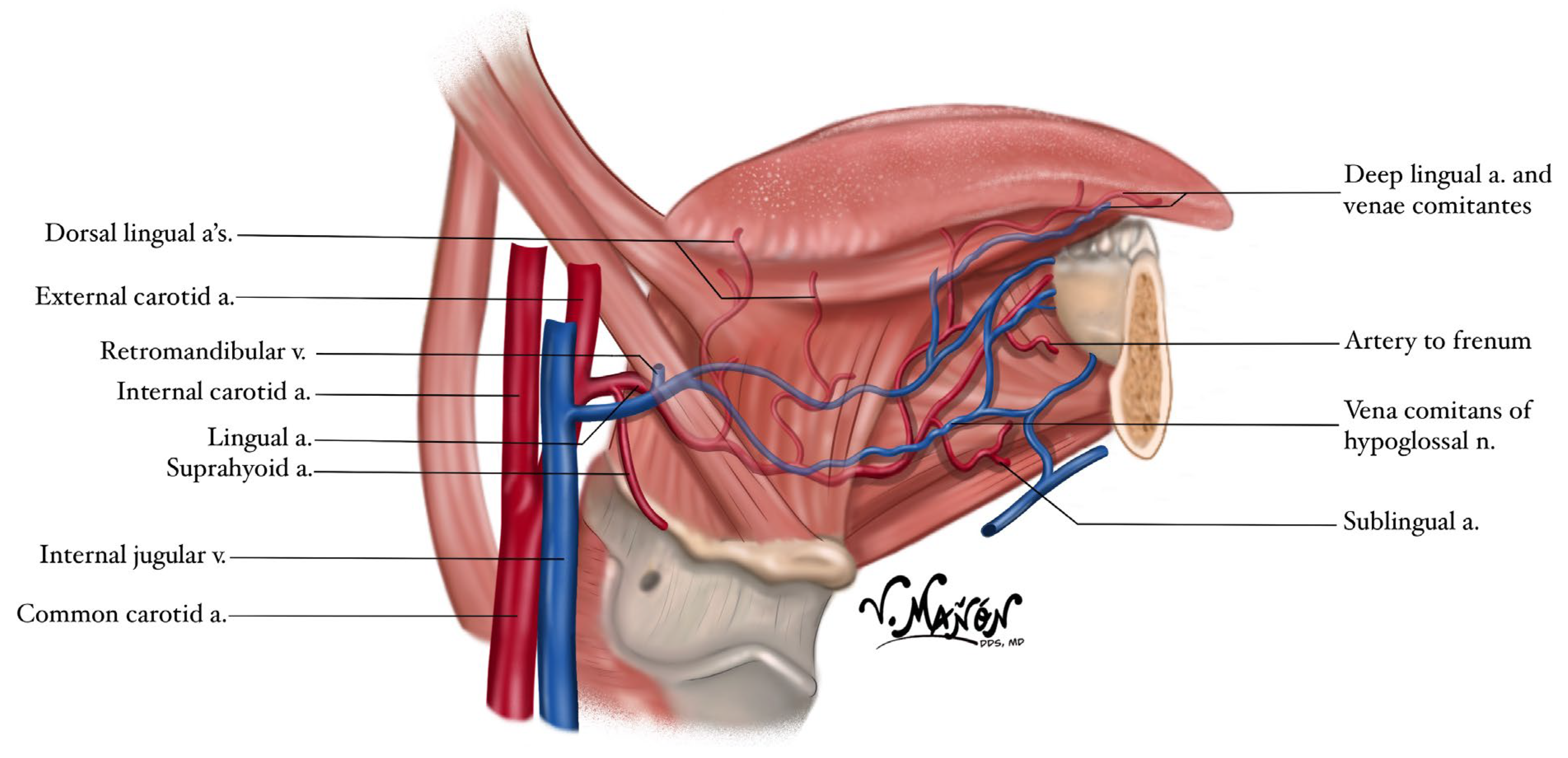
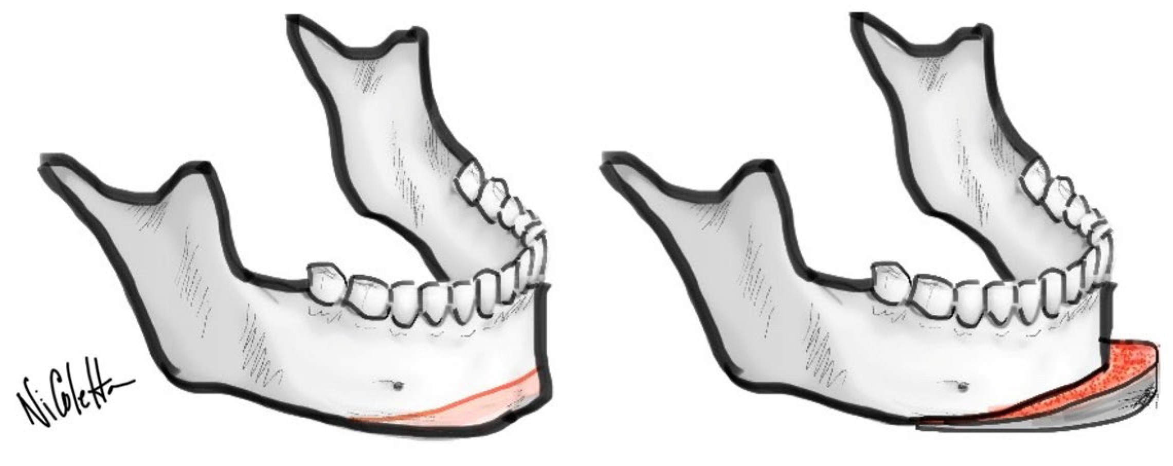
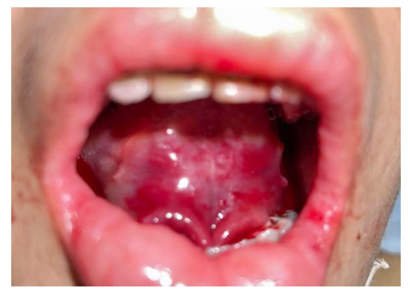
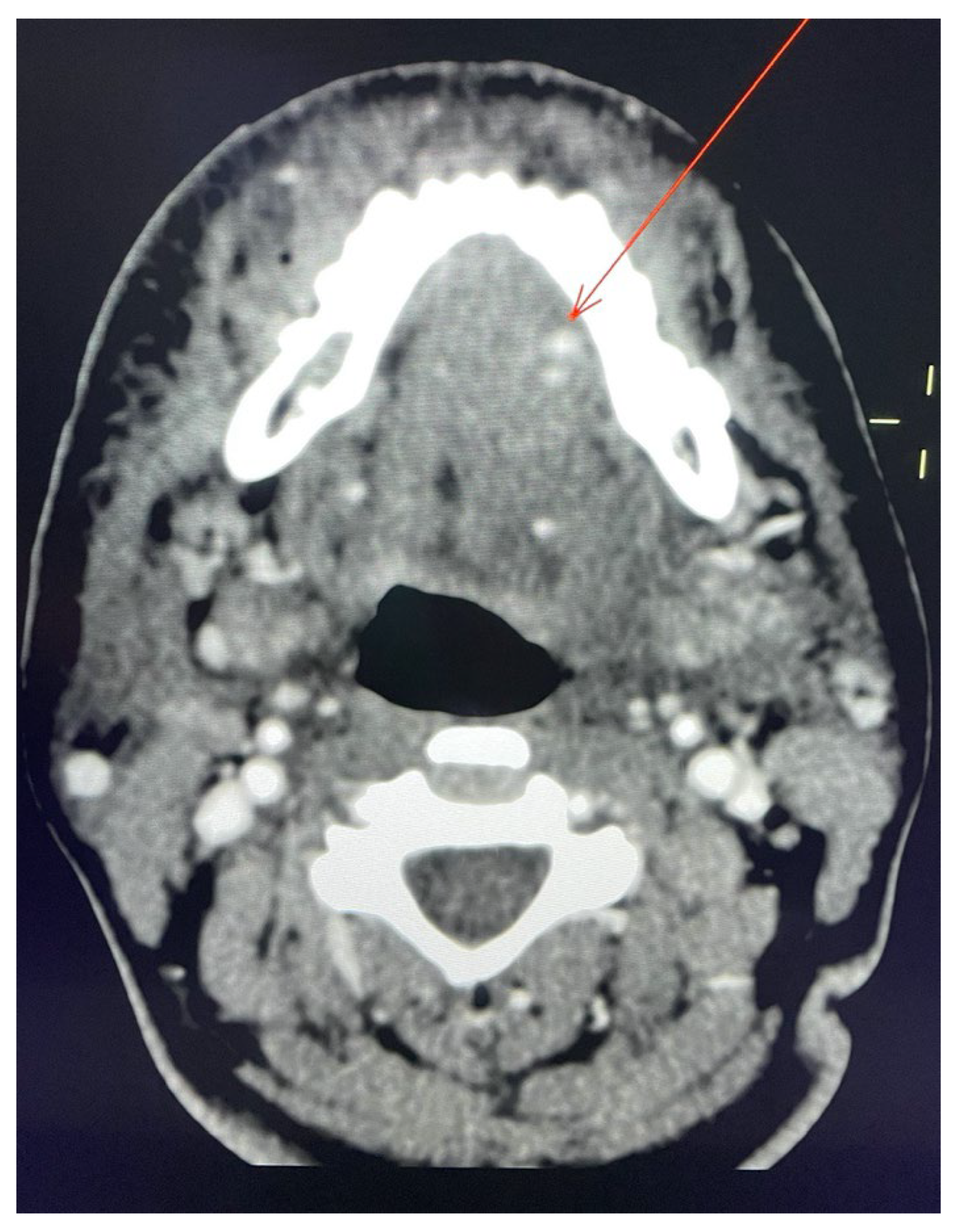
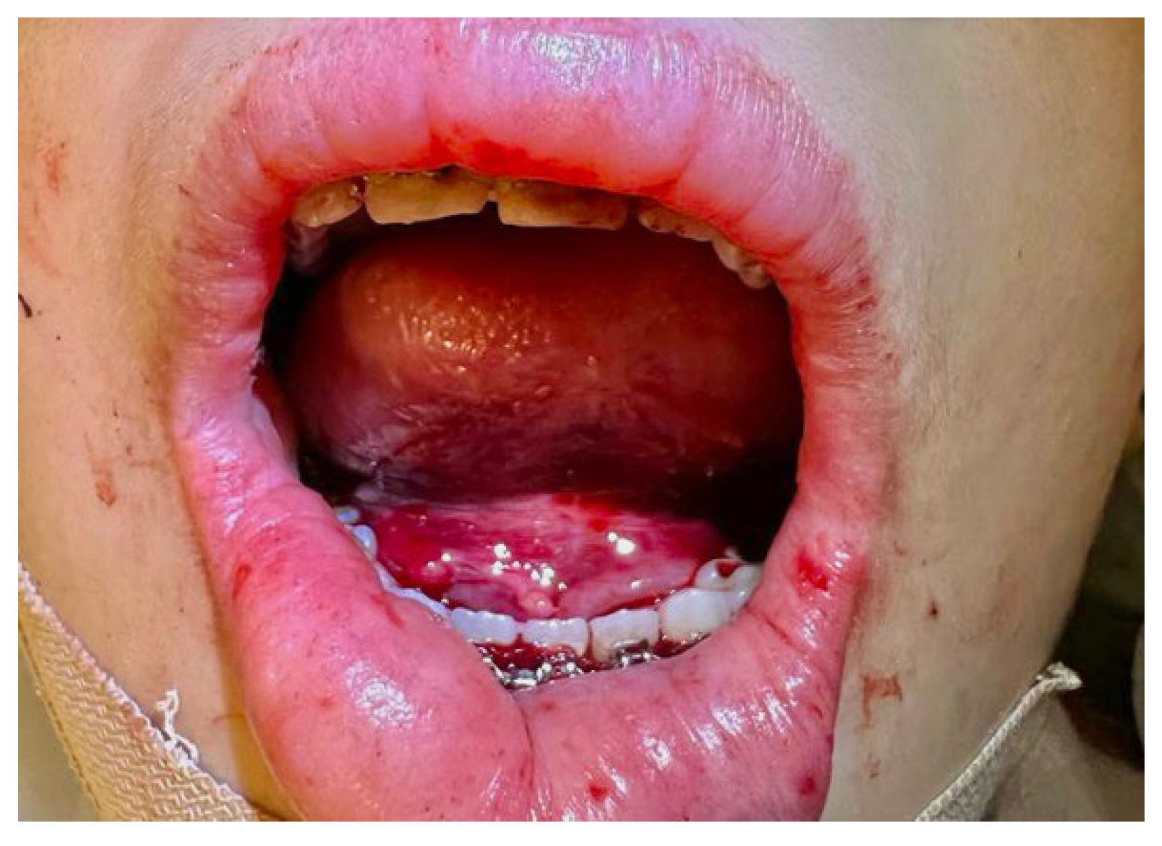
2. Case Report

| Complication | Type of Surgery | Study |
|---|---|---|
| Immediate postoperative pain | IVRO, ASO Md, ASO Mx, GeP | Hsu et al. [1] |
| Inferior alveolar nerve injury | BSSO | Choi et al. [2] |
| Cranial nerve injury involving CN II, VI, ophthalmic (CN V1) and maxillary nerve (CN V2) | Le Fort I osteotomy | Kim et al. [3] |
| Upper and lower lip hypoesthesia | Le Fort I osteotomy in combination with SSRO or IVRO | Ueki et al. [4] |
| Ocular palsy due to injury of CN III and CN VI | Le Fort I osteotomy | Newlands et al. [5] |
| Mandibular infections; Mandibular pseudoarthrosis; Cranial base fractures; Hemorrhage; Amaurosis | Bimaxillary osteotomy, Le Fort I osteotomy, BSSO, GeP, Bimaxillary with GeP, Le Fort I with GeP, BSSO with GeP | Ferri et al. [6] |
| Hemorrhage: | Mandibularosteotomies (alone or in combination): | Lanigan et al. [7] |
| Retromandibular vein | BSRO | |
| Aberrant venous plexus | Le Fort I, BSRO | |
| Right inferior alveolar artery and vein | BSRO, Reduction GeP | |
| Maxillary artery | Le Fort I, BSRO, GeP | |
| Maxillary artery | BSRO | |
| Facial artery | BSRO | |
| Facial artery | Le Fort I, BSRO, GeP | |
| Retromandibular vein | IVRO | |
| Facial artery | IVRO | |
| Masseteric or maxillary arteries | IVRO | |
| Maxillary artery | Le Fort I, IVRO | |
| Maxillary artery | IVRO | |
| Artery to mylohyoid muscle | BSRO, advancement GeP | |
| Capillary, venous | BSRO, advancement GeP | |
| Lingual artery hemorrhage | Biopsy of a lesion in the floor of the mouth | Burke et al. [8] |
| Lingual artery hemorrhage | Procedures involving carcinoma of the tongue | Li et al. [9] |
| Hemorrhage: | Maxillary osteotomies (alone or in combination): | Lanigan et al. [12,13] |
| Left descending palatine artery | Le Fort I | |
| Right sphenopalatine artery | Le Fort I, BSRO | |
| Descending palatine artery | Le Fort I | |
| Left pterygoid plexus | Le Fort I | |
| Left pterygoid plexus | Left hemi-Le Fort I with RPE | |
| A terminal branch of Left maxillary artery | Le Fort I, bilateral tubinate ectomies | |
| Descending palatine or sphenopalatine artery | Le Fort I | |
| Right maxillary artery | Le Fort I, BSRO | |
| A terminal branch of Left maxillary artery | Le Fort I | |
| A terminal branch of Right maxillary artery | Le Fort I | |
| Left descending palatine artery | Le Fort I, BSRO, GeP | |
| Major branch of Right maxillary artery | Le Fort I | |
| Left maxillary artery or a major branch | Le Fort I, BSRO | |
| Right maxillary artery | Le Fort I | |
| Maxillary artery or a major branch | Le Fort I (previous Le Fort III fx.) | |
| Major branch of Right maxillary artery | Le Fort I, BVRO, GeP | |
| Left sphenopalatine artery | Le Fort I, BVRO, GeP | |
| Hemorrhage; Inadvertent bone-cut; Nerve damage; Inability to retroposition; Placement of condylar fragment; Non-union; Infection; Extrusion of teeth; Relapse | IVRO | Tuinzing et al. [14] |
| Varying degrees of fracture of the mandible; Displacement of the mandibular condyle out of the glenoid fossa; Bilateral neuropathy of the inferior alveolar dental nerves; Transient lingual nerve paresthesia; Slight to severe lip and face swelling with moderate respiratory embarrassment; Unusual dislocation of a proximal fragment anteriorly; Dehiscence of the wound due to hematoma formation secondary to bleeding from the osteotomy site, followed by low-grade infection and drainage; Infection; Swelling and localized osteitis in an extraction site; Subperiosteal swelling and abscess formation; Anesthetic complication with a complex fulminating pyrexia; Non-controllable postoperative nausea and vomit; Relapse of the occlusion | Intraoral sagittal osteotomy in the mandibular rami | Guernsey et al. [15] |
| Near-fatal airway obstruction secondary to sublingual bleeding and hematoma possibly due to injury of submental, sublingual, and/or mylohyoid arteries | Routine dental implant placement | Niamtu et al. [16] |
| Massive epistaxis resulting in hemorrhagic shock due to a pseudoaneurysm from a distal branch of the maxillary artery, probably the sphenopalatine artery | Le Fort I | Lanigan et al. [17] |
| Repeated epistaxis due to a pseudoaneurysm of the sphenopalatine artery | Le Fort I, GeP | |
| Epistaxis due to a small false aneurysm of the left maxillary artery | Le Fort I due to a previous Le Fort II fx. in an MVA | |
| Chemosis of the left eye, mild left ptosis, diplopia, and an inability to move the left eye laterally from the midline due to a left VI nerve palsy secondary to extensive left carotid- cavernous fistula | Le Fort I with advancement and a bone graft to the alveolar cleft site due to unilateral cleft lip and palate deformity | |
| Early formation of pseudoaneurysm of a lingual artery branch due to tongue trauma | Traumatic tongue bleed | Rathod et al. [18] |
3. Discussion
Author Contributions
Funding
Institutional Review Board Statement
Informed Consent Statement
Data Availability Statement
Acknowledgments
Conflicts of Interest
References
- Hsu, H.J.; Hsu, K.J. Investigation of Immediate Postoperative Pain following Orthognathic Surgery. Biomed. Res. Int. 2021, 2021, 9942808. [Google Scholar] [CrossRef] [PubMed]
- Choi, B.K.; Lo, L.J.; Oh, K.S.; Yang, E.J. The influence of reduction mandibuloplasty history on the incidence of inferior alveolar nerve injury during sagittal split osteotomy. Plast. Reconstr. Surg. 2013, 131, 231e–237e. [Google Scholar] [CrossRef] [PubMed]
- Kim, J.W.; Chin, B.R.; Park, H.S.; Lee, S.H.; Kwon, T.G. Cranial nerve injury after Le Fort I osteotomy. Int. J. Oral Maxillofac. Surg. 2011, 40, 327–329. [Google Scholar] [CrossRef] [PubMed]
- Ueki, K.; Nakagawa, K.; Marukawa, K.; Shimada, M.; Yoshida, K.; Hashiba, Y.; Shimizu, C.; Yamamoto, E. Evaluation of upper lip hypoesthesia with a trigeminal somatosensory-evoked potential following Le Fort I osteotomy in combination with mandibular osteotomy. Oral Surg. Oral Med. Oral Pathol. Oral Radiol. Endod. 2007, 103, 169–174. [Google Scholar] [CrossRef] [PubMed] [Green Version]
- Newlands, C.; Dixon, A.; Altman, K. Ocular palsy following Le Fort 1 osteotomy: A case report. Int. J. Oral Maxillofac. Surg. 2004, 33, 101–104. [Google Scholar] [CrossRef] [PubMed]
- Ferri, J.; Druelle, C.; Schlund, M.; Bricout, N.; Nicot, R. Complications in orthognathic surgery: A retrospective study of 5025 cases. Int. Orthod. 2019, 17, 789–798. [Google Scholar] [CrossRef] [PubMed]
- Lanigan, D.T.; Hey, J.; West, R.A. Hemorrhage following mandibular osteotomies: A report of 21 cases. J. Oral Maxillofac. Surg. 1991, 49, 713–724. [Google Scholar] [CrossRef] [PubMed]
- Burke, R.H.; Masch, G.L. Lingual artery hemorrhage. Oral Surg. Oral Med. Oral Pathol. 1986, 62, 258–261. [Google Scholar] [CrossRef] [PubMed]
- Li, Z.P.; Meng, J.; Wu, H.J.; Zhang, J.; Gu, Q.P. Application of superselective lingual artery embolization in treatment of severe hemorrhange in patients with carcinoma of tongue. Zhonghua Kou Qiang Yi Xue Za Zhi 2018, 53, 425–427. [Google Scholar] [PubMed]
- Hollinshead, W.H. Anatomy For Surgeons: Volume 1—The Head And Neck. Plast. Reconstr. Surg. 1968, 42, 593. [Google Scholar] [CrossRef]
- Abadi, M.; Pour, O.B. Genioplasty. Facial Plast. Surg. 2015, 31, 513–522. [Google Scholar] [PubMed]
- Lanigan, D.T.; Hey, J.H.; West, R.A. Major vascular complications of orthognathic surgery: Hemorrhage associated with Le Fort I osteotomies. J. Oral Maxillofac. Surg. 1990, 48, 561–573. [Google Scholar] [CrossRef] [PubMed]
- Seki, S.; Sumida, K.; Yamashita, K.; Baba, O.; Kitamura, S. Gross anatomical classification of the courses of the human lingual artery. Surg. Radiol. Anat. 2017, 39, 195–203. [Google Scholar] [CrossRef] [PubMed]
- Tuinzing, D.B.; Greebe, R.B. Complications related to the intraoral vertical ramus osteotomy. Int. J. Oral Surg. 1985, 14, 319–324. [Google Scholar] [CrossRef] [PubMed]
- Guernsey, L.H.; DeChamplain, R.W. Sequelae and complications of the intraoral sagittal osteotomy in the mandibular rami. Oral Surg. Oral Med. Oral Pathol. 1971, 32, 176–192. [Google Scholar] [CrossRef] [PubMed]
- Niamtu, J., 3rd. Near-fatal airway obstruction after routine implant placement. Oral Surg. Oral Med. Oral Pathol. Oral Radiol. Endod. 2001, 92, 597–600. [Google Scholar] [CrossRef] [PubMed] [Green Version]
- Lanigan, D.T.; Hey, J.H.; West, R.A. Major vascular complications of orthognathic surgery: False aneurysms and arteriovenous fistulas following orthognathic surgery. J. Oral Maxillofac. Surg. 1991, 49, 571–577. [Google Scholar] [CrossRef] [PubMed]
- Rathod, R.; Choudhary, N.; Hosur, B.; Bansal, S. Early presentation of traumatic pseudoaneurysm of deep lingual artery as a massive oral bleed. BMJ Case Rep. 2021, 14, e240928. [Google Scholar] [CrossRef] [PubMed]


Disclaimer/Publisher’s Note: The statements, opinions and data contained in all publications are solely those of the individual author(s) and contributor(s) and not of MDPI and/or the editor(s). MDPI and/or the editor(s) disclaim responsibility for any injury to people or property resulting from any ideas, methods, instructions or products referred to in the content. |
© 2023 by the authors. Licensee MDPI, Basel, Switzerland. This article is an open access article distributed under the terms and conditions of the Creative Commons Attribution (CC BY) license (https://creativecommons.org/licenses/by/4.0/).
Share and Cite
Vargas, N.; Donado, D.; Sifuentes-Cervantes, J.S.; Castro-Núñez, J.; Guerrero, L.M.; Ferrer-Nuin, L. Life-Threatening Hemorrhage from the Lingual Artery after a Genioplasty—Case Report and Review of Possible Complications Associated with Orthognathic Surgeries. Oral 2023, 3, 92-100. https://doi.org/10.3390/oral3010009
Vargas N, Donado D, Sifuentes-Cervantes JS, Castro-Núñez J, Guerrero LM, Ferrer-Nuin L. Life-Threatening Hemorrhage from the Lingual Artery after a Genioplasty—Case Report and Review of Possible Complications Associated with Orthognathic Surgeries. Oral. 2023; 3(1):92-100. https://doi.org/10.3390/oral3010009
Chicago/Turabian StyleVargas, Nikoletta, Dasha Donado, José S. Sifuentes-Cervantes, Jaime Castro-Núñez, Lidia M. Guerrero, and Luis Ferrer-Nuin. 2023. "Life-Threatening Hemorrhage from the Lingual Artery after a Genioplasty—Case Report and Review of Possible Complications Associated with Orthognathic Surgeries" Oral 3, no. 1: 92-100. https://doi.org/10.3390/oral3010009