Article

Masticatory Functionality in Post-Acute-COVID-Syndrome (PACS) Patients with and without Sarcopenia

by
Bruno Davide Pugliese
1,
Giovanna Garuti
1,*,
Lucia Bergamini
1,
Riccardo Karim Khamaisi
2,
Giovanni Guaraldi
3,
Ugo Consolo
1 and
Pierantonio Bellini
1
1
Unit of Dentistry & Oral-Maxillo-Facial Surgery, Department of Surgery, Medicine, Dentistry and Morphological Sciences, University of Modena and Reggio Emilia, 41125 Modena, Italy
2
Department of Engineering “Enzo Ferrari”, University of Modena and Reggio Emilia, 41125 Modena, Italy
3
Infectious Diseases Unit, Department of Surgery, Medicine, Dentistry and Morphological Sciences, University of Modena and Reggio Emilia, 41125 Modena, Italy
*
Author to whom correspondence should be addressed.
Oral 2023, 3(1), 77-91; https://doi.org/10.3390/oral3010008
Submission received: 19 December 2022 / Revised: 19 January 2023 / Accepted: 30 January 2023 / Published: 3 February 2023
(This article belongs to the Special Issue Exclusive Papers of the Editorial Board Members of Oral)

Abstract

:
Musculoskeletal symptoms are common in both acute COVID-19 disease and post-acute sequelae (Post-Acute COVID Syndrome). The purpose of this study is to investigate whether there are reduced levels of masticatory function in patients with PACS (Post Acute COVID Syndrome) who suffer from sarcopenia, under the hypothesis that the latter may also involve the masticatory muscles. This study includes 23 patients hospitalized for COVID-19 between February 2020 and April 2021 and currently suffering from PACS. Among these PACS patients, 13/23 (56%) suffer from sarcopenia, 5/23 (22%) complain of asthenia but do not suffer from sarcopenia and the remaining 5/23 (22%) do not present muscle symptoms (non-asthenic non-sarcopenic). Oral health indices of all patients were collected. The masticatory strength was assessed with a gnathodynamometer based on piezoresistive sensors, and the masticatory effectiveness was measured by administering the “chewing gum mixing ability test” by having patients perform 20 masticatory cycles on a two-color chewing gum and analyzing the outcome through the ViewGum© software. Moreover, we gathered data with a hand grip test and gait speed test. The data collected in this study show that PACS sarcopenic patients have decreased masticatory effectiveness and strength compared to PACS asthenic non-sarcopenic patients and PACS non-asthenic non-sarcopenic patients.

1. Introduction

ACE2 (angiotensin-converting enzyme) receptors are recognized as the prime target of SARS-CoV-2 (Severe Acute Respiratory Syndrome Coronavirus 2) [1], and the cellular infection causes the release of inflammatory cytokines [2,3,4]. As ACE2 receptors are present in multiple organs (lungs, trachea, intestines, skin, kidneys, pancreas, brain, heart and salivary glands [1,4,5]), the acute damage of COVID-19 (Coronavirus Disease 2019), caused by uncontrolled hyperinflammation, [3,6] is defined as multiorgan [7,8,9]. The World Health Organization has defined post-acute COVID-19 syndrome as long-term effects present for 3 months after a COVID-19 onset which cannot be explained by an alternative diagnosis [10]. Due to the relapsing/remitting nature of post-COVID symptoms, the following classification has been proposed: Transition Phase (symptoms potentially related to infection; 4–5 weeks), Phase 1 (acute post-COVID symptoms; 5–12 weeks), Phase 2 (symptoms long post-COVID; 12–24 weeks) and Phase 3 (persistent post-COVID symptoms; >24 weeks). Post-acute COVID-19 syndrome (PACS) is considered in patients with persistent post-COVID symptoms (Phase 3) [11]. (Figure 1).
PACS has been described as an expression of a modified aging trajectory induced by SARS-CoV-2 [12]. Aging, defined as the accumulation of unrepaired changes generated in different cells, tissues and organs, depends both on internal and external adaptation mechanisms [13]. At the immunological level, aging is an interaction between the innate immune system, mainly represented by an inflammation cascade [14], and the adaptive immune system, represented by T-lymphocytes [15]. The interaction between inflam-aging and immunosenescence may underlie the pathogenesis of PACS. The accumulation of senescent cells that acquire a secretory phenotype associated with senescence (SASP) [16] has been described, and it is suggested this a phenotypic change is the result of cellular stress secondary to cellular homeostatic mechanisms compromised by SARS-CoV-2. SASP cells release cytokines, chemokines, proteases, reactive metabolites, growth factors, non-coding nucleotides [17], thus effectively activating a chronic inflammatory state.
From hospitalization through to Phase 3, multi-organ signs and symptoms have been reported, including pulmonary [18], cardiovascular [19], metabolic [20], neurocognitive [21], sensory, gastrointestinal [22], psychological [23], dermatological and muscular [11,24]. The pro-inflammatory state associated with COVID-19 and PACS become chronic, [25] provoking cellular senescence [13,16,26]. Muscle weakness, asthenia, sarcopenia, and intolerance to physical exercise [27] in PACS patients is caused by systemic inflammation [28,29], viral infiltration [30], muscle disuse [31], hypoxia [32], malnutrition [33] and adverse drug effects [34].
Further, respiratory fatigue associated with PACS may also be due to respiratory muscle dysfunction, especially the diaphragm [35]. Therefore, it has been suggested that skeletal muscle may be the most affected tissue by the effects of a severe COVID-19 infection. Hence, it is hypothesized that sarcopenia in PACS patients can also affect the masticatory muscles causing fatigue in chewing and possible masticatory distress.
Previous studies have highlighted a link between masticatory dysfunction and sarcopenia. Yoshida et al. reported in 2021 that almost half of the elderly living in Kyoto, Japan has oral hypofunction, defined as a disease not only influenced by aging but also by various factors related to diseases and disorders significantly related to sarcopenia and “frailty” [36]. Kugimiya, Y. et al. in a study of more than 800 elderly (76.5 ± 8.3 years) reported that sarcopenia is observed with a higher frequency in patients diagnosed with oral hypofunction compared to those without; consequently, oral hypofunction appears to be significantly associated with sarcopenia [37].
However, there is currently no evidence in the literature regarding the involvement of the stomatognathic system in PACS patients. We aim to measure the bite force and masticatory performance in PACS patients hospitalized at our center between February 2020 and April 2021.

2. Materials and Methods

We offered a dedicated odontoiatric consultancy to patients diagnosed with PACS attending a dedicated multidisciplinary clinic at Modena University. Patients were selected independently of gravity of PACS and presence of dental signs and symptoms.
Data obtained from medical screening visits of PACS patients included Depression Anxiety Stress Scale (DASS) [38], Body Mass Index (BMI) [39], “dominant hand grip test” (measurement of hand grip strength thanks to a digital dynamometer), “chair stand test” (seconds used for getting up and sitting down 5 times) and “gait speed test” (seconds employed for walking 5 m). The latter two tests are widely used in geriatrics as indicators of motility and frailty among the elderly [40,41].
Asthenia was detected using a predefined checklist of symptoms in which the patient is asked to identify presence and intensity (low, moderate or severe) of muscular symptoms. Sarcopenia was defined according to the European Working Group on Sarcopenia in Older People as low muscle strength, low muscle quantity or quality and low physical performance, adjusted for age and sex [42].
All patients enrolled in the study have PACS symptoms. In our paper, patients are divided into 3 groups: (1) PACS patients with sarcopenia (sarcopenic patients); (2) PACS patients who complain of muscle fatigue but do not fulfill the criteria for diagnosis of sarcopenia (asthenic non-sarcopenic patients); (3) PACS patients without sarcopenia and who do not complain of muscle fatigue (non-asthenic non-sarcopenic patients).

2.1. Short Medical History Interview

Patients were initially subjected to a short medical history associated with COVID-19, including hospitalization, persistence of PACS symptoms, possible presence of temporomandibular disorders and presence of parafunctions.

2.2. Anatomo-Functional Analysis

A palpatory analysis was performed, according to Slavicek [43], to assess the presence of pain in the head and neck muscles and TMJ (Temporo Mandibular Joint) dysfunctions. An extra-oral palpation of the shoulders, neck, atlanto-occipital region, sternocleidomastoid, homohyoid, TMJ in static and opening, posterior joint space, anterior temporal, median and posterior, superficial and digastric masseter was performed.
Intra-oral palpation of deep masseter, medial pterygoid, lateral pterygoid, mylohyoid and tongue was also performed.
The palpation of the TMJ was carried out both at a superficial and intra-articular level to assess the posterior joint space. Any clicks and squeaks heard during jaw opening movements were noted.
The mandibular limit movements of each patient were assessed with the aid of a ruler. Zero was positioned at the incisal edge between 1.1 and 2.1; the mm of maximum opening, right and left lateral movement and protrusion were measured. Maximum opening of >40 mm was considered normal. Laterality assessments were personalized (single patient comparison) according to the movement of the jaw in either direction. Protrusion was measured in mm without any value considered normal. All measurements considered the patients’ overjet. Any sagittal axis deviations in the opening and closing route were recorded.

2.3. Intra-Oral Examination

An intra-oral physical examination was performed for each patient. Any missing teeth, prosthetic teeth (crowns on natural teeth or implants, veneers, bridges, pontic elements or teeth in resin belonging to removable partial prothesis [RPP]), decayed teeth, filled teeth, teeth with non-carious cervical lesion (NCCL), heavily abraded teeth, the level of oral hygiene (excellent, good or poor) and the state of the mucous membranes (presence inflammation, erythematous or hyperplastic areas) were noted. Pockets or pathologies of the periodontium were assessed with periodontal probing. Probing <3.5 mm was considered physiological [44].

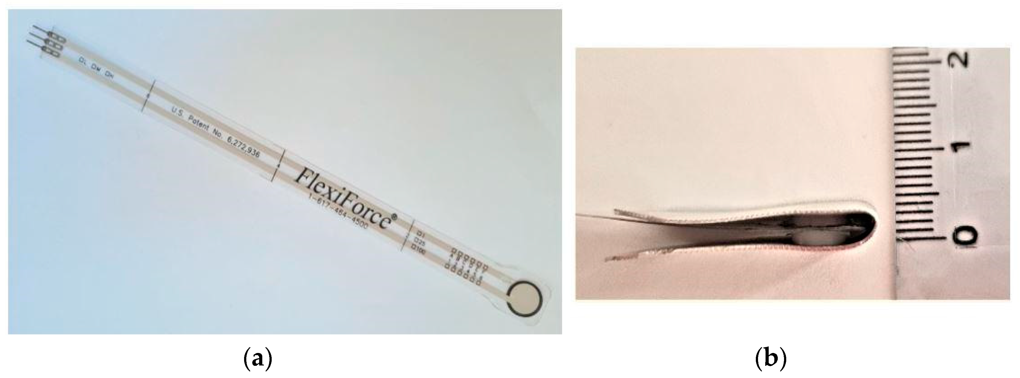
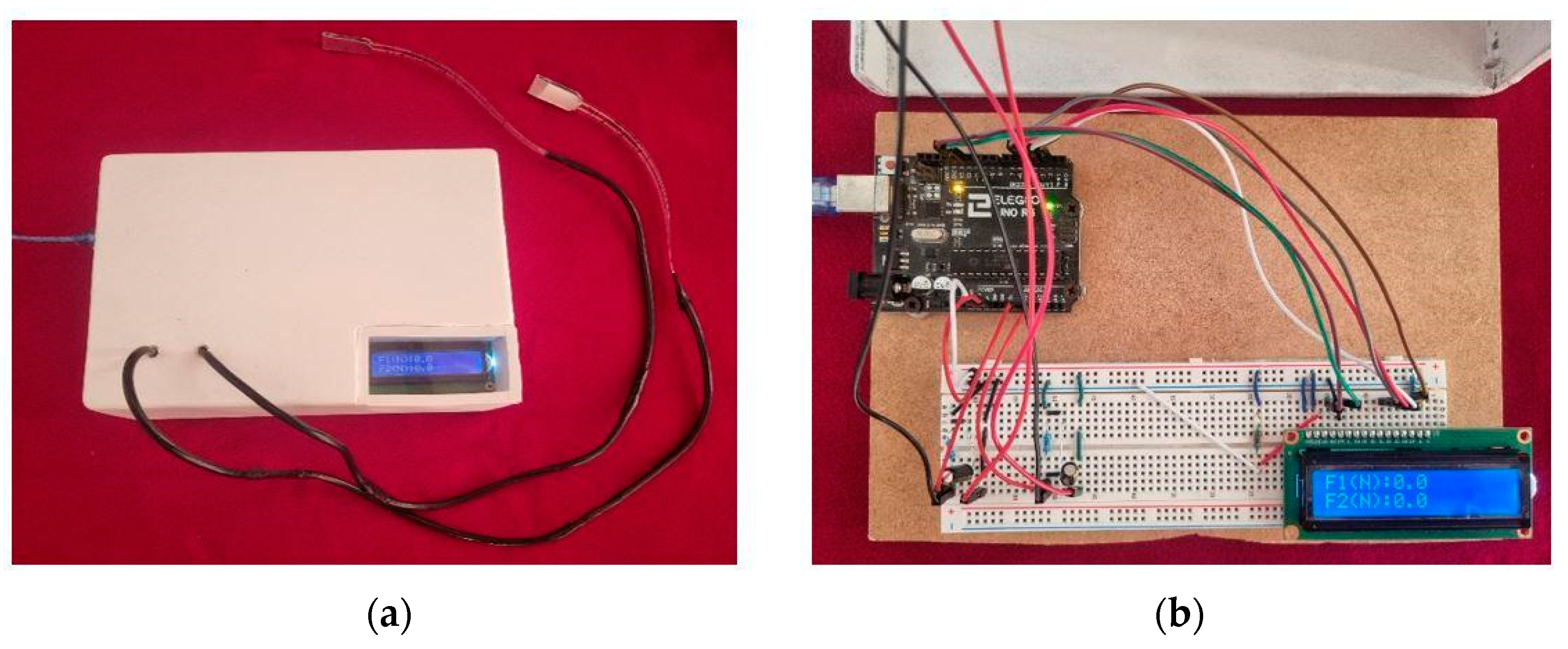
2.4. Bite Force Measurement

We measured bite force with the FlexiForce® A201 piezoresistive force transducer (Tekscan, Boston, MA, USA), see Figure 2a. The transducer has a load range up to 440 N, which is suitable for use with adults, and a sensitivity of 0.01 V/N [45]. The force transducer is inserted into a homemade “sandwich structure,” consisting of two 8 × 1 mm discs, made of thermoformed plastic material covered with an aluminum sheet, see Figure 2b. Discs are placed above and below the FlexiForce® force sensor, held in place by a layer of double-sided adhesive tape, according to the manufacturer’s recommendation in the FlexiForce® user manual [46]. The function of the plastic disc is to ensure that all lines of force between the upper and lower teeth are conveyed through that area. Because the sensor does not tolerate heat and sterilization [45], the FlexiForce® strip is placed in a disposable plastic shield, used in dentistry for digital intra-oral radiographs. Adequate expression of force is maintained by the maximum thickness of 4 mm, enabling muscle fiber movement at an optimal length [47,48]. Calibration of the sensor was based on data in the literature [45].
The two FlexiForce® sensors were connected to an electrical circuit with a voltage of 5 V, powered by a lithium battery. The circuit consists of an “Arduino Uno” controller, to which an LCD screen is connected and displays pressure data. This corresponds to the force expressed in Newtons (N) exerted by the patient during the chewing test (Figure 3).
During the design phase of the device, an update frequency of 1.5 Hz for the chewing value was established, and the numerical precision was set to a single decimal digit without approximation. A threshold of 20 N was determined to return a null value [45] and, given the instrumental linear response, measurable and admissible values without losing generality range between 20–320 N.
The sensors were bitten by the patients between the first molars, if present, or between the most posterior teeth. Measurements were taken in the rest position and the maximal chewing force, which was recorded three times.

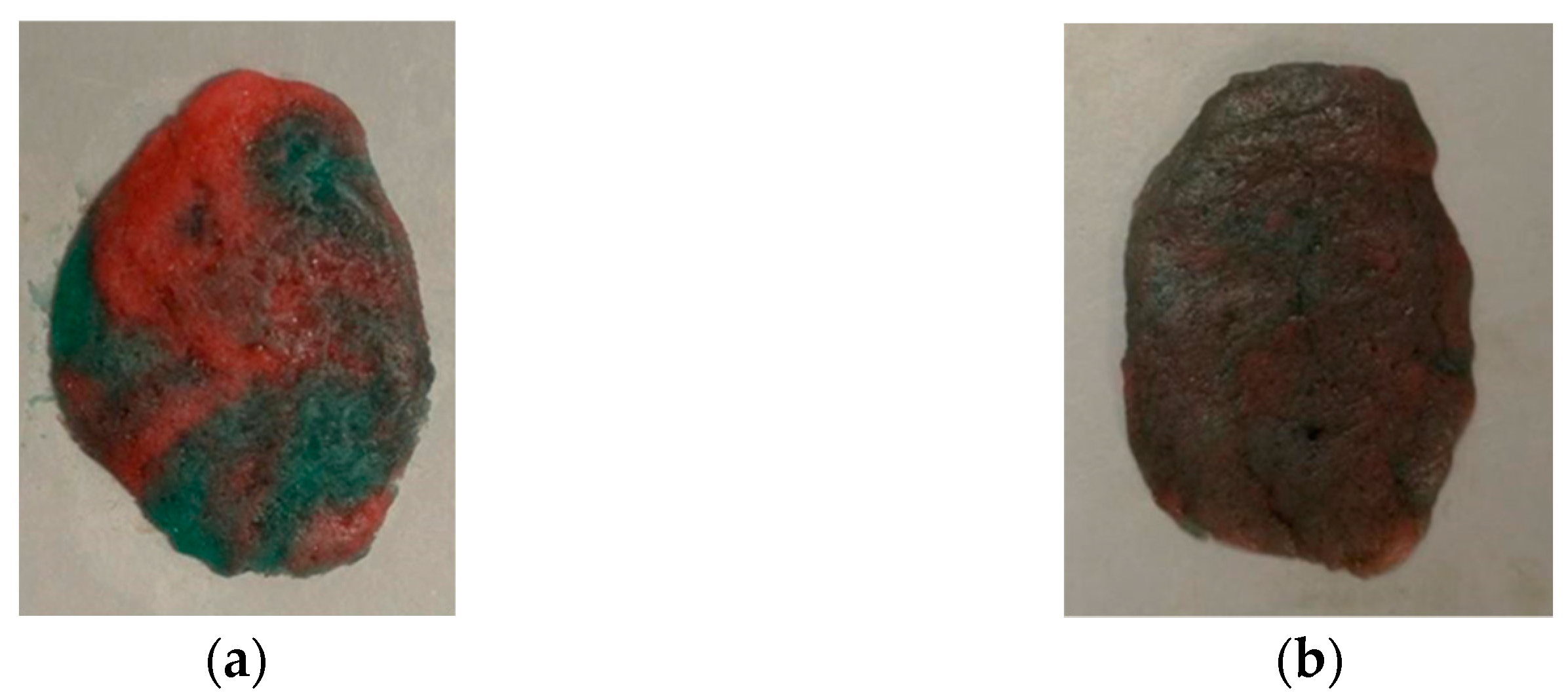
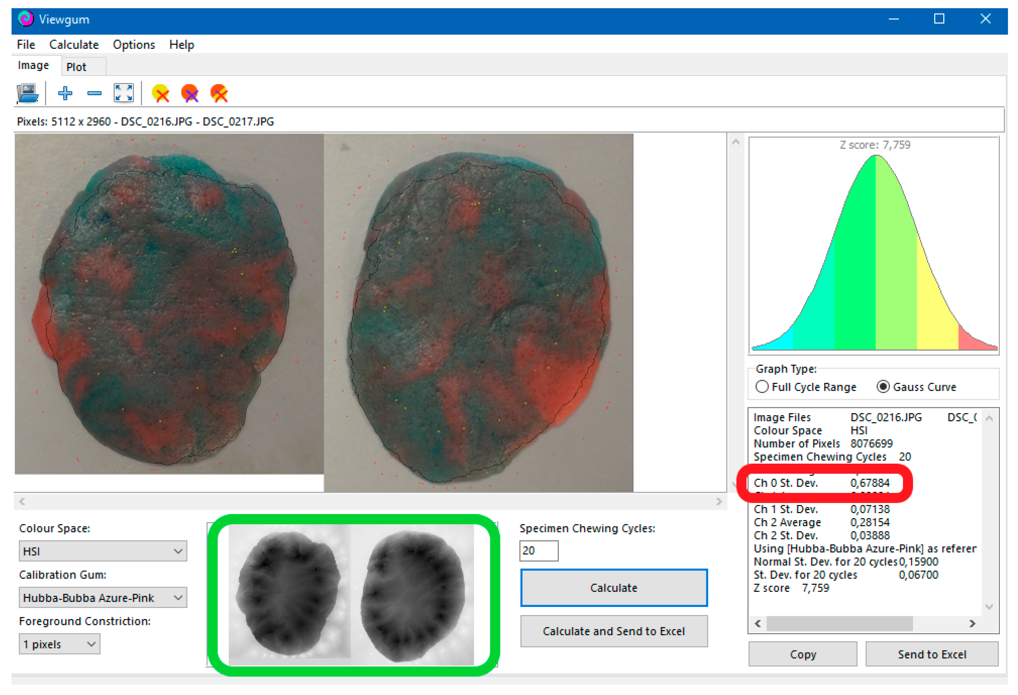
2.5. Chewing Gum Mixing Ability Test

Garfield® 30 mm long strips of “blue raspberry” (blue) and “all fruits” (red) flavor gum were manually stuck together and used for the chewing gum test. Patients were asked to chew the two strips of gum for 20 mastication cycles [49,50,51] in a seated upright position. The chewed sample was spat in a plastic bag and temporarily stored in a refrigerator (16 °C).
All gum samples were prepared for analysis by flattening them to a 1 mm thick disk. Then they were photographed from both sides with a Nikon reflex camera (300 dpi resolution) at a standard distance of 10 cm in the same room and with the same lighting conditions [52]. Digitalization of the chewed samples has always been performed within a few hours of the chewing test. Figure 4 shows two examples of chewed gum.
We performed an opto-electrical analysis of the gum photographs to evaluate the degree of color mixing with the ViewGum© software [53] (Figure 5). Variance of hue (VOH), an indication of the logarithmic association with the number of chewing cycles, was used to assess the gum samples; a high VOH indicates poorly mixed colors from poor chewing, and a low VOH indicates well mixed colors from adequate chewing [52]. The results were displayed as “Ch 0 St. Dev” in the ViewGum© software [53].

2.6. Statistical Analysis

Statistical analysis was performed using STATA® software version 17 (StataCorp. 2021. Stata Statistical Software: Release 17. College Station, TX, USA: StataCorp LLC.). The KolmogorovSmirnov test was used to evaluate the normality of the data, and Levene tests were used to assess the homogeneity of variances. We used the parametric tests when assuming the normality of the data distribution and homogeneity of variances. Descriptive statistics were presented for baseline demographic clinical characteristics for the entire group. Continuous variables were presented as mean, standard deviation (SD), minimum (min) and maximum (max) and were compared between subgroups using ANOVA test. A p < 0.05 was considered statistically significant.

3. Results

A total of 23 patients out of 35 approached consented to participate in the current study; 12 patients refused. Most were male (n = 17/23; 73%), and the mean patient age was 62 ± 7.4 (range 50–75) years old. Patients were hospitalized for COVID-19 infection between February 2020–April 2021. According to PACS muscular symptoms, patients were grouped as sarcopenic (n = 13/23; 57%), asthenic non-sarcopenic (n = 5/23; 22%) and non-asthenic non-sarcopenic (n = 5/23; 22%).
Anxiety (DASS A), depression (DASS D) and stress (DASS S) indices in most patients were within normal ranges [38] (Table 1). Mean patient BMI was 29.5, with value ranges between 41.7 and 21.6. Interestingly, there was no relationship found between BMI and chewing efficacy/efficiency. There was also no relevant difference in BMI between sarcopenic and non-sarcopenic patients. The dominant hand grip test mean values were lower in sarcopenic subjects compared to non-asthenic non-sarcopenic ones (Table 2).
Along with asthenic patients, sarcopenic patients had slower and more strenuous movements, as evidenced in lower gait speed test values compared to non-asthenic non-sarcopenic patients p = 0.001, see Figure 6 and Table 2.

3.1. Short Medical History Interview

Only 9/23 (39%) were admitted to the Intensive Care Unit (ICU). All ICU admitted patients had post-intensive care syndrome (PICS), reporting general motor difficulties within the first few weeks after discharge [54,55].
Parafunctions were recorded in 8/23 (35%) patients, and one of them reported that nocturnal bruxism began right after discharge, when post-COVID symptoms arose. The reason for the onset cannot be precisely defined, but it can be assumed that it is caused by anxiety and stress—in fact, this patient had very high DASS values. Table 1 reports results about the Depression, Anxiety, and Stress Scale (DASS) questionnaire.
Twenty-three percent of patients reported TMDs (Temporo Mandibular Disorders) before admission, and all of them belong to sarcopenic or asthenic groups. Among the 13 sarcopenic patients, 3 reported spontaneous or chewing pain in the masseter, which appeared shortly after discharge. Two of those offered additional information, specifying motor difficulties associated with the opening and closure of the jaw.

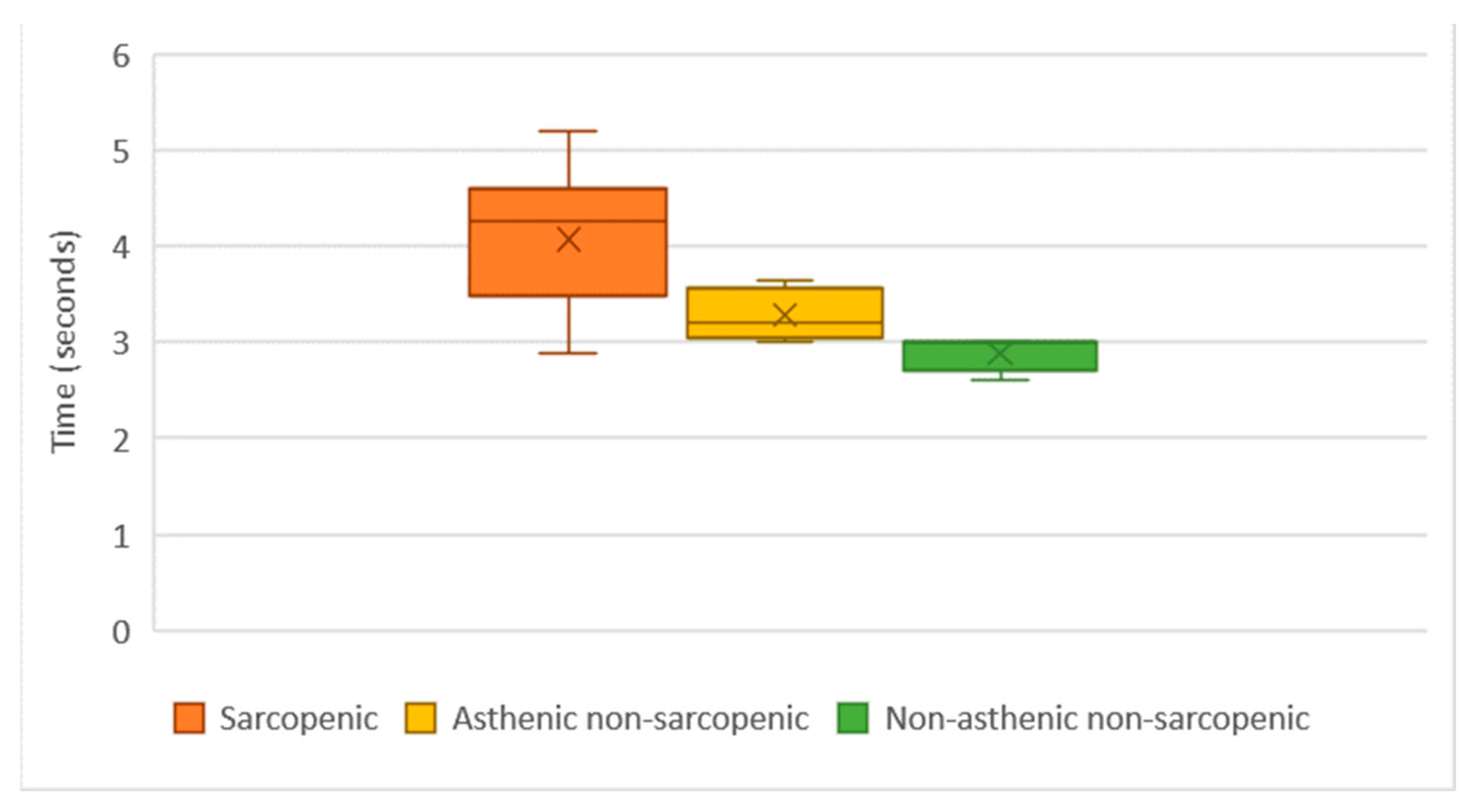
3.2. Anatomo-Functional Analysis

Pain on palpation was recorded according to the muscle location; sternocleidomastoid/occipital/trapexious in 26% (6/23 patients), the mid-anterior temporal in 22% (5/23 patients), mylohyoid in 13% (3/23 patients) and lateral pterygoid in 9% (2/23 patients). No particular areas of muscle tension or inflammation were detected. No patients reported muscle pain during the bite force measurement, so the pain on palpation reported by the subjects is not so severe that it could significantly affect the bite force measurement.
During mandibular movements, noises from the TMJ (Temporo Mandibular Joint) were detected in 11/23 patients (48%), and pain during the jaw opening and closing was recorded in 3/23 (13%). Mandibular opening was normal in 20/23 (87%) patients. Lateral movements were symmetrical, and protrusion was perceivable for most patients; only one patient (sarcopenic) had difficult control of all mandibular movements. There were no differences among the patient groups revealed in terms of any anatomo-functional analysis.

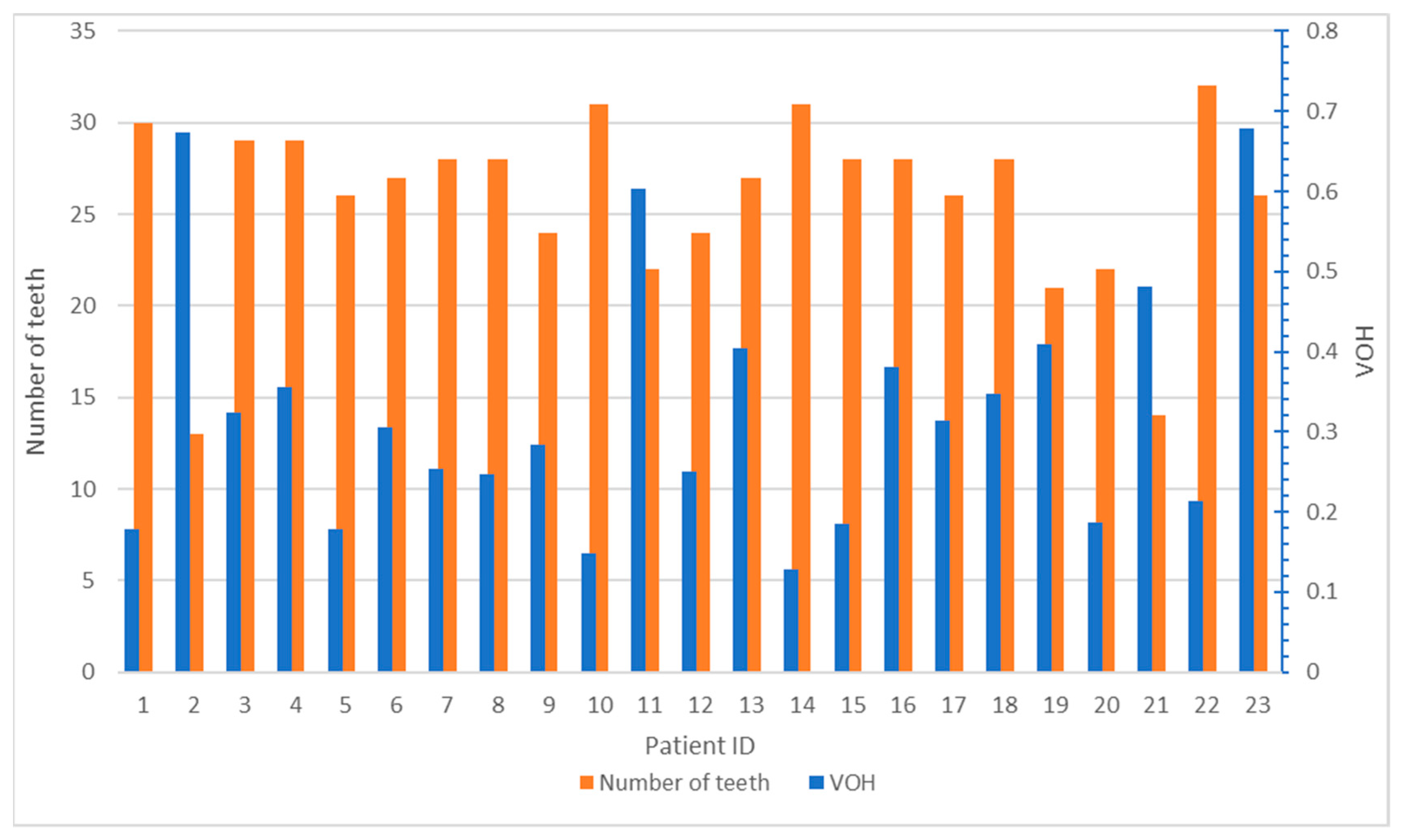
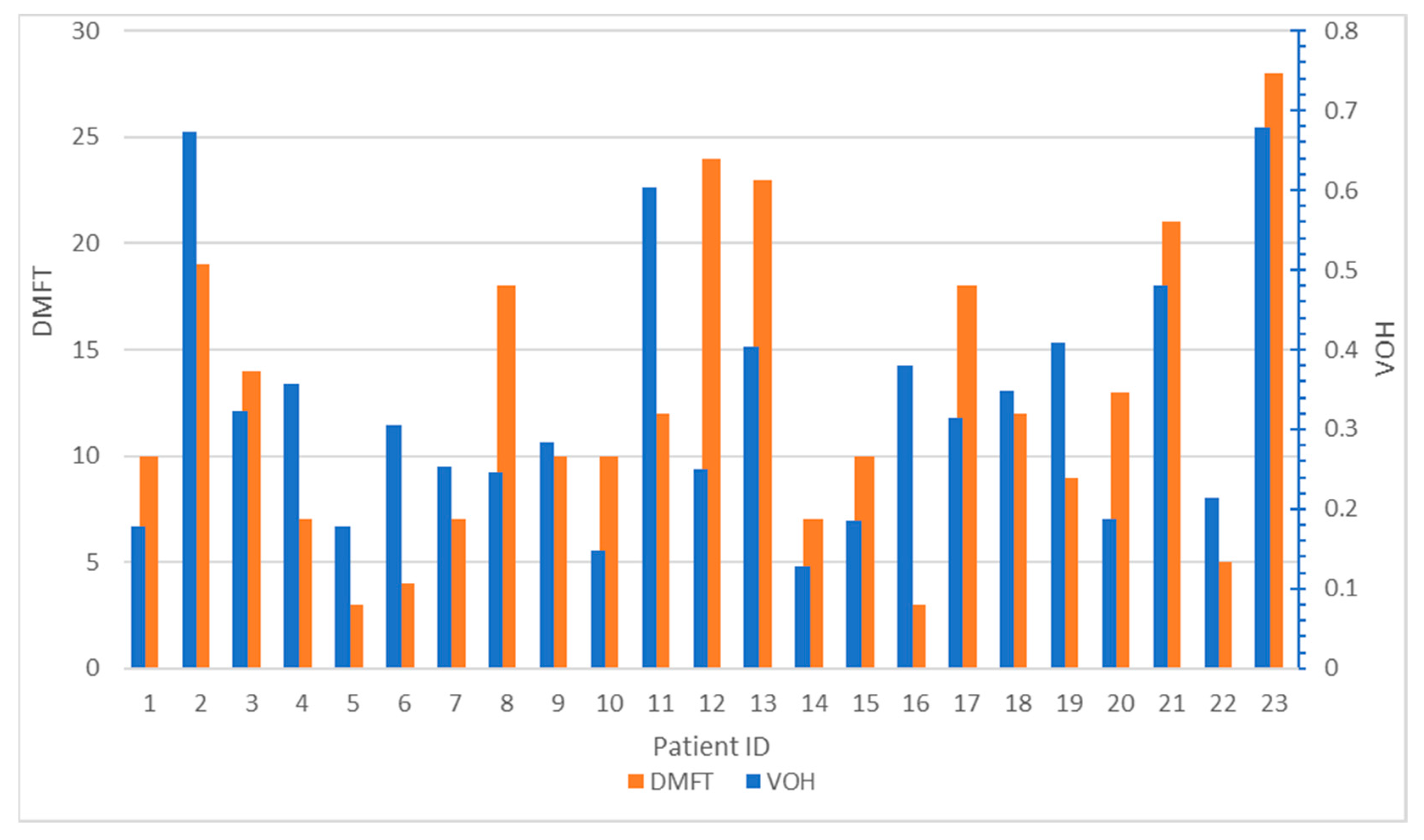
3.3. Intra-Oral Examination

The intra-oral dental examination revealed various critical issues. Oral mucosa showed a normal trophism. Marginal gingivitis was found in 6/23 (26%) patients characterized by the presence of plaque and calculus. Almost half of all patients (n = 11/23; 47.8%) had a poor level of oral hygiene, although periodontal probes were non-physiological in only 4/23 (17%) patients. Table 1 outlines the quantitative measurements from the oral investigations. The overall mean DMFT (Decayed Missing Filled Teeth) was 12.5 ± 7.1. According to study groups, sarcopenic patients displayed a worse overall dental health compared to asthenic non sarcopenic and non-asthenic non-sarcopenic patients.
Comparing the index of each subject, DMFT was higher in sarcopenic patients; no correlation between NCCL and symptoms of sarcopenia or asthenia in PACS patients was found.

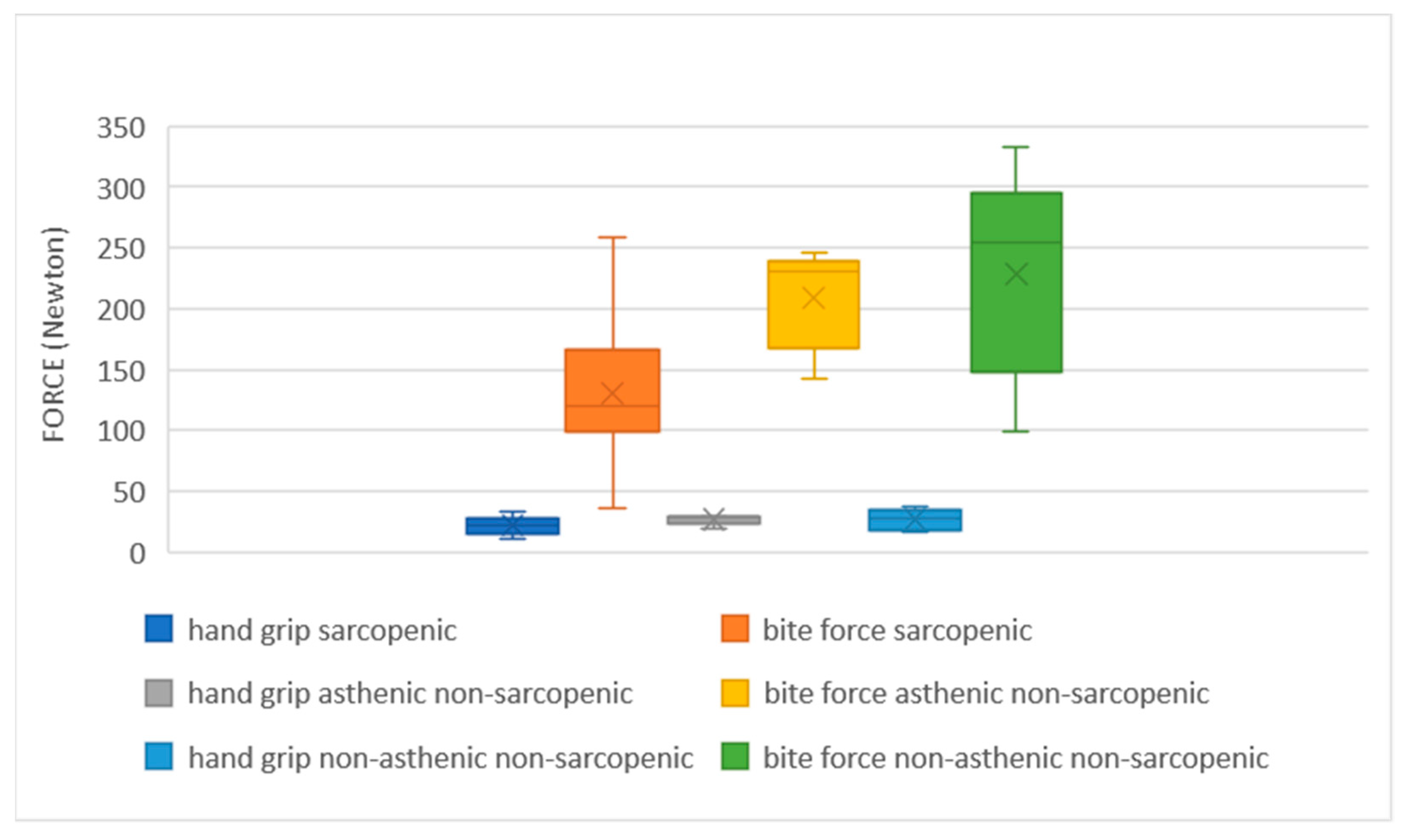
3.4. Bite Force Mesurament

According to patient groups, bite force was lowest among sarcopenic patients and highest among the non-asthenic non-sarcopenic patients (Figure 7 and Table 2). In particular, sarcopenic patients have an average decreased bite force of 125.8 N compared to non-asthenic non-sarcopenic subjects and a decreased bite force of 86.1 N compared to asthenic non-sarcopenic subjects. Figure 7 shows the variations in bite force compared to dominant hand force. The difference in bite force among the three groups was much more evident than the hand force. We have registered a maximum bite force value of 332.8 N, while with the hand grip test, the maximum value measured was 47.2 N.

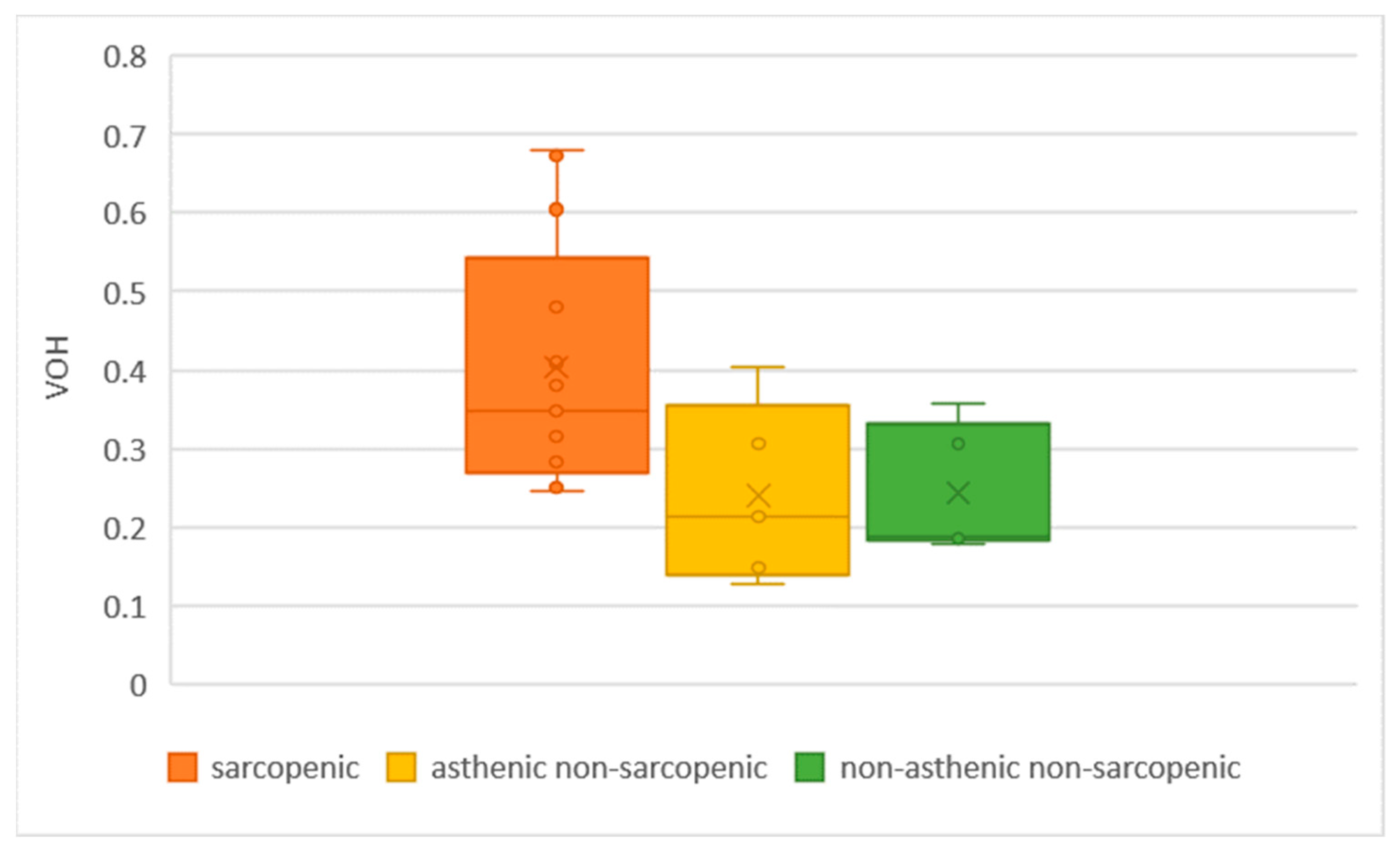
3.5. Chewing Gum Mixing Ability Test

The VOH ranged between 0.127 and 0.678. The highest values (inefficient chewing) were observed in sarcopenic patients, compared to non-asthenic non-sarcopenic and asthenic non-sarcopenic subjects (Figure 8 and Table 2). Sarcopenic patients showed an average VOH of 0.403; non-asthenic non-sarcopenic patients showed an average VOH value of 0.22. This difference is statistically significant.
We also compared the VOH with the DMFT and the number of teeth in the arch (prosthetic or natural). Figure 9 and Figure 10 demonstrate how oral health, especially of the teeth, was correlated with patients’ masticatory performance.

4. Discussion

This research includes both intra-oral and extra-oral physical examination. This choice is motivated by the need to observe a variety of possible effects of PACS or COVID-19 in the areas of dental interest (teeth, mucous membranes and periodontium).

4.1. Short Medical History Interview

Stress, anxiety and emotional factors can create parafunctional problems, such as nocturnal grinding or locking, or exacerbate those already present [56,57], and it is important to consider psychological factors when investigating dysfunctional problems.
The severity of COVID-19 affects the symptoms and incidence of PACS [12]. With the anamnestic interview, the patients who were admitted to intensive care unit reported the difficulties encountered after discharge. It is, therefore, evident that post-intensive care syndrome (PISC) can affect the stomatognathic system and can also leave chewing problems.

4.2. Anatomo-Functional Analysis

Some patients responded positively to muscle palpation, indicating specific areas of pain. In many of them, dysfunctional signs and symptoms were found, often mild. In general, it can be said that the TMJ does not perform its function perfectly in all subjects. It is not known exactly the reason behind the slight dysfunctions highlighted in these PACS subjects because there is no data of the anatomo-functional analysis before the COVID-19 infection. It can be hypothesized that the pain in the cervico-facial muscles found post-COVID may be part of the musculoskeletal symptoms characteristic of PACS; nevertheless, further studies are needed to confirm this hypothesis.

4.3. Intra-Oral Examination

The surveys did not reveal periodontal problems in most patients. The only non-physiological probes (>3 mm) were observed in those patients with a poor level of oral hygiene and were attributable to gingival inflammation due to tartar and plaque. No cases of obvious inflammatory gingival problems have been found, so it can be said that the systemic inflammation underlying PACS does not affect oral soft tissues.
Third molars were not taken into consideration, thus reducing the maximum DMFT value to 28. The mean DMFT was 12.5 ± 7.1, hence the oral health status of the PACS subjects under examination is worse than the values found in the literature [58]. The reason may be that the PACS patient shifted attention to more important health problems—even hospitalization did not help to maintain proper oral hygiene. Many of these patients had to wear an oxygen insufflation mask for some time and were intubated [59]. Most patients reported that they did not go to the dentist prior to the COVID-19 infection.
The NCCL results are in line with a 2020 meta-analysis [60]. Many studies were not able to confirm a positive association between occlusal loading and abfraction. The literature suggests that dentin demineralization promotes NCCL formation from an early stage, while occlusal stress is an etiological factor contributing to the progression of these lesions [61].

4.4. Bite Force Measurement

The FlexiForce® device has already been used in the related literature [62,63]. When inserting a device between the first or second molar, the mouth opens no more than 2–3 mm interincisal distance. This means that the condyles remain almost centered in the temporal fossa, i.e., in a centric position.
During the measurement, it was difficult to ensure that the sensor remained correctly interposed between the cusps of the teeth. The occlusal table is not as flat as the sensor turns out to be. The thermoplastic material of the discs, which is elastic, helps to overcome this problem. The aluminum sheet allows the correct positioning of the cusps with respect to the sensor, but it can also represent a disadvantage; if during the first detection the thermo-molded disc is moved outside the sensitive area of the Flexi-Force®, the subsequent measurements are invalid. To address this problem, when the aluminum housing undergoes a clear plastic deformation, it is replaced.
The measurement takes place simultaneously on the right and left side, thus allowing one to quantify any masticatory asymmetries. However, for the purposes of the study, the average value of bite force between the right and the left was considered of most interest.

4.5. Chewing Gum Mixing Ability Test

The most important predictors of chewing efficiency are the number of pairs of occluding teeth, the bite force, the flow of saliva, prosthetic reconstructions, the strength and coordination of the tongue and cheeks, as well as age and sex [64,65]. Cognitive status and intra-oral sensitivity are also associated with chewing function [52]. This study only considers age, number of teeth and bite force.
The two-color chewing gum test is documented as a simple and effective test that can be used in the clinical setting [66]. The literature recommends the chewing gum mixing test to evaluate interindividual differences in chewing efficiency in clinical and research settings, in both prosthetic and non-prosthetic patients [67]. The simplicity of the optoelectronic assessment could help establish widespread screening for masticatory deficiencies. Furthermore, application in geriatrics or special care could help visualize oral-functionality or dental comorbidities.
From the data obtained through Viewgum©, it is evident that sarcopenic patients have a lower degree of masticatory performance than those without muscle symptoms (non-asthenic non-sarcopenic patients). This may be due to both decreased muscle function, caused by PACS, but also an increased DMFT. Comparisons of the VOH against the DMFT (Figure 10) or the number of teeth (Figure 9) highlight that chewing efficiency depends on the state of the oral health. Since it can be difficult to increase the muscular performance of sarcopenic PACS patients, it is advisable to improve the state of oral health and to rehabilitate missing teeth.

4.6. Limits of the Study

The lack of data on the chewing performance of patients before COVID-19 infection and the small number of subjects prevent proving that PACS was the actual cause of the decreased chewing performance. Moreover, this study does not allow us to detect improvement or deterioration in muscle performance over time.
We do not have the possibility to do an X-ray examination. For this reason, we only conducted an intra-oral examination that does not allow us to collect data about interproximal caries, periapical lesions and root fractures.
In this study, we used Garfield® chewing gums. We remark that these chewing gums are not the same used and validated in the previous studies. Validated chewing gums (either Hubba Bubba® or HueCheck®) were not readily available in our country at the moment of the investigation. We selected Garfield® chewing gums because they are the most similar to Hubba Bubba®.
We remark that bite force is different from masticatory force because the clenching movement of the mouth is different from the chewing movement. For this reason, the study cannot affirm the correlation between PACS and masticatory force.

5. Conclusions

In conclusion, the study showed that PACS people with sarcopenia have an average bite force of 122.4 Newton (N). This value is 86.1 N lower than asthenic non-sarcopenic patients (average bite force of 208.5 N) and 125.8 N lower than non-asthenic non-sarcopenic patients (average bite force of 248.2 N), As regards chewing efficiency, sarcopenic patients showed an average VOH increase of 0.18 compared to non-asthenic non-sarcopenic ones. Patients who complained only of asthenia (asthenic non-sarcopenic) were found to have lower values of bite force and masticatory efficacy than those who did not have muscular symptoms (non-asthenic non-sarcopenic) but higher values than those who suffered from sarcopenia (sarcopenic).
The piezoresistive sensor gnathodynamometer proved to be a valid bite force measurement tool. This tool can be a good alternative to other motor tests used to evaluate muscle effectiveness (such as the dominant hand grip test). Further studies on larger groups of subjects will be needed to validate their clinical use.
Oral health in most PACS patients appears to be compromised; it is, therefore, advisable to direct these patients towards a multi-disciplinary-rehabilitation path, addressing dental issues as well as all functional problems already present or occurring in the post-hospitalization.

Author Contributions

Conceptualization, B.D.P. and G.G. (Giovanni Guaraldi); methodology, G.G. (Giovanni Guaraldi), B.D.P.; software, R.K.K.; validation, L.B. and P.B.; formal analysis, R.K.K. and L.B.; investigation, L.B. and B.D.P.; resources, G.G. (Giovanni Guaraldi) and R.K.K.; writing—original draft preparation, L.B. and G.G. (Giovanna Garuti); writing—review and editing, L.B. and G.G. (Giovanna Garuti); supervision, U.C., P.B. and G.G. (Giovanna Garuti); project administration, U.C. and P.B. All authors have read and agreed to the published version of the manuscript.

Funding

This research received no external funding.

Institutional Review Board Statement

All subjects gave informed consent for inclusion before participating in the study. This study was conducted in accordance with the Declaration of Helsinki, and the protocol was approved by the ethic committee of “Area Vasta Emilia Nord” with the project identification code number 396/2020.

Informed Consent Statement

Informed consent was obtained from all subjects involved in the study.

Data Availability Statement

Not applicable.

Conflicts of Interest

The authors declare no conflict of interest.

References

  1. Imai, K.; Tanaka, H. SARS-CoV-2 Infection and Significance of Oral Health Management in the Era of “the New Normal with COVID-19”. Int. J. Mol. Sci. 2021, 22, 6527. [Google Scholar] [CrossRef]
  2. Huang, N.; Pérez, P.; Kato, T.; Mikami, Y.; Okuda, K.; Gilmore, R.C.; Conde, C.D.; Gasmi, B.; Stein, S.; Beach, M.; et al. SARS-CoV-2 infection of the oral cavity and saliva. Nat. Med. 2021, 27, 892–903. [Google Scholar] [CrossRef] [PubMed]
  3. Mulchandani, R.; Lyngdoh, T.; Kakkar, A.K. Deciphering the COVID-19 cytokine storm: Systematic review and meta-analysis. Eur. J. Clin. Investig. 2021, 51, e13429. [Google Scholar] [CrossRef] [PubMed]
  4. Sakaguchi, W.; Kubota, N.; Shimizu, T.; Saruta, J.; Fuchida, S.; Kawata, A.; Yamamoto, Y.; Sugimoto, M.; Yakeishi, M.; Tsukinoki, K. Existence of SARS-CoV-2 Entry Molecules in the Oral Cavity. Int. J. Mol. Sci. 2020, 21, 6000. [Google Scholar] [CrossRef] [PubMed]
  5. Xu, H.; Zhong, L.; Deng, J.; Peng, J.; Dan, H.; Zeng, X.; Li, T.; Chen, Q. High expression of ACE2 receptor of 2019-nCoV on the epithelial cells of oral mucosa. Int. J. Oral Sci. 2020, 12, 8. [Google Scholar] [CrossRef] [PubMed]
  6. Mason, R.J. Pathogenesis of COVID-19 from a cell biology perspective. Eur. Respir. J. 2020, 55, 2000607. [Google Scholar] [CrossRef] [PubMed]
  7. Gu, S.X.; Tyagi, T.; Jain, K.; Gu, V.W.; Lee, S.H.; Hwa, J.M.; Kwan, J.M.; Krause, D.S.; Lee, A.I.; Halene, S.; et al. Thrombocytopathy and endotheliopathy: Crucial contributors to COVID-19 thromboinflammation. Nat. Rev. Cardiol. 2020, 18, 194–209. [Google Scholar] [CrossRef]
  8. Fabrizi, F.; Alfieri, C.M.; Cerutti, R.; Lunghi, G.; Messa, P. COVID-19 and Acute Kidney Injury: A Systematic Review and Meta-Analysis. Pathogens 2020, 9, 1052. [Google Scholar] [CrossRef]
  9. Sultan, S.; Altayar, O.; Siddique, S.M.; Davitkov, P.; Feuerstein, J.D.; Lim, J.K.; Falck-Ytter, Y.; El-Serag, H.B. AGA Institute Rapid Review of the Gastrointestinal and Liver Manifestations of COVID-19, Meta-Analysis of International Data, and Recommendations for the Consultative Management of Patients with COVID-19. Gastroenterology 2020, 159, 320–334.e27. [Google Scholar] [CrossRef]
  10. Herridge, M.S.; Tansey, C.; Matté, A.; Tomlinson, G.; Diaz-Granados, N.; Cooper, A.; Guest, C.; Mazer, D.; Mehta, S.; Stewart, T.; et al. Functional Disability 5 Years after Acute Respiratory Distress Syndrome. N. Engl. J. Med. 2011, 364, 1293–1304. [Google Scholar] [CrossRef] [Green Version]
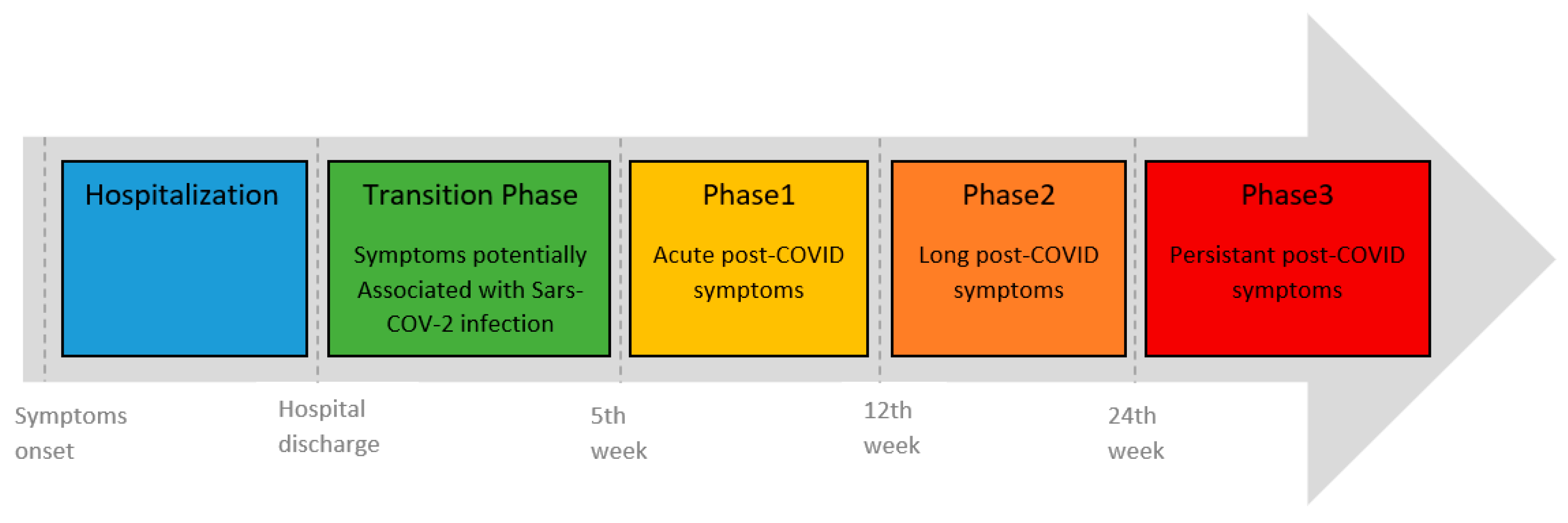
  11. Fernández-De-Las-Peñas, C.; Palacios-Ceña, D.; Gómez-Mayordomo, V.; Cuadrado, M.L.; Florencio, L.L. Defining Post-COVID Symptoms (Post-Acute COVID, Long COVID, Persistent Post-COVID): An Integrative Classification. Int. J. Environ. Res. Public Health 2021, 18, 2621. [Google Scholar] [CrossRef]
  12. Taquet, M.; Dercon, Q.; Luciano, S.; Geddes, J.R.; Husain, M.; Harrison, P.J. Incidence, co-occurrence, and evolution of long-COVID features: A 6-month retrospective cohort study of 273,618 survivors of COVID-19. PLoS Med. 2021, 18, e1003773. [Google Scholar] [CrossRef] [PubMed]
  13. Cohen, A.A. Complex systems dynamics in aging: New evidence, continuing questions. Biogerontology 2016, 17, 205–220. [Google Scholar] [CrossRef] [PubMed]
  14. 2021 Alzheimer’s Disease Facts and Figures. Available online: https://alz-journals.onlinelibrary.wiley.com/doi/10.1002/alz.12328#:~:text=An%20estimated%206.2%20million%20Americans,prevent%2C%20slow%20or%20cure%20AD (accessed on 17 December 2022).
  15. COVID-19 Associated with Long-Term Cognitive Dysfunction, Acceleration of Alzheimer’s Symptoms. Available online: https://aaic.alz.org/releases_2021/covid-19-cognitive-impact.asp (accessed on 19 January 2023).
  16. Fulop, T.; Larbi, A.; Dupuis, G.; Le Page, A.; Frost, E.H.; Cohen, A.A.; Witkowski, J.M.; Franceschi, C. Immunosenescence and Inflamm-Aging as Two Sides of the Same Coin: Friends or Foes? Front. Immunol. 2018, 8, 1960. [Google Scholar] [CrossRef] [PubMed]
  17. Coppé, J.-P.; Patil, C.K.; Rodier, F.; Sun, Y.; Muñoz, D.P.; Goldstein, J.; Nelson, P.S.; Desprez, P.-Y.; Campisi, J. Senescence-Associated Secretory Phenotypes Reveal Cell-Nonautonomous Functions of Oncogenic RAS and the p53 Tumor Suppressor. PLoS Biol. 2008, 6, e301. [Google Scholar] [CrossRef]
  18. Salehi, S.; Reddy, S.; Gholamrezanezhad, A. Long-term Pulmonary Consequences of Coronavirus Disease 2019 (COVID-19): What we know and what to expect. J. Thorac. Imaging 2020, 35, W87–W89. [Google Scholar] [CrossRef]
  19. Puntmann, V.O.; Carerj, M.L.; Wieters, I.; Fahim, M.; Arendt, C.; Hoffmann, J.; Shchendrygina, A.; Escher, F.; Vasa-Nicotera, M.; Zeiher, A.M.; et al. Outcomes of Cardiovascular Magnetic Resonance Imaging in Patients Recently Recovered from Coronavirus Disease 2019 (COVID-19). JAMA Cardiol. 2020, 5, 1265–1273. [Google Scholar] [CrossRef]
  20. Sathish, T.; Anton, M.C.; Sivakumar, T. New-onset diabetes in ‘long COVID’. J. Diabetes 2021, 13, 693–694. [Google Scholar] [CrossRef]
  21. Wang, F.; Kream, R.M.; Stefano, G.B. Long-term respiratory and neurological sequelae of COVID-19. Med. Sci. Monit. 2020, 26, e928996. [Google Scholar] [CrossRef]
  22. Phipps, M.M.; Barraza, L.H.; Lasota, E.D.; Sobieszczyk, M.E.; Pereira, M.R.; Zheng, E.X.; Fox, A.N.; Zucker, J.; Verna, E.C. Acute Liver Injury in COVID-19: Prevalence and Association with Clinical Outcomes in a Large U.S. Cohort. Hepatology 2020, 72, 807–817. [Google Scholar] [CrossRef]
  23. Troyer, E.A.; Kohn, J.N.; Hong, S. Are we facing a crashing wave of neuropsychiatric sequelae of COVID-19? Neuropsychiatric symptoms and potential immunologic mechanisms. Brain. Behav. Immun. 2020, 87, 34–39. [Google Scholar] [CrossRef]
  24. Karaarslan, F.; Demircioğlu Güneri, F.; Kardeş, S. Postdischarge rheumatic and musculoskeletal symptoms following hospitalization for COVID-19: Prospective follow-up by phone interviews. Rheumatol. Int. 2021, 41, 1263–1271. [Google Scholar] [CrossRef]
  25. Bektas, A.; Schurman, S.H.; Franceschi, C.; Ferrucci, L. A public health perspective of aging: Do hyper-inflammatory syndromes such as COVID-19, SARS, ARDS, cytokine storm syndrome, and post-ICU syndrome accelerate short- and long-term inflammaging? Immun. Ageing 2020, 17, 23. [Google Scholar] [CrossRef]
  26. Delgado-Alonso, C.; Valles-Salgado, M.; Delgado-Álvarez, A.; Yus, M.; Gómez-Ruiz, N.; Jorquera, M.; Polidura, C.; Gil, M.J.; Marcos, A.; Matías-Guiu, J.; et al. Cognitive dysfunction associated with COVID-19: A comprehensive neuropsychological study. J. Psychiatr. Res. 2022, 150, 40–46. [Google Scholar] [CrossRef] [PubMed]
  27. van den Borst, B.; Peters, J.B.; Brink, M.; Schoon, Y.; Bleeker-Rovers, C.P.; Schers, H.; van Hees, H.W.H.; van Helvoort, H.; van den Boogaard, M.; van der Hoeven, H.; et al. Comprehensive Health Assessment 3 Months After Recovery from Acute Coronavirus Disease 2019 (COVID-19). Clin. Infect. Dis. 2021, 73, e1089. [Google Scholar] [CrossRef] [PubMed]
  28. Bloch, S.; Polkey, M.I.; Griffiths, M.; Kemp, P. Molecular mechanisms of intensive care unit-acquired weakness. Eur. Respir. J. 2012, 39, 1000–1011. [Google Scholar] [CrossRef]
  29. Wåhlin-Larsson, B.; Wilkinson, D.J.; Strandberg, E.; Hosford-Donovan, A.; Atherton, P.J.; Kadi, F. Mechanistic Links Underlying the Impact of C-Reactive Protein on Muscle Mass in Elderly. Cell. Physiol. Biochem. 2017, 44, 267–278. [Google Scholar] [CrossRef] [PubMed]
  30. Aschman, T.; Schneider, J.; Greuel, S.; Meinhardt, J.; Streit, S.; Goebel, H.-H.; Büttnerova, I.; Elezkurtaj, S.; Scheibe, F.; Radke, J.; et al. Association Between SARS-CoV-2 Infection and Immune-Mediated Myopathy in Patients Who Have Died. JAMA Neurol. 2021, 78, 948–960. [Google Scholar] [CrossRef]
  31. de Andrade-Junior, M.C.; de Salles, I.C.D.; de Brito, C.M.M.; Pastore-Junior, L.; Righetti, R.F.; Yamaguti, W.P. Skeletal Muscle Wasting and Function Impairment in Intensive Care Patients with Severe COVID-19. Front. Physiol. 2021, 12, 640973. [Google Scholar] [CrossRef]
  32. McKenna, H.T.; Murray, A.J.; Martin, D.S. Human adaptation to hypoxia in critical illness. J. Appl. Physiol. 2020, 129, 656–663. [Google Scholar] [CrossRef]
  33. Wierdsma, N.J.; Kruizenga, H.M.; Konings, L.A.; Krebbers, D.; Jorissen, J.R.; Joosten, M.-H.I.; van Aken, L.H.; Tan, F.M.; van Bodegraven, A.A.; Soeters, M.R.; et al. Poor nutritional status, risk of sarcopenia and nutrition related complaints are prevalent in COVID-19 patients during and after hospital admission. Clin. Nutr. Espen. 2021, 43, 369. [Google Scholar] [CrossRef] [PubMed]
  34. Soares, M.N.; Eggelbusch, M.; Naddaf, E.; Gerrits, K.H.L.; van der Schaaf, M.; van den Borst, B.; Wiersinga, W.J.; van Vugt, M.; Weijs, P.J.M.; Murray, A.J.; et al. Skeletal muscle alterations in patients with acute COVID-19 and post-acute sequelae of COVID-19. J. Cachexia Sarcopenia Muscle 2022, 13, 11–22. [Google Scholar] [CrossRef] [PubMed]
  35. Shepherd, S.; Batra, A.; Lerner, D.P. Review of Critical Illness Myopathy and Neuropathy. Neurohospitalist 2017, 7, 41–48. [Google Scholar] [CrossRef]
  36. Yoshida, M.; Hiraoka, A.; Takeda, C.; Mori, T.; Maruyama, M.; Yoshikawa, M.; Tsuga, K. Oral hypofunction and its relation to frailty and sarcopenia in community-dwelling older people. Gerodontology 2022, 39, 26–32. [Google Scholar] [CrossRef] [PubMed]
  37. Kugimiya, Y.; Iwasaki, M.; Ohara, Y.; Motokawa, K.; Edahiro, A.; Shirobe, M.; Watanabe, Y.; Obuchi, S.; Kawai, H.; Fujiwara, Y.; et al. Relationship between Oral Hypofunction and Sarcopenia in Community-Dwelling Older Adults: The Otassha Study. Int. J. Environ. Res. Public Health 2021, 18, 6666. [Google Scholar] [CrossRef]
  38. Višnjić, A.; Veličković, V.; Sokolović, D.; Stanković, M.; Mijatović, K.; Milošević, Z.; Radulović, O. Relationship between the manner of mobile phone use and depression, anxiety, and stress in university students. Int. J. Environ. Res. Public Health 2018, 15, 697. [Google Scholar] [CrossRef]
  39. Calcolo Indice Massa Corporea—IMC (BMI—Body Mass Index). Available online: https://www.salute.gov.it/portale/nutrizione/dettaglioIMCNutrizione.jsp?lingua=italiano&id=5479&area=nutrizione&menu=vuoto (accessed on 17 December 2022).
  40. Fried, L.P.; Tangen, C.M.; Walston, J.; Newman, A.B.; Hirsch, C.; Gottdiener, J.; Seeman, T.; Tracy, R.; Kop, W.J.; Burke, G.; et al. Frailty in Older AdultsEvidence for a Phenotype. J. Gerontol. Ser. A 2001, 56, M146–M157. [Google Scholar] [CrossRef]
  41. Giornale di Cardiologia. Available online: https://www.giornaledicardiologia.it/archivio/1261/articoli/13935/#:~:text=Inparticolare%2Cunavelocità%3E1.0,instabilieridottaautonomia24 (accessed on 17 December 2022).
  42. Cruz-Jentoft, A.J.; Bahat, G.; Bauer, J.; Boirie, Y.; Bruyère, O.; Cederholm, T.; Cooper, C.; Landi, F.; Rolland, Y.; Aihie Sayer, A.; et al. Sarcopenia: Revised European consensus on definition and diagnosis. Age Ageing 2019, 48, 16–31. [Google Scholar] [CrossRef]
  43. Sondaggio Parodontale. Available online: https://www.gengive.org/glossario/sondaggio-parodontale/ (accessed on 17 December 2022).
  44. Testa, M.; Di Marco, A.; Pertusio, R.; Van Roy, P.; Cattrysse, E.; Roatta, S. A validation study of a new instrument for low cost bite force measurement. J. Electromyogr. Kinesiol. 2016, 30, 243–248. [Google Scholar] [CrossRef]
  45. FlexiForce User Manual | Tekscan. Available online: https://www.tekscan.com/support/faqs/flexiforce-user-manual (accessed on 16 May 2022).
  46. Manns, A.; Miralles, R.; Palazzi, C. EMG, bite force, and elongation of the masseter muscle under isometric voluntary contractions and variations of vertical dimension. J. Prosthet. Dent. 1979, 42, 674–682. [Google Scholar] [CrossRef]
  47. Âudio, C.; Fernandes, P.; Glantz, J.; Svensson, S.A.; Bergmark, A. A Novel Sensor for Bite Force Determinations. Available online: http://www.elsevier.com/locate/dental (accessed on 17 December 2022).
  48. Anastassiadou, V.; Heath, M.R.; Bartholomew, S. The development of a simple objective test of mastication suitable for older people, using chewing gums. Gerodontology 2001, 18, 79–86. [Google Scholar] [CrossRef]
  49. Prinz, J.F. Quantitative evaluation of the effect of bolus size and number of chewing strokes on the intra-oral mixing of a two-colour chewing gum. J. Oral Rehabil. 1999, 26, 243–247. [Google Scholar] [CrossRef] [PubMed]
  50. Schimmel, M.; Leemann, B.; Herrmann, F.R.; Kiliaridis, S.; Schnider, A.; Müller, F. Masticatory function and bite force in stroke patients. J. Dent. Res. 2011, 90, 230–234. [Google Scholar] [CrossRef]
  51. Buser, R.; Ziltener, V.; Samietz, S.; Fontolliet, M.; Nef, T.; Schimmel, M. Validation of a purpose-built chewing gum and smartphone application to evaluate chewing efficiency. J. Oral Rehabil. 2018, 45, 845–853. [Google Scholar] [CrossRef] [PubMed]
  52. Fankhauser, N.; Kalberer, N.; Müller, F.; Leles, C.R.; Schimmel, M.; Srinivasan, M. Comparison of smartphone-camera and conventional flatbed scanner images for analytical evaluation of chewing function. J. Oral Rehabil. 2020, 47, 1496–1502. [Google Scholar] [CrossRef]
  53. ViewGum. Available online: http://www.dhal.com/viewgum.html (accessed on 17 December 2022).
  54. Chippa, V.; Aleem, A.; Anjum, F. Post Acute Coronavirus (COVID-19) Syndrome. StatPearls. 2022. Available online: https://www.ncbi.nlm.nih.gov/books/NBK570608/ (accessed on 17 December 2022).
  55. PICS. Available online: https://postintensiva.it/la-pics-post-intensive-care-syndrome/ (accessed on 17 December 2022).
  56. Kuhn, M.; Türp, J.C.; Türp, J.C.; Myoarthropathien, M.A.A. Risk factors for bruxism. SWISS Dent. J. SSO 2018, 128, 118–124. [Google Scholar]
  57. Ohrbach, R.; Michelotti, A. The Role of Stress in the Etiology of Oral Parafunction and Myofascial Pain. Oral Maxillofac. Surg. Clin. N. Am. 2018, 30, 369–379. [Google Scholar] [CrossRef]
  58. Kit, A.; Tamrakar, M.; Jiang, C.M.; Man Lo, E.C.; Man Leung, K.C.; Chu, C.H. A Systematic Review on Caries Status of Older Adults. Int. J. Environ. Res. Public Health 2021, 18, 10662. [Google Scholar] [CrossRef]
  59. Mańka-Malara, K.; Gawlak, D.; Hovhannisyan, A.; Klikowska, M.; Kostrzewa-Janicka, J. Dental trauma prevention during endotracheal intubation—Review of literature. Anaesthesiol. Intensive Ther. 2015, 47, 425–429. [Google Scholar] [CrossRef]
  60. Teixeira, D.N.R.; Thomas, R.Z.; Soares, P.V.; Cune, M.S.; Gresnigt, M.M.M.; Slot, D.E. Prevalence of noncarious cervical lesions among adults: A systematic review. J. Dent. 2020, 95, 103285. [Google Scholar] [CrossRef]
  61. Nascimento, M.M.; Dilbone, D.A.; Pereira, P.N.; Duarte, R.; Geraldeli, S.; Delgado, A.J. Clinical, Cosmetic and Investigational Dentistry Abfraction lesions: Etiology, diagnosis, and treatment options. Clin. Cosmet. Investig. Dent. 2016, 8, 79–87. [Google Scholar] [CrossRef] [PubMed] [Green Version]
  62. Testa, M.; Rolando, M. Control of jaw-clenching forces in dentate subjects. J. Orofac. Pain 2011, 25, 250–260. [Google Scholar] [PubMed]
  63. Testa, M.; Geri, T.; Signori, A.; Roatta, S. Visual Feedback of Bilateral Bite Force to Assess Motor Control of the Mandible in Isometric Condition. Mot. Control. 2015, 19, 312–324. [Google Scholar] [CrossRef]
  64. Ikebe, K.; Matsuda, K.-I.; Kagawa, R.; Enoki, K.; Yoshida, M.; Maeda, Y.; Nokubi, T. Association of masticatory performance with age, gender, number of teeth, occlusal force and salivary flow in Japanese older adults: Is ageing a risk factor for masticatory dysfunction? Arch. Oral Biol. 2011, 56, 991–996. [Google Scholar] [CrossRef]
  65. Yamada, A.; Kanazawa, M.; Komagamine, Y.; Minakuchi, S. Association between tongue and lip functions and masticatory performance in young dentate adults. J. Oral Rehabil. 2015, 42, 833–839. [Google Scholar] [CrossRef] [PubMed]
  66. Schimmel, M.; Christou, P.; Miyazaki, H.; Halazonetis, D.; Herrmann, F.R.; Müller, F. A novel colourimetric technique to assess chewing function using two-coloured specimens: Validation and application. J. Dent. 2015, 43, 955–964. [Google Scholar] [CrossRef] [PubMed]
  67. Silva, L.C.; Nogueira, T.E.; Rios, L.F.; Schimmel, M.; Leles, C.R. Reliability of a two-colour chewing gum test to assess masticatory performance in complete denture wearers. J. Oral Rehabil. 2018, 45, 301–307. [Google Scholar] [CrossRef]
Figure 1. Integrative model of post-COVID symptoms in hospitalized patients showing the Transition Phase (green) and Phases 1 (yellow), 2 (orange) and 3 (red) of post-COVID symptoms [11].
Oral 03 00008 g001
Figure 2. (a) FlexiForce® piezoresistive sensor. (b) The designed housing with the thermo-molded discs covered with an aluminum sheet.
Oral 03 00008 g002
Figure 3. (a) External view of the gnathodynamometer with piezoresistive sensors. (b) Gnathodynamometer’s electronics.
Oral 03 00008 g003
Figure 4. (a) chewing gum chewed by a patient with poor chewing efficiency. (b) Chewing gum chewed by a patient with excellent chewing efficiency.
Oral 03 00008 g004
Figure 5. ViewGum© software user interface immediately after loading two images of a chewed gum sample for 20 mastication cycles. The images were obtained by scanning both sides of the flattened gum. The software separates the area of interest from the background as shown by the thumbnail images (green circle). Mouse tracks (yellow and red dots in the main images) can be added or deleted for segmentation. The numerical results are displayed on the right. The VOH value (displayed as “Ch 0 St. Dev” and indicated by an orange box) are calculated; higher VOH correspond to poorer chewing capacity (low color mix).
Oral 03 00008 g005
Figure 6. Walking time of the three patient groups: sarcopenic (orange), asthenic non-sarcopenic (yellow) and non-asthenic non-sarcopenic (green).
Oral 03 00008 g006
Figure 7. Comparison between hand-grip force and bite force for the three groups of patients.
Oral 03 00008 g007
Figure 8. Chewing efficiency: comparison of VOH values between sarcopenic, asthenic non-sarcopenic and non-asthenic non-sarcopenic subjects.
Oral 03 00008 g008
Figure 9. Comparison between VOH (in blue) and the number of prosthetic/natural teeth present in the arches (in orange). As can be seen from the graph, patients with fewer teeth tend to have a higher VOH score.
Oral 03 00008 g009
Figure 10. Comparison between VOH (in blue) and DMFT (in orange). From the figure, it is possible to appreciate how the higher the DMFT index, the higher the VOH.
Oral 03 00008 g010
Table 1. Intra-oral dental visit and DASS questionnaire results: medium values of missing teeth, crowns, pontic elements or part of Removable Partial Prosthesis (RPP), caries, filled teeth, teeth with Non-Carious Cervical Lesion (NCCL) and Decay Missing Filled Teeth (DMFT); DASS-A; DASS-D; DASS-S.
Total (n = 23)Sarcopenic (n = 13, 56.5)Asthenic Non-Sarcopenic
(n = 5, 21.7)
Non Asthenic Non-Sarcopenic
(n = 5, 21.7)
p-Value
Missing teeth5.5 ± 6.8 (0–28)8.1 ± 7.9 (1–28)2.0 ± 2.9 (0–7)2.4 ± 2.7 (0–7)0.118
Crown4.3 ± 6.7 (0–28)4.7 ± 8.1 (0–28)4.0 ± 6.3 (0–15)3.6 ± 3.5 (0–9)0.944
Pontic elements or part of RPP2.0 ± 4.5 (0–18)3.0 ± 5.7 (0–18)1.2 ± 1.7 (0–4)0.4 ± 0.5 (0–1)0.507
Teeth present (natural or prosthetic)25.8 ± 4.8 (13–32)23.9 ± 5.2 (13–29)29.6 ± 2.4 (27–32)27.0 ± 3.1 (22–30)0.063
Decayed teeth1.4 ± 1.8 (0–8)2.0 ± 2.1 (0–8)0.8 ± 0.8 (0–2)0.6 ± 0.5 (0–1)0.235
Filled teeth2.3 ± 2.6 (0–7)2.3 ± 2.9 (0–7)3.0 ± 1.6 (1–5)2.0 ± 2.8 (0–6)0.834
Teeth with NCCL2.9 ± 4.1 (0–14)1.6 ± 3.8 (0–14)3.4 ± 3.4 (0–8)5.8 ± 4.6 (0–11)0.145
DMFT12.5 ± 7.1 (3–28)15.0 ± 7.1 (3–28)9.8 ± 7.7 (4–23)8.6 ± 3.7 (3–13)0.143
DASS-A4.6 ± 4.4 (0–14)5.3 ± 5.0 (0–14)6.0 ± 2.7 (4–9)1.5 ± 2.3 (0–5)0.293
DASS-D4.9 ± 5.1 (0–18)5.6 ± 5.5 (0–18)6.3 ± 5.1 (2–12)2.0 ± 4.0 (0–8)0.441
DASS-S6.6 ± 4.7 (0–17)8.0 ± 4.7 (3–17)7.7 ± 2.5 (5–10)2.0 ± 3.4 (0–7)0.078
Table 2. Comparison of the measurements obtained: DMFT, Value of Hue (VOH), hand grip test, gait speed test, chair stand test and bite force measured in the three groups of patients.
Total (n = 23)Sarcopenic
(n = 13, 56.5)
Asthenic Non-Sarcopenic
(n = 5, 21.7)
Non-Asthenic Non-Sarcopenic
(n = 5, 21.7)
p-Value
DMFT12.5 ± 7.1 (3–28)15.0 ± 7.1 (3–28)9.8 ± 7.7 (4–23)8.6 ± 3.7 (3–13)0.143
VOH0.33 ± 0.16 (0.13–0.68)0.40 ± 0.14 (0.25–0.68)0.24 ± 0.11 (0.12–0.4)0.22 ± 0.07 (0.18–0.35)0.02
Hand grip test24.2 ± 7.0 (9.2–35.1)23.4 ± 6.5 (10.1–34.4)27.6 ± 4.9 (19.2–31.9)23.1 ± 10.1 (9.2–35.1)0.501
Gait speed test *3.6 ± 0.7 (2.6–5.2)4.1 ± 0.7 (2.9–5.2)3.2 ± 0.2 (3.0–3.6)2.8 ± 0.2 (8.5–14.3)0.001
Chair stand test12.7 ± 2.7 (8.5–18.7)13.2 ± 2.9 (9.5–18.7)13.5 ± 2.2 (10.4–16.3)10.7 ± 2.3 (8.5–14.3)0.189
Bite force *168.5 ± 69.9
(35.5–332.8)
122.4 ± 41.9
(35.5–173.5)
208.5 ± 41.8
(142.3–246.1)
248.2 ± 55.2
(197.5–332.8)
<0.001
* Non-asthenic non-sarcopenic vs. sarcopenic; non-asthenic non-sarcopenic vs. asthenic non-sarcopenic.
Disclaimer/Publisher’s Note: The statements, opinions and data contained in all publications are solely those of the individual author(s) and contributor(s) and not of MDPI and/or the editor(s). MDPI and/or the editor(s) disclaim responsibility for any injury to people or property resulting from any ideas, methods, instructions or products referred to in the content.

