
Biological Impact of the Ratio of E-Cigarette Liquid Base Constituents, Propylene Glycol and Vegetable Glycerin, on Primary Human Melanocytes

1
Department of Biomedical Engineering, Stony Brook University, Stony Brook, NY 11794-5281, USA
2
Department of Biochemistry & Cell Biology, Stony Brook University, Stony Brook, NY 11794-5215, USA
Oral 2023, 3(1), 40-56; https://doi.org/10.3390/oral3010005
Submission received: 11 November 2022
/
Revised: 5 January 2023
/
Accepted: 10 January 2023
/
Published: 16 January 2023
Abstract
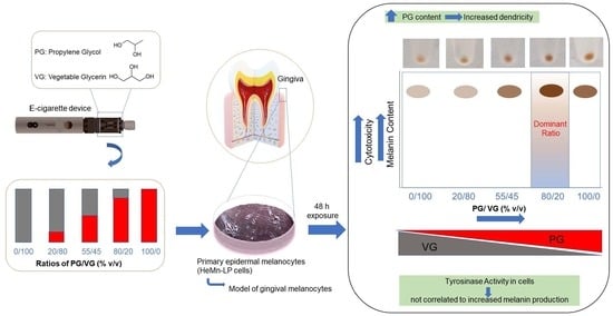
:Oral cavity is the first site to encounter e-cigarette (EC) or tobacco smoke. Increased gingival pigmentation can lead to aesthetic concerns and hinder successful outcomes of gingival depigmentation procedures as well as lead to color alterations in patients with dental restorations. While the effects of tobacco smoke and nicotine in increasing pigmentation in the gingiva of the smoker have been well-documented, the effects of EC on pigmentation have not been explored. Due to large variations in e-liquids from different sources, this study focused on the effects of EC liquid base constituents, propylene glycol (PG) and vegetable glycerin (VG), which are a universal constituent of all e-liquids. Effects of PG and VG solutions mixed at different ratios (0/100, 20/80, 55/45, 80/20, and 100/0 % v/v) were examined using primary human melanocytes obtained from neonatal foreskin; this cell model is representative of the physiological model of gingival melanocytes and has been used in our previous study. Results showed significant concentration-dependent cytotoxicity for all groups, although mixtures with higher PG content showed higher cytotoxicity to cells as compared to those with VG. Melanogenesis was robustly activated by PG-containing mixtures with the greatest effect obtained for 80/20 PG/VG mixture as compared to other ratios, while VG by itself did not activate melanogenesis. The activation of melanin synthesis within cells was not correlated to intracellular tyrosinase activity as that was suppressed by PG at higher ratios. Morphological changes of a multidendritic phenotype were observed in cells exposed to all PG/VG mixtures, with markedly greater effects for groups with higher PG content. Taken together, the results of this pilot study demonstrate for the first time that EC base constituents possess the capacity to significantly activate melanogenesis in human melanocytes at nontoxic concentrations, with the dominant effect obtained at a PG/VG ratio of 80/20, indicative of a nonlinear response with increasing concentrations of PG. Moreover, further studies to address the impact of PG/VG with the addition of nicotine and the effects of different EC flavors are underway. Future studies to elucidate mechanisms of increased pigmentation as well as further investigate effects in melanocytes with the presence of other oral cell types and other components of the oral microenvironment such as saliva and bacterial flora are warranted. This research emphasizes the need to reconsider the regulation of EC base constituents PG and VG as different ratios of these compounds can cause differential effects.
1. Introduction
E-cigarette (EC) is a nicotine-delivery system that contains an e-liquid primarily consisting of propylene glycol (PG) and vegetable glycerin (VG) as solvent carriers in different ratios, with or without nicotine and different flavorings [1]. The aerosol generated from the heating of e-liquid contained within the EC cartridge contact oral cavity cells as the first entry port. EC use has been increasing worldwide on account of its perception as a harm-reduction device as compared to tobacco smoking [2,3,4]. Nevertheless, EC exposure has been shown to affect oral health in adverse ways causing gingival diseases, periodontitis, inflammation, and alterations of the oral microbiome as demonstrated before [5,6]. Another study [7] compared the effects of EC aerosols and cigarette smoke on human gingival epithelial cells and found similar results of upregulated inflammatory cytokines and cytotoxicity, although the authors used EC that contained nicotine salt and flavorings. Recently, EC aerosol from tobacco-flavored nicotine-free and nicotine-rich EC was shown to induce proinflammatory cytokine secretion and cause damage in a 3D-human gingival mucosal model [8]. In addition, increased gingival inflammation was noted for tobacco smokers who switched to vaping from smoking [9].
Throat-hit that mimics the effects of cigarette smoking as experienced by EC users is primarily ascribed to PG which has been also shown to act as a carrier for multiple flavorings used in ECs [10,11]. On the other hand, VG-based e-liquids have a sweet taste and produce visibly large aerosol vape clouds imparting a “cloud-hit” that is desired by users [12,13] and also adolescents who engage in the generation of large clouds as a competitive hobby [13]. Despite having a less pleasant taste than VG, PG-based e-liquids deliver higher amounts of nicotine systematically [14], in particular, the yield of nicotine has been shown to be almost doubled by PG-containing e-liquids as compared to VG-based e-liquids [15]. PG and VG are considered Generally Recognized As Safe (GRAS) by the Food and Drug Administration (FDA) and have been approved for cosmetic and pharmaceutical use. A study conducted in mice that were exposed to either 100% PG or 100% VG unflavored EC aerosol for 4 weeks demonstrated that PG and not VG induced adverse cardiovascular effects in the aorta [16]. PG was shown to induce toxicity to proximal tubule cells of the kidney [17] and cause renal and liver toxicity upon inhalation [18], while upon intravenous administration it caused neurotoxicity as well as renal toxicity [19]. Other previous studies have shown adverse effects of PG and VG on lung surfactants’ with implications for compromised lung function [20], and demonstrated molecular interactions of PG and VG aerosols with key phospholipids of the lung [21]. Previous studies that have tested different ratios of PG/VG have either tested them in unvaped form or vaped form. For example, Beklen et al. reported augmented release of pro-inflammatory mediators IL-6 and IL-8 with upregulated MMP-9 in gingival epithelial cells exposed to unvaped e-liquids of different ratios of PG/VG [22]. In another study, volunteers that had never smoked or vaped were exposed to aerosols from flavoring-free and nicotine-free VG-containing e-liquids for 7 d and were shown to have ion channel dysfunction and airway inflammation while aerosols from PG-containing e-liquids did not show adverse effects [23].
Melanocytes are the melanin producing specialized cells that are present in the basal and/or spinous layer of the oral epithelium. Melanin is exported from mature melanocytes to neighboring keratinocytes via multiple dendrites of a melanocyte [24]. A keratinocyte-melanocyte unit is formed as one melanocyte connects to multiple keratinocytes with typical ratios ranging from 1:10 to 1:15 [25]. Tyrosinase is one of the primary enzymes in the melanogenesis process that catalyzes tyrosine to DOPA and its further oxidation to DOPAquinone through a two-step reaction sequence within melanosomes [26,27,28]. A recent study showed the correlation of degree of gingival pigmentation in human subjects with the gene expression of tyrosinase [29]. Human oral mucosal pigmentation can be affected by melanin produced within melanocytes, maturation of melanosomes, or the export of melanosomes from melanocytes to keratinocytes [30]. Oral pigmentation can occur either due to endocrine factors (systemic diseases, periodontitis, genetics) or due to exogenous factors such as smoking, drugs, metal exposure, or due to postsurgical gingival depigmentation [31] and has been shown to have higher prevalence in three groups: males, fair-skinned people, and cigarette smokers based on a recent meta-analysis study [32]. Melanosis caused by smoking, commonly referred as smoker’s melanosis, has been shown to occur in 21.5% of smokers [33]. Bardellini et al. [34] for the first time demonstrated oral mucosal lesions (OML) in EC users; the authors reported that volunteers who used EC for six months showed a higher prevalence of three types of inflammatory lesions in oral cavity (hyperplastic candidiasis, nicotine stomatitis, and hairy tongue) as compared to former smokers. Interestingly, the authors also noted the presence of common lesion of smokers melanosis in both groups, which indicates that EC use contributed to pigmentation, although it was not possible to attribute the effects to specific EC constituents as the user might have vaped EC that contained nicotine or flavors. Despite the well-established effects of nicotine or tobacco smoking on melanogenesis [35], the effects of EC base constituents PG/VG on melanogenesis are relatively underappreciated. Hence, in this study, the effects of unvaped e-liquids containing different ratios of PG and VG were examined using melanocytes from human foreskin as an in vitro model of oral melanocytes.
2. Materials and Methods
2.1. Materials
PG (USP Kosher, CAS# 57-55-6, 99.5%) and VG (USP Kosher, CAS# 56-85-1, 99.7%) were mixed at different ratios (0/100, 20/80, 55/45, 80/20, and 100/0) by volume to generate the e-liquid samples that were stored in the dark at 4 °C for the duration of the study. PG/VG ratios of 0/100 and 100/0 refer to pure VG and pure PG, respectively. Commercial EC liquids typically contain PG/VG mixtures in volume ratios % v/v [36], hence, in this study, PG/VG groups with different ratios were prepared in volume ratios similar to those reported in previous studies [14,20,37,38]. CellTiter 96® AQueous One Solution Cell Proliferation Assay (MTS) was obtained from Promega Corporation (Madison, WI, USA). L-DOPA was purchased from Sigma-Aldrich (St. Louis, MO, USA). Bicinchoninic acid (BCA) protein assay kit was purchased from Thermo Fisher Scientific (Bothell, WA, USA).
2.2. Cell Culture
Human epidermal melanocytes from lightly pigmented neonatal donor (HEMn-LP) were obtained from Cascade Biologics (Portland, OR, USA) and were cultured using Medium 254 containing 1% human melanocyte growth supplement (HMGS) and 1% penicillin-streptomycin. These cells were maintained at 37 °C in a humidified incubator (95% air-5% CO2). All experiments were conducted with melanocytes between passage five to ten.
2.3. Cytotoxicity Assay
Cell viability was estimated using the 3-(4,5-dimethylthiazol-2-yl)-5-(3-carboxymethoxyphenyl)-2-(4-sulfophenyl)-2H-tetrazolium (MTS) assay. Cell suspensions (8.2 × 10⁴ cells/well) were seeded onto 96-well plates (200 µL/well) and cultured for 72 h, after which the culture medium was replaced with fresh medium containing different concentrations of PG/VG groups. The plates were then incubated for a duration of 48 h at 37 °C in a humidified incubator with 5% CO2. At the end of exposure duration, the culture medium was aspirated, and subsequently, 100 µL of culture medium containing 20 µL of MTS solution was added to each well and incubated for 1.5 h. After incubation, the absorbance of 100 µL aliquots was measured at 490 nm with a Versamax® microplate reader. Percentage cell viability was determined from the absorbance readings that were normalized to the negative control group (set as 100%).
2.4. Estimation of Cellular Melanin Content
Cells (1.3 × 10⁵ cells/well in 1 mL complete medium) were seeded in 12-well plates and incubated for 72 h. After this, the culture medium was replaced with fresh medium containing nontoxic concentrations of compounds and the cultures were maintained for a period of 48 h. At the end of 48 h, the cells were detached, centrifuged, and pellets were washed in PBS buffer. Subsequently, cell pellets were dissolved in 75 µL of 1 N sodium hydroxide at 70 °C to solubilize melanin. Next, aliquots were transferred to a 96-well plate and the absorbance was read at 405 nm, the wavelength at which melanin absorbs light [39] using a microplate reader. The absorbance was normalized to negative control and expressed as percentage.
2.5. Microscopic Observation
Cells were imaged using a Nikon Labphot microscope equipped with a digital camera at 20× magnification in phase-contrast mode to observe morphology of dendrites. For some groups, cells were also imaged under bright-field microscopy at 40× magnification to identify deposition of melanin aggregates.
2.6. Intracellular Tyrosinase Activity
5 × 10⁴ cells (in 0.5 mL complete medium) were seeded onto each well of a 24-well plate and after 72 h, the culture medium was replaced with fresh medium containing various concentrations of e-liquid samples and the culture plates were incubated for 48 h at 37 °C. After the incubation period, cells were harvested and processed for determination of tyrosinase activity, similar to the method reported in our previous study [40]. Briefly, absorbances of mixture of 25 µL lysate and 75 µL L-DOPA solution were recorded in a microplate reader under kinetic mode at 475 nm; slopes were used to estimate tyrosinase activity that was normalized to total protein contents.
2.7. Statistical Analysis
One-way analysis of variance (ANOVA) with Dunnett’s post-hoc test or unpaired t-test was run using GraphPad Prism software (version 9.4.1, San Diego, CA, USA). Differences were considered statistically significant at p < 0.05 and all data are reported as mean ± SD.
3. Results
3.1. Effect of E-Liquids on Cell Viability
Exposure of melanocytes to PG/VG at a ratio of 0/100 induced cytotoxicity at concentrations >3%; cell viability was significantly diminished to 65.35% and 14.52% at PG/VG concentrations of 5% and 10%, respectively (Figure 1A). A similar result was obtained for the group PG/VG at a ratio of 20/80 as well as PG/VG at a ratio of 55/45 where concentrations up to 3% were noncytotoxic, while higher concentrations induced cytotoxicity. The viability of cells was significantly diminished to 75.06% and 10.58% at PG/VG (20/80) concentrations of 5% and 10%, respectively (Figure 1B), while the viability was significantly diminished to 62.64% and 10.19% at PG/VG (55/45) concentrations of 5% and 10%, respectively (Figure 1C). Next, the exposure of cells to PG/VG 80/20 group induced greater cytotoxicity with significant diminution starting from concentration of 3% onwards. Cell viability was significantly diminished to 56.78%, 39.58%, and 10.22% at concentrations of 3%, 5%, and 10%, respectively (Figure 1D). Lastly, cell viability was significantly diminished to 44.24% and 10.67% at PG/VG (100/0) concentrations of 5% and 10%, respectively with no significant change at concentrations up to 3% (Figure 1E).
The half-maximal inhibitory concentrations (IC50) values were determined for all groups and results are summarized in Table 1. Based on the results, it can be confirmed that mixtures with higher PG have augmented cytotoxicity. Interestingly, the mean IC50 values for cytotoxicity of the PG/VG (80/20) group was significantly lower than that of PG/VG (20/80) group.
Taken together, these results demonstrate that PG, and not VG, is the key driver of greater cytotoxicity to melanocytes, as mixtures containing higher amounts of PG displayed greater cytotoxicity to cells with the PG/VG (80/20) mixture exhibiting greatest cytotoxicity amongst all groups.
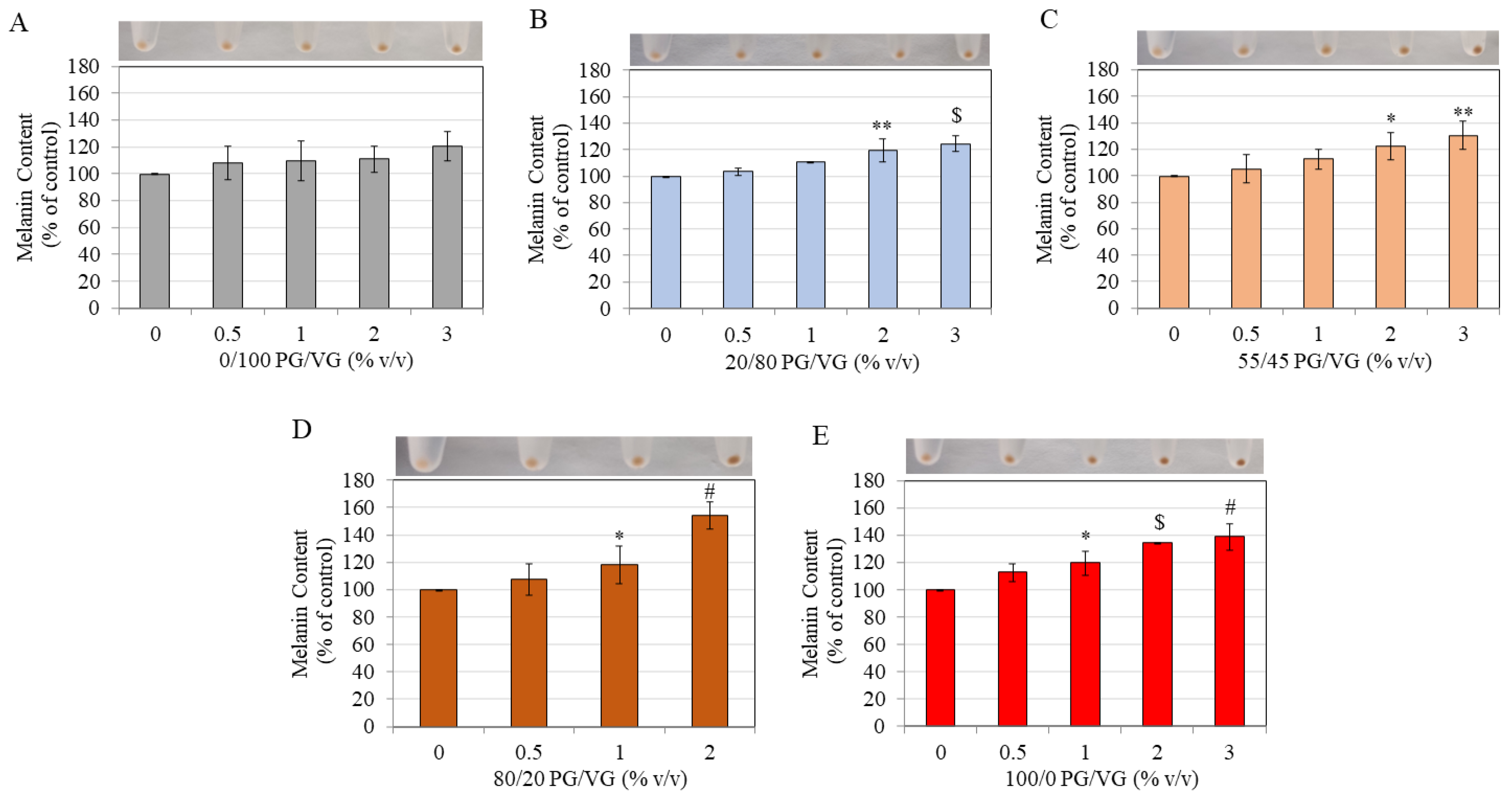
3.2. Effect of E-Liquids on Cellular Melanin Content
Cell pellets were visually inspected for qualitative changes in coloration of pellets as compared to negative control group for each group, after which the melanin levels were quantitated by spectrophotometric methods. Exposure of melanocytes to PG/VG at a ratio of 0/100 showed no alterations in the levels of melanin content of cells at any concentration, although a trend for increased melanin levels were noted at 3%, but levels did not reach statistical significance (Figure 2A). However, treatment with PG/VG at 20/80 ratio showed significant increases in melanin contents at concentrations 2% and 3% with melanin contents increased by 19.66% (p < 0.01) and 24.84% (p < 0.001), respectively (Figure 2B).
The results of the exposure of melanocytes to PG/VG at a ratio of 55/45 showed similar results as that of PG/VG (20/80), with melanin content significantly increased by 22.79% (p < 0.05) and 30.71% (p < 0.01) at concentration 2% and 3%, respectively (Figure 2C). Next, the exposure of melanocytes to PG/VG at a ratio of 80/20 showed significant increases of 18.37% (p < 0.05) and 54.40% (p < 0.0001) at concentration 1% and 2%, respectively (Figure 2D), with the cell pellets exhibiting darkest color at 2% based on visual observation (panel, Figure 2D). Lastly, the exposure of melanocytes to PG/VG at a ratio of 100/0 showed visibly darker coloration of pellets at the concentrations (1, 2 and 3%) as compared to negative control (panel; Figure 2E). Analysis of relative melanin contents further confirmed the qualitative results as PG/VG at 100/0 ratio induced a concentration-dependent stimulation of melanin synthesis that was significantly higher by 20.08% (p < 0.05), 34.44% (p < 0.001), and 39% (p < 0.0001) at concentrations of 1, 2 and 3%, respectively (Figure 2E).
Taken together, these results show that PG/VG groups containing high PG demonstrated a significant capacity to stimulate melanin production in a concentration-dependent manner, with robust melanin increase obtained by PG/VG 80/20 group at 2% that was 2.76-fold greater than that achieved by the equivalent concentration (2%) of PG/VG 20/80 group.
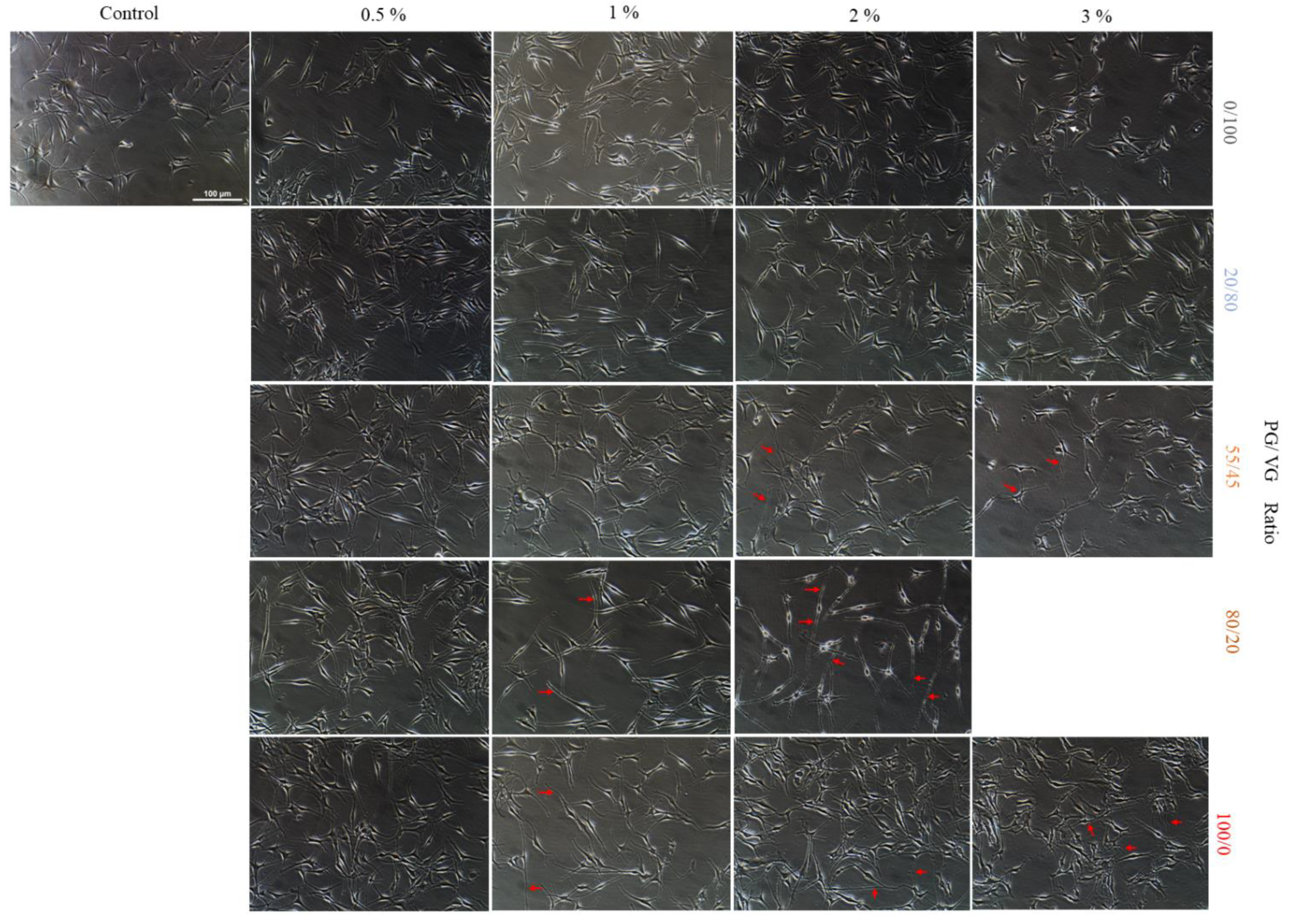
3.3. Effect of E-Liquids on Cellular Morphology
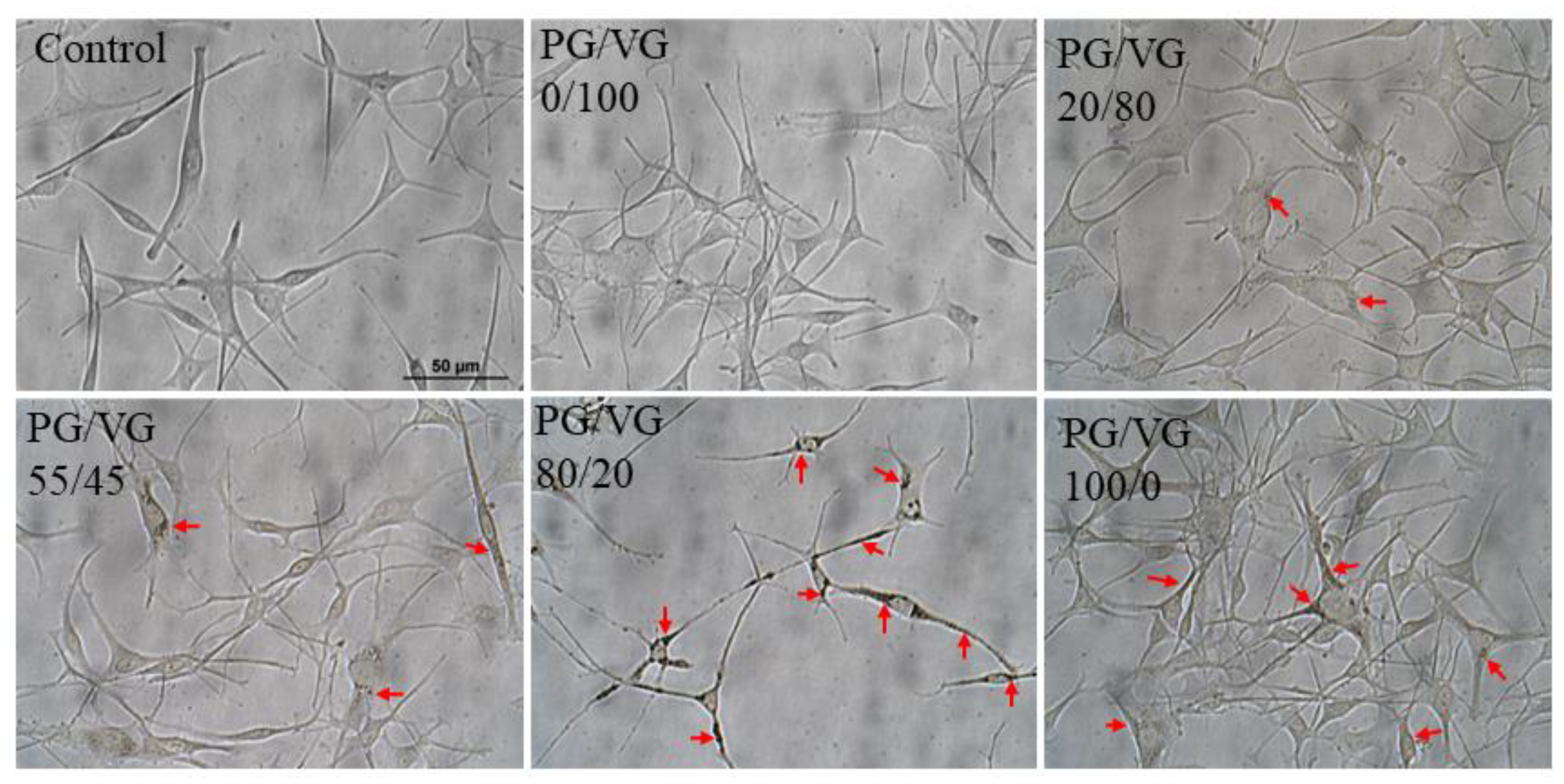
Cells were observed after exposure to various groups to identify any morphological changes in dendritic structures. As shown in Figure 3, qualitative evaluation revealed that groups with higher PG content showed morphological changes characterized by longer dendrites that was more evident as concentration of PG/VG in a group increased. Particularly, PG/VG (80/20) at 2% showed very long dendrites with appearance of new dendritic growth that could be noted from the small dendrite spine-like projections, which was not present in melanocytes of control group (Figure 3).
The cellular morphology of melanocytes was also compared after exposure to all e-liquids at the concentration of 2% (Figure 4) where marked deposits of melanin pigment could be observed under bright-field imaging at higher magnification. Presence of increased melanin pigment deposits could be discernible within cytoplasm of cells from the PG/VG 20/80 group onwards with an increase in groups with higher PG content. Interestingly, the cells in PG/VG 80/20 group showed most marked deposition of melanin synthesis with extended dendrites that also contained pigment deposits indicative of active process of export of melanin pigment, plus these dendrites appeared longer as compared to other groups (Figure 4). Collectively, these results corroborate earlier results of increased melanin content within cells for groups with higher PG, with the group PG/VG 80/20 showing the most prominent result.
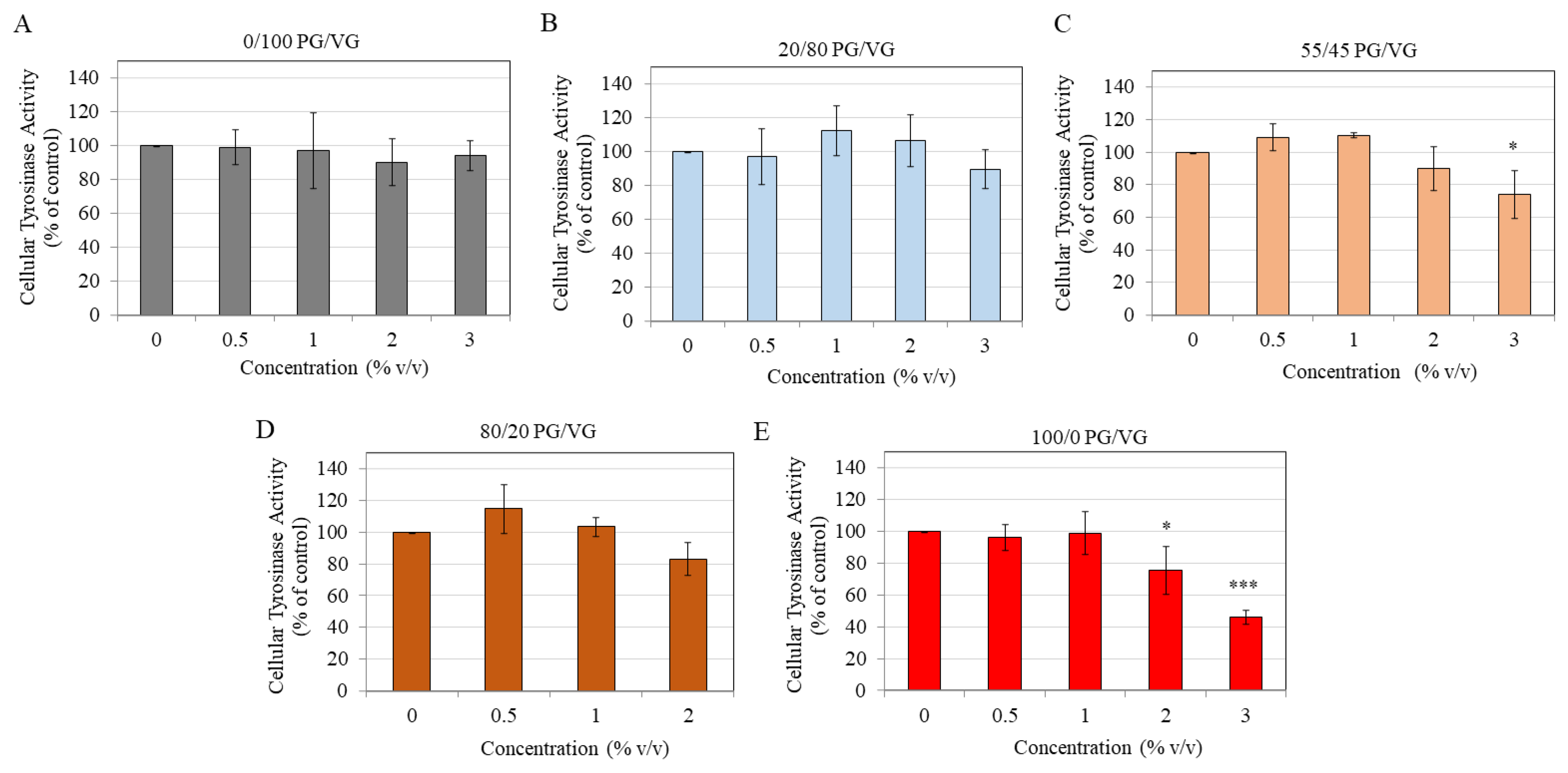
3.4. Effect of E-Liquids on Cellular Tyrosinase Activity
Exposure of melanocytes to PG/VG at a ratio of 0/100 (Figure 5A) as well as ratio 20/80 showed no changes in tyrosinase activity at any tested concentration (Figure 5B). In the case of PG/VG at ratio 55/45, the tyrosinase activity was significantly inhibited by 26% at the highest concentration of 3% with no change at lower concentrations (Figure 5C).
Next, treatment with PG/VG at 80/20 ratio showed no significant change at any concentration as compared to untreated control (Figure 5D). Lastly, PG/VG at 100/0 ratio showed a concentration-dependent inhibition of tyrosinase activity; a significant inhibition of 24.62% and 53.73% were obtained at concentrations of 2% and 3%, respectively (Figure 5E).
Overall, these results indicate that increased melanogenesis in cells is not related to activity of the enzyme tyrosinase, as instead of stimulation, a significant suppression was obtained by groups with higher PG. Surprisingly, for PG/VG 80/20 group, no changes were seen at any concentration.
4. Discussion
Although there is a wide variety of EC products containing diverse flavorings and nicotine formulations that are available for sale, all EC liquids contain PG and VG in some ratio. In fact, about 80–90% of total volume of e-liquids in ECs comprises solvent mixture PG/VG [41]. Hence, the primary objective of this study was to elucidate the effects of combinations of major constituents PG and VG. Results of this study demonstrate the novel finding that PG-containing base ingredients in e-liquids markedly activate melanin production even in the absence of nicotine and flavorings, that has implications for oral pigmentation of EC users, similar to that observed with tobacco smoking. Accumulating evidence shows that pigmentation in the gingiva not only causes social distress to those who have excessive display of gums while smiling, but it also hinders success outcomes of periodontal treatments. For instance, it was shown previously that melanin in gingival tissue impairs efficacy of antimicrobial photodynamic therapy, necessitating the need to revise irradiation protocols [42]. Moreover, former smokers who underwent gingival depigmentation surgeries to get rid of dark gingiva [43], might have a recurrence of pigmentation if they switch to the use of EC perceiving them as less harmful than cigarettes. This in turn, would lead to poor success outcomes of therapeutic procedures for gingival depigmentation where recurrence of pigmentation is often a challenge [44,45]. Furthermore, it has been documented that in 30% of cases, oral mucosal melanoma was shown to develop at the oral sites where pigmentation was higher [46,47].
The cell model of epidermal melanocytes used in this study has been shown to be a physiological model of oral melanocytes as it exhibits similar ultrastructure of melanosomes and histology to that of melanocytes in the gingiva [48,49]. Additionally, this cell model has been used in our previous study to evaluate the effects of the dental agent fluoride [50]. One key strength of this study in terms of identification of EC toxicity is that the confounding effects of prior history of smoking or any other oral pathology such as periodontitis, can be effectively ruled out as these cells were derived from neonatal foreskin and not adult donors and can be considered as healthy. Regardless, it would be very interesting to conduct future studies where melanocytes isolated from smokers were used to examine the impact of EC liquids (PG/VG). A recent interesting study demonstrated that small airway epithelial cells obtained from chronic obstructive pulmonary disease (COPD) patients (which already had cigarette smoke exposure leading to COPD development) showed greater toxicity by PG than cells obtained non-COPD patients [51], thus highlighting that PG might display higher sensitivity and induce greater adverse effects in cells derived from previous smokers than cells from never smokers.
Results of nonlinear response of PG/VG ratio on cellular viability is in agreement with earlier studies [52,53] that also showed similar responses in bronchial epithelial cells. For example, Leslie et al. [53] showed that aerosolized PG/VG at 80/20 ratio induced greatest cytotoxicity as compared to other ratios in bronchial epithelial cells, while another study reported that PG/VG at 70/30 ratio demonstrated significant toxicity with no effect at other ratios [52]. Results of higher cytotoxicity by PG and not VG are in agreement with multiple studies which similarly demonstrated that PG was the driver of cytotoxicity while VG caused lesser effects [22,54,55]. Structurally, PG and VG differ only by an OH group where PG has two OH groups while VG has three OH groups, thus PG is more hydrophobic as compared to VG. Hence, results of higher cytotoxicity by PG might be attributable to the ability of PG to cross cellular membrane more easily as compared to VG. Another possibility is that PG/VG might affect membrane rheology and change protein diffusion, similar to the effects shown in a prior study [56]. The contribution of osmotic stress might explain cell death at higher concentrations (5 and 10%) for PG and VG groups, as it was shown that hyperosmotic shock due to high PG content induced cytotoxicity in lung epithelial cells [55]. Elsewhere, it was also shown that high osmotic stress due to higher PG content in PG/VG mixtures caused selective alterations in functions of lung cells [57]. In order to examine if osmotic changes might have contributed to the enhanced melanin contents of melanocytes, mannitol was used as an osmotic control at 408 mM which has similar osmolarity to 3% PG/VG 55/45 group based on a previous study [58] that also tested for osmotic effects. Results (Figure S1 photos) showed darker-colored pellets for melanocytes exposed to 3% PG/VG 55/45 as compared to control expectedly, but pellets for mannitol were visibly lighter than PG/VG 55/45 and similar to control group thereby confirming that the stimulation of melanin synthesis by PG/VG 55/45 was independent of osmotic stress (quantitated by melanin content analysis; Figure S1 plot). This is because if osmotic stress per se induced melanin production, both groups which were theoretically isosmotic should have similar levels of melanin content, which was not the case. Moreover, a previous study has shown that higher osmotic stress suppressed melanin synthesis in cells resulting in lightening of pellets [59]. Hence, we believe that osmotic stress may not be a causative factor in the stimulation of cellular melanin production. Results of stimulation of melanin production in melanocytes is in agreement with a previous study [60] that showed robust increase in melanin production in primary human melanocytes treated with PG over a concentration range of 100–300 mM for a duration of 5 d, although the authors showed increased melanogenesis without any effects on cellular tyrosinase activity (although tyrosinase activity in the extracellular particulate fraction extract was enhanced) which is partly similar to our findings, although we did not evaluate extracellular tyrosinase activity. Interestingly, the authors of this study [60] validated the skin-darkening effects of PG using a guinea pig in vivo model and also showed evidence of particulate melanin fraction in the extracellular medium of melanocytes treated with PG. We also noted a similar occurrence of dark melanin particulates both within the dendrite body and at the dendrite tip as well as the shedding of pigment globules in the culture medium for the PG/VG 80/20 group (Figure S2). It should be emphasized that as PG shares structural similarity with ortho-diphenols such as catechol, the possibility of the formation of quinone-like structures that might induce adverse effects of melanocytotoxicity that are characteristic of catechols warrant detailed investigation in future studies, although the study by Brown et al. [60] discussed that the effect of PG was not similar to catechol or ortho-diphenols. Our results of increased melanin production by PG-based e-liquids are reminiscent of that of minocycline, a tetracycline-based antibiotic drug, that was previously shown to increase melanin content in an vitro model of HEMn-LP cells [61]. Interestingly, minocycline has also been shown to induce oral pigmentation in vivo [62,63]. Given our current findings of marked melanin production by PG-containing e-liquids in vitro, we reason that similar results might also occur in vivo, although further studies incorporating organotypic models of oral mucosa and in vivo studies will be necessary.
In order to analyze whether stimulation of melanogenesis within cells exposed to PG/VG groups could be explained, at least in part, by stimulation of the activity of the enzyme tyrosinase, further experiments were conducted to determine tyrosinase activity in lysates of cells exposed to different PG/VG groups. Unexpectedly, results revealed no correlation as tyrosinase activity was either unchanged or suppressed in the groups. Previous studies have also shown a similar discrepancy including our prior study [64] where cellular melanin content was increased but tyrosinase activity in cells was not affected. For example, hyperosmotic stress was shown to diminish melanin synthesis in B16F10 mouse melanoma cells (model of melanocytes) in the absence of any effects on the cellular tyrosinase activity, indicative of an alternative mechanism that might diminish melanogenesis [59]. This is not entirely surprising since in addition to tyrosinase, tyrosinase-related protein 1 (TRP-1), and tyrosinase-related protein 2 (TRP-2) are two other enzymes that participate in the process of melanogenesis at different steps [65,66].
Melanocytes are known to co-express Ca+2 permeable cation channels, transient receptor potential ankyrin 1 (TRPA1), and transient receptor potential vanilloid 1 (TRPV1). Qi et al. [67] demonstrated that TRPA1 and TRPV1 expressions were enhanced in physiological or pathological conditions of hyperpigmentation, and human melanocytic lesions. Moreover, TRPA1 positively regulated melanogenesis in primary human melanocytes by regulating the pH of melanosomes [68] and stimulated phagocytosis of melanosomes by keratinocytes [69], indicative of its effect on further steps in the melanogenesis pathway. Additionally, TRPA1 activation was shown to be involved in UV radiation-induced enhancement of melanogenesis [70]. High osmolarity is known to activate TRPA1 and TRPV1 [71,72] and PG has been shown to be a skin-irritant [73]. Jordt et al. [74] demonstrated a similar irritant response of 90/10 PG/VG vapors of flavoring- and nicotine-free EC to that of cinnamaldehyde vapors, a classic irritant compound. Moreover, PG and VG at high millimolar concentrations were shown to have a moderate activation effect on the irritant receptor TRPA1, indicative of their irritant effects. Furthermore, PG at subcytotoxic concentrations sensitized human TRPV1 to further enhance response to capsaicin, while VG was shown to diminish the response of TRPA1 to cinnamaldehyde. Previously, Florian et al. [75] showed that PG up to 10 v% concentrations was shown to exhibit irritating effects that were not related to its high osmolarity, but rather were related it its interactions with receptor proteins of TRPA1 and TRPV1. Moreover, PG at higher concentrations of 35 v% was also shown to stimulate both TRPA1 and TRPV1. We hypothesize that TRPA1 and TRPV1 might be involved in the melanogenesis-stimulating effects of PG-containing e-liquids, although future studies to validate this hypothesis are warranted.
Some studies have reported that higher PG content in EC was the primary determinant of the release of proinflammatory cytokine IL-6 in gingival epithelial cells [22] or 3D skin keratinocyte model [54]. Elsewhere, it was shown that under stimulation by bacterial lipopolysaccharide (LPS), the release of cytokine IL-8 by macrophages was suppressed by aerosol from a 50/50 mixture of PG/VG [76]. We preliminary examined levels of IL-6 in supernatants of cultures that were exposed to all five PG/VG groups at 2% v/v in the presence of 10 µg/mL LPS. Data showed that while LPS stimulated the production of IL-6 in culture, the presence of PG/VG compounds at any ratio had no appreciable effect in further augmenting or impairing IL-6 secretion. (Figure S3). Previously, higher secretion of cytokine IL-1α was demonstrated after PG treatment in a 3D keratinocyte model (EpiDerm™) under basal conditions [54], while in this study IL-6 levels were evaluated under LPS-stimulated conditions, thus whether PG/VG can affect cytokine levels under basal conditions in melanocytes, will need to be determined in future.
Although oxidative stress markers such as reactive oxygen species (ROS) were not evaluated in this study as it was not the primary focus, prior research has demonstrated that a higher PG content increased oxidation of biologically relevant lipids in the EC aerosol, with a 3-fold higher level of generation of free radicals for PG/VG 100/0 as compared to PG/VG 0/100 [77]. These data suggest that PG content is associated with free radical production that can induce oxidative stress. EC aerosol from VG-based liquids due to their thick consistency remains adhered to teeth’s fissures and pits in the oral cavity and promote bacterial attachment [78]. Although we did not obtain any significant changes in melanogenesis by VG in this study, it is a possibility that VG might still affect other steps of the melanogenesis cycle in melanocytes that were not presently studied. Interestingly, a higher VG content (lower PG/VG ratio) has been shown to be associated with increased emission of fine particulate matter (PM2.5) in recent studies [37,38]. PM2.5 is found in EC emissions similar to that of cigarette smoke emissions [79,80,81] and has been shown to increase melanogenesis in the skin in multiple previous studies [82,83,84]. As environmental tobacco smoke has been shown to be correlated to oral pigmentation [31], it would be very interesting in the future to explore whether second-hand exposure to EC emissions (second-hand vaping) might also cause an increase in oral pigmentation of passive smokers.
PG has a lower boiling point than VG [85] hence its volatile nature causes it to be present in the gas phase of EC aerosol. The ratio of PG/VG in e-liquid after aerosol generation is known to remain unaltered, rather enriched in PG [86], hence we believe that our biological results will continue to be valid after vaping. Degradation of solvents PG and VG can occur by oxidation or dehydration reactions, the chemical pathways of which have been described previously; interestingly dihydroxyacetone, a known skin tanning agent, was generated as one of the chemical products [87]. Small oligomers generated in PG/VG aerosol have also been reported for the first time by Escobar et al. [88]; authors speculated that these might have contributed to cytotoxicity. Whether the production of such oligomers might exacerbate melanocytotoxicity will be interesting for future investigations. In a study by Peace et al. [36], 4 of 27 e-liquids that were analyzed showed erroneous labeling of PG/VG, where the labeled ratio of 50/50 was actually found to be 80/20; the authors pointed out that although the percentage was low, chances that even PG/VG ratio might be mislabeled and consequently expose the user to higher PG, raise concern. In addition, another study showed that EC users who can typically distinguish extreme end ratios (100/0 and 0/100) due to the cloud- and throat-hit, were unable to distinguish intermediate ratios (30/70, 50/50, and 70/30) [89], thus indicating that beginners as well as experienced vapers might not pay attention to intermediate ratios while purchase and can be exposed to higher PG ratios (such as 80/20 vs. 20/80) which in our study showed significantly different responses. This study focused on the effects of PG/VG e-liquids on melanocytes that were used as a representative model for gingival melanocytes. However, as melanocytes are in close contact with gingival keratinocytes constituting a unit that enables crosstalk and transfer of melanin [25], the effects of e-liquid base constituents (PG/VG) on gingival keratinocytes will also be necessary. Encouragingly, a recent study by Beklen et al. [22] that is most fitting in the context of the experimental design of our study, examined the effects of three PG/VG mixtures (PG/VG 20/80, 50/50, and 80/20) on primary gingival keratinocytes and documented enhanced cytotoxicity in the presence of nicotine (18 mg/mL) for all e-liquids as compared to nicotine-free e-liquids, although a limitation of their study was that the effects of PG or VG alone were not examined. To the best of our knowledge, the in vitro effects of PG/VG e-liquids on other cells in the oral cavity as immediately exposed cells in the mouth such as squamous cells, gingival fibroblasts, or gingival stem cells have not yet been explored and would be fitting for future investigations.
Prior studies by Dalrymple et al. [90,91] proposed that smokers who switched to EC might impart cosmetic benefits as the authors showed that exposure to EC aerosols resulted in minimal enamel staining as compared to cigarette smoke, although the authors used nicotine-rich and tobacco-flavored EC in their experiments. Results of the current study, although need to be validated with further studies and ultimately in vivo studies point out that EC use can induce gingival pigmentation, even in the absence of nicotine or flavors. Consequently, this might be a risk factor for individuals who switch to EC from cigarette smoking, as it might still create another cosmetic concern, that of a darker gingiva. Moreover, as the results of this study showed a robust increase of melanin production for PG/VG 80/20 group within 48 h duration, the possibility of further increase by repetitive exposures over longer durations is likely. The cytotoxicity of e-liquid has shown a good correlation to the cytotoxicity of its generated aerosol as shown previously [92]. Hence, the results of this study, although conducted with neat, un-aerosolized e-liquids, can be extrapolated to that of the cytotoxicity of e-liquid after vaping. Although it should be emphasized that the microenvironment of the oral cavity which has other surrounding cell types along with the presence of salivary pellicle layer, and bacterial flora, was missing in our in vitro experiments that were performed with only a single-cell monolayer.
The effects of nicotine dissolved in PG/VG solutions of different ratios on melanocyte cytotoxicity and functions were not investigated in this study as it was beyond the scope and was not the primary focus of this study. In the literature, the effects of nicotine dissolved in PG/VG is rather complex and conflicting and dependent on cell type, nicotine concentration, and PG/VG ratios, as few studies have shown that nicotine-containing PG/VG augmented cytotoxicity as compared to PG/VG [22,55], while another [93] showed rescue of cytotoxicity and increased viability by nicotine-containing PG/VG and another study showed that cytotoxicity of nicotine-containing PG/VG vs. PG/VG was similar [94]. For example, the cytotoxicity of nicotine-containing PG/VG was higher than PG/VG in gingival epithelial cells, irrespective of PG content [22]. Similarly, another study [55] showed that the EC50 values of a 24 h exposure of lung epithelial cells to pure PG, 70/30 PG/VG, and pure VG e-liquids were 3.41%, 3.92%, and 6.30%, respectively, while the EC50 values of same e-liquids containing nicotine at 18 mg/mL were 2.09%, 1.95%, and 2.47%, respectively. Clearly, these results imply that there is no correlation of cytotoxicity to PG content in the presence of nicotine as all e-liquids had nearly similar EC50 values, in contrast to values in absence of nicotine that showed PG to be the driver of cytotoxicity. Another study [93] that used e-liquid condensates (generated from aerosolizing e-liquid and collecting vapors in a medium) reported conflicting results, which showed that nicotine at 18 mg/mL rescued high cytotoxicity by 50/50 PG/VG in airway epithelial cells, although the rescue effect was diminished by nicotine at 60 mg/mL. Moreover, another study [94] compared the effects of aerosolized e-liquids of 70/30 PG/VG, PG/VG with nicotine (12 mg/mL), PG/VG with ten different flavors, and PG/VG with nicotine and ten different flavors, in three immortalized oral epithelial cell lines, showed low or negligible cytotoxicity of unflavored nicotine-containing PG/VG and nicotine-containing PG/VG with flavors groups. A prior study [95] showed that the EC50 value of nicotine cytotoxicity after a 24 h exposure to HEMn-LP cells (same cells as used in our study) was 7.43 mM, while nicotine at 5 mM significantly lowered the viability of LP cells by 35.4%. As the concentration of nicotine in the saliva of smokers has been reported to be 5 mM [96], it is likely that nicotine can induce cytotoxicity to these cells by itself, and the presence of nicotine in PG/VG mixtures of e-liquid may further amplify cytotoxicity. Flavorings that are added to e-liquids have also been shown to have a cytotoxic effect on several mammalian cells, that were independent of nicotine [97]. Currently, the effects of PG/VG with nicotine, PG/VG with flavorings, as well as PG/VG with nicotine and flavorings on melanocytes remain unknown. Hence, the examination of diverse flavorings with PG/VG as well as nicotine (at varying concentrations) with PG/VG will be essential for future investigations. Importantly, it will also be necessary to examine whether the addition of nicotine/flavorings might have additive or synergistic effects in stimulating melanin production.
The adverse effects of PG have been described as causing xerostomia in the oral cavity [98,99], while another study documented the induction of genotoxicity by e-liquids that were predominantly PG based (≥70% PG and 100% PG) [100]. Based on these reports, future studies should also address if PG-based e-liquids might be genotoxic to melanocytes. Moreover, a recent study reported the use of 1,3 propane diol as a substitute for PG in ECs [101], hence the effects of this substitute vehicle on melanocytes would be interesting to explore to identify if it can have less impact on melanocyte toxicity and functions as compared to PG.
5. Conclusions
In summary, the results of this study demonstrate that different ratios of EC liquid base constituent PG and VG possess the capacity to significantly activate melanogenesis in cells at concentrations where they did not affect cellular viability. While the molecular mechanism(s) for these effects remain to be determined, these data suggest that exposure of melanocytes to nicotine- and flavoring-free e-liquids containing only PG or predominantly PG may cause adverse outcomes by activating cellular melanogenic activity. Based on the results of this study, it is likely that pure VG (PG/VG 0/100) or high VG-containing e-liquids (PG/VG 20/80) have the least impact on melanocyte cytotoxicity and pigmentation as compared to pure PG or high PG-based e-liquids. Hence, regulation of PG content in e-liquids and use of only VG or predominantly VG-based e-liquids might seem like a plausible strategy for regulatory agencies, although we acknowledge that more in-depth studies will need to be conducted in the future as it is still premature to propose any regulatory policies and future directions. Taken together, the results of this study could provide a critical initial step in the understanding of EC use and melanocyte oral pigmentation and can open new avenues to expand further assessment of e-liquid constituent hazard in a physiological model with the presence of other components of the oral cavity.
Supplementary Materials
The following supporting information can be downloaded at: https://www.mdpi.com/article/10.3390/oral3010005/s1, Figure S1. Intracellular melanin levels of HEMn-LP cells treated with 3% PG/VG 55/45 or mannitol (7.4% w/v) for a duration of 48 h with the corresponding photos of cell pellets also shown; data is mean ± SD of duplicate values; * p < 0.05 vs. Ctrl; ** p < 0.01 vs. PG/VG 55/45; p > 0.05 for Ctrl vs. mannitol; pairwise unpaired t-test with Welch’s correction. Figure S2. Two representative photomicrographs (A) and (B) taken from different regions at 40× magnification in culture well of HEMn-LP cells treated with 2% PG/VG 80/20 for a duration of 48 h; white arrows indicate the released pigment globules; dark melanin pigment aggregates in dendrite body and tips are visible in both images. Figure S3: IL-6 levels measured in culture supernatants of cells treated with 10 µg/mL LPS alone or co-treated with LPS and PG/VG at 0/100, 20/80, 55/45, 80/20, and 100/0, all at concentration of 2% v/v for a duration of 48 h; cytokine data is expressed as fold-change and was normalized to negative control (Ctrl). Data is mean ± SD of triplicate determinations. (# p < 0.0001 vs. Ctrl; one-way ANOVA with Tukey’s test). Reference [102] is cited in the supplementary materials.
Funding
This research was funded, in part, by unrestricted funds through the Stony Brook Foundation.
Institutional Review Board Statement
Not applicable.
Informed Consent Statement
Not applicable.
Data Availability Statement
The data used in this work is available from the corresponding author upon reasonable request.
Acknowledgments
The author would like to acknowledge the use of lab facilities under Sanford Simon at Stony Brook University.
Conflicts of Interest
The author declares no conflict of interest.
References
- Brown, C.J.; Cheng, J.M. Electronic cigarettes: Product characterisation and design considerations. Tob. Control. 2014, 23, ii4–ii10. [Google Scholar] [CrossRef] [PubMed] [Green Version]
- Levy, D.T.; Borland, R.; Lindblom, E.N.; Goniewicz, M.L.; Meza, R.; Holford, T.R.; Yuan, Z.; Luo, Y.; O’Connor, R.J.; Niaura, R. Potential deaths averted in USA by replacing cigarettes with e-cigarettes. Tob. Control. 2018, 27, 18–25. [Google Scholar] [CrossRef] [PubMed] [Green Version]
- Stratton, K.; Kwan, L.Y.; Eaton, D.L. Public Health Consequences of E-Cigarettes: Consensus Study Report; National Academies Press: Washington, DC, USA, 2018. [Google Scholar]
- Farsalinos, K.E.; Polosa, R. Safety evaluation and risk assessment of electronic cigarettes as tobacco cigarette substitutes: A systematic review. Ther. Adv. Drug Saf. 2014, 5, 67–86. [Google Scholar] [CrossRef] [PubMed] [Green Version]
- Yang, I.; Sandeep, S.; Rodriguez, J. The oral health impact of electronic cigarette use: A systematic review. Crit. Rev. Toxicol. 2020, 50, 97–127. [Google Scholar] [CrossRef]
- Szumilas, P.; Wilk, A.; Szumilas, K.; Karakiewicz, B. The Effects of E-Cigarette Aerosol on Oral Cavity Cells and Tissues: A Narrative Review. Toxics 2022, 10, 74. [Google Scholar] [CrossRef]
- Ramenzoni, L.L.; Schneider, A.; Fox, S.C.; Meyer, M.; Meboldt, M.; Attin, T.; Schmidlin, P.R. Cytotoxic and Inflammatory Effects of Electronic and Traditional Cigarettes on Oral Gingival Cells Using a Novel Automated Smoking Instrument: An In Vitro Study. Toxics 2022, 10, 179. [Google Scholar] [CrossRef]
- Alanazi, H.; Rouabhia, M. Effect of e-cigarette aerosol on gingival mucosa structure and proinflammatory cytokine response. Toxicol. Rep. 2022, 9, 1624–1631. [Google Scholar] [CrossRef] [PubMed]
- Wadia, R.; Booth, V.; Yap, H.; Moyes, D. A pilot study of the gingival response when smokers switch from smoking to vaping. Br. Dent. J. 2016, 221, 722–726. [Google Scholar] [CrossRef]
- Harvanko, A.; Kryscio, R.; Martin, C.; Kelly, T. Stimulus effects of propylene glycol and vegetable glycerin in electronic cigarette liquids. Drug Alcohol Depend. 2019, 194, 326–329. [Google Scholar] [CrossRef]
- Smith, T.T.; Heckman, B.W.; Wahlquist, A.E.; Cummings, K.M.; Carpenter, M.J. The impact of e-liquid propylene glycol and vegetable glycerin ratio on ratings of subjective effects, reinforcement value, and use in current smokers. Nicotine Tob. Res. 2020, 22, 791–797. [Google Scholar] [CrossRef]
- Clapp, P.W.; Jaspers, I. Electronic cigarettes: Their constituents and potential links to asthma. Curr. Allergy Asthma Rep. 2017, 17, 79. [Google Scholar] [CrossRef] [PubMed]
- Baassiri, M.; Talih, S.; Salman, R.; Karaoghlanian, N.; Saleh, R.; El Hage, R.; Saliba, N.; Shihadeh, A. Clouds and “throat hit”: Effects of liquid composition on nicotine emissions and physical characteristics of electronic cigarette aerosols. Aerosol Sci. Technol. 2017, 51, 1231–1239. [Google Scholar] [CrossRef] [PubMed] [Green Version]
- Spindle, T.R.; Talih, S.; Hiler, M.M.; Karaoghlanian, N.; Halquist, M.S.; Breland, A.B.; Shihadeh, A.; Eissenberg, T. Effects of electronic cigarette liquid solvents propylene glycol and vegetable glycerin on user nicotine delivery, heart rate, subjective effects, and puff topography. Drug Alcohol Depend. 2018, 188, 193–199. [Google Scholar] [CrossRef] [PubMed] [Green Version]
- Talih, S.; Salman, R.; El-Hage, R.; Karaoghlanian, N.; El-Hellani, A.; Saliba, N.; Shihadeh, A. Effect of free-base and protonated nicotine on nicotine yield from electronic cigarettes with varying power and liquid vehicle. Sci. Rep. 2020, 10, 16263. [Google Scholar] [CrossRef] [PubMed]
- Frazier, J.; Coblentz, T.; Bruce, J.; Nassabeh, S.; Plants, R.; Burrage, E.; Mills, A.; Chantler, P.; Olfert, I. Effect of E-liquid Base Solution (Vegetable Glycerin or Propylene Glycol) on Aortic Function in Mice. FASEB J. 2021, 35. [Google Scholar] [CrossRef]
- Morshed, K.M.; Jain, S.K.; McMartin, K.E. Propylene glycol-mediated cell injury in a primary culture of human proximal tubule cells. Toxicol. Sci. 1998, 46, 410–417. [Google Scholar] [CrossRef]
- Doi, A.M.; Roycroft, J.H.; Herbert, R.A.; Haseman, J.K.; Hailey, J.R.; Chou, B.J.; Dill, J.A.; Grumbein, S.L.; Miller, R.A.; Renne, R.A. Inhalation toxicology and carcinogenesis studies of propylene glycol mono-t-butyl ether in rats and mice. Toxicology 2004, 199, 1–22. [Google Scholar] [CrossRef]
- Yaucher, N.E.; Fish, J.T.; Smith, H.W.; Wells, J.A. Propylene glycol-associated renal toxicity from lorazepam infusion. Pharmacother. J. Hum. Pharmacol. Drug Ther. 2003, 23, 1094–1099. [Google Scholar] [CrossRef]
- Sosnowski, T.R.; Jabłczyńska, K.; Odziomek, M.; Schlage, W.K.; Kuczaj, A.K. Physicochemical studies of direct interactions between lung surfactant and components of electronic cigarettes liquid mixtures. Inhal. Toxicol. 2018, 30, 159–168. [Google Scholar] [CrossRef] [Green Version]
- Hayeck, N.; Zoghzoghi, C.; Karam, E.; Salman, R.; Karaoghlanian, N.; Shihadeh, A.; Eissenberg, T.; Zein El Dine, S.; Saliba, N.A. Carrier solvents of electronic nicotine delivery systems alter pulmonary surfactant. Chem. Res. Toxicol. 2021, 34, 1572–1577. [Google Scholar] [CrossRef]
- Beklen, A.; Uckan, D. Electronic cigarette liquid substances propylene glycol and vegetable glycerin induce an inflammatory response in gingival epithelial cells. Hum. Exp. Toxicol. 2021, 40, 25–34. [Google Scholar] [CrossRef] [PubMed]
- Kim, M.; Chung, S.; Dennis, J.; Yoshida, M.; Aguiar, C.; Aller, S.; Mendes, E.; Schmid, A.; Sabater, J.; Baumlin, N. Vegetable glycerin e-cigarette aerosols cause airway inflammation and ion channel dysfunction. Front. Pharmacol. 2022, 13, 1012723. [Google Scholar] [CrossRef]
- Natesan, S.C.; Ramakrishnan, B.P.; Krishnapillai, R.; Thomas, P. Biophysiology of oral mucosal melanocytes. J. Health Sci. 2019, 2, 47–51. [Google Scholar] [CrossRef]
- Feller, L.; Masilana, A.; Khammissa, R.A.; Altini, M.; Jadwat, Y.; Lemmer, J. Melanin: The biophysiology of oral melanocytes and physiological oral pigmentation. Head Face Med. 2014, 10, 8. [Google Scholar] [CrossRef] [PubMed]
- Suryaningsih, B.E. Melanogenesis and its associated signalings. Bali Med. J. 2020, 9, 327–331. [Google Scholar] [CrossRef]
- Lin, J.Y.; Fisher, D.E. Melanocyte biology and skin pigmentation. Nature 2007, 445, 843–850. [Google Scholar] [CrossRef]
- Iozumi, K.; Hoganson, G.E.; Pennella, R.; Everett, M.A.; Fuller, B.B. Role of tyrosinase as the determinant of pigmentation in cultured human melanocytes. J. Investig. Dermatol. 1993, 100, 806–811. [Google Scholar] [CrossRef] [Green Version]
- Sanadi, R.M.; Deshmukh, R.S. Expression of tyrosinase gene in gingiva: A pilot study. J. Oral Maxillofac. Pathol. 2022, 26, 422. [Google Scholar]
- Moneim, R.A.A.; El Deeb, M.; Rabea, A.A. Gingival pigmentation (cause, treatment and histological preview). Future Dent. J. 2017, 3, 1–7. [Google Scholar] [CrossRef]
- Firoozi, P.; Noormohammadi, R.; Rafieyan, S. Effect of environmental tobacco smoke on oral pigmentation: A systematic review. J. Oral Health Oral Epidemiol. 2020, 9, 1–6. [Google Scholar]
- Rotbeh, A.; Kazeminia, M.; Rajati, F. Full title: Global prevalence of oral pigmentation and its related factors: A systematic review and meta-analysis. J. Stomatol. Oral Maxillofac. Surg. 2022, 123, e411–e424. [Google Scholar] [CrossRef] [PubMed]
- Tadakamadla, J.; Kumar, S.; Nagori, A.; Tibdewal, H.; Duraiswamy, P.; Kulkarni, S. Effect of smoking on oral pigmentation and its relationship with periodontal status. Dent. Res. J. 2012, 9, S112. [Google Scholar]
- Bardellini, E.; Amadori, F.; Conti, G.; Majorana, A. Oral mucosal lesions in electronic cigarettes consumers versus former smokers. Acta Odontol. Scand. 2018, 76, 226–228. [Google Scholar] [CrossRef] [PubMed]
- Araki, S.; Murata, K.; Ushio, K.; Sakai, R. Dose-response relationship between tobacco consumption and melanin pigmentation in the attached gingiva. Arch. Environ. Health Int. J. 1983, 38, 375–378. [Google Scholar] [CrossRef] [PubMed]
- Peace, M.R.; Baird, T.R.; Smith, N.; Wolf, C.E.; Poklis, J.L.; Poklis, A. Concentration of nicotine and glycols in 27 electronic cigarette formulations. J. Anal. Toxicol. 2016, 40, 403–407. [Google Scholar] [CrossRef] [PubMed] [Green Version]
- Li, L.; Lee, E.S.; Nguyen, C.; Zhu, Y. Effects of propylene glycol, vegetable glycerin, and nicotine on emissions and dynamics of electronic cigarette aerosols. Aerosol Sci. Technol. 2020, 54, 1270–1281. [Google Scholar] [CrossRef] [PubMed]
- Eversole, A.; Crabtree, M.; Spindle, T.R.; Baassiri, M.; Eissenberg, T.; Breland, A. E-cigarette Solvent Ratio and Device Power Influence Ambient Air Particulate Matter. Tob. Regul. Sci. 2021, 7, 177–183. [Google Scholar] [CrossRef] [PubMed]
- Ozeki, H.; Ito, S.; Wakamatsu, K.; Thody, A.J. Spectrophotometric characterization of eumelanin and pheomelanin in hair. Pigment. Cell Res. 1996, 9, 265–270. [Google Scholar] [CrossRef] [PubMed]
- Goenka, S.; Simon, S.R. Depigmenting effect of Xanthohumol from hop extract in MNT-1 human melanoma cells and normal human melanocytes. Biochem. Biophys. Rep. 2021, 26, 100955. [Google Scholar] [CrossRef] [PubMed]
- Health, U.D.o.; Services, H. E-cigarette use among youth and young adults: A report of the Surgeon General. JAMA Pediatr. 2016, 171, 209–210. [Google Scholar]
- Fruet-Arruda, R.T.; Anselmo, G.G.; Tortamano, A.C.A.; Rossi, A.L.; Biffi, M.B.; Marco, R.L.; Kato, I.T.; Nuñez, S.C.; Prates, R.A. Melanin pigmented gingival tissue impairs red-light lateral scattering for antimicrobial photodynamic therapy. Photodiagn. Photodyn. Ther. 2021, 33, 102135. [Google Scholar] [CrossRef]
- Syaify, A. Depigmentation of Gingival Smoker’s Melanosis Using Scalpel Surgical Technique: A Case Report. KnE Med. 2022, 270–281. [Google Scholar] [CrossRef]
- Prasad, D.; Sunil, S.; Mishra, R. Treatment of gingival pigmentation: A case series. Indian J. Dent. Res. 2005, 16, 171. [Google Scholar]
- Mokeem, S.A. Management of gingival hyperpigmentation by surgical abrasion: Report of three cases. Saudi Dent. J. 2006, 18, 162–166. [Google Scholar]
- Mihajlovic, M.; Vlajkovic, S.; Jovanovic, P.; Stefanovic, V. Primary mucosal melanomas: A comprehensive review. Int. J. Clin. Exp. Pathol. 2012, 5, 739. [Google Scholar] [PubMed]
- Pai, A.; Prasad, S.; Patil, B.A.; Dyasanoor, S.; Hedge, S. Oral pigmentation: Case report and review of malignant melanoma with flow charts for diagnosis and treatment. Gen. Dent. 2012, 60, 410–416; quiz 417. [Google Scholar] [PubMed]
- Eisen, D. Disorders of pigmentation in the oral cavity. Clin. Dermatol. 2000, 18, 579–587. [Google Scholar] [CrossRef]
- Nilima, S.; Vandana, K. Melanin: A scavenger in gingival inflammation. Indian J. Dent. Res. 2011, 22, 38. [Google Scholar]
- Goenka, S.; Simon, S.R. Effects of Fluoride Exposure on Primary Human Melanocytes from Dark and Light Skin. Toxics 2020, 8, 114. [Google Scholar] [CrossRef]
- Komura, M.; Sato, T.; Yoshikawa, H.; Nitta, N.A.; Suzuki, Y.; Koike, K.; Kodama, Y.; Seyama, K.; Takahashi, K. Propylene glycol, a component of electronic cigarette liquid, damages epithelial cells in human small airways. Respir. Res. 2022, 23, 216. [Google Scholar] [CrossRef]
- Sinha, I.; Goel, R.; Bitzer, Z.T.; Trushin, N.; Liao, J.; Sinha, R. Evaluating electronic cigarette cytotoxicity and inflammatory responses in vitro. Tob. Induc. Dis. 2022, 20, 1–13. [Google Scholar] [CrossRef]
- Leslie, L.J.; Vasanthi Bathrinarayanan, P.; Jackson, P.; Mabiala Ma Muanda, J.A.; Pallett, R.; Stillman, C.J.; Marshall, L.J. A comparative study of electronic cigarette vapor extracts on airway-related cell lines in vitro. Inhal. Toxicol. 2017, 29, 126–136. [Google Scholar] [CrossRef] [PubMed] [Green Version]
- Khachatoorian, C.; Luo, W.; McWhirter, K.J.; Pankow, J.F.; Talbot, P. E-cigarette fluids and aerosol residues cause oxidative stress and an inflammatory response in human keratinocytes and 3D skin models. Toxicol. Vitr. 2021, 77, 105234. [Google Scholar] [CrossRef] [PubMed]
- Gonzalez-Suarez, I.; Marescotti, D.; Martin, F.; Scotti, E.; Guedj, E.; Acali, S.; Dulize, R.; Baumer, K.; Peric, D.; Frentzel, S. In vitro systems toxicology assessment of nonflavored e-cigarette liquids in primary lung epithelial cells. Appl. Vitr. Toxicol. 2017, 3, 41–55. [Google Scholar] [CrossRef] [Green Version]
- Ghosh, A.; Coakley, R.C.; Mascenik, T.; Rowell, T.R.; Davis, E.S.; Rogers, K.; Webster, M.J.; Dang, H.; Herring, L.E.; Sassano, M.F. Chronic e-cigarette exposure alters the human bronchial epithelial proteome. Am. J. Respir. Crit. Care Med. 2018, 198, 67–76. [Google Scholar] [CrossRef]
- Woodall, M.; Jacob, J.; Kalsi, K.; Schroeder, V.; Davis, E.; Kenyon, B.; Khan, I.; Garnett, J.; Tarran, R.; Baines, D. E-cigarette constituents propylene glycol and vegetable glycerin decrease glucose uptake and its metabolism in airway epithelial cells in vitro. Am. J. Physiol. Lung Cell. Mol. Physiol. 2020, 319, L957–L967. [Google Scholar] [CrossRef]
- Ghosh, A.; Coakley, R.D.; Ghio, A.J.; Muhlebach, M.S.; Esther, C.R., Jr.; Alexis, N.E.; Tarran, R. Chronic e-cigarette use increases neutrophil elastase and matrix metalloprotease levels in the lung. Am. J. Respir. Crit. Care Med. 2019, 200, 1392–1401. [Google Scholar] [CrossRef]
- Bin, B.-H.; Bhin, J.; Yang, S.H.; Choi, D.-H.; Park, K.; Shin, D.W.; Lee, A.-Y.; Hwang, D.; Cho, E.-G.; Lee, T.R. Hyperosmotic stress reduces melanin production by altering melanosome formation. PLoS ONE 2014, 9, e105965. [Google Scholar] [CrossRef] [Green Version]
- Brown, D.A.; Ren, W.-Y.; Khorlin, A.; Lesiak, K.; Conklin, D.; Watanabe, K.A.; Seidman, M.M.; George, J. Aliphatic and alicyclic diols induce melanogenesis in cultured cells and guinea pig skin. J. Investig. Dermatol. 1998, 110, 428–437. [Google Scholar] [CrossRef] [Green Version]
- Rok, J.; Rzepka, Z.; Kowalska, J.; Banach, K.; Beberok, A.; Wrześniok, D. Molecular and biochemical basis of minocycline-induced Hyperpigmentation—The study on normal human melanocytes exposed to UVA and UVB radiation. Int. J. Mol. Sci. 2021, 22, 3755. [Google Scholar] [CrossRef]
- Wang, J.; Brown, I.; Goodarzi, H. Minocycline-Induced Gum Pigmentation during Treatment for Acne Vulgaris. Case Rep. Pediatr. 2022, 2022, 9493061. [Google Scholar] [CrossRef] [PubMed]
- Tosios, K.I.; Kalogirou, E.-M.; Sklavounou, A. Drug-associated hyperpigmentation of the oral mucosa: Report of four cases. Oral Surg. Oral Med. Oral Pathol. Oral Radiol. 2018, 125, e54–e66. [Google Scholar] [CrossRef] [PubMed]
- Goenka, S.; Simon, S.R. Comparative study of curcumin and its hydrogenated metabolites, tetrahydrocurcumin, hexahydrocurcumin, and octahydrocurcumin, on melanogenesis in B16F10 and MNT-1 cells. Cosmetics 2021, 8, 4. [Google Scholar] [CrossRef]
- Slominski, A. Coming of age of melanogenesis-related proteins. Arch. Pathol. Lab. Med. 2002, 126, 775–777. [Google Scholar] [CrossRef] [PubMed]
- Del Marmol, V.; Beermann, F. Tyrosinase and related proteins in mammalian pigmentation. FEBS Lett. 1996, 381, 165–168. [Google Scholar] [CrossRef]
- Jia, Q.; Tian, W.; Li, B.; Chen, W.; Zhang, W.; Xie, Y.; Cheng, N.; Chen, Q.; Xiao, J.; Zhang, Y. Transient Receptor Potential channels, TRPV1 and TRPA1 in melanocytes synergize UV-dependent and UV-independent melanogenesis. Br. J. Pharmacol. 2021, 178, 4646–4662. [Google Scholar] [CrossRef]
- Wu, W.; Wang, Y.; Liu, Y.; Guo, H.; Li, Z.; Zou, W.; Liu, J.; Song, Z. TRPA1 Promotes UVB-Induced Skin Pigmentation by Regulating Melanosome Luminal pH. Exp. Dermatol. 2022. [Google Scholar] [CrossRef]
- Wang, Y.; Li, Z.; Wu, W.; Liu, Y.; Xiao, Y.; Qi, D.; Zhao, G.; Zhou, M.; Wang, H.; Liu, J. TRPA1 promotes melanosome phagocytosis in keratinocytes via PAR-2/CYLD axis. J. Dermatol. Sci. 2022, 106, 181–188. [Google Scholar] [CrossRef]
- Bellono, N.W.; Kammel, L.G.; Zimmerman, A.L.; Oancea, E. UV light phototransduction activates transient receptor potential A1 ion channels in human melanocytes. Proc. Natl. Acad. Sci. USA 2013, 110, 2383–2388. [Google Scholar] [CrossRef] [Green Version]
- Nishihara, E.; Hiyama, T.Y.; Noda, M. Osmosensitivity of transient receptor potential vanilloid 1 is synergistically enhanced by distinct activating stimuli such as temperature and protons. PLoS ONE 2011, 6, e22246. [Google Scholar] [CrossRef] [Green Version]
- Zhang, X.F.; Chen, J.; Faltynek, C.R.; Moreland, R.B.; Neelands, T.R. Transient receptor potential A1 mediates an osmotically activated ion channel. Eur. J. Neurosci. 2008, 27, 605–611. [Google Scholar] [CrossRef]
- Lessmann, H.; Schnuch, A.; Geier, J.; Uter, W. Skin-sensitizing and irritant properties of propylene glycol: Data analysis of a multicentre surveillance network (IVDK*) and review of the literature. Contact Dermat. 2005, 53, 247–259. [Google Scholar] [CrossRef]
- Jordt, S.; Jabba, S.; Ghoreshi, K.; Smith, G.; Morris, J. Propylene Glycol and Glycerin in E-Cigarettes Elicit Respiratory Irritation Responses and Modulate Human Sensory Irritant Receptor Function. In B107. Effects of E-Cigarettes and Their Components on Respiratory Dysfunction, Inflammation, and Repair; American Thoracic Society: New York, NY, USA, 2019; p. A4169. [Google Scholar]
- Niedermirtl, F.; Eberhardt, M.; Namer, B.; Leffler, A.; Nau, C.; Reeh, P.W.; Kistner, K. Etomidate and propylene glycol activate nociceptive TRP ion channels. Mol. Pain 2018, 14, 1744806918811699. [Google Scholar] [CrossRef]
- Bell, R.L.; McAuley, D.F.; Shyamsundar, M.; O’Kane, C.M.; Dombrowski, Y. E-cigarette vapour from base components propylene glycol and vegetable glycerine inhibits inflammatory response in macrophages and epithelial cells. bioRxiv 2022. [Google Scholar] [CrossRef]
- Bitzer, Z.T.; Goel, R.; Reilly, S.M.; Foulds, J.; Muscat, J.; Elias, R.J.; Richie, J.P., Jr. Effects of solvent and temperature on free radical formation in electronic cigarette aerosols. Chem. Res. Toxicol. 2018, 31, 4–12. [Google Scholar] [CrossRef]
- Kim, S.A.; Smith, S.; Beauchamp, C.; Song, Y.; Chiang, M.; Giuseppetti, A.; Frukhtbeyn, S.; Shaffer, I.; Wilhide, J.; Routkevitch, D. Cariogenic potential of sweet flavors in electronic-cigarette liquids. PLoS ONE 2018, 13, e0203717. [Google Scholar] [CrossRef] [Green Version]
- Schober, W.; Szendrei, K.; Matzen, W.; Osiander-Fuchs, H.; Heitmann, D.; Schettgen, T.; Jörres, R.A.; Fromme, H. Use of electronic cigarettes (e-cigarettes) impairs indoor air quality and increases FeNO levels of e-cigarette consumers. Int. J. Hyg. Environ. Health 2014, 217, 628–637. [Google Scholar] [CrossRef]
- Nguyen, C.; Li, L.; Sen, C.A.; Ronquillo, E.; Zhu, Y. Fine and ultrafine particles concentrations in vape shops. Atmos. Environ. 2019, 211, 159–169. [Google Scholar] [CrossRef]
- Ingebrethsen, B.J.; Cole, S.K.; Alderman, S.L. Electronic cigarette aerosol particle size distribution measurements. Inhal. Toxicol. 2012, 24, 976–984. [Google Scholar] [CrossRef]
- Yang, X.; Peng, F.; Huang, J.; Chen, Z.; Zhang, J. Particulate matter 2.5 induced hyperpigmentation in reconstructed human epidermis model (MelaKutis®). Chin. Med. J. 2022, 135, 502–504. [Google Scholar] [CrossRef]
- Ahn, Y.; Lee, E.J.; Luo, E.; Choi, J.; Kim, J.Y.; Kim, S.; Kim, S.-H.; Bae, Y.J.; Park, S.; Lee, J. Particulate Matter Promotes Melanin Production through Endoplasmic Reticulum Stress—Mediated IRE1α Signaling. J. Investig. Dermatol. 2022, 142, 1425–1434.e1426. [Google Scholar] [CrossRef] [PubMed]
- Peng, F.; Tsuji, G.; Zhang, J.-Z.; Chen, Z.; Furue, M. Potential role of PM2.5 in melanogenesis. Environ. Int. 2019, 132, 105063. [Google Scholar] [CrossRef] [PubMed]
- Duell, A.K.; Pankow, J.F.; Gillette, S.M.; Peyton, D.H. Boiling points of the propylene glycol+ glycerol system at 1 atmosphere pressure: 188.6–292 C without and with added water or nicotine. Chem. Eng. Commun. 2018, 205, 1691–1700. [Google Scholar] [CrossRef] [PubMed]
- Kerber, P.J.; Duell, A.K.; Peyton, D.H. Ratio of Propylene Glycol to Glycerol in E-Cigarette Reservoirs Is Unchanged by Vaping as Determined by 1H NMR Spectroscopy. Chem. Res. Toxicol. 2021, 34, 1846–1849. [Google Scholar] [CrossRef]
- Jensen, R.P.; Strongin, R.M.; Peyton, D.H. Solvent chemistry in the electronic cigarette reaction vessel. Sci. Rep. 2017, 7, 42549. [Google Scholar] [CrossRef] [Green Version]
- Escobar, Y.-N.H.; Nipp, G.; Cui, T.; Petters, S.S.; Surratt, J.D.; Jaspers, I. In vitro toxicity and chemical characterization of aerosol derived from electronic cigarette humectants using a newly developed exposure system. Chem. Res. Toxicol. 2020, 33, 1677–1688. [Google Scholar] [CrossRef]
- Schneller, L.M.; Vanderbush, T.S.; O’Connor, R.J. Can Established Vapers Distinguish Different PG: VG Ratios? A Pilot Study. Tob. Regul. Sci. 2018, 4, 73. [Google Scholar] [CrossRef]
- Dalrymple, A.; Bean, E.-J.; Badrock, T.C.; Weidman, R.A.; Thissen, J.; Coburn, S.; Murphy, J. Enamel staining with e-cigarettes, tobacco heating products and modern oral nicotine products compared with cigarettes and snus: An in vitro study. Am. J. Dent. 2021, 34, 3–9. [Google Scholar]
- Dalrymple, A.; Badrock, T.C.; Terry, A.; Barber, M.; Hall, P.J.; Thorne, D.; Gaca, M.D.; Coburn, S.; Proctor, C. Assessment of enamel discoloration in vitro following exposure to cigarette smoke and emissions from novel vapor and tobacco heating products. Am. J. Dent. 2018, 31, 227–233. [Google Scholar]
- Behar, R.Z.; Wang, Y.; Talbot, P. Comparing the cytotoxicity of electronic cigarette fluids, aerosols and solvents. Tob. Control. 2018, 27, 325–333. [Google Scholar] [CrossRef]
- McAlinden, K.D.; Lu, W.; Ferdowsi, P.V.; Myers, S.; Markos, J.; Larby, J.; Chia, C.; Weber, H.C.; Haug, G.; Eapen, M.S. Electronic cigarette aerosol is cytotoxic and increases ACE2 expression on human airway epithelial cells: Implications for SARS-CoV-2 (COVID-19). J. Clin. Med. 2021, 10, 1028. [Google Scholar] [CrossRef]
- Tellez, C.S.; Juri, D.E.; Phillips, L.M.; Do, K.; Yingling, C.M.; Thomas, C.L.; Dye, W.W.; Wu, G.; Kishida, S.; Kiyono, T. Cytotoxicity and genotoxicity of E-cigarette generated aerosols containing diverse flavoring products and nicotine in oral epithelial cell lines. Toxicol. Sci. 2021, 179, 220–228. [Google Scholar] [CrossRef]
- Delijewski, M.; Beberok, A.; Otręba, M.; Wrześniok, D.; Rok, J.; Buszman, E. Effect of nicotine on melanogenesis and antioxidant status in HEMn-LP melanocytes. Environ. Res. 2014, 134, 309–314. [Google Scholar] [CrossRef]
- Feyerabend, C.; Higenbottam, T.; Russell, M. Nicotine concentrations in urine and saliva of smokers and non-smokers. Br. Med. J. (Clin. Res. Ed.) 1982, 284, 1002–1004. [Google Scholar] [CrossRef] [Green Version]
- Yu, V.; Rahimy, M.; Korrapati, A.; Xuan, Y.; Zou, A.E.; Krishnan, A.R.; Tsui, T.; Aguilera, J.A.; Advani, S.; Crotty Alexander, L.E.; et al. Electronic cigarettes induce DNA strand breaks and cell death independently of nicotine in cell lines. Oral Oncol. 2016, 52, 58–65. [Google Scholar] [CrossRef] [Green Version]
- Bestman, E.G.; Brooks, J.K.; Mostoufi, B.; Bashirelahi, N. What every dentist needs to know about electronic cigarettes. Gen. Dent. 2021, 69, 31–35. [Google Scholar]
- Sultan, A.S.; Jessri, M.; Farah, C.S. Electronic nicotine delivery systems: Oral health implications and oral cancer risk. J. Oral Pathol. Med. 2021, 50, 316–322. [Google Scholar] [CrossRef]
- Smart, D.J.; Helbling, F.R.; McHugh, D.; Vanscheeuwijck, P. Baseline effects of non-flavored e-liquids in the in vitro micronucleus assay. Toxicol. Res. Appl. 2019, 3, 2397847319887904. [Google Scholar] [CrossRef] [Green Version]
- Bertrand, P.; Bonnarme, V.; Piccirilli, A.; Ayrault, P.; Lemée, L.; Frapper, G.; Pourchez, J. Physical and chemical assessment of 1, 3 Propanediol as a potential substitute of propylene glycol in refill liquid for electronic cigarettes. Sci. Rep. 2018, 8, 10702. [Google Scholar] [CrossRef] [Green Version]
- Goenka, S. Effects of serotype and species dependency of bacterial lipopolysaccharides in human melanocytes from lightly and darkly-pigmented skin. BBA Adv. 2022, 2, 100042. [Google Scholar] [CrossRef]
Figure 1.
Viabilities of human melanocytes after 48 h exposure to e-liquids containing PG/VG at a ratio of (A) 0/100; (B) 20/80; (C) 55/45; (D) 80/20 and; (E) 100/0, at concentrations: 0, 1, 2, 3, 5, 10% v/v; (One-way ANOVA with Dunnett’s post hoc test; * p < 0.05, ** p < 0.01, $ p < 0.001 and # p < 0.0001 vs. untreated control). All data is mean ± SD of at least three independent experiments.

Figure 2.
Melanin content in lysates of human melanocytes after 48 h exposure to e-liquids containing PG/VG at a ratio of (A) 0/100; (B) 20/80; (C) 55/45; (D) 80/20 and; (E) 100/0, the panel in each graph shows the corresponding photos of pellets of melanocytes exposed to compounds at each concentration; (One-way ANOVA with Dunnett’s post hoc test; * p < 0.05, ** p < 0.01, $ p < 0.001 and # p < 0.0001 vs. untreated control). Data for (D) is mean ± SD of four independent experiments while all other data is mean ± SD of three independent experiments.

Figure 3.
Representative phase-contrast micrographs (20× magnification) of human melanocytes after 48 h exposure to e-liquids containing PG/VG at a ratio of 0/100, 20/80, 55/45, 80/20, and 100/0, red arrows in images indicate the extended dendrites of melanocytes in various groups after treatments.
Figure 3.
Representative phase-contrast micrographs (20× magnification) of human melanocytes after 48 h exposure to e-liquids containing PG/VG at a ratio of 0/100, 20/80, 55/45, 80/20, and 100/0, red arrows in images indicate the extended dendrites of melanocytes in various groups after treatments.

Figure 4.
Bright-field images of human melanocytes at 40× magnification after 48 h exposure to a 2% v/v e-liquid solution containing PG/VG at a ratio of 0/100, 20/80, 55/45, 80/20, and 100/0. Red arrows denote the melanin granules within cells.
Figure 4.
Bright-field images of human melanocytes at 40× magnification after 48 h exposure to a 2% v/v e-liquid solution containing PG/VG at a ratio of 0/100, 20/80, 55/45, 80/20, and 100/0. Red arrows denote the melanin granules within cells.

Figure 5.
Tyrosinase activity in lysates of human melanocytes after 48 h exposure to e-liquids containing PG/VG at a ratio of (A) 0/100; (B) 20/80; (C) 55/45; (D) 80/20 and; (E) 100/0; (One-way ANOVA with Dunnett’s post hoc test; * p < 0.05 and *** p < 0.001 vs. untreated control). All data is mean ± SD of at least three independent experiments.


Table 1.
IC50 values of cytotoxicity induced by different PG/VG ratios in melanocytes.
| PG/VG | IC50 (% v/v) |
|---|---|
| 0/100 | 5.96 ± 0.97 |
| 20/80 | 6.18 ± 0.61 |
| 55/45 | 5.47 ± 1.34 |
| 80/20 | 3.76 ± 0.28 a |
| 100/0 | 4.70 ± 1.13 |
Values expressed as mean ± SD; value indicated with lowercase letter a present a statistically significant difference (p = 0.0105) when compared to PG/VG 20/80 group, using unpaired t-test with Welch’s correction.
Disclaimer/Publisher’s Note: The statements, opinions and data contained in all publications are solely those of the individual author(s) and contributor(s) and not of MDPI and/or the editor(s). MDPI and/or the editor(s) disclaim responsibility for any injury to people or property resulting from any ideas, methods, instructions or products referred to in the content. |
© 2023 by the author. Licensee MDPI, Basel, Switzerland. This article is an open access article distributed under the terms and conditions of the Creative Commons Attribution (CC BY) license (https://creativecommons.org/licenses/by/4.0/).
Share and Cite
MDPI and ACS Style
Goenka, S. Biological Impact of the Ratio of E-Cigarette Liquid Base Constituents, Propylene Glycol and Vegetable Glycerin, on Primary Human Melanocytes. Oral 2023, 3, 40-56. https://doi.org/10.3390/oral3010005
AMA Style
Goenka S. Biological Impact of the Ratio of E-Cigarette Liquid Base Constituents, Propylene Glycol and Vegetable Glycerin, on Primary Human Melanocytes. Oral. 2023; 3(1):40-56. https://doi.org/10.3390/oral3010005
Chicago/Turabian StyleGoenka, Shilpi. 2023. "Biological Impact of the Ratio of E-Cigarette Liquid Base Constituents, Propylene Glycol and Vegetable Glycerin, on Primary Human Melanocytes" Oral 3, no. 1: 40-56. https://doi.org/10.3390/oral3010005