Article

Scanning Electron and Atomic Force Microscopic Analysis of Erythrocytes in a Cohort of Atopic Asthma Patients—A Pilot Study

Department of Physiology, School of Medicine, Faculty of Health Sciences, University of Pretoria, Gauteng 0028, South Africa
*
Author to whom correspondence should be addressed.
Hemato 2023, 4(1), 90-99; https://doi.org/10.3390/hemato4010009
Submission received: 9 January 2023 / Revised: 8 February 2023 / Accepted: 10 March 2023 / Published: 14 March 2023

Abstract

:
Background: Non-communicable diseases are often associated with chronic inflammation, placing patients suffering from these conditions at a higher risk of thrombosis and other complications. The pathophysiology of asthma and/or atopic asthma is also linked to chronic inflammation, which consequently may alter blood parameters including erythrocyte structure and function. Methodology: The objective of this study was to evaluate differences in erythrocytes between patients with atopic asthma (n = 30) and healthy individuals (n = 30) by evaluating routine haematological parameters; structures and axial ratios of erythrocytes using light microscopy; erythrocyte membrane elasticity using atomic force microscopy; and erythrocyte ultrastructure using scanning electron microscopy. Results: The haematological findings of healthy participants and patients suffering from asthma were within normal clinical ranges together with significantly higher levels of circulating monocytes (p = 0.0066), erythrocytes (p = 0.0004), haemoglobin (p = 0.0057), and haematocrit (p = 0.0049) in asthma patients. The analysis of eosin-stained erythrocytes by light microscopy showed more echinocytes, acanthocytes, and ovalocytes compared to controls and a significant difference in axial ratios (p < 0.0001). Atomic force microscopy findings showed reduced erythrocyte membrane elasticity in asthmatic erythrocytes (p = 0.001). Ultrastructural differences in erythrocytes were visible in the asthma group compared to controls. Conclusion: Altered erythrocyte ultrastructural morphology and a significant change in the haematological profile are evident in atopic asthma and may influence common complications associated with asthma. The impact of these changes on the physiological mechanisms of coagulation and the pathophysiology of asthma needs to be further elucidated.

1. Background

Asthma, a chronic inflammatory disorder [1], was responsible for the loss of 21.6 million (95% UI 17.1–27.0) disability-adjusted life years (DALYs) in 2019, accounting for 20.8% (17.5–24.7) of total DALYs from chronic respiratory disease [2]. Death rates from asthma were highest in countries of low and middle social deprivation index (SDI), while prevalence was highest in high SDI countries. Considering the prevalence, extent, and duration of episodes, asthma is the fourteenth most significant chronic disease in the world [3]. The prevalence of childhood asthma in southern Africa is variable, but it has increased over the last four decades, with an increase from 3.17% to over 21% in South Africa [4].
Asthma is characterised by airway eosinophilic inflammation and structural changes (remodelling), which are associated with an irreversible loss in lung function [5,6]. Allergic asthma is a heterogeneous disease which involves the intricate interplay between genetic and environmental factors, leading to an altered immune response [7,8,9]. The genetic complexity is evident from the number of genes identified as contributors to the symptomatic manifestations in asthma [10]. The strongest risk factor for developing asthma is atopy. When considering non-genetic risk factors of asthma, it is important to distinguish between the triggers of asthma attacks and the causes of the underlying pathological processes.
Exposure to air pollutants and tobacco smoke promotes immunoglobulin (Ig) E synthesis, resulting in allergen sensitisation and increasing the risk of developing asthma [11,12]. In atopic asthma, allergen exposure is an aggravating factor in both children and adults [13,14].
A complex network of pro-inflammatory mediators is linked to allergic inflammation in asthma. The inflammatory cells release mediators such as lymphokines, proinflammatory cytokines, chemokines, growth factors, and eicosanoids, which promote allergic inflammation in asthma [15]. Some of the most prevalent pro-inflammatory mediators in allergic inflammation have been found to be interleukin (IL) 4 (IL-4), IL-13, IL-9, IL-5, IL-33, IL-1β, and tumour necrosis factor (TNF) [16,17]. These mediators are capable of inducing inflammation through the accumulation of immune cells and plasma extravasation from small vessels into the interstitial space in inflamed tissue, thus causing bronchoconstriction [18]. Pro-inflammatory markers, such as TNF and IL-1β, have been shown to have an impact on erythrocyte structure and contribute to an altered viscoelasticity profile [19,20].
Erythrocytes play an important role in haemostasis. Erythrocytes require a robust and highly deformable membrane to withstand shear stresses in the microvasculature [21,22]. The deformability of erythrocytes is a determinant of blood viscosity, which contributes significantly to blood flow [23], is directly linked to membrane properties such as elasticity, rigidity, and stiffness, and influences the dynamics of erythrocyte function [24,25]. The functional and physiological properties of erythrocytes depend on their morphology and elastic properties [26]. The elastic properties of erythrocytes are determined by the structure of the membrane, which allows erythrocytes to undergo extensive deformability without cell fragmentation [27]. Alterations in erythrocyte deformability due to changes in structure contribute to the pathophysiology of many diseases [24,28,29].
Pathological changes in the morphology of erythrocytes are associated with oxidative stress and inflammation [30,31]. Erythrocytes play a fundamental role in the inflammatory process by binding inflammatory mediators to surface receptors and having changed deformability, rheology, or sedimentation rate [32,33]. Although asthma is most prevalent in low to middle income countries, this is the first observational study on erythrocyte ultrastructure, morphology, and elasticity conducted in a cohort of atopic asthma patients in South Africa.

2. Study Design and Methods

2.1. Patient Recruitment

Following approval from the Research Ethics Committee of the University of Pretoria (Ethical clearance number: 463/2013), whole blood samples were obtained from 60 consenting male and female volunteers of any race aged ≥17 years. The sampling was non-random and conducted on a convenience basis.
  • 30 healthy individuals (control group)
  • 30 patients (experimental group) diagnosed with atopic asthma from Steve Biko Academic Hospital, Lung Unit, South Africa.
The control group included healthy individuals that were not suffering from any inflammatory diseases, including asthma, and were not on any chronic medication.
The experimental group included patients that were diagnosed with persistent asthma [according to global initiative for asthma (GINA) 2011] for a period of at least six months prior to screening as well as patients with atopic asthma, as diagnosed historically by either a skin prick test (≥3 mm diameter above background) or positive specific IgE test (≥0.35 IU eq./mL). Patients were also included who had reversible airway obstruction, airway hyper reactivity, or had shown either of such responses in previous tests within the last year. The exclusion criteria of the experimental group included patients with a history of smoking or inhaled tobacco products within the six month period prior to screening; chronic lung diseases other than asthma, including chronic obstructive pulmonary disease, bronchiectasis, sarcoidosis, interstitial lung disease, cystic fibrosis, and tuberculosis; ischemic heart disease and hypo- and hyperthyroidism; malignancy of any organ system; neurodegenerative diseases, rheumatoid arthritis, type 2 diabetes mellitus, and other autoimmune diseases; acute illness; or were HIV positive, on the basis of patient information records.

2.2. Blood Sample Collection

Following informed consent, blood was collected in two 5 mL Vacucare® citrate tubes (1:9 3.2% sodium citrate, BD Biosciences, Johannesburg, South Africa) from the study participants via venipuncture. One sample from each patient was used for haematological analysis using the LabGeoHC10 (Samsung, Johannesburg, South Africa).

2.3. Methods

2.3.1. Light Microscopy

Whole blood (10 microlitre (µL)) was placed on a glass slide (Labotec, Johannesburg, South Africa) and a thin smear of the blood was made across the slide using a glass cover slip. The sample was allowed to air dry, then placed in 100% methanol (Merck, Lethabong, South Africa) for five minutes (min), and allowed to air dry again. Samples were stained with methylene blue (Sigma-Aldrich, Johannesburg, South Africa) for 5 min, rinsed with running tap water until the water was clear of the stain, and left to air dry again. The dried sample was then placed in eosin (Sigma-Aldrich, Johannesburg, South Africa) for 30 s (sec), rinsed with tap water until the water was cleared of the stain, and left to air dry again. A coverslip was mounted over the dried sample with mounting solution (Entellan, Merck, Lethabong, South Africa). Smears were viewed under 100× magnification using a Nikon Trans Optiphot Light Microscope (Nikon, Tokyo, Japan). Micrographs of erythrocytes were obtained. These micrographs were used to determine the axial ratios with the Red Blood Cell AnalyzerTM program, and a comparison was made between the control and experimental groups. At least three images per patient were captured and 50 cells per image were used to determine the axial ratio.

2.3.2. Atomic Force Microscopy

Whole blood was centrifuged at 268 times gravity (× g) for 30 sec to obtain a pellet of erythrocytes. Erythrocytes were prepared for AFM by fixing in 4% formaldehyde (made up in PBS) for 30 min at room temperature (22 °C). The sample was rinsed three times with 0.075 M phosphate-buffered saline (PBS, Sigma-Aldrich, Johannesburg South Africa) for 10 min each (the mixture was centrifuged, the supernatant discarded, and the pellet resuspended) before being fixed with 1% osmium tetroxide (OsO4, Sigma-Aldrich, Johannesburg, South Africa) for 10 min. The samples were rinsed three times with 0.075 M PBS for 3 min each. The samples were dehydrated with a series of ethanol (Merck, Lethabong, South Africa), including 30%, 50%, 70%, and 90% ethanol, and three times with 100% ethanol for 3 min each. The sample was centrifuged again, the supernatant was discarded, and the pellet was resuspended in hexamethyldisilazane (HMDS, Sigma-Aldrich, Johannesburg, South Africa) for 30 min. After 30 min, the sample was centrifuged, the supernatant was discarded, and the sample was resuspended again in HMDS. Then, 10 µL of the suspended material was placed onto a glass cover-slip in such a way to ensure an even distribution of cells. The sample was left to air dry. An atomic force microscope (AFM) (Dimension Icon, Bruker, CA, USA) was used to study the membrane elasticity of the erythrocytes. Data obtained from the AFM as a summary of “Force Curves” were used to determine the elasticity differences between the experimental and control groups. A rapid force-distance curve was recorded at each pixel. Calibration of the cantilever’s deflection sensitivity and spring constant allowed the rapid quantitative analysis of these force-distance curves on a number of different areas on the sample. The curve obtained was used to calculate the Young’s modulus. Deformation was calculated using the variation between zero and the maximum force applied. Energy dissipation was calculated by determining the area between the approach and the development of the retract curve [34].

2.3.3. Statistical Analysis of AFM Data

The elasticity measurements were performed on erythrocytes using the Young’s modulus values of 50 randomly selected force-distance curves with good fit on each erythrocyte, and these were compared between the two groups.

2.3.4. Preparation of Blood Pellet for Scanning Electron Microscopy

For scanning electron microscopy (SEM), following centrifugation of the citrated blood at 1250× g, 400 μL of the thick blood pellet was transferred to a glass vial and immediately fixed in a mixture of PBS, distilled water, 2.5% glutaraldehyde, and formaldehyde in the ratio of 5:3:1:1, respectively. After 30 min of fixation, the sample was rinsed thrice in 0.075 M PBS (pH = 7.4) for 5 min before being placed in secondary fixative and a 1% osmium tetroxide solution for 30 min. Following secondary fixation, the sample was rinsed three times in 0.075 M PBS (pH = 7.4) for 5 min. Subsequently, the sample was dehydrated in 30%, 50%, 70%, and 90% ethanol, and three changes of 100% ethanol. The sample remained in the glass vial for the whole procedure. After each step, the sample was centrifuged and the solvent drawn off and discarded. The SEM procedure was completed by drying the sample for 30 min. After 30 min, 200 μL of the sample and HMDS were placed on a cover slip. The cover slip was left to air dry and then coated with carbon. The sample was examined using a Zeiss Ultra plus FEG SEM (Carl Zeiss Microscopy GmbH, Gottingen, Germany).

2.3.5. Statistical Analysis

Differences between asthma and control erythrocyte parameters were determined using a two-tailed non-parametric t-test (Mann-Whitney U test) using GraphPad Prism software (Version 8.0, San Diego, CA, USA). The differences were considered to be significant when the p-value < 0.05.

3. Results

3.1. Patient Demographics and Biochemical Parameters

The healthy control group (n = 30) consisted of both males (n = 19) and females (n = 11), with an age range of 17 to 55 years.
Patients suffering from atopic asthma (n = 30) in this study included males (n = 9) and females (n = 21), with an age range of 19 to 55 years. All patients who participated in this study presented with atopic asthma. Common asthma treatment included several medications prescribed in the form of inhalers, oral medications, and the use of nebulizers or breathing machines. The medications mainly focused on decreasing airway inflammation, which relieves the symptoms of the asthma attack. The types and doses of prescribed asthma medications depended on age, symptoms, and severity of the condition [3]. Table 1 summarises the prescribed medications of the recruited asthma patients. The medications included long-acting bronchodilators, corticosteroids, various anti-leukotrienes, anti-IgEs, and phosphodiesterase-4 inhibitors, corticosteroids, and immunosuppressant drugs, a class of drugs that inhibit or reduce the activity of the immune system of the body, thus reducing pro-inflammatory cytokines.

3.2. Haematology Analysis

A standard full blood count analysis was performed for all samples (Table 2) using a haematology analyser (LabGeoHC10, Samsung, Johannesburg, South Africa).
Six asthma patients had higher haemoglobin (Hb) index values between 22 and 24.5 g/dL, higher erythrocyte counts ranging from 7.2 × 10¹² to 8.46 × 10¹²/L, and higher haematocrit (Hct) index values between 60% and 70%. In all of these six patients, the Hct index increased with high Hb index and erythrocyte count index.

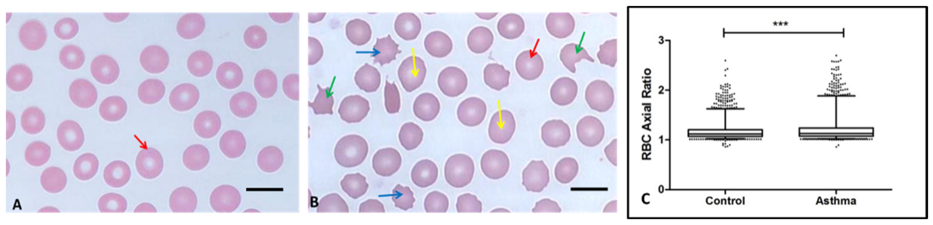
3.3. Light Microscopy and Axial Ratio

Morphological analysis of the erythrocytes by light microscopy showed round and disc-shaped cells, which is consistent with normal discocyte morphology. Most erythrocytes in the blood of asthma patients displayed normal morphology, but they showed increased aggregation. In two of the patients, a variety of abnormally shaped erythrocytes (poikilocytes) were noticed, including echinocytes, acanthocytes, and ovalocytes.
The axial ratios of erythrocytes in both the control and patient groups were measured and the mean axial ratio for asthma patients was higher than that of controls (1.2 ± 0.22 nanometer (nm) versus 1.165 ± 0.16 nm, p < 0.0001). Figure 1 clearly shows much higher axial ratios for asthma patients, with distribution, outliers, and density in data points indicating altered erythrocyte morphology. This indicated that the patient group had an increased number of deformed erythrocytes.

3.4. Atomic Force Microscopy on Erythrocyte Membrane Elasticity

The Young’s modulus values of erythrocytes, generated by AFM, were higher in erythrocytes from asthma patients than those obtained from controls (p-value < 0.001) (Table 3), indicating a reduction in membrane elasticity or deformability of the cells.

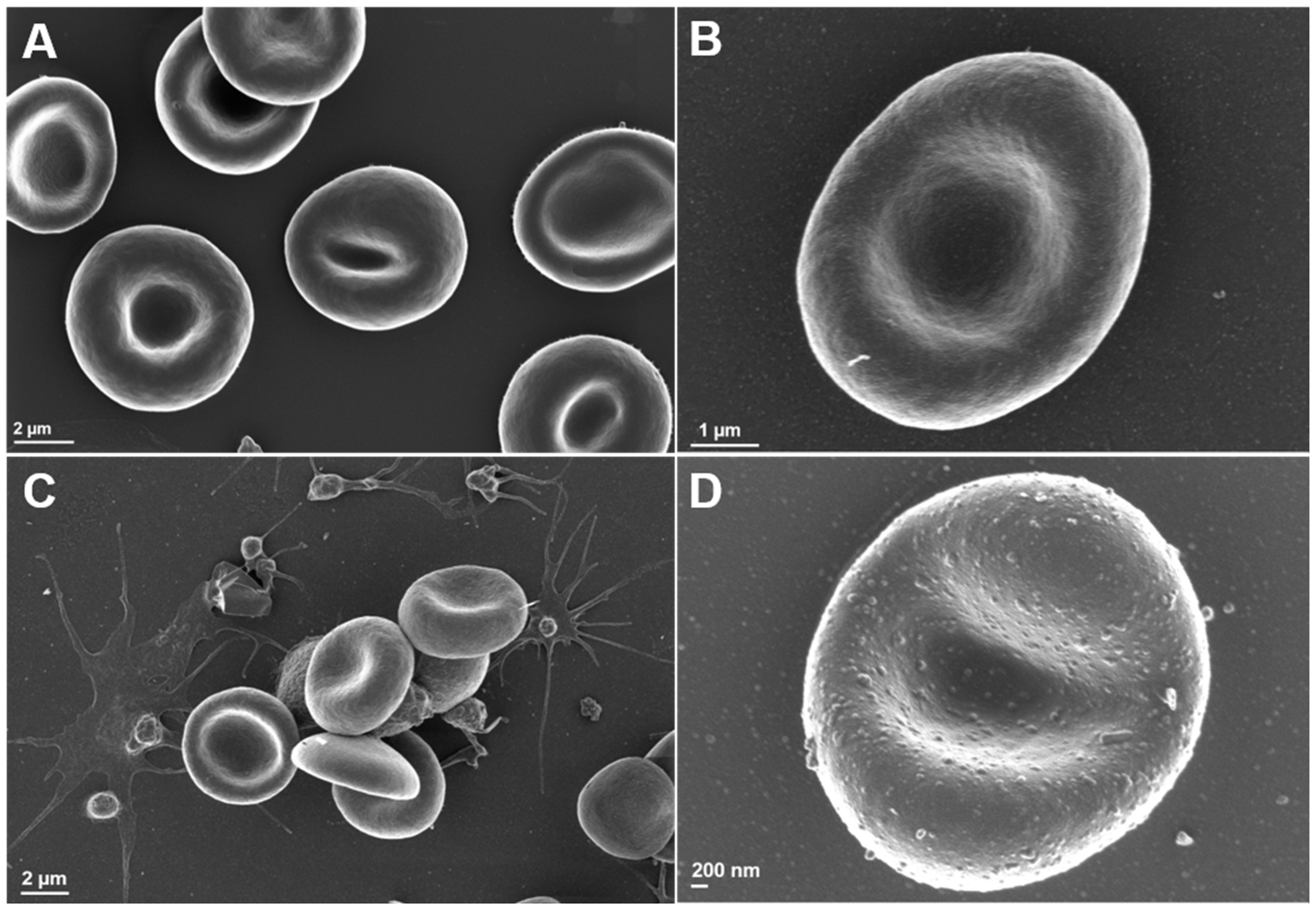
3.5. Scanning Electron Microscopy Analysis of Erythrocyte Ultrastructure

Erythrocytes in the control group showed normal morphology with a biconcave disc shape, as shown in Figure 2A. The erythrocyte membrane was also studied, appearing smooth and granular, as depicted in Figure 2A. Representative SEM images were obtained to illustrate the erythrocytes of the patients suffering from asthma. Erythrocytes from asthma patients showed shape changes similar to those shown in images obtained by light microscopy (Figure 2C,D).

4. Discussion

Erythrocyte morphology plays an integral role in haemostasis and thrombosis. Haemorheology is the study of the flow of blood in a blood vessel, which is mainly focused on the dynamics of erythrocytes in the vascular system [35]. Erythrocytes have the capacity to alter their shape due to a flexible membrane that allows them to enter capillaries with a very small diameter [35]. The biodynamics of erythrocytes, especially their deformation and orientation, have an impact on blood fluidity and subsequently on blood flow influencing tissue perfusion [25]. The mechanical properties of the cytoskeleton and membrane lipid bilayer are both vital to deformation in erythrocytes [36]. In several disease processes, blood flow may be severely impaired due to alterations in rheology by genetic factors, parasites, or by changes in the microenvironment [25]. The function of erythrocytes depends on their shape and biomechanical characteristics and their altered rheology can affect haemostasis.
The morphological profile of erythrocytes of asthma patients identified via imaging studies using LM and SEM showed some shape variations, from discocytes to poikilocytes. Poikilocytes are abnormally shaped erythrocytes found in blood. SEM was employed to determine whether the shape changes were also visible under higher magnification. Micrographs from the SEM showing erythrocytes supported the findings obtained from LM. The presence of defective and aggregated erythrocytes in the blood of patients with asthma can reduce the oxygen content in the blood, leading to lethargy and decreased tissue perfusion. The morphological changes detected in this study could be a combined effect of chronic inflammation and the drugs used for treatment of asthma. Increased inflammatory conditions cause both loss of membrane asymmetry via phosphatidylserine (PS) externalisation and the release of microparticles in erythrocytes [36]. Microparticles also contribute to the potential of cells to agglutinate. Increased levels of erythrocyte microparticles are associated with decreased clotting time due to procoagulant proteins [37], resulting in hypercoagulability in asthma (unpublished data). The effects of drugs used for asthma treatment on erythrocyte morphology need to be investigated.
The increased axial ratios of erythrocytes in asthma are indicative of structural changes also visualised in the ultrastructural study. The normal discocytic shape of erythrocytes is transformed into the echinocytic shape along with increased aggregation of cells [38]. Hence, the morphopathology of erythrocytes may be attributed to cell membrane abnormalities. It has been reported that the formation of echinocytes or acanthocytes can alter the membrane bilayer leaflet, which can be induced by lysophopholipids, fatty acids, or diverse chemical agents [39,40].
The results obtained from the study of erythrocytes show a clear relationship between elasticity and erythrocyte morphology. The elasticity or deformability of erythrocytes can also affect the viscoelastic properties of blood clots [41]. Research shows that normal-shaped erythrocytes are more elastic than poikilocytes [42]. In recent literature, AFM, which produces a Young’s modulus, is a validated methodology used to support changes observed in SEM images [22,43,44,45]. Statistical analysis of AFM data showed that erythrocytes in asthmatics possessed significantly reduced membrane elasticity relative to that of erythrocytes from healthy controls. Since Young’s modulus is inversely proportional to the elasticity of erythrocytes [46], the higher Young’s modulus values obtained for erythrocytes in asthma patients indicate a decrease in membrane elasticity of the cells. This decrease in membrane elasticity may be the cause of the shape changes observed with LM and SEM. Reduced cellular life span is associated with decreased erythrocyte membrane elasticity [46] because a decrease in elasticity would reduce the ability of the red blood cell to return to its original shape once it has been deformed.
The change in the biophysical profile of erythrocytes detected in this study may be ascribed to the modulation of the cell membrane due to the presence of oxidative damage or inflammatory conditions or it may be due to the effects of drugs that are used in asthma treatment, which requires further research.
Although the current study was completed before the SARS-CoV-2 pandemic, recent literature shows that in addition to extrinsic and intrinsic factors such as inflammation, stress, anxiety, and fear have the potential to trigger asthma attacks. It was recommended that patient compliance to medication should continue regardless of infection status [47]. A systematic and meta-analysis review also indicated that asthma was not associated with higher COVID-19 severity or worse prognosis [48,49,50]. The effect of SARS-CoV-2 on erythrocytes in patients with atopic asthma is also unknown.

5. Conclusions

Although the visible structural changes in erythrocytes in asthma seem minor, the structural differences were observed when measuring the mechanical and morphological properties, suggesting that even small structural differences can have a major influence on pathophysiology. Furthermore, this study demonstrated that erythrocyte cell stiffness, measured quantitatively by AFM, was altered in asthma. Since the elasticity (deformability) of erythrocytes influences their biomechanical properties, these results also suggest that the increased stiffness due to decreased elasticity found in asthmatic erythrocytes might influence the viscosity and flow dynamics of blood. Of note, morphologic pathology affects the deformability of erythrocytes and can also influence the elastic properties of the thrombus by modulating fibrinolytic properties in asthma. Therefore, this finding may be taken into consideration for treatment strategies and disease management, especially in patients with co-morbidities. The exact effect of altered erythrocytes in asthma pathology needs to be further elucidated.

6. Study Limitations

As a pilot study, only 30 controls and 30 atopic asthma patients were recruited. The recruitment was conducted on a convenience basis with no discrimination between race or ethnicity. Although it is considered a mixed population, the samples are a true representation of the diverse South African population. The majority of the asthmatic study group was on treatment for asthma and none of the control group was taking those medications that might contribute to the changes observed. The authors acknowledge that it would be ideal to exclude medication; however, due to disease management and ethical implications, it is highly unlikely that this would be possible. Thus, an investigation could consider a younger cohort, such as children who first present with symptoms of asthma and are newly diagnosed. In addition, measurements regarding the impact of altered morphology/deformability to lysis parameters (e.g., haemolysis, mechanical fragility) would provide more robust evidence to confirm the morphological and deformability changes observed in this study.

Author Contributions

S.A.: Completed the experiments and wrote the first draft of the manuscript. M.J.v.R.: Co-supervision of the project, editing of the manuscript. J.B.: Microscopy imaging, data analysis, and manuscript writing. C.G. is a clinician who provided clinical insight and assisted with phlebotomy. A.P.: Co-supervision of the project, laboratory assay supervision, statistical analysis, interpretation of data, and manuscript editing. All authors have read and agreed to the published version of the manuscript.

Funding

No funding was obtained specifically for this project.

Institutional Review Board Statement

Not applicable.

Informed Consent Statement

Not applicable.

Data Availability Statement

Availability of data and material; Data and material are available upon request.

Acknowledgments

Sthembikile Mbotwe is acknowledged for technical assistance.

Conflicts of Interest

The authors declare no conflict of interest.

Ethics Approval and Consent to Participate

Ethical approval and consent was obtained as outlined in Section 2.1.

Consent for Publication

All authors provided consent for publication.

References

  1. Lundbäck, B.; Backman, H.; Lötvall, J.; Rönmark, E. Is asthma prevalence still increasing? Expert Rev. Respir. Med. 2016, 10, 39–51. [Google Scholar] [CrossRef] [PubMed]
  2. Diseases, G.B.D.; Injuries, C. Global burden of 369 diseases and injuries in 204 countries and territories, 1990-2019: A systematic analysis for the Global Burden of Disease Study 2019. Lancet 2020, 396, 1204–1222. [Google Scholar] [CrossRef]
  3. Bruno, F.; Spaziano, G.; Liparulo, A.; Roviezzo, F.; Nabavi, S.M.; Sureda, A.; Filosa, R.; D’Agostino, B. Recent advances in the search for novel 5-lipoxygenase inhibitors for the treatment of asthma. Eur. J. Med. Chem. 2017, 114, 70–77. [Google Scholar] [CrossRef]
  4. Mwanga, H.H.; Baatjies, R.; Singh, T.; Jeebhay, M.F. Work-related allergy and asthma associated with cleaning agents in health workers in Southern African tertiary hospitals. Am. J. Ind. Med. 2022, 65, 382–395. [Google Scholar] [CrossRef]
  5. Saglani, S.; Lloyd, C.M. Novel concepts in airway inflammation and remodelling in asthma. Eur. Respir. J. 2015, 46, 1796–1804. [Google Scholar] [CrossRef] [Green Version]
  6. Bergeron, C.; Tulic, M.K.; Hamid, Q. Airway remodelling in asthma: From benchside to clinical practice. Can. Respir. J. 2010, 17, e85–e93. [Google Scholar] [CrossRef] [PubMed] [Green Version]
  7. Vercelli, D. Gene-environment interactions in asthma and allergy: The end of the beginning? Curr. Opin. Allergy Clin. Immunol. 2010, 10, 145–148. [Google Scholar] [CrossRef] [Green Version]
  8. Skadhauge, L.R.; Christensen, K.; Kyvik, K.O.; Sigsgaard, T. Genetic and environmental influence on asthma: A population-based study of 11,688 Danish twin pairs. Eur. Respir. J. 1999, 13, 8–14. [Google Scholar] [CrossRef] [PubMed]
  9. Koeppen-Schomerus, G.; Stevenson, J.; Plomin, R. Genes and environment in asthma: A study of 4 year old twins. Arch. Dis. Child. 2001, 85, 398–400. [Google Scholar] [CrossRef] [PubMed] [Green Version]
  10. Ober, C.; Yao, T.C. The genetics of asthma and allergic disease: A 21st century perspective. Immunol. Rev. 2011, 242, 10–30. [Google Scholar] [CrossRef]
  11. Feleszko, W.; Ruszczynski, M.; Jaworska, J.; Strzelak, A.; Zalewski, B.M.; Kulus, M. Environmental tobacco smoke exposure and risk of allergic sensitisation in children: A systematic review and meta-analysis. Arch. Dis. Child. 2014, 99, 985–992. [Google Scholar] [CrossRef]
  12. Hallit, S.; Bou Assy, T.; Hallit, R.; Salameh, P. Allergic Diseases, Smoking and Environmental Exposure Among University Students in Lebanon. J. Asthma Off. J. Assoc. Care Asthma 2017, 55, 35–42. [Google Scholar] [CrossRef]
  13. Nelson, H.S. The importance of allergens in the development of asthma and the persistence of symptoms. Dis. Mon. DM 2001, 47, 5–15. [Google Scholar] [CrossRef] [PubMed]
  14. Kelly, F.J.; Mudway, I.S.; Fussell, J.C. Air Pollution and Asthma: Critical Targets for Effective Action. Pulm. Ther. 2021, 7, 9–24. [Google Scholar] [CrossRef] [PubMed]
  15. Regateiro, F.S.; Botelho Alves, P.; Moura, A.L.; Azevedo, J.P.; Regateiro, D.T. The diverse roles of T cell subsets in asthma. Eur. Ann. Allergy Clin. Immunol. 2021, 53, 201–208. [Google Scholar] [CrossRef] [PubMed]
  16. Han, X.; Krempski, J.W.; Nadeau, K. Advances and novel developments in mechanisms of allergic inflammation. Allergy 2020, 75, 3100–3111. [Google Scholar] [CrossRef]
  17. Ahmad, S.; Azid, N.A.; Boer, J.C.; Lim, J.; Chen, X.; Plebanski, M.; Mohamud, R. The key role of TNF-TNFR2 interactions in the modulation of allergic inflammation: A review. Front. Immunol. 2018, 9, 2572. [Google Scholar] [CrossRef] [Green Version]
  18. Moldoveanu, B.; Otmishi, P.; Jani, P.; Walker, J.; Sarmiento, X.; Guardiola, J.; Saad, M.; Yu, J. Inflammatory mechanisms in the lung. J. Inflamm. Res. 2009, 2, 1–11. [Google Scholar] [PubMed]
  19. Bester, J.; Pretorius, E. Effects of IL-1β, IL-6 and IL-8 on Erythrocytes, Platelets and Clot Viscoelasticity. Sci. Rep. 2016, 6, 32188. [Google Scholar] [CrossRef] [Green Version]
  20. Page, M.J.; Bester, J.; Pretorius, E. The inflammatory effects of TNF-α and complement component 3 on coagulation. Sci. Rep. 2018, 8, 1812. [Google Scholar] [CrossRef] [Green Version]
  21. Buys, A.V.; Van Rooy, M.J.; Soma, P.; Van Papendorp, D.; Lipinski, B.; Pretorius, E. Changes in red blood cell membrane structure in type 2 diabetes: A scanning electron and atomic force microscopy study. Cardiovasc. Diabetol. 2013, 12, 25. [Google Scholar] [CrossRef] [PubMed] [Green Version]
  22. Jeongho, K.; HoYoon, L.; Sehyun, S. Advances in the measurement of red blood cell deformability: A brief review. J. Cell. Biotechnol. 2015, 1, 63–79. [Google Scholar]
  23. AlMomani, T.D.; Vigmostad, S.C.; Chivukula, V.K.; Al-zube, L.; Smadi, O.; BaniHani, S. Red blood cell flow in the cardiovascular system: A fluid dynamics perspective. Crit. Rev. Biomed. Eng. 2012, 40, 427–440. [Google Scholar] [CrossRef]
  24. Pretorius, E.; Kell, D.B. Diagnostic morphology: Biophysical indicators for iron-driven inflammatory diseases. Integr. Biol. Quant. Biosci. Nano Macro 2014, 6, 486–510. [Google Scholar] [CrossRef] [PubMed] [Green Version]
  25. Dupire, J.; Socol, M.; Viallat, A. Full dynamics of a red blood cell in shear flow. Proc. Natl. Acad. Sci. USA 2012, 109, 20808–20813. [Google Scholar] [CrossRef] [Green Version]
  26. Zuk, A.; Targosz-Korecka, M.; Szymonski, M. Effect of selected drugs used in asthma treatment on morphology and elastic properties of red blood cells. Int. J. Nanomed. 2011, 6, 249–257. [Google Scholar] [CrossRef] [Green Version]
  27. Mohandas, N.; Chasis, J.A. Red blood cell deformability, membrane material properties and shape: Regulation by transmembrane, skeletal and cytosolic proteins and lipids. Semin. Hematol. 1993, 30, 171–192. [Google Scholar]
  28. Delaunay, J. The molecular basis of hereditary red cell membrane disorders. Blood Rev. 2007, 21, 1–20. [Google Scholar] [CrossRef]
  29. Moutzouri, A.G.; Skoutelis, A.T.; Gogos, C.A.; Missirlis, Y.F.; Athanassiou, G.M. Red blood cell deformability in patients with sepsis: A marker for prognosis and monitoring of severity. Clin. Hemorheol. Microcirc. 2007, 36, 291–299. [Google Scholar] [PubMed]
  30. Gyawali, P.; Richards, R.S.; Bwititi, P.T.; Nwose, E.U. Association of abnormal erythrocyte morphology with oxidative stress and inflammation in metabolic syndrome. Blood Cells Mol. Dis. 2015, 54, 360–363. [Google Scholar] [CrossRef]
  31. Karbiner, M.S.; Sierra, L.; Minahk, C.; Fonio, M.C.; Bruno, M.P.; Jerez, S. The role of oxidative stress in alterations of hematological parameters and inflammatory markers induced by early hypercholesterolemia. Life Sci. 2013, 93, 503–508. [Google Scholar] [CrossRef]
  32. Telen, M.J. Red blood cell surface adhesion molecules: Their possible roles in normal human physiology and disease. Semin. Hematol. 2000, 37, 130–142. [Google Scholar] [CrossRef]
  33. Lopes de Almeida, J.P.; Oliveira, S.; Saldanha, C. Erythrocyte as a biological sensor. Clin. Hemorheol. Microcirc. 2012, 51, 1–20. [Google Scholar] [CrossRef] [PubMed]
  34. Ciasca, G.; Papi, M.; Di Claudio, S.; Chiarpotto, M.; Palmieri, V.; Maulucci, G.; Nocca, G.; Rossi, C.; De Spirito, M. Mapping viscoelastic properties of healthy and pathological red blood cells at the nanoscale level. Nanoscale 2015, 7, 17030–17037. [Google Scholar] [CrossRef] [PubMed]
  35. Cho, Y.I.; Cho, D.J. Hemorheology and microvascular disorders. Korean Circ. J. 2011, 41, 287–295. [Google Scholar] [CrossRef] [Green Version]
  36. Gillespie, A.H.; Doctor, A. Red Blood Cell Contribution to Hemostasis. Front. Pediatr. 2021, 9, 629824. [Google Scholar] [CrossRef]
  37. Litvinov, R.I.; Weisel, J.W. Role of red blood cells in haemostasis and thrombosis. ISBT Sci. Ser. 2017, 12, 176–183. [Google Scholar] [CrossRef] [Green Version]
  38. Chung, S.M.; Bae, O.N.; Lim, K.M.; Noh, J.Y.; Lee, M.Y.; Jung, Y.S.; Chung, J.H. Lysophosphatidic acid induces thrombogenic activity through phosphatidylserine exposure and procoagulant microvesicle generation in human erythrocytes. Arterioscler. Thromb. Vasc. Biol. 2007, 27, 414–421. [Google Scholar] [CrossRef] [PubMed] [Green Version]
  39. Lim, H.W.G.; Wortis, M.; Mukhopadhyay, R. Stomatocyte-discocyte-echinocyte sequence of the human red blood cell: Evidence for the bilayer- couple hypothesis from membrane mechanics. Proc. Natl. Acad. Sci. USA 2002, 99, 16766–16769. [Google Scholar] [CrossRef] [Green Version]
  40. Pretorius, E.; Olumuyiwa-Akeredolu, O.O.; Mbotwe, S.; Bester, J. Erythrocytes and their role as health indicator: Using structure in a patient-orientated precision medicine approach. Blood Rev. 2016, 30, 263–274. [Google Scholar] [CrossRef]
  41. Gersh, K.C.; Nagaswami, C.; Weisel, J.W. Fibrin network structure and clot mechanical properties are altered by incorporation of erythrocytes. Thromb. Haemost. 2009, 102, 1169–1175. [Google Scholar] [CrossRef] [PubMed] [Green Version]
  42. Park, Y.; Best, C.A.; Badizadegan, K.; Dasari, R.R.; Feld, M.S.; Kuriabova, T.; Henle, M.L.; Levine, A.J.; Popescu, G. Measurement of red blood cell mechanics during morphological changes. Proc. Natl. Acad. Sci. USA 2010, 107, 6731–6736. [Google Scholar] [CrossRef] [PubMed] [Green Version]
  43. Tomaiuolo, G. Biomechanical properties of red blood cells in health and disease towards microfluidics. Biomicrofluidics 2014, 8, 051501. [Google Scholar] [CrossRef] [Green Version]
  44. Baier, D.M.T.; Mohr, T.; Windberger, U. Red Blood Cell Stiffness and Adhesion Are Species-Specific Properties Strongly Affected by Temperature and Medium Changes in Single Cell Force Spectroscopy. Molecules 2021, 26, 2771. [Google Scholar] [CrossRef] [PubMed]
  45. Diez-Silva, M.; Dao, M.; Han, J.; Lim, C.T.; Suresh, S. Shape and Biomechanical Characteristics of Human Red Blood Cells in Health and Disease. MRS Bull. 2010, 35, 382–388. [Google Scholar] [CrossRef] [PubMed]
  46. Olumuyiwa-Akeredolu, O.O.; Soma, P.; Buys, A.V.; Debusho, L.K.; Pretorius, E. Characterizing pathology in erythrocytes using morphological and biophysical membrane properties: Relation to impaired hemorheology and cardiovascular function in rheumatoid arthritis. Biochim. Biophys. Acta 2017, 1859, 2381–2391. [Google Scholar] [CrossRef]
  47. Benfante, A.; Scichilone, N. Prioritizing care for severe asthma during SARS-CoV-2 pandemic. Pulmonology 2021, 27, 189–190. [Google Scholar] [CrossRef]
  48. Liu, S.; Cao, Y.; Du, T.; Zhi, Y. Prevalence of Comorbid Asthma and Related Outcomes in COVID-19: A Systematic Review and Meta-Analysis. J. Allergy Clin. Immunol. Pract. 2021, 9, 693–701. [Google Scholar] [CrossRef]
  49. Kalemci, S.; Sarihan, A.; Zeybek, A. Does asthma affect outcomes of patients with COVID-19 infections? J. Allergy Clin. Immunol. Pract. 2021, 9, 594. [Google Scholar] [CrossRef]
  50. Heffler, E.; Detoraki, A.; Contoli, M.; Papi, A.; Paoletti, G.; Malipiero, G.; Brussino, L.; Crimi, C.; Morrone, D.; Padovani, M.; et al. COVID-19 in Severe Asthma Network in Italy (SANI) patients: Clinical features, impact of comorbidities and treatments. Allergy 2021, 76, 887–892. [Google Scholar] [CrossRef]
Figure 1. Light microscopy images of erythrocytes in (A) healthy controls and (B) experimental group (patients with asthma). Red arrow: normal discoidal cell; Blue arrow: echinocyte; Green arrow: acanthocyte; Yellow arrow: ovalocyte. Magnification: ×100, scale bar: 10 µm. (C) A comparison of erythrocyte axial ratios between controls and asthma patients. Data is represented as a dot plot with the mean and standard error of the mean. *** p < 0.0001, Mann-Whitney U Test.
Hemato 04 00009 g001
Figure 2. Scanning electron micrographs representing erythrocytes of healthy controls and asthma patients. Micrograph (A) is a low magnification of erythrocytes with a normal biconcave shape with limited interaction and no visible platelet activity. Micrograph (B) is a high magnification of a single erythrocyte from the control group, showing a biconcave shape with a slightly granular membrane. Micrograph (C) is a low magnification that shows the erythrocyte aggregation and platelet activity of asthma patients. Micrograph (D) is a higher magnification of a single erythrocyte that shows a slightly changed shape with a much more granular membrane.
Hemato 04 00009 g002
Table 1. Demographic and medication usage of study participants.
ControlAsthma
Age [Mean (range)]53 (17–55)45 (19–55) ns
Gender (M/F)19/119/21
Medication (n)
Corticosteroids Immunosuppressant-28
Bronchodilator, Class I B2 adrenergic R antagonist-19
Bronchodilator, Class II B2 adrenergic R antagonist-22
Leukotriene receptor antagonist-14
Anti-histamine-15
Competitive nonselective phosphodiesterase inhibitor-9
ns: where p = 0.2365 using the Kolmogorov-Smirnov test to compare the ages of controls and asthma subjects.
Table 2. Whole blood analysis of controls and asthma patients.
ParameterNormal RangeControl (n = 30)Asthma (n = 30)
MeanIQR RangeMeanIQR Rangep Value
White Blood Cells3.92–9.886.465.99–7.097.145.41–8.38ns
Lymphocytes * 1091–42.301.93–2.572.041.42–2.79ns
Monocytes * 1090.18–10.360.24–0.420.690.29–0.960.0066
Granulocytes * 1092–7.53.813.52–4.014.432.92–5.64ns
Erythrocyte * 10124.7–6.14.724.42–5.025.724.79–6.410.0004
Haemoglobin (g/dL)13.5–1814.2013.2–14.916.6713.6–20.10.0057
Haematocrit (%)38.8–5041.7538.5–44.948.6840.6–57.00.0049
Platelets * 109150–450237193–265192107–278ns
* Level of significance: p < 0.05, Mann-Whitney U test. Where ns: not significantly different.
Table 3. Summary of erythrocyte Young’s Modulus statistics in asthma and control subjects.
ControlAsthma
Mean Young’s Modulus46,71150,760 **
Standard Error749896
Median34,74532,749
Mode13,0822689
Standard Deviation39,21156,216
Sample Variance1,537,498,6203,160,222,538
** p = 0.001, Mann-Whitney U Test.
Disclaimer/Publisher’s Note: The statements, opinions and data contained in all publications are solely those of the individual author(s) and contributor(s) and not of MDPI and/or the editor(s). MDPI and/or the editor(s) disclaim responsibility for any injury to people or property resulting from any ideas, methods, instructions or products referred to in the content.

