Review

Merkel Cell Carcinoma in Kidney Transplant Recipients

1
Renal Research Laboratory, Kolling Institute of Medical Research, Royal North Shore Hospital and The University of Sydney, St. Leonards, Sydney, NSW 2065, Australia
2
Dermatology Clinical and Translational Medicine Laboratory, Kolling Institute of Medical Research, Royal North Shore Hospital and The University of Sydney, St. Leonards, Sydney, NSW 2065, Australia
3
Department of Renal Medicine, Northern Care Alliance NHS Foundation Trust, Salford M6 8HD, UK
*
Author to whom correspondence should be addressed.
Dermato 2023, 3(1), 25-50; https://doi.org/10.3390/dermato3010003
Submission received: 6 December 2022 / Revised: 28 December 2022 / Accepted: 19 January 2023 / Published: 30 January 2023
(This article belongs to the Special Issue Feature Review Papers in Dermato)

Abstract

:
Merkel cell carcinoma (MCC) is an uncommon form of skin neoplasm with poor histological differentiation and an aggressive disease process, leading to high recurrence and mortality. There are multiple risk factors in which being in an immunocompromised state is a significant factor, and the discovery of Merkel cell polyomavirus (MCPyV) since 2008 has strengthened causal associations between MCC and immunosuppression. Individuals who have undergone kidney transplantation are therefore more susceptible to having MCC, secondary to post-transplant immunosuppression which plays a vital role in reducing the risk of transplant kidney rejection. Over recent years a rise in the incidence of MCC following kidney transplantation is noted, with increased reporting of such cases. Whilst localized MCC is observed, MCC metastasis to the lymphatic system, brain, bone, liver, lung, and heart has been previously observed in patients with transplanted kidneys. Kidney metastasis is less common and has been only reported in recent years with greater frequency. The management of aggressive, metastatic MCC has historically been palliative, and prognosis is poor. Recently, the use of immune checkpoint inhibitors for metastatic MCC in multi-center phase II clinical trials have shown promising survival outcomes and have been approved for use in countries such as the United States as a first-line treatment. In this review we will explore the potential pathophysiological processes of MCC manifesting post-kidney transplantation. We will then evaluate the epidemiology of MCC within the context of kidney transplantation, before discussing the various clinical presentations, diagnostic measures, surveillance strategies, and current treatment options as well as future directions to best manage MCC in kidney transplant recipients.

1. Introduction

Merkel Cell Carcinoma (MCC) is a neuroendocrine cancer of the skin [1]. Although this diagnosis is relatively rare compared to other forms of skin neoplasm, MCC is the second most frequent cause of death from skin malignancy after melanoma [2]. Found mostly in sun-exposed areas of the skin such as the head and neck, MCC originates from nerve-associated Merkel cells which lie in the basal epidermal layer [3,4]. Certain demographic characteristics such as older age, being of Caucasian ethnicity, extensive exposure to ultraviolet (UV) radiation and/or immunosuppression are deemed significant risk factors of MCC development [3,4,5,6,7].
Historically, a poor prognosis is expected because of aggressive tumor progression, poor histological differentiation and high recurrence rates [8,9]. Outcomes data over the years have demonstrated the clinical course to be variable, even if these factors were present [3,10,11,12,13]. The presence or absence of metastasis is often considered the most important prognostic marker in MCC [14]. Regional and distant metastasis to various sites—from the lymphatic system, brain, bone, liver, lung, heart and more recently, the kidneys—has been reported in patients with metastatic MCC [15,16,17,18,19,20].
Patients with organ transplants are generally at higher risk of developing MCC and other forms of skin cancers compared to the non-transplanted population, with the risk estimated to be between 66 and 182 fold [21]. In particular, a more aggressive clinical course of MCC is observed for kidney transplant recipients where MCC diagnosis at an earlier age is expected [22,23]. Such patients usually present with localized disease in the form of a red or violaceous nodular lesion in sun-exposed skin before lymphovascular invasion and metastasis.
The aggressive nature of MCC development in kidney transplant recipients is primarily explained by these patients almost universally requiring post-transplant immunosuppression. Systemic immunosuppression displayed the strongest association with poor survival prognosis, irrespective of MCC stage and metastatic status compared to other established risk factors of MCC which occur following kidney transplantation [24]. Previous studies highlighted that MCC-specific mortality in immunosuppressed individuals nearly doubles that of non-immunosuppressed individuals over 3 years follow-up [25]. UV radiation-induced immunosuppression, Human Immunodeficiency Virus (HIV), autoimmune disorders, lymphoproliferative disorders and more importantly specific to this scenario, Merkel cell polyomavirus (MCPyV), are other risk factors of MCC which compound the effects from immunosuppression in kidney transplant recipients [26,27,28]. Conditions of immunosuppression might increase the viral replication activity of MCPyV. This increase could lead, in turn, to an increased risk of MCPyV-driven MCC. In a similar fashion, MCPyV-positive MCC has been reported to arise in rheumatoid arthritis patients under iatrogenic immunosuppressive therapy [29], so the connecting link between MCPyV and MCC may be the anti-viral immune system which is unable to control the oncogenic activity of MCPyV oncoproteins [30].
Though radiotherapy or cytotoxic chemotherapy followed by palliative care if these treatments fail are traditionally mainstays of management, significant developments have been made on the medical treatment options for advanced stage and metastatic MCC, particularly with the advent of immunotherapies for this condition. Positive findings demonstrated in numerous phase 2 multi-center clinical trials have led to the European Association of Dermato-Oncology (EADO) proposing the potential use of medications such as Avelumab, Pembrolizumab or Nivolumab as first-line standard treatment for metastatic MCC [31,32,33,34]. The United States Food and Drug Administration (FDA) approved the use of Avelumab and Pembrolizumab for this purpose [35,36]. Other novel therapeutic options include the use of epigenetic-based therapies such as histone deacetylase (HDAC) inhibitors [37]. Continued investigation is warranted to fully validate the use of medications as well as other treatment options for MCC.
This review article aims to discuss the possible etiologies and pathophysiological pathways of MCC following kidney transplantation. The epidemiological patterns reported amongst kidney transplant recipients with MCC will be elucidated. This article will then evaluate the variations in clinical presentation, diagnostic and surveillance measures undertaken to confirm a post-transplant MCC diagnosis. Following this, current preventative measures and current therapeutic options will be reviewed, as well as the exploration of future avenues in research to optimize treatment of MCC in kidney transplant recipients.

2. Etiology and Pathophysiology of Merkel Cell Carcinoma

MCC was thought to originate from Merkel cell precursors which are likely derived from epidermal or hair follicle stem cells, pre-B/pro-B cells, or dermal fibroblasts, though the cells of origin in MCC remains not fully established at present [38,39,40,41]. Since normal Merkel cells are terminally differentiated and do not undergo cell division, they are deemed unlikely to be the cell of origin for MCC development [42].
The carcinogenesis process of MCCs is primarily linked to two main etiologies—clonal integration of MCPyV and long-term sunlight exposure leading to ultraviolet-mediated DNA damage (Figure 1) [43,44]. MCPyV is a recently identified human polyomavirus that is clonally integrated into the genome of MCC cells, as determined by whole-transcriptome sequencing [45]. In earlier studies, Southern blot patterns of the primary tumor and a metastatic lymph node isolated from the same patient appeared identical, suggesting the MCPyV integration event was clonal and likely occurred in early phases of the tumorigenic process [45]. MCPyV can usually be acquired during childhood and is detected in the skin of most healthy individuals [46,47]. Despite the widespread and lifelong infection with MCPyV in most individuals, very few MCPyV-exposed subjects actually have MCC [48]. Antibodies against MCPyV viral capsid proteins, particularly immunoglobulin G (IgG), are detected in between 60 and 80% of healthy, immunocompetent adults [49,50,51,52]. Maternally derived antibodies might account for the seropositivity in newborn babies and are probably effective in preventing primary infection [46]. When the maternal antibodies are no longer present by around 18 months of age, children are susceptible to de novo infection and are capable to mount antibody responses of their own [46].
MCPyV-specific T-cell responses detected in the serum blood samples of post-transplant patients with MCC are characterized by CD4+ helper cells, which react to a broad range of peptides derived from viral capsid and oncoproteins [53]. The action of IgG antibodies against small T (ST) and large T (LT) antigens of MCC are relatively specific, with this mechanism observed in more than 40% of post-transplant patients with MCC but less than 1% of normal controls [54]. It has been shown that the levels of ST and LT antibodies correlate to tumor mass in MCPyV-positive MCC and will increase in the event of spread or metastatic disease [54]. It should be taken into account that surveillance for MCPyV-positive MCC is not only mediated by humoral immunity and CD4+ T-helper cells, but also by cell-mediated immunity [55]. MCPyV-specific CD8+ T-lymphocytes were found in serum blood samples for over half of MCPyV-positive MCC patients, in which its levels correlate with disease progression and degree of remission following MCC treatment [55]. It is known that MCPyV-positive MCC contain increased numbers of tumor-infiltrating CD8+ and CD3+ lymphocytes, natural killer cells, macrophages and Fox P3+ regulatory T-cells, when compared to MCPyV-negative MCC [56,57]. The tumor-infiltrating CD8+ lymphocytes are associated with a favorable prognosis of MCC [56,58]. Another important feature relating to the immune surveillance of MCC cells is that they are able to employ certain mechanisms to evade tumor surveillance by tumor-infiltrating lymphocytes (TILs) [10,42,59]. The loss of vascular E-selectin expression, an important factor in T-cell entry to the skin, displays significant association with poor intra-tumoral CD8+ infiltration and worsened prognosis of MCC cells [60]. A decreased activity of TILs in MCC signifies the decreased expression of co-stimulatory signal molecules, as well as expression of specific T-cell exhaustion markers [61]. Restriction of T-cell entry into tumor cells and reduction in T-cell function might be considerable and targetable forms of immuno-evasion in MCC [61]. Besides clonal integration, chronic expression of the two MCPyV oncoproteins also contributes significantly to MCC pathophysiology. This probably occurs due to the loss of expression of the MCPyV miRNA that negatively regulates MCPyV LT transcript [62].
Medium- to long-term ultraviolet exposure may result in the manifestation of MCPyV-positive MCC as chronic sunlight exposure leads to local immunosuppression [42,59]. This is explained by the fact that ultraviolet radiation induces the expression of inflammatory mediators and functional alterations in the antigen-presenting dendritic cells, resulting in a cascade of events that modulate immune sensitivity [63]. Nevertheless, the frequency of DNA mutations occurring in ultraviolet-induced MCPyV-negative MCC is significantly higher (between 25 and 90-fold) compared to MCPyV-positive MCC, in similarity with other ultraviolet-induced skin cancers such as melanoma and squamous cell carcinoma [64,65,66,67,68]. This finding further distinguishes the MCPyV-positive and negative subtypes of MCC, according to DNA sequencing studies of MCC samples which rely on sequencing of cancer-specific genes, whole exomes or whole genomes. The MCPyV-negative MCC that is typically characterized by numerous mutations reflecting DNA damage from ultraviolet exposure, and MCPyV-positive MCC containing integrated MCPyV DNA, few somatic mutations and scarce evidence of ultraviolet-induced damage [64]. Amongst MCPyV-negative MCC cells, the mutational patterns frequently reflected faulty repair of pyrimidine dimers induced by UV radiation [66,67]. MCPyV-positive MCC cells usually had low mutation numbers in the range of 0.4 per megabase [42,68].
Within the context of kidney transplantation, iatrogenic immunosuppression is frequently observed due to the medications administered to prevent graft rejection. Whilst details regarding the impact of each individual immunosuppressant medication on MCC development in kidney transplant recipients are not fully known, it is established that calcineurin inhibitors and Azathioprine use significant increase risk of non-melanoma skin cancer including MCC [28,69]. Calcineurin inhibitors such as Cyclosporine and Tacrolimus were shown to display tumorigenic effects through interference with DNA repair and other mutational changes, raising risks of non-melanoma skin cancer by up to 200-fold even in previously immunocompetent individuals [70,71]. Pathophysiological associations between immunosuppressant use and MCPyV-positive and negative MCC disease activity are supported by findings that amongst patients who developed metastatic MCC following kidney transplantation, regression of MCC following withdrawal of immunosuppressants was observed although remission did not persist for more than 12 months in reported cases [72,73].

3. Epidemiology of Merkel Cell Carcinoma in Kidney Transplant Recipients

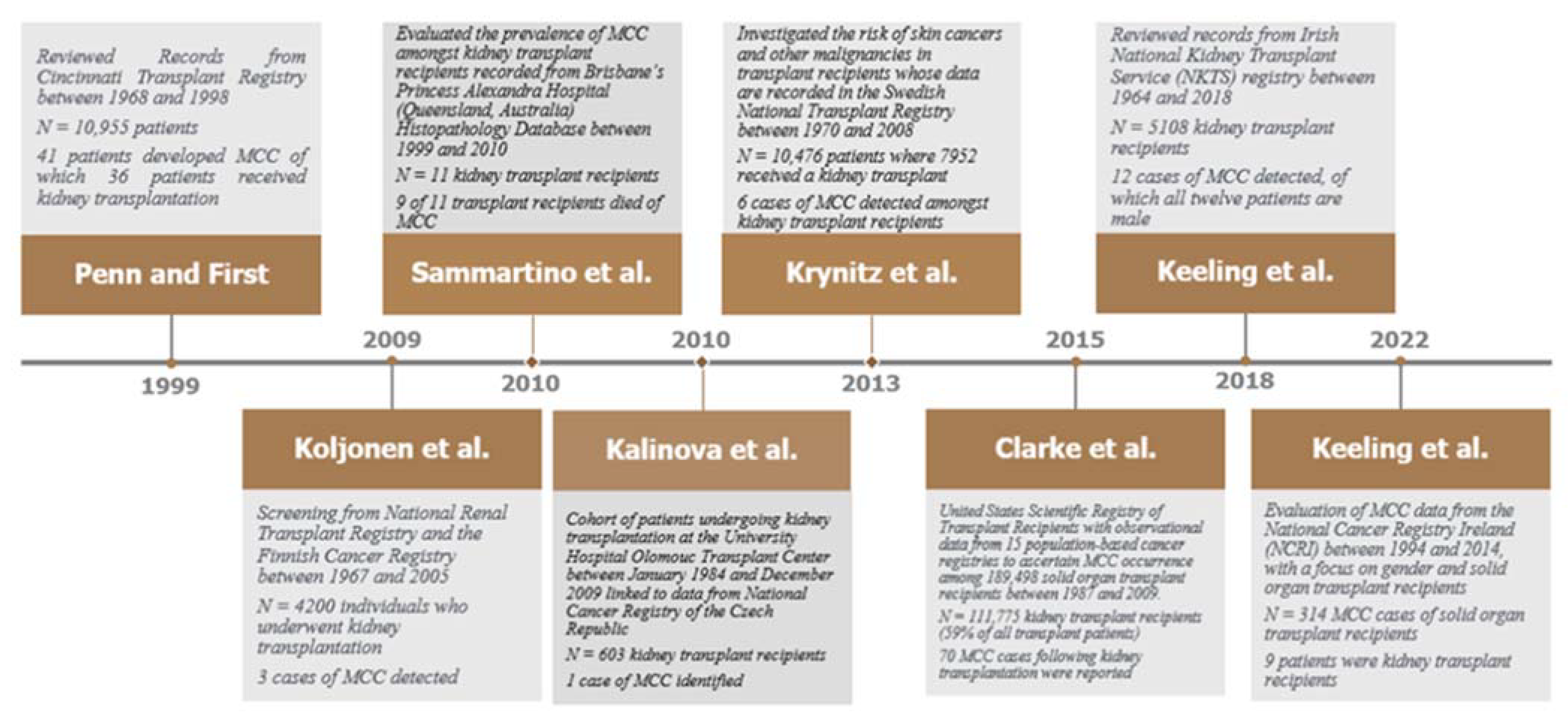
3.1. Methods of Systematic Search for Epidemiological Data

Between the 2000s and 2010s the incidence of MCC has increased almost 2-fold globally, despite being known as a rare condition compared to other forms of skin malignancies [5]. This is most likely explained by a rise in the global aging population, as MCC typically occurs amongst elderly patients age >65 years. Nevertheless, uncertainty as to the global epidemiology of MCC amongst kidney transplant recipients remains, with a paucity of observational data from registry cohorts reporting the incidence and prevalence of MCC within the transplanted population. Following a systematic search encompassing the input of the following search terms: “Merkel Cell Carcinoma” AND “Kidney Transplantation” using PubMed, Web of Science, EMBASE, Google Scholar, and Medline-ProQuest, we note the majority of published articles relating to this topic appeared in the form of isolated case reports or case series (Table 1) [74,75,76,77,78,79,80,81,82,83,84,85,86,87,88,89,90,91,92]. Otherwise, there were seven epidemiological cohort studies published, which recorded specific MCC characteristics in kidney transplant recipients over decades of follow-up (Figure 2) [22,26,93,94,95,96,97,98].

3.2. Epidemiological Cohort Studies

The earliest publication reviewed records of 10,955 patients from the Cincinnati Transplant Tumor Registry between 1968 and 1998. This article authored by Penn and First [22] in 1999 described 41 patients who developed MCC following solid organ transplantation of which 36 patients received a kidney transplant, 1 patient receiving heart and kidney transplantation simultaneously and another patient with simultaneous liver and kidney transplantation. Amongst these 38 patients, the mean age was 53.3 years, and there were 27 male and 11 female patients. Mean time of MCC appearance following transplant surgery was 97 months. The majority of MCC first formed in the head and neck region (16 cases) followed by the upper limbs (11 cases), the trunk (7 cases) and lower limbs (3 cases). In total, 19 patients developed other malignancies during follow-up where in 17 of these cases they were other skin malignancies, i.e., squamous cell carcinoma, basal cell carcinoma (BCC), or melanoma. By the end of 1998, 15 of the 38 patients were still living whilst 23 patients had died after treatments that included wide local excision, radical node dissection, radiotherapy, and chemotherapy. Mean length of follow-up following MCC diagnosis was 15.8 months.
The second epidemiological study came from work by Koljonen and colleagues [93], in which they screened for MCC cases amongst individuals who underwent kidney transplantation between 1967 and 2005, according to data from the National Renal Transplant Registry and the Finnish Cancer Registry. Three cases of MCC were detected among 4200 individuals who underwent kidney transplantation from 1967 to 2005 (expected number 0.05, standardized incidence ratio 66, 95%CI 14–194, p < 0.001). The first patient was a 68-year old man who received kidney transplantation due to chronic autoimmune glomerulonephritis. Receiving cyclosporin A, methylprednisolone for post-transplant immunosuppression, he was diagnosed with left cheek MCC 8 years following transplantation, in which radical excision of the MCC and adjuvant radiotherapy was administered. Unfortunately, neck metastases were found 6 months following radical excision, and the patient died 8 months after initial MCC diagnosis. The second patient was a 66-year old man who also received kidney transplantation as a result of chronic autoimmune glomerulonephritis. Receiving azathioprine and methylprednisolone as part of the post-transplantation immunosuppression, the patient developed MCC at the right earlobe 19 years after his kidney transplantation in which regional lymph node and distant metastasis was identified. Despite receiving radical excision and neck dissection followed by post-operative radio- and chemotherapy, the patient died 6 months after his metastatic MCC diagnosis. The third patient is a 44-year old man with a background history of rheumatoid arthritis, amyloidosis and nephrotic syndrome who received kidney transplantation. His post-transplant immunosuppression regime included azathioprine, cyclosporin A, and methylprednisolone. The patient developed a right cheek MCC 6 years following transplantation, and radical excision (alongside sentinel node biopsy) together with adjuvant radiotherapy was administered. He developed repeated local recurrences of the tumor and eventually died 2 years after his initial MCC diagnosis.
There was an abstract presented in the 2010 Australian and New Zealand Society of Nephrology Conference which evaluated the prevalence of MCC amongst kidney transplant recipients recorded from Brisbane’s Princess Alexandra Hospital (Queensland, Australia) Histopathology Database between 1999 and 2010. Sammartino and colleagues [94] reported 11 kidney transplant recipients who were diagnosed with MCC during this period, where mean age was 72 years. Mean time from kidney transplant to MCC diagnosis was 15 years, and 9 of the 11 kidney transplant recipients eventually died of metastatic MCC in which the median time to death from diagnosis was 8.9 months.
The third reported epidemiological cohort study, by Kalinova and colleagues [95], linked patients who underwent kidney transplantation at the University Hospital Olomouc Transplant Center between 1984 and 2009 to reported data from the National Cancer Registry of the Czech Republic, identifying patients who were diagnosed with skin cancer post-transplant. There was one patient who was diagnosed with MCC, a 59-year old man who had developed a reddish–brown nodule sized approximately 20 × 30 mm on his right buttock four years after kidney transplantation. Despite wide surgical excision of the MCC lesion, chemotherapy and modification of his immunosuppression regime (replacing Cyclosporine with mTOR inhibitor Rapamycin) the patient died due to tumor advancement 7 months following his MCC diagnosis.
The fourth publication was an observational study which investigated the risk of skin cancers and other malignancies in kidney, liver, heart and lung transplant recipients whose data are recorded in the Swedish National Transplant Registry between 1970 and 2008 [21]. Out of 10,476 transplant recipients during this period, 7952 people received a kidney transplant. There were 6 cases of MCC during follow-up, in which the standardized incidence ratio is 52 (95%CI 19–113). The fifth epidemiological study was an observational analysis on the risk factors of MCC following solid organ transplantation. Clarke and colleagues [26] linked the United States Scientific Registry of Transplant Recipients with data from 15 population-based cancer registries to ascertain MCC occurrence among 189,498 solid organ transplant recipients between 1987 and 2009. Risks for MCC following transplantation were compared to that with the general population using standardized incidence ratios, and Poisson regression was used to compare incidence rates according to key patient and transplant characteristics. Kidney transplantation recipients formed the majority of the transplants undertaken during this period (111,775 kidney transplant recipients, 59% of all transplant patients included) and 70 MCC cases following kidney transplantation were reported (13.8 incidence rate per 100,000 years). The authors concluded from this study that overall risk of MCC was increased by 23.8-fold following solid organ transplantation, where adjusted risks were highest amongst older transplant recipients, increased with time since transplantation. These risks varied by organ type in which older patients with kidney transplantation were at higher risk compared to those receiving liver, heart, lung and combined organ transplantations. Furthermore, azathioprine, cyclosporin, and mTOR inhibitors such as sirolimus given for maintenance post-transplant immunosuppression significantly increased MCC risk, and non-Hispanic white recipients on cyclosporine and azathioprine experienced increased MCC risk if they resided in lower latitudes with higher UV light exposure (p = 0.012).
The sixth study, reviewing MCC cases following kidney transplantation between 1964 and 2018 from records in the Irish National Kidney Transplant Service (NKTS) registry, was presented as a letter to the editor manuscript by Keeling and colleagues [96] in 2019. Twelve individuals from 5108 kidney transplant recipients were identified during this period. All were male patients, and the median age was 67 years (range between 49 and 86 years old). The mean time from kidney transplantation to diagnosis of MCC was 15 years, in which 8 of the 12 patients with MCC were diagnosed between 2008 and 2018. Median survival from the time of MCC diagnosis was 14 months amongst the 12 patients, and only 3 of them have survived beyond 5 years of follow-up. The standardized incidence ratio for MCC was 97.0 (95%CI 44.3–184.2).
Another recent publication by Keeling and colleagues [97] examined cases of MCC between 1994 and 2014 from the National Cancer Registry Ireland (NCRI), with a focus on gender and solid organ transplant recipients. Out of 314 MCC cases during that period, 10 were solid organ transplant recipients of which nine people received kidney transplantation. Mean age at diagnosis was 65.1 years (compared to 79.0 years in non-transplanted patients). Amongst the ten patients, the average time from transplantation to the development of MCC was 14.1 years. Seven patients developed MCCs on the head and neck. All 9 patients who received kidney transplantation developed other non-melanoma skin malignancies in addition to MCC. Seven of the 9 patients died from advancing MCC in which the median survival of those who died was 0.14 years.
Ultimately, as Keeling and colleagues have alluded to in their publications, there may be underestimation of the true incidence of MCC amongst kidney transplant recipients across the currently available database studies [96,97]. Considering that an international classification of diseases-10 (ICD-10) code for MCC was only first created in 2009, and cytokeratin-20 (CK-20) staining, a specific diagnostic marker of MCC was only established in 1995, some MCC cases could have been missed from earlier years. Given the relative lack of clarity at present regarding its epidemiology, there remains a need to establish national and international registry databases to record MCC incidence and prevalence amongst kidney transplant recipients going forwards.

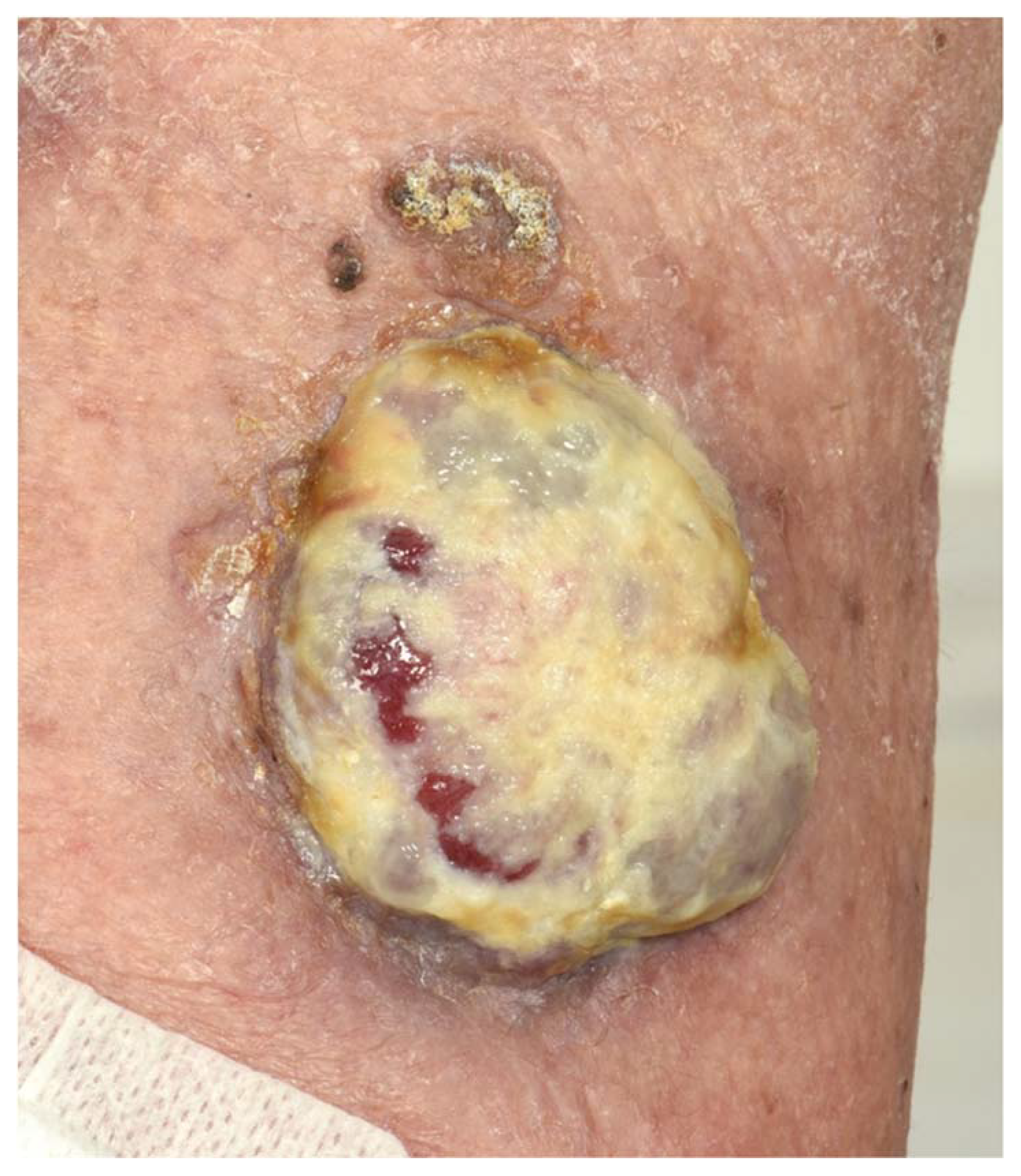
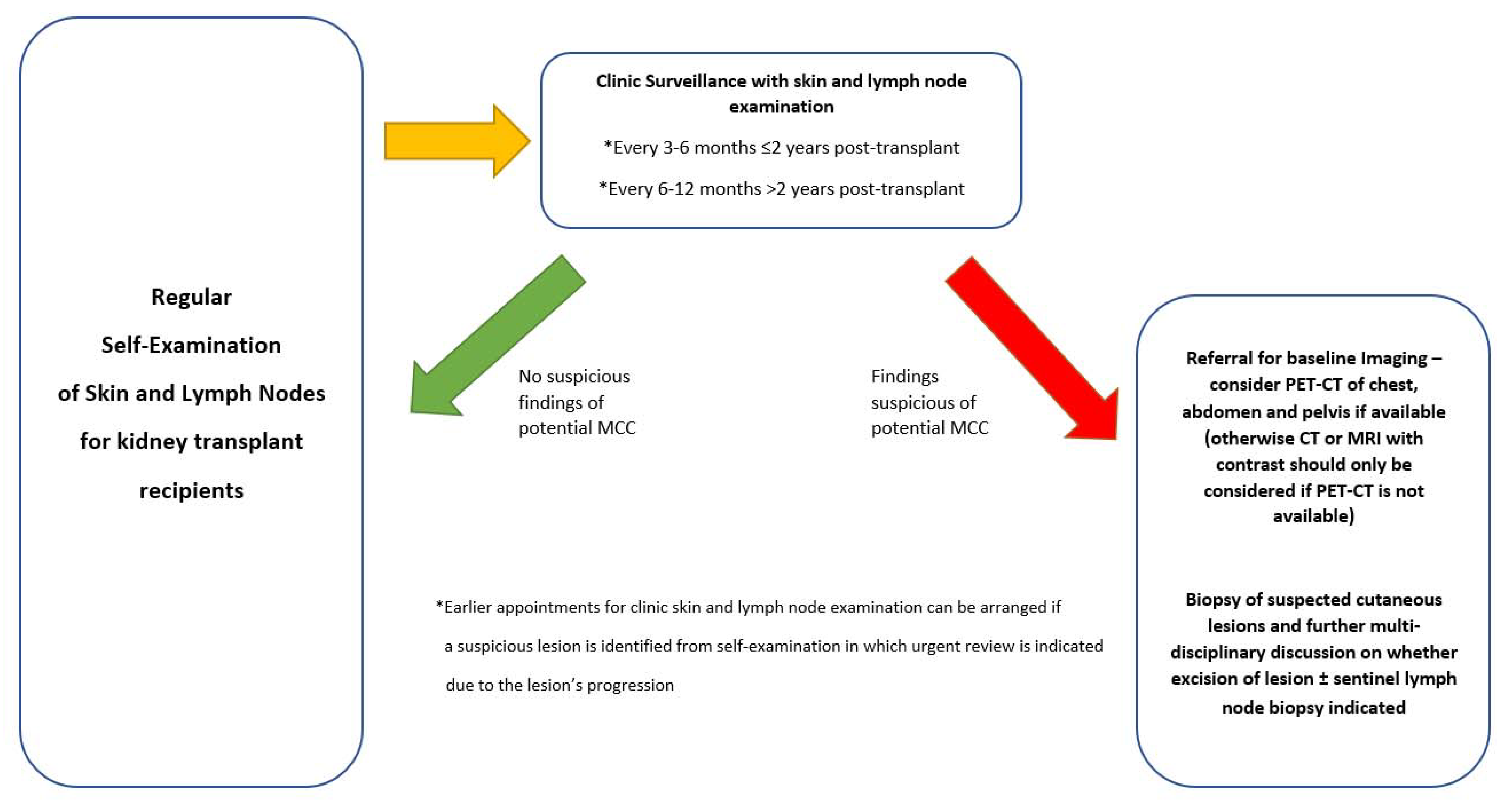
4. Clinical Presentation, Diagnostic Features and Surveillance of Merkel Cell Carcinoma

MCC typically presents itself as a rapidly progressing solitary tumor which lies in cutaneous or subcutaneous tissue [98,99]. It is mainly located around sun-exposed areas of the skin, such as the head and neck regions, but could also appear in the extremities and buttock region though with less frequency (Figure 3) [98,99,100]. It remains unclear whether there are differences in clinical appearances between MCPyV-positive and MCPyV-negative MCC, and they both present as red-to-violet nodular lesions that may be misdiagnosed as benign dermatological disease (i.e., cysts, infected or inflamed skin lesions) or other forms of skin malignancy (i.e., SCC, skin complications of lymphoma or metastatic disease) [23]. Differences in clinical appearance between MCC in transplant recipients and non-transplant patients are also not currently known [23,100]. Classic MCC lesions usually do not present with ulceration, and it is uncommon that multiple lesions stemming from various body sites are elucidated [101].
Given MCC lesions are usually asymptomatic, and progression of its clinical presentation non-specific, formal diagnosis of the condition would be delayed. Structured guidance to ensure the timely identification of MCC lesions have been discussed over the years, in which the development of the AEIOU (Asymptomatic, Expanding rapidly, Immunosuppressed, >50 years of age, UV-exposed) system has been touted as a useful way to consider whether the patient’s clinical presentation and demographic status are of high risk towards a MCC diagnosis, where greater attention should be placed for kidney transplant recipients [23]. Histopathological confirmation is necessary, given the challenges to confirm MCC through clinical means only [42,98,99,100]. It should be acknowledged that a morphological diagnosis may be difficult to obtain, as MCC cells are very sensitive to drying artefacts that can occur during the preparation of the sample especially with small biopsies [42]. Suspected MCC biopsies with phenotypic aberrations would require a comprehensive immunohistochemical expression profile work-up to confirm the diagnosis.
MCC is one of the diagnoses which encompass the group of tumors known as the ‘small, blue round cell’ tumors [102]. It is composed of dermal and/or subcutaneous nodules or sheets of small, monomorphic, round-to-oval cells with a vesicular nucleus and scanty cytoplasm [102]. There are three major classification types of MCCs: small-cell, trabecular and intermediate, though most MCCs present with an overlap between these three types [102]. Neoplastic MCC cells can emerge large in size, particularly after recurrence of disease following radiotherapy, and display a more explicit pleomorphic morphological appearance [98,102]. In these cells, the nucleoli are usually not prominent and multiply within the cell, and cellular necrosis is common [102]. Histopathological features such as large tumor thickness, high mitotic rate, an infiltrative (rather than circumscribed) growth pattern and the presence of lymphovascular invasion have been associated with an increased risk of microscopic nodal metastases and a poor prognostic outlook [102]. A total of 10% of MCC cases would display epidermotropism though purely intra-epidermal tumors are rarely described [103]. Despite this, it is not uncommon to observe intra-lymphatic invasion. High rate of local MCC reoccurrence following initial surgical treatment may be explained by the presence of intra-lymphatic complexes and isolated tumor cells close to the surgical margins [103]. These features should be diligently searched for and accordingly documented during histopathological evaluation.
Previous reports note frequent observations where MCCs are contiguous to or intermingled with other skin malignancies, in particular SCC and Bowen’s disease [104,105]. The pathophysiological association and progression between MCC and SCC are explained that both tumors originate from a common multipotent stem cell, and following this have divergent differentiation of neoplastic cells and eventually the simultaneous growth of two unrelated malignancies [106]. In scenarios where there is a combined MCC and SCC tumor, p53 is usually overexpressed [106].
Due to uncertainties when determining whether a lesion is genuinely MCC through its histopathological features alone, immunohistochemical markers are needed to confirm MCC diagnoses. MCC has a characteristic immunohistochemical profile, in terms of antigens expressed and expression patterns [42,59]. MCC cells conventionally express several type I or type II cytoskeletal keratins, in particular cytokeratin (CK) 20 but also CK8, CK18 and CK19 [59,107,108]. Less than 10% of MCC cases stain negative for CK20 [100]. These cases are usually characterized by a high mutational burden and likely MCPyV-negative MCCs [109]. Neuroendocrine markers such as synaptophysin and others could also be expressed [59,107,108]. A large subset of MCCs typically stains positive for the MCPyV T antigens, consistent with genetic findings [42]. Many MCC cases display positivity to the oncoprotein huntingtin-interacting protein 1 whilst one-third would likely stain positive for tumor protein 63 [110,111,112]. MCCs are usually negative for thyroid transcription factor 1, mammalian achaete-scute homologue 1, vimentin, S100 calcium-binding protein B and CK7 [42,59,98,99,100]. It is acknowledged that whilst the use of immunohistochemical markers is useful to confirm a MCC diagnosis, it is unable to selectively differentiate between MCPyV-positive and MCPyV-negative MCC [42]. Whilst positive staining for MCPyV large T antigen likely suggests MCPyV-positive MCC, negative staining does not necessarily rule it out [42]. Ultimately in most cases, having a clinical presentation and morphological features similar to MCC, positive staining for CK20 and neuroendocrine markers and negative staining for TTF1, CK7 and lymphoid markers are sufficient to confirm the diagnosis. Other MCC diagnostic markers such as AE1/AE3, CAM5.2 and CD46 should also be considered.
Following confirmation of MCC through histopathological evaluation, all positive cases should undertake further imaging to screen for the presence of extra-cutaneous disease [34]. Ultrasonography should be utilized at the first instance to screen the lymph node basin. MCC usually spreads to lymph nodes first [100]. Identification of such spread would indicate sentinel lymph node biopsy to be performed, with this procedure being an important component for staging purposes [13,113]. This should be followed by Positron Emission Tomography-Computed Tomography (PET-CT), which has replaced CT and magnetic resonance imaging (MRI) scanning as the primary imaging option for MCC staging [100]. Previous studies, albeit conducted in a single center setting, have noted changes to the MCC stage classification in 33% of patients and to management in 43% of patients [114]. The most updated MCC staging classification is based on the 2018 American Joint Committee on Cancer (AJCC) staging classification—Stage 0 (in situ); Stage I (localized MCC with primary lesion ≤2 cm); Stage II (localized disease with primary lesion >2 cm); Stage III (lymph node spread); Stage IV (Metastatic disease beyond the local lymph nodes) [115]. Due to improved planning on identifying MCC within the clinical setting, most initial MCC diagnoses occur during Stage I or II [42]. Whilst survival depends on stage at initial diagnosis, it is with optimism that MCC may regress spontaneously, and this is associated with improved prognosis [116]. Local or distant recurrences usually occur in previously treated MCC within the first 3 years following initial diagnosis [42]. Patients whose MCC has not recurred by 3 years are likely to have significantly diminished risks of disease recurrence.
Regular screening for MCC in immunosuppressed patients through the diagnostic measures described in this section, particularly for kidney transplant and other solid organ transplant recipients, has been touted as a potentially important part of post-transplant management (Figure 4) [34]. In addition to regular self-examination, clinic surveillance with skin and lymph node physical examination every 3 to 6 months in the first 2 years post-transplant, and every 6 to 12 months thereafter, is recommended [34,117]. Biopsy of suspicious cutaneous lesions should not be postponed, particularly in high risk patients. Current guidelines suggest referral for imaging when clinically indicated, and more frequent imaging through PET-CT (CT or MRI with contrast should only be considered if PET-CT is not available) is indicated for patients deemed with high risk for MCC [118].
Further work on the design of MCC surveillance programs for the post-transplant cohort is anticipated. There remain scarcely any data detailing the association between specific immunosuppressive treatments and the development of MCC [119]. The use of serum biomarkers as indicators of MCC disease severity and tumor burden to replace the need for regular invasive histopathological evaluation in suspected disease requires greater study. Current conclusions in employing MCPyV anti small T antigen antibodies for this purpose are inconsistent [54,120]. Further validation of this method across different patient cohorts and centers is needed before consideration for implementation within the clinical setting.

5. Prevention and Management Options for Merkel Cell Carcinoma

Encouraging primary preventative measures to reduce risks of MCC as much as possible for kidney transplant recipients would be the ideal scenario, though difficult to achieve. Lowering ultraviolet radiation exposure and reducing immunosuppressive therapy with MCC considerations may be important components of preventative management, but the efficacy of these measures to reduce MCC risks remain debatable [34,42]. Ultraviolet radiation from sunlight and/or artificial light sources has been associated with increased MCC risks [23,121,122]. It may be the most easily preventable risk factor of MCC through avoidance of sunlight by staying indoors, seeking shade when outdoors, and applying ultraviolet protection by wearing hats, clothing, and sunscreens, but the effectiveness of these measures to prevent MCC has been questioned [123]. On the other hand, the lack of ultraviolet radiation exposure may have detrimental effects on vitamin D synthesis, which is particularly significant for those with kidney disease [123,124]. Ultraviolet radiation plays a role in the cutaneous synthesis of vitamin D and the impact of chronic sunscreen use resulting in low serum 25-hydroxyvitamin D levels should be recognized [123]. Optimization of post-transplant immunosuppression in view of MCC risks is challenging, given simultaneous risks of transplant rejection if inadequate immunosuppression is prescribed [119]. Multidisciplinary management to appropriately balance the risks and benefits alongside a personalized immunosuppressive regime for each individual patient is recommended [28,34,119]. The use of statins, with these being immunosuppressive agents, should be cautioned for kidney transplant recipients given statins are linked to increased risks of MCC development in immunosuppressed individuals [28,125].
If feasible, wide excision of the primary MCC tumor is the ideal treatment [34,42]. Given the majority of MCCs manifest in the head and neck region, wide excision may lead to functional and cosmetic complications [126]. For kidney transplant recipients, their immunosuppressed state and co-morbidities may affect eligibility for extensive surgery, considering general anesthetic and other post-operative risks. Unfortunately, local recurrence rate of MCC is significantly higher with small excisions is high particularly in cases of positive surgical resection margins. Available literature is limited on associations between excision margins and recurrence risk [127]. Current guidelines generally recommend a 1 to 2 cm excision margin down to the muscle fascia or the pericranium for MCCs in the head region, regardless of tumor size [34,42]. Microscopic surgery and complete histological inspection of the margins of the excised material to confirm complete resection of the tumor can be considered when there are functional risks [127,128]. The utility of these techniques is relatively premature within the MCC context. For early stage MCCs, recent studies have suggested advantages of utilizing Mohs surgery, though further work will be needed to validate this approach [129]. Surgical reconstruction of the excision site should be postponed until negative margins and sentinel lymph node biopsy is performed, if this is indicated [34,126]. Whether to immediately proceed with surgical reconstruction or not should also consider if post-operative radiotherapy is required.
It would be ideal to perform sentinel lymph node biopsy simultaneously with wide excision if the lymph nodes of the draining basin appear clinically negative. It may be challenging to always identify nodal micro-metastases, considering up to 30% of MCCs present sub-clinically with lymph node spread which could progress to clinical lymph node metastases if untreated from an early stage [130]. Treatment options may include complete lymph node dissection and/or loco-regional radiotherapy to the draining lymph node basin [126]. Since current American and European guidelines advise for adjuvant radiotherapy to the primary MCC lesion once this is excised, there are considerable benefits in numerous cases to apply loco-regional radiotherapy as well in reducing lymph node spread or recurrence [34,131,132]. MCC cases would have good responses towards radiotherapy, and single-modality radiotherapy is usually considered in MCC cases which are inoperable [133]. Previous studies found that radiotherapy in primary tumors and positive lymph nodes can control disease activity in between 75 and 85% of cases [42,133]. Its mechanism of reducing the size of an advancing MCC lesion and potentially delaying or preventing fungation, which results in ulceration and bleeding, may provide immense impact on improving a patient’s quality of life. For MCC patients who are deemed to be palliative with advanced metastases, a single (i.e., 8 Gy) fraction of radiotherapy has been shown to relieve debilitating skeletal pain symptoms significantly [134]. Despite its advantages, application of radiotherapy in elderly patients and those with co-morbidities including the post-transplant and immunosuppressed populations would require meticulous management. Common adverse effects observed include fatigue, cutaneous desquamation and site-specific adverse effects (e.g., xerostomia and taste dysfunction with radiotherapy to MCC in the parotid region). In elderly patients and those with multi-morbidities and/or poor functional baseline, a shorter hypo-fractionated radiotherapy course is ideal. Many patients can tolerate radiotherapy, given treatment is usually applied superficially and ipsilaterally [42]. A multidisciplinary approach to make treatment decisions on an individual basis whether radiotherapy is suitable has been advocated, to balance between the benefits of treatment versus potential long-term adverse effects [28]. Regular follow-up with clinical examination and imaging whilst on radiotherapy is recommended to guide treatment decisions [34,126].
Prior to the advent of immunotherapies, chemotherapy is the most common systemic treatment for metastatic MCCs which are not amenable to surgical cure. Chemotherapeutic regimens applied for MCC are similar to those used for small-cell carcinoma given their similar cellular morphology [28,42]. Platinum-based therapies, etoposide, taxanes, and anthracyclines are used as single or combined regimens [135]. Previous reports on positive MCC response are variable across published studies, ranging between 20 and 61% [34,135]. Better responses are observed where chemotherapy was used as first-line rather than second-line treatment [135]. Ultimately, chemotherapy-related toxicities are very common, and the implications are more severe for patients with liver and kidney impairment and those who are immunosuppressed [136]. Common adverse effects of aggressive chemotherapy may include: myelosuppression, sepsis, fatigue, alopecia, nausea, vomiting, and acute kidney injury [136]. Therefore, the recent availability of immunotherapy options for MCC management suggests immunotherapy as the more optimal choice of systemic treatment for kidney transplant recipients. Chemotherapy may only be considered as a palliative strategy after failure or contraindication to immunotherapy [137].
The development and progressive application of therapeutics from the PD-1/PD-L1 immune checkpoint pathway is a key breakthrough for the treatment of metastatic MCC, alongside various types of cancers [138]. There are numerous reasons why this is a viable systemic treatment option. First of all, MCC can be identified as an immunogenic cancer, on the basis of increased incidence and poorer prognosis amongst immunosuppressed patients such as the kidney transplant population [24]. Furthermore, immune responses to MCPyV T antigens are present in serum samples of MCC patients and tumor-infiltrating T cells which may be specific to MCPyV proteins enriched in MCCs [55,139]. The constitutive expression of viral proteins in MCPyV-positive MCCs and very high frequencies of DNA mutations associated with ultraviolet damage in MCPyV-negative MCCs may explain for MCC immunogenicity [64].
Several PD-1/PD-L1 inhibitors have been investigated and published in completed phase 1 and/or 2 clinical trials, namely Avelumab, Pembrolizumab, and Nivolumab (Table 2) [31,32,140,141,142,143].
Avelumab, a fully human anti-PD-L1 antibody, has been extensively evaluated in the phase II JAVELIN Merkel 200 trial [31,140]. Administering this drug at 10 mg/kg every 2 weeks with a median follow-up of 65.1 months, an overall response rate of 33.0% (95%CI 23.3–43.8%) and a complete response rate of 11.4% was found [31]. Median progression-free survival was 2.7 months (95%CI 1.4–6.9). With the median duration of treatment response being 40.5 months, it shows responding patients benefit over the long-term with Avelumab, something not seen with conventional chemotherapies [140]. Whilst median overall survival and 3-year survival was noted to be 12.6 months (95%CI 7.5–17.1 months) and 32% (95%CI 23–42%), respectively, according to the original publication of trial results, an updated publication of study findings stated the 5-year survival as 26% (95%CI 17–36%) [31,141]. This further confirms the durable responses and potential survival benefits of Avelumab in an indirect comparison to chemotherapies. Based on its efficacy and that there were no particular safety issues from this trial, Avelumab has been approved for use in many countries globally including the US and across Europe. An expanded access program study, documenting real-world experience with Avelumab in MCC patients has been published. In this study which was conducted following FDA and EADO approval, Walker and colleagues [142] confirmed efficacy and safety data from the original registrational study.
Pembrolizumab is a humanized IgG4 antibody directed against PD-1. Keynote-017 [32] is a multi-center phase II trial which administered Pembrolizumab 2 mg/kg every 3 weeks for up to 2 years in 50 patients with treatment-naïve advanced MCC, to evaluate its efficacy. The study concluded that median follow-up time was 14.9 months and overall response rate was 56% (95%CI 41.3–70.0%). Median duration of response to Pembrolizumab was not reached and median progression-free survival was 16.8 months, with 2-year overall survival being 68.7%. Overall, these results confirm a good efficacy of anti-PD-1/PD-L1 blockade in treatment-naïve MCC patients.
Nivolumab is a fully human IgG4 antibody acting against PD-1. In the Checkmate-358 trial [143], the authors evaluated the use of Nivolumab in patients with previously untreated advanced MCC or those previously treated with 1 or 2 systemic therapies. A group of 25 patients enrolled in this trial were given Nivolumab at 240 mg every 2 weeks with a median follow-up of 26 weeks (the range being between 5 and 35 weeks). Overall response rate was 68% to 71% for treatment-naïve patients and 63% for pre-treated patients. Over a short follow-up period of 3 months, progression-free survival and overall survival were 82 and 92%, respectively. Further results are anticipated to determine the medium- to long-term outcomes of MCC patients receiving Nivolumab treatment.
Though trial findings are encouraging for the utility of PD-1/PD-L1 inhibitors in MCC management, there remain uncertainties and knowledge gaps in relation to the optimal application of these therapeutics in clinical practice. Biomarkers with potential to measure immunotherapy response have been investigated—this includes changes in PD-L1 status, tumor mutational burden and tumor MCPyV status [31,32]. Current results remain inconclusive, and more work is needed to evaluate the viability of utilizing these biomarkers reliably in measuring treatment response [31,32]. An optimal duration of immunotherapy treatment is still uncertain, with currently available studies unable to establish a common pre-defined treatment duration and predictors for long-term response to immunotherapy in MCC [31,32,140,141,143]. Early data evaluating the rate of immunotherapy discontinuation suggest immunotherapy responses in metastatic MCC do not appear to be as durable off-treatment as in other cancers, including those patients who have achieved a complete response. This requires further investigation [141].
Another topic of interest with currently pending results is the administration of immunotherapy in adjuvant and neoadjuvant settings. Ipililumab, a monoclonal antibody activates the immune system by targeting cytotoxic T-lymphocyte-associated protein 4, was given as an adjuvant treatment and compared to observation alone in a randomized DeCOG phase II trial (‘ADMEC’) [144]. This study, conducted in Germany, was prematurely ended as the result of a futility analysis. Amongst the 40 patients included no differences in progression-free survival were identified whilst Ipilimumab caused significant toxicities. Going forwards, results from the subsequent randomized phase-2 trial of the DeCOG (“ADMEC-O’) are hugely anticipated, where this trial compares the efficacy of Nivolumab versus observation alone in 180 patients randomized in a 2:1 ratio (NCT02196961). Numerous trials comparing adjuvant immunotherapy to observation alone are ongoing (NCT04291885, NCT03271372, NCT03712605). There is only one notable study published now in relation to the administration of immunotherapy in a neoadjuvant setting. In a sub-study of the Checkmate-358 trial [145], patients with resectable MCC received Nivolumab 240 mg intravenously on days 1 and 15 of the study, and resection of the primary lesions was planned to occur on day 29. In total, 39 patients with AJCC stages IIA to IV resectable MCC received 1 or more Nivolumab doses. There were 3 patients who did not undergo surgery because of tumor progression or adverse events. Among the 36 patients who underwent MCC resection, 17 (47.2%) patients achieved a complete pathologic response. Out of 33 patients who underwent imaging evaluations following surgery, 18 (54.5%) patients had tumor size reductions of 30% or more. These responses were observed regardless of MCPyV, PD-1 or tumor mutational burden statuses. Although recurrence-free survival significantly correlated with complete pathologic and radiographic response at the time of resection, median recurrence-free and overall survival were not reached at the median follow-up of 20.3 months. It was positive that no patient with a complete pathologic response had MCC relapse during the observation period.

6. Conclusions

MCC, although uncommon compared to other skin malignancies, may have dire consequences if timely intervention is not commenced due to rapid progression of the disease. Clinicians should have greater awareness of this condition as a differential diagnosis when patients present with skin lesions similar to the descriptions provided above, particularly for kidney transplant recipients receiving long-term immunosuppression. All kidney transplant recipients should be provided with advice to undertake primary measures as much as possible in reducing the risk of developing MCC. There have been positive developments over the past decade in our understanding on the etiologies and pathophysiological processes of MCC in kidney transplant recipients. Initiatives for a structured approach to assess potential MCC manifestations improved our ability to detect disease at an earlier stage, particularly for sub-clinical presentations. Better guidance on surgical management and radiotherapy has improved patient outcomes over recent times, and the advancement of immunotherapy options provided new dimensions on systemic treatment strategy in metastatic MCC. Further evidence is still required to unravel the remaining uncertainties surrounding our understanding of MCC and how to optimize its management, especially in complex patients such as those receiving long-term suppression following kidney transplantation where the risk versus benefits balance of treatment need to be meticulously considered. Going forwards, greater multi-disciplinary collaboration is needed to enhance research efforts in this area and improve patient outcomes within the clinical setting.

Author Contributions

Conceptualization, H.H.L.W.; resources, H.H.L.W.; writing—original draft preparation, H.H.L.W.; writing—review and editing, I.P. and R.C.; visualization, H.H.L.W. and R.C.; supervision, R.C. All authors have read and agreed to the published version of the manuscript.

Funding

This research received no external funding.

Institutional Review Board Statement

Not applicable.

Informed Consent Statement

Written informed consent was obtained from the deceased patient’s legal next-of-kin (patient’s son) to present Figure 1 and Figure 2 in the article.

Data Availability Statement

Not applicable.

Conflicts of Interest

The authors declare no conflict of interest.

References

  1. Goessling, W.; McKee, P.H.; Mayer, R.J. Merkel cell carcinoma. J. Clin. Oncol. 2002, 20, 588–598. [Google Scholar] [CrossRef] [PubMed]
  2. Schadendorf, D.; Lebbé, C.; Zur Hausen, A.; Avril, M.F.; Hariharan, S.; Bharmal, M.; Becker, J.C. Merkel cell carcinoma: Epidemiology, prognosis, therapy and unmet medical needs. Eur. J. Cancer 2017, 71, 53–69. [Google Scholar] [CrossRef] [PubMed] [Green Version]
  3. Albores-Saavedra, J.; Batich, K.; Chable-Montero, F.; Sagy, N.; Schwartz, A.M.; Henson, D.E. Merkel cell carcinoma demographics, morphology, and survival based on 3870 cases: A population based study. J. Cutan. Pathol. 2010, 37, 20–27. [Google Scholar] [CrossRef] [PubMed]
  4. Brissett, A.E.; Olsen, K.D.; Kasperbauer, J.L.; Lewis, J.E.; Goellner, J.R.; Spotts, B.E.; Weaver, A.L.; Strome, S.E. Merkel cell carcinoma of the head and neck: A retrospective case series. Head Neck 2002, 24, 982–988. [Google Scholar] [CrossRef]
  5. Paulson, K.G.; Park, S.Y.; Vandeven, N.A.; Lachance, K.; Thomas, H.; Chapuis, A.G.; Harms, K.L.; Thompson, J.A.; Bhatia, S.; Stang, A.; et al. Merkel cell carcinoma: Current US incidence and projected increases based on changing demographics. J. Am. Acad Dermatol. 2018, 78, 457–463. [Google Scholar] [CrossRef]
  6. Ezaldein, H.H.; Ventura, A.; DeRuyter, N.P.; Yin, E.S.; Giunta, A. Understanding the influence of patient demographics on disease severity, treatment strategy, and survival outcomes in merkel cell carcinoma: A surveillance, epidemiology, and end-results study. Oncoscience 2017, 4, 106–114. [Google Scholar] [CrossRef] [Green Version]
  7. Tetzlaff, M.T.; Harms, P.W. Danger is only skin deep: Aggressive epidermal carcinomas. An overview of the diagnosis, demographics, molecular-genetics, staging, prognostic biomarkers, and therapeutic advances in Merkel cell carcinoma. Mod. Pathol. 2020, 33, 42–55. [Google Scholar] [CrossRef]
  8. Llombart, B.; Monteagudo, C.; Lopez-Guerrero, J.A.; Carda, C.; Jorda, E.; Sanmartín, O.; Almenar, S.; Molina, I.; Martin, J.M.; Llombart-Bosch, A. Clinicopathological and immunohistochemical analysis of 20 cases of Merkel cell carcinoma in search of prognostic markers. Histopathology 2005, 46, 622–634. [Google Scholar] [CrossRef]
  9. Güler-Nizam, E.; Leiter, U.; Metzler, G.; Breuninger, H.; Garbe, C.; Eigentler, T.K. Clinical course and prognostic factors of Merkel cell carcinoma of the skin. Br. J. Dermatol. 2009, 161, 90–94. [Google Scholar] [CrossRef]
  10. Koljonen, V.; Sahi, H.; Bohling, T.; Mäkisalo, H. Post-transplant Merkel cell carcinoma. Acta. Dermato Venereol. 2016, 96, 442–447. [Google Scholar] [CrossRef]
  11. Tarantola, T.I.; Vallow, L.A.; Halyard, M.Y.; Weenig, R.H.; Warschaw, K.E.; Grotz, T.E.; Jakub, J.W.; Roenigk, R.K.; Brewer, J.D.; Weaver, A.L.; et al. Prognostic factors in Merkel cell carcinoma: Analysis of 240 cases. J. Am. Acad Dermatol. 2013, 68, 425–432. [Google Scholar] [CrossRef]
  12. Muñoz, I.P.; Masferrer, J.P.; Vegas, J.O.; Montalvo, M.S.; Díaz, R.J.; Casas, A.M. Merkel cell carcinoma from 2008 to 2012, reaching a new level of understanding. Cancer Treat. Rev. 2013, 39, 421–429. [Google Scholar] [CrossRef]
  13. Lemos, B.D.; Storer, B.E.; Iyer, J.G.; Phillips, J.L.; Bichakjian, C.K.; Fang, L.C.; Johnson, T.M.; Liegeois-Kwon, N.J.; Otley, C.C.; Paulson, K.G.; et al. Pathologic nodal evaluation improves prognostic accuracy in Merkel cell carcinoma: Analysis of 5823 cases as the basis of the first consensus staging system. J. Am. Acad Dermatol. 2010, 63, 751–761. [Google Scholar] [CrossRef] [Green Version]
  14. Lewis, C.W.; Qazi, J.; Hippe, D.S.; Lachance, K.; Thomas, H.; Cook, M.M.; Juhlin, I.; Singh, N.; Thuesmunn, Z.; Takagishi, S.R.; et al. Patterns of distant metastases in 215 Merkel cell carcinoma patients: Implications for prognosis and surveillance. Cancer Med. 2020, 9, 1374–1382. [Google Scholar] [CrossRef]
  15. Ikawa, F.; Kiya, K.; Uozumi, T.; Yuki, K.; Takeshita, S.; Hamasaki, O.; Arita, K.; Kurisu, K. Brain metastasis of Merkel cell carcinoma. Neurosurg. Rev. 1999, 22, 54–57. [Google Scholar] [CrossRef]
  16. Kamijo, A.; Koshino, T.; Hirakawa, K.; Saito, T. Merkel cell carcinoma with bone metastasis: A case report. J. Orthop. Sci. 2002, 7, 574–577. [Google Scholar] [CrossRef]
  17. An, K.P.; Ratner, D. Merkel cell carcinoma in the setting of HIV infection. J. Am. Acad Dermatol. 2001, 45, 309–312. [Google Scholar] [CrossRef]
  18. Pellitteri, P.K.; Takes, R.P.; Lewis, J.S., Jr.; Devaney, K.O.; Harlor, E.J.; Strojan, P.; Rodrigo, J.P.; Suárez, C.; Rinaldo, A.; Medina, J.E.; et al. Merkel cell carcinoma of the head and neck. Head Neck 2012, 34, 1346–1354. [Google Scholar] [CrossRef]
  19. Yamana, N.; Sueyama, H.; Hamada, M. Cardiac metastasis from Merkel cell skin carcinoma. Int. J. Clin. Oncol. 2004, 9, 210–212. [Google Scholar] [CrossRef]
  20. Motaouakil, A.; Boukhannous, I.; Chennoufi, M.; El Moudane, A.; Mokhtari, M.; Barki, A. Kidney metastasis in a case of Merkel cell carcinoma. Urol. Case. Rep. 2021, 37, 101704. [Google Scholar] [CrossRef]
  21. Krynitz, B.; Edgren, G.; Lindelöf, B.; Baecklund, E.; Brattström, C.; Wilczek, H.; Smedby, K.E. Risk of skin cancer and other malignancies in kidney, liver, heart and lung transplant recipients 1970 to 2008—A Swedish population-based study. Int. J. Cancer 2013, 132, 1429–1438. [Google Scholar] [CrossRef] [PubMed]
  22. Penn, I.; First, M.R. Merkel’s cell carcinoma in organ recipients: Report of 41 cases1. Transplantation 1999, 68, 1717–1721. [Google Scholar] [CrossRef] [PubMed]
  23. Heath, M.; Jaimes, N.; Lemos, B.; Mostaghimi, A.; Wang, L.C.; Peñas, P.F.; Nghiem, P. Clinical characteristics of Merkel cell carcinoma at diagnosis in 195 patients: The AEIOU features. J. Am. Acad Dermatol. 2008, 58, 375–381. [Google Scholar] [CrossRef] [Green Version]
  24. Paulson, K.G.; Iyer, J.G.; Blom, A.; Warton, E.M.; Sokil, M.; Yelistratova, L.; Schuman, L.; Nagase, K.; Bhatia, S.; Asgari, M.M.; et al. Systemic immune suppression predicts diminished merkel cell carcinoma–specific survival independent of stage. J. Investig. Dermatol. 2013, 133, 642–646. [Google Scholar] [CrossRef] [PubMed] [Green Version]
  25. Cook, M.; Baker, K.; Redman, M.; Lachance, K.; Nguyen, M.; Parvathaneni, U.; Bhatia, S.; Nghiem, P.T.; Tseng, Y.D. Differential outcomes among immunosuppressed patients with Merkel cell carcinoma: Impact of immunosuppression type on cancer-specific and overall survival. Am. J. Clin. Oncol. 2019, 42, 82–88. [Google Scholar] [CrossRef]
  26. Clarke, C.A.; Robbins, H.A.; Tatalovich, Z.; Lynch, C.F.; Pawlish, K.S.; Finch, J.L.; Hernandez, B.Y.; Fraumeni, J.F., Jr.; Madeleine, M.M.; Engels, E.A. Risk of merkel cell carcinoma after solid organ transplantation. J. Natl. Cancer Inst. 2015, 107, dju382. [Google Scholar] [CrossRef] [Green Version]
  27. Sahi, H.; Kukko, H.; Böhling, T.; Tukiainen, E.; Sihto, H.; Joensuu, H.; Koljonen, V. Unusually young Merkel cell carcinoma patients are Merkel cell polyomavirus positive and frequently immunocompromised. Eur. J. Plast. Surg. 2010, 33, 349–353. [Google Scholar] [CrossRef]
  28. Ma, J.E.; Brewer, J.D. Merkel cell carcinoma in immunosuppressed patients. Cancers 2014, 6, 1328–1350. [Google Scholar] [CrossRef]
  29. Rotondo, J.C.; Bononi, I.; Puozzo, A.; Govoni, M.; Foschi, V.; Lanza, G.; Gafà, R.; Gaboriaud, P.; Touzé, F.A.; Selvatici, R.; et al. Merkel Cell Carcinomas Arising in Autoimmune Disease Affected Patients Treated with Biologic Drugs, Including Anti-TNFMCC in Autoimmune Disease Patients. Clin. Cancer. Res. 2017, 23, 3929–3934. [Google Scholar] [CrossRef] [Green Version]
  30. Schlemeyer, T.; Ohnezeit, D.; Virdi, S.; Körner, C.; Weißelberg, S.; Starzonek, S.; Schumacher, U.; Grundhoff, A.; Indenbirken, D.; Albertini, S.; et al. Merkel cell carcinoma and immune evasion: Merkel cell polyomavirus small T-antigen induced surface changes can be reverted by therapeutic intervention. J. Investig. Dermatol. 2022, 142, 3071–3081.e13. [Google Scholar] [CrossRef]
  31. D’Angelo, S.P.; Bhatia, S.; Brohl, A.S.; Hamid, O.; Mehnert, J.M.; Terheyden, P.; Shih, K.C.; Brownell, I.; Lebbé, C.; Lewis, K.D.; et al. Avelumab in patients with previously treated metastatic Merkel cell carcinoma: Long-term data and biomarker analyses from the single-arm phase 2 JAVELIN Merkel 200 trial. J. Immunother. Cancer 2020, 8, e000674. [Google Scholar] [CrossRef]
  32. Nghiem, P.; Bhatia, S.; Lipson, E.J.; Sharfman, W.H.; Kudchadkar, R.R.; Brohl, A.S.; Friedlander, P.A.; Daud, A.; Kluger, H.M.; Reddy, S.A.; et al. Durable tumor regression and overall survival in patients with advanced Merkel cell carcinoma receiving pembrolizumab as first-line therapy. J. Clin. Oncol. 2019, 37, 693–702. [Google Scholar] [CrossRef]
  33. Kim, S.; Wuthrick, E.; Blakaj, D.; Eroglu, Z.; Verschraegen, C.; Thapa, R.; Mills, M.; Dibs, K.; Liveringhouse, C.; Russell, J.; et al. Combined nivolumab and ipilimumab with or without stereotactic body radiation therapy for advanced Merkel cell carcinoma: A randomised, open label, phase 2 trial. Lancet 2022, 400, 1008–1019. [Google Scholar] [CrossRef]
  34. Gauci, M.L.; Aristei, C.; Becker, J.C.; Blom, A.; Bataille, V.; Dreno, B.; Del Marmol, V.; Forsea, A.M.; Fargnoli, M.C.; Grob, J.J.; et al. Diagnosis and treatment of Merkel cell carcinoma: European consensus-based interdisciplinary guideline—Update 2022. Eur. J. Cancer. 2022, 171, 203–231. [Google Scholar] [CrossRef]
  35. U.S. Food and Drug Administration. FDA Approves First Treatment for Rare form of Skin Cancer. Available online: https://www.fda.gov/news-events/press-announcements/fda-approves-first-treatment-rare-form-skin-cancer (accessed on 15 November 2022).
  36. U.S. Food and Drug Administration. FDA Approves Pembrolizumab for Merkel Cell Carcinoma. Available online: https://www.fda.gov/drugs/fda-approves-pembrolizumab-merkel-cell-carcinoma (accessed on 15 November 2022).
  37. Song, L.; Bretz, A.C.; Gravemeyer, J.; Spassova, I.; Muminova, S.; Gambichler, T.; Sriram, A.; Ferrone, S.; Becker, J.C. The HDAC Inhibitor domatinostat promotes cell-cycle arrest, induces apoptosis, and increases immunogenicity of Merkel cell carcinoma cells. J. Investig. Dermatol. 2021, 141, 903–912.e4. [Google Scholar] [CrossRef]
  38. Zur Hausen, A.; Rennspiess, D.; Winnepenninckx, V.; Speel, E.J.; Kurz, A.K. Early B-cell differentiation in Merkel cell carcinomas: Clues to cellular ancestry. Cancer Res. 2013, 73, 4982–4987. [Google Scholar] [CrossRef] [PubMed] [Green Version]
  39. Tilling, T.; Wladykowski, E.; Failla, A.V.; Houdek, P.; Brandner, J.M.; Moll, I. Immunohistochemical analyses point to epidermal origin of human Merkel cells. Histochem. Cell Biol. 2014, 141, 407–421. [Google Scholar] [CrossRef]
  40. Tilling, T.; Moll, I. Which are the cells of origin in merkel cell carcinoma? J. Skin Cancer 2012, 2012, 680410. [Google Scholar] [CrossRef] [Green Version]
  41. Sunshine, J.C.; Jahchan, N.S.; Sage, J.; Choi, J. Are there multiple cells of origin of Merkel cell carcinoma? Oncogene 2018, 37, 1409–1416. [Google Scholar] [CrossRef]
  42. Becker, J.C.; Stang, A.; DeCaprio, J.A.; Cerroni, L.; Lebbé, C.; Veness, M.; Nghiem, P. Merkel cell carcinoma. Nat. Rev. Dis. Prim. 2017, 3, 1–7. [Google Scholar] [CrossRef]
  43. Duncavage, E.J.; Zehnbauer, B.A.; Pfeifer, J.D. Prevalence of Merkel cell polyomavirus in Merkel cell carcinoma. Mod. Pathol. 2009, 22, 516–521. [Google Scholar] [CrossRef] [PubMed] [Green Version]
  44. Popp, S.; Waltering, S.; Herbst, C.; Moll, I.; Boukamp, P. UV-B-type mutations and chromosomal imbalances indicate common pathways for the development of Merkel and skin squamous cell carcinomas. Int. J. Cancer 2002, 99, 352–360. [Google Scholar] [CrossRef] [PubMed]
  45. Feng, H.; Shuda, M.; Chang, Y.; Moore, P.S. Clonal integration of a polyomavirus in human Merkel cell carcinoma. Science 2008, 319, 1096–1100. [Google Scholar] [CrossRef] [Green Version]
  46. Martel-Jantin, C.; Pedergnana, V.; Nicol, J.T.; Leblond, V.; Trégouët, D.A.; Tortevoye, P.; Plancoulaine, S.; Coursaget, P.; Touzé, A.; Abel, L.; et al. Merkel cell polyomavirus infection occurs during early childhood and is transmitted between siblings. J. Clin. Virol. 2013, 58, 288–291. [Google Scholar] [CrossRef] [Green Version]
  47. Loyo, M.; Guerrero-Preston, R.; Brait, M.; Hoque, M.O.; Chuang, A.; Kim, M.S.; Sharma, R.; Liégeois, N.J.; Koch, W.M.; Califano, J.A.; et al. Quantitative detection of Merkel cell virus in human tissues and possible mode of transmission. Int. J. Cancer 2010, 126, 2991–2996. [Google Scholar] [CrossRef] [Green Version]
  48. Liu, W.; You, J. Molecular mechanisms of merkel cell polyomavirus transformation and replication. Ann. Rev. Virol. 2020, 7, 289–307. [Google Scholar] [CrossRef]
  49. Tolstov, Y.L.; Pastrana, D.V.; Feng, H.; Becker, J.C.; Jenkins, F.J.; Moschos, S.; Chang, Y.; Buck, C.B.; Moore, P.S. Human Merkel cell polyomavirus infection II. MCV is a common human infection that can be detected by conformational capsid epitope immunoassays. Int. J. Cancer 2009, 125, 1250–1256. [Google Scholar] [CrossRef] [Green Version]
  50. Zhang, C.; Liu, F.; He, Z.; Deng, Q.; Pan, Y.; Liu, Y.; Zhang, C.; Ning, T.; Guo, C.; Liang, Y.; et al. Seroprevalence of Merkel cell polyomavirus in the general rural population of Anyang, China. PLoS ONE 2014, 9, e106430. [Google Scholar] [CrossRef]
  51. Vahabpour, R.; Aghasadeghi, M.R.; Salehi-Vaziri, M.; Mohajel, N.; Keyvani, H.; Nasimi, M.; Esghaei, M.; Monavari, S.H. Prevalence of Merkel cell polyomavirus in Tehran: An age-specific serological study. Iran Red. Crescent Med. J. 2016, 18, e26097. [Google Scholar] [CrossRef] [Green Version]
  52. Van der Meijden, E.; Bialasiewicz, S.; Rockett, R.J.; Tozer, S.J.; Sloots, T.P.; Feltkamp, M.C. Different serologic behavior of MCPyV, TSPyV, HPyV6, HPyV7 and HPyV9 polyomaviruses found on the skin. PloS ONE 2013, 8, e81078. [Google Scholar] [CrossRef] [PubMed]
  53. Iyer, J.G.; Afanasiev, O.K.; McClurkan, C.; Paulson, K.; Nagase, K.; Jing, L.; Marshak, J.O.; Dong, L.; Carter, J.; Lai, I.; et al. Merkel Cell Polyomavirus-Specific CD8+ and CD4+ T-cell Responses Identified in Merkel Cell Carcinomas and Blood T-cell Responses against Merkel Cell Polyomavirus. Clin. Cancer Res. 2011, 17, 6671–6680. [Google Scholar] [CrossRef] [PubMed] [Green Version]
  54. Paulson, K.G.; Carter, J.J.; Johnson, L.G.; Cahill, K.W.; Iyer, J.G.; Schrama, D.; Becker, J.C.; Madeleine, M.M.; Nghiem, P.; Galloway, D.A. Antibodies to merkel cell polyomavirus T antigen oncoproteins reflect tumor burden in merkel cell carcinoma patients. Cancer Res. 2010, 70, 8388–8397. [Google Scholar] [CrossRef] [Green Version]
  55. Afanasiev, O.K.; Yelistratova, L.; Miller, N.; Nagase, K.; Paulson, K.; Iyer, J.G.; Ibrani, D.; Koelle, D.M.; Nghiem, P. Merkel Polyomavirus-Specific T Cells Fluctuate with Merkel Cell Carcinoma Burden and Express Therapeutically Targetable PD-1 and Tim-3 Exhaustion Markers Fluctuating and Exhausted CD8 T Cells in MCC. Clin. Cancer Res. 2013, 19, 5351–5360. [Google Scholar] [CrossRef] [PubMed] [Green Version]
  56. Harms, P.W.; Patel, R.M.; Verhaegen, M.E.; Giordano, T.J.; Nash, K.T.; Johnson, C.N.; Daignault, S.; Thomas, D.G.; Gudjonsson, J.E.; Elder, J.T.; et al. Distinct gene expression profiles of viral-and nonviral-associated merkel cell carcinoma revealed by transcriptome analysis. J. Investig. Dermatol. 2013, 133, 936–945. [Google Scholar] [CrossRef] [PubMed] [Green Version]
  57. Sihto, H.; Joensuu, H. Tumor-infiltrating lymphocytes and outcome in Merkel cell carcinoma, a virus-associated cancer. Oncoimmunology 2012, 1, 1420–1421. [Google Scholar] [CrossRef] [Green Version]
  58. Paulson, K.G.; Iyer, J.G.; Tegeder, A.R.; Thibodeau, R.; Schelter, J.; Koba, S.; Schrama, D.; Simonson, W.T.; Lemos, B.D.; Byrd, D.R.; et al. Transcriptome-wide studies of merkel cell carcinoma and validation of intratumoral CD8+ lymphocyte invasion as an independent predictor of survival. J. Clin. Oncol. 2011, 29, 1539–1546. [Google Scholar] [CrossRef] [Green Version]
  59. Hernandez, L.E.; Mohsin, N.; Yaghi, M.; Frech, F.S.; Dreyfuss, I.; Nouri, K. Merkel cell carcinoma: An updated review of pathogenesis, diagnosis, and treatment options. Dermatol. Ther. 2022, 35, e15292. [Google Scholar] [CrossRef]
  60. Afanasiev, O.K.; Nagase, K.; Simonson, W.; Vandeven, N.; Blom, A.; Koelle, D.M.; Clark, R.; Nghiem, P. Vascular E-selectin expression correlates with CD8 lymphocyte infiltration and improved outcome in Merkel cell carcinoma. J. Investig. Dermatol. 2013, 133, 2065–2073. [Google Scholar] [CrossRef] [Green Version]
  61. Dowlatshahi, M.; Huang, V.; Gehad, A.E.; Jiang, Y.; Calarese, A.; Teague, J.E.; Dorosario, A.A.; Cheng, J.; Nghiem, P.; Schanbacher, C.F.; et al. Tumor-specific T cells in human Merkel cell carcinomas: A possible role for Tregs and T-cell exhaustion in reducing T-cell responses. J. Investig. Dermatol. 2013, 133, 1879–1889. [Google Scholar] [CrossRef] [Green Version]
  62. Theiss, J.M.; Günther, T.; Alawi, M.; Neumann, F.; Tessmer, U.; Fischer, N.; Grundhoff, A. A comprehensive analysis of replicating Merkel cell polyomavirus genomes delineates the viral transcription program and suggests a role for mcv-miR-M1 in episomal persistence. PLoS Pathog. 2015, 11, e1004974. [Google Scholar] [CrossRef]
  63. Harms, P.W.; Vats, P.; Verhaegen, M.E.; Robinson, D.R.; Wu, Y.M.; Dhanasekaran, S.M.; Palanisamy, N.; Siddiqui, J.; Cao, X.; Su, F.; et al. The distinctive mutational spectra of polyomavirus-negative Merkel cell carcinoma. Cancer Res. 2015, 75, 3720–3727. [Google Scholar] [CrossRef] [Green Version]
  64. Goh, G.; Walradt, T.; Markarov, V.; Blom, A.; Riaz, N.; Doumani, R.; Stafstrom, K.; Moshiri, A.; Yelistratova, L.; Levinsohn, J.; et al. Mutational landscape of MCPyV-positive and MCPyV-negative Merkel cell carcinomas with implications for immunotherapy. Oncotarget 2016, 7, 3403–3415. [Google Scholar] [CrossRef] [Green Version]
  65. Starrett, G.J.; Marcelus, C.; Cantalupo, P.G.; Katz, J.P.; Cheng, J.; Akagi, K.; Thakuria, M.; Rabinowits, G.; Wang, L.C.; Symer, D.E.; et al. Merkel cell polyomavirus exhibits dominant control of the tumor genome and transcriptome in virus-associated Merkel cell carcinoma. MBio 2017, 8, e02079-16. [Google Scholar] [CrossRef] [Green Version]
  66. Del Carmen Gonzalez-Vela, M.; Curiel-Olmo, S.; Derdak, S.; Beltran, S.; Santibanez, M.; Martinez, N.; Castillo-Trujillo, A.; Gut, M.; Sanchez-Pacheco, R.; Almaraz, C.; et al. Shared oncogenic pathways implicated in both virus-positive and UV-induced Merkel cell carcinomas. J. Investig. Dermatol. 2017, 137, 197–206. [Google Scholar] [CrossRef] [Green Version]
  67. Wong, S.Q.; Waldeck, K.; Vergara, I.A.; Schröder, J.; Madore, J.; Wilmott, J.S.; Colebatch, A.J.; De Paoli-Iseppi, R.; Li, J.; Lupat, R.; et al. UV-associated mutations underlie the etiology of MCV-negative Merkel cell carcinomas. Cancer Res. 2015, 75, 5228–5234. [Google Scholar] [CrossRef] [Green Version]
  68. Ahmed, M.M.; Cushman, C.H.; DeCaprio, J.A. Merkel cell polyomavirus: Oncogenesis in a stable genome. Viruses 2021, 14, 58. [Google Scholar] [CrossRef]
  69. Bottomley, M.J.; Thomson, J.; Harwood, C.; Leigh, I. The role of the immune system in cutaneous squamous cell carcinoma. Int. J. Mol. Sci. 2019, 20, 2009. [Google Scholar] [CrossRef] [Green Version]
  70. Harwood, C.A.; Mesher, D.; McGregor, J.M.; Mitchell, L.; Leedham-Green, M.; Raftery, M.; Cerio, R.; Leigh, I.M.; Sasieni, P.; Proby, C.M. A surveillance model for skin cancer in organ transplant recipients: A 22-year prospective study in an ethnically diverse population. Am. J. Transplant. 2013, 13, 119–129. [Google Scholar] [CrossRef]
  71. Kuschal, C.; Thoms, K.M.; Schubert, S.; Schäfer, A.; Boeckmann, L.; Schön, M.P.; Emmert, S. Skin cancer in organ transplant recipients: Effects of immunosuppressive medications on DNA repair. Exp. Dermatol. 2012, 21, 2–6. [Google Scholar] [CrossRef]
  72. Muirhead, R.; Ritchie, D.M. Partial regression of Merkel cell carcinoma in response to withdrawal of azathioprine in an immunosuppression-induced case of metastatic Merkel cell carcinoma. Clin. Oncol. 2007, 19, 96. [Google Scholar] [CrossRef]
  73. Friedlaender, M.M.; Rubinger, D.; Rosenbaum, E.; Amir, G.; Siguencia, E. Temporary regression of Merkel cell carcinoma metastases after cessation of cyclosporine. Transplantation 2002, 73, 1849–1850. [Google Scholar] [CrossRef] [PubMed]
  74. Formica, M.; Basolo, B.; Funaro, L.; Mazzucco, G.; Segoloni, G.P.; Piccoli, G. Merkel cell carcinoma in renal transplant recipient. Nephron 1994, 68, 399. [Google Scholar] [CrossRef] [PubMed]
  75. Douds, A.C.; Mellotte, G.J.; Morgan, S.H. Fatal Merkel-cell tumour (cutaneous neuroendocrine carcinoma) complicating renal transplantation. Nephrol. Dial. Transplant. 1995, 10, 2346–2348. [Google Scholar] [CrossRef] [PubMed]
  76. Gooptu, C.; Woollons, A.; Ross, J.; Price, M.; Wojnarowska, F.; Morris, P.J.; Wall, S.; Bunker, C.B. Merkel cell carcinoma arising after therapeutic immunosuppression. Br. J. Dermatol. 1997, 137, 637–641. [Google Scholar] [CrossRef] [PubMed]
  77. Williams, R.H.; Morgan, M.B.; Mathieson, I.M.; Rabb, H. Merkel cell carcinoma in a renal transplant patient: Increased incidence? Transplantation 1998, 65, 1396–1397. [Google Scholar] [CrossRef]
  78. Urbatsch, A.; Sams, W.M., Jr.; Urist, M.M.; Sturdivant, R. Merkel cell carcinoma occurring in renal transplant patients. J. Am. Acad Dermatol. 1999, 41, 289–291. [Google Scholar] [CrossRef]
  79. Silvestris, N.; D’Elia, F.; Tarantino, G.; Lucarelli, A. Merkel cell carcinoma in a renal transplant patient. J. Exp. Clin. Cancer Res. 2000, 19, 399–400. [Google Scholar]
  80. Bartsch, C.; Mechtersheimer, G.; Helmchen, B.; Krempien, B. Merkel-Zell-Karzinom nach Nierentransplantation. Extreme Lebensumstände als mittelbare Todesursache. Pathologe 2002, 23, 308–312. [Google Scholar] [CrossRef]
  81. Bucci, J.; Popovsky, J.; Wikas, S. Merkel Cell Carcinoma in a Renal-Transplant Patient. J. Resi. Dermatol. 2003, 7, 19–22. [Google Scholar]
  82. Morris, K.L.; Williams, B.; Kennedy, G.A. Images in haematology. Heavy bone marrow involvement with metastatic Merkel cell tumour in an immunosuppressed renal transplant recipient. Br. J. Haematol. 2005, 128, 133. [Google Scholar] [CrossRef]
  83. Ferreira, P.; Pinho, C.; Cardoso, A.; Pereira, J.M.; Cunha, R.; Rodrigues, J.; Amarante, J. Unusual Merkel cell carcinoma in a renal transplant recipient: Case report and review of the literature. Eur. J. Plast. Surg. 2006, 28, 426–431. [Google Scholar] [CrossRef]
  84. Kanitakis, J.; Euvrard, S.; Chouvet, B.; Butnaru, A.C.; Claudy, A. Merkel cell carcinoma in organ-transplant recipients: Report of two cases with unusual histological features and literature review. J. Cutan. Pathol. 2006, 33, 686–694. [Google Scholar] [CrossRef]
  85. Lau, P.P.; Lui, P.C. Fine needle aspiration diagnosis of metastatic Merkel cell carcinoma with multinucleated bizarre tumour cells in a post-renal transplant patient. Pathology 2006, 38, 456–458. [Google Scholar] [CrossRef]
  86. Kaisar, M.O.; Mahadevan, K.; Faull, R.J. Primary presentation with metastatic merkel cell carcinoma in a renal transplant recipient. Nephrology 2007, 12, 420–421. [Google Scholar] [CrossRef]
  87. Kurnatowska, I.; Zawiasa, A.; Narbutt, J.; Wagrowska-Danielewicz, M.; Stempien, M.; Nowicki, M. Merkel cell carcinoma in a kidney transplant patient: Case report and update on management. Ann. Transplant. 2010, 15, 66–70. [Google Scholar]
  88. Krejci, K.; Tichy, T.; Horak, P.; Ciferska, H.; Hajduch, M.; Srovnal, J.; Trojanec, R.; Zezulová, M.; Zlevorová, M.; Kalinová, L.; et al. Merkel cell carcinoma of the gluteal region with ipsilateral metastasis into the pancreatic graft of a patient after combined kidney-pancreas transplantation. Onkologie 2010, 33, 520–524. [Google Scholar] [CrossRef]
  89. Singh, P.; Von, J.V.; Prosek, J.; Rovin, B.; Pesavento, T.E.; Olencki, T.; Pandey, D. Preserved renal allograft function and successful treatment of metastatic Merkel cell cancer post nivolumab therapy. Transplantation 2019, 103, e52–e53. [Google Scholar] [CrossRef]
  90. Bystrup Boyles, T.; Schødt, M.; Hendel, H.W.; Krarup-Hansen, A.; Junker, N. Pembrolizumab as first line treatment of Merkel cell carcinoma patients—A case series of patients with various co-morbidities. Acta Oncol. 2020, 59, 793–796. [Google Scholar] [CrossRef]
  91. Rizzo, J.M.; Harms, P.W.; Harms, K.L.; Plaska, A.; Brenner, C.; Durham, A.B. Unknown primary Merkel cell carcinoma in the immunosuppressed patient: Case series. JAAD Case Rep. 2021, 8, 19–22. [Google Scholar] [CrossRef]
  92. Wu, H.H.L.; Jeyalan, V.; Ponnusamy, A. An Unusual Cause of AKI in a Kidney Transplant Patient with Merkel Cell Cancer. Kidney360 2021, 2, 2040–2041. [Google Scholar] [CrossRef]
  93. Koljonen, V.; Kukko, H.; Tukiainen, E.; Böhling, T.; Sankila, R.; Pukkala, E.; Sihto, H.; Joensuu, H.; Kyllönen, L.; Mäkisalo, H. Incidence of Merkel cell carcinoma in renal transplant recipients. Nephrol. Dial. Transplant. 2009, 24, 3231–3235. [Google Scholar] [CrossRef] [PubMed] [Green Version]
  94. Sammartino, C.; Edey, M.; Barraclough, K.; Bialasiewicz, S.; Rockett, R.; Sloots, T.; Oliver, K.; Strutton, G.; Griffin, A.; Campell, S.; et al. Outcome of Merkel cell carcinoma in renal transplant recipients. Nephrology 2010, 15, 63–64. [Google Scholar]
  95. Kalinova, L.; Majek, O.; Stehlik, D.; Krejci, K.; Bachleda, P. Skin cancer incidence in renal transplant recipients-a single center study. Biomed. Pap. Med. Fac. Univ. Palacky. Olomouc. Czech. Repub. 2010, 154, 257–260. [Google Scholar] [CrossRef] [PubMed] [Green Version]
  96. Keeling, E.; Murray, S.L.; Williams, Y.; Sexton, D.J.; O’Kelly, P.; Deady, S.; O’Leary, E.; Dorman, A.; Roche, M.; Ni Raghallaigh, S.; et al. Merkel cell carcinoma in kidney transplant recipients in Ireland 1964–2018. Br. J. Dermatol. 2019, 181, 1314–1315. [Google Scholar] [CrossRef] [PubMed]
  97. Keeling, E.; O’Leary, E.; Deady, S.; ONeill, J.P.; Conlon, P.J.; Moloney, F.J. Gender and immunosuppression impact on Merkel cell carcinoma diagnosis and prognosis. A population based cohort study. Skin. Health Dis. 2022, 2, e80. [Google Scholar] [CrossRef]
  98. Becker, J.C.; Kauczok, C.S.; Ugurel, S.; Eib, S.; Bröcker, E.B.; Houben, R. Merkel cell carcinoma: Molecular pathogenesis, clinical features and therapy. J. Dtsch. Dermatol. Ges. 2008, 6, 709–719. [Google Scholar] [CrossRef]
  99. Lien, M.H.; Baldwin, B.T.; Thareja, S.K.; Fenske, N.A. Merkel cell carcinoma: Clinical characteristics, markers, staging and treatment. J. Drugs. Dermatol. 2010, 9, 779–784. [Google Scholar]
  100. Llombart, B.; Requena, C.; Cruz, J. Update on Merkel cell carcinoma: Epidemiology, etiopathogenesis, clinical features, diagnosis, and staging. Actas Dermosifiliogr. 2017, 108, 108–119. [Google Scholar] [CrossRef]
  101. Sparks, J.; Sparks, M.; Malone, J.C. Cutaneous Merkel cell carcinoma: Multiple asynchronous primary lesions in a patient on immunosuppressive therapy. J. Cutan. Pathol. 2017, 44, 309–312. [Google Scholar] [CrossRef]
  102. Fried, I.; Cerroni, L. Merkel cell carcinoma. Pathologe 2014, 35, 467–475. [Google Scholar] [CrossRef]
  103. Calder, K.B.; Smoller, B.R. New insights into merkel cell carcinoma. Adv. Anat. Pathol. 2010, 17, 155–161. [Google Scholar] [CrossRef]
  104. Narisawa, Y.; Koba, S.; Inoue, T.; Nagase, K. Histogenesis of pure and combined Merkel cell carcinomas: An immunohistochemical study of 14 cases. J. Dermatol. 2015, 42, 445–452. [Google Scholar] [CrossRef]
  105. Iacocca, M.V.; Abernethy, J.L.; Stefanato, C.M.; Allan, A.E.; Bhawan, J. Mixed Merkel cell carcinoma and squamous cell carcinoma of the skin. J. Am. Acad. Dermatol. 1998, 39, 882–887. [Google Scholar] [CrossRef]
  106. Lai, J.H.; Fleming, K.E.; Ly, T.Y.; Pasternak, S.; Godlewski, M.; Doucette, S.; Walsh, N.M. Pure versus combined Merkel cell carcinomas: Immunohistochemical evaluation of cellular proteins (p53, Bcl-2, and c-kit) reveals significant overexpression of p53 in combined tumors. Human. Pathol. 2015, 46, 1290–1296. [Google Scholar] [CrossRef]
  107. Sakata, Y.; Inaba, Y.; Kunimoto, K.; Kaminaka, C.; Yamamoto, Y.; Iwahashi, Y.; Murata, S.I.; Asamura, S.; Jinnin, M. The clinical significance of cytokeratin 20 staining pattern in Merkel cell carcinoma. Drug. Discov. Ther. 2021, 15, 162–165. [Google Scholar] [CrossRef]
  108. Pasternak, S.; Carter, M.D.; Ly, T.Y.; Doucette, S.; Walsh, N.M. Immunohistochemical profiles of different subsets of Merkel cell carcinoma. Human. Pathol. 2018, 82, 232–238. [Google Scholar] [CrossRef]
  109. Miller, N.J.; Church, C.D.; Dong, L.; Crispin, D.; Fitzgibbon, M.P.; Lachance, K.; Jing, L.; Shinohara, M.; Gavvovidis, I.; Willimsky, G.; et al. Tumor-Infiltrating Merkel Cell Polyomavirus-Specific T Cells Are Diverse and Associated with Improved Patient Survival A02-Restricted T-cell Receptors in Merkel Cell Carcinoma. Cancer Immunol. Res. 2017, 5, 137–147. [Google Scholar] [CrossRef] [Green Version]
  110. Ames, H.M.; Bichakjian, C.K.; Liu, G.Y.; Oravecz-Wilson, K.I.; Fullen, D.R.; Verhaegen, M.E.; Johnson, T.M.; Dlugosz, A.A.; Ross, T.S. Huntingtin-interacting protein 1, a Merkel cell carcinoma marker that interacts with c-Kit. J. Investig. Dermatol. 2011, 131, 2113–2120. [Google Scholar] [CrossRef] [Green Version]
  111. Fleming, K.E.; Ly, T.Y.; Pasternak, S.; Godlewski, M.; Doucette, S.; Walsh, N.M. Support for p63 expression as an adverse prognostic marker in Merkel cell carcinoma: Report on a Canadian cohort. Human. Pathol. 2014, 45, 952–960. [Google Scholar] [CrossRef]
  112. Stetsenko, G.Y.; Malekirad, J.; Paulson, K.G.; Iyer, J.G.; Thibodeau, R.M.; Nagase, K.; Schmidt, M.; Storer, B.E.; Argenyi, Z.B.; Nghiem, P. p63 expression in Merkel cell carcinoma predicts poorer survival yet may have limited clinical utility. Am. J. Clin. Pathol. 2013, 140, 838–844. [Google Scholar] [CrossRef] [Green Version]
  113. Gupta, S.G.; Wang, L.C.; Penas, P.F.; Gellenthin, M.; Lee, S.J.; Nghiem, P. Sentinel lymph node biopsy for evaluation and treatment of patients with Merkel cell carcinoma: The Dana-Farber experience and meta-analysis of the literature. Arch. Dermatol. 2006, 142, 685–690. [Google Scholar] [CrossRef] [PubMed]
  114. Concannon, R.; Larcos, G.S.; Veness, M. The impact of 18F-FDG PET-CT scanning for staging and management of Merkel cell carcinoma: Results from Westmead Hospital, Sydney, Australia. J. Am. Acad. Dermatol. 2010, 62, 76–84. [Google Scholar] [CrossRef] [PubMed]
  115. Harms, K.L.; Healy, M.A.; Nghiem, P.; Sober, A.J.; Johnson, T.M.; Bichakjian, C.K.; Wong, S.L. Analysis of prognostic factors from 9387 Merkel cell carcinoma cases forms the basis for the new 8th edition AJCC staging system. Ann. Surg. Oncol. 2016, 23, 3564–3571. [Google Scholar] [CrossRef] [PubMed]
  116. Walsh, N.M. Complete spontaneous regression of Merkel cell carcinoma (1986–2016): A 30 year perspective. J. Cutan. Pathol. 2016, 43, 1150–1154. [Google Scholar] [CrossRef]
  117. Bichakjian, C.K.; Olencki, T.; Aasi, S.Z.; Alam, M.; Andersen, J.S.; Blitzblau, R.; Bowen, G.M.; Contreras, C.M.; Daniels, G.A.; Decker, R.; et al. Merkel cell carcinoma, version 1.2018, NCCN clinical practice guidelines in oncology. J. Natl. Compr. Canc. Netw. 2018, 16, 742–774. [Google Scholar] [CrossRef]
  118. Hawryluk, E.B.; O’Regan, K.N.; Sheehy, N.; Guo, Y.; Dorosario, A.; Sakellis, C.G.; Jacene, H.A.; Wang, L.C. Positron emission tomography/computed tomography imaging in Merkel cell carcinoma: A study of 270 scans in 97 patients at the Dana-Farber/Brigham and Women’s Cancer Center. J. Am. Acad Dermatol. 2013, 68, 592–599. [Google Scholar] [CrossRef]
  119. Locke, F.L.; Rollison, D.E.; Sondak, V.K. Merkel cell carcinoma and immunosuppression: What we still need to know. J. Natl. Cancer Inst. 2015, 107, dju422. [Google Scholar] [CrossRef] [Green Version]
  120. Paulson, K.G.; Lewis, C.W.; Redman, M.W.; Simonson, W.T.; Lisberg, A.; Ritter, D.; Morishima, C.; Hutchinson, K.; Mudgistratova, L.; Blom, A.; et al. Viral oncoprotein antibodies as a marker for recurrence of Merkel cell carcinoma: A prospective validation study. Cancer 2017, 123, 1464–1474. [Google Scholar] [CrossRef] [Green Version]
  121. Calzavara-Pinton, P.; Monari, P.; Manganoni, A.M.; Ungari, M.; Rossi, M.; Gualdi, G.; Venturini, M.; Sala, R. Merkel cell carcinoma arising in immunosuppressed patients treated with high-dose ultraviolet A1 (320–400 nm) phototherapy: A report of two cases. Photodermatol. Photoimmunol. Photomed. 2010, 26, 263–265. [Google Scholar] [CrossRef]
  122. Harms, P.W.; Collie, A.; Hovelson, D.H.; Cani, A.K.; Verhaegen, M.E.; Patel, R.M.; Fullen, D.R.; Omata, K.; Dlugosz, A.A.; Tomlins, S.A.; et al. Next generation sequencing of Cytokeratin 20-negative Merkel cell carcinoma reveals ultraviolet-signature mutations and recurrent TP53 and RB1 inactivation. Modern. Pathol. 2016, 29, 240–248. [Google Scholar] [CrossRef] [Green Version]
  123. Mancebo, S.E.; Hu, J.Y.; Wang, S.Q. Sunscreens: A review of health benefits, regulations, and controversies. Dermatol. Clin. 2014, 32, 427–438. [Google Scholar] [CrossRef]
  124. Samimi, M.; Touzé, A.; Laude, H.; Le Bidre, E.; Arnold, F.; Carpentier, A.; Gardair, C.; Carlotti, A.; Maubec, E.; Dupin, N.; et al. Vitamin D deficiency is associated with greater tumor size and poorer outcome in Merkel cell carcinoma patients. J. Eur. Acad Dermatol. Venereol. 2014, 28, 298–308. [Google Scholar] [CrossRef]
  125. Sahi, H.; Koljonen, V.; Böhling, T.; Neuvonen, P.J.; Vainio, H.; Lamminpää, A.; Kyyrönen, P.; Pukkala, E. Increased incidence of Merkel cell carcinoma among younger statin users. Cancer Epidemiol. 2012, 36, 421–424. [Google Scholar] [CrossRef]
  126. Lebbe, C.; Becker, J.C.; Grob, J.J.; Malvehy, J.; Del Marmol, V.; Pehamberger, H.; Peris, K.; Saiag, P.; Middleton, M.R.; Bastholt, L.; et al. Diagnosis and treatment of Merkel Cell Carcinoma. European consensus-based interdisciplinary guideline. Eur. J. Cancer 2015, 51, 2396–2403. [Google Scholar] [CrossRef]
  127. Ellis, D.L.; Davis, R.S. Evidence-based management of primary and localized Merkel cell carcinoma: A review. Int. J. Dermatol. 2013, 52, 1248–1258. [Google Scholar] [CrossRef]
  128. Kline, L.; Coldiron, B. Mohs micrographic surgery for the treatment of Merkel cell carcinoma. Dermatol. Surg. 2016, 42, 945–951. [Google Scholar] [CrossRef]
  129. Terushkin, V.; Brodland, D.G.; Sharon, D.J.; Zitelli, J.A. Mohs surgery for early-stage Merkel cell carcinoma (MCC) achieves local control better than wide local excision ± radiation therapy with no increase in MCC-specific death. Int. J. Dermatol. 2021, 60, 1010–1012. [Google Scholar] [CrossRef]
  130. Gunaratne, D.A.; Howle, J.R.; Veness, M.J. Sentinel lymph node biopsy in Merkel cell carcinoma: A 15-year institutional experience and statistical analysis of 721 reported cases. Br. J. Dermatol. 2016, 174, 273–281. [Google Scholar] [CrossRef]
  131. Bhatia, S.; Storer, B.E.; Iyer, J.G.; Moshiri, A.; Parvathaneni, U.; Byrd, D.; Sober, A.J.; Sondak, V.K.; Gershenwald, J.E.; Nghiem, P. Adjuvant radiation therapy and chemotherapy in Merkel cell carcinoma: Survival analyses of 6908 cases from the National Cancer Data Base. J. Natl. Cancer. Inst. 2016, 108, djw042. [Google Scholar] [CrossRef] [Green Version]
  132. Nugent, S.T.; Lukowiak, T.M.; Cheng, B.; Stull, C.; Miller, C.J.; Aizman, L.; Perz, A.M.; Etzkorn, J.; Sobanko, J.F.; Shin, T.M.; et al. High compliance with National Comprehensive Cancer Network guidelines and no local recurrences for patients receiving Mohs micrographic surgery for Merkel cell carcinoma: A single-center retrospective case series. J. Am. Acad Dermatol. 2022. [Google Scholar] [CrossRef]
  133. Veness, M.; Howle, J. Radiotherapy alone in patients with Merkel cell carcinoma: The Westmead Hospital experience of 41 patients. Australas. J. Dermatol. 2015, 56, 19–24. [Google Scholar] [CrossRef] [PubMed]
  134. Iyer, J.G.; Parvathaneni, U.; Gooley, T.; Miller, N.J.; Markowitz, E.; Blom, A.; Lewis, C.W.; Doumani, R.F.; Parvathaneni, K.; Anderson, A.; et al. Single-fraction radiation therapy in patients with metastatic Merkel cell carcinoma. Cancer. Med. 2015, 4, 1161–1170. [Google Scholar] [CrossRef] [PubMed]
  135. Nghiem, P.; Kaufman, H.L.; Bharmal, M.; Mahnke, L.; Phatak, H.; Becker, J.C. Systematic literature review of efficacy, safety and tolerability outcomes of chemotherapy regimens in patients with metastatic Merkel cell carcinoma. Future. Oncol. 2017, 13, 1263–1279. [Google Scholar] [CrossRef] [PubMed] [Green Version]
  136. Voog, E.; Biron, P.; Martin, J.P.; Blay, J.Y. Chemotherapy for patients with locally advanced or metastatic Merkel cell carcinoma. Cancer 1999, 85, 2589–2595. [Google Scholar] [CrossRef]
  137. Tello, T.L.; Coggshall, K.; Yom, S.S.; Siegrid, S.Y. Merkel cell carcinoma: An update and review: Current and future therapy. J. Am. Acad Dermatol. 2018, 78, 445–454. [Google Scholar] [CrossRef]
  138. Topalian, S.L.; Drake, C.G.; Pardoll, D.M. Immune checkpoint blockade: A common denominator approach to cancer therapy. Cancer Cell. 2015, 27, 450–461. [Google Scholar] [CrossRef] [Green Version]
  139. Lyngaa, R.; Pedersen, N.W.; Schrama, D.; Thrue, C.A.; Ibrani, D.; Met, Ö.; Nghiem, P.; Becker, J.C.; Hadrup, S.R. T-cell Responses to Oncogenic Merkel Cell Polyomavirus Proteins Distinguish Patients with Merkel Cell Carcinoma from Healthy DonorsT-cell Epitopes in MCPyV-Encoded Proteins. Clin. Cancer Res. 2014, 20, 1768–1778. [Google Scholar] [CrossRef] [Green Version]
  140. Kaufman, H.L.; Russell, J.S.; Hamid, O.; Bhatia, S.; Terheyden, P.; D’Angelo, S.P.; Shih, K.C.; Lebbé, C.; Milella, M.; Brownell, I.; et al. Updated efficacy of avelumab in patients with previously treated metastatic Merkel cell carcinoma after ≥1 year of follow-up: JAVELIN Merkel 200, a phase 2 clinical trial. J. Immunother. Cancer 2018, 6, 1–7. [Google Scholar] [CrossRef] [Green Version]
  141. D’Angelo, S.; Lebbé, C.; Mortier, L.; Brohl, A.; Fazio, N.; Grob, J.J.; Prinzi, N.; Hanna, G.; Hassel, J.; Kiecker, F.; et al. 604 First-line avelumab treatment in patients with metastatic Merkel cell carcinoma: 4-year follow-up from the JAVELIN Merkel 200 trial. J. Immunother. Cancer 2022, 10, 604. [Google Scholar]
  142. Walker, J.W.; Lebbé, C.; Grignani, G.; Nathan, P.; Dirix, L.; Fenig, E.; Ascierto, P.A.; Sandhu, S.; Munhoz, R.; Benincasa, E.; et al. Efficacy and safety of avelumab treatment in patients with metastatic Merkel cell carcinoma: Experience from a global expanded access program. J. Immunother. Cancer 2020, 8, e000313. [Google Scholar] [CrossRef] [Green Version]
  143. Topalian, S.L.; Bhatia, S.; Hollebecque, A.; Awada, A.; Boer, J.P.; Kudchadkar, R.R.; Goncalves, A.; Delord, J.P.; Martens, U.M.; Picazo, J.M.; et al. Abstract CT074, Non-comparative, open-label, multiple cohort, phase 1/2 study to evaluate nivolumab (NIVO) in patients with virus-associated tumors (CheckMate 358): Efficacy and safety in Merkel cell carcinoma (MCC). Cancer Res. 2017, 77, CT074. [Google Scholar] [CrossRef]
  144. Becker, J.C.; Hassel, J.C.; Menzer, C.; Kähler, K.C.; Eigentler, T.K.; Meier, F.E.; Berking, C.; Gutzmer, R.; Mohr, P.; Kiecker, F.; et al. Adjuvant ipilimumab compared with observation in completely resected Merkel cell carcinoma (ADMEC): A randomized, multicenter DeCOG/ADO study. J. Clin. Oncol. 2018, 36, 9527. [Google Scholar] [CrossRef]
  145. Topalian, S.L.; Bhatia, S.; Amin, A.; Kudchadkar, R.R.; Sharfman, W.H.; Lebbé, C.; Delord, J.P.; Dunn, L.A.; Shinohara, M.M.; Kulikauskas, R.; et al. Neoadjuvant nivolumab for patients with resectable Merkel cell carcinoma in the CheckMate 358 trial. J. Clin. Oncol. 2020, 38, 2476–2487. [Google Scholar] [CrossRef]
Figure 1. Clonal integration of MCPyV and UV-mediated DNA damage from sunlight exposure carcinogenesis models of MCC. DNA: Deoxyribonucleic acid; MCC: Merkel Cell Carcinoma; MCPyV: Merkel Cell Polyomavirus; UV: Ultraviolet.
Dermato 03 00003 g001
Figure 2. Timeline of epidemiological cohort studies reporting MCC characteristics in kidney transplant recipients. MCC: Merkel Cell Carcinoma.
Dermato 03 00003 g002
Figure 3. MCC in the Right Anteromedial Thigh of a Kidney Transplant Recipient.
Dermato 03 00003 g003
Figure 4. Simplified strategy for screening and diagnosis of MCC in Kidney Transplant Recipients.
Dermato 03 00003 g004
Table 1. Case Reports and Case Series of MCC in Kidney Transplant Recipients.
Author(s),
Country of Report, Journal and Year of Publication
Age (Years) and Sex of Patient(s)Previous MalignanciesPost-Transplant
Immunosuppression Received
Location and Spread of MCCTime from Transplantation to MCC
Diagnosis
Treatment Received Clinical Outcome
Formica et al.,
Italy
Nephron, 1994 [74]
54, Male Prostatic highly differentiated in situ adenocarcinoma Prednisolone
Cyclosporine A
Chest Skin Region,
Contralateral supraclavicular lymph node metastases,
Left subcostal area MCC
3 yearsRegional Lymphadenectomy followed by radiotherapy; Eventual tapering of immunosuppressant dose as disease spread increasesNot specified
Douds et al.,
United Kingdom
Nephrol. Dial. Transplant., 1995 [75]
67, Male NilPrednisolone
Cyclosporine A
Upper Left Thigh Skin Region
Metastatic MCC at the mediastinum
2 yearsWide surgical Excision followed by radiotherapy initially; Further radiotherapy given when metastatic MCC diagnosedPassed away 2 weeks following diagnosis of large mediastinal mass suggestive of metastatic MCC
Gooptu et al.,
United Kingdom
Br. J. Dermatol.
1997 [76]
Case 1: 56, Female
Case 2: 55, Male
Case 1: Nil
Case 2: Numerous episodes of Squamous Cell Carcinoma post-transplant
Case 1: Cyclosporine, Azathioprine
Case 2: Cyclosporine, Azathioprine
Case 1: Skin area over left shin and enlarged lymph node at left groin. Pelvic and para-aortic lymphadenopathy confirmed
Case 2: Skin area at back of neck initially, axillary and cervical lymphadenopathy prior to spine and brain metastases
Case 1: 4 years
Case 2: 15 years
Case 1: Six courses of chemotherapy (etoposide and vincristine)
Case 2: Surgical excision followed by radiotherapy
Case 1: Patient died 1 year after presentation
Case 2: Patient died 6 months after presentation
Williams et al.,
United States
Transplantation, 1998 [77]
65, Male NilPrednisolone
Cyclosporine
Azathioprine
Left lower back Skin Region6 yearsWide surgical Excision followed by radiotherapy and two cycles of chemotherapy (carboplatin and etoposide);
Modification of immunosuppression regime to Prednisolone and Cyclosporine only
At 13 months after treatment, the patient continued to be in good health, without evidence of either allograft rejection or MCC recurrence
Urbatsch et al.,
United States
J. Am. Acad. Dermatol., 1999 [78]
Case 1: 40, Male
Case 2: 48, Male
Case 3: 60, Male
Case 1: SCC in upper extremities, neck, back and face
Case 2: Multiple BCC and SCCs of arms and neck
Case 3: Multiple BCC and SCCs in various sites
Case 1: Prednisolone
Cyclosporine
Azathioprine
Case 2: Prednisolone
Cyclosporine
Azathioprine
Case 3: Prednisolone Cyclosporine
Case 1: Skin area of right parotid gland
Case 2: Skin area of AV fistula at right arm. SCC overlying MCC at this site. Right axillary lymphadenopathy found
Case 3: Skin area over left forearm. Left axillary lymphadenopathy suggestive of metastatic MCC
Case 1: 3 years
Case 2: 4 years
Case 3: 7 years
Case 1: Radiotherapy to affected areas administered
Case 2: Wide surgical excisions and right axillary lymphadenectomy, prior to radiotherapy and 3 cycles of chemotherapy (cyclophosphamide, Adriamycin and Vincristine)
Case 3: Wide surgical excision initially performed to excise lesion. Due to metastatic spread, patient referred to oncology. Radiotherapy and Chemotherapy deemed unsuitable, for palliative treatment
Case 1: Achieved remission. No evidence of recurrence
Case 2: Achieved remission. No evidence of recurrence
Case 3: Not specified
Silvestris et al.,
Italy
J. Exp. Clin. Cancer Res., 2000 [79]
90, Female Resected Colorectal CancerPrednisoloneSkin area over the left anterior thigh. Sentinel lymph node biopsy negative. Not specified Wide surgical excision performed. Patient declined post-operative radiotherapy6 months follow-up did reveal patient was in remission, with no MCC recurrence
Bartsch et al.,
Germany
Pathologe, 2002 [80]
59, Male Post-transplant urinary bladder adenocarcinoma
Multiple metachronic pre-/neoplastic skin lesions
Prednisolone
Cyclosporine
Skin area over the Left Auricular Helix 27 yearsSurgical treatment of lesion was performed, but patient died 6 months later before further treatment could be initiatedPatient died of hematogenous spread of MCC 6 months following surgical excision of MCC lesion
Morris et al.,
Australia
Br. J. Haematol., 2005 [81]
72, MaleNot specifiedPrednisolone
Cyclosporine
Mycophenolate Mofetil
Skin area over Right Shoulder with Right Axillary Lymphadenopathy 4 yearsSurgical excision of primary lesion. By the time metastatic MCC was identified, the patient was considered for palliative chemotherapyPatient died before palliative chemotherapy was instituted
Bucci et al.,
United States
J. Resid. Dermatol., 2006 [82]
55, Male Multiple SCCsNot specified. MCC diagnosed 4 weeks after kidney transplantationSkin area over Central Forehead, with multiple satellite nodules and spread to right cheek and metastatic disease to head and neck regions4 weeksMohs micrographic surgery was initially performed to clear tumor burden, and the patient also went through bilateral, radical neck dissectionsTumor recurrence occurred months after initial resection and lymph node dissection. Disease advanced quickly and the patient died 6 months following diagnosis
Ferreira et al., Portugal
Eur. J. Plastic Surg.,
2006 [83]
72, Female Forehead BCC removed 8 months prior to MCC presentationPrednisolone
Cyclosporine
Azathioprine
Primary lesion over the Medial Canthus of the Right and Left Eyes. Stage II lymphatic and distant metastasis was identified initially, before liver metastases was found11 yearsSurgical removal to the level of the periosteum of the nasal bone initially. Plan for adjuvant radiotherapy and chemotherapy following diagnosis of liver metastasesRapid clinical deterioration with advancing liver metastases before radiotherapy and chemotherapy could be initiated. Patient died 10 months following initial diagnosis
Kanitakis et al., France
J. Cutan. Pathol., 2006 [84]
47, MaleNil Steroids
Cyclosporine
Skin area over Left Shoulder. Patient also had an atypical lentiginous junctional nevus of the chest 5 yearsLesion surgically excised. Progressive discontinuation of immunosuppression medications Achieved remission, but patient developed chronic graft rejection and therefore required HD. Lost to follow-up
Lau et al.,
China (Hong Kong)
Pathology,
2006 [85]
57, Female Nil Prednisolone
Cyclosporine A
Skin area over Right Temporal Region. Later developed two enlarged lymph nodes
over the ipsilateral Head and Neck region, one over the
preauricular region and one over level II of the Cervical
lymphatic chain
7 yearsWide local clear excision of the primary lesion was performed, followed by fine needle biopsy of the enlarged lymph nodes. Further treatment received, if any, was not specified. Not specified
Kaisar et al.,
Australia
Nephrology (Carlton),
2007 [86]
67, Female Frequent occurrences of SCC and BCC post-transplantPrednisolone
Cyclophosphamide
No primary skin lesions detected, axillary lymphadenopathy and Hepatic metastases identified from initial diagnosis 6 years Patient opted for palliation due to the extent of metastatic diseaseDied soon after diagnosis
Kurnatowska et al.,
Poland
Ann. Transplant.,
2010 [87]
62, Male Nil Prednisolone
Cyclosporine A
Primary lesion at the buttock, eventually developing disseminating skin lesions and multiple metastases5 yearsSurgical excision of lesions and adjuvant radiotherapy received. Switch from Cyclosporine A to mTOR inhibitorPatient eventually died due to advancement of disease with multiple metastases
Krejci et al.,
Czech Republic
Onkologie, 2010 [88]
62, Male Nil Prednisolone
Tacrolimus
Mycophenolate Mofetil
Skin area over Right Gluteal Region, eventual inguinal lymph node spread and Head of Pancreas metastasis 8 yearsWide surgical excision of initial skin lesion, and sentinel lymph node biopsy/lymph node dissection performed.
Adjuvant radiotherapy given. Immunosuppressive regime was adjusted (Mycophenolate Mofetil was discontinued, and Tacrolimus treatment was
changed to Sirolimus with a low dose of prednisone). 3 cycles of chemotherapy (doxorubicin and cyclophosphamide) were administered when metastatic disease was identified
Patient died of complicating
obstructive ileus caused by the tumor mass and pneumonia 9 months
after the primary diagnosis.
Singh et al.,
United States
Transplantation, 2019 [89]
71, Female Nil Anti-thymoglobulin
Glucocorticoid
Tacrolimus
Mycophenolate Mofetil
Skin area over Left Neck, with MCC then metastasizing to Liver and Spine 12 yearsInitially underwent left lateral neck dissection and radiotherapy. Adjustment of immunosuppression regime (discontinued Tacrolimus, halved Mycophenolate Mofetil dose, and reduced Prednisolone dose to 5mg/day). Eventual commencement on PD-1 therapy (Nivolumab) with 13 cycles of treatmentSignificant improvement in quality of life outcomes after commencement of PD-1 therapy. No further progression of MCC and kidney function was stabilized
Brystrup Boyles et al.,
Denmark
Acta Oncologica,
2020 [90]
72, Female Not specified Not specified, though noted patient was on long-term immunosuppressive therapy Primary lesion is arm. AJCC IV grade MCC noted, though area affected by metastases not specifiedNot specified PD-1 therapy (Pembrolizumab) administered as first-line treatment for MCCHad stable disease and partial response whilst receiving Pembrolizumab, but this was discontinued after four doses and disease progressed again. Mortality outcome not specified.
Rizzo et al.,
United States
JAAD Case Rep., 2021 [91]
48, Male Not specifiedNot specified, though noted patient was on immunosuppressive therapy Primary lesion over skin area in Right Groin, leading to Right Groin Adenopathy. Liver and then brain metastases eventually diagnosedNot specified Treated with chemotherapy (carboplatin and etoposide) followed by PD-1 therapy (Pembrolizumab). When brain metastases were identified, stereotactic surgery was performed and the patient then continued with PembrolizumabThe patient’s condition deteriorated despite active and aggressive treatment. He was transitioned into hospice care and died of the disease 9 months following diagnosis
Wu et al.,
United Kingdom
Kidney360
2021 [92]
64, Female Not specifiedNot specified, though noted patient was on immunosuppressive therapyPrimary lesion over skin area over Right Anteromedial Thigh. Metastases to the right transplanted kidney was identified10 yearsPalliative care as patient deemed unsuitable for immunotherapy treatment Patient passed away during the same hospital admission in which the diagnosis was made
AJCC: American Joint Committee on Cancer; AV: Arteriovenous; BCC: Basal Cell Carcinoma; HD: Hemodialysis; MCC: Merkel Cell Carcinoma; mTOR: Mammalian Target Of Rapamycin; PD-1: Programmed Death-1; SCC: Squamous Cell Carcinoma.
Table 2. Current developments of utilizing Avelumab, Pembrolizumab and Nivolumab for treatment of MCC.
PD-1/PD-L1 InhibitorCurrent Developments
Avelumab [31,140,141,142]
  • Phase II JAVELIN Merkel 200 trial: administered Avelumab at 10mg/kg every 2 weeks with a median follow-up of 65.1 months found an overall response rate of 33.0% (95%CI 23.3–43.8%), and a complete response rate of 11.4%
  • Median progression-free survival was 2.7 months (95%CI 1.4–6.9). Median duration of treatment response was 40.5 months
  • Median overall survival and 3-year survival was noted to be 12.6 months (95%CI 7.5–17.1 months) and 32% (95%CI 23–42%), respectively. The 5-year survival is 26% (95%CI 17–36%)
  • Avelumab has been approved for use in many countries globally including the US and across Europe
  • In an expanded access program study documenting real-world experience conducted following FDA and EADO approval, results were similar to the original registrational study in terms of efficacy and safety
Pembrolizumab [32]
  • Keynote-017 trial: multi-center phase II trial which administered Pembrolizumab 2 mg/kg every 3 weeks for up to 2 years in 50 patients with treatment-naïve advanced MCC to evaluate its efficacy
  • Median follow-up time was 14.9 months and overall response rate was 56% (95%CI 41.3–70.0%)
  • Median duration of response to Pembrolizumab was not reached
  • Median progression-free survival was 16.8 months, with 2-year overall survival being 68.7%
Nivolumab [143]
  • Checkmate-358 trial: patients with previously untreated advanced MCC or those previously treated with 1 or 2 systemic therapies were evaluated. Patients enrolled in this trial were given Nivolumab at 240 mg every 2 weeks with a median follow-up of 26 weeks (the range being between 5 and 35 weeks)
  • Overall response rate was 68% to 71% for treatment-naïve patients and 63% for pre-treated patients
  • Progression-free survival and overall survival were 82 and 92%, respectively, over 3-month follow-up
  • Awaiting further results in relation to the medium to long-term outcomes of MCC patients receiving Nivolumab treatment
EADO: European Association of Dermato-Oncology; FDA: Food and Drug Administration; MCC: Merkel Cell Carcinoma; PD-1: Programmed Cell Death Protein 1; PD-L1: Programmed Death-Ligand 1; US: United States.
Disclaimer/Publisher’s Note: The statements, opinions and data contained in all publications are solely those of the individual author(s) and contributor(s) and not of MDPI and/or the editor(s). MDPI and/or the editor(s) disclaim responsibility for any injury to people or property resulting from any ideas, methods, instructions or products referred to in the content.

