Article

Effects of Bifidobacterium animalis subsp. lactis Bl-04 on Skin Wrinkles and Dryness: A Randomized, Triple-Blinded, Placebo-Controlled Clinical Trial

1
IFF Health and Biosciences, Sokeritehtaantie 20, 02460 Kantvik, Finland
2
DERMAPRO Ltd., 61 Seocho-daero, Seocho-gu, Seoul 06570, Korea
3
Department of Dermatology, College of Medicine, Kyung Hee University, Kyung Hee Dae Ro 23, Dongdaemun-gu, Seoul 02447, Korea
4
Oy 4Pharma Ltd., Tykistökatu 4D, 20520 Turku, Finland
5
Skin and Allergy Hospital, Helsinki University Hospital, 00250 Helsinki, Finland
*
Author to whom correspondence should be addressed.
Dermato 2022, 2(2), 30-52; https://doi.org/10.3390/dermato2020005
Submission received: 31 March 2022 / Revised: 11 May 2022 / Accepted: 12 May 2022 / Published: 18 May 2022

Abstract

:
The effects of orally consumed probiotics on skin wrinkles and dryness are not fully known. A randomized, placebo-controlled, triple-blinded study was conducted with 148 healthy Korean female volunteers aged between 33 and 60 years, who were administered 1.75 × 10⁹ colony-forming units (CFUs) of Bifidobacterium animalis subsp. lactis Bl-04 (Bl-04) (N = 74) or matching placebo (N = 74) for 12 weeks. Facial wrinkles (with 3-dimensional (3D) imaging), skin hydration, transepidermal water loss (TEWL), elasticity, and gloss were assessed at baseline and after every 4 weeks of the intervention. Questionnaire-based subjective evaluations of product efficacy and usability were also analyzed. The consumption of Bl-04 was safe and ameliorated significantly facial skin wrinkle parameters (total wrinkle area and volume, average depth of wrinkles, and arithmetic average roughness (Ra)) versus placebo at 4 weeks, but there were no differences at Week 8 or 12 between groups. Skin hydration, TEWL, elasticity, and gloss were similar between treatment groups, as were the subjective evaluation scores. Oral consumption of Bl-04 indicated promising short-term effects on skin appearance from the winter toward the spring. In future study designs, special attention should be paid to environmental conditions as well as to the skin condition and age of the participants.

1. Introduction

Healthy skin is a barrier between the internal and external environment, and its chief function is to protect the body from external insults and prevent water from evaporating from the inside [1,2]. The dermis and multilayered epidermis are important for maintaining the integrity of this barrier [3]. The stratum corneum of the epidermis is the primary layer that mediates the permeability of the skin barrier; the dermis, which lies beneath the stratum corneum, comprises connective tissue that gives the skin its elasticity, strength, and ability to resist external forces [3,4].
Skin is metabolically active and regenerates throughout the human lifespan, but the capacity for the latter declines with age [2]. Various external and internal factors affect the appearance of the skin and the rate at which it ages [5]. Skin aging can be divided into chronological aging, caused by biological factors, and photoaging, which evolves due to exposure to ultraviolet (UV) radiation; both subtypes manifest as dryness, loss of elasticity, wrinkling, laxity, reduced gloss, and increased pigmentation [4].
Dietary factors and balanced nutrition have been suggested to influence skin aging [4]. The gut–skin axis is a loosely defined term that describes the interplay between the gut, immune system, and the skin. Diseases such as inflammatory bowel disease, celiac disease, psoriasis, acne, and atopic dermatitis (AD) have been shown to have etiological links to the gut and skin, confirming the existence of the gut–skin axis [6,7].
Probiotics are defined as “live microorganisms that, when administered in adequate amounts, confer a health benefit on the host” [8]. Probiotics have provided benefits in the treatment and prevention of gastrointestinal diseases and have several mechanisms for modulating the mucosal immune system, which has been implicated as the key element in mediating these health benefits [9,10]. Because certain probiotics function through maintenance of the gut’s barrier and immune system, they have been suggested as one means of preserving healthy skin via the gut–skin axis [6]. Evidence for the efficacy of orally consumed probiotics in preserving healthy skin has been found in studies with regard to various outcomes, including skin hydration, transepidermal water loss (TEWL), elasticity, or wrinkles [11,12,13], hydration in capsaicin-induced sensitivity assay [13], self-reported skin roughness and dryness [13], and cutaneous immune system after UV exposure [14].
Bifidobacterium animalis ssp. lactis Bl-04 (Bl-04) has long been considered to be safe and suitable for human consumption [15]. It can tolerate gastrointestinal conditions in vitro [16,17,18] and remains viable when consumed [19,20]. The immunological effects of Bl-04 have gained particular interest. The ability of Bl-04 to modulate certain immune functions in humans has been assessed in a vaccination study and in an experimental rhinovirus challenge model [21,22,23,24].
The probiotic effects of Bl-04 have thus been evaluated in several clinical studies, but its effects on the skin have remained unexamined. The purpose of this randomized, triple-blinded, placebo-controlled, parallel-group clinical trial was to explore the effects of orally consumed Bl-04 on two primary objectives—skin wrinkles and hydration—and several secondary objectives: TEWL, elasticity, gloss, and subjective questionnaires on product efficacy and usability, among females participants with facial wrinkles and dry skin, and aged between 30 and 60 years per their Korean age (corresponding to 29 to 59 years by international age; at birth a Korean is already 1 year old, turning 1 year older on January 1).

2. Materials and Methods
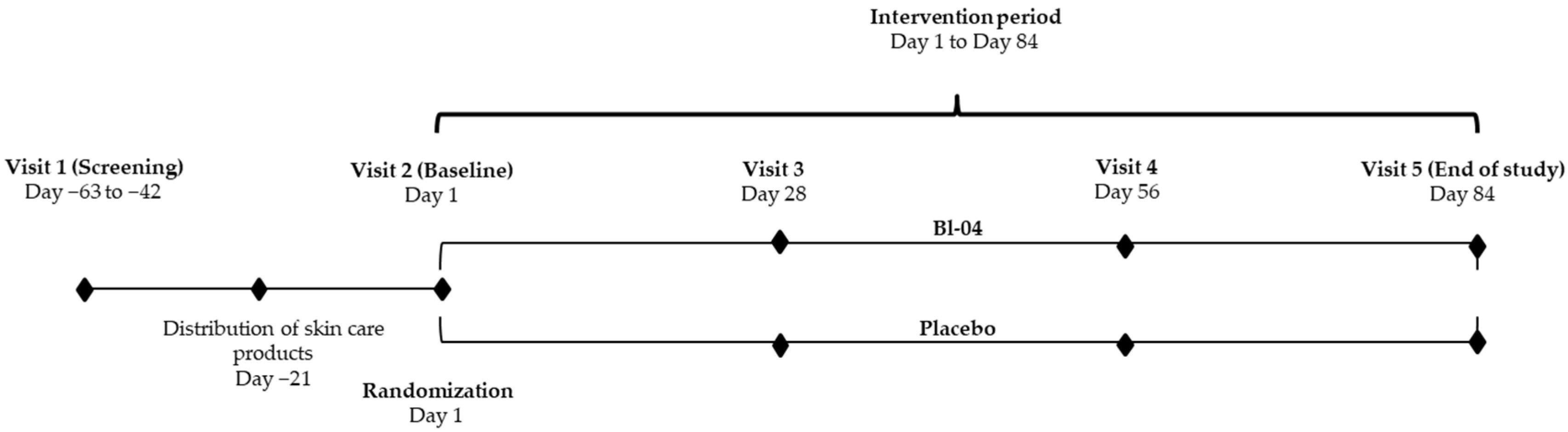
2.1. Trial Design

This randomized, triple-blind, placebo-controlled, parallel-group clinical trial was performed at Dermapro Ltd. Skin Research Center (Seoul, Korea) according to the Good Clinical Practice (GCP) guidelines (Integrated addendum to ICH E6(R1): Guideline for Good Clinical Practice) E6(R2) and the WMA Declaration of Helsinki (64th WMA General Assembly, Fortaleza, Brazil, October 2013).
The study was conducted between November 2020 and April 2021 and comprised 5 visits: a screening visit (V1); a baseline visit, during which the participants were randomized (V2); and 3 follow-up visits (V3 on Day 28 ± 2, V4 on Day 56 ± 2, and V5 on Day 84 ± 2) during the intervention (Figure 1). At V1, all participants were informed about the content and purpose of the study and signed written consent forms to participate in the trial.
Eligibility criteria were checked at V1, including an assessment of facial skin wrinkles by visual scale, skin hydration of the cheek with a Corneometer®, and a blood safety test. Demographic data, relevant medical history, and concomitant medications (CMs) were also collected.
Skin care products devoid of probiotics were distributed to the participants for use during the study, which the subjects were advised to use from 3 weeks before V2 (from Day-21) until the end of the study. They were also advised to maintain their lifestyle habits, including diet and exercise; use sunscreen when exposed to sunlight; avoid unusual levels of exposure to sunlight; avoid the use of functional cosmetics or probiotics as dietary supplements, food, or beverage products; and avoid medical and esthetic treatments of the test areas. The participants were asked to record their compliance with the investigational products (IPs) daily in a diary and inform the investigator of any adverse events (AEs) or CM use during the study. Outcome measures for facial wrinkles, hydration, TEWL, elasticity, gloss, and facial photographing were performed at V2–V5. Subjective questionnaires on skin improvement were completed at V3, V4, and V5, and product usability at V5. Blood safety tests were performed at V5 (in addition to V1), and body composition was analyzed at baseline and V5.

2.2. Ethical Approval and Registry

The ethical and scientific validity of this study was reviewed by the Dermapro Ltd. institutional review board (IRB) (Seoul, Korea; Approval 1-220777-A-N-01-DICN20181 dated 6 August 2020), and the study was performed with the voluntary consent of the participants. The trial was registered with ISRCTN (ISRCTN10924200) and CRIS (KCT0005565) before its outset.

2.3. Sample Size

The sample size was calculated by a two-group t-test with a two-sided significance level of 5%. Calculations were made separately for both primary outcomes, skin wrinkles, and hydration. The estimations were based on studies with comparable study designs, populations, and endpoint definitions ([11] for skin wrinkles and [11,25,26,27,28,29,30] for skin hydration).
With 80% power to detect a difference with 95% confidence, 64 evaluable participants per group were necessary to observe a difference of 7.5 µm in maximum profile valley depth (Rv) (representing facial wrinkles) in the treatment group and a standard deviation of 15 µm.
For skin hydration, to detect a 5-unit difference with a standard deviation of 8 units in the treatment group, 42 evaluable participants per group were required, with 80% power and 95% confidence.
Thus, considering an attrition rate of 30%, a total of 170 randomized participants were to be allocated to the study to ultimately obtain 128 evaluable participants.

2.4. Inclusion and Exclusion Criteria

A total of 170 healthy Korean female volunteers were screened for entrance into the study. The inclusion and exclusion criteria were as follow:
Inclusion criteria
  • Korean female subjects aged 30 to 60 years (by Korean age).
  • Dry skin on cheek (hydration value < 48 arbitrary units (AU) on a Corneometer®).
  • Greater than grade 3 skin wrinkle per a DERMAPRO standard photograph.
  • No chronic or acute disease, including skin disease.
  • Signed informed consent for study participation.
  • Cooperation and availability for follow-up during the study period.
Exclusion criteria
  • Consumption of probiotics as dietary supplements, food, or beverage during the previous 2 weeks.
  • Pregnancy, planned pregnancy, or lactation.
  • Irritation or symptomatic allergy to food, including ingredients of cosmetic, medical, and test products.
  • Use of oral or topical antibiotics during the previous 3 months.
  • Use of oral retinoid/steroid drug or topical steroid application during the previous 6 months.
  • Use of functional cosmetics to improve skin wrinkles, hydration, or elasticity within the past 3 months.
  • Having had skin treatment on the test site (e.g., decortication and Botox treatment).
  • Participation in a previous study without an appropriate intervening period (3 months) between studies.
  • Presence of disease that could affect the aim of the study (e.g., cardiovascular, kidney, liver, thyroid, gastrointestinal disease, gout).
  • Presence of any skin disease (e.g., AD) on test site.
  • Presence of any chronic disease (e.g., diabetes, asthma, high blood pressure) or psychiatric disorder (e.g., depression, schizophrenia).
  • Use of medication for obesity (e.g., antidepressants, anorectics), contraceptives, hormones, or diuretics.
  • Excessive alcohol use (over 30 g alcohol per day) or drug abuse.
  • Sensitive or hypersensitive skin.
  • Damaged skin in or around the test area (including sunburn, tattoos, scars, or other disfiguration of the test area).
  • Abnormal clinical chemical analysis result at V1, per a medical specialist.
  • Presence of a problem that could interfere with the aim of the study, based on the judgment of the principal investigator.

2.5. Interventions

Participants received one daily capsule of 1.75 × 10⁹ colony-forming units (CFUs) Bl-04 (ATCC SD5219) in microcrystalline cellulose with magnesium stearate and silicon dioxide as flow agents as a probiotic supplement or a comparable capsule without Bl-04 as a placebo for 12 weeks. Participants were instructed to consume one capsule orally before breakfast on the following morning after V2 to the morning of V5. The IP was manufactured by Danisco USA Inc. (Madison, WI, USA).

2.6. Randomization and Blinding

Randomization to the Bl-04 and placebo arms was performed at a 1:1 ratio using nQuery Advisor 7.0, version 7.0.1490.0 (San Diego, CA, USA), applying a random seed and a block size of 4 units. No stratification was conducted. In the randomization, eligible participants were enrolled sequentially by the study investigator at V2 and assigned the next free randomization number that was indicated on the IP label. Participants who dropped out of the study following the randomization were not replaced.
The study was conducted and analyzed in a triple-blind manner (i.e., the investigator, participants, and statistician were blinded). The randomization list (randomization numbers 001 to 200) was created by the IP manager, who was responsible for packaging and labeling the IP and creating individual emergency unblinding envelopes at Danisco USA Inc. (Madison, WI, USA; the IP manager did not otherwise participate in the study). The intactness of the emergency unblinding envelopes was monitored over the course of the study. After the database lock and blind data review, the randomization code was opened as Groups A and B for the statistical analyses without revealing the allocation. After the analyses were completed and the clinical study report was signed, the treatment codes were revealed as Bl-04 and placebo.

2.7. Study Assessments

All skin assessments were performed after 30 min of stabilization at a controlled temperature of 22 ± 2 °C and a relative humidity of 50 ± 5%. In addition, before each study visit, participants were advised not to have applied makeup for 12 h.

2.7.1. Primary Outcomes: Facial Wrinkles and Skin Hydration

Skin wrinkles were measured from the corner of the eye at V2–V5 using the PRIMOS® Premium 3-dimensional (3D) imaging system, software version 5.8 (GFMesstechnik GmbH, Teltow, Germany). Nine parameters were analyzed automatically from images that were taken from the same area of the corner of the eye at each visit: average depth of wrinkles, mean depth of the largest wrinkle, maximum depth of the largest wrinkle, total wrinkle area, total wrinkle volume, the total length of wrinkles, arithmetic mean deviation of the profile (Ra), the maximum height of the profile (Ry), and 10-point height of irregularities (Rz). To analyze the exact same location, the images that were taken during subsequent visits were aligned to the baseline image using the device software.
Skin hydration was measured with a Corneometer® CM 825 (Courage & Khazaka, Cologne, Germany) and a Moisturemeter® D (Delfin, Kuopio, Finland) on the cheek (crossing area between the center of the pupil and the tip of the nose), the middle of the forearm (10 cm above the wrist, avoiding visible blood vessels), and the back of the hand (crossing area between the middle finger and wrist, avoiding bone and visible blood vessels). The S15 probe was used with the Moisturemeter® D to measure skin hydration up to a depth of 1.5 mm. With both devices, each location was measured in triplicate, and the average value was used in the analysis.

2.7.2. Secondary Outcomes

TEWL was measured from the cheek, middle of the forearm, and back of the hand (the same locations as the measurements for skin hydration) with a Tewameter® TM300 (Courage & Khazaka). These measurements were repeated within 30 s, and the average of the last 3 stabilized values was used in the analysis.
Skin elasticity was recorded with a Cutometer® MPA580 (Courage & Khazaka) at the cheek (crossing area between the center of the pupil and the tip of the nose). The measurement was performed with a 2-s on/off pulse of a constant 450-mbar negative pressure, drawing the skin into the aperture of the probe, followed by its release. Three cycles of suction and release constituted one measurement cycle. The parameter R2 (gross elasticity, Ua/Uf) was used for the primary analysis.
Skin gloss was measured with a SkinGlossMeter® (Delfin) on the cheek (crossing area between the center of the pupil and the tip of the nose). The measurement was repeated three times, and the average value was used for analysis.
Facial images were taken with a VISIA® CR (Canfield Scientific, Parsippany, NJ, USA) with standard 2 and parallel-polarized setting.
Participants answered the subjective evaluation questionnaires on product efficacy and usability using a 5-point Likert scale: 1. Difficult/Dissatisfied/Disagree; 2. Somewhat difficult/dissatisfied/disagree; 3. No opinion; 4. Somewhat easy/satisfied/agree; and 5. Easy/Satisfied/Agree. The frequency and percentage of positive answers (Responses 4 and 5) were analyzed for each questionnaire.
Body composition parameters—weight, fat mass, body mass index, body fat percentage, and waist–hip ratio—were measured with an Inbody® 270 (Biospace, Seoul, Korea).

2.7.3. Safety Outcomes and Other Evaluations

Blood glucose, hemoglobin, serum glutamic-oxaloacetic transaminase (SGOT), serum glutamic-pyruvic transaminase (SGPT), gamma-glutamyl transpeptidase (γ-GTP), total cholesterol (T-cholesterol), triglycerides, high-density lipoprotein (HDL-cholesterol), low-density lipoprotein (LDL-cholesterol), and creatinine were analyzed with regard to safety at V1 and V5 (Daehang Hospital, Seoul, Korea).
AEs were recorded based on clinical observation by the investigator and as reported by the participants. The frequency, duration, and intensity of the AEs and their relationship with the test product were estimated by the investigator.
Product compliance was calculated, based on the number of leftover capsules in the container that was returned to the clinic.
In an ancillary evaluation, outdoor temperature (mean, min, max), mean relative humidity, and mean UV radiation (Korea Meteorological Administration, Seoul, Korea) on weekdays between the first participant’s initial visit to the last participant’s final visit were recorded.
Participant age, medical history, relevant CM use, skin, and lifestyle characteristics were collected as demographic data. The participants evaluated the following aspects of skin and lifestyle: skin type on the face and body (dry, normal, oily, dry and oily, or problematic with acne or other skin disease), skin hydration on the face and body (sufficient, normal, deficient), skin sebum on the face and body (glossy, normal, deficient), skin surface roughness on the face and body (smooth, normal, rough), facial skin thickness (thin, normal, thick), average daily time of UV exposure (less than 1 h, 1–3 h, over 3 h), average hours of sleep per day (less than 5, 5–8, over 8), the average amount of daily smoking (none, fewer than 10 pieces, over 10 pieces), the sensitivity of skin (yes, no), development of stinging sensation within 30 min of applying cosmetics (yes, no), and having experienced adverse reactions from cosmetics in the past 12 months? (yes, no).

2.8. Statistical Methods

The statistical analyses were conducted using SPSS® (IBM, Chicago, IL, USA). All data were analyzed for intention-to-treat (ITT) and per-protocol (PP) populations; the primary population was PP. Participants who had missing endpoint data, had a compliance rate of less than 80% IP use, used prohibited CMs, or encountered other major deviations that could have affected the primary endpoints were excluded from the PP population. Normality was determined by Kurtosis and Skewness test, and for the between-group analyses, homogeneity at baseline was tested by independent t-test.
For the within-group comparisons between time points (V2 vs. V3, V4, or V5), repeated measures analysis of variance (RM-ANOVA) was applied for parametric variables, and the Friedman test and Wilcoxon Signed-rank test with Bonferroni correction were used for nonparametric variables. For between-group comparisons, RM-ANOVA was performed for parametric values, and Mann–Whitney U-test was used for nonparametric values.
Clinical blood tests and body composition parameters from V1 and V2, respectively, were compared with V5 by paired t-test.
The subjective questionnaires on IP efficacy and usability were analyzed by chi-square test and Fisher’s Exact test.
This study was an exploratory study in nature, and no adjustments for multiplicity were performed. A statistically significant difference was set at p < 0.05.

2.9. Post Hoc Analyses

The primary study results were examined further by post hoc analysis, evaluating the effects in subgroups with the appropriate participant number, and correlations between a skin variable and weather parameter were evaluated by scatterplot and Pearson correlation. The post hoc analyses were conducted using SAS® for Windows, version 9.4 (SAS Institute Inc., Cary, NC, USA). The post hoc analysis results are shown only for the subgroups by age: 30–44 years and 45–60 years by international age.
Repeated measures analysis of covariance (RM-ANCOVA) was applied for continuous variables to analyze differences between groups and the changes within groups. The models included fixed effects for baseline value, treatment group, repeating factor week (Week 4 (V3), Week 8 (V4), Week 12 (V5)), and the interaction between treatment group and week. ‘Participant’ was the random effect in the model. An unstructured covariance matrix was assumed for the repeated measures for each participant over time. The contrasts between and within groups were estimated from the models, with 95% confidence intervals (CIs), using least square means. Two-sided p-values were calculated from the model. Further modeling was performed, including an adjustment factor as an additional covariate in the model.
Model fit was evaluated by testing residual normality using the Shapiro–Wilk test. If residual normality was not achieved with standard transformations (e.g., log, square root, inverse), nonparametric methods were applied. In such cases, an overall comparison between treatment groups was performed by Friedman test, within-group differences were analyzed by time point using the Wilcoxon Signed-rank test, and between-group differences were examined by time point using Mann–Whitney U-test.
Correlations between two continuous variables were evaluated using scatter plots and Pearson correlation coefficients.
A statistically significant difference was set at p < 0.05 and two-sided 95% CIs were produced for the estimated differences. Missing values were not imputed in the analyses.

3. Results

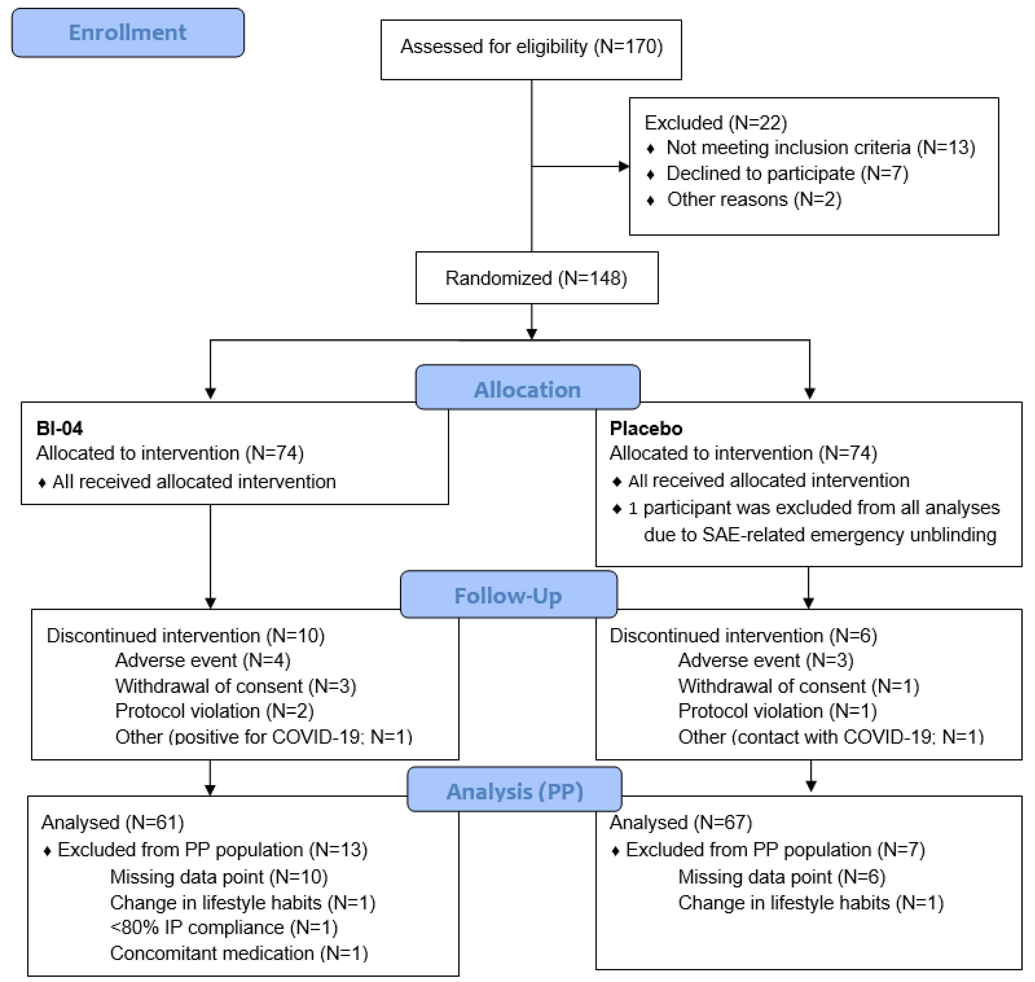
3.1. Recruitment and Study Populations

Volunteers were recruited through advertisements and clinic volunteer registry, followed by prescreening phone calls to schedule screening visits. Of the 170 volunteers who were screened, 148 were randomly allocated into two parallel, equal-sized intervention groups—Bl-04 and placebo—and 132 completed the study. The ITT population (N = 147; 74 active and 73 placebo) comprised all randomized participants who had consumed the IP, except for one participant who was subjected to emergency unblinding during the study. The PP analysis included 128 participants (61 and 67 in the Bl-04 and placebo groups, respectively; Figure 2). The safety population was composed of all randomized participants who had consumed the IP, including the participant who underwent emergency unblinding (Figure 2).

3.2. Baseline Data

A total of 147 females participated in the study, aged between 33 and 60 (49.8 ± 6.0 years (mean ± SD)). In the Bl-04 group, the average age was 49.2 ± 6.1 years (mean ± SD, N = 74), versus 50.4 ± 5.9 years (N = 73) in the placebo group. Skin hydration (by Corneometer®) at V1 was 43.7 ± 3.8 AU (mean ± SD) in the Bl-04 group and 45.0 ± 3.1 AU in the placebo group. Self-evaluated skin and lifestyle characteristics at the screening (Table 1 and Supplementary Material Table S1) and the skin parameters (wrinkle parameters, hydration, TEWL, skin elasticity, and skin gloss) at baseline (Table 2, Table 3 and Table 4, and Supplementary Material Tables S2–S4) were comparable between groups.
Of the body composition parameters (Supplementary Material Table S6), only body weight differed significantly at baseline between the Bl-04 and placebo groups (p = 0.042), which had mean weights of 61.0 kg and 57.7 kg, respectively.

3.3. Primary Outcomes

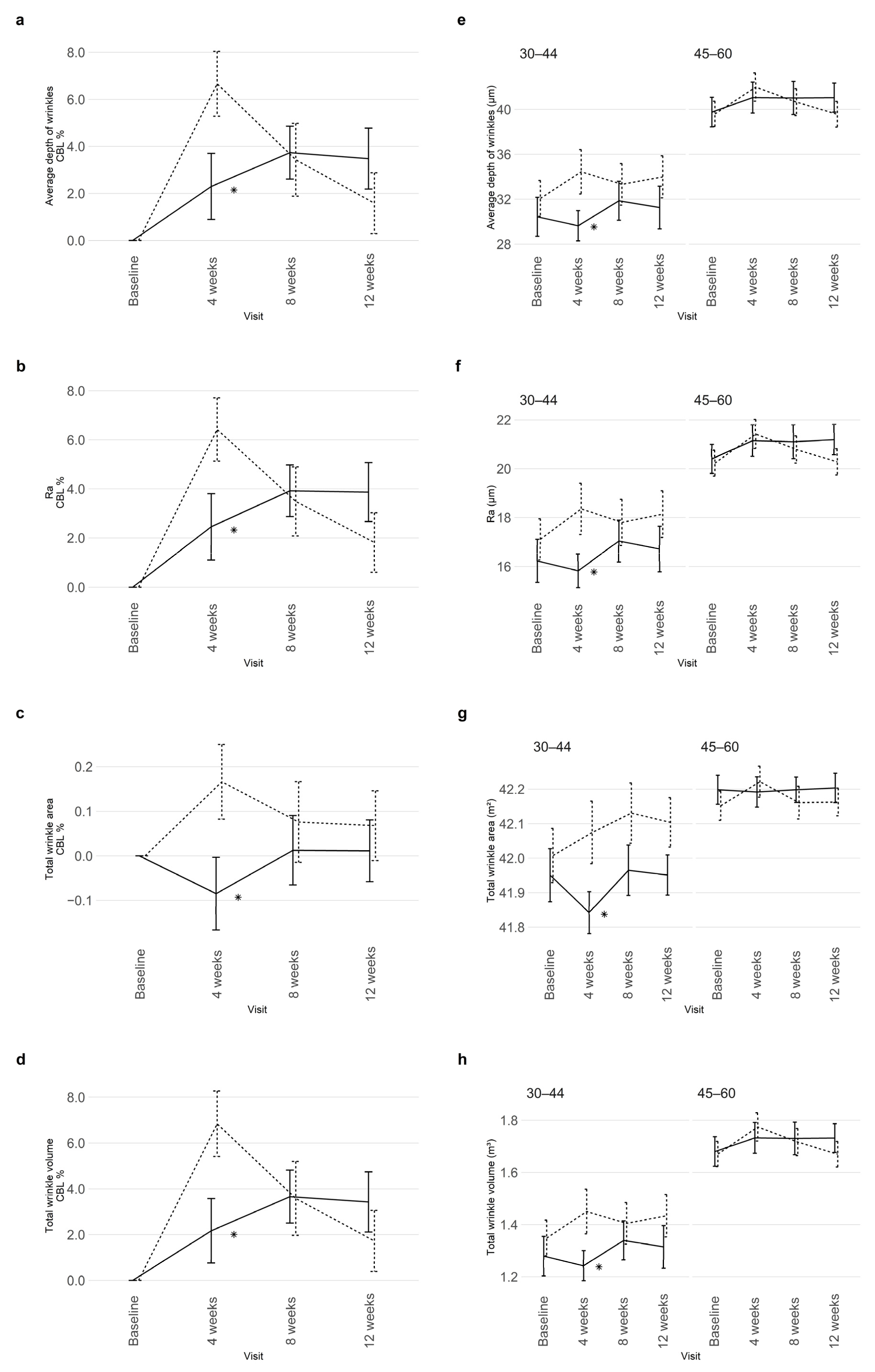
3.3.1. Skin Wrinkles

The effect of Bl-04 on facial wrinkles in the corner of the eye was analyzed by optical 3D imaging PRIMOS® premium at baseline and Weeks 4, 8, and 12 (Table 2). The mean values at each time point (Table 2) were used to calculate relative changes from baseline (Figure 3a–d).
Statistically significant differences between the Bl-04 and placebo groups were observed in wrinkle parameters that were related to general roughness in the corner of the eye at Week 4 (Table 2, Figure 3).
The average depth of wrinkles increased to a lesser extent from baseline (p = 0.030) with Bl-04 (+2.30%) versus placebo (+6.66%) at Week 4 (Figure 3a). At 8 weeks within the Bl-04 group, the average depth was increased further from baseline, but began to decline by Week 12 (Table 2, Figure 3a). Within placebo, the increase from baseline in the average depth of wrinkles had reduced at Week 8 (around to the level of Bl-04), and further at Week 12, not differing from baseline. At Weeks 8 and 12, there were no significant differences between Bl-04 and placebo.
Ra and total wrinkle volume had similar effect patterns as the average depth of wrinkles between Bl-04 and placebo (Table 2). At 4 weeks, the increases from baseline were significantly higher in the placebo group (+6.42%; p = 0.027 and +6.84%; p = 0.023 in Ra and total wrinkle volume, respectively) compared to Bl-04 (+2.46% and +2.17%, respectively) (Figure 3b, d). Placebo effected a significant within-group increase in Ra still at Week 8 versus baseline (p = 0.030) (Table 2).
In contrast to the pattern, Bl-04 significantly decreased (p = 0.036) the total wrinkle area by 0.09% from baseline at Week 4, whereas the area rose 0.17% with placebo (Figure 3c). Within Bl-04 and placebo groups, there were no statistically significant differences against baseline at any time point (Table 2).
In the post hoc analysis, in which the study population was subdivided by age into 30–44 years (total subgroup N = 31; Bl-04 N = 18, placebo N = 13) and 45–60 years (total subgroup N = 97; Bl-04 N = 43, placebo N = 54), older participants had more wrinkles and deeper wrinkles than the younger subjects. The benefits of Bl-04 on the average depth of wrinkles (Figure 3e), Ra (Figure 3f), total wrinkle area (Figure 3g), and total wrinkle volume (Figure 3h) were statistically significant at Week 4 compared to placebo in the younger subgroup (p = 0.017, p = 0.016, p = 0.021, and p = 0.022, respectively) but not at Week 8 or 12. In the older subgroup, there were no statistically significant differences between Bl-04 and placebo at any time point.
No statistically significant differences were observed in the mean depth of the largest wrinkle, maximum depth of the largest wrinkle, the total length of wrinkles, Ry, or Rz between Bl-04 and placebo (Table 2). Compared to baseline, at Week 4, there were significant within-group increases in Ry in the Bl-04 and placebo groups (p = 0.010 and p = 0.007, respectively) and in Rz at Week 8 with Bl-04 (p = 0.019) and Week 4 with placebo (p = 0.007), after which Ry and Rz values reverted to baseline values.

3.3.2. Skin Hydration

To determine the effects of Bl-04 on skin hydration, we measured the water content from the cheek, middle of the forearm, and back of the hand using a Corneometer® CM 825 (Table 3) and Moisturemeter® D (Table 4). The corneometer measured hydration at the surface of the skin to a depth of 10 µm of the stratum corneum, whereas the moisturemeter with S15 probe measured up to 1.5 mm of the dermis. With the corneometer, a statistically significant decrease in hydration was observed on the hand in the Bl-04 and placebo groups in the within-group analysis at 4 (p < 0.001 and p = 0.005, respectively), 8 (p = 0.002 and p = 0.019), and 12 (p = 0.001 and p < 0.001) weeks compared to baseline (Table 3). Hydration in the forearm was significantly decreased only at 12 weeks in the placebo group (p = 0.003) compared to baseline values. With the moisturemeter (Table 4), hydration decreased on the forearm in the Bl-04 and placebo groups in the within-group analysis at 8 weeks (p = 0.041 and p = 0.026, respectively) and at 12 weeks in the placebo arm (p = 0.016) compared to baseline values. No other significant observations were noted.

3.4. Secondary Outcomes

The effects of Bl-04 on TEWL from the cheek, middle of the forearm, and back of the hand were examined with Tewameter® TM300. A significant decrease in TEWL from the forearm was observed in the Bl-04 and placebo groups in the within-group analysis at 4 (p < 0.001 and p = 0.030, respectively), 8 (p < 0.001 and p = 0.007), and 12 weeks (p < 0.001 and p < 0.001) compared to baseline values (Supplementary Material Table S2). Similarly, TEWL from the hand declined at 4 (p = 0.005 and p < 0.001), 8 (p < 0.001 and p < 0.001), and 12 weeks (p < 0.001 and p < 0.001) in these groups versus baseline values. On the cheek, TEWL decreased in the Bl-04 and placebo groups at 8 (p = 0.047 and p = 0.002, respectively) and 12 weeks (p < 0.001 and p < 0.001) against baseline values. No other significant observations were noted.
Skin gross elasticity (R2 parameter) at the cheek, examined using Cutometer® MPA580, increased significantly at Week 4 with Bl-04 and placebo (p < 0.001 and p = 0.028, respectively), Week 8 for placebo only (p = 0.008), and Week 12 for Bl-04 only (p = 0.007) compared to baseline values (Supplementary Material Table S3). No other significant changes in skin elasticity (R2) were observed.
A statistically significant decrease in skin gloss was observed at Week 4 with placebo (p = 0.036) versus baseline value (Supplementary Material Table S4). No other significant differences in skin gloss were noted.
None of the subjective efficacy or usability items differed significantly between the Bl-04 and placebo groups (Supplementary Material Table S5). The proportion of participants who responded positively (4. Somewhat agree/satisfied/easy or 5. Agree/satisfied/easy) on items of subjective efficacy rose in both groups from Week 4 to Weeks 8 and 12 (within-group significance not tested).

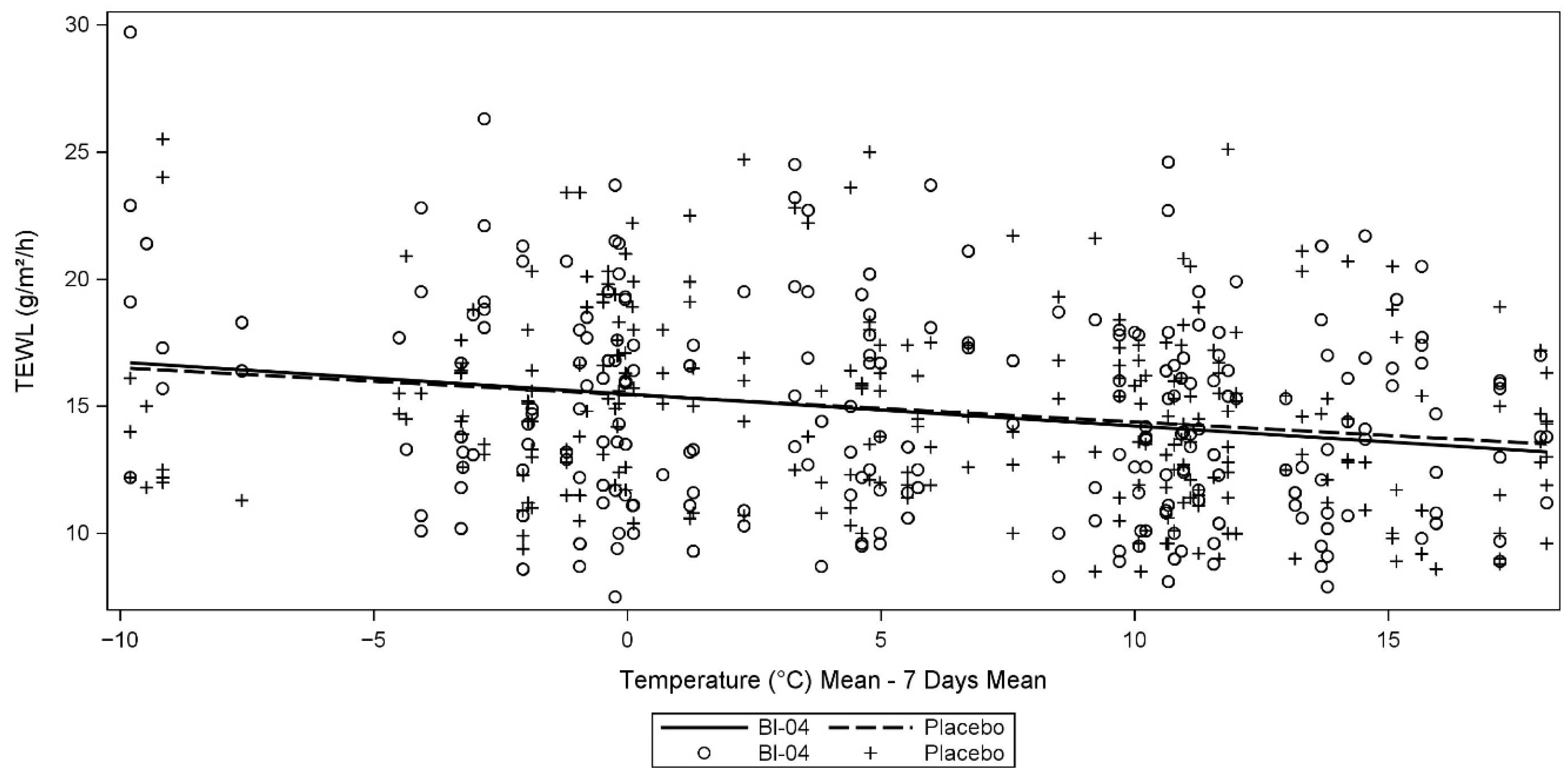
3.5. Ancillary Analyses

Descriptive statistics were calculated for all recorded weather parameters (Supplementary Material Tables S8 and S9). Nonparametric methods were used to evaluate the differences between and within treatment groups by visit.
Significant changes in average humidity, average temperature, and UV index from baseline were apparent for almost all within treatment comparisons by visit (Supplementary Material Table S10). The 7-day mean, maximum, and minimum temperature and 7-day mode and maximum UV index rose during the study, and average daily humidity was 58% to 59% at baseline and Week 8 and ranged from 52% to 54% at Weeks 4 and Week 12. There were no statistically significant differences in weather parameters between the Bl-04 and placebo groups at any visit.
TEWL from the cheek correlated negatively with 7-day mean temperature in overall (Pearson r = −0.211, p < 0.0001), and in the Bl-04 (r = −0.214, p = 0.0008) and placebo groups (r = −0.209, p= 0.0006) (Figure 4). In the subgroup analysis, this correlation was diluted and just exceeded statistical significance in the 30–44-year-old subgroup (total subgroup N = 31; Bl-04 N = 18, placebo N = 13) (r = −0.182, p = 0.0430), whereas in the older subgroup of those aged 45 to 60 years (total subgroup N = 97; Bl-04 N = 43, placebo N = 54), it became more pronounced (r = −0.220, p < 0.0001).
This pattern was also seen for the 7-day minimum and maximum temperatures against TEWL (r = −0.214, p < 0.0001 and r = −0.205, p < 0.0001, respectively); this correlation was more robust in the older age group (r = −0.185, p = 0.0393 and r = −0.165, p = 0.0674 in 30–44-year-olds and r = −0.223, p < 0.0001 and r = −0.217, p < 0.0001 in 45–60-year-olds, respectively). No correlation between TEWL and relative humidity was observed (data not shown).

3.6. Adverse Events

Six and five participants in the Bl-04 and placebo groups, respectively, experienced AEs during the study. All AEs were encountered during the consumption of the IP. One AE in the placebo group was classified as a serious adverse event (SAE) and was evaluated as probably not related to the IP. Emergency unblinding was performed for the SAE, resulting in the participant’s exclusion from the blinded analyses. All other AEs were considered mild and definitively unrelated to the IP. Three AEs in the placebo group and four AEs in the Bl-04 group resulted in the discontinuation of the study.
Although several statistically significant changes in the blood safety tests were observed in both groups from the initial screening to the end of the study (Supplementary Material Table S7), none was considered to be clinically relevant.

4. Discussion

Daily intake of Bl-04 for 12 weeks significantly decreased the total wrinkle area at the corner of the eye at 4 weeks compared to placebo, after which there was no significant difference between groups. In addition, with Bl-04, the average depth of wrinkles, Ra, and total wrinkle volume underwent smaller increases at Week 4 versus placebo; at Weeks 8 and 12, there were no significant differences between groups. Further, there was a difference noticed in subgroups by age. Overall, those who received Bl-04 experienced less fluctuation in the measured wrinkle parameters than placebo-treated subjects during the intervention.
The demonstrated immune system modulation benefits of the Bl-04 strain were one of the key characteristics due to which the strain was selected for this study. Bl-04 is rather anti-inflammatory in nature, inducing high IL-10 production and low levels of the pro-inflammatory IL-12 from human peripheral blood mononuclear cells ex vivo and protecting from colitis in vivo [31]; based on these properties, we surmised that this probiotic could act beneficially for the mucosal immune system and the intestinal barrier protection. Greater intestinal permeability has been associated with several health conditions, including autoimmune diseases, liver-related diseases, diabetes, and neurological and gastrointestinal conditions [32,33]. The gut–skin axis comprises the crosstalk between the gut microbiota, gut mucosal barrier, and immune system, which is then translated to a distal site: the skin [6]. Dysbiosis of the gut microbiota have been noticed to associate with several skin diseases, such as acne vulgaris, AD, and psoriasis [34]. Various factors, such as a fatty-acid-rich diet, alcohol, various medications, smoking, and stress, can cause defects in the intestinal barrier and the leakage of luminal components [35], activating the mucosal immune response and inducing the production of pro-inflammatory cytokines, such as tumor necrosis factor α (TNF-α) and interferon γ (IFN-γ), which disrupt the barrier further [36,37]. Conversely, an anti-inflammatory milieu, through its production of growth factors and anti-inflammatory cytokines, such as interleukin 10 (IL-10) and transforming growth factor β (TGF-β), has been suggested to be protective for the intestinal barrier [37].
The exact mechanisms by which gut barrier permeability manifests in the skin are incompletely understood, but the metabolites that are produced by the gut microbiota might function in such processes. Gut microbiota can metabolize nondigestible dietary components and cellular debris and produce beneficial and harmful, even toxic, metabolites as end products, which can affect gut permeability and, ultimately, skin health [34]. Certain metabolites, such as short-chain fatty acids (SCFAs), particularly butyrate, can benefit gut health by serving as an energy source for colonocytes, improving barrier permeability, and having anti-inflammatory properties [38,39]. SCFAs are readily absorbed, and the amount that is absorbed into peripheral circulation is low compared to the colonic lumen and depends on diet [40].
Butyrate has been shown to increase regulatory T cell numbers, which maintain immune system homeostasis in the skin, as in the gut [41,42]. Bifidobacteria do not produce butyrate per se but instead synthesize acetate and lactate, which are converted into butyrate by other colonic bacteria through cross-feeding interactions [43]. Butyrate has also been shown to stimulate in vitro in L929 murine fibroblasts TGF-β1 and collagen production, which are important in maintaining the dermal extracellular matrix [44]. The short-chain acids that are absorbed into systemic circulation could function peripherally in the skin, as shown for Bifidobacterium longum with or without galacto-oligosaccharides in UV-induced wrinkle formation [45], constituting another mechanism through which Bl-04 could function against skin wrinkling and warranting additional studies.
Skin wrinkles are forming as a result of internal and external factors creating changes in epidermal thickness, lack of hyaluronan, flattening of the dermo-epidermal junction, loss and disorganization of collagen due to the upregulation of collagen-degrading matrix metalloproteases (MMPs), degradation of elastic fibers, and decreased water-holding capacity [5,46,47]. In addition, cellular senescence affects skin aging and wrinkle formation through the secretion of pro-inflammatory cytokines, MMPs, and chemokines that can lead to inflammaging—chronic low-grade inflammation that is associated with aging [48].
Parameters describing the features of deeper wrinkles did not show major changes in this study (Table 2). The development of deep wrinkles is a cumulative, long-term process that takes years [49]. In our study, the intervention was 12 weeks; thus, a longer treatment period might be needed for deeper wrinkles.
Few studies have examined the effects of an oral probiotic supplement as a single active ingredient on skin wrinkles and hydration with a similar methodology [50], which makes the comparison of the results challenging. When our results were compared to a study conducted with a similar design and methods [11], we noted that the baseline values of several comparable wrinkle parameters where considerably higher in volunteers in our study than in Lee et al., in our study, 10-point average roughness (Rz) was over 230 μm and a maximum depth of biggest wrinkle was over 150 μm compared to Lee et al. values of Rz under 90 μm and maximum profile valley depth (Rv) less than 80 μm, respectively,—perhaps partially explaining the observations in our study. Younger participants, in whom the beneficial effects of Bl-04 on skin wrinkles over placebo were especially noticed, had fewer and shallower wrinkles than the older subjects (Figure 3e–h). The younger participants were also closer to the population used in the Lee et al. considering the wrinkle status; Ra describing average roughness was in both under 20 μm. Nonetheless, we could observe visual decreases in several wrinkle features also in participants in the older Bl-04-treated subgroup (Supplementary Material Figure S1).
As with the younger subgroup, in subsets of subjects with self-reported deficient facial skin hydration and deficient facial skin sebum (Table 1), wrinkle volume, an average depth of wrinkles, and Ra increased to a lesser extent with Bl-04 than placebo at Week 4 compared to baseline (data not shown). In addition, in a subgroup with more than 1 h of self-reported daily UV exposure, Bl-04 resulted in a decrease in total wrinkle area, and smaller increases total wrinkle volume, and the average depth of wrinkles versus baseline than placebo at Week 4 (data not shown). As in the older age subgroup, no statistically significant differences between Bl-04 and placebo were seen in total wrinkle volume and area, average depth of wrinkles and Ra in subgroups with self-reported normal skin hydration, normal skin sebum, and less than 1 h of daily UV exposure (Table 1) at any time point (data not shown). No statistically significant differences were observed in total wrinkle volume and area, average depth of wrinkles and Ra in skin roughness subgroups—normal or rough—at any time point (data not shown). This may indicate that participants with suboptimal skin conditions might benefit from Bl-04 use.
In this study, we noted increases in many wrinkle parameters in both groups at Week 4, which then declined at varying rates, whereas in Lee et al. [11], these values decreased quite steadily in both groups throughout the intervention. In a study by Fanian et al. [51], which was also performed from winter to spring, reported similar effects as ours: several parameters of skin roughness increased in the middle of the study period in both groups, but the increase was milder in the treatment group. Thus, as Fanian et al. due to the milder increase, we propose that the treatment may protect the skin against dryness during winter. Winter seasons can vary with regard to weather conditions, and few skin health-related studies have reported weather conditions and the timing of the intervention, further hindering a comparison of the results.
In Korean women, skin scaliness and elasticity correlated with environmental factors, such as air temperature, pressure, and peak precipitation [52]. Short-term exposure to low humidity has been shown to induce aggravation of skin texture and the formation of fine lines [53]. In our study, TEWL did not correlate with 7-day mean relative humidity—overall or when stratifying by age (data not shown). There are no general guidelines on how to evaluate correlations with the weather in skin research. Relative humidity, rather than absolute humidity, is the most common method of measuring moisture content in the air, but the association of skin parameters with absolute humidity should be considered as well [54]. Our study was conducted during the dry winter season, and indoor heating might have further decreased the humidity, affecting additional dryness of the skin and most likely exacerbating fine line wrinkling [55].
Skin barrier function is negatively affected by climate, and there are clear seasonal differences in hydration of the skin, peaking during the summer and bottoming in the winter, causing winter xerosis [56,57,58]. Skin appearance can be influenced by controlling the hydration of the skin, and dry skin with alkaline pH has been linked to faster persistent wrinkling in an 8-year longitudinal study [49]. The amount of water accumulated in the outermost corneal layer is defined as skin hydration, which depends on natural hygroscopic agents in corneocytes, natural moisturizing factors, and intercellular lipids [1]. Daily insults from the environment, such as low humidity, surfactants, wind, and sun, can lower the water content in the stratum corneum, causing improper desquamation and the appearance of dry, rough, flaky skin [59,60]. Lifestyle factors, including water intake, can also affect the hydration of the skin, especially if an individual has low prior water consumption [61].
Conversely, TEWL is the diffusion of a small proportion of water molecules through the stratum corneum. It can be used as an indirect measure of skin permeability, and because it is affected by factors in the surrounding microclimate, such as environmental humidity, temperature, and airflow, it should be measured under controlled conditions [62,63,64], as was performed for all skin measurements in our study, including hydration and TEWL. TEWL values have been shown to correlate inversely with hydration in diseased skin, such as in AD and psoriasis [65]. Hydration has been suggested to decrease with aging in Korean females and during the winter season [66,67].
We did not observe significant changes in hydration of the cheek (Table 3), even when participants were stratified by age, which is consistent with Baek et al. [68]. Instead, we noted significant within-group decreases in TEWL from the forearm, hand, and cheek in the Bl-04 and placebo groups (Supplementary Material Table S2), as reported by Lee et al., who showed a significant decrease with a probiotic versus placebo [11]. The lack of an effect of the probiotic over placebo in our study might be related to differences in the study populations between ours and the study by Lee et al., as described above. TEWL has been suggested to remain constant or decrease during aging [69], and a decline has also been noted in Korean females with age [66,67,68]. Environmental temperature correlated negatively with TEWL in the overall cohort and the 45–60-year-old subgroup and to a lesser extent in those aged up to 44 years.
TEWL has been previously shown to be dependent on environmental temperature, becoming higher during winter versus in summer, but there are contradictory results regarding this pattern [70,71,72,73]. In addition, humidity, affecting skin dryness at higher temperatures, might have a greater effect on TEWL than temperature itself [71].
It is poorly understood how TEWL affects wrinkle formation. TEWL correlates with skin hydration and skin surface patterns in the short-term [55]; however, because wrinkle formation is a slow process [49], the longer-term effects of increased permeability should be determined to understand this relationship. Skin hydration is a rather understudied topic and warrants further investigation [55], especially with regard to wrinkle formation.
This study was conducted during the global severe acute respiratory syndrome coronavirus 2 (SARS-CoV-2, or COVID-19) pandemic, during which the use of protective masks was widely recommended for the general public. Recent results demonstrated that only 3-h use of a fabric or disposable medical mask increased skin hydration in the general population, whereas such changes in TEWL and pH were not observed [74]. Even after short-term 1-h or 6-h use, skin temperature, redness, and sebum secretion rose, with varying results for TEWL and no significant change in skin hydration or elasticity in the cheek area [75]. In contrast, long-term use, 6-h daily use for 2 weeks, decreased skin elasticity and increased skin pores and acne, whereas skin temperature, redness, TEWL, and sebum content did not change significantly [76]. Additionally, mask use for 6 h daily for 4 weeks was shown to enhance wrinkle formation and roughness in nasolabial slopes [77].
In our study, skin hydration, TEWL, elasticity, and gloss were measured in the area of the cheek that would be covered by a mask, and it is unknown whether mask use would also have affected wrinkle parameters at the corner of the eye. Despite the pre-measurement stabilization in a temperature and humidity-controlled room, it remains unknown how putative short- or long-term mask use would have impacted the measured values, warranting further studies. In addition, we noted a decrease in skin hydration on the hand in both groups (Table 3), perhaps due to long-term, frequent use of hand sanitizer and hand-washing with soap during the pandemic [78].
We did not observe any statistically significant effects in the data on the subjective questionnaires. Subjective data that are collected with questionnaires are highly prone to placebo effects and, by nature, measure perceived effects; thus, they should not be compared directly with data that are gathered using objective measurements.
This study provides important novel data on the effects of probiotic supplementation on skin health and is the first report to examine the effects of oral consumption of Bl-04 on skin wrinkle and hydration characteristics. The study was a triple-blinded, rigorously conducted, randomized, placebo-controlled trial, with proper inclusion and exclusion criteria. Seasonal weather conditions were collected from the local meteorological office to analyze the study outcomes. Moreover, skin parameters were evaluated with objective measurements using reliable quantitative methods, the use of which remains limited in dermatological studies.
Studies on probiotics encounter the same issues as other interventional studies, in that the participants are non-responders or responders [79]. We suggest that future similar studies would prescreen participants not only for wrinkles but also for either low-grade inflammation or gut permeability, if the mode of action of an IP is proposed to be linked to the gut–skin axis. Low-grade inflammation could be measured, for instance, with high-sensitivity C-reactive protein or interleukins, such as IL-6 [80,81,82]. Permeability of the gut is ideally measured by the urinary recovery of molecular markers that are administered, such as non-metabolizable sugars, but this approach would require non-eligible participants to consume such substances, increasing the complexity of the study. Noninvasive biomarkers could be used instead, including urinary or serum fatty acid-binding protein, urinary or serum glutathione S-transferase, serum zonulin, and serum lipopolysaccharide or D-lactate that is derived from gut bacteria, but with caution [35,83].
The gut microbiota is unique to each individual, and with microbially derived metabolites, it might also determine which individual is a responder [79]. Bl-04 has been shown to possess gut microbiota modulatory functions, but this activity is debated [24,84]. It remains unknown whether Bl-04 could influence the composition of the gut microbiota, as has been recently suggested for Lactiplantibacillus plantarum HY7714 [85]; however, as a prescreening tool, the use of gut microbiota would be difficult because dysbiosis in the gut microbiota is observed in intestinal and extraintestinal disorders but not in healthy individuals, who presumably have healthy and unique gut microbiota [86]. Biological samples for efficacy parameters were not collected in our exploratory study, but the results warrant collecting and investigating those in future trials to illustrate the interaction between gut and skin.
Our study has several limitations. The trial was conducted at a single center and included only females, which challenges the generalization of our results to males. The study was short—12 weeks—considering the slow development of wrinkles. Further, the dose of Bl-04 of 1.75 × 10⁹ CFU was over 5-fold lower than in the corresponding Korean study [11], complicating a comparison between these otherwise similar studies. In the stratification by age, the younger group (30 to 44 years) was smaller than the older subgroup (45 to 60 years), raising some statistical uncertainty with the results.
In addition, we did not take into consideration the menopausal status of the participants, as entering into menopause has been shown to accelerate wrinkle formation [49]. This phenomenon could have diluted the potential beneficial effects of Bl-04 on skin wrinkles if many of the participants were entering menopause during the study.
Further, based on the instructions that were given to the participants, consumption of other functional foods that contain probiotics was prohibited, but it was not followed by a self-reported diary or by other measures, which would have improved the quality of the study. Participants might have been celebrating the Korean New Year during the intervention, which could have introduced changes to their dietary habits. The study participants were eligible for the study based on the measurement of dry skin on the cheek and subjective visual subjective inspection of skin wrinkles at the corner of the eye. The latter could have been measured with an objective quantitative method to obtain a more homogeneous population.

5. Conclusions

This randomized, placebo-controlled, triple-blinded clinical trial has demonstrated that the consumption of Bl-04 (1.75 × 10⁹ CFU) for 12 weeks is safe. This probiotic may alleviate facial wrinkling at 4 weeks, especially in the younger population; however, its effects on wrinkle parameters did not differ significantly at later stages of the intervention, when environmental conditions became more favorable for the skin. No statistically significant differences in hydration parameters were detected between Bl-04 and placebo during the study. This study suggests that environmental conditions should be considered more carefully in skin intervention trials and that skin condition, age, and unpredictable confounding factors, such as the use of protective masks, may have effects on the measured parameters.

Supplementary Materials

The following supporting information can be downloaded at: https://www.mdpi.com/article/10.3390/dermato2020005/s1, Table S1: Participants’ self-evaluated lifestyle and skin characteristics in Bifidobacterium animalis subsp. lactis Bl-04 (Bl-04) and placebo groups in the per-protocol (PP) population; Table S2: Transepidermal water loss (TEWL) from the skin in the PP population; Table S3: Skin elasticity according to the R2 parameter (E/mm) in the PP population; Table S4: Skin gloss values as skin gloss units (SGUs), measured from the cheek in the PP population; Table S5: Subjective evaluation of investigational product efficacy and usability in the PP population; Table S6: Body composition of the participants in the PP population; Table S7: Blood safety test values of the participants in the safety population; Table S8: Descriptive statistics of 7-day average daily mean temperature (°C), minimum temperature (°C), maximum temperature (°C), and mean relative humidity (%) by treatment and visit for the PP population; Table S9: Descriptive statistics of 7-day mode of ultraviolet (UV) index and 7-day maximum of UV index for the PP population; Table S10: Analysis of 7-day average daily mean temperature (°C), minimum temperature (°C), maximum temperature (°C), mean relative humidity (%), and maximum UV index for the PP population between time points and groups; Figure S1: Three-dimensional (3D) image of the corner of the eye area of a participant in the Bl-04 group.

Author Contributions

Conceptualization, L.H., H.A., A.L. (Anna Lyra), A.I., and K.T.; methodology, L.H., A.L. (Anna Lyra), H.A., and K.T.; validation, J.R. (Jahyun Ryu) and Y.S.; formal analysis, J.R. (Jahyun Ryu), E.L., H.J., and T.P.; investigation, E.L.; resources, K.T.; data curation, A.L. (Anna Lyra), L.H., H.A., T.P., and J.R. (Jenni Reimari); writing—original draft preparation, L.H. and H.A.; writing—review and editing, A.L. (Anna Lyra), A.I., K.T., A.L. (Antti Lauerma), J.R. (Jenni Reimari), and T.P.; visualization, T.P.; supervision, J.B., M.S., A.I., A.L. (Antti Lauerma), and K.T.; project administration, J.R. (Jahyun Ryu), L.H., and K.T. All authors have read and agreed to the published version of the manuscript.

Funding

This research was fully sponsored by Danisco Sweeteners Oy (a subsidiary of International Flavors & Fragrances, IFF), Kantvik, Finland. No public funding was received to conduct this trial.

Institutional Review Board Statement

This study was conducted in accordance with the Declaration of Helsinki and approved by the Dermapro Ltd. Institutional Review Board (Seoul, Korea; Approval 1-220777-A-N-01-DICN20181, dated 6 August 2020).

Informed Consent Statement

Informed consent was obtained from all subjects who were involved in the study.

Data Availability Statement

Not applicable.

Acknowledgments

The authors wish to thank Tad Stewart, IFF Health and Biosciences, Madison WI, USA, for management of the IP manufacturing; Sujin Ha, IFF Health and Biosciences, Seoul, Korea, for supporting the IP logistics; Jenni Perämäki, 4Pharma, Turku, Finland, for her contribution to the post hoc statistical analyses; and Markus Lehtinen and Arthur Ouwehand, IFF Health and Biosciences, Kantvik, Finland, for their valuable insights to the manuscript content.

Conflicts of Interest

This study was funded by IFF, which manufactures and sells Bl-04. The employees of IFF were salaried staff who participated in the design of the study, the analysis and interpretation of the data, writing of the manuscript, and the decision making with regard to publishing the results. Otherwise, the authors declare no conflict of interest.

References

  1. Boer, M.; Duchnik, E.; Maleszka, R.; Marchlewicz, M. Structural and biophysical characteristics of human skin in maintaining proper epidermal barrier function. Postepy Dermatol. Alergol. 2016, 33, 1–5. [Google Scholar] [CrossRef] [PubMed]
  2. Fore, J. A review of skin and the effects of aging on skin structure and function. Ostomy Wound Manag. 2006, 52, 24–35. [Google Scholar] [PubMed]
  3. Liska, D.; Mah, E.; Brisbois, T.; Barrios, P.L.; Baker, L.B.; Spriet, L.L. Narrative Review of Hydration and Selected Health Outcomes in the General Population. Nutrients 2019, 11, 70. [Google Scholar] [CrossRef] [PubMed] [Green Version]
  4. Cao, C.; Xiao, Z.; Wu, Y.; Ge, C. Diet and Skin Aging-From the Perspective of Food Nutrition. Nutrients 2020, 12, 870. [Google Scholar] [CrossRef] [Green Version]
  5. Krutmann, J.; Bouloc, A.; Sore, G.; Bernard, B.A.; Passeron, T. The skin aging exposome. J. Dermatol. Sci. 2017, 85, 152–161. [Google Scholar] [CrossRef] [Green Version]
  6. O’Neill, C.A.; Monteleone, G.; McLaughlin, J.T.; Paus, R. The gut-skin axis in health and disease: A paradigm with therapeutic implications. Bioessays 2016, 38, 1167–1176. [Google Scholar] [CrossRef]
  7. Szanto, M.; Dozsa, A.; Antal, D.; Szabo, K.; Kemeny, L.; Bai, P. Targeting the gut-skin axis-Probiotics as new tools for skin disorder management? Exp. Dermatol. 2019, 28, 1210–1218. [Google Scholar] [CrossRef] [Green Version]
  8. Hill, C.; Guarner, F.; Reid, G.; Gibson, G.R.; Merenstein, D.J.; Pot, B.; Morelli, L.; Canani, R.B.; Flint, H.J.; Salminen, S.; et al. Expert consensus document. The International Scientific Association for Probiotics and Prebiotics consensus statement on the scope and appropriate use of the term probiotic. Nat. Rev. Gastroenterol. Hepatol. 2014, 11, 506–514. [Google Scholar] [CrossRef] [Green Version]
  9. Ritchie, M.L.; Romanuk, T.N. A meta-analysis of probiotic efficacy for gastrointestinal diseases. PLoS ONE 2012, 7, e34938. [Google Scholar] [CrossRef] [Green Version]
  10. Maldonado Galdeano, C.; Cazorla, S.I.; Lemme Dumit, J.M.; Velez, E.; Perdigon, G. Beneficial Effects of Probiotic Consumption on the Immune System. Ann. Nutr. Metab. 2019, 74, 115–124. [Google Scholar] [CrossRef]
  11. Lee, D.E.; Huh, C.S.; Ra, J.; Choi, I.D.; Jeong, J.W.; Kim, S.H.; Ryu, J.H.; Seo, Y.K.; Koh, J.S.; Lee, J.H.; et al. Clinical Evidence of Effects of Lactobacillus plantarum HY7714 on Skin Aging: A Randomized, Double Blind, Placebo-Controlled Study. J. Microbiol. Biotechnol. 2015, 25, 2160–2168. [Google Scholar] [CrossRef] [PubMed]
  12. Liu, C.; Tseng, Y.P.; Chan, L.P.; Liang, C.H. The potential of Streptococcus thermophiles (TCI633) in the anti-aging. J. Cosmet. Dermatol. 2021, 1–13. [Google Scholar] [CrossRef] [PubMed]
  13. Gueniche, A.; Philippe, D.; Bastien, P.; Reuteler, G.; Blum, S.; Castiel-Higounenc, I.; Breton, L.; Benyacoub, J. Randomised double-blind placebo-controlled study of the effect of Lactobacillus paracasei NCC 2461 on skin reactivity. Benef. Microbes 2014, 5, 137–145. [Google Scholar] [CrossRef]
  14. Peguet-Navarro, J.; Dezutter-Dambuyant, C.; Buetler, T.; Leclaire, J.; Smola, H.; Blum, S.; Bastien, P.; Breton, L.; Gueniche, A. Supplementation with oral probiotic bacteria protects human cutaneous immune homeostasis after UV exposure-double blind, randomized, placebo controlled clinical trial. Eur. J. Dermatol. 2008, 18, 504–511. [Google Scholar] [CrossRef] [PubMed]
  15. Cox, A.J.; West, N.P.; Horn, P.L.; Lehtinen, M.J.; Koerbin, G.; Pyne, D.B.; Lahtinen, S.J.; Fricker, P.A.; Cripps, A.W. Effects of probiotic supplementation over 5 months on routine haematology and clinical chemistry measures in healthy active adults. Eur. J. Clin. Nutr. 2014, 68, 1255–1257. [Google Scholar] [CrossRef]
  16. Ding, W.K.; Shah, N.P. Acid, bile, and heat tolerance of free and microencapsulated probiotic bacteria. J. Food Sci. 2007, 72, M446–M450. [Google Scholar] [CrossRef] [PubMed]
  17. Forssten, S.D.; Ouwehand, A.C. Simulating colonic survival of probiotics in single-strain products compared to multi-strain products. Microb Ecol Health Dis 2017, 28, 1378061. [Google Scholar] [CrossRef] [Green Version]
  18. Forssten, S.; Ouwehand, A.C. Dose-Response Recovery of Probiotic Strains in Simulated Gastro-Intestinal Passage. Microorganisms 2020, 8, 112. [Google Scholar] [CrossRef] [Green Version]
  19. Ouwehand, A.C.; Nermes, M.; Collado, M.C.; Rautonen, N.; Salminen, S.; Isolauri, E. Specific probiotics alleviate allergic rhinitis during the birch pollen season. World J. Gastroenterol. 2009, 15, 3261–3268. [Google Scholar] [CrossRef]
  20. Taverniti, V.; Koirala, R.; Dalla Via, A.; Gargari, G.; Leonardis, E.; Arioli, S.; Guglielmetti, S. Effect of Cell Concentration on the Persistence in the Human Intestine of Four Probiotic Strains Administered through a Multispecies Formulation. Nutrients 2019, 11, 285. [Google Scholar] [CrossRef] [Green Version]
  21. Paineau, D.; Carcano, D.; Leyer, G.; Darquy, S.; Alyanakian, M.A.; Simoneau, G.; Bergmann, J.F.; Brassart, D.; Bornet, F.; Ouwehand, A.C. Effects of seven potential probiotic strains on specific immune responses in healthy adults: A double-blind, randomized, controlled trial. FEMS Immunol. Med Microbiol. 2008, 53, 107–113. [Google Scholar] [CrossRef] [Green Version]
  22. Turner, R.B.; Woodfolk, J.A.; Borish, L.; Steinke, J.W.; Patrie, J.T.; Muehling, L.M.; Lahtinen, S.; Lehtinen, M.J. Effect of probiotic on innate inflammatory response and viral shedding in experimental rhinovirus infection—A randomised controlled trial. Benef. Microbes 2017, 8, 207–215. [Google Scholar] [CrossRef] [PubMed] [Green Version]
  23. Muehling, L.M.; Turner, R.B.; Brown, K.B.; Wright, P.W.; Patrie, J.T.; Lahtinen, S.J.; Lehtinen, M.J.; Kwok, W.W.; Woodfolk, J.A. Single-Cell Tracking Reveals a Role for Pre-Existing CCR5+ Memory Th1 Cells in the Control of Rhinovirus-A39 After Experimental Challenge in Humans. J. Infect. Dis. 2018, 217, 381–392. [Google Scholar] [CrossRef] [PubMed] [Green Version]
  24. Lehtinen, M.J.; Hibberd, A.A.; Männikkö, S.; Yeung, N.; Kauko, T.; Forssten, S.; Lehtoranta, L.; Lahtinen, S.J.; Stahl, B.; Lyra, A.; et al. Nasal microbiota clusters associate with inflammatory response, viral load, and symptom severity in experimental rhinovirus challenge. Sci. Rep. 2018, 8, 1–12. [Google Scholar] [CrossRef] [Green Version]
  25. Ogawa, M.; Saiki, A.; Matsui, Y.; Tsuchimoto, N.; Nakakita, Y.; Takata, Y.; Nakamura, T. Effects of oral intake of heat-killed Lactobacillus brevis SBC8803 (SBL88) on dry skin conditions: A randomized, double-blind, placebo-controlled study. Exp. Ther. Med. 2016, 12, 3863–3872. [Google Scholar] [CrossRef]
  26. Kim, H.; Kim, H.R.; Jeong, B.J.; Lee, S.S.; Kim, T.R.; Jeong, J.H.; Lee, M.; Lee, S.; Lee, J.S.; Chung, D.K. Effects of oral intake of kimchi-derived Lactobacillus plantarum K8 lysates on skin moisturizing. J. Microbiol. Biotechnol. 2015, 25, 74–80. [Google Scholar] [CrossRef] [Green Version]
  27. Kimoto-Nira, H.; Aoki, R.; Sasaki, K.; Suzuki, C.; Mizumachi, K. Oral intake of heat-killed cells of Lactococcus lactis strain H61 promotes skin health in women. J. Nutr. Sci. 2012, 1, e18. [Google Scholar] [CrossRef] [Green Version]
  28. Saito, Y.; Mihara, T.; Maruyama, K.; Saito, J.; Ikeda, M.; Tomonaga, A.; Kumagai, T. Effects of intake of Lactobacillus casei subsp. casei 327 on skin conditions: A randomized, double-blind, placebo-controlled, parallel-group study in women. Biosci. Microbiota Food Health 2017, 36, 111–120. [Google Scholar] [CrossRef] [Green Version]
  29. Hong, Y.H.; Chang, U.J.; Kim, Y.S.; Jung, E.Y.; Suh, H.J. Dietary galacto-oligosaccharides improve skin health: A randomized double blind clinical trial. Asia Pac. J. Clin. Nutr. 2017, 26, 613–618. [Google Scholar] [CrossRef]
  30. Kim, D.U.; Chung, H.C.; Choi, J.; Sakai, Y.; Lee, B.Y. Oral Intake of Low-Molecular-Weight Collagen Peptide Improves Hydration, Elasticity, and Wrinkling in Human Skin: A Randomized, Double-Blind, Placebo-Controlled Study. Nutrients 2018, 10, 826. [Google Scholar] [CrossRef] [Green Version]
  31. Foligne, B.; Nutten, S.; Grangette, C.; Dennin, V.; Goudercourt, D.; Poiret, S.; Dewulf, J.; Brassart, D.; Mercenier, A.; Pot, B. Correlation between in vitro and in vivo immunomodulatory properties of lactic acid bacteria. World J. Gastroenterol. 2007, 13, 236–243. [Google Scholar] [CrossRef] [PubMed] [Green Version]
  32. Leech, B.; Schloss, J.; Steel, A. Association between increased intestinal permeability and disease: A systematic review. Adv. Integr. Med. 2019, 6, 23–34. [Google Scholar] [CrossRef]
  33. Hanning, N.; Edwinson, A.L.; Ceuleers, H.; Peters, S.A.; De Man, J.G.; Hassett, L.C.; De Winter, B.Y.; Grover, M. Intestinal barrier dysfunction in irritable bowel syndrome: A systematic review. Therap. Adv. Gastroenterol. 2021, 14, 1756284821993586. [Google Scholar] [CrossRef] [PubMed]
  34. De Pessemier, B.; Grine, L.; Debaere, M.; Maes, A.; Paetzold, B.; Callewaert, C. Gut-Skin Axis: Current Knowledge of the Interrelationship between Microbial Dysbiosis and Skin Conditions. Microorganisms 2021, 9, 353. [Google Scholar] [CrossRef]
  35. Vancamelbeke, M.; Vermeire, S. The intestinal barrier: A fundamental role in health and disease. Expert Rev. Gastroenterol. Hepatol. 2017, 11, 821–834. [Google Scholar] [CrossRef]
  36. Farre, R.; Fiorani, M.; Abdu Rahiman, S.; Matteoli, G. Intestinal Permeability, Inflammation and the Role of Nutrients. Nutrients 2020, 12, 1185. [Google Scholar] [CrossRef]
  37. Suzuki, T. Regulation of intestinal epithelial permeability by tight junctions. Cell. Mol. Life Sci. 2013, 70, 631–659. [Google Scholar] [CrossRef]
  38. Gill, P.A.; van Zelm, M.C.; Muir, J.G.; Gibson, P.R. Review article: Short chain fatty acids as potential therapeutic agents in human gastrointestinal and inflammatory disorders. Aliment. Pharmacol. Ther. 2018, 48, 15–34. [Google Scholar] [CrossRef] [Green Version]
  39. Martin-Gallausiaux, C.; Marinelli, L.; Blottière, H.M.; Larraufie, P.; Lapaque, N. SCFA: Mechanisms and functional importance in the gut. Proc. Nutr. Soc. 2021, 80, 37–49. [Google Scholar] [CrossRef]
  40. Canfora, E.E.; Jocken, J.W.; Blaak, E.E. Short-chain fatty acids in control of body weight and insulin sensitivity. Nat. Rev. Endocrinol. 2015, 11, 577–591. [Google Scholar] [CrossRef]
  41. Egawa, G.; Honda, T.; Kabashima, K. SCFAs Control Skin Immune Responses via Increasing Tregs. J. Invest. Dermatol. 2017, 137, 800–801. [Google Scholar] [CrossRef] [PubMed] [Green Version]
  42. Schwarz, A.; Bruhs, A.; Schwarz, T. The Short-Chain Fatty Acid Sodium Butyrate Functions as a Regulator of the Skin Immune System. J. Invest. Dermatol. 2017, 137, 855–864. [Google Scholar] [CrossRef] [PubMed] [Green Version]
  43. Riviere, A.; Selak, M.; Lantin, D.; Leroy, F.; De Vuyst, L. Bifidobacteria and Butyrate-Producing Colon Bacteria: Importance and Strategies for Their Stimulation in the Human Gut. Front. Microbiol. 2016, 7, 979. [Google Scholar] [CrossRef] [PubMed] [Green Version]
  44. Castro, P.R.; Bittencourt, L.F.F.; Larochelle, S.; Andrade, S.P.; Mackay, C.R.; Slevin, M.; Moulin, V.J.; Barcelos, L.S. GPR43 regulates sodium butyrate-induced angiogenesis and matrix remodeling. Am. J. Physiol. Heart Circ. Physiol. 2021, 320, H1066–H1079. [Google Scholar] [CrossRef]
  45. Kim, D.; Lee, K.R.; Kim, N.R.; Park, S.J.; Lee, M.; Kim, O.K. Combination of Bifidobacterium longum and Galacto-Oligosaccharide Protects the Skin from Photoaging. J. Med. Food 2021, 24, 606–616. [Google Scholar] [CrossRef]
  46. Krutmann, J.; Schikowski, T.; Morita, A.; Berneburg, M. Environmentally-Induced (Extrinsic) Skin Aging: Exposomal Factors and Underlying Mechanisms. J. Invest. Dermatol. 2021, 141, 1096–1103. [Google Scholar] [CrossRef]
  47. Humbert, P.; Viennet, C.; Legagneux, K.; Grandmottet, F.; Robin, S.; Oddos, T.; Muret, P. In the shadow of the wrinkle: Theories. J. Cosmet. Dermatol. 2012, 11, 72–78. [Google Scholar] [CrossRef]
  48. Lee, Y.I.; Choi, S.; Roh, W.S.; Lee, J.H.; Kim, T.G. Cellular Senescence and Inflammaging in the Skin Microenvironment. Int. J. Mol. Sci. 2021, 22, 3849. [Google Scholar] [CrossRef]
  49. Hillebrand, G.G.; Liang, Z.; Yan, X.; Yoshii, T. New wrinkles on wrinkling: An 8-year longitudinal study on the progression of expression lines into persistent wrinkles. Br. J. Dermatol. 2010, 162, 1233–1241. [Google Scholar] [CrossRef]
  50. Huuskonen, L.; Anglenius, H.; Tiihonen, K.; Ouwehand, A.C. Probiotics and Their Various Forms Supporting Skin Health. In Probiotic Research in Therapeutics: Volume 3: Probiotics and Gut Skin Axis–Inside Out and Outside In; Beri, K., Deol, P.K., Sandhu, S.K., Eds.; Springer: Singapore, 2022; pp. 57–109. [Google Scholar]
  51. Fanian, F.; Mac-Mary, S.; Jeudy, A.; Lihoreau, T.; Messikh, R.; Ortonne, J.P.; Sainthillier, J.M.; Elkhyat, A.; Guichard, A.; Kenari, K.H.; et al. Efficacy of micronutrient supplementation on skin aging and seasonal variation: A randomized, placebo-controlled, double-blind study. Clin. Interv. Aging 2013, 8, 1527–1537. [Google Scholar] [CrossRef] [Green Version]
  52. Nam, G.W.; Baek, J.H.; Koh, J.S.; Hwang, J.K. The seasonal variation in skin hydration, sebum, scaliness, brightness and elasticity in Korean females. Skin Res. Technol. 2015, 21, 1–8. [Google Scholar] [CrossRef] [PubMed]
  53. Egawa, M.; Oguri, M.; Kuwahara, T.; Takahashi, M. Effect of exposure of human skin to a dry environment. Skin Res. Technol. 2002, 8, 212–218. [Google Scholar] [CrossRef] [PubMed]
  54. Uter, W.; Gefeller, O.; Schwanitz, H.J. An epidemiological study of the influence of season (cold and dry air) on the occurrence of irritant skin changes of the hands. Br. J. Dermatol. 1998, 138, 266–272. [Google Scholar] [CrossRef] [PubMed]
  55. Goad, N.; Gawkrodger, D.J. Ambient humidity and the skin: The impact of air humidity in healthy and diseased states. J. Eur. Acad. Dermatol. Venereol. 2016, 30, 1285–1294. [Google Scholar] [CrossRef]
  56. Black, D.; Del Pozo, A.; Lagarde, J.M.; Gall, Y. Seasonal variability in the biophysical properties of stratum corneum from different anatomical sites. Skin Res. Technol. 2000, 6, 70–76. [Google Scholar] [CrossRef]
  57. Wan, M.J.; Su, X.Y.; Zheng, Y.; Gong, Z.J.; Yi, J.L.; Zhao, Y.; Guan, X.M.; Lai, W. Seasonal variability in the biophysical properties of forehead skin in women in Guangzhou City, China. Int. J. Dermatol. 2015, 54, 1319–1324. [Google Scholar] [CrossRef]
  58. Engebretsen, K.A.; Johansen, J.D.; Kezic, S.; Linneberg, A.; Thyssen, J.P. The effect of environmental humidity and temperature on skin barrier function and dermatitis. J. Eur. Acad. Dermatol. Venereol. 2016, 30, 223–249. [Google Scholar] [CrossRef]
  59. Verdier-Sévrain, S.; Bonté, F. Skin hydration: A review on its molecular mechanisms. J. Cosmet. Dermatol. 2007, 6, 75–82. [Google Scholar] [CrossRef]
  60. Dabrowska, A.K.; Adlhart, C.; Spano, F.; Rotaru, G.M.; Derler, S.; Zhai, L.; Spencer, N.D.; Rossi, R.M. In vivo confirmation of hydration-induced changes in human-skin thickness, roughness and interaction with the environment. Biointerphases 2016, 11, 031015. [Google Scholar] [CrossRef] [Green Version]
  61. Akdeniz, M.; Tomova-Simitchieva, T.; Dobos, G.; Blume-Peytavi, U.; Kottner, J. Does dietary fluid intake affect skin hydration in healthy humans? A systematic literature review. Skin Res. Technol. 2018, 24, 459–465. [Google Scholar] [CrossRef]
  62. Rogiers, V. EEMCO guidance for the assessment of transepidermal water loss in cosmetic sciences. Skin Pharmacol. Appl. Skin Physiol. 2001, 14, 117–128. [Google Scholar] [CrossRef] [PubMed]
  63. Berardesca, E. EEMCO guidance for the assessment of stratum corneum hydration: Electrical methods. Skin Res. Technol. 1997, 3, 126–132. [Google Scholar] [CrossRef] [PubMed]
  64. Berardesca, E.; Loden, M.; Serup, J.; Masson, P.; Rodrigues, L.M. The revised EEMCO guidance for the in vivo measurement of water in the skin. Skin Res. Technol. 2018, 24, 351–358. [Google Scholar] [CrossRef] [PubMed]
  65. Proksch, E.; Brandner, J.M.; Jensen, J.M. The skin: An indispensable barrier. Exp. Dermatol. 2008, 17, 1063–1072. [Google Scholar] [CrossRef]
  66. Cho, C.; Cho, E.; Kim, N.; Shin, J.; Woo, S.; Lee, E.; Hwang, J.; Ha, J. Age-related biophysical changes of the epidermal and dermal skin in Korean women. Skin Res. Technol. 2019, 25, 504–511. [Google Scholar] [CrossRef]
  67. Galzote, C.; Estanislao, R.; Suero, M.O.; Khaiat, A.; Mangubat, M.I.; Moideen, R.; Tagami, H.; Wang, X. Characterization of facial skin of various Asian populations through visual and non-invasive instrumental evaluations: Influence of age and skincare habits. Skin Res. Technol. 2013, 19, 454–465. [Google Scholar] [CrossRef]
  68. Baek, J.H.; Lee, M.Y.; Koh, J.S. Relationship between clinical features of facial dry skin and biophysical parameters in Asians. Int. J. Cosmet. Sci. 2011, 33, 222–227. [Google Scholar] [CrossRef]
  69. Akdeniz, M.; Gabriel, S.; Lichterfeld-Kottner, A.; Blume-Peytavi, U.; Kottner, J. Transepidermal water loss in healthy adults: A systematic review and meta-analysis update. Br. J. Dermatol. 2018, 179, 1049–1055. [Google Scholar] [CrossRef]
  70. Galzote, C.; Estanislao, R.; Suero, M.O.; Khaiat, A.; Mangubat, M.I.; Moideen, R.; Tagami, H.; Wang, X. Characterization of facial skin of various Asian populations through visual and non-invasive instrumental evaluations: Influence of seasons. Skin Res. Technol. 2014, 20, 453–462. [Google Scholar] [CrossRef]
  71. Cravello, B.; Ferri, A. Relationships between skin properties and environmental parameters. Skin Res. Technol. 2008, 14, 180–186. [Google Scholar] [CrossRef]
  72. Singh, B.; Maibach, H. Climate and skin function: An overview. Skin Res. Technol. 2013, 19, 207–212. [Google Scholar] [CrossRef] [PubMed]
  73. Abe, T.; Mayuzumi, J.; Kikuchi, N.; Arai, S. Seasonal variations in skin temperature, skin pH, evaporative water loss and skin surface lipid values on human skin. Chem Pharm Bull 1980, 28, 387–392. [Google Scholar] [CrossRef] [PubMed] [Green Version]
  74. Tasic-Kostov, M.; Martinovic, M.; Ilic, D.; Cvetkovic, M. Cotton versus medical face mask influence on skin characteristics during COVID-19 pandemic: A short-term study. Skin Res. Technol. 2022, 28, 66–70. [Google Scholar] [CrossRef]
  75. Park, S.R.; Han, J.; Yeon, Y.M.; Kang, N.Y.; Kim, E. Effect of face mask on skin characteristics changes during the COVID-19 pandemic. Skin Res. Technol. 2021, 27, 554–559. [Google Scholar] [CrossRef]
  76. Kim, J.; Yoo, S.; Kwon, O.S.; Jeong, E.T.; Lim, J.M.; Park, S.G. Influence of quarantine mask use on skin characteristics: One of the changes in our life caused by the COVID-19 pandemic. Skin Res. Technol. 2021, 27, 599–606. [Google Scholar] [CrossRef] [PubMed]
  77. Park, M.; Kim, H.; Kim, S.; Lee, J.; Kim, S.; Byun, J.W.; Hwang-Bo, J.; Park, K.H. Changes in skin wrinkles and pores due to long-term mask wear. Skin Res. Technol. 2021, 27, 785–788. [Google Scholar] [CrossRef] [PubMed]
  78. Montero-Vilchez, T.; Cuenca-Barrales, C.; Martinez-Lopez, A.; Molina-Leyva, A.; Arias-Santiago, S. Skin adverse events related to personal protective equipment: A systematic review and meta-analysis. J. Eur. Acad. Dermatol. Venereol. 2021, 35, 1994–2006. [Google Scholar] [CrossRef]
  79. Reid, G.; Gaudier, E.; Guarner, F.; Huffnagle, G.B.; Macklaim, J.M.; Munoz, A.M.; Martini, M.; Ringel-Kulka, T.; Sartor, B.; Unal, R.; et al. Responders and non-responders to probiotic interventions: How can we improve the odds? Gut Microbes 2010, 1, 200–204. [Google Scholar] [CrossRef] [Green Version]
  80. Mazidi, M.; Rezaie, P.; Ferns, G.A.; Vatanparast, H. Impact of Probiotic Administration on Serum C-Reactive Protein Concentrations: Systematic Review and Meta-Analysis of Randomized Control Trials. Nutrients 2017, 9, 20. [Google Scholar] [CrossRef]
  81. Calder, P.C.; Bosco, N.; Bourdet-Sicard, R.; Capuron, L.; Delzenne, N.; Dore, J.; Franceschi, C.; Lehtinen, M.J.; Recker, T.; Salvioli, S.; et al. Health relevance of the modification of low grade inflammation in ageing (inflammageing) and the role of nutrition. Ageing Res. Rev. 2017, 40, 95–119. [Google Scholar] [CrossRef]
  82. Van den Munckhof, I.C.L.; Kurilshikov, A.; Ter Horst, R.; Riksen, N.P.; Joosten, L.A.B.; Zhernakova, A.; Fu, J.; Keating, S.T.; Netea, M.G.; de Graaf, J.; et al. Role of gut microbiota in chronic low-grade inflammation as potential driver for atherosclerotic cardiovascular disease: A systematic review of human studies. Obes. Rev. 2018, 19, 1719–1734. [Google Scholar] [CrossRef] [PubMed] [Green Version]
  83. Ajamian, M.; Steer, D.; Rosella, G.; Gibson, P.R. Serum zonulin as a marker of intestinal mucosal barrier function: May not be what it seems. PLoS ONE 2019, 14, e0210728. [Google Scholar] [CrossRef] [PubMed]
  84. Grubb, D.S.; Wrigley, S.D.; Freedman, K.E.; Wei, Y.; Vazquez, A.R.; Trotter, R.E.; Wallace, T.C.; Johnson, S.A.; Weir, T.L. PHAGE-2 Study: Supplemental Bacteriophages Extend Bifidobacterium animalis subsp. lactis BL04 Benefits on Gut Health and Microbiota in Healthy Adults. Nutrients 2020, 12, 2474. [Google Scholar] [CrossRef] [PubMed]
  85. Nam, B.; Kim, S.A.; Park, S.D.; Kim, H.J.; Kim, J.S.; Bae, C.H.; Kim, J.Y.; Nam, W.; Lee, J.L.; Sim, J.H. Regulatory effects of Lactobacillus plantarum HY7714 on skin health by improving intestinal condition. PLoS ONE 2020, 15, e0231268. [Google Scholar] [CrossRef] [PubMed]
  86. Rinninella, E.; Raoul, P.; Cintoni, M.; Franceschi, F.; Miggiano, G.A.D.; Gasbarrini, A.; Mele, M.C. What is the Healthy Gut Microbiota Composition? A Changing Ecosystem across Age, Environment, Diet, and Diseases. Microorganisms 2019, 7, 14. [Google Scholar] [CrossRef] [Green Version]
Figure 1. Study outline. Visit 1 (V1) was a screening visit at which all potential participants were informed about the content of the study, written consent forms to participate in the study were signed, and eligibility was confirmed. The baseline visit (Visit 2; V2) and all follow-up visits (Visits 3 to 5; V3 to V5) included assessments of skin wrinkles, hydration, transepidermal water loss (TEWL), elasticity, gloss, facial photography, and a questionnaire on the perceived effects on the skin (only on V3 to V5). Questionnaire on product usability was filled on V5. Compliance with the study product, adverse events (AEs), and concomitant medication (CM) use were also recorded.
Dermato 02 00005 g001
Figure 2. CONSORT flow diagram. Of the 170 volunteers who were screened for eligibility, 148 were randomly allocated into Bl-04 and placebo and 132 completed the study. The intention-to-treat (ITT) population included only 147 participants due to one participant being subjected to emergency unblinding during the study. According to subject classification criteria, the participants in the per-protocol (PP) population were to have at least an 80% compliance rate with the investigational product (IP), a minimum of 14 days of use of the provided skin care products prior to randomization, less than −5 or +7 days deviation from the scheduled visits, no missing endpoint data on any visit, under 10% acclimatation condition deviation prior to skin measurements, no treatment of or reaction on the skin test site, no considerable lifestyle change during the study, and no use of prohibited concomitant medications.
Dermato 02 00005 g002
Figure 3. Skin wrinkle parameters from baseline to 12 weeks in the Bl-04 (black line) and placebo (dashed line) groups in the PP population: average depth of wrinkles, Ra, total wrinkle area, and total wrinkle volume, measured from the corner of the eye. Change from baseline in percentage (CBL %) of (a) average depth of wrinkles, (b) Ra, (c) total wrinkle area, and (d) total wrinkle volume at Weeks 4, 8, and 12 versus the respective baseline and actual values for (e) average depth of wrinkles, (f) Ra, (g) total wrinkle area, and (h) total wrinkle volume in subgroups by age—30–44 years and 45–60 years (international age)—at baseline and Weeks 4, 8, and 12. Figure shows mean ± standard error of the mean (SEM); significant differences between Bl-04 and placebo (p < 0.05) are marked with an asterisk (*).
Dermato 02 00005 g003
Figure 4. Correlation of TEWL from the cheek and 7-day mean temperature during the 12-week intervention in the Bl-04 and placebo groups. Black and dashed lines represent the correlation of TEWL and temperature in the Bl-04 and placebo groups, respectively. Individual data points are denoted as circles (Bl-04) and crosses (placebo).
Dermato 02 00005 g004
Table 1. Self-evaluated baseline skin and lifestyle characteristics in the PP population.
ItemClassificationBl-04 (N = 61)
N (%) 1
Placebo (N = 67)
N (%) 1
Total (N = 128)
N (%) 1
Facial skin typeDry35 (57.4)41 (61.2)76 (59.4)
Normal19 (31.1)18 (26.9)37 (28.9)
Oily1 (1.6)1 (1.5)2 (1.6)
Dry and oily6 (9.8)7 (10.4)13 (10.2)
Problematic (acne or other skin disease)0 (0.00)0 (0.00)0 (0.00)
Body skin typeDry35 (57.4)39 (58.2)74 (57.8)
Normal23 (37.7)24 (35.8)47 (36.7)
Oily0 (0.00)0 (0.00)0 (0.00)
Dry and oily3 (4.9)4 (6.0)7 (5.5)
Problematic (acne or other skin disease)0 (0.00)0 (0.00)0 (0.00)
Facial skin hydration 2Sufficient1 (1.6)0 (0.0)1 (0.8)
Normal27 (44.3)25 (37.3)52 (40.6)
Deficient32 (52.5)42 (62.7)74 (57.8)
Body skin hydration 3Sufficient0 (0.0)1 (1.5)1 (0.8)
Normal28 (45.9)30 (44.8)58 (45.3)
Deficient32 (52.5)35 (52.2)67 (52.3)
Facial skin sebumGlossy3 (4.9)2 (3.0)5 (3.9)
Normal29 (47.5)37 (55.2)66 (51.6)
Deficient29 (47.5)28 (41.8)57 (44.5)
Body skin sebumGlossy1 (1.6)1 (1.5)2 (1.6)
Normal27 (44.3)37 (55.2)64 (50.0)
Deficient33 (54.1)29 (43.3)62 (48.4)
Facial skin surface roughnessSmooth9 (14.8)20 (29.9)29 (22.7)
Normal47 (77.0)37 (55.2)84 (65.6)
Rough5 (8.2)10 (14.9)15 (11.7)
Body skin surface roughnessSmooth8 (13.1)13 (19.4)21 (16.4)
Normal43 (70.5)46 (68.7)89 (69.5)
Rough10 (16.4)8 (11.9)18 (14.1)
Facial skin thicknessThin17 (27.9)25 (37.3)42 (32.8)
Normal40 (65.6)40 (59.7)80 (62.5)
Thick4 (6.6)2 (3.0)6 (4.7)
Average daily UV exposure timeLess than 1 h25 (41.0)32 (47.8)57 (44.5)
1–3 h33 (54.1)33 (49.3)66 (51.6)
More than 3 h3 (4.9)2 (3.0)5 (3.9)
1 N (Frequency) = Number of answers, % (Percentage) = Number of answers/Total number of subjects (61 or 67) × 100. 2 One participant in the Bl-04 group was excluded due to supplying an answer that was not in the classification. 3 One participant each in the Bl-04 and placebo groups was excluded due to supplying an answer that was not in the classification.
Table 2. Skin wrinkle parameters by optical 3-dimensional (3D) imaging system PRIMOS® premium, from the corner of the eye in the PP population every 4 weeks from baseline to Week 12.
ParameterTime PointBl-04 (N = 61)Placebo (N = 67)p-Value 2
Mean (SD) 4p-Value 1Mean (SD) 4p-Value 1
Average depth of wrinkles, µmBaseline37.01 (9.28)-38.14 (8.37)-0.470
4 weeks37.69 (9.71)0.23740.52 (9.30)<0.001 *0.026 *
8 weeks38.32 (9.92)0.003 *39.23 (8.88)0.0560.755
12 weeks38.16 (9.45)0.031 *38.48 (8.37)0.4660.252
Mean depth largest wrinkle, µmBaseline52.43 (20.00)-56.20 (19.12)-0.279
4 weeks53.79 (20.34)0.38558.03 (20.84)0.1490.810
8 weeks53.81 (20.67)0.31058.32 (18.97)0.0970.685
12 weeks53.92 (20.08)0.31456.31 (19.94)0.9120.447
Max. depth largest wrinkle, µmBaseline152.84 (77.98)-157.18 (66.18)-0.734
4 weeks163.05 (88.98)0.747 3163.23 (74.72)0.2110.607
8 weeks153.87 (81.68)2.497 3165.34 (70.37)0.0690.365
12 weeks153.47 (71.88)2.572 3163.59 (77.12)0.2790.499
Total wrinkle area, mm2Baseline42.13 (0.31)-42.12 (0.31)-0.982
4 weeks42.09 (0.32)0.29242.19 (0.33)0.0540.034 *
8 weeks42.13 (0.28)0.89742.16 (0.34)0.4180.598
12 weeks42.13 (0.29)0.89442.15 (0.29)0.4060.597
Total wrinkle volume, mm3Baseline1.56 (0.40)-1.61 (0.36)-0.490
4 weeks1.59 (0.42)0.2801.71 (0.40)<0.001 *0.019 *
8 weeks1.61 (0.42)0.004 *1.66 (0.38)0.0570.863
12 weeks1.61 (0.40)0.041 *1.62 (0.36)0.4450.309
Total length of wrinkles, mmBaseline109.23 (16.26)-106.00 (11.80)-0.205
4 weeks108.46 (15.05)0.367105.42 (11.64)0.4410.778
8 weeks108.74 (15.22)0.511104.99 (12.32)0.1630.388
12 weeks108.08 (15.45)0.203105.70 (11.36)0.7240.787
Ra, µmBaseline19.17 (4.28)-19.62 (3.92)-0.533
4 weeks19.58 (4.59)0.13820.83 (4.41)<0.001 *0.031 *
8 weeks19.90 (4.65)0.001 *20.21 (4.14)0.030 *0.661
12 weeks19.87 (4.49)0.005 *19.86 (3.93)0.2940.170
Ry, µmBaseline307.84 (109.65)-311.52 (91.50)-0.836
4 weeks324.39 (116.72)0.010 *3328.49 (104.48)0.007 *0.963
8 weeks316.17 (108.98)0.654 3314.40 (93.84)0.6360.530
12 weeks310.13 (94.70)1.372 3314.02 (95.41)0.7190.983
Rz, µmBaseline234.86 (57.64)-240.68 (53.42)-0.554
4 weeks241.84 (59.58)0.053249.61 (55.36)0.007 *0.684
8 weeks241.89 (61.75)0.019 *242.73 (51.97)0.4970.238
12 weeks240.32 (57.54)0.108241.23 (50.45)0.8690.298
1 Within-group comparisons to baseline by Repeated measures analysis of variance (RM-ANOVA). Significant p-values (p < 0.05) are marked with an asterisk (*). 2 Between-group comparisons at baseline by independent t-test and at Weeks 4, 8, and 12 by RM-ANOVA. Significant p-values (p < 0.05) are marked with an asterisk (*). 3 Wilcoxon Signed-rank test with Bonferroni correction was applied when values were not normally distributed. 4 Values shown as mean ± standard deviation (SD).
Table 3. Skin hydration values in the PP population, as measured with the Corneometer® and expressed as arbitrary units (AUs). Skin hydration was measured from the cheek, forearm, and hand every 4 weeks from baseline to Week 12.
LocationTime PointBl-04 (N = 61)Placebo (N = 67)p-Value 2
Mean (SD)p-Value 1Mean (SD)p-Value 1
Cheek, AUBaseline44.87 (2.68)-45.24 (2.01)-0.697 4
4 weeks45.47 (6.47)2.940 346.27 (6.16)1.045 30.615 4
8 weeks45.97 (7.96)2.488 346.87 (7.35)0.587 30.580 4
12 weeks46.12 (6.94)0.797 346.67 (7.12)0.485 30.780 4
Forearm, AUBaseline35.63 (7.89)-37.40 (8.61)-0.227
4 weeks34.51 (7.20)0.15435.60 (6.65)0.0710.588
8 weeks34.37 (6.78)0.15336.01 (7.53)0.1560.917
12 weeks33.98 (7.20)0.06134.28 (6.83)0.003 *0.277
Hand, AUBaseline40.36 (8.39)-40.44 (7.41)-0.957
4 weeks37.34 (7.24)<0.001 *38.41 (7.35)0.005 *0.349
8 weeks37.59 (7.23)0.002 *38.51 (7.95)0.019 *0.473
12 weeks37.69 (6.90)0.001 *36.81 (7.69)<0.001 *0.386
1 Within-group comparisons to baseline by RM-ANOVA. Significant p-values (p < 0.05) are marked with an asterisk (*). 2 Between-group comparisons at baseline by independent t-test and at Weeks 4, 8, and 12 by RM-ANOVA. Significant p-values (p < 0.05) are marked with an asterisk (*). 3 Wilcoxon Signed-rank test with Bonferroni correction was applied when values were not normally distributed. 4 Mann–Whitney U-test was applied when values were not normally distributed.
Table 4. Skin hydration values in the PP population, as measured with the Moisturemeter® and expressed as AU. Hydration was measured from the cheek, forearm, and hand every 4 weeks from baseline to Week 12.
LocationTime PointBl-04 (N = 61)Placebo (N = 67)p-Value 2
Mean (SD)p-Value 1Mean (SD)p-Value 1
Cheek, AUBaseline44.20 (3.41)-44.33 (3.56)-0.832
4 weeks44.83 (4.40)0.19145.74 (5.03)0.087 30.288
8 weeks43.67 (4.79)0.38844.82 (4.74)1.060 30.213
12 weeks45.00 (3.97)0.15045.47 (4.41)0.049 *30.634
Forearm, AUBaseline31.18 (2.98)-31.35 (3.07)-0.754
4 weeks30.95 (3.07)0.47031.19 (3.25)0.5940.872
8 weeks30.52 (2.98)0.041 *30.61 (3.34)0.026 *0.864
12 weeks30.61 (3.15)0.08130.53 (3.38)0.016 *0.600
Hand, AUBaseline38.65 (4.52)-37.96 (4.20)-0.371
4 weeks38.70 (4.53)0.90338.60 (4.75)0.1060.303
8 weeks38.54 (4.20)0.75437.89 (4.66)0.8710.930
12 weeks38.70 (4.25)0.91737.74 (4.77)0.5760.644
1 Within-group comparisons to baseline by RM-ANOVA. Significant p-values (p < 0.05) are marked with an asterisk (*). 2 Between-group comparisons at baseline by independent t-test and at Weeks 4, 8, and 12 by RM-ANOVA. Significant p-values (p < 0.05) are marked with an asterisk (*). 3 Wilcoxon Signed-rank test with Bonferroni correction was applied when values were not normally distributed.
Publisher’s Note: MDPI stays neutral with regard to jurisdictional claims in published maps and institutional affiliations.

