
Electrical Impedance Spectroscopy Improves Skin Cancer Detection and Reduces the Number of Biopsies
by
 ,
,
Christoph Liebich
1,* ,
,
Jana Nadine Bartsch
2,
Irene Schubert
1,
Marie-Luise von Bruehl
1 and
Christian Sander
3 1
Dermazent, Dermatologie im Zentrum, Hackenstr. 2, 80331 Munich, Germany
2
Faculty of Sports and Health Sciences, TUM4Health, Technical University of Munich, Uptown Munich, Campus D, Georg-Brauchle-Ring 60/62, 80992 Munich, Germany
3
Department of Dermatology, Asklepios Klinik St. Georg, 20099 Hamburg, Germany
*
Author to whom correspondence should be addressed.
Dermato 2022, 2(2), 21-29; https://doi.org/10.3390/dermato2020004
Submission received: 17 February 2022
/
Revised: 22 April 2022
/
Accepted: 25 April 2022
/
Published: 28 April 2022
Abstract
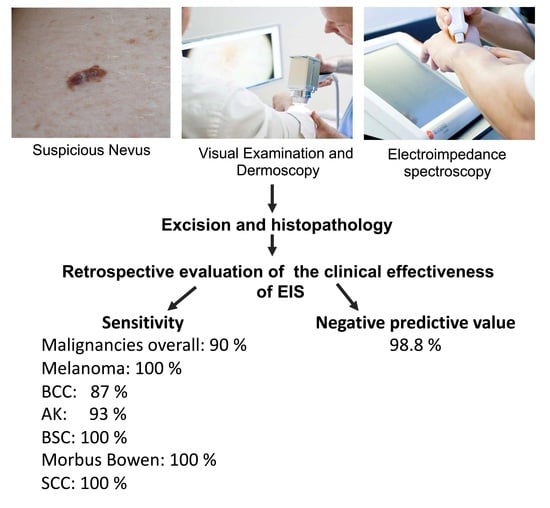
:There has recently been an increased interest in the use of novel automated technologies for the early detection of skin malignancies. We performed a retrospective analysis of the clinical effectiveness of electrical impedance spectroscopy (EIS) in detecting malignancies in an everyday clinical setting. After a thorough visual examination and dermoscopy, 909 abnormal lesions suspicious for malignancy were detected in 481 patients who presented in a private dermatology practice between 2015 and 2017 and evaluated with the EIS system. The histopathological results of the excised lesions were compared to the Neviscore, the output of the device. In total, 443 lesions (49%) received a negative Neviscore and were thus benign. On the other hand, 466 lesions received a positive Neviscore, indicating aberrations of the physiological cell structure. Of the 909 lesions, 45% were excised after visual and EIS examination. Of the excised lesions, 16% were diagnosed histopathologically as malignant. The EIS detected melanomas with 100% accuracy. The number needed to excise decreased from 17.5 to 7.8. The predictive value for a negative Neviscore was 98.9% (true negative results). EIS was found to be a valuable adjunct support tool when making clinical assessments of potentially malignant lesions.
1. Introduction
Melanomas and non-melanocytic skin cancer, primarily basal and squamous cell carcinoma, are the main types of malignant skin cancer in humans. Early detection and excision remain the most important prognostic factors for the treatment of all types of skin cancer, especially melanoma [1]. Detection continues to be challenging and melanoma-related mortality is still high. In 2020, an estimated 65,168 people were diagnosed with melanoma [2] and 179,219 with non-melanoma skin cancer [3] in Western Europe. There has been a significant interest in new technologies aimed at augmenting the detection rate achieved with the clinical diagnosis of skin cancer [4]. In the clinical risk assessment of patients with suspicious skin lesions, electrical impedance spectroscopy (EIS) proved to be a safe and accurate technical means of support, making additional information available to the physician responsible for deciding whether biopsy or excision of the lesion was indicated. EIS proved to increase detection efficacy in melanoma diagnostics [5]. Furthermore, a good discriminative power to distinguish NMSC from benign skin tissue was shown [6,7]. This retrospective study was conducted to investigate the accuracy of a three-step approach involving visual examination, dermoscopy, and EIS integrated into the cancer diagnosis carried out by a dermatological practice as part of its everyday clinical routine. Validation of the Neviscore (NS), the output of the EIS device, was performed by comparing the results of the NS with histopathological findings. The sensitivity and accuracy of the technique in detecting malignancies was assessed on the basis of this comparison. The positive and practical consequence for the patients is that there may be a large reduction in the number of excisions in the future.
2. Materials and Methods
The study involved the retrospective examination of 909 lesions detected in 481 patients from a dermatological practice, who had been diagnosed from 2015 to 2017 with the help of EIS. The ethical committee of the Bavarian Medical Association reviewed the study and exempted it from ethical approval.
This study included only lesions presented in the course of the cancer consultation of the respective practice. To evaluate the nature of a lesion, a thorough visual examination was performed, followed by dermoscopy in accordance with the current dermatological standards. Lesions were evaluated using the ABCD score as described by Stolz [8]. If the benign nature of a lesion was uncertain, EIS was used as an additional diagnostic tool.
Normal healthy skin tissue, in contrast to atypical diseased tissue, has a different cell size, shape, orientation, compactness, and structure of the cell membranes. These changes affect the cell’s ability to conduct and store electricity, a measurable property called electrical impedance. Thus, if a nevus is exposed to electrical signals during examination with Nevisense, changes can be detected using EIS and thus, melanoma and its precursors can be detected or ruled out. The technique is based on a harmless electrical signal emitted by the device which can detect and analyze these changes, allowing microinvasive bioimpedance measurements. EIS is a painless and non-invasive technology that measures the resistance in the tissue in the upper layers of the skin. Per measurement over 225 measurement points are taken and changes can be detected that indicate abnormalities in cell structure, orientation, size, molecular composition, and cell wall integrity. Incoming data are processed and classified using a complex algorithm. The EIS classifier provides an EIS score, the so-called Neviscore, on a scale of 0–10, that reflects the degree of atypia identified by the method. If the score is between 0 and 3 (negative NS), the lesion can be considered non-malignant and the patient may not have to have it removed. A score of 4 to 10 rates as positive NS, so that excision of the lesion is advisable. The examination takes only a few minutes and can be easily integrated into the normal preventive examination. During the examination, a stamp-shaped electrode is pressed twice onto the skin for each birthmark. The result in the form of the EIS score is available immediately. This makes it possible to make a decision based on reliable facts.
In this study, 909 lesions were suspicious for malignancy, and in addition to visual examination and dermoscopy, examined with EIS. The EIS was used as an objective, unbiased, and automated source of supportive information, and the score was taken into consideration when deciding on how to further manage the lesion. Nonetheless, the final decision about whether to operate was made on the basis of the doctor’s clinical expertise. All excised samples were sent for histopathological evaluation. In a retrospective approach, the NS of all excised lesions was compared to the definite histopathological classification to cross-check the accuracy of the assigned NS.
The next step involved taking (i) the number needed to excise (NNE), (ii) the sensitivity, and (iii) the negative predictive value (NPV) to assess the efficiency of diagnosing melanoma using the practical approach described previously [6]. The NNE is a metric providing information about the number of biopsies performed by the dermatological practice for every malignancy diagnosed. It was calculated by dividing the total number of lesions removed by the number of confirmed malignancies (including melanoma, basal cell carcinoma (BCC), squamous cell carcinoma (SCC), morbus Bowen, and basosquamous carcinoma). By comparing the NNE both with and without the EIS result, the increase in diagnostic accuracy through using EIS could be determined.
The sensitivity shows the proportion of malignant lesions correctly identified by receiving a positive Neviscore in the clinical setting. Sensitivity was calculated by dividing the total number of malignancies with a positive Nevisense score by the total number of malignancies (Supplementary Figure S1).
The NPV gives the ratio of true negative results compared to all negative results. Thus, the NPV was calculated as all benign lesions with a negative score (NS 0 to 3) divided by all lesions with a negative score. In this setting, the NPV shows the ratio of negatively scored lesions that were excised but were not malignant (Supplementary Figure S1).
Lesions included in this study had to meet particular criteria to be considered suitable for evaluation with EIS. Hence, the exclusion criteria for the use of EIS based on the manufacturer’s specifications for the device were in accordance with previous studies [6]. Included were all patients regardless of sex and age who presented to the dermatologist in the course of nevus screening or due to another reason for examination, whose nevi were classified as potentially malignant due to previous visual inspection, anamnesis, and dermoscopy. Suspicious lesions were additionally examined with Nevisense. In accordance with the manufacturer’s guidelines, some lesions were excluded from EIS evaluation. The exclusion criteria for the lesions studied were: (i) metastases of a pre-existing malignancy, (ii) a diameter of less than 2 mm or more than 20 mm, (iii) lesions on obviously non-intact skin, for example, in the form of eczema, scars, sunburn, previous injuries, trauma, etc., (iv) lesions on extremely hairy skin such as the scalp or beard, (v) lesions on the genital area or other mucosal location, (vi) lesions on tattoos, or in the presence of other foreign bodies or unphysiological interfering factors, and (vii) pedunculated lesions.
Limitations of the Study
Not all lesions that were considered suspicious for malignancy and that had a positive Neviscore were excised. This was due to the fact that some patients declined having an intervention. Most lesions that scored negative after EIS examination were not excised in order to spare the patient the intervention. Nonetheless, patients with skin abnormalities were monitored closely and asked to return for repeated examination in short intervals. However, because of the initial study design, the result of follow-up examinations was not reported and not considered when analyzing the data. Thus, no objective statement can be made whether malignant changes were present in lesions with a negative Neviscore.
3. Results
3.1. EIS Score Classification
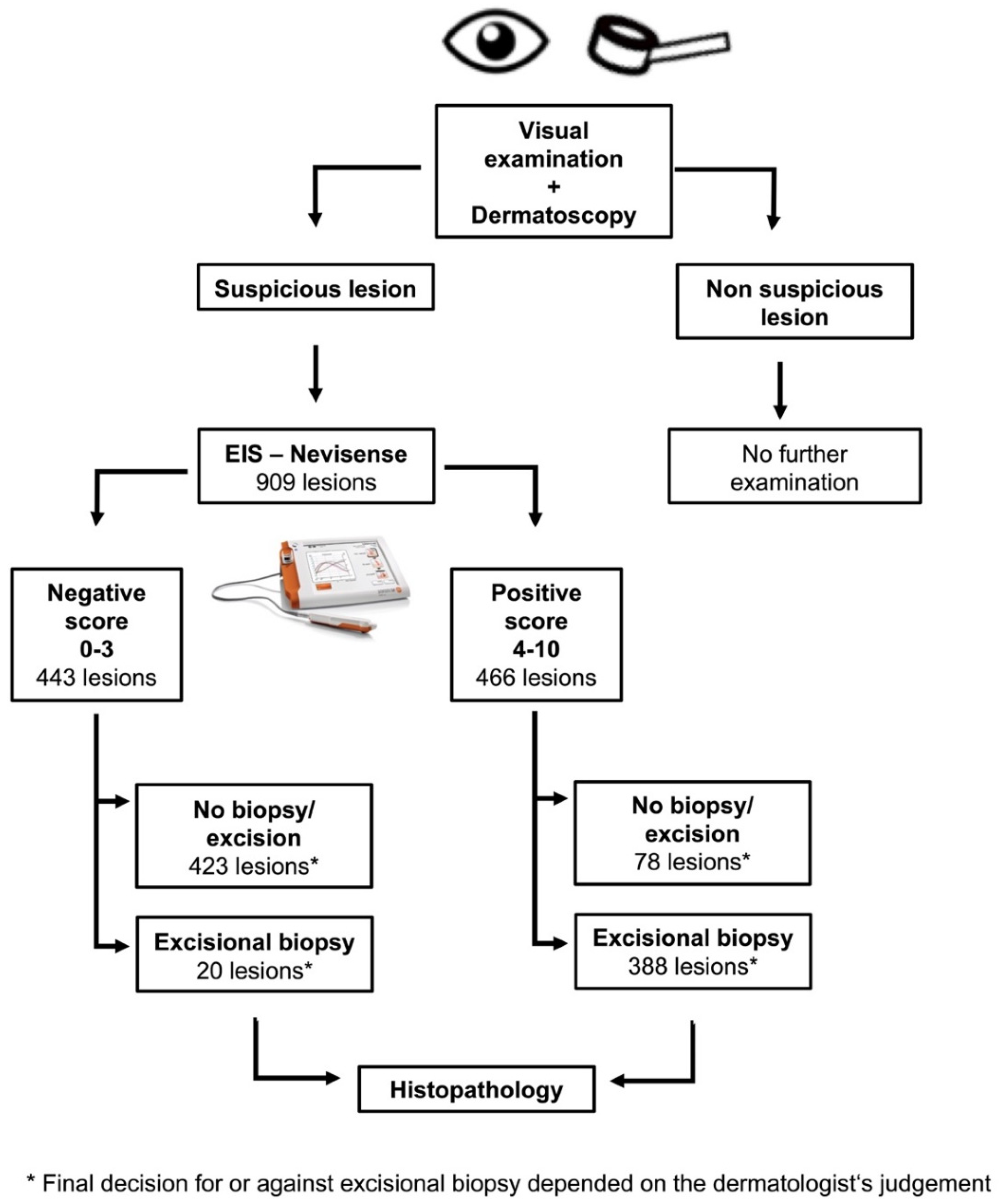
In total, 909 lesions presented for cancer diagnosis and suspicious for malignancy were included in the retrospective analysis. A thorough visual examination was performed, followed by dermoscopy and an EIS measurement (Figure 1). Of 909 suspicious lesions, 443 (48.7%) scored between 0 and 3 (negative NS) and 466 scored between 4 and 10 (positive NS) (Figure 1).
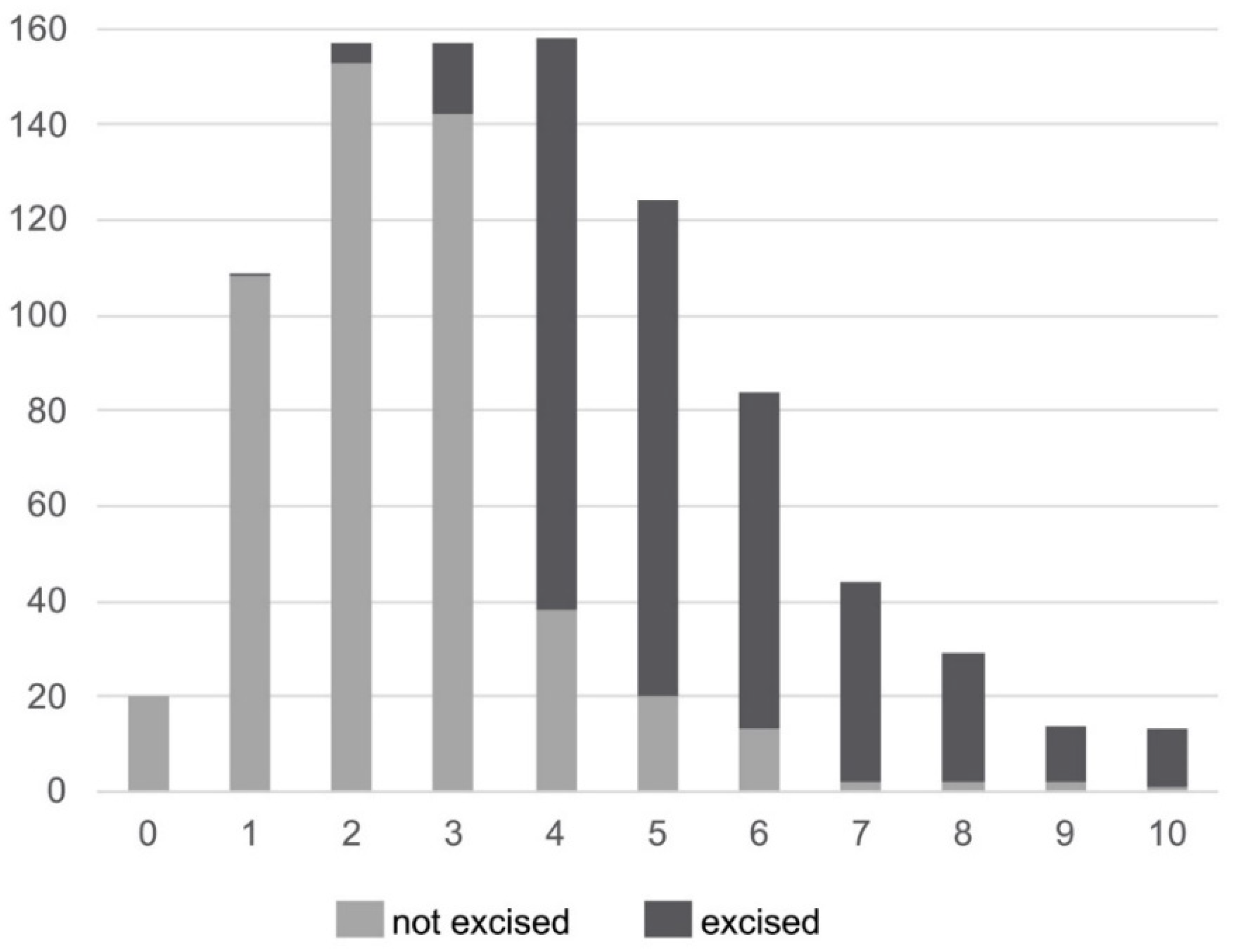
On the basis of this three-step approach, 408 (44.9%) lesions were excised and 501 lesions (55.1%) were left. Most of the lesions had a score in the middle, and only a few showed a strongly positive or a strongly negative NS. The number of lesions with the same NS was determined (Figure 2).
Of the 443 lesions with a negative NS (0 to 3), 20 were excised (Figure 1 and Figure 2) based on the judgement of the physician. Of the 466 lesions with a positive NS (4–10), 78 (17%) were left and 388 lesions (83%) were excised (Figure 1 and Figure 2). Ultimately, the decision whether to excise was based on the medical experience and expertise of the physician or determined by the explicit wish of the patient.
Furthermore, 423 (84.4%) of the 501 lesions not excised received an NS score of 0 to 3 and 78 (15.6%) received an NS score of 4 to 10 (Figure 2).
3.2. Histopathological Examination
The histopathological examinations of the excised lesions produced the following results: of 408 excised lesions, 66 (16%) were found to be malignant or pre-malignant and 342 (84%) were benign or showed various degrees of dysplasia. The histopathological examination of 408 excised lesions revealed 246 dysplastic nevi, of which 58 were of mild, 178 of moderate, three of severe, and seven of unknown severity. Additionally, 96 lesions were classified as benign (Table 1).
By means of histopathological diagnosis, 14 cases of actinic keratosis (AK), 38 basal cell carcinomas (BCC), seven cases of Bowen’s disease, three squamous cell carcinomas (SCC), three melanomas, and one basosquamous carcinoma were detected (Figure 3). The histopathological findings showed that moderate dysplastic lesions formed the highest percentage of abnormalities (43.6%), and melanomas were detected in 0.7% of cases (Table 1).
3.3. Histopathological Diagnosis and EIS Score
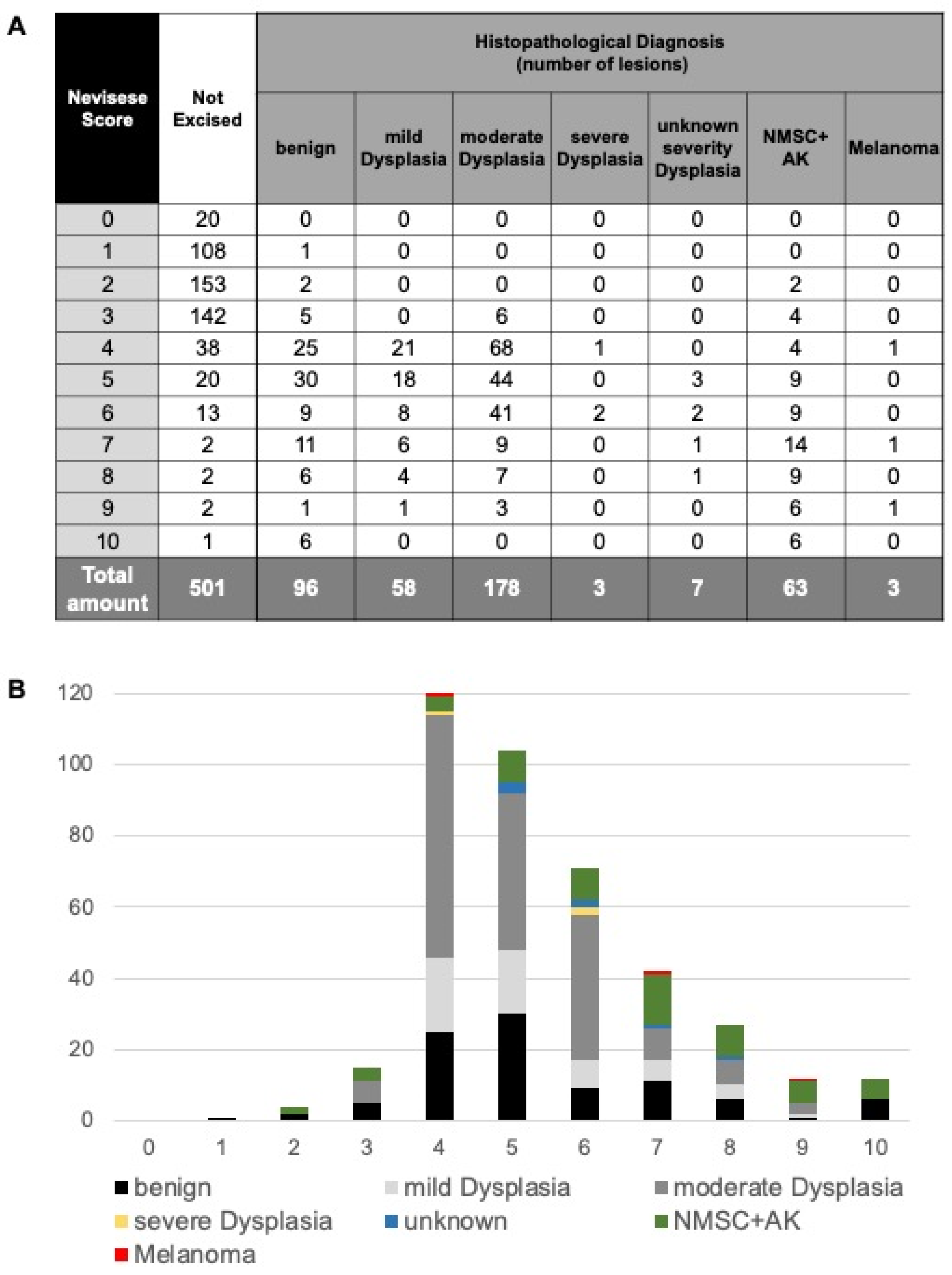
The final step involved comparing the output of the EIS system, the so-called Neviscore (NS), with the histopathological findings. Of 20 negatively scored (NS 0 to 3) but nevertheless excised lesions, histopathological diagnosis revealed two BCCs but no melanomas (Figure 4).
Histopathological analysis of all 388 positively scored (NS 4–10) excised lesions revealed 88 (23%) to be benign (Figure 4). All three histopathologically diagnosed melanomas as well as 57 (86%) of all other malignant and pre-malignant skin lesions were found in the group of positively scored lesions (Figure 4). Furthermore, 246 (63.4%) of all 388 positively scored excised lesions were found to display various degrees of dysplasia (Figure 4).
3.4. Accuracy Parameters for the Three-Step Approach
Based on the three-step approach of (i) visual observation, (ii) dermoscopy, and the (iii) EIS system, 408 lesions were excised. Taking all excised lesions with a definite histopathological diagnosis as the reference group, the overall sensitivity of the system in detecting malignancies was calculated to be 90%. Broken down into the individual malignancies, the sensitivity varied between 87% for BCC, 93% for AK, and 100% for melanoma, basosquamous carcinoma, Morbus Bowen, and spinocellular carcinoma (Supplementary Figure S1). Severe dysplastic skin lesions were also detected with a sensitivity of 100%. The negative predictive value (NPV) of the EIS system in detecting malignant skin tumors (melanoma, BCC, SCC, Bowen’s disease, and basosquamous carcinoma) was calculated to be 98.9% (Supplementary Figure S1); therefore, it was highly probable that lesions scoring an NS of 0–3 were not malignant.
When calculating the number needed to excise (NNE), we focused on all the malignancies including melanoma, BCC, spinocellular and basosquamous carcinoma, as well as Bowen’s disease. In the previous procedure (without EIS), all lesions classified as suspicious were biopsied. Therefore, all 909 suspicious lesions were considered for the calculation of NNE without EIS. NNE with EIS refers to the reduced number of only 408 lesions that were biopsied after additional EIS examination. For 52 malignant lesions diagnosed in the context of this study, the NNE without EIS was 17.5. Using the EIS as a second opinion to help the clinician decide whether surgical removal of a suspicious lesion was necessary, the NNE dropped to 7.8, thus dividing the number of excisions needed to correctly diagnose and remove a melanoma in half.
4. Discussion
This study was based on the everyday procedures of a private dermatology practice. In total, 45% of suspicious lesions were removed after visual and EIS testing; 16% of these were found through histopathological examination to be malignant. The EIS testing process proved to be 100% accurate in identifying melanomas, although these were extremely rare. This retrospective study showed how EIS was integrated into the everyday clinical routine and how rare melanomas were reliably detected through the additional use of EIS. Patients could be spared a high percentage of the excisions performed in the course of skin cancer prevention.
The EIS technology and its use in cancer detection has been investigated in several previous studies. In a prospective blinded clinical trial on the efficacy and safety of EIS [9] that involved 1943 eligible equivocal lesions that would have been excised on the basis of visual evaluation, EIS achieved a sensitivity for melanoma of 96.6% and for non-melanoma skin cancer of 100%, and a specificity of 34.1%. In another study, EIS was used in a protocol for routine sequential digital dermoscopy imaging for a total of 160 lesions [10]. The addition of EIS to the protocol reduced the need for sequential monitoring by 47% and also identified 83% of the melanomas three months earlier than the standard sequential monitoring protocol. In a reader study, 164 clinicians were shown clinical images of lesions, and made a total of 7380 clinical decisions with and without the EIS output [11]. When the EIS results were included, the mean sensitivity improved from 80.7% to 95.2% and the mean specificity from 50.4% to 58.6%. The non-melanoma skin cancer assessment was also highly accurate [9,10,12]. In accordance with previous findings, the results of this study confirm that the EIS system is highly reliable with regard to the detection of malign skin aberrations, especially melanoma.
The NNE is a key indicator of the clinical utility of EIS for the dermatologist and of the benefits for the patients. In this investigation, the NNE necessary for identifying one malignant lesion dropped significantly, while the efficiency in detecting malignant lesions increased. If the malignant potential of a suspicious lesion was in doubt, the EIS system predicted with a degree of high sensitivity whether a lesion needed to be excised. For the patients, this means that fewer lesions that are benign or mildly to moderately dysplastic are excised in the first place. Fewer surgical interventions are necessary, reducing the burden for the patients.
A more current guideline for the integration of EIS in the screening process for skin cancer was described in 2018 [13], and this process is in addition supported by Onkoderm—a German dermato-oncology association. According to the guideline, lesions scoring an NS of 0 to 3 can be considered as benign. Lesions with an NS of 4 to 6 are not in need of immediate excision but should be followed-up. The guideline recommends that lesions with an NS of 7 to 10 should be excised immediately. These recommendations can of course be overthrown by the expert opinion of the dermatologist. Our results support this approach and indicate the usefulness of integrating EIS into the screening process for skin cancer as described in the guideline.
This study provided insight into the outcome of using EIS in a single private practice. It is important to emphasize that the cohort of patients and lesions included in this study was seen during normal medical consultation over a defined time period. The prevalence of malignant lesions was lower than in the previous controlled clinical studies but considering the patient population in the clinic and the usage of the device on lesions with a lower suspicion of malignancy or dysplasia as well, this was to be expected. Since this was a retrospective analysis, the definite diagnosis of lesions that were left untreated was unknown and no reliable value for false negative results could be calculated. We were also confronted with a further limitation: several patients refused to undergo invasive treatment and have positively scored lesions excised. Another limitation of the study is the lack of follow-up. There was no evaluation available on the non-biopsy patients whether malignant skin cancer was detected at a later stage. This impeded an all-encompassing statistical evaluation with respect to, for example, the correlation between NS and histopathological diagnosis. Nonetheless, we found it important to include the data in its entirety to portray the everyday routine of a clinical practice. The EIS device nevertheless provided useful information to support a doctor’s recommendation regarding the surgical removal of a lesion [14,15]. The doctor’s decision is ultimately based on his or her experience. The unbiased and objective additional information of the NS can be helpful for deciding which lesions to excise and can contribute to reducing unnecessary excisions. This is particularly beneficial for people with several suspicious lesions and poor wound healing. As extensive studies have confirmed, the automated and highly standardized determination of the NS in addition to the subjective visual examination is a quick and reliable means of indicating the probability of malignancy. This makes the additional examination using EIS a suitable instrument for cancer screening and for monitoring the progress of suspicious lesions.
Supplementary Materials
The following supporting information can be downloaded at: https://www.mdpi.com/article/10.3390/dermato2020004/s1, Figure S1: Histopathological diagnosis, EIS score, and sensitivity. The table in Figure S1 is used to explain how the NNE values and the sensitivity were calculated.
Author Contributions
Author contributions are as follows: C.L. was responsible for the conceptualization of the study, and together with J.N.B., for project administration. Data collection and investigation were performed by C.L. and his team in his dermatological practice; he wrote and reported with input from all the authors. Data curation and formal analysis were conducted by J.N.B., I.S. and M.-L.v.B. and C.S. interpreted the data and provided assistance with preparing this manuscript. C.S. was the diagnosing histopathologist. He and his team assessed the histological samples and evaluated them. All authors have significantly contributed to the study’s design and conduction, have been closely involved in writing the manuscript and approved the final version of the manuscript. All authors have read and agreed to the published version of the manuscript.
Funding
This research did not receive any specific grant from funding agencies in the public, commercial, or not-for-profit sectors, thus no external funding.
Institutional Review Board Statement
Ethical review and approval were waived for this study because fully anonymized tissue was used in accordance with German legal requirements regarding ethical review of health research projects on the use of anonymized human material for research purposes.
Informed Consent Statement
Patient consent was waived due to the use of fully anonymized tissue in accordance with German legal requirements regarding ethical review of health research projects on the use of anonymized human material for research purposes.
Data Availability Statement
All data needed to evaluate the conclusions made in this paper are included in the main body of the manuscript. Additional data can be made available by the corresponding author upon reasonable request.
Acknowledgments
We thank all study investigators, participants, and patients who participated in this study.
Conflicts of Interest
The authors declare no conflict of interest.
References
- Glazer, A.M.; Rigel, D.S.; Winkelmann, R.R.; Farberg, A.S. Clinical Diagnosis of Skin Cancer: Enhancing Inspection and Early Recognition. Dermatol. Clin. 2017, 35, 409–416. [Google Scholar] [CrossRef] [PubMed]
- GLOBOCAN. Melanoma of Skin. Available online: https://gco.iarc.fr/today/data/factsheets/cancers/16-Melanoma-of-skin-fact-sheet.pdf (accessed on 15 March 2021).
- GLOBOCAN. Non-Melanoma Skin Cancer. Available online: https://gco.iarc.fr/today/data/factsheets/cancers/17-Non-melanoma-skin-cancer-fact-sheet.pdf (accessed on 15 March 2021).
- Winkelmann, R.R.; Farberg, A.S.; Glazer, A.M.; Cockerell, C.J.; Sober, A.J.; Siegel, D.M.; Leachman, S.A.; High, W.A.; Markowitz, O.; Berman, B.; et al. Integrating Skin Cancer-Related Technologies into Clinical Practice. Dermatol. Clin. 2017, 35, 565–576. [Google Scholar] [CrossRef] [PubMed]
- Svoboda, R.M.; Frankco, A.I.; Rigel, D.S. Electrical impedance spectroscopy versus clinical inspection approaches: Melanoma efficacy detection comparison. SKIN J. Cutan. Med. 2018, 2, 162–167. [Google Scholar] [CrossRef] [Green Version]
- Liebich, C.; von Bruehl, M.L.; Schubert, I.; Oberhoffer, R.; Sander, C. Retrospective evaluation of the performance of the electrical impedance spectroscopy system Nevisense in detecting keratinocyte cancers. Skin Res. Technol. 2021, 27, 723–729. [Google Scholar] [CrossRef] [PubMed]
- Sarac, E.; Meiwes, A.; Eigentler, T.; Forchhammer, S.; Kofler, L.; Hafner, H.M.; Garbe, C. Diagnostic accuracy of electrical impedance spectroscopy in non-melanoma skin cancer. Acta Derm. Venereol. 2020, 100, adv00328. [Google Scholar] [CrossRef] [PubMed]
- Nachbar, F.; Stolz, W.; Merkle, T.; Cognetta, A.B.; Vogt, T.; Landthaler, M.; Bilek, P.; Braun-Falco, O.; Plewig, G. The ABCD rule of dermatoscopy: High prospective value in the diagnosis of doubtful melanocytic skin lesions. J. Am. Acad. Dermatol. 1994, 30, 551–559. [Google Scholar] [CrossRef] [Green Version]
- Malvehy, J.; Hauschild, A.; Curiel-Lewandrowski, C.; Mohr, P.; Hofmann-Wellenhof, R.; Motley, R.; Berking, C.; Grossman, D.; Paoli, J.; Loquai, C.; et al. Clinical performance of the Nevisense system in cutaneous melanoma detection: An international, multicentre, prospective and blinded clinical trial on efficacy and safety. Br. J. Dermatol. 2014, 171, 1099–1107. [Google Scholar] [CrossRef] [PubMed]
- Rocha, L.; Menzies, S.W.; Lo, S.; Avramidis, M.; Khoury, R.; Jackett, L.; Guitera, P. Analysis of an electrical impedance spectroscopy system in short-term digital dermoscopy imaging of melanocytic lesions. Br. J. Dermatol. 2017, 177, 1432–1438. [Google Scholar] [CrossRef] [PubMed] [Green Version]
- Svoboda, R.M.; Prado, G.; Mirsky, R.S.; Rigel, D.S. Assessment of clinician accuracy for diagnosing melanoma on the basis of electrical impedance spectroscopy score plus morphology versus lesion morphology alone. J. Am. Acad. Dermatol. 2019, 80, 285–287. [Google Scholar] [CrossRef] [PubMed] [Green Version]
- Braun, R.P.; Mangana, J.; Goldinger, S.; French, L.; Dummer, R.; Marghoob, A.A. Electrical Impedance Spectroscopy in Skin Cancer Diagnosis. Dermatol. Clin. 2017, 35, 489–493. [Google Scholar] [CrossRef] [PubMed]
- Welzel, J.; Reinhold, U. Atypien von Hautveränderungen präzise messen. Der. Dtsch. Dermatol. 2018, 847–851. [Google Scholar] [CrossRef]
- Litchman, G.H.; Teplitz, R.W.; Marson, J.; Rigel, D.S. Impact of Electrical Impedance Spectroscopy on Dermatologists’ Number-Needed-to-Biopsy Metric and Biopsy Decisions for Pigmented Skin Lesions. J. Am. Acad. Dermatol. 2020, 85, 976–979. [Google Scholar] [CrossRef] [PubMed]
- Waldmann, A.; Nolte, S.; Geller, A.C.; Katalinic, A.; Weinstock, M.A.; Volkmer, B.; Greinert, R.; Breitbart, E.W. Frequency of excisions and yields of malignant skin tumors in a population-based screening intervention of 360,288 whole-body examinations. Arch. Dermatol. 2012, 148, 903–910. [Google Scholar] [CrossRef] [PubMed] [Green Version]
Figure 1.
Flow chart of the individual examination steps.

Figure 2.
The EIS score and the number of lesions excised and left.

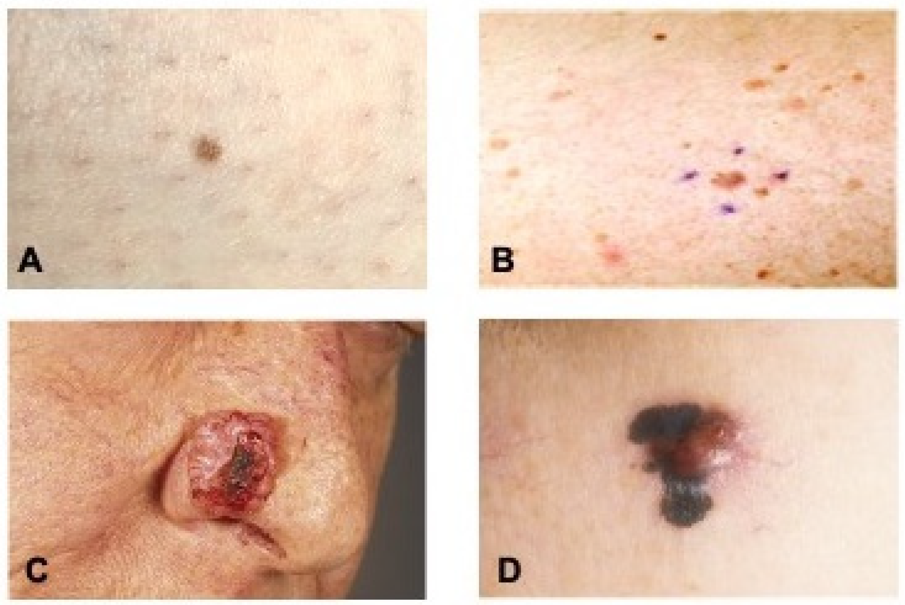
Figure 3.
Macroscopic images of the depicted skin lesions including the result of the EIS measurement. (A) Nevus (Neviscore 2). (B) Dysplastic lesion (Neviscore 5). (C) Basal cell carcinoma (Neviscore 6). (D) Malignant melanoma (Neviscore 10).
Figure 3.
Macroscopic images of the depicted skin lesions including the result of the EIS measurement. (A) Nevus (Neviscore 2). (B) Dysplastic lesion (Neviscore 5). (C) Basal cell carcinoma (Neviscore 6). (D) Malignant melanoma (Neviscore 10).

Figure 4.
Number of not excised and excised lesions for each EIS score. NMSC+AK (non-melanocytic skin cancer and actinic keratosis). (A) Excised lesions assigned to their histopathological classification and EIS score. (B) The number of histopathologically diagnosed lesions per EIS score.
Figure 4.
Number of not excised and excised lesions for each EIS score. NMSC+AK (non-melanocytic skin cancer and actinic keratosis). (A) Excised lesions assigned to their histopathological classification and EIS score. (B) The number of histopathologically diagnosed lesions per EIS score.


Table 1.
The excised lesions were diagnosed by histopathological examination.
| Diagnosis | Number of Lesions | % of Lesions |
|---|---|---|
| Melanoma | 3 | 0.3 |
| Basal Cell Carcinoma | 38 | 4.2 |
| Squamous Cell Carcinoma | 3 | 0.3 |
| Bowen’s Disease | 7 | 0.8 |
| Basosquamous Carcinoma | 1 | 0.1 |
| Actinic Keratosis | 14 | 1.5 |
| Dysplasia Severe | 3 | 0.3 |
| Dysplasia Moderate | 178 | 19.6 |
| Dysplasia Mild | 58 | 6.4 |
| Dysplasia Unknown Severity | 7 | 0.8 |
| Benign | 96 | 10.6 |
| Not Excised | 501 | 55.1 |
| TOTAL | 909 | 100% |
Publisher’s Note: MDPI stays neutral with regard to jurisdictional claims in published maps and institutional affiliations. |
© 2022 by the authors. Licensee MDPI, Basel, Switzerland. This article is an open access article distributed under the terms and conditions of the Creative Commons Attribution (CC BY) license (https://creativecommons.org/licenses/by/4.0/).
Share and Cite
MDPI and ACS Style
Liebich, C.; Bartsch, J.N.; Schubert, I.; von Bruehl, M.-L.; Sander, C. Electrical Impedance Spectroscopy Improves Skin Cancer Detection and Reduces the Number of Biopsies. Dermato 2022, 2, 21-29. https://doi.org/10.3390/dermato2020004
AMA Style
Liebich C, Bartsch JN, Schubert I, von Bruehl M-L, Sander C. Electrical Impedance Spectroscopy Improves Skin Cancer Detection and Reduces the Number of Biopsies. Dermato. 2022; 2(2):21-29. https://doi.org/10.3390/dermato2020004
Chicago/Turabian StyleLiebich, Christoph, Jana Nadine Bartsch, Irene Schubert, Marie-Luise von Bruehl, and Christian Sander. 2022. "Electrical Impedance Spectroscopy Improves Skin Cancer Detection and Reduces the Number of Biopsies" Dermato 2, no. 2: 21-29. https://doi.org/10.3390/dermato2020004