Review

Digital Subtraction Angiography (DSA) Technical and Diagnostic Aspects in the Study of Lower Limb Arteries

1
Department of Diagnostic Imaging, Oncologic Radiotherapy and Hematology—A. Gemelli University Hospital Foundation IRCCS, L.go A. Gemelli 8, 00168 Rome, Italy
2
Department of Radiology, Fatebenefratelli Hospital Foundation for Health Research and Education, Via di Ponte Quattro Capi 39, 00186 Rome, Italy
3
Fondazione Policlinico Universitario “Agostino Gemelli” IRCCS, Catholic University of the Sacred Heart, L.go F. Vito 1, 00168 Rome, Italy
*
Author to whom correspondence should be addressed.
Radiation 2022, 2(4), 376-386; https://doi.org/10.3390/radiation2040028
Submission received: 8 September 2022 / Revised: 16 October 2022 / Accepted: 17 October 2022 / Published: 1 November 2022

Abstract

:

Simple Summary

Digital subtraction angiography has great utility in endovascular arterial interventions in the lower limbs. Knowledge of angiographic findings, both pathologic and iatrogenic, and how to interpret them is mandatory to obtain a good outcome and limit complications.

Abstract

Cardiovascular diseases represent one of the most frequent diseases worldwide; among these, lower limb ischemia is a threatening condition, which can lead to permanent disability if not promptly and correctly diagnosed and treated. A patient’s clinical evaluation and diagnostic imaging (e.g., color-Doppler ultrasound, computed tomography angiography (CTA), and magnetic resonance imaging (MRI)) are mandatory to carefully assess arterial lesion extension and severity. Digital subtraction angiography (DSA) is a minimally invasive technique that represents the gold standard for percutaneous revascularization treatment of symptomatic patients who are refractory to medical management. However, when dealing with patients with lower limb terminal ischemia, the correct interpretation of diagnostic DSA findings is mandatory for treatment re-planning and to effectively evaluate post-treatment results and complications. The purpose of this review is to provide interventional radiologists and endovascular practitioners with an up-to-date practical guide to diagnostic angiography of the lower limbs, which is mandatory to address correct treatment decisions and post-treatment evaluation.

1. Introduction

Lower limb peripheral artery disease represents a common pathology, affecting more than 200 million adults worldwide and leading to adverse clinical outcomes and physical impairments, such as pain, soft tissue ulceration, osteomyelitis, claudication, and eventually amputation [1]. Common risk factors are represented by diabetes, dyslipidemia, cigarette smoking, and arterial hypertension.
Diagnostic catheter angiography (DCA) was once considered the gold standard for the diagnosis and assessment of peripheral arterial disease lower limb pathology; however, as noninvasive and panoramic techniques such as computed tomography angiography (CTA) and magnetic resonance angiography (MRA) became more and more available, giving lower risk of complications and similar diagnostic efficacy, DCA was progressively replaced [2,3]. Multidetector CTA is a validated diagnostic imaging method for the evaluation of patients with claudication and limb ischemia, to grade disease severity and plan treatment. CTA is currently considered as the most feasible, useful, reliable, and accurate diagnostic imaging technique in the assessment of lower limb artery disease, with an overall sensitivity of 92–99%, specificity of 93–98%, accuracy of 94–98%, positive predictive value of 97%, and negative predictive value of 99% [4,5,6]. CTA is noninvasive and has a low effective radiation dose; however, CTA usefulness can be limited in determining the degree of stenosis in the case of highly and extensively calcified lesions [7]. Contrast-enhanced MRA appears to be more specific than CTA in ruling out arterial stenosis greater than 50%, and more sensitive than duplex ultrasound (US) in ruling in arterial stenosis smaller than 50% [8]. CTA and MRA are characterized by low or absent use of ionizing radiation, three-dimensional volumetric display, direct plaque visualization, identification of abnormal nonvascular findings, as well as multiplanarity [2]. Nowadays, when it comes to endovascular treatment of arterial lower limb pathology, digital subtraction angiography (DSA) is the imaging method of choice [9]. Noninvasive imaging such as MRA and CTA before DSA or surgery is validated and commonly used in order to help the planning of the endovascular intervention [10]. While noninvasive diagnostic imaging is mandatory for disease identification, diagnosis, and stratification, in order to address every patient to the most useful and correct treatment, as well as for treatment planning, DSA can be used for both diagnostic and therapeutic purposes simultaneously: in particular, during every DSA for lower limb ischemia treatment, a diagnostic evaluation must always be performed to correctly determine the pathologic site that will therefore be treated.

2. Digital Subtraction Angiography Technique

Nowadays, the standard for contrast arteriography of the lower limb is intra-arterial DSA, which replaced the previously used conventional film-screen angiography (FSA) with cut-film radiographs and rapid film changers [11]. Conventional angiography is based on image acquisition after contrast medium injection: the obtained image is made up of blood vessels, bones, and other structures; this superimposition of nonvascular structures makes it difficult to accurately evaluate the blood vessels. Therefore, to obtain better visualization of vascular structures, a pre-contrast image (so-called “mask image”) is firstly acquired and is digitally subtracted to the post-contrast images, granting the removal of distracting structures [12]. DSA grants the production of good and diagnostic angiograms, evaluating vessel stenosis and opacification, in particular when used for single-limb lower extremity angiography [13]. DSA is able to visualize small vessels with little use of contrast medium; this is especially useful in diabetes-related arterial disease [14].

2.1. Pre-Procedural Evaluation

Before performing DSA, it is mandatory to collect data from patients affected by lower-limb pathology, regarding history and physical examination, laboratory study, as well as noninvasive imaging of lower limb arteries in order to determine the appropriateness of DSA and the most appropriate endovascular treatment. In particular, it is advised to elicit an accurate description of the patient’s symptoms and the consequent impairment in daily activities. Symptoms that do not compromise common daily activities contraindicate an invasive procedure such as DSA. Physical examination and patient’s history most of the time will identify nonvascular etiologies for a patient’s symptoms (usually originating from musculoskeletal or neurologic diseases); in addition, a pre-procedural patient’s evaluation allows the categorization of lower limb ischemia as acute versus chronic, and the assessment of the degree of ischemia severity [15]. Atopy or allergy history as well as any prior adverse reaction to iodinated contrast media must be known prior to DSA.

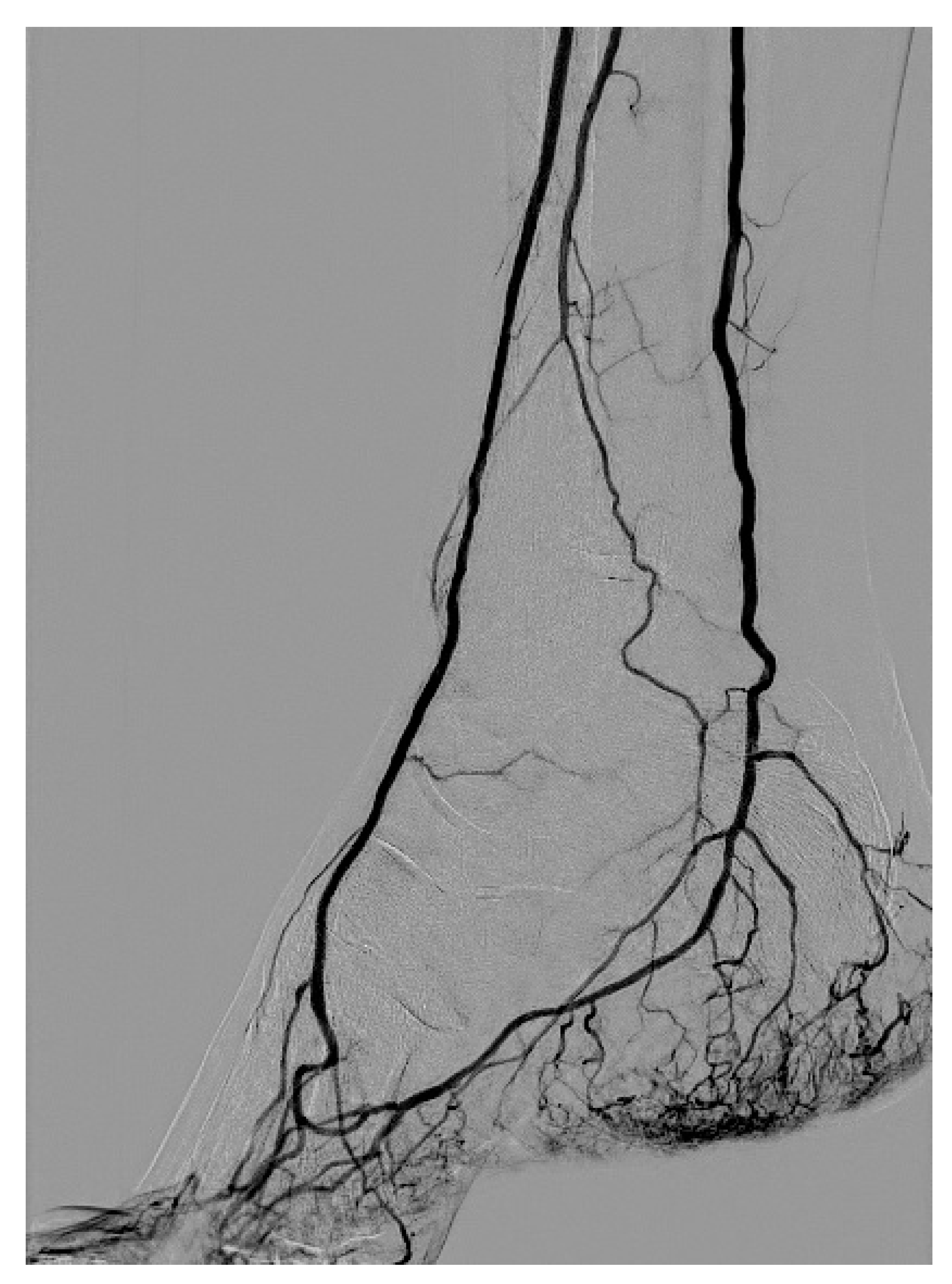
2.2. Angiography Technique

DSA is usually performed through an antegrade approach, puncturing the common femoral artery and placing a 5 French vascular sheath. Diagnostic angiography can be obtained by injecting iodinated contrast medium through the vascular sheath, or using a diagnostic straight catheter. Injection can be performed manually or with an automatic injector, which can provide a controlled flow rate and contrast medium dose. The injection volume and flow rate varies depending on the examined site and on the diagnostic catheter position. In the case of iliac artery disease, a retrograde approach is performed through the common femoral artery. When an ipsilateral common femoral artery approach is not feasible due to vessel occlusion or highly calcified plaques, the target limb can be reached using a contralateral retrograde common femoral artery access and using a “cross-over” technique, which consists of reaching the aortic lumen and then catheterizing the contralateral iliac artery using a pigtail diagnostic catheter.
Apart from procedural steps, it is essential to know that new angiography machines have automated sequences, which set the frame speed and exposure values according to different segments studied, such as the abdomen, brain, and lower extremities. Advanced computer processing power has made the vastly used techniques such as subtraction, pixel shifting, re-masking, or view tracing simple and feasible. The operator at the angiographic table can easily perform road mapping (superimposition of the fluoroscopic image on a DSA image; Figure 1) and un-subtracted image referencing (fluoro save mode). Collimation, magnification, edge filters, table positioning, and the gantry angle can be changed by the operator in order to optimize visualization of the arteries. An important technical improvement has been the introduction of high resolution flat panel image intensifiers, which provide great image quality minimizing parallax distortion. New flat panels have a large field of view (FOV) that can cover large body segments, bringing lower contrast medium usage and the reduction in radiation exposure both for the patient and the operator. DSA imaging evaluation can be performed on adequate portable C-arm machines, in the case of patients requiring endovascular treatment in the operating room, or on ceiling-fixed angiography machines in the angiographic suite.
To optimize and improve arterial vessel visualization in the lower limb, endovascular administration of vasodilator drugs can be performed, the most used being nitroglycerin and papaverine [16,17,18].
Optimal visualization of the arterial vessel is mandatory to evaluate the pathologic segment and to guide the endovascular guidewire and catheters. Therefore, the use of oblique projections of the C-arm or of the angiography machine is essential: while the superficial femoral artery and popliteal artery can be investigated using a frontal projection, below the knee arteries are evaluated with a slight anterior oblique projection, usually no more than 10 degrees, in which the proximal tibio-peroneal joint is clearly seen (Figure 2). Distal tibial arteries, the pedidial artery, and plantar arch can be evaluated using a pure frontal and, especially, a lateral projection (Figure 3). In the case of common and external iliac arteries, oblique projections help to identify the origin of the internal iliac (or hypogastric) artery, which is mandatory in the case of iliac stenting; identification of the hypogastric artery is performed in the same way as in the case of uterine fibroid embolization, and consists of a 20 degree ipsilateral anterior oblique projection [19].

3. Angiographic Findings

DSA findings can be represented both by pathological findings and procedural iatrogenic complications.

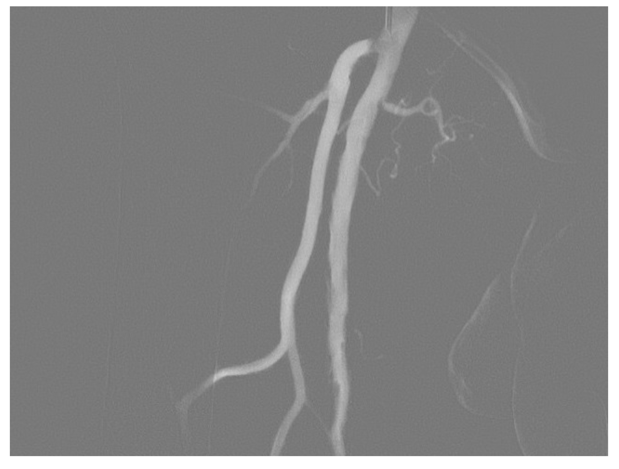
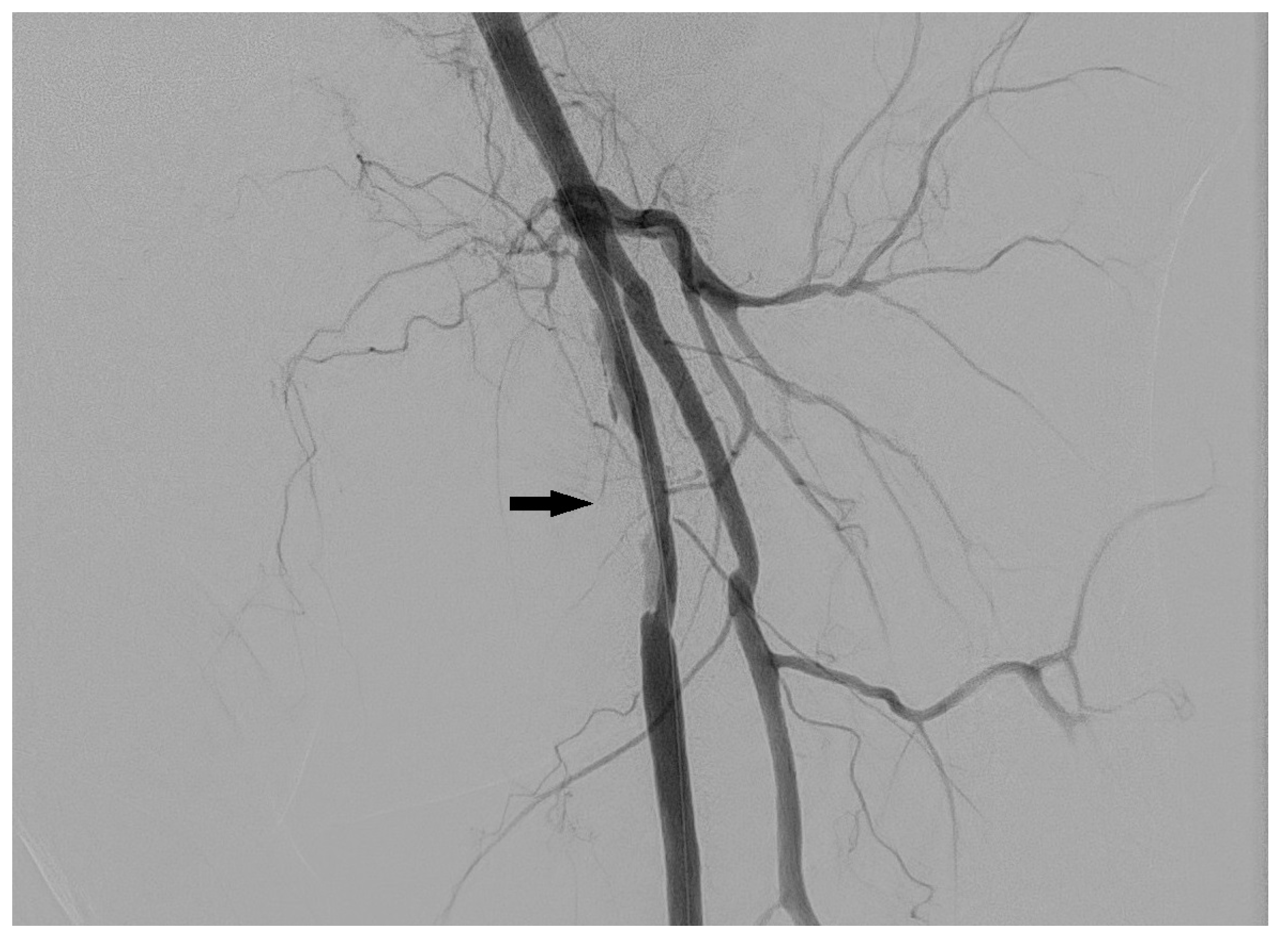
3.1. Stenosis and Occlusion
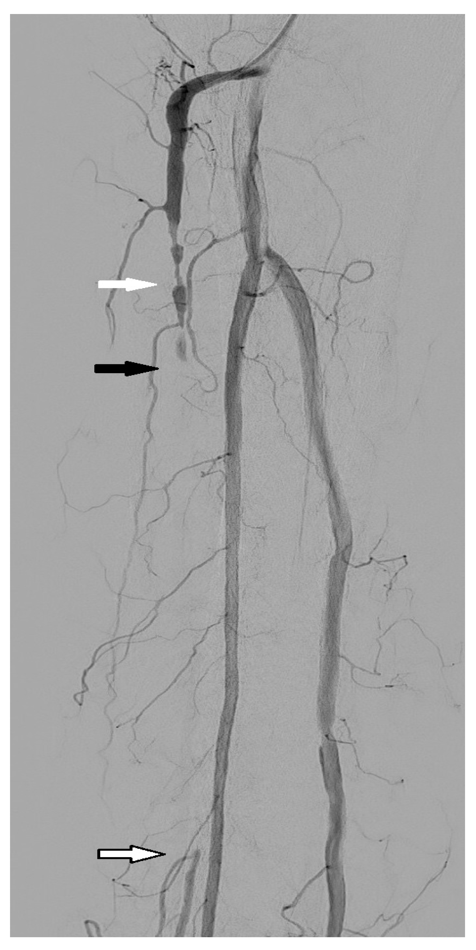
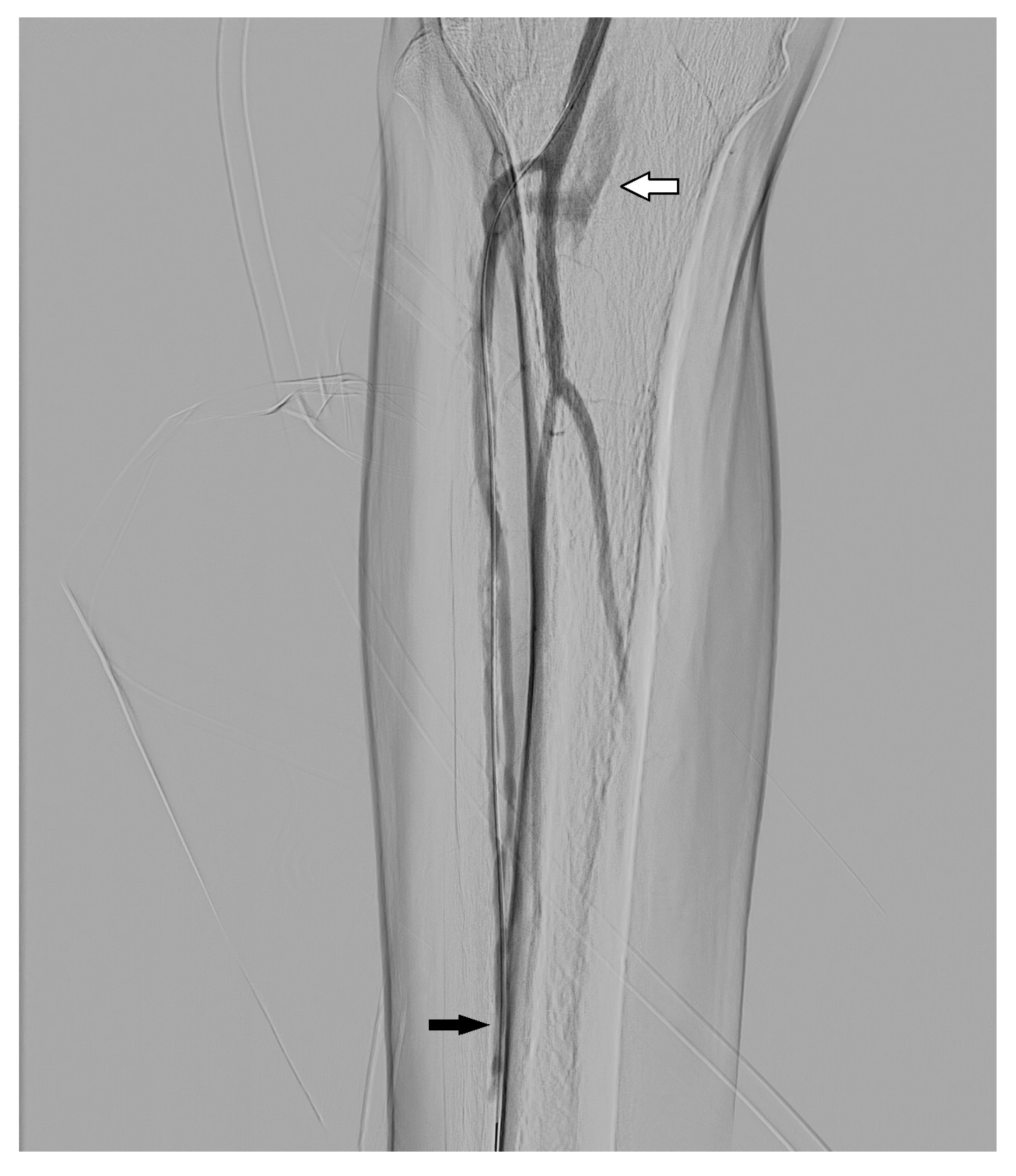
Arterial stenosis is characterized by a narrowing of the arterial lumen, which reduces the blood flow to the lower extremity (Figure 4); stenosis can be focal, short, or long. On the other hand, occlusion is characterized by a blockage of the artery, which stops the blood flow to the limb extremity (Figure 5). Occlusion can be focal, short, or long, as well as acute or chronic; in the case of chronic occlusion, collateral circulation usually provides inflow to the distal artery. Causes of stenosis and occlusion can be various, ranging from thrombus to atherosclerotic or calcified plaque [20]. The Transatlantic Inter-Society Consensus for the management of peripheral artery disease classified femoropopliteal stenoses and obstructions based on their number and length [21].
Acute occlusion (Figure 6) is represented by a sudden blood flow blockage of one or more vessels, and can be due to an anomalous coagulation of blood after injury of the vessel’s intima layer, after catheter or guidewire passage, or after angioplasty balloon inflation. As the guidewire or diagnostic catheter or angioplasty balloon hits a thrombus or a mural plaque, or when a clot forms attached to the catheter and is afterwards dislodged, the consequences can be acute embolization and ischemia.

3.2. Dissection

Arterial dissection (Figure 7) happens when an intimal tear allows blood to enter the media layer, leading to a second channel filled with blood within the wall. The so-called “true lumen” is the one lined by the intima, while the “false lumen” is the channel filled with blood in the media. The pressure in the false lumen is higher compared to the true lumen because the blood has poor outflow. This leads to the compression of the true lumen and causes an impairment of the arterial flow in the artery in the limb extremity (flow-limiting dissection). Iatrogenic arterial dissection can be caused by passage of the guidewire (as the tip of the guidewire can advance in the subintimal space with very little resistance), or as a result of angioplasty balloon inflation.

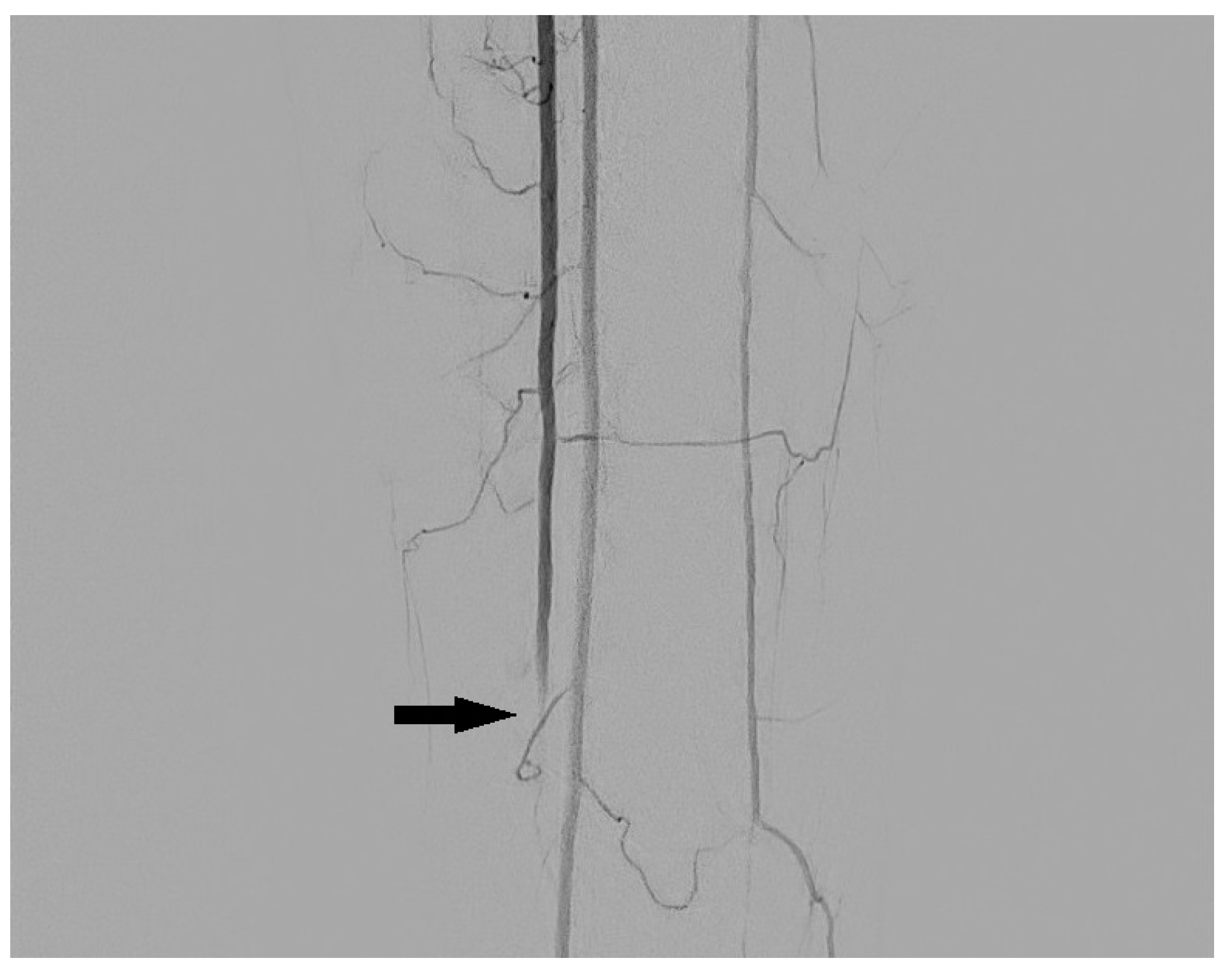
3.3. Arteriovenous Fistula

The arteriovenous fistula (Figure 8) is an anomalous communication between an artery and a vein, with consequent blood flow from the structure at a higher pressure (artery) to a structure at a lower pressure (vein). It can be iatrogenic, occurring after guidewire passage through a damaged vessel wall, or after angioplasty of calcified plaques. Arteriovenous fistula is identified after contrast medium injection as an early opacification of a vessel that is not referable to an artery, with a blood flow going in the opposite direction with respect to the arterial flow. A prompt identification of an arteriovenous fistula is mandatory, particularly to exclude a blood theft from the distal artery, which could cause or worsen the acute limb ischemia.

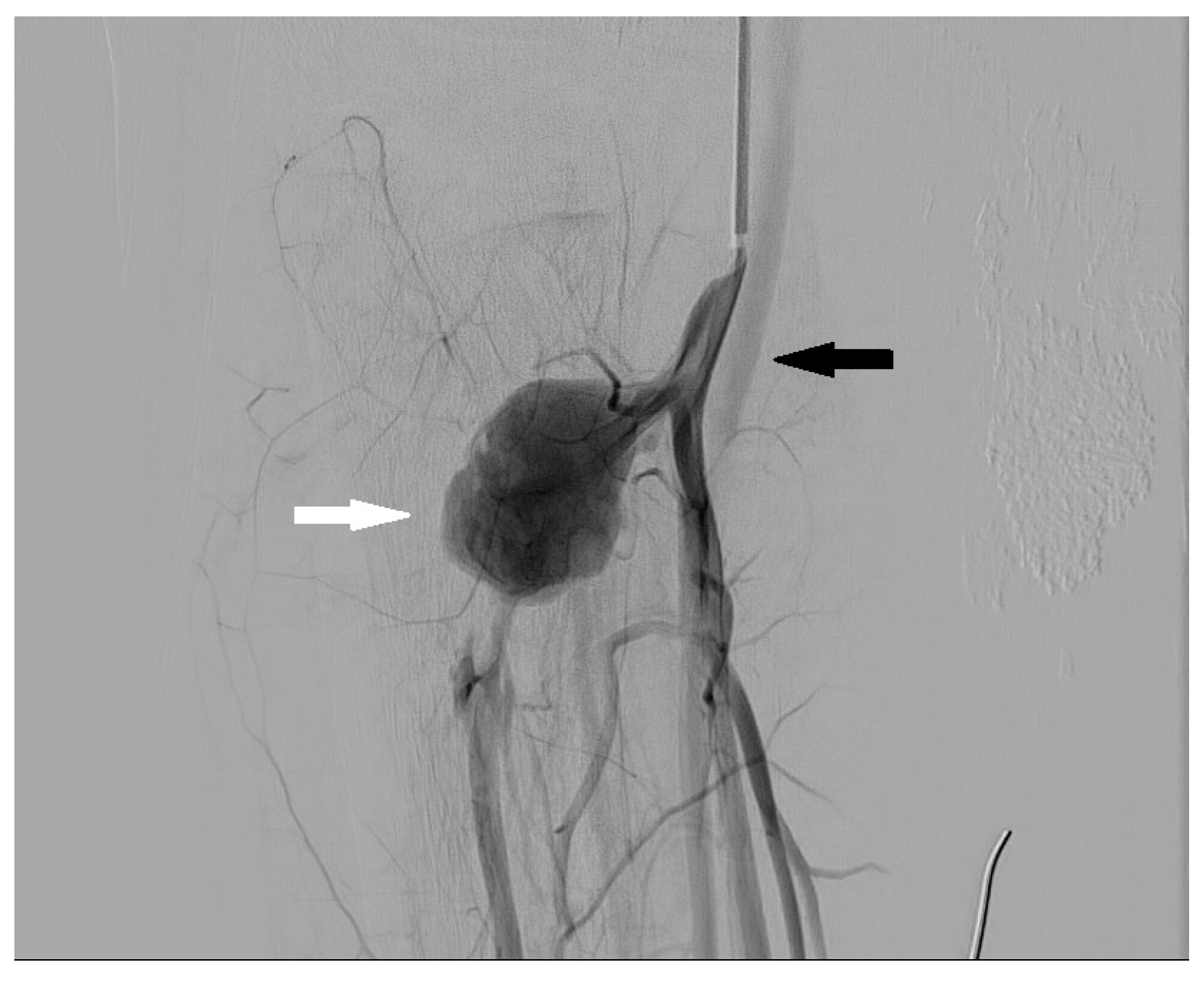
3.4. Aneurysm and Pseudoaneurysm

An aneurysm is characterized by an enlargement of the vessel diameter more than 50%, compared to a proximal healthy segment, with integrity of the vessel wall. On the other side, a pseudoaneurysm (Figure 9) is formed when the vessel wall is damaged and the blood occupies the space between the intima-media and the other layers, leading to a focal enlargement of the vessel. Usually, a vessel dilatation occurring during/after interventional endovascular procedures of a lower limb is to be considered pseudo-aneurismatic (until proven otherwise), linked to a wall injury.

4. Risks of DSA

4.1. Percutaneous Arterial Access-Related Complications

DSA represents an invasive imaging modality for both the diagnosis and treatment of PAD; however, it bears a nonnegligible complication rate (about 10%) [6]. Angiographic procedures are well tolerated by the patient, but there is always a risk for morbidity and mortality [15]. Many complications are self-limited and mild, with little to no treatment required. Apart from the abovementioned dissection, acute thrombosis, pseudoaneurysm, and arteriovenous fistula, the most common complication related to the percutaneous arterial puncture is the hematoma formation at the puncture site. The use of small bore catheters (3 to 5 French) for the diagnostic workup actively helps to reduce this kind of complication; however, the increase in endovascular treatments, which require a larger working sheath (from 5 to 11 French), increases the risks of hematoma formation. A hematoma usually arises immediately after sheath removal while applying pressure at the puncture site or shortly after the patient leaves the angio-suite. Since hematomas occur early, almost all can be noticed and kept under control before intervention is needed. Little hematomas tend to resolve spontaneously in a few days. Performing good hemostasis is difficult when a puncture is performed cranially to the inguinal ligament, as there is no bony structure under the femoral artery, which can provide support during compression at the puncture site. Moreover, retroperitoneal bleeding can happen and its early detection can be difficult and challenging. To avoid or at least reduce hematoma and bleeding risks, it is advised to identify the femoral head using fluoroscopy prior to femoral puncture.

4.2. Contrast Medium-Related Risks

The use of iodinated contrast media is associated with rare but worrying complications. Most important ones are contrast-induced nephropathy and fatal systemic reactions [9]. Many patients with lower limb peripheral artery disease are old and/or affected by diabetes, with an impaired renal function; in those patients, the nephrotoxicity is important to consider [22]. Other imaging modalities (such as MRA) or carbon dioxide angiography should be considered when the estimated glomerular filtration rate is inferior to 45 mL/min/1.73 m2 [15,23].

4.3. Radiation Dose

DSA is associated with a high radiation dose with the consequent risk of radiation-induced malignancy. It is mandatory to use post-processing imaging to improve DSA image quality and at the same time reduce the radiation dose [9]. Another important factor is that many patients repeat DSA because of their disease (as PAD often involves both lower limbs), therefore limiting radiation exposure has to be considered.
The Society of Interventional Radiology (SIR) published in 2012 a document containing guidelines for patient radiation doses in interventional radiology [24]. Current European guidelines underline that the patient’s total dose during procedures should be recorded as part of the programs related to quality and safety. Literature is available on the topic to point interventional physicians to the best clinical practice [25]. Some precautions include appropriate training in radiation protection, usage of the available dose-reduction instruments and the lowest acceptable fluoroscopy rates, avoidance of magnification whenever possible, usage of last image hold and recorded loops, as well as the routine and smart utilization of collimators.

5. Conclusions and Future Perspectives

Digital variance angiography (DVA) is a particular type of kinetic imaging that enables the viewing of motion on picture sequences produced by penetrating radiations. DVA allows for the creation of angiographic image series using contrast agents and fluoroscopy, and offers noticeably better picture quality than DSA imaging for lower limb angiography with metal implants also reducing radiation exposure [26]. DVA can be used for carbon dioxide angiography. A prospective study by Gyánó et al. compared the quality of DVA and DSA images of patients undergoing lower limb angiography with iodinated contrast medium. Signal-to-noise ratio (SNR) and image quality were evaluated, showing higher SNR in DVA images, meaning that DVA could better visualize blood vessels; DVA was also considered better in 69% of cases regarding image quality, particularly for popliteal and foot regions, highlighting its usefulness on small vessels. The authors concluded that DVA can provide angiographic images with the same quality as DSA, but the dosage of radiation delivered and/or the amount of contrast material needed to obtain the same vascular visibility might be reduced [27]. Óriás et al. investigated the feasibility of lower limb DVA using carbon dioxide, comparing SNR and image quality to DSA, finding that DVA provided higher quality images regardless of the anatomical site [28,29]. Software for the perfusion analysis of bi-dimensional digital DSA images are available [30]. They allow quantification of perfusion parameters such as blood volume, blood flow and mean transit time on subtraction images. The method allows a quantitative analysis of post-angioplasty treatment based on pre- and post-perfusion of foot tissue vascularization. A possible downside is the requirement of the patient’s immobility of the lower limbs to avoid motion artifacts.

Author Contributions

Conceptualization, A.P. and A.T.; methodology, A.P.; investigation, F.A.; resources, V.L. and G.M.; data curation, P.B. and V.L.; writing—original draft preparation, L.S., F.A. and A.P.; writing—review and editing, A.P. and A.T.; visualization, P.B., L.S. and A.P.; supervision, R.I.; project administration, A.P. All authors have read and agreed to the published version of the manuscript.

Funding

This research received no external funding.

Institutional Review Board Statement

This study was conducted in accordance with the Declaration of Helsinki. The figures originating from A. Gemelli University Hospital Foundation IRCCS, were taken from the hospital database and were fully anonymized. The internal meeting of the Integrated Research Ethics Board of the hospital determined that this study did not need ethical approval as it was only retrospectively depicting physiological and pathological conditions seen at DSA examinations. The present publication was conceived late after the complete resolution of the patients’ disease.

Informed Consent Statement

Not applicable.

Data Availability Statement

The data presented in this study are available in this article.

Conflicts of Interest

The authors declare no conflict of interest.

References

  1. Dua, A.; Lee, C.J. Epidemiology of Peripheral Arterial Disease and Critical Limb Ischemia. Tech. Vasc. Interv. Radiol. 2016, 19, 91–95. [Google Scholar] [CrossRef] [PubMed]
  2. Levin, D.C.; Rao, V.M.; Parker, L.; Frangos, A.J.; Sunshine, J.H. The effect of the introduction of MR and CT angiography on the utilization of catheter angiography for peripheral arterial disease. J. Am. Coll. Radiol. 2007, 4, 457–460. [Google Scholar] [CrossRef] [PubMed]
  3. Patel, M.C.; Levin, D.C.; Parker, L.; Rao, V.M. Have CT and MR Angiography Replaced Catheter Angiography in Diagnosing Peripheral Arterial Disease? J. Am. Coll. Radiol. 2015, 12, 909–914. [Google Scholar] [CrossRef] [PubMed]
  4. Heijenbrok-Kal, M.H.; Kock, M.C.; Hunink, M.G. Lower extremity arterial disease: Multidetector CT angiography meta-analysis. Radiology 2007, 245, 433–439. [Google Scholar] [CrossRef] [PubMed]
  5. Fotiadis, N.; Kyriakides, C.; Bent, C.; Vorvolakos, T.; Matson, M. 64-section CT angiography in patients with critical limb ischaemia and severe claudication: Comparison with digital subtractive angiography. Clin. Radiol. 2011, 66, 945–952. [Google Scholar] [CrossRef]
  6. Al-Rudaini, H.E.A.; Han, P.; Liang, H. Comparison Between Computed Tomography Angiography and Digital Subtraction Angiography in Critical Lower Limb Ischemia. Curr. Med. Imaging Rev. 2019, 15, 496–503. [Google Scholar] [CrossRef]
  7. Willmann, J.K.; Baumert, B.; Schertler, T.; Wildermuth, S.; Pfammatter, T.; Verdun, F.R.; Seifert, B.; Marincek, B.; Böhm, T. Aortoiliac and lower extremity arteries assessed with 16-detector row CT angiography: Prospective comparison with digital subtraction angiography. Radiology 2005, 236, 1083–1093. [Google Scholar] [CrossRef] [Green Version]
  8. Collins, R.; Burch, J.; Cranny, G.; Aguiar-Ibáñez, R.; Craig, D.; Wright, K.; Berry, E.; Gough, M.; Kleijnen, J.; Westwood, M. Duplex ultrasonography, magnetic resonance angiography, and computed tomography angiography for diagnosis and assessment of symptomatic, lower limb peripheral arterial disease: Systematic review. BMJ 2007, 334, 1257. [Google Scholar] [CrossRef] [Green Version]
  9. Sun, Z. Digital Variance Angiography: A Promising Alternative Technology to Traditional Angiography for Improvement of Image Quality with Reduction of Radiation and Contrast Medium Doses. Cardiovasc. Interv. Radiol. 2021, 44, 460–461. [Google Scholar] [CrossRef]
  10. Weiss, C.R.; Azene, E.M.; Majdalany, B.S.; AbuRahma, A.F.; Collins, J.D.; Francois, C.; Gerhard-Herman, M.D.; Gornik, H.L.; Moriarty, J.M.; Norton, P.T.; et al. ACR Appropriateness Criteria® Sudden Onset of Cold, Painful Leg. J. Am. Coll. Radiol 2017, 14, S307–S313. [Google Scholar] [CrossRef]
  11. Kaufman, S.L.; Chang, R.; Kadir, S.; Mitchell, S.E.; White, R.I., Jr. Intraarterial digital subtraction angiography in diagnostic arteriography. Radiology 1984, 151, 323–327. [Google Scholar] [CrossRef] [PubMed]
  12. Jeans, W.D. The development and use of digital subtraction angiography. Br. J. Radiol. 1990, 63, 161–168. [Google Scholar] [CrossRef] [PubMed]
  13. Smith, T.P.; Cragg, A.H.; Berbaum, K.S.; Nakagawa, N. Comparison of the efficacy of digital subtraction and film-screen angiography of the lower limb: Prospective study in 50 patients. Am. J. Roentgenol. 1992, 158, 431–436. [Google Scholar] [CrossRef] [PubMed] [Green Version]
  14. Pomposelli, F. Arterial imaging in patients with lower extremity ischemia and diabetes mellitus. J. Vasc. Surg. 2010, 52, 81S–91S. [Google Scholar] [CrossRef] [PubMed] [Green Version]
  15. Murray, K.K.; Hawkins, I.F. Jr. Angiography of the lower extremity in atherosclerotic vascular disease. Current techniques. Surg. Clin. N. Am. 1992, 72, 767–789. [Google Scholar] [CrossRef]
  16. Cohen, M.I.; Vogelzang, R.L. A comparison of techniques for improved visualization of the arteries of the distal lower extremity. Am. J. Roentgenol. 1986, 147, 1021–1024. [Google Scholar] [CrossRef] [Green Version]
  17. Hoh, B.L.; Ogilvy, C.S. Endovascular treatment of cerebral vasospasm: Transluminal balloon angioplasty, intra-arterial papaverine, and intra-arterial nicardipine. Neurosurg. Clin. N. Am. 2005, 16, 501–516. [Google Scholar] [CrossRef]
  18. El-Zammar, Z.M.; Latorre, J.G.; Wang, D.; Satyan, S.; Elnour, E.; Kamel, A.; Devasenapathy, A.; Lodi, Y.M. Intra-arterial vasodilator use during endovascular therapy for acute ischemic stroke might improve reperfusion rate. Ann. N. Y. Acad. Sci 2012, 1268, 134–140. [Google Scholar] [CrossRef]
  19. Cina, A.; Steri, L.; Barbieri, P.; Contegiacomo, A.; Amodeo, E.M.; Di Stasi, C.; Morasca, A.; Romualdi, D.; Ciccarone, F.; Manfredi, R. Optimizing the Angiography Protocol to Reduce Radiation Dose in Uterine Artery Embolization: The Impact of Digital Subtraction Angiographies on Radiation Exposure. Cardiovasc. Interv. Radiol. 2022, 45, 249–254. [Google Scholar] [CrossRef]
  20. Ross, R. Atherosclerosis--an inflammatory disease. N. Engl. J. Med. 1999, 340, 115–126. [Google Scholar] [CrossRef]
  21. Norgren, L.; Hiatt, W.R.; Dormandy, J.A.; Nehler, M.R.; Harris, K.A.; Fowkes, F.G.; TASC II Working Group; Bell, K.; Caporusso, J.; Durand-Zaleski, T.; et al. Inter-Society Consensus for the Management of Peripheral Arterial Disease (TASC II). Eur. J. Vasc. Endovasc. Surg. 2007, 33, S1–S75. [Google Scholar] [CrossRef] [PubMed] [Green Version]
  22. Hentsch, A.; Aschauer, M.A.; Balzer, J.O.; Brossmann, J.; Busch, H.P.; Davis, K.; Douek, P.; Ebner, F.; Van Engelshoven, J.M.A.; Gregor, M.; et al. Gadobutrol-enhanced moving-table magnetic resonance angiography in patients with peripheral vascular disease: A prospective, multi-centre blinded comparison with digital subtraction angiography. Eur. Radiol. 2003, 13, 2103–2114. [Google Scholar] [CrossRef] [PubMed]
  23. American College of Radiology. Manual on contrast media. Available online: http://www.acr.org/Quality-Safety/Resources/Contrast-Manual (accessed on 16 August 2022).
  24. Miller, D.L.; Balter, S.; Dixon, R.G.; Nikolic, B.; Bartal, G.; Cardella, J.F.; Dauer, L.T.; Stecker, M.S.; Society of Interventional Radiology Standards of Practice Committee. Quality improvement guidelines for recording patient radiation dose in the medical record for fluoroscopically guided procedures. J. Vasc. Interv. Radiol. 2012, 23, 11–18. [Google Scholar] [CrossRef] [PubMed]
  25. Bartal, G.; Vano, E.; Paulo, G.; Miller, D.L. Management of patient and staff radiation dose in interventional radiology: Current concepts. Cardiovasc. Interv. Radiol. 2014, 37, 289–298. [Google Scholar] [CrossRef] [PubMed]
  26. Bastian, M.B.; König, A.M.; Viniol, S.; Gyánó, M.; Szöllősi, D.; Góg, I.; Kiss, J.P.; Osvath, S.; Szigeti, K.; Mahnken, A.H.; et al. Digital Variance Angiography in Lower-Limb Angiography with Metal Implants. Cardiovasc. Interv. Radiol. 2021, 44, 452–459. [Google Scholar] [CrossRef]
  27. Gyánó, M.; Góg, I.; Óriás, V.I.; Ruzsa, Z.; Nemes, B.; Csobay-Novák, C.; Oláh, Z.; Nagy, Z.; Merkely, B.; Szigeti, K.; et al. Kinetic Imaging in Lower Extremity Arteriography: Comparison to Digital Subtraction Angiography. Radiology 2019, 290, 246–253. [Google Scholar] [CrossRef]
  28. Óriás, V.I.; Gyánó, M.; Góg, I.; Szöllősi, D.; Veres, D.S.; Nagy, Z.; Csobay-Novák, C.; Zoltán, O.; Kiss, J.P.; Osváth, S.; et al. Digital Variance Angiography as a Paradigm Shift in Carbon Dioxide Angiography. Investig. Radiol. 2019, 54, 428–436. [Google Scholar] [CrossRef]
  29. Gyánó, M.; Csobay-Novák, C.; Berczeli, M.; Góg, I.; Kiss, J.P.; Szigeti, K.; Osváth, S.; Nemes, B. Initial Operating Room Experience with Digital Variance Angiography in Carbon Dioxide-Assisted Lower Limb Interventions: A Pilot Study. Cardiovasc. Interv. Radiol. 2020, 43, 1226–1231. [Google Scholar] [CrossRef]
  30. Kagadis, G.C.; Tsantis, S.; Gatos, I.; Spiliopoulos, S.; Katsanos, K.; Karnabatidis, D. 2D perfusion DSA with an open-source, semi-automated, color-coded software for the quantification of foot perfusion following infrapopliteal angioplasty: A feasibility study. Eur. Radiol. Exp. 2020, 4, 47. [Google Scholar] [CrossRef]
Figure 1. Subtracted image (roadmap) showing common femoral artery and its bifurcation.
Radiation 02 00028 g001
Figure 2. Frontal DSA acquisition showing physiological appearance of tibio-peroneal trunk.
Radiation 02 00028 g002
Figure 3. Lateral oblique DSA acquisition showing physiological vascularization of distal lower limb with good visualization of pedidial artery and plantar arch.
Radiation 02 00028 g003
Figure 4. Chronic severe stenosis of distal femoral superficial artery (black arrow).
Radiation 02 00028 g004
Figure 5. Multiple focal critical stenosis of proximal anterior tibial artery (white arrow) followed by segmental occlusion of the vessel (black arrow) and distal reperfusion (contoured white arrow).
Radiation 02 00028 g005
Figure 6. Acute occlusion of distal anterior tibial artery due to popliteal plaque embolization (black arrow).
Radiation 02 00028 g006
Figure 7. Post-angioplasty superficial femoral artery acute dissection (black arrow).
Radiation 02 00028 g007
Figure 8. Iatrogenic arteriovenous fistula (black arrow) between anterior tibial artery and anterior tibial vein with early opacification of popliteal vein (white arrow).
Radiation 02 00028 g008
Figure 9. Post-traumatic pseudoaneurysm of proximal anterior tibial artery (white arrow) associated with arteriovenous fistula (black arrow).
Radiation 02 00028 g009
Publisher’s Note: MDPI stays neutral with regard to jurisdictional claims in published maps and institutional affiliations.

