

Ethnomedicinal Use, Phytochemistry, Pharmacology, and Toxicology of Euphorbia resinifera O. Berg. (B): A Review


 , ,
, ,
Abstract
:1. Introduction
2. Materials and Methods
3. Results and Discussion
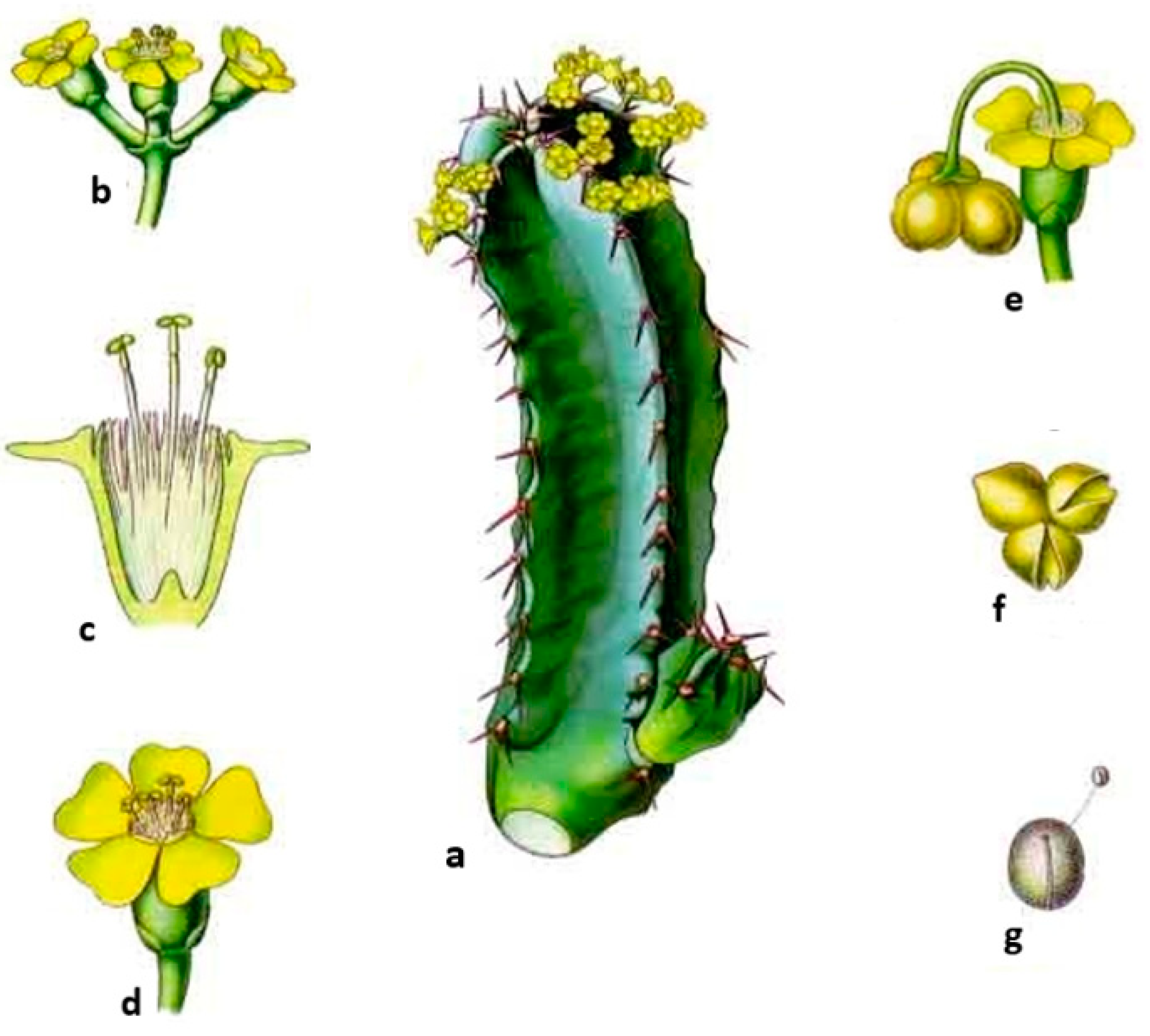
3.1. Botanical Description
3.2. Taxonomy and Geographical Distribution
3.3. Population Status
3.4. Possible Threats
3.5. Conservation
3.6. Ethnomedicinal Use

| Area of Study | Used Composition/Part | Mode of Preparation | Traditional Use | References |
|---|---|---|---|---|
| Central High Atlas of Morocco | Stems Fruits Flowers | Poultice Infusion Powder | Venomous stings | [5] |
| Against paralysis (kneaded with flour or semolina and egg white) | ||||
| Abortifacient | ||||
| Antidiabetic | ||||
| Tinghir South, Ouarzazate, and Center of the atlas chain of Azilal North | Flowers, latex | Infusion, latex in water | Antidiabetic | [12] |
| Marrakech | Dried plants, euphorbia honey | Not reported | Antidiabetic | [13] |
| Mountain chains of the Middle Atlas, Ifrane National Park, and Khenifra National Park | Entire plant (dry and fresh) | Not reported | Laxative | [9] |
| Anti-inflammatory | ||||
| Hypoglycemic | ||||
| Antitumor activity | ||||
| Cosmetic purposes | ||||
| Greater Casablanca, Morocco | Roots, leaves, stem, and bark | Decoction | Cancer | [6] |
| The Northern Azilal located in the center of the Atlas Mountains (Morocco) | Not reported | Not reported | Inflammation | [7] |
| Skin | ||||
| Tumor | ||||
| Teeth treatment | ||||
| Skin care and hair | ||||
| Azilal and Beni Mellal located in Atlas Mountains (Morocco) | Warm water mixed with latex, and honey | External use | Skin inflammation | [8] |
| Oral | Intoxications | |||
| External use and/or oral | Snakebites and scorpion stings | |||
| Aerial parts (powder) with honey | Oral | Goiter | ||
| Latex | External use | Skin cancers | ||
| Warts | ||||
| Toothache | ||||
| Stem (without latex or bark) and milk juice | Oral | Cysts of the female genital tract | ||
| Female genital tract cancer | ||||
| Breast cancer | ||||
| Ground aerial parts mixed with honey | Oral | Cancers | ||
| Seeds (powder) with honey | Oral | Digestive issues | ||
| Honey | Oral | Respiratory diseases (flu, asthma, allergies, etc.) | ||
| Circulatory disorder | ||||
| Metabolic disorders | ||||
| Digestive diseases | ||||
| Diseases of the reproductive system | ||||
| Headache | ||||
| Weakness and yellowing | ||||
| Cancers | ||||
| Angina | ||||
| Honey | External use | Skin conditions | ||
| Rabat | Aerial parts | Ground with honey (oral) | Cancers | [10] |
| Ksar Lakbir district | Seeds | Mixed with Lawsonia inermis and kneaded with water | Hair care | [45] |
| Melted in olive oil | Hair tonic | |||
| Casablanca | Resin | Natural | Cancers (breast, colon, lung, uterus, ORL, leukemia, stomatology) | [11] |
| Beni Mellal | Leaf stems | Decoction | Antidiabetic | [44] |
3.7. Phytochemistry
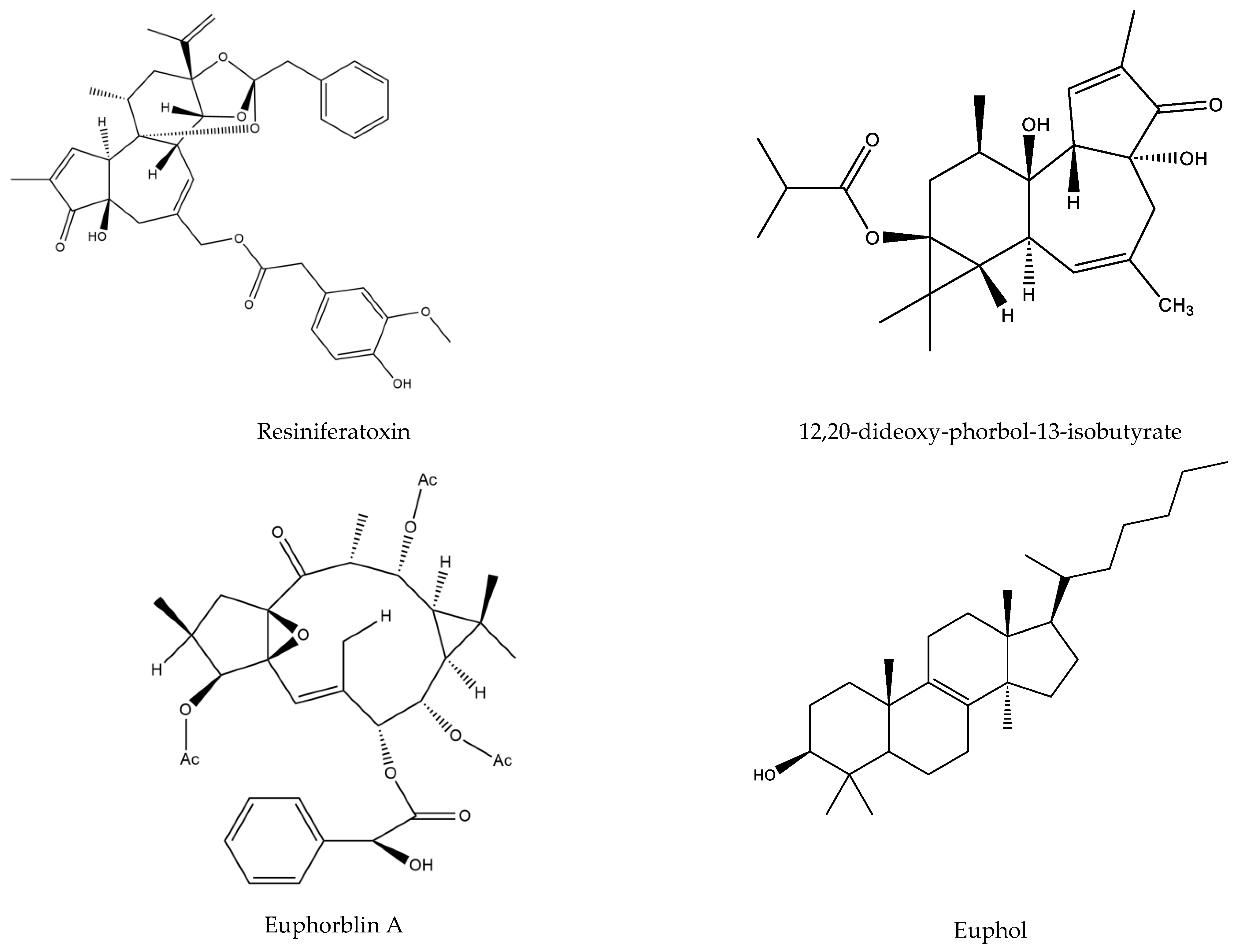
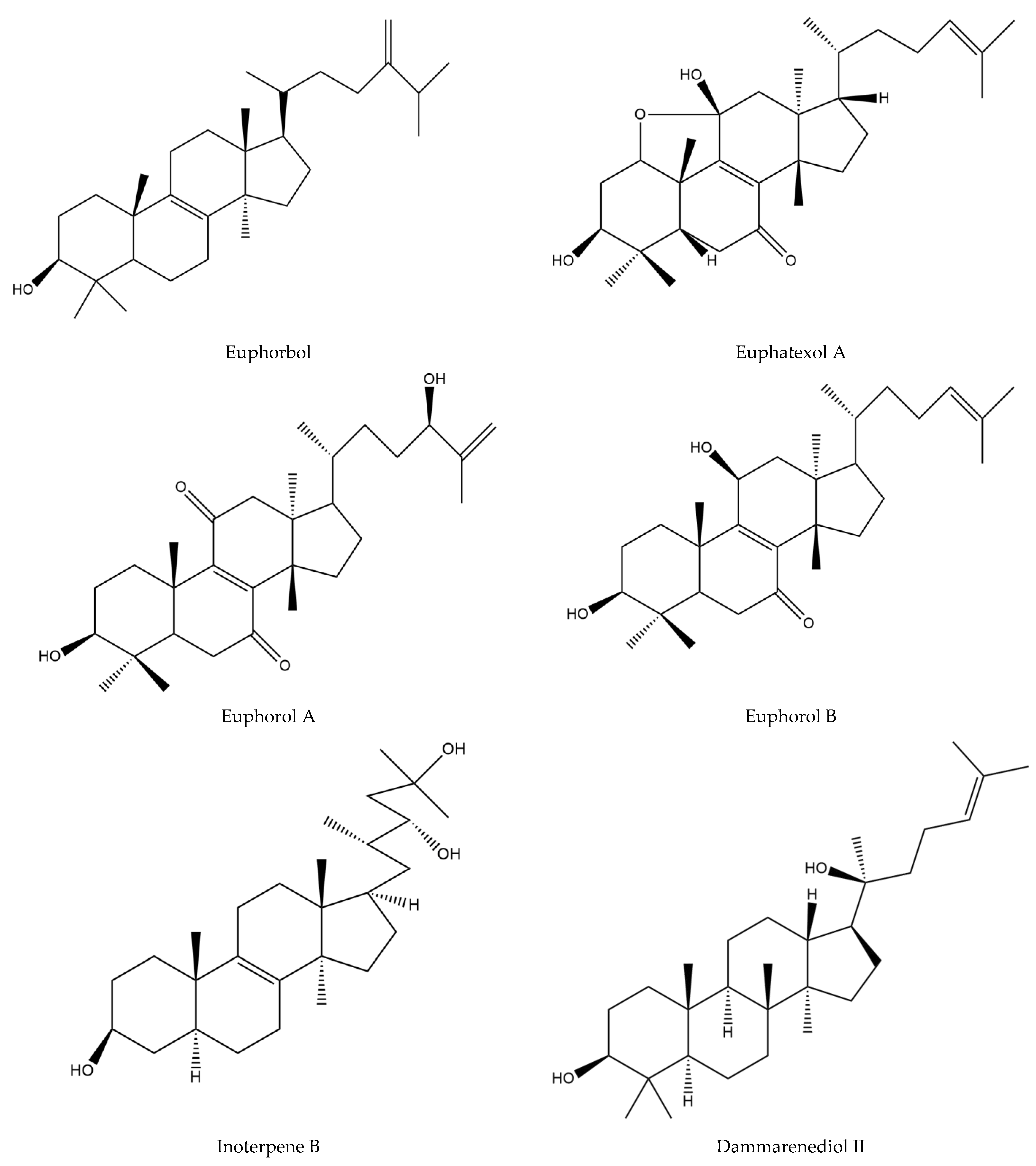
3.7.1. Terpenoids
3.7.2. Honey Composition
3.8. Pharmacological Investigation
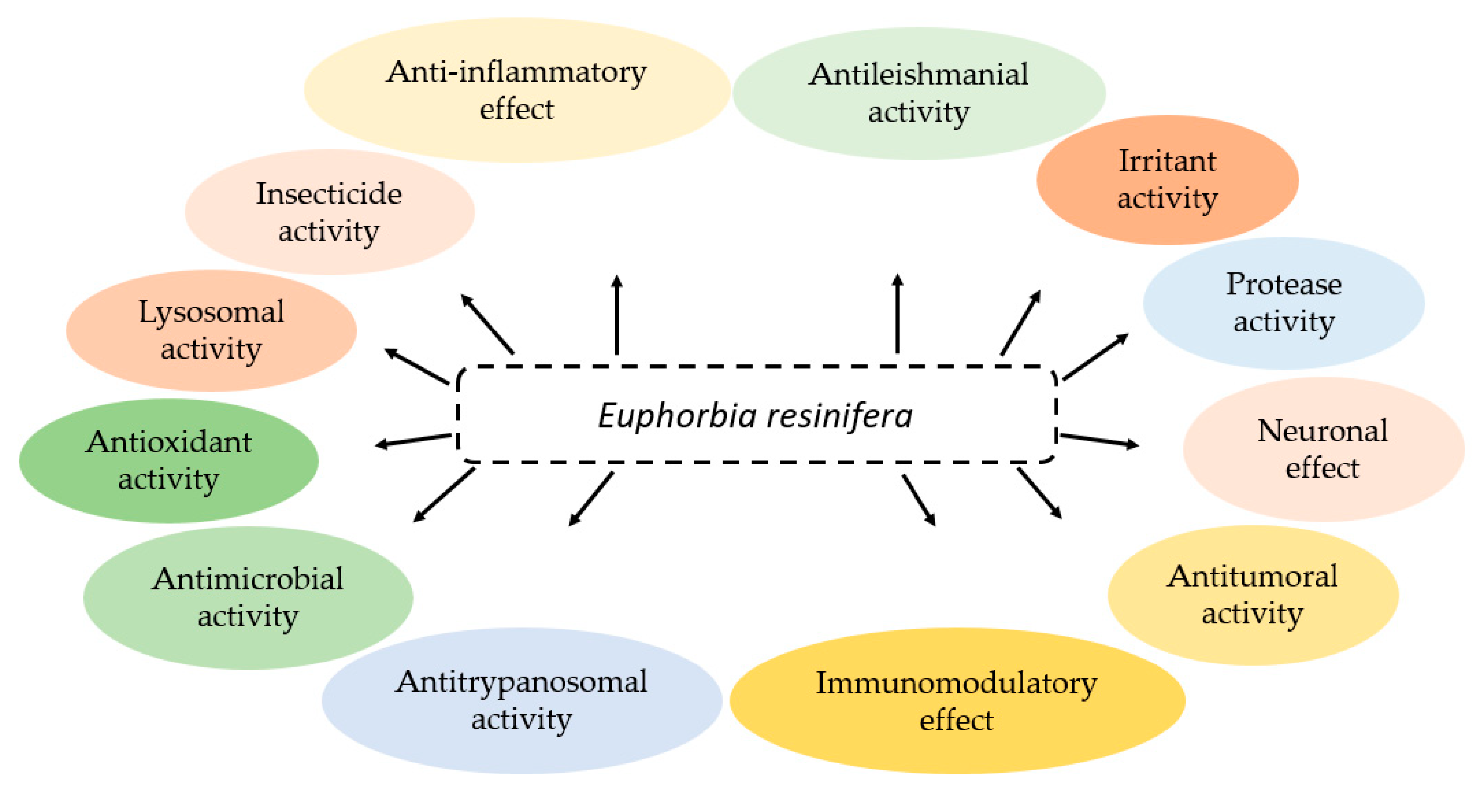
3.8.1. Antimicrobial Activity
Antibacterial Activity
Antifungal Activity
3.8.2. Antioxidant Activity
3.8.3. Antitumoral Activity
3.8.4. Anti-Inflammatory Effect
3.8.5. Antileishmanial Activity
3.8.6. Antitrypanosomal Activity
3.8.7. Protease Activity
3.8.8. Anti-Xanthine Oxidase, Antilipoxygenase, Antiacetylcholinesterase, and Antityrosinase Activities
3.8.9. Neuronal Effect
3.8.10. Immunomodulatory Effect
3.8.11. Irritant Activity
3.8.12. Lysosomal Activity
3.8.13. Toxic Effect
3.8.14. Insecticide Activity
| Activities | Use Part | Extracts | Experimental Approach | Key Results | References |
|---|---|---|---|---|---|
| Antileishmanial activity | Latex | Methanol extract | MTT assays against Leishmania infantum (strain PB75) | ED50 < 10 μg/mL | [20] |
| Antitrypanosomal activity | Latex | Methanol extract | MTT assays against Trypanosoma cruzi (strain Y) | ED50 < 10 μg/mL | [20] |
| Protease activity | Latex | Crude extract | Activity of purified protease against blood-circulating protease inhibitors | No inhibition of protease activity | [80] |
| In vitro effect of purified enzyme on human fibrin clots using SDS-PAGE under reducing conditions | Hydrolysis of all chains of the fibrin clot by purified protease with concentrations ≥ 0.5 µM | ||||
| In vitro anticoagulant activity of purified protease using PT and APTT assays | APTT and PT prolongation inducted in both in the presence of purified protease | ||||
| Activity of purified protease against human platelet aggregation using light transmission aggregometry | At concentrations ≥ 1 µM, purified protease abolishes platelet function | ||||
| Latex | Crude extract | Optimum pH: activity of purified protease was assayed at different pH values (1–14) | Optimum pH = pH 5.0 | [80] | |
| Optimum temperature: activity of purified protease was assayed at temperature between 15 and 95 °C | Optimum temperature = 45 °C | ||||
| pH stability: purified protease incubated at different pH (1–14) at 37 °C during a period of 24 h | Purified protease was stable in broad pH range (1–14) | ||||
| Temperature stability: purified protease was incubated at temperature between 15 and 95 °C for 2 h | Purified protease was stable at temperatures up to 65–66 °C | ||||
| Kinetic studies: Km, Vmax, and Vmax/Km of the fibrinogenolytic activity of purified protease determined by incubating 2 µg of the enzyme with various amounts of human fibrinogen (0–32 µM) | Km = 4.95 µM | ||||
| Vmax = 578.1 ng/min | |||||
| Vmax/Km = 116.8 ng µM−1 min−1 | |||||
| Enzyme inhibitory potential | Aerial parts and honey | Aqueous extracts and honey | Percentage of inhibition of lipoxygenase, xanthine oxidase, tyrosinase, and acetylcholinesterase calculated by the determination of IC50 | Higher in vitro activities of aqueous extracts observed than in the respective honey samples | [17] |
| Immunomodulatory effect | Latex | Crude extract | In vitro studies of 12-deoxyphorbol esters, ingol esters, and triterpenes | Only the 12-deoxyphorbol esters showed a stimulation of lymphocytes proliferation | [85] |
| Aerial parts | Aqueous extract | In vivo acute toxicity (at dose 5 g/kg body weight) and sub-acute toxicity tests (at dose 0.1, 0.5, 1.0, 2.5, or 5 g/kg) | A significant increase in antibody titer values in the group treated without toxic dose (1 g/kg) compared to the control | [31] | |
| Increase in the DTH reactivity response (37%) compared to control animals (25%) at a dose of 1g/kg | |||||
| Irritant activity | Latex and resin | Methanol extract | Irritant activity of purified compounds of E. resinifera resin and latex was investigated in vivo on the ear of a mouse; irritant activity of these compounds was estimated as irritation units | Two esters of ingol did not exhibit irritant activity | [50] |
| Two 12,20-dideoxyphorbol esters were also practically nonirritant | |||||
| Esters of 12-deoxyphorbol, 12-deoxy-16-hydroxyphorbol, resiniferonol, and ingenol showed less or more irritant activity | |||||
| Resin | Methanol extract | Irritant activity of isolated 13,20-diesters and 13-monoesters from resin of E. resinifera was investigated in vivo on the ear of a mouse; irritant activity of these compounds was estimated as irritation units | The 13,20-diesters generally have a lower irritant activity than the corresponding 13-monoesters | [23] | |
| Liver effect | Propolis | Oil extract | Activities of the SGPT and SGOT were analyzed before, during, and after 10 months of the beginning of the treatment | A significant reduction of SGPT and SGOT concentrations of all patients | [24] |
| Lysosomal activity | Latex | Methanol extract | Different concentrations of the isolated diterpenoids (10, 20, 40, and 60 µM) were tested for the lysosomal effect at different time intervals (1, 3, 6, and 9 h) using HeLa cell line using LysoTracker Red staining to observe the induction of lysosome | The isolated diterpenoids showed notable abilities to induce lysosome biosynthesis in concentrations and time-dependent manner | [25] |
| Skin effect | Resin | Alcohol extract | Sixty human patients with verrucae plantares treated with alcohol-resin solution (95–30%) on the central keratotic area of the verruca | After 96 h, a complete removal of the central keratotic area of the verrucae was observed | [26] |
| Toxic effect | Aerial parts | Aqueous extract | In vivo acute toxicity (at dose 5 g/kg body weight) and sub-acute toxicity tests (at dose 0.1, 0.5, 1.0, 2.5, or 5 g/kg) during a period of 28 days; biochemical analysis (creatine, urea, ALAT and ASAT) and histopathological examination of liver, kidney, and spleen were investigated | At the doses of 0.1 and 0.5 g/kg, creatinine and urea did not show any differences | [31] |
| A significant dose-dependent increase in ASAT and ALAT | |||||
| At dose lower than 2.5 g/kg, no detectable alteration in spleen tissues or kidney | |||||
| At 5 g/kg, alteration was reported in kidney and spleen tissues | |||||
| At doses of 0.5 and 1 g/kg, some low hepatocytes lesions were observed | |||||
| Using 5 g/kg, massive hepatocytes lesions were observed |
3.8.15. Clinical Trials
4. Conclusions
Author Contributions
Funding
Institutional Review Board Statement
Data Availability Statement
Conflicts of Interest
References
- Moujanni, A.; Partida, L.; Essamadi, A.K.; Hernanz, D.; Heredia, F.J.; Terrab, A. Physicochemical characterization of unique unifloral honey: Euphorbia resinifera . CyTA J. Food 2017, 16, 27–35. [Google Scholar] [CrossRef] [Green Version]
- Chakir, A.; Romane, A.; Marcazzan, G.L.; Ferrazzi, P. Physicochemical properties of some honeys produced from different plants in Morocco. Arab. J. Chem. 2016, 9, S946–S954. [Google Scholar] [CrossRef] [Green Version]
- Benmehdi, H.; Bounoua, N.; Amrouche, A.; Lahcene, D.; Maazouzi, A. PHYTOCHEMICAL STUDY, antioxidant and antimicrobial activities of Euphorbia resinifera. Int. Res. J. Pharm. 2013, 2, 44–50. [Google Scholar] [CrossRef]
- Belakhdar, J. La pharmacopée marocaine traditionnelle. In Médecine Arabe Ancienne Savoirs Pop; Ibis Press: Paris, French, 1997; p. 348. [Google Scholar]
- Belhaj, S.; Dahmani, J.; Belahbib, N.; Zidane, L. Ethnopharmacological and Ethnobotanical study of Medicinal plants in the Central High Atlas, Morocco. Ethnobot. Res. Appl. 2020, 20, 1–40. [Google Scholar] [CrossRef]
- Bourhia, M.; Shahat, A.A.; Almarfadi, O.M.; Naser, F.A.; Abdelmageed, W.M.; Said, A.A.H.; El Gueddari, F.; Naamane, A.; Benbacer, L.; Khlil, N. Ethnopharmacological Survey of Herbal Remedies Used for the Treatment of Cancer in the Greater Casablanca-Morocco. Evidence-Based Complement. Altern. Med. 2019, 2019, 1613457. [Google Scholar] [CrossRef] [PubMed] [Green Version]
- El Alami, A.; Farouk, L.; Chait, A. Etude ethnobotanique sur les plantes médicinales spontanées poussant dans le versant nord de l’Atlas d’Azilal (Maroc). Alger. J. Nat. Prod. 2016, 4, 271–282. [Google Scholar]
- EL ALAMI, A.; Fattah, A.; Chait, A. Medicinal plants used for the prevention purposes during the covid-19 pandemic in Morocco. J. Anal. Sci. Appl. Biotechnol. 2020, 2, 4–11. [Google Scholar]
- El Houssine Bouiamrine, L.B.; Ibijbijen, J.; Nassiri, L. Use of medicinal plants in Middle Atlas of Morocco: Potential health risks and indigenous knowledge in a Berber community. J. Med. Plants 2017, 5, 338–342. [Google Scholar]
- Kabbaj, F.; Meddah, B.; Cherrah, Y.; Faouzi, E. Ethnopharmacological profile of traditional plants used in Morocco by cancer patients as herbal therapeutics. Phytopharmacology 2012, 2, 243–256. [Google Scholar]
- Samouh, Y.; Lemrani, A.; Mimouni, H.; Mohamad, J.; Said, A.A.H. Ethnopharmacological Study of Herbal Medicines used to treat Cancer in Morocco. J. Phytopharm. 2019, 8, 135–141. [Google Scholar] [CrossRef]
- Belhaj, S.; Chaachouay, N.; Zidane, L. Ethnobotanical and toxicology study of medicinal plants used for the treatment of diabetes in the High Atlas Central of Morocco. J. Pharm. Pharmacogn. Res. 2021, 9, 619–662. [Google Scholar] [CrossRef]
- Errajraji, A.; Ouhdouch, F.; El-Anssari, N. Usage des plantes médicinales dans le traitement du diabète de type 2 au Maroc: Use of medicinal plants for type 2 diabetes treatment, in Morocco. Médecine Mal. Métaboliques 2010, 4, 301–304. [Google Scholar] [CrossRef]
- Kemboi, D.; Peter, X.; Langat, M.; Tembu, J. A review of the ethnomedicinal uses, biological activities, and triterpenoids of Euphorbia species. Molecules 2020, 25, 4019. [Google Scholar] [CrossRef]
- Merzouki, A.; Ed-Derfoufi, F.; Molero Mesa, J. Contribución al Conocimiento de la Medicina Rifeña Tradicional III: Fitoterapia de la Diabetes en la Provincia de Chefchaouen (norte de Marruecos); Universidad de Granada, Facultad de Farmacia: Granada, Spain, 2003. [Google Scholar]
- Boutoub, O.; El-Guendouz, S.; Estevinho, L.M.; Paula, V.B.; Aazza, S.; El Ghadraoui, L.; Rodrigues, B.; Raposo, S.; Carlier, J.; Costa, M.C.; et al. Antioxidant activity and enzyme inhibitory potential of Euphorbia resinifera and E. officinarum honeys from Morocco and plant aqueous extracts. Environ. Sci. Pollut. Res. 2020, 28, 503–517. [Google Scholar] [CrossRef]
- Hanane, F.; Abdellah, E.C.; Abdeslam, L. In vitro antioxidant and antibacterial activity of the root extract of Euphorbia resinifera. J. Pharmacogn. Phytochem. 2014, 2, 161–163. [Google Scholar]
- Talbaoui, A.; El Hamdaoui, L.; Bouyahya, A.; El Moussaouiti, M.; Bakri, Y. Chemical composition, in vitro cytotoxic, and antibacterial activities of Moroccan medicinal plants Euphorbia resinifera and Marrubium vulgare. Biointerface Res. Appl. Chem. 2020, 10, 7343–7355. [Google Scholar]
- Benjamaa, R.; Moujanni, A.; Terrab, A.; Eddoha, R.; Benbachir, M.; Moujahid, A.; Nasser, B.; Darkaoui, S.; Zyate, N.; Talmi, A.; et al. Relationship Among Antibiotic Residues And Antibacterial Activity Of The Endemic Spurge Honey (Euphorbia Resinifera O. Berg) From Morocco. Emir. J. Food Agric. 2020, 32, 795–807. [Google Scholar] [CrossRef]
- Mazoir, N.; Benharref, A.; Bailén, M.; Reina, M.; González-Coloma, A.; Martínez-Díaz, R.A. Antileishmanial and antitrypanosomal activity of triterpene derivatives from latex of two Euphorbia species. Z. Für Nat. C 2011, 66, 360–366. [Google Scholar] [CrossRef] [PubMed] [Green Version]
- Ezzanad, A.; Gómez-Oliva, R.; Escobar-Montano, F.; Díez-Salguero, M.; Geribaldi-Doldan, N.; Dominguez-Garcia, S.; Botubol-Ares, J.M.; de los Reyes, C.; Durán-Patrón, R.; Nunez-Abades, P.; et al. Phorbol Diesters and 12-Deoxy-16-hydroxyphorbol 13, 16-Diesters Induce TGFα Release and Adult Mouse Neurogenesis. J. Med. Chem. 2021, 64, 6070–6084. [Google Scholar] [CrossRef]
- Furst, D.E. Pharmacokinetics of hydroxychloroquine and chloroquine during treatment of rheumatic diseases. Lupus 1996, 5, 11–15. [Google Scholar] [CrossRef]
- Zayed, S.; Sorg, B.; Hecker, E. Structure activity relations of polyfunctional diterpenes of the tigliane type, VI. Irritant and tumor promoting activities of semisynthetic mono and diesters of 12-deoxyphorbol. Planta Med. 1984, 50, 65–69. [Google Scholar] [CrossRef]
- Mohamed, F.P. Serum Glutamate Transaminases Decrease in Chronic Hepatitis C Patients By Propolis From Euphorbia Resinifera. In Proceedings of the 5th German Apitherapy Congress, Passau, Germany, 23–25 March 2007. [Google Scholar]
- Zhao, N.D.; Ding, X.; Song, Y.; Yang, D.Q.; Yu, H.L.; Adelakun, T.A.; Qian, W.D.; Zhang, Y.; Di, Y.T.; Gao, F.; et al. Identification of ingol and rhamnofolane diterpenoids from Euphorbia resinifera and their abilities to induce lysosomal biosynthesis. J. Nat. Prod. 2018, 81, 1209–1218. [Google Scholar] [CrossRef]
- Goldblum, R.W.; Curtis, A.C. Effects of the resin of Euphorbium on verrucae plantares; human and animal experimentation. J. Investig. Dermatol. 1953, 20, 45–50. [Google Scholar] [CrossRef] [PubMed] [Green Version]
- Kuehn, B. Plant-Chemical Shows Promise for Pain Relief. JAMA 2018, 319, 760. [Google Scholar] [CrossRef] [PubMed]
- Mancera, N.; Wadia, H.P. Corneal edema associated with systemic dopaminergic agents. Cornea 2019, 38, 1040–1042. [Google Scholar] [CrossRef] [PubMed]
- Renukadevi, K.P.; Sultana, S.S. Determination of antibacterial, antioxidant and cytotoxicity effect of Indigofera tinctoria on lung cancer cell line NCI-h69. Int. J. Pharmacol. 2011, 7, 356–362. [Google Scholar] [CrossRef] [Green Version]
- Zissu, R. Aborder la deuxième prescription: Exemples cliniques. La Rev. D’homéopathie 2011, 2, 141–147. [Google Scholar] [CrossRef]
- Issiki, Z.; Daoudi, F.; Mtairag, E.M.; Rais, S.; Marnissi, F.; Zaid, Y.; Naya, A. and Oudghiri, M. Acute a sub-chronic toxicity and immunomodulatory activity of an aqueous extract of Euphorbia resinifera in rodents. Int. J. Pharm. Sci. Res. 2019, 10, 2962–2969. [Google Scholar]
- Vindt, J. Monographie des Euphorbiacées du Maroc: 1. Partie, Revision et Systématique; Travaux de l’institut Scientifique Cherifien: Tangier, Moroco, 1953. [Google Scholar]
- Boullard, B. Plantes Médicinales du Monde: Réalités et Croyances; Estem: Casablanca, Morocco, 2001. [Google Scholar]
- Greenish, H.G. A Text Book of Pharmacognosy: Being an Account of the More Important Crude Drugs of Vegetable and Animal Origin; J. & A. Churchill: London, UK, 1933. [Google Scholar]
- Bruyns, P.V.; Klak, C.; Hanáček, P. Age and diversity in Old World succulent species of Euphorbia (Euphorbiaceae). Taxon 2011, 60, 1717–1733. [Google Scholar] [CrossRef]
- Terrab, A.; Moujanni, A.; Essamadi, A.K.; Hernanz, D.; Díez, M.J.; Berjano, R. A palynological and geographical characterization of labeled resin spurge honey: Euphorbia resinifera. Palynology 2022, 46, 1–10. [Google Scholar] [CrossRef]
- Webster, G.L. Classification of the Euphorbiaceae. Ann. Mo. Bot. Gard. 1994, 81, 3–32. [Google Scholar] [CrossRef]
- Vasas, A.; Orbán-Gyapai, O.; Hohmann, J. The Genus Rumex: Review of traditional uses, phytochemistry and pharmacology. J. Ethnopharmacol. 2015, 175, 198–228. [Google Scholar] [CrossRef] [PubMed]
- Vasas, A.; Hohmann, J. Euphorbia diterpenes: Isolation, structure, biological activity, and synthesis (2008–2012). Chem. Rev. 2014, 114, 8579–8612. [Google Scholar] [CrossRef] [Green Version]
- Ettaqy, A.; Taha, A.; ElGhiouane, A.; ElKhou, A.; Boulli, A.; Abbas, Y. New data on the ecological distribution of Euphorbia resinifera O. Berg in the Beni Mellal-Khenifra region. E3S Web Conf. 2020, 183, 01001. [Google Scholar] [CrossRef]
- Lawant, P.; Winthagen, D. Euphorbia resinifera portrayed in a manuscript herbal nearly fifteen hundred years ago. Bradleya 2001, 2001, 3–14. [Google Scholar] [CrossRef]
- Sabbahi, R. Economic value of insect pollination of major crops in Morocco. Int. J. Trop. Insect Sci. 2021, 42, 1275–1284. [Google Scholar] [CrossRef]
- Basak, S.K.; Bakshi, P.K.; Basu, S.; Basak, S. Keratouveitis caused by Euphorbia plant sap. Indian J. Ophthalmol. 2009, 57, 311–313. [Google Scholar] [CrossRef]
- Fouad, Z.; Lahcen, Z. Antidiabetic medicinal plants in Morocco: Ethnobotanical survey of the population of Beni Mellal. Plant Arch. 2020, 20, 337–343. [Google Scholar]
- Merzouki, A.; Ed-Derfoufi, F.; Mesa, J.M. Contribution to the knowledge of Rifian traditional medicine. II: Folk medicine in Ksar Lakbir district (NW Morocco). Fitoterapia 2000, 71, 278–307. [Google Scholar] [CrossRef] [PubMed]
- Farah, H.; Ech-Chahad, A.; Lamiri, A. Antioxidant, antimicrobial and phytochemical investigations of polar extracts of Euphorbia resinifera Beg. roots, stems and flowers. Am. J. Adv. Drug Deliv. 2014, 6, 776–785. [Google Scholar]
- Fattorusso, E.; Lanzotti, V.; Taglialatela-Scafati, O.; Tron, G.C.; Appendino, G. Bisnorsesquiterpenoids from Euphorbia resinifera Berg. and an expeditious procedure to obtain resiniferatoxin from its fresh latex. Eur. J. Org. Chem. 2002, 2002, 71–78. [Google Scholar] [CrossRef]
- Girin, M.; Paphassarang, S.; David-Eteve, C.; Chaboud, A.; Raynaud, J. Determination of ingenol in homoeopathic mother tinctures of Euphorbia species by high-performance liquid chromatography. J. Chromatogr. A 1993, 637, 206–208. [Google Scholar] [CrossRef]
- Hergenhahn, M.; Adolf, W.; Hecker, E. Resiniferatoxin and other esters of novel polyfuncticnal diterpenes from Euphorbia resinifera and unispina. Tetrahedron Lett. 1975, 16, 1595–1598. [Google Scholar] [CrossRef]
- Hergenhahn, M.; Kusumoto, S.; Hecker, E. On the active principles of the spurge family (Euphorbiaceae) Extremely skin-irritant and moderately tumor-promoting diterpene esters from Euphorbia resinifera Berg. J. Cancer Res. Clin. Oncol. 1984, 108, 98–109. [Google Scholar] [CrossRef]
- Li, M.M.; Qi, Y.R.; Feng, Y.P.; Liu, W.; Yuan, T. Four new lanostane triterpenoids from latex of Euphorbia resinifera. Zhongguo Zhong Yao Za Zhi Zhongguo Zhongyao Zazhi China J. Chin. Mater. Med. 2021, 46, 4744–4748. [Google Scholar] [CrossRef]
- Li, M.M.; Qi, Y.R.; Feng, Y.P.; Liu, W.; Yuan, T. Euphatexols C- G, five new triterpenoids from the latex of Euphorbia resinifera. J. Asian Nat. Prod. Res. 2022, 24, 311–320. [Google Scholar] [CrossRef]
- Li, Y.J.; Ji, T.F.; Zhao, J.; Gu, Z.Y. Chemical constituents of triterpenoids from Euphorbia resinifera. Zhongguo Zhong Yao Za Zhi Zhongguo Zhongyao Zazhi China J. Chin. Mater. Med. 2021, 46, 4433–4437. [Google Scholar] [CrossRef]
- Mallon, R.; Onrubia, M.; Lota, F.; Corral, P.; Lorrain, B.; Michoux, F. In vitro propagation methods of the succulent plant Euphorbia resinifera and quantification of its major compounds. Planta Med. 2014, 80, P2F2. [Google Scholar] [CrossRef]
- Mazoir, N.; Auhmani, A.; Daoubi, M.; Collado, I.G.; Benharref, A. Hemisynthesis of New Triterpene Derivatives Using Oxidation by CrO3 and NaIO4-(RuCl3, 3H2O). Cheminform 2007, 38, 1289–1299. [Google Scholar] [CrossRef]
- Nordal, A.; Benson, A.A. Phorbic Acid Biosynthesis in the Latex Vessel System of Euphorbia. Plant Physiol. 1969, 44, 78–84. [Google Scholar] [CrossRef]
- Ourhzif, E.-M.; Ketatni, E.M.; Akssira, M.; Troin, Y.; Khouili, M. Crystal structure, Hirshfeld surface analysis and DFT studies of Euphorbioside monohydrate a major bisnorsesquiterpene isolated from Euphorbia resinifera latex. J. Mol. Struct. 2021, 1241, 130511. [Google Scholar] [CrossRef]
- Qi, Y.; Liu, W.; Chen, Y.; Guan, M.; Yuan, T. Euphatexols A and B, two unusual euphane triterpenoids from the latex of Euphorbia resinifera. Tetrahedron Lett. 2019, 60, 151303. [Google Scholar] [CrossRef]
- Wang, S.; Liang, H.; Zhao, Y.; Wang, G.; Yao, H.; Kasimu, R.; Wu, Z.; Li, Y.; Huang, J.; Wang, J. New triterpenoids from the latex of Euphorbia resinifera Berg. Fitoterapia 2016, 108, 33–40. [Google Scholar] [CrossRef] [PubMed]
- Wang, S.-Y.; Huang, C.; Sun, R.-K.; Lu, L.-N.; Liang, H.-G.; Gao, L.; Huang, J.; Wang, J.-H.; Yang, B.-F. New tirucallane triterpenoids from the dried latex of Euphorbia resinifera. Phytochem. Lett. 2018, 29, 220–224. [Google Scholar] [CrossRef]
- Wang, S.-Y.; Li, G.-Y.; Zhang, K.; Wang, H.-Y.; Liang, H.-G.; Huang, C.; Huang, J.; Wang, J.-H.; Yang, B.-F. New ingol-type diterpenes from the latex of Euphorbia resinifera . J. Asian Nat. Prod. Res. 2019, 21, 1075–1082. [Google Scholar] [CrossRef] [PubMed]
- Farah, H.; Echchahad, A.; Lamiri, A. Semi-synthesis and antimicrobial activities of some new euphorbioside derivatives. Int. J. Chem. Tech. Res. 2014, 6, 763–767. [Google Scholar]
- Wang, S.-Y.; Li, G.-Y.; Zhang, K.; Wang, H.-Y.; Liang, H.-G.; Huang, C.; Huang, J.; Wang, J.-H.; Yang, B.-F. Triterpenoids from Uygur medicine latex of Euphorbia resinifera. China J. Chin. Mater. Medica 2018, 43, 3688–3693. [Google Scholar] [CrossRef]
- Zhao, N.-D.; Li, Y.-L.; Song, Y.; Yang, B.-J.; Ding, X.; Gao, F.; Ye, J.; Hao, X.-J.; Zhang, Y.; Li, S.-L. Ten new nortriterpenes from Euphorbia resinifera and their anti-tomato yellow leaf curl virus activities. Fitoterapia 2021, 153, 104989. [Google Scholar] [CrossRef]
- Malika, N.; Mohamed, F.; Chakib, E.A. Microbiological and physicochemical properties of Moroccan honey. Int. J. Agric. Biol. 2005, 7, 773–776. [Google Scholar]
- Moujanni, A.; Terrab, A.; Eddoha, R.; Nasser, B.; Benbachir, M.; Tannaoui, M.; Zouaoui, A.; Essamadi, A.K. Quantification of heavy metals and pesticides residues in labeled Moroccan Euphorbia resinifera honey from Tadla-Azilal. J. Mater. Env. Sci. 2017, 8, 1826–1836. [Google Scholar]
- Crittenden, A.N. The Importance of Honey Consumption in Human Evolution. Food Foodways 2011, 19, 257–273. [Google Scholar] [CrossRef]
- Abderrahim, L.A.; Taïbi, K.; Abderrahim, N.A.; Boussaid, M.; Rios-Navarro, C.; Ruiz-Saurí, A. Euphorbia honey and garlic: Biological activity and burn wound recovery. Burns 2019, 45, 1695–1706. [Google Scholar] [CrossRef]
- Alami, A.E.; Fattah, A.; Aboufatima, R.; Chait, A. Ethnopharmacological Survey of Euphorbia resinifera in the Atlas Mountains of Azilal-Beni Mellal, Morocco. Int. J. Pharma Sci. Res. 2020, 11, 240–245. [Google Scholar]
- Moujanni, A.; Terrab, A.; Eddoha, R.; Nasser, B.; Benbachir, M.; Chaouqy, N.E.; Bouzid, T.; Essamadi, A.K. Microbiological Quality Of Moroccan Labeled Euphorbia Resinifera Honey. J. Microbiol. Biotechnol. Food Sci. 2017, 6, 1188–1194. [Google Scholar] [CrossRef]
- Bouyahya, A.; Bakri, Y.; Et-Touys, A.; Talbaoui, A.; Khouchlaa, A.; Charfi, S.; Abrini, J.; Dakka, N. Resistance to antibiotics and mechanisms of action of essential oils against bacteria. Phytothérapie 2017, 1–11. [Google Scholar]
- El Idrissi, A.E.Y.; Khouchlaa, A.; Bouyahya, A.; Bakri, Y.; Tijane, M.H. Phytochemical Characterization, In Vitro Antioxidant, Cytotoxic, and Antibacterial Effects of Aristolochia longa L.; AMG Transcend Association: Bucharest, Romania, 2021; Volume 11, pp. 8129–8140. [Google Scholar] [CrossRef]
- Khouchlaa, A.; Et-Touys, A.; Lakhdar, F.; Laasri, F.E.; El Idrissi, A.E.Y.; Zaakour, F. Ethnomedicinal use, phytochemistry, pharmacology, and toxicology of Salvia verbenaca L.: A review. Biointerface Res. Appl. Chem. 2021, 12, 1437–1469. [Google Scholar]
- Sousa, J.M.; De Souza, E.L.; Marques, G.; Meireles, B.; de Magalhães Cordeiro, Â.T.; Gullón, B.; Pintado, M.M.; Magnani, M. Polyphenolic profile and antioxidant and antibacterial activities of monofloral honeys produced by Meliponini in the Brazilian semiarid region. Food Res. Int. 2016, 84, 61–68. [Google Scholar] [CrossRef]
- Engwa, G.A. Free Radicals and the Role of Plant Phytochemicals as Antioxidants Against Oxidative Stress-Related Diseases. In Phytochemicals: Source of Antioxidants and Role in Disease Prevention; IntechOpen: London, UK, 2018. [Google Scholar] [CrossRef] [Green Version]
- Samaranayake, N.; Fernando, S.D.; Neththikumara, N.F.; Rodrigo, C.; Karunaweera, N.D.; Dissanayake, V.H.W. Association of HLA class I and II genes with cutaneous leishmaniasis: A case control study from Sri Lanka and a systematic review. BMC Infect. Dis. 2016, 16, 292. [Google Scholar] [CrossRef] [PubMed] [Green Version]
- Bahmani, M.; Abbasi, N.; Hosseini, M.; Rafieian-Kopaei, M. Concise review: Medicinal plants are effective against leishmaniasis. Biomed. Res. Ther. 2017, 4, 1775. [Google Scholar] [CrossRef] [Green Version]
- Mazzeti, A.L.; Capelari-Oliveira, P.; Bahia, M.T.; Mosqueira, V.C.F. Review on Experimental Treatment Strategies Against Trypanosoma cruzi. J. Exp. Pharmacol. 2021, ume 13, 409–432. [Google Scholar] [CrossRef]
- Ali, M.; Salim Hossain, M.; Islam, M.; Arman, S.I.; Sarwar Raju, G.; Dasgupta, P.; Noshin, T.F. Aspect of thrombolytic therapy: A review. Sci. World J. 2014, 2014, 586510. [Google Scholar] [CrossRef]
- Siritapetawee, J.; Khunkaewla, P.; Thumanu, K. Roles of a protease from Euphorbia resinifera latex in human anticoagulant and antithrombotic activities. Chem. Interactions 2020, 329, 109223. [Google Scholar] [CrossRef]
- Gulati, V.; Harding, I.H.; Palombo, E.A. Enzyme inhibitory and antioxidant activities of traditional medicinal plants: Potential application in the management of hyperglycemia. BMC Complement. Altern. Med. 2012, 12, 77. [Google Scholar] [CrossRef] [PubMed] [Green Version]
- Weiss, N.; Couraud, P.O. Thérapie cellulaire par les cellules souches neurales—Neural stem cell therapy in neurological diseases. Mise Au Point 2008. [Google Scholar]
- Szallasi, A. Vanilloid (Capsaicin) Receptors in Health and Disease. Am. J. Clin. Pathol. 2002, 118, 110–121. [Google Scholar] [CrossRef] [Green Version]
- Basu, P.; Maier, C.; Averitt, D. (427) Novel effects of Euphorbia bicolor (Euphorbiaceae) latex extract on nociceptors. J. Pain 2016, 17, S81. [Google Scholar] [CrossRef]
- Kreher, B.; Neszmélyi, A.; Jurcic, K.; Wagner, H. New Phytochemical and Immunological Investigations ofEuphorbia resiniferaandDaphne mezereum. Planta Med. 1990, 56, 572–573. [Google Scholar] [CrossRef]
- Therre, C. Gentoxizität und Antigentoxizität von Daphne Mezereum, Euphorbia Resinifera und Ruta Graveolens (HAB2000) in Humanen, Metabolisch Kompetenten Hepatomzellen (HepG2). Ph.D Thesis, Heidelberg University, Heidelberg, Germany, 2004. [Google Scholar]
- Smaili, A.; Mazoir, N.; Rifai, L.A.; Koussa, T.; Makroum, K.; Belfaiza, M.; Benharref, A.; Faize, M. Induced resistance to wild fire disease of Nicotiana benthamiana using seed treated with triterpene derivatives from Euphorbia. J. Plant Pathol. 2018, 100, 75–83. [Google Scholar] [CrossRef]
- Sultana, A.; Begum, M.; Sultana, S.; Asma, K. Usefulness of polyherbal Unani formulation for cervical ripening and induction of labour: A uncontrolled study. Altern. Integr. Med. 2015, 4, 1000184. [Google Scholar]


| Used Part/Country | Extract | Compound Groups | Main Compound | References |
|---|---|---|---|---|
| - (Spain) | Latex extract | Terpenoids | 12-Deoxy-16 hydroxyphorbol 13,16-diesters | [21] |
| - (Morocco) | Latex extract | Terpenoids | Euphorbioside | [62] |
| Stem (Italy) | Latex extract | Terpenoids | Euphorbioside A | [47] |
| Euphorbioside B | ||||
| Aerial part (France) | Latex extract | Terpenoids | Ingenol (16.71 μg/mL) | [48] |
| Aerial parts (Morocco) | Latex extract | Terpenoids | Resiniferatoxin | [49] |
| Proresiniferatoxin | ||||
| Resiniferonol | ||||
| 12-deoxy-phorbol-13-angelate 12-deoxy-phorbol-13-isobutyrate | ||||
| - (Morocco) | Latex extract | Terpenoids | Ingenol-3-acylates | [50] |
| 12-deoxyphorbol-13-ester-20-acetates | ||||
| - (China) | Latex extract | Terpenoids | 3β-hydroxy-12α-methoxy-24-methylene-lanost-7,9(11)-dien; | [51] |
| 3β-hydroxy-12α-methoxylanosta-7,9(11),24-triene; | ||||
| 3,7-dioxo-lanosta-8,24-diene; | ||||
| 3,7-dioxo-24-methylene-lanost-8-en | ||||
| - (China) | Ethanol extract | Terpenoids | Cycloartan-1,24-diene-3-ol; | [51] |
| Cycloartan-1,24-diene-3-one; | ||||
| 3β-hydroxy-lanosta-8,24-diene-11-one; | ||||
| Eupha-8,24-diene-3β-ol-7,11-dione; | ||||
| Eupha-8,24-diene-3β,11β-diol-7-one; | ||||
| Eupha-24-methylene-8-ene-3β-ol-7,11-dione; | ||||
| - (China) | Latex (methanol extract) | Terpenoids | Euphatexol C | [52] |
| Euphatexol D | ||||
| Euphatexol E | ||||
| Euphatexol F | ||||
| Euphatexol G | ||||
| - (Morocco) | Latex extract | Terpenoids | Euphol | [54] |
| Euphorbol | ||||
| - (Morocco) | Latex extract | Terpenoids | Lanosta-8,24-dien-3b-ol; | [53] |
| (3S,5S,10S,13S,14S,17S)3b-acetyl-25,26,27-trisnorlanost-8-en-24-oate | ||||
| Stem (USA) | Latex extract | - | Phorbic Acid | [56] |
| Stem (USA) | Latex extract | Terpenoids | Glycoside | |
| Lipid | ||||
| Stem (Morocco) | Latex extract | Terpenoids | Euphorbioside C | [56] |
| - (China) | Latex Extract | Terpenoids | Euphatexol A | [57] |
| Euphatexol B | ||||
| Aerial parts (Morocco) | Hexane extract | Hexane compounds | Ethyl linoleate (13%) | [18] |
| 1,3,4-Trimethyl-3-cyclohexanyl-1-carboxaldehyde (14%) | ||||
| Heptacosane (26%) | ||||
| Aerial parts (Morocco) | Dichloromethane extract | Dichloromethane compounds | 1,3,4-Trimethyl-3-cyclohexanyl-1-carboxaldehyde | [18] |
| Ledane (13%) | ||||
| 1,4-bis-(2ˊ-cyclopropyl-2ˊ-methylcyclopropyl)-but-2-en-1-one (5%) | ||||
| Cis-Z-α-Bisabolene epoxide (7%) | ||||
| Aerial parts (Morocco) | Methanolic extract | Methanolic compounds | Cis-Z-α-Bisabolene epoxide (5%) | [18] |
| Methyl arachidonate (9%) | ||||
| Methyl ester 9,11-(1,1ˊ-bicyclopropyl)-octanoic acid (4%) | ||||
| - (China) | Methanolic extract | Terpenoids | Euphorols A-I | [59] |
| - (China) | Methanolic extract | Terpenoids | Iso-maticadienediol; | [63] |
| 3β-hydroxy-25,26,27-trinor eupha-8-ene-24-oate; | ||||
| Dammarendiol II; | ||||
| 25,26,27-trinorTirucall-8-ene-3β-ol-4-acid; | ||||
| eupha-8,24-diene-3-ol-26-al | ||||
| Inonotusane C; | ||||
| Eupha-8,24-diene-3β-ol-7,11-dione | ||||
| Inoterpenes A-B; | ||||
| Eupha-24-methylene-8-ene-3β-ol-7,11-dione | ||||
| - (China) | Methanolic extract | Terpenoids | Euphorol K | [60] |
| Euphorol J | ||||
| Kansuinone | ||||
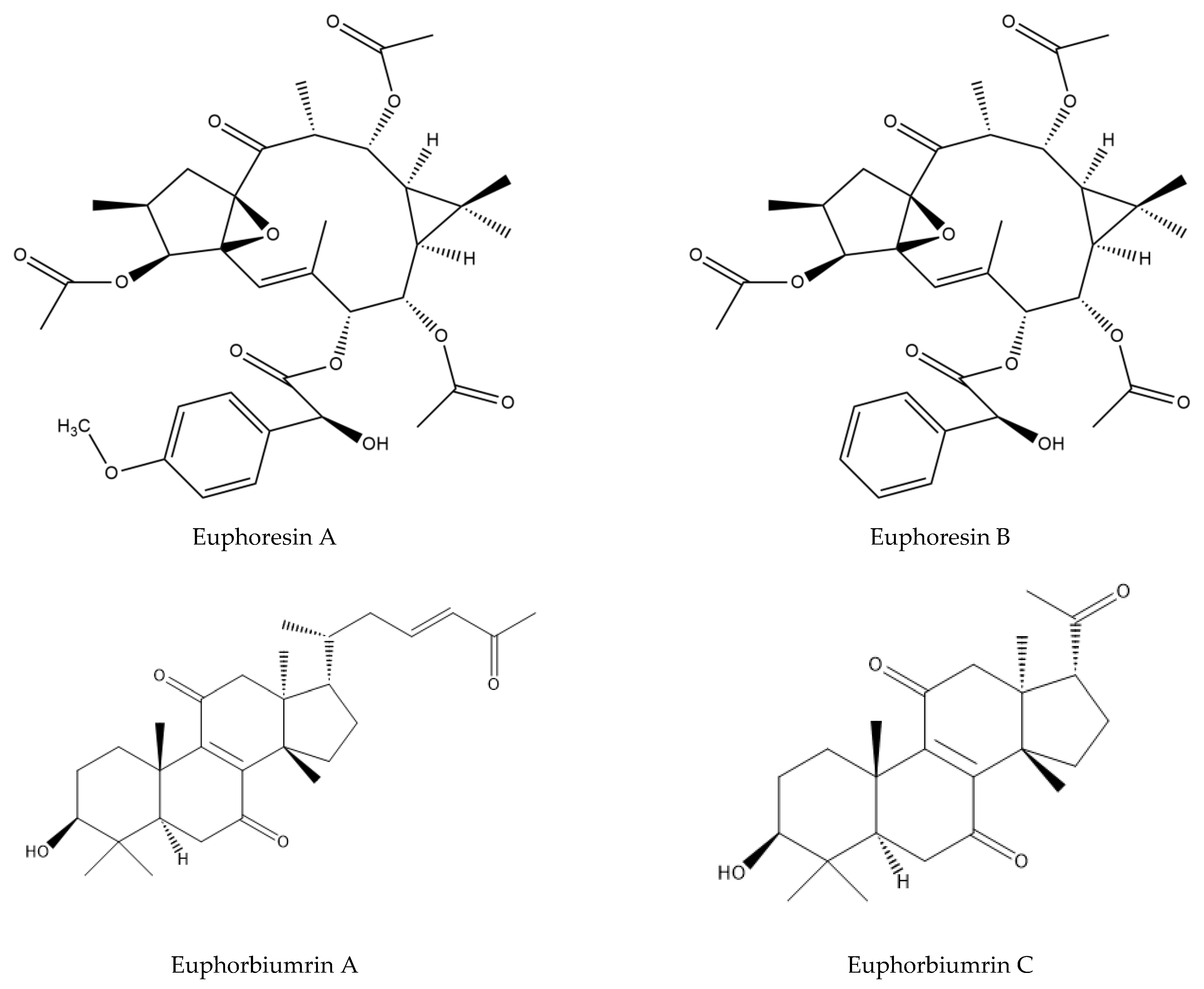
| - (China) | Methanolic extract | Terpenoids | Euphoresins A | [61] |
| Euphoresins B | ||||
| - (China) | Methanolic extract | Terpenoids | Euphorblin A | [64] |
| Euphorblin B | ||||
| Euphorblin C | ||||
| Euphorblin D | ||||
| Euphorblin E | ||||
| Euphorblin F | ||||
| Euphorblin G | ||||
| Euphorblin H | ||||
| Euphorblin I | ||||
| Euphorblin J | ||||
| Euphorblin K | ||||
| Euphorblin L | ||||
| Euphorblin M | ||||
| Euphorblin N | ||||
| Euphorblin O | ||||
| Euphorblin P | ||||
| Euphorblin Q | ||||
| - (China) | Methanolic extract | Terpenoids | Euphorbiumrin A | [64] |
| Euphorbiumrin B | ||||
| Euphorbiumrin C | ||||
| Euphorbiumrin D | ||||
| Euphorbiumrin E | ||||
| Euphorbiumrin F | ||||
| Euphorbiumrin G | ||||
| Euphorbiumrin H | ||||
| Euphorbiumrin I | ||||
| Euphorbiumrin J |
| Use Part | Extracts | Used Method | Tested Strains | Key Results | References |
|---|---|---|---|---|---|
| Aerial parts | Aqueous extract | Agar direct contact method | S. aureus ATCC 25923 | - | [3] |
| P. aeruginosa ATCC 27853 | - | ||||
| E. coli ATCC 25922 | + | ||||
| Roots | Acetone extract | Microdilution technique | E. coli ATCC 35210 | MIC = 0.5 ± 0.0 mg/mL | [17] |
| MBC = 0.5 ± 0.2 mg/mL | |||||
| S. aureus ATCC 29213 | MIC = 0.5 ± 0.0 mg/mL | ||||
| MBC =1.0 ± 0.2 mg/mL | |||||
| S. typhimurium ATCC 13311 | MIC = 1.0 ± 0.1 mg/mL | ||||
| MBC = 1.5 ± 0.2 mg/mL | |||||
| B. subtilis ATCC 10907 | MIC = 1.0 ± 0.2 mg/mL | ||||
| MBC = 1.5 ± 0.0 mg/mL | |||||
| S. epidermidis ATCC 12228 | MIC = 0.5 ± 0.0 mg/mL | ||||
| MBC = 1.0 ± 0.2 mg/mL | |||||
| Ethanol extract | Microdilution technique | E. coli ATCC 35210 | MIC = 0.3 ± 0.0 mg/mL | ||
| MBC = 0.5 ± 0.0 mg/mL | |||||
| S. aureus ATCC 29213 | MIC = 0.5 ± 0.0 mg/mL | ||||
| MBC = 1.0 ± 0.2 mg/mL | |||||
| S. typhimurium ATCC 13311 | MIC = 0.5 ± 0.0 mg/mL | ||||
| MBC = 1.0 ± 0.2 mg/mL | |||||
| B. subtilis ATCC 10907 | MIC = 1.0 ± 0.2 mg/mL | ||||
| MBC = 1.0 ± 0.0 mg/mL | |||||
| S. epidermidis ATCC 12228 | MIC = 0.5 ± 0.0 mg/mL | ||||
| MBC = 1.0 ± 0.2 mg/mL | |||||
| Ethyl acetate | Microdilution technique | E. coli ATCC 35210 | MIC = 0.4 ± 0.0 mg/mL | ||
| MBC = 0.3 ± 0.0 mg/mL | |||||
| S. aureus ATCC 29213 | MIC = 0.2 ± 0.0 mg/mL | ||||
| MBC = 0.3 ± 0.2 mg/mL | |||||
| S. typhimurium ATCC 13311 | MIC = 0.5 ± 0.0 mg/mL | ||||
| MBC = 1.0 ± 0.2 mg/mL | |||||
| B. subtilis ATCC 10907 | MIC = 0.3 ± 0.0 mg/mL | ||||
| MBC = 0.1 ± 0.0 mg/mL | |||||
| S. epidermidis ATCC 12228 | MIC = 0.2 ± 0.1 mg/mL | ||||
| MBC = 0.6 ± 0.2 mg/mL | |||||
| Dichloromethane extract | Microdilution technique | E. coli ATCC 35210 | MIC = 0.5 ± 0.0 mg/mL | ||
| MBC = 1.0 ± 0.2 mg/mL | |||||
| S. aureus ATCC 29213 | MIC = 0.5 ± 0.0 mg/mL | ||||
| MBC = 1.0 ± 0.2 mg/mL | |||||
| S. typhimurium ATCC 13311 | MIC = 1.0 ± 0.2 mg/mL | ||||
| MBC = 0.5 ± 0.3 mg/mL | |||||
| B. subtilis ATCC 10907 | MIC = 0.5 ± 0.0 mg/mL | ||||
| MBC = 1.0 ± 0.2 mg/mL | |||||
| S. epidermidis ATCC 12228 | MIC = 0.5 ± 0.0 mg/mL | ||||
| MBC = 1.0 ± 0.2 mg/mL | |||||
| Roots | Methanolic extract | Microdilution technique | E. coli ATCC 35210 | MIC ≥ 16 ± 0 mg/mL | [17] |
| MBC ≥ 16 ± 0 mg/mL | |||||
| S. aureus ATCC 29213 | MIC = 2 ± 0 mg/mL | ||||
| MBC = 2 ± 0 mg/mL | |||||
| Ethyl acetate extract | S. typhimurium ATCC 13311 | MIC = 0.5 ± 0 mg/mL | |||
| MBC = 0.5 ± 0 mg/mL | |||||
| B. subtilis ATCC 10907 | MIC = 0.5 ± 0 mg/mL | ||||
| MBC = 0.5 ± 0 mg/mL | |||||
| Stems | Methanolic extract | Microdilution technique | S. aureus ATCC 29 213 | MIC ≥ 16 ± 0 mg/mL | [46] |
| MBC ≥ 16 ± 0 mg/mL | |||||
| B. subtilis ATCC 3366 | MIC = 4 ± 0 mg/mL | ||||
| MBC = 4 ± 0 mg/mL | |||||
| Ethyl acetate extract | S. aureus ATCC 29 213 | MIC = 2 ± 0 mg/mL | |||
| MBC = 2 ± 0 mg/mL | |||||
| B. subtilis ATCC 3366 | MIC = 4 ± 0 mg/mL | ||||
| MBC = 2 ± 0 mg/mL | |||||
| Flowers | Methanolic extract | Microdilution technique | S. aureus ATCC 29 213 | MIC = 2 ± 0 mg/mL | [46] |
| MBC = 2 ± 0 mg/mL | |||||
| B. subtilis ATCC 3366 | MIC = 4 ± 0 mg/mL | ||||
| MBC = 4 ± 0 mg/mL | |||||
| Ethyl acetate extract | S. aureus ATCC 29 213 | MIC = 4 ± 0 mg/mL | |||
| MBC = 4 ± 0 mg/mL | |||||
| B. subtilis ATCC 3366 | MIC = 4 ± 0 mg/mL | ||||
| MBC = 4 ± 0 mg/mL | |||||
| Latex | Isolated euphorbioside | Serial dilution methode | E. coli ATCC 25922 | [62] | |
| S.aureus ATCC 19433 | |||||
| P. aeruginosa ATCC 27853 | |||||
| B. subtilis ATCC 6633 | |||||
| Aerial parts | Hexanic extract | Well agar diffusion method | Rhodococcus equi | ɸ= 18 mm | [17] |
| Dichloromethane extract | Well agar diffusion method | Rhodococcus equi | ɸ = 18 mm | ||
| Rhodococcus sp GK1 | ɸ = 15 mm | ||||
| Honey | - | Well agar diffusion and dilution range | S. aureus ATCC 6538 | ɸ = 25.98 ± 0.11 mm | [19] |
| E. coli ATCC 10536 | ɸ = 13.84 ± 1.10 mm |
| Use Part | Extracts Used | Used Method | Test Strains | Key Results | References |
|---|---|---|---|---|---|
| Aerial parts | Aqueous extract | Growth radial technique on solid medium | A. Flavus MTTC 2799 | I = 64.14% to 85.51% | [3] |
| P. expansum MTTC 1344 | I = 60.14% to 85.51% |
| Used Part | Extracts | Used Method | Key Results | References |
|---|---|---|---|---|
| Aerial parts | Methanolic extract | DPPH | IC50 = 0.0086 mg/mL | [3] |
| Flavonoids extract | IC50 = 0.378 mg/mL | |||
| Alkaloids extract | IC50 = 1.171 mg/mL | |||
| Aerial parts | Aqueous extract | DPPH | IC50 = 0.370 mg/mL | [17] |
| Honey | - | DPPH | IC50 = 80.1 ± 1.1 mg/mL | [17] |
| Superoxide | IC50 = 3.70 ± 0.0 mg/mL | |||
| Nitric oxide | IC50 = 88.2 ± 0.8 mg/mL | |||
| Roots | Dichloromethane extract | DPPH | SC50 = 122.15 ± 0.52 μg/mL | [17] |
| Ethyl acetate extract | SC50 = 18.20 ± 0.41 μg/mL | |||
| Ethanol extract | SC50 = 65.01 ± 0.32 μg/mL | |||
| Acetone extract | SC50 = 98.44 ± 0.13 μg/mL | |||
| Roots | Methanol extract | DPPH | IC50 = 10.01 ± 0.17 µg/mL | [46] |
| Ethyl acetate extract | IC50 = 18.85 ± 0.12 µg/mL |
| Part Used | Extracts | Cells Lines | Key Lines | References |
|---|---|---|---|---|
| Aerial parts | Hexane extract | Animal cells Vero | IC50= 266.43 µg/mL | [18] |
| Human cells RD | IC50= 50.7 µg/mL | |||
| Dichloromethane extract | Animal cells BSR | IC50= 77.2 µg/mL | ||
| Vero | IC50= 79.2 µg/mL | |||
| Methanolic extract | Human cells RD | IC50= 67.57 µg/mL | ||
| Animal cells BSR | IC50= 200 µg/mL |
| Part Used | Compounds Isolated | Experimental Approach | Key Lines | References |
|---|---|---|---|---|
| Latex | Euphatexol C | NO * production inhibition assay | IC50 = 22.30 μM | [52] |
| Euphatexol D | IC50 = 48,04 μM | |||
| Euphatexol E | IC50 = 21.89 μM | |||
| Euphatexol F | IC50 = 38.15 μM | |||
| Euphatexol G | IC50 = 41.15 μM |
Disclaimer/Publisher’s Note: The statements, opinions and data contained in all publications are solely those of the individual author(s) and contributor(s) and not of MDPI and/or the editor(s). MDPI and/or the editor(s) disclaim responsibility for any injury to people or property resulting from any ideas, methods, instructions or products referred to in the content. |
© 2023 by the authors. Licensee MDPI, Basel, Switzerland. This article is an open access article distributed under the terms and conditions of the Creative Commons Attribution (CC BY) license (https://creativecommons.org/licenses/by/4.0/).
Share and Cite
Hmidouche, O.; Bouftini, K.; Chafik, A.; Khouri, S.; Rchid, H.; Rahimi, A.; Mimouni, M.; Maarouf, E.; Zaakour, F.; Nmila, R.; et al. Ethnomedicinal Use, Phytochemistry, Pharmacology, and Toxicology of Euphorbia resinifera O. Berg. (B): A Review. J. Zool. Bot. Gard. 2023, 4, 364-395. https://doi.org/10.3390/jzbg4020029
Hmidouche O, Bouftini K, Chafik A, Khouri S, Rchid H, Rahimi A, Mimouni M, Maarouf E, Zaakour F, Nmila R, et al. Ethnomedicinal Use, Phytochemistry, Pharmacology, and Toxicology of Euphorbia resinifera O. Berg. (B): A Review. Journal of Zoological and Botanical Gardens. 2023; 4(2):364-395. https://doi.org/10.3390/jzbg4020029
Chicago/Turabian StyleHmidouche, Oumaima, Khadija Bouftini, Abdelbasset Chafik, Sara Khouri, Halima Rchid, Abdessadek Rahimi, Mostafa Mimouni, Elbekay Maarouf, Fatna Zaakour, Rachid Nmila, and et al. 2023. "Ethnomedicinal Use, Phytochemistry, Pharmacology, and Toxicology of Euphorbia resinifera O. Berg. (B): A Review" Journal of Zoological and Botanical Gardens 4, no. 2: 364-395. https://doi.org/10.3390/jzbg4020029