Article

Comparing Predictors and Outcomes of Higher Allostatic Load across Zoo-Housed African Great Apes

1
Department of Reproductive and Behavioral Sciences, Saint Louis Zoo, St. Louis, MO 63110, USA
2
Center for Species Survival, Smithsonian National Zoo and Conservation Biology Institute, Front Royal, VA 22630, USA
3
North of England Zoological Society, Chester Zoo, Upton by Chester CH2 1LH, UK
4
Smithsonian Global Health Program, Smithsonian National Zoo and Conservation Biology Institute, Washington, DC 20008, USA
5
National Museum of Natural History, Smithsonian Institution, Washington, DC 20560, USA
6
Department of Epidemiology of Microbial Disease, Yale School of Public Health, New Haven, CT 06520, USA
7
Veterinary Initiative for Endangered Wildlife, Bozeman, MT 59715, USA
8
Department of Anthropology, The Ohio State University, Columbus, OH 43210, USA
9
College of Public Health, The Ohio State University, Columbus, OH 43210, USA
10
Department of Clinical Sciences, Colorado State University, Fort Collins, CO 80523, USA
11
Wildlife Health Sciences, Smithsonian National Zoo and Conservation Biology Institute, Washington, DC 20008, USA
*
Author to whom correspondence should be addressed.
J. Zool. Bot. Gard. 2023, 4(1), 158-175; https://doi.org/10.3390/jzbg4010016
Submission received: 30 November 2022 / Revised: 17 February 2023 / Accepted: 20 February 2023 / Published: 1 March 2023
(This article belongs to the Special Issue Innovative Techniques Used to Improve Animal Health and Welfare)

Abstract

:
Stressors over the lifespan can contribute to physiological dysregulation, or allostatic load. Allostatic load has been studied in humans using allostatic load indices (ALIs) for over 25 years, but the same methods are rarely applied to other species. We constructed an ALI for zoo-housed western lowland gorillas, chimpanzees, and bonobos and tested potential predictors of and health outcomes associated with allostatic load. Allostatic load scores ranged from 0–6 for gorillas and chimpanzees and 0–7 for bonobos. Age was significantly associated with allostatic load in gorillas and chimpanzees but not bonobos. Cumulative stressful events were positively associated with allostatic load in chimpanzees. Wild-caught gorillas had higher allostatic load than zoo-born conspecifics, but rearing differences between zoo-born animals were not significant for any species. Age may affect associations of allostatic load with stressful events and birthplace as results change when it is included as a covariate. Allostatic load was not retained in best-fit models for risk of all-cause morbidity, cardiac disease, or mortality risk. Some analyses herein were limited by the use of retrospective data, such as reason for sample collection and length of records provided for individual animals. Nevertheless, these data indicate additional research is needed to optimize ALIs for non-human primates.

1. Introduction

Experiences across the lifespan can contribute to disease risk in animals. In mammalian taxa, each time an event is perceived as stressful, a cascade of physiological changes is triggered within the body [1,2]. For example, when stressors activate the hypothalamic-pituitary-adrenal (HPA) axis, corticotropin-releasing hormone (CRH) travels from the hypothalamus to the anterior pituitary, inducing the synthesis and release of adrenocorticotropin hormone (ACTH) into the bloodstream. When ACTH reaches the adrenal glands, it triggers the adrenal cortex to release glucocorticoids into systemic circulation. Glucocorticoids such as cortisol, the primary glucocorticoid in primates, induce physiological changes such as gluconeogenesis, lipolysis, and increased blood pressure. Glucocorticoids, therefore, are frequently measured to quantify stress in zoo-housed animals. However, interpreting glucocorticoid data can be difficult [3,4,5]. A number of other physiological changes also occur in response to stress, including changes in circulating catecholamines (e.g., epinephrine, norepinephrine) [1,2] and immune parameters (e.g., interleukin-6 [IL-6], tumor necrosis factor-alpha [TNF-α]) [6,7], but these are not measured as frequently as glucocorticoids when assessing potential stress responses.
Individually, stress responses are a healthy, adaptive mechanism that evolved to stimulate behavioral and physiological changes that promote survival. However, over the lifespan, experiencing repeated and/or chronic stressors can result in the physiological dysregulation of biomarkers across somatic systems that eventually increases disease risk [8]. For example, elevations in blood pressure in response to stress are part of normal coping mechanisms, but after repeated elevations, physiological dysregulation may occur where levels do not return to baseline following stressor abatement. Over time, this results in a new, shifted baseline that is higher than the original, and with repeated shifts over time even baseline levels may reach the threshold for the diagnosis of hypertension.
This cumulative physiological “wear-and-tear” over time is known as allostatic load [8]. While it is not possible to measure allostatic load across every system, it can be estimated using an allostatic load index (ALI) composed of biomarkers from multiple systems (e.g., neuroendocrine, cardiovascular, metabolic, immune) that are integrated to provide a single score for an individual. Within a population, those that have higher estimated allostatic load have greater physiological dysregulation than those with lower allostatic load. The first ALI was developed in human clinical research in 1997 [9] and in the more than 25 years since, there have been a wealth of studies with ALIs constructed using dozens of different biomarker combinations [10,11,12]. In humans, allostatic load can mediate the pathway from lifetime experiences to disease risk. For example, lower socioeconomic status [12,13], early life adversity [14,15], and stressful experiences [16,17] have been shown to contribute to higher allostatic load. In turn, higher allostatic load is associated with health outcomes such as cancer [18,19], cardiac disease [13,20], diabetes [13,21], and mortality [9,22,23].
Given its potential to both reflect previous life experiences and predict future health outcomes, allostatic load is of broad interest to those who study animals. Since 2003, more than 500 papers published on various animal taxa have referenced allostasis and/or allostatic load [24]. However, despite the widespread interest of researchers who study animals, developing ALIs to estimate allostatic load outside of humans remains infrequent [24]. Previously, we constructed the first non-human ALIs and applied them to western lowland gorillas (Gorilla gorilla gorilla). In our pilot work on gorillas at one zoo [25,26], we observed positive associations between age and cumulative stressful events (e.g., number of immobilizations, transfers, etc.,) over the lifespan, higher allostatic load in females than males, and higher allostatic load in wild-caught gorillas compared to zoo-born mother- and nursery-reared conspecifics. When we expanded that research to include two additional zoos, we observed these same relationships as well as an association with mortality risk [27]. We have since explored other iterations of the ALI. For example, we have found indices that include cholesterol and triglycerides improve model predictions of health outcomes [28] and we reported on a possible method for refining selection of biomarkers to include in the index, which unfortunately was not effective at identifying a better ALI for improving model predictions [29].
Herein, we compare allostatic load across three zoo-housed African ape species: western lowland gorillas, chimpanzees (Pan troglodytes), and bonobos (P. paniscus). Research on humans has been criticized in the past because ALIs constructed with different biomarkers are difficult to compare [12,30,31]. As such, we only included biomarkers that we had measured in all three species, resulting in an eight-biomarker ALI including albumin, cholesterol, cortisol, dehydroepiandrosterone-sulfate (DHEA-S), glucose, IL-6, triglycerides, and TNF-α. While all of these biomarkers have been included in one or more previous ALIs constructed for gorillas, this particular combination is new to allow direct comparisons between the three species. The gorilla dataset also was expanded to include individuals from an additional zoo. Once allostatic load scores were estimated for each species, we investigated potential predictors (e.g., age, rearing) and health outcomes (e.g., risk of all-cause morbidity and mortality) associated with higher allostatic load. As the data in this study were collected retrospectively, there are inherent limitations to some of our analyses and interpretations. These limitations are reviewed extensively in the Discussion section.

2. Materials and Methods

2.1. Subjects

Through collaborations with 25 North American zoological associations accredited by the Association of Zoos and Aquariums (AZA), we obtained serum samples collected during previous immobilizations on western lowland gorillas (108 samples from 71 unique individuals), chimpanzees (238 samples from 162 unique individuals), and bonobos (44 samples from 38 unique individuals). Samples collected prior to 7 years of age were excluded to reduce variation in biomarkers due to growth and development. Only one sample per animal was included for analysis. When possible, the sample for each individual came from a routine immobilization (e.g., annual health examination); samples from routine immobilizations are preferred because many biomarkers included in the ALI are responsive to acute events and allostatic load is meant to estimate physiological dysregulation outside of acute responses. However, some animals only had samples collected during non-routine immobilizations (i.e., in response to injury or illness) or the reason for immobilization was unknown. Although an immobilization itself may represent an acute event that has an impact on circulating biomarkers, we used the purpose for immobilization to differentiate between animals that may have changes in biomarkers for reasons other than anesthesia in case there was an additive effect of multiple stressors (e.g., injury + anesthesia) on circulating biomarker levels. In cases where an individual had multiple routine or non-routine/unknown reason samples, two criteria were used to select a single sample. First, as it was not always possible to measure all biomarkers in every sample due to limited volume, samples with fewer missing data were selected (e.g., if a sample from one date had five of eight biomarkers and a sample from a second date had seven of eight biomarkers, the latter would be selected for inclusion). Second, if the same number of biomarkers were analyzed from each sample, then the sample collected at the animal’s oldest age was selected (e.g., a sample collected at age 17 years would be selected over a sample collected at age 15 years). This resulted in datasets of 68 gorillas from four zoos (aged 7–52 years, x ¯ = 23.3, SD = 12.3, 34 males), 148 chimpanzees from 17 zoos (aged 7–73 years, x ¯ = 27.5, SD = 12.5, 60 males), and 33 bonobos from five zoos (aged 7–48 years, x ¯ = 22.2, SD = 11.5, 18 males). Some subjects had missing biomarker data due to limited sample volume, including 13 gorillas (1 biomarker missing: n = 1; 2 biomarkers missing: n = 12), 16 chimpanzees (1 biomarker missing: n = 8; 2 biomarkers missing: n = 3; 3 biomarkers missing: n = 1; 4 biomarkers missing: n = 4), and one bonobo (1 biomarker missing). For these individuals, ALIs were calculated in the standard manner as described in Section 2.3, but the maximum score totaled the number of biomarkers measured. This study was approved by each participating institution and adheres to the American Society of Primatologists’ Principles for the Ethical Treatment of Primates.

2.2. Biomarker Analyses

Serum samples were collected between 1984 and 2019 and cryopreserved at −80 °C until analysis. Sample degradation can occur when samples remain in storage over the long-term, but studies indicate steroid hormones (cortisol, DHEA-S) [32,33] and inflammatory cytokines (IL-6, TNF-α) [34] maintain their integrity when cryopreserved without multiple freeze-thaw cycles, and it has been demonstrated that degradation over 10 or more years has minimal effect when using lipids (cholesterol, triglycerides) to study disease risk [35].
Biomarker analyses occurred at three different time periods: the original pilot project studying allostatic load in gorillas from three zoos (2014, 2019) [25,26,27,28,29], inclusion of data from a fourth zoo holding gorillas (2019), and collection of data from chimpanzees and bonobos for a multi-species comparison (2019–2020). For gorillas in the original pilot project, albumin and glucose values were obtained from medical records from the same immobilization as the serum samples used to assay additional biomarkers. Otherwise, albumin and glucose for the remaining gorillas as well as the chimpanzees and bonobos, along with total cholesterol and triglycerides for all individuals, were analyzed at the Smithsonian Conservation Biology Institute in 2019 using an RX Daytona automated clinical chemistry analyzer (Randox Industries-US Ltd., Kearneysville, WV, USA). Commercially available reagents (albumin: AB3800, glucose: GL3981, cholesterol: CH3810, triglycerides: TR3823), calibrators (all: CAL2351), and two-level controls (all: HN1530, HN1532) were purchased from Randox Industries-US Ltd. The technical ranges were 3.20–50.6 g/L for albumin, 0.200–35.5 mmol/L for glucose, 0.865–16.6 mmol/L for cholesterol, and 0.134–12.7 mmol/L for triglycerides. Serum was generally run neat but was diluted 1:5 with saline (SA3854) when needed. The analyzer was subject to routine quality control measurements throughout the study. Normal and elevated controls for each analyte were maintained within two standard deviations of the respective target value.
Cortisol, DHEA-S, IL-6, and TNF-α were analyzed using solid-phase enzyme immunoassays (EIAs), with one exception. All samples were analyzed in duplicate with coefficients of variation (CVs) maintained below 10% and inter-assay CVs below 15% for high and low concentration controls. Assays for gorillas from the three pilot study zoos were completed at The Ohio State University Center for Clinical and Translation Science: Clinical Research Center in 2014. Cortisol (LKCO1) and DHEA-S (LKDS1) were measured using competitive chemiluminescent EIAs with an Immulite 1000 (Siemens Healthcare Diagnostics Inc., Deerfield, IL, USA). The calibration range for cortisol was 1–1000 µg/dL and for DHEA-S was 15–1000 µg/dL. The IL-6 (HS600B) and TNF-α (HSTA00D) EIAs were from the same manufacturer (R and D Systems, Minneapolis, MN, USA). The assay range for IL-6 was 0.20–10.0 pg/mL with a sensitivity of 0.11 pg/mL. The assay range for TNF-α was 0.5–32.0 pg/mL with a sensitivity of 0.106 pg/mL.
Assays for the remaining gorillas and all chimpanzees and bonobos were completed at the Smithsonian Conservation Biology Institute in 2019–2020. Cortisol for gorillas from the fourth zoo was measured using a solid-phase 125I radioimmunoassay (Corti-Cote RIA; 06B-256440, MP Biomedicals, Santa Ana, CA, USA) with modifications as described in [36], using 25 µL cortisol standards (0.25–60 µg/dL), controls (BioRad Lyphocheck Immunoassay Plus Control, Levels 1,2 and 3; REF370), and samples (diluted 1:20 with 0 standard), in duplicate. The RIA was validated biochemically for measuring cortisol in gorilla serum through parallelism and matrix interference assessments, and subsequent regression analyses. Serial two-fold dilutions of serum yielded a displacement curve parallel to the standard curve (y = 0.837x − 4.517, R2 = 0.974, F1,6 = 229.276, p < 0.001). There was no evidence of matrix interference, as addition of appropriately diluted serum to assay standards did not alter the amount observed (y = 0.491x − 5.574, R2 = 0.903, F1,5 = 46.910, p = 0.001). For all chimpanzees and bonobos, cortisol was measured using a double antibody EIA with a secondary goat-anti rabbit IgG antibody (A009, Arbor Assays, Ann Arbor, MI, USA) and polyclonal rabbit anti-cortisol antibody (R4866, C. Munro, University of California, Davis, CA, USA). Our protocol for this non-commercially available assay has been previously described [37]. This EIA was validated biochemically for measuring cortisol in chimpanzee and bonobo serum through parallelism and matrix interference assessments, and subsequent regression analyses. Serial two-fold dilutions of serum yielded a displacement curve parallel to the standard curve (chimp: y = 1.019x − 2.114, R2 = 0.998, F1,4 = 1753.968, p < 0.001 and bonobo: y = 0.986x + 1.449, R2 = 0.996, F1,6 = 1566.448, p < 0.001). There was no evidence of matrix interference, as addition of appropriately diluted serum (chimpanzees 1:150; bonobos 1:100) to assay standards did not alter the amount observed (chimpanzees: y = 1.183x − 13.972, R2 = 0.979, F1,7 = 321.043, p < 0.001; bonobos: y = 1.060x − 11.286, R2 = 0.992, F1,7 = 849.689, p < 0.001).
DHEA-S was analyzed using a competitive colorimetric EIA (K054, Arbor Assays, An Arbor, MI, USA) with a range of 9.6–6000 µg/dL. Samples were analyzed at a 1:100 dilution for gorillas, 1:250 for chimpanzees, and 1:200 for bonobos. The DHEA-S EIA was validated biochemically in western lowland gorilla, chimpanzee, and bonobo serum through parallelism and matrix assessment interference, and subsequent regression analyses. Serial five-fold dilutions of serum yielded a displacement curve parallel to the standard curve (gorillas: y = 1.088x − 6.520, R2 = 0.984, F1,3 = 184.242, p < 0.001; chimpanzees: y = 0.975x + 8.855, R2 = 0.994, F1,3 = 525.601, p < 0.001; bonobos: y = 0.985x + 11.015, R2 = 0.997, F1,3 = 983.034, p < 0.001). There was no evidence of matrix interference for any species, as the addition of appropriately diluted serum to assay standards did not alter the amount observed (gorillas: y = 0.748x + 110.722, R2 = 0.998, F1,3 = 1772.389, p < 0.001; chimpanzees: y = 0.915x + 653.511, R2 = 0.995, F1,3 = 566.412, p < 0.001; bonobos: y = 0.860x − 402.019, R2 = 0.999, F1,3 = 3812.135, p < 0.001).
IL-6 and TNF-α were measured using EIAs from the same manufacturer as the pilot study gorillas (R and D Systems, Minneapolis, MN, USA), but the original kits had been discontinued and replaced with an updated kit with increased sensitivity. The assay range for IL-6 (HS600C) was 0.20–10.0 pg/mL with a sensitivity of 0.09 pg/mL. Serum samples were generally diluted 1:5 for gorillas and 1:2 for chimpanzees and bonobos; any sample that exceeded the highest standard was further diluted (up to 1:40 for gorillas, up to 1:30 for chimpanzees and bonobos) until they were within the range of the standard curve. The IL-6 assay was validated via linearity (gorillas: 82.9%, chimpanzees: 96.9%, bonobos: 107.9%) and spike and recovery (gorillas: 110.3%, chimpanzees: 99.6%, bonobos: 105.7%) assessment within the range of dilutions used. For TNF-α (HSTA00E), the assay range was 0.2–10.0 pg/mL with a sensitivity of 0.049 pg/mL. Serum samples for all three species were analyzed undiluted, although some chimpanzee samples exceeded the highest standard and were further diluted up to 1:5. The TNF-α assay was validated via linearity for chimpanzees (102.7%), as the only species for which some samples needed dilution, and spike and recovery assessment for all three species (gorillas: 124%, chimpanzees: 85.6%, bonobos: 89.8%).

2.3. Allostatic Load Index

The ALI was constructed and subsequent allostatic load scores were estimated following methodologies used in our previous research on gorillas [25,26,27,28,29]. Allostatic load scores are typically estimated by determining a high-risk threshold, or cut-point, for each biomarker and then counting the number of biomarkers each individual has above or below that cut-point, depending on the biomarker. Prior to determining the cut-points for each biomarker, biomarker values for all zoos were pooled into a single dataset per species. Once pooled, data for each biomarker were analyzed to determine if there were differences between males and females (α = 0.05). Most biomarkers were right-skewed, so we used generalized linear models (GLMs) with a Gamma distribution and log-link function to examine differences by sex. Albumin for all three species and cholesterol for chimpanzees were normally distributed, so a linear regression was used in these cases. The reason for sample collection (routine, non-routine, unknown) was included as a fixed effect in all analyses. For biomarkers with significant differences between males and females, sex-specific cut-points were used when estimating allostatic load.
Once biomarkers with significant sex differences were identified, we calculated the high-risk cut-points using the quartile methodology originally proposed by Seeman and colleagues [9], with one modification. In the quartile method, the distribution of each biomarker is divided into quartiles and either the first or fourth quartile is designated as high-risk, depending on the biomarker [9]. For many biomarkers, physiological dysregulation is reflected by elevated levels, making the fourth quartile high-risk, but for albumin and DHEA-S, the first quartile (i.e., low concentrations) is considered high-risk. However, for some biomarkers the direction of the dysregulation-induced shift varies. For example, both low and high cortisol levels may indicate physiological dysregulation [38,39,40] and both high and low levels of total cholesterol have been associated with negative health outcomes [41,42,43,44]. Therefore, for cortisol and total cholesterol, we modified the original methodology and used a two-tailed split quartile (top and bottom 12.5% of the distribution) to reflect that dysregulation of these biomarkers can be characterized by either high or low concentrations. Statistical associations of each biomarker with sex as well as the cut-points assigned for each biomarker by species are presented in Table 1. As this ALI contains eight biomarkers, allostatic load scores for each individual could range anywhere from 0, meaning there are no biomarkers within the high-risk quartile, to 8, indicating all biomarkers are within the high-risk quartile. For animals with some missing values, those biomarkers were not counted toward an animal’s allostatic load score (e.g., if an animal was missing values for two biomarkers, its maximum allostatic load would be 6).

2.4. Predictors and Outcomes of Higher Allostatic Load

Data on potential predictors and outcomes of higher allostatic load were obtained from ZIMS [45] and/or husbandry and medical records provided by each participating zoo. Potential predictors of higher allostatic load included age, sex, the cumulative number of stressful events experienced over the lifespan, differences in parity for females, and rearing history. Stressful events were defined as immobilizations, transfers between holding institutions, aggression resulting in wounding or injury, and pregnancies for females. While zoo-housed great apes likely experience other events as being stressful, such as agonistic interactions without wounding, disruptions to the social dominance hierarchy, or changes in group composition, these events are not consistently recorded in zoo records and so could not be analyzed in this retrospective study. When possible, stressful events were counted from date of birth up until the date of sample collection for each individual. However, digital daily record keeping has only been a practice for the past two or three decades and records often do not accompany animals when they are transferred between institutions, so retrospective data may be incomplete for older individuals or those who have moved from their birth institution. Each individual was assigned one of five rearing histories: wild-caught or, for zoo-born animals, mother-reared, nursery-reared (i.e., reared by human caretakers), surrogate-reared (i.e., reared by a different female of the same species), or peer-reared (i.e., reared with other members of the same species without a maternal figure). Risk of chronic all-cause morbidity, cardiac disease, and all-cause mortality were included as outcome variables. Examples of conditions identified as chronic morbidities include osteoarthritis, hypothyroidism, neoplasia, obesity, and cardiac disease. Those with cardiac disease were analyzed as a subset of all-cause morbidity due to the high prevalence of this condition within zoo-housed great apes [46,47,48]. Records were not provided for seven bonobos; these individuals were excluded from all analyses except for those obtainable via ZIMS [45], including age, sex, number of transfers, and age at death (if applicable).

2.5. Quantitative Analyses
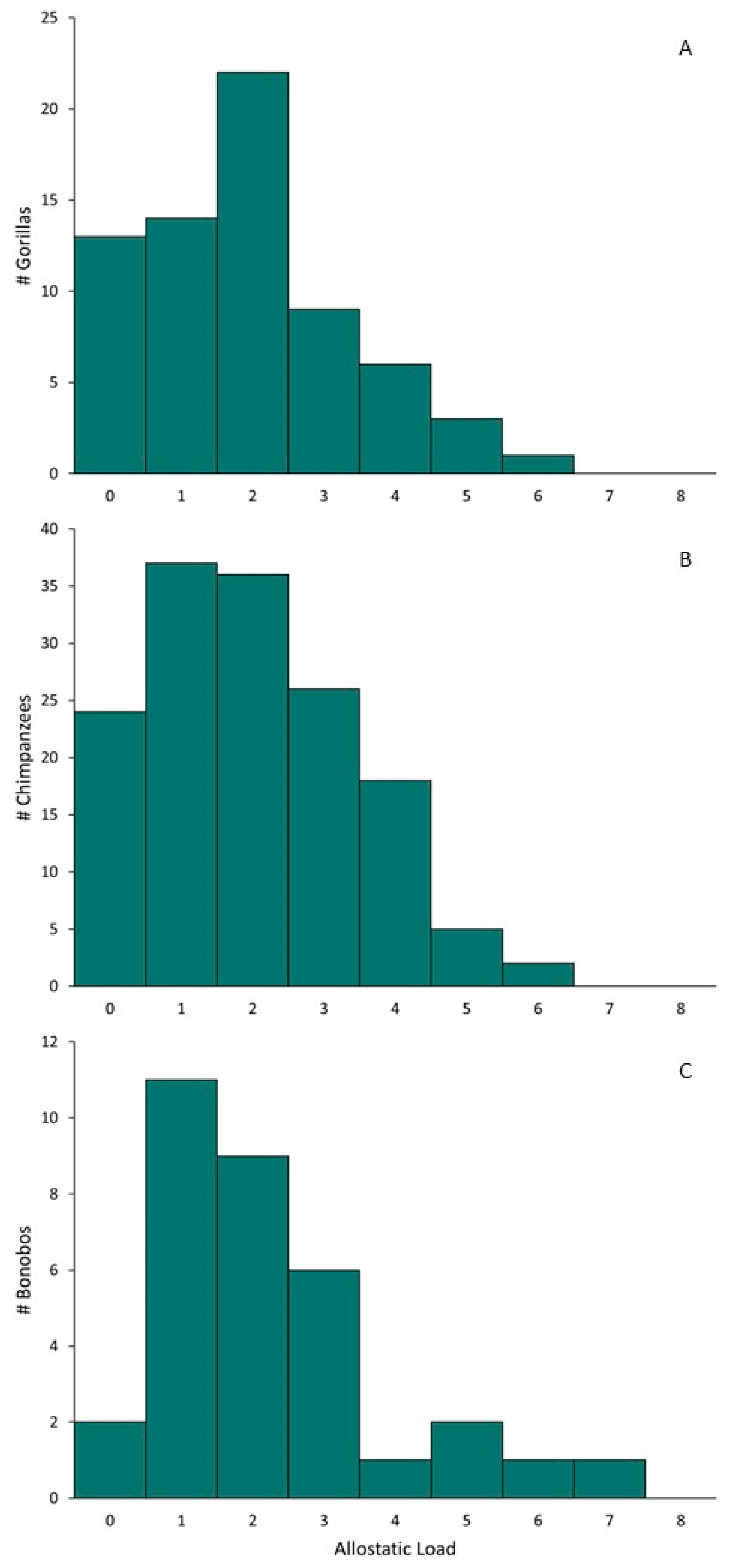
To test whether age, sex, stressful events, parity, or rearing history predicted higher allostatic load in gorillas, chimpanzees, or bonobos, we used generalized linear mixed models (GLMMs) with a Poisson distribution and a log-link function. The reason for sample collection was included as a fixed effect to account for any differences in scores obtained from non-routine immobilizations. Zoo was included as a random effect in all predictor models. To determine if allostatic load is predictive of health outcomes, we used binomial GLMMs with logit links to assess risk of all-cause morbidity and cardiac disease. We used Cox proportional hazards models based on time from sample collection to date of death to analyze whether allostatic load predicts mortality risk. Censored animals were still alive as of October 15, 2022. For both GLMMs and Cox proportional hazards models, we first analyzed models containing allostatic load, age, sex, or age and sex. Out of these four models, if the model containing age, sex, or age and sex had the best model fit, as determined by the lowest Akaike’s information criterion adjusted for small sample sizes (AICc), a fifth model would be analyzed with allostatic load added to determine if its inclusion improved model fit over having age and/or sex alone. As with predictor models, the reason for sample collection was included as a fixed effect. All GLMMs were analyzed using the “lme4” package [49] and mortality risk was estimated using “survival” [50], with AICc values for all models determined using the “MuMIn” package [51], in R [52].

3. Results

Descriptive statistics for predictor (e.g., stressful events, rearing history) and outcome (e.g., chronic conditions, mortality) variables by species are presented in Table 2. Allostatic load ranged from 0–6 ( x ¯ = 1.91, SD = 1.45) in gorillas, 0–6 ( x ¯ = 2.00, SD = 1.45) in chimpanzees, and 0–7 ( x ¯ = 2.24, SD = 1.64) in bonobos (Figure 1). Allostatic load was significantly lower for animals whose samples were collected during routine examinations compared to those collected during non-routine immobilizations for gorillas (β = −0.759, SE = 0.169, p < 0.001) and chimpanzees (β = −0.309, SE = 0.152, p = 0.042), but not for bonobos (β = −0.533, SE = 0.350, p = 0.128), supporting the inclusion of immobilization reason as a covariate in our analyses.

3.1. Predictors of Higher Allostatic Load

Model estimates, standard errors, and p-values for each possible predictor of higher allostatic load are available by species in Table 3. Variation in allostatic load for dichotomous variables (e.g., male vs. female, wild-caught vs. zoo-born) is presented in Table 4. After taking into account the reason for immobilization, older age was significantly associated with higher allostatic load in gorillas and chimpanzees but not bonobos. Sex was not significantly associated with allostatic load in any species. Cumulative stressful events had a significant positive association with allostatic load in chimpanzees but not gorillas or bonobos. When types of stressful events were examined individually, the number of transfers, wounding events, or immobilizations were not associated with allostatic load in gorillas, chimpanzees, or bonobos. There was no association between parity and allostatic load in female gorillas or chimpanzees and there were insufficient data to analyze this variable in female bonobos. Finally, being zoo-born as opposed to wild-caught was associated with a 46.4% reduction in allostatic load in gorillas, but this variable was not significantly associated with allostatic load in chimpanzees or bonobos. Among zoo-born individuals, differences between mother- and nursery-reared conspecifics were not significant for any species. As older animals are likely to have experienced more stressful events and pregnancies and were more likely to be wild-caught or reared by hand, we analyzed additional models for stressful events (cumulative and separately by type), parity, birthplace and rearing history with age included as a covariate. For gorillas, when age was included as a covariate, there was no longer a significant difference between zoo-born and wild-caught animals (β = −0.404, SE = 0.320, p = 0.207). In chimpanzees, the association between total stressful events was no longer significant when age was included as a covariate (β = −0.004, SE = 0.002, p = 0.080). Additionally, the three results in chimpanzees that approached significance no longer did so after age was included as a covariate (immobilizations: β = 0.004, SE = 0.009, p = 0.651; parity: β = 0.427, SE = 0.347, p = 0.219); birthplace: β = 0.004, SE = 0.186, p = 0.982).

3.2. Outcomes of Higher Allostatic Load

Variation in allostatic load for the dichotomous health outcome variables is presented in Table 5. Including allostatic load in models did not improve the relative goodness of fit for predicting health outcomes for any species. Risk of all-cause morbidity was best explained by age and sex in gorillas and age alone in chimpanzees and bonobos (Table 6). Each 5-year increase in age was associated with an increase in all-cause morbidity risk of 60% for gorillas, 30% for chimpanzees, and 105% for bonobos. In gorillas, males were nearly ten-times more likely to be affected by a chronic condition than females. The same pattern for all-cause morbidity was also observed with cardiac disease, with risk best explained by age and sex in gorillas and only age in chimpanzees and bonobos (Table 7). Risk of cardiac disease increased 45%, 15%, and 50% with every 5-year increase in age in gorillas, chimpanzees, and bonobos, respectively, and male gorillas were 13-times more likely to be affected than females. In gorillas and bonobos, age alone was the best predictor of mortality risk, although the variable was not significant in the best-fit model for bonobos, while both age and sex best predicted mortality in chimpanzees (Table 8). In both gorillas and bonobos, every 5-year increase in age was associated with a 35% increase in mortality risk. In chimpanzees, a 5-year increase in age was associated with a 25% increased risk of mortality and males had two-fold higher mortality risk than females.

4. Discussion

Based on research in humans and our past studies in gorillas, fewer variables than expected predicted higher allostatic load in gorillas, chimpanzees, and bonobos. Age was significantly associated with allostatic load in gorillas and chimpanzees but not in bonobos. Numerous studies in humans have reported positive associations between allostatic load and age [53,54,55] and we have previously observed this with some gorilla studies [25,27]. Allostatic load was lower in males across the three species but the differences were not significant. Sex differences are inconsistent in human populations, with different studies reporting men have higher [21,56,57], lower [58,59,60], or similar [17,61,62] allostatic load relative to women. Although there was a positive relationship between cumulative stressful events and allostatic load for all three species, it was only significant for chimpanzees. When analyzed individually, no single type of stressful event was significantly associated with allostatic load in any species. These results may indicate that the events we identified as stressful may not be perceived that way or contribute to allostatic load in these species and other events may have more salient effects. For example, negative social interactions irrespective of wounding may be more important to these species than wounding alone. Despite allostatic load theory suggesting that increased stressful events will directly contribute to higher allostatic load [8,55], this relationship is not as well tested in humans as those of allostatic load with many other predictors and outcomes. A few studies have reported a positive association between stressful events or experiences and allostatic load [17,63,64,65], but others have not [66,67,68,69]. Previously, we observed stressful events were significantly associated with allostatic load in gorillas at one zoo [25] but not others [27]. Allostatic load did not vary significantly between parous and nulliparous females for gorillas or chimpanzees in this study. This particular variable has not yet been analyzed in women and our previous reports on gorillas also did not document differences in female allostatic load based on parity [25,27], perhaps suggesting that although there are physiological costs of reproduction, these costs do not result in cumulative wear and tear over the lifetime. Zoo-born gorillas had significantly lower allostatic load than wild-caught individuals, but the difference was not significant for chimpanzees or bonobos. Given the age at which it occurs and the nature of capture from the wild, it may represent a form of early life adversity that zoo-born conspecifics do not experience. Similarly, adverse childhood experiences are associated with higher allostatic load in humans [15,68,70,71]. Among zoo-born individuals, there were no significant differences between mother- and nursery-reared gorillas, chimpanzees, or bonobos. We previously observed higher allostatic load in wild-caught compared to mother- and nursery-reared gorillas but not between mother- and nursery-reared individuals [13,20,21,70,71,72]. Differing results when age was included as a covariate suggest the significant and near significant associations with allostatic load that were observed for some variables may be better explained by age.
Allostatic load by itself did not significantly predict any outcome variable other than mortality risk in gorillas, and the variable did not improve relative goodness of fit for predicting all-cause morbidity, cardiac disease, or mortality risk for gorillas, chimpanzees, or bonobos. Instead, age and sex best predicted all-cause morbidity and cardiac disease in gorillas, and age alone best predicted these two health outcomes in chimpanzees and bonobos. Mortality risk was best predicted by age in gorillas and bonobos, and age and sex in chimpanzees. Numerous studies in humans have documented the connection between higher allostatic load and poor health outcomes, including cardiac disease [13,20,21,72,73,74] and others such as diabetes, arthritis, obesity, periodontal disease, and cancer [13,18,19,21,74,75,76]. Higher allostatic load in humans also is associated with increased mortality risk [9,19,22,23,77,78,79,80,81,82,83]. In previous work with gorillas using similar quantitative approaches, allostatic load was retained in best-fit models for mortality risk but not for risk of all-cause morbidity or cardiac disease [53].
Given the few associations reported herein and in some previous work with gorillas, it is possible that the ALIs constructed thus far are not the “right fit” for gorillas, chimpanzees, or bonobos, and that other combinations of biomarkers or perhaps other methods of constructing or determining the cut-points may be better suited to estimating allostatic load in these species. As physiological decline is naturally observed with aging for many biomarkers [12,30,31], an association between allostatic load and age may be one way of validating that the constructed index is measuring physiological dysregulation. As such, the association between allostatic load and age for gorillas and chimpanzees indicates that the index should be sensitive to physiological dysregulation; there is no reason to expect differing results in bonobos and so we suspect sample size and variability may be an issue. However, even if the index is reflective of physiological dysregulation, it still fails to vary in relation to most predictor variables and does not predict health-related outcomes, indicating that the ALI herein may be insufficient for measuring physiological dysregulation in African great apes. Despite criticism [30,31], there still is no set standard ALI for human research, nor an established method for identifying which biomarkers are best to include. While the initial iteration of allostatic load in humans was not intended to be a “gold standard” and the authors encouraged continued biomarker discovery [9], lack of consensus or method for identifying the best biomarkers presents a challenge for researchers working with new species using limited funds as well as limited sample availability and volume. Alternatively, while our group and others see the potential of applying allostatic load indices to animals to help advance studies of health and welfare, we must point out it is also possible that, although a valid tool in human research, allostatic load is not appropriate for other species, even those most closely related to us. Differences between the results reported here and those we previously reported in gorillas may also partially be explained by an increase in the sophistication and complexity of statistical approaches used; for example, we previously did not include the reason for immobilization in our analyses or use Poisson regressions when examining effects of predictors on allostatic load.
There were multiple limitations during this study that must be considered when interpreting the results. First, the differences between allostatic load scores estimated using samples collected during routine exams with those collected during non-routine immobilizations suggest that estimates of allostatic load may be more accurate if using only samples from routine immobilizations. As this was a retrospective study and we were often unable to obtain routine immobilization samples for some individuals, we included it as a covariate in the models, but prospective projects may be better able to control for this variation. Additionally, because animals move between institutions and their records often do not travel with them, the historical data we could obtain for each individual were highly variable. For some, we have daily or near daily notes from time of birth up to the date of sample. For others who are older and were born before regular digital record keeping and/or who were transferred, we may be missing years or even decades of data that contribute to our counts of cumulative stressful events. Furthermore, as previously mentioned, the perception of events as stressors is highly individualized and it is possible that the events we selected as stressors were not those with the most substantial impact on allostatic load development in these species. Social factors such as position in the dominance hierarchy, which could not be examined here due to the limitations of retrospective research (i.e., records of hierarchies are not maintained at all institutions), may be an important predictor and/or mediator of allostatic load in gregarious species like great apes and as such be a critical covariate that we are missing. Similar variables in humans, such as socioeconomic status and educational attainment [12,13,19,54,61,84,85], predict allostatic load and can mediate the pathway from stressful events to later life disease. These limitations may contribute to the lack of significant associations between stressful events and allostatic load, both cumulative and by type. Finally, while all animals in this study were under the care of veterinary teams, there may be undiagnosed or incipient conditions in those currently marked as having no chronic conditions that impact these results. Furthermore, there may be differences in allostatic load between those with no or single conditions and those that suffer from co-morbidities, as observed in humans [15,84]. How long an individual has experienced a condition, as chronic conditions themselves can represent stressors [85], and whether the condition is medically controlled also may contribute to allostatic load.
Given the wide-ranging body of research on allostatic load in humans suggesting allostatic load may be a pathway through which events over the lifespan contribute to health outcomes in later life, there is considerable interest among those who study animals in the possibility of applying allostatic load theory and methods to non-human species. Herein, we constructed an ALI for gorillas, chimpanzees, and bonobos using the same eight biomarkers and compared predictors and outcomes of higher allostatic load between the three species. Higher allostatic load was predicted by older age in gorillas and chimpanzees, more stressful events in chimpanzees, and being wild-caught for gorillas. However, these latter results also correlated with age. In turn, allostatic load was not included in the best-fit models for predicting risk of all-cause morbidity, cardiac disease, or mortality risk for gorillas, chimpanzees, or bonobos. Although African great apes are our closest relatives, many of the results reported herein are inconsistent with the large body of allostatic load research conducted in humans. As those developed so far do not seem to be the best fit, additional research may help optimize ALIs for these species. Developing a method to identify biomarkers that would be best to include in species-specific ALIs is a necessary next step for advancing this research area.

Author Contributions

Conceptualization: A.N.E.; methodology: A.N.E. and K.L.E.; validation: A.N.E. and K.L.E.; formal analysis: A.N.E. and K.L.E.; resources: A.N.E., K.L.E. and D.Z.; data curation: A.N.E., K.L.E., D.Z. and B.J.; writing—original draft preparation: A.N.E. and K.L.E.; writing—review and editing: A.N.E., K.L.E., D.Z., B.J., D.E.C., B.A.W., D.L.N. and J.L.B.; visualization: A.N.E.; project administration: A.N.E. and K.L.E.; funding acquisition: A.N.E., K.L.E., D.Z., D.E.C., B.A.W., D.L.N. and J.L.B. All authors have read and agreed to the published version of the manuscript.

Funding

This research was supported by funding from an Association of Zoos and Aquariums Conservation Grants Fund award, a Columbus Zoo and Aquarium Conservation Fund award, a Sacramento Zoo Conservation Committee Small Grants award, Friends of the National Zoo, the Endocrinology Research Laboratory at the Smithsonian Conservation Biology Institute, and the Department of Anthropology at The Ohio State University. The automated clinical chemistry analyzer was supported by funds from the Smithsonian Women’s Committee.

Institutional Review Board Statement

This research was approved by The Ohio State University (IACUC #2013A00000147) and the Smithsonian National Zoo as well as each participating institution.

Data Availability Statement

Data are available upon reasonable request from the corresponding author and agreement from the cooperating zoological institution.

Acknowledgments

This research would not have been possible without the assistance of veterinarians, keepers, and staff at the 25 participating zoological institutions: ABQ BioPark, Chattanooga Zoo, Cincinnati Zoo, Columbus Zoo and Aquarium, Dallas Zoo, Detroit Zoological Society, Jacksonville Zoo, Los Angeles Zoo, Lincoln Park Zoo, Lion Country Safari Park, Little Rock Zoo, Louisville Zoo, Maryland Zoo, Memphis Zoo, Milwaukee County Zoo, North Carolina Zoo, Oklahoma City Zoo, Omaha’s Henry Doorly Zoo, Oregon Zoo, Sacramento Zoo, Saint Louis Zoo, Sedgwick County Zoo, Smithsonian National Zoo, Tulsa Zoo, and Zoo Knoxville. We thank Matthew Krcmarik and Steve Paris for their assistance in the laboratory. We also thank research assistants Drew Arbogast, Zara Bittner, Myra Brooks, Rachael Figueroa, Michelle Forman, Erica Greene, Stephanie Kaufman, Megan Lucas, Rebecca Makii, Julia Monk, Daniel Neale, Michelle Pollowitz, Jordan Reichenbach, and Jessica Schuster for their help collating project data. Thank you to three anonymous reviewers for your thoughtful feedback and suggestions on how to improve this manuscript.

Conflicts of Interest

The authors declare no conflict of interest.

References

  1. Seaward, B.L. The Physiology of Stress. In Managing Stress: Principles and Strategies for Health and Well Being; Seaward, B.L., Ed.; Jones and Bartlett Publishers: Boston, MA, USA, 2006; pp. 31–48. [Google Scholar]
  2. Everly, G.S.; Lating, J.M. The Anatomy and Physiology of the Human Stress Response. In A Clinical Guide to the Treatment of the Human Stress Response; Everly, G.S., Lating, J.M., Eds.; Springer: New York, NY, USA, 2013; pp. 53–65. ISBN 9781461455370. [Google Scholar]
  3. Cockrem, J.F. Individual Variation in Glucocorticoid Stress Responses in Animals. Gen. Comp. Endocrinol. 2013, 181, 45–58. [Google Scholar] [CrossRef] [PubMed]
  4. MacDougall-Shackleton, S.A.; Bonier, F.; Romero, L.M.; Moore, I.T. Glucocorticoids and “Stress” Are Not Synonymous. Integr. Org. Biol. 2019, 1, obz017. [Google Scholar] [CrossRef] [PubMed] [Green Version]
  5. McEwen, B.S. What Is the Confusion with Cortisol? Chronic Stress 2019, 3, 2470547019833647. [Google Scholar] [CrossRef] [PubMed]
  6. Glaser, R.; Kiecolt-Glaser, J.K. Stress-Induced Immune Dysfunction: Implications for Health. Nat. Rev. Immunol. 2005, 5, 243–261. [Google Scholar] [CrossRef]
  7. Hawkley, L.C.; Bosch, J.A.; Engeland, C.G.; Marucha, P.T.; Cacioppo, J.T. Loneliness, Dysphoria, Stress, and Immunity: A Role for Cytokines. In Cytokines: Stress and Immunity; Plotnikoff, N.P., Faith, R.E., Murgo, A.J., Good, R.A., Eds.; CRC Press: Boca Raton, FL, USA, 2007; pp. 67–85. [Google Scholar]
  8. McEwen, B.S.; Stellar, E. Stress and the Individual: Mechanisms Leading to Disease. Arch. Intern. Med. 1993, 153, 2093–2101. [Google Scholar] [CrossRef]
  9. Seeman, T.E.; Singer, B.H.; Rowe, J.W.; Horwitz, R.I.; McEwen, B.S. Price of Adaptation—Allostatic Load and Its Health Consequences. Arch. Intern. Med. 1997, 157, 2259–2268. [Google Scholar] [CrossRef]
  10. Juster, R.-P.; McEwen, B.S.; Lupien, S.J. Allostatic Load Biomarkers of Chronic Stress and Impact on Health and Cognition. Neurosci. Biobehav. Rev. 2010, 35, 2–16. [Google Scholar] [CrossRef]
  11. Edes, A.N.; Crews, D.E. Allostatic Load and Biological Anthropology. Am. J. Phys. Anthropol. 2017, 162 (Suppl. 63), 44–70. [Google Scholar] [CrossRef] [Green Version]
  12. Johnson, S.C.; Cavallaro, F.L.; Leon, D.A. A Systematic Review of Allostatic Load in Relation to Socioeconomic Position: Poor Fidelity and Major Inconsistencies in Biomarkers Employed. Soc. Sci. Med. 2017, 192, 66–73. [Google Scholar] [CrossRef]
  13. Guidi, J.; Lucente, M.; Sonino, N.; Fava, G.A. Allostatic Load and Its Impact on Health: A Systematic Review. Psychother. Psychosom. 2020, 90, 11–27. [Google Scholar] [CrossRef]
  14. Christensen, D.S.; Flensborg-Madsen, T.; Garde, E.; Hansen, Å.M.; Masters Pedersen, J.; Mortensen, E.L. Early Life Predictors of Midlife Allostatic Load: A Prospective Cohort Study. PLoS ONE 2018, 13, e0202395. [Google Scholar] [CrossRef] [PubMed] [Green Version]
  15. Atkinson, L.; Joshi, D.; Raina, P.; Griffith, L.E.; MacMillan, H.; Gonzalez, A. Social Engagement and Allostatic Load Mediate between Adverse Childhood Experiences and Multimorbidity in Mid to Late Adulthood: The Canadian Longitudinal Study on Aging. Psychol. Med. 2021, 1–11. [Google Scholar] [CrossRef]
  16. Glei, D.A.; Goldman, N.; Chuang, Y.-L.; Weinstein, M. Do Chronic Stressors Lead to Physiological Dysregulation? Testing the Theory of Allostatic Load. Psychosom. Med. 2007, 69, 769–776. [Google Scholar] [CrossRef] [PubMed] [Green Version]
  17. Cave, L.; Cooper, M.N.; Zubrick, S.R.; Shepherd, C.C.J. Racial Discrimination and Allostatic Load among First Nations Australians: A Nationally Representative Cross-Sectional Study. BMC Public Health 2020, 20, 1881. [Google Scholar] [CrossRef]
  18. Shen, J.; Fuemmeler, B.F.; Guan, Y.; Zhao, H. Association of Allostatic Load and All Cancer Risk in the SWAN Cohort. Cancers 2022, 14, 3044. [Google Scholar] [CrossRef]
  19. Akinyemiju, T.; Wilson, L.E.; Deveaux, A.; Aslibekyan, S.; Cushman, M.; Gilchrist, S.; Safford, M.; Judd, S.; Howard, V. Association of Allostatic Load with All-Cause and Cancer Mortality by Race and Body Mass Index in the REGARDS Cohort. Cancers 2020, 12, 1695. [Google Scholar] [CrossRef]
  20. Karlamangla, A.S.; Singer, B.H.; McEwen, B.S.; Rowe, J.W.; Seeman, T.E. Allostatic Load as a Predictor of Functional Decline: MacArthur Studies of Successful Aging. J. Clin. Epidemiol. 2002, 55, 696–710. [Google Scholar] [CrossRef]
  21. Mattei, J.; Demissie, S.; Falcon, L.M.; Ordovas, J.M.; Tucker, K. Allostatic Load Is Associated with Chronic Conditions in the Boston Puerto Rican Health Study. Soc. Sci. Med. 2010, 70, 1988–1996. [Google Scholar] [CrossRef] [Green Version]
  22. Bruun-Rasmussen, N.E.; Napolitano, G.; Christiansen, C.; Bojesen, S.E.; Ellervik, C.; Jepsen, R.; Rasmussen, K.; Lynge, E. Allostatic Load as Predictor of Mortality: A Cohort Study from Lolland-Falster, Denmark. BMJ Open 2022, 12, e057136. [Google Scholar] [CrossRef]
  23. Parker, H.W.; Abreu, A.M.; Sullivan, M.C.; Vadiveloo, M.K. Allostatic Load and Mortality: A Systematic Review and Meta-Analysis. Am. J. Prev. Med. 2022, 63, 131–140. [Google Scholar] [CrossRef]
  24. Seeley, K.E.; Proudfoot, K.L.; Edes, A.N. The Application of Allostasis and Allostatic Load in Animal Species: A Scoping Review. PLoS ONE 2022, 17, e0273838. [Google Scholar] [CrossRef] [PubMed]
  25. Edes, A.N.; Wolfe, B.A.; Crews, D.E. Assessing Stress in Zoo-Housed Western Lowland Gorillas (Gorilla Gorilla Gorilla) Using Allostatic Load. Int. J. Primatol. 2016, 37, 241–259. [Google Scholar] [CrossRef]
  26. Edes, A.N.; Wolfe, B.A.; Crews, D.E. Rearing History and Allostatic Load in Adult Western Lowland Gorillas (Gorilla Gorilla Gorilla) in Human Care. Zoo Biol. 2016, 35, 167–173. [Google Scholar] [CrossRef] [PubMed]
  27. Edes, A.N.; Wolfe, B.A.; Crews, D.E. The First Multi-Zoo Application of an Allostatic Load Index to Western Lowland Gorillas (Gorilla Gorilla Gorilla). Gen. Comp. Endocrinol. 2018, 266, 135–149. [Google Scholar] [CrossRef] [PubMed]
  28. Edes, A.N.; Edwards, K.L.; Wolfe, B.A.; Brown, J.L.; Crews, D.E. Allostatic Load Indices With Cholesterol and Triglycerides Predict Disease and Mortality Risk in Zoo-Housed Western Lowland Gorillas (Gorilla Gorilla Gorilla). Biomark. Insights 2020, 15, 1–13. [Google Scholar] [CrossRef] [PubMed]
  29. Edes, A.N.; Wolfe, B.A.; Crews, D.E. Testing a Method to Improve Predictions of Disease and Mortality Risk in Western Lowland Gorillas (Gorilla Gorilla Gorilla) Using Allostatic Load. Stress 2021, 24, 76–86. [Google Scholar] [CrossRef]
  30. Gallo, L.C.; Fortmann, A.L.; Mattei, J. Allostatic Load and the Assessment of Cumulative Biological Risk in Biobehavioral Medicine: Challenges and Opportunities. Psychosom. Med. 2014, 76, 478–480. [Google Scholar] [CrossRef] [PubMed] [Green Version]
  31. Duong, M.T.; Bingham, B.A.; Aldana, P.C.; Chung, S.T.; Sumner, A.E. Variation in the Calculation of Allostatic Load Scores: 21 Examples from NHANES. J. Racial Ethn. Health Disparities 2017, 4, 455–461. [Google Scholar] [CrossRef]
  32. Harder, J.D. Reproduction and Hormones. In The Wildlife Techniques Manual; Silvy, N.J., Ed.; John Hopkins University Press: Baltimore, MD, USA, 2012; Volume 1, pp. 502–525. [Google Scholar]
  33. Orentreich, N.; Brind, J.L.; Rizer, R.L.; Vogelman, J.H. Age Changes and Sex Differences in Serum Dehydroepiandrosterone Sulfate Concentrations throughout Adulthood. J. Clin. Endocrinol. Metab. 1984, 59, 551–555. [Google Scholar] [CrossRef]
  34. Tworoger, S.S.; Hankinson, S.E. Collection, Processing, and Storage of Biological Samples in Epidemiological Studies: Sex Hormones, Carotenoids, Inflammatory Markers, and Proteomics as Examples. CEBP Focus Biorepos. Biospecim. Sci. 2006, 15, 1578–1581. [Google Scholar]
  35. Arts, E.E.A.; Popa, C.D.; Smith, J.P.; Arntz, O.J.; van de Loo, F.A.; Donders, R.; Semb, A.G.P.; Kitas, G.D.; van Riel, P.L.C.M.; Fransen, J. Serum Samples That Have Been Stored Long-Term (>10 Years) Can Be Used as a Suitable Data Source for Developing Cardiovascular Risk Prediction Models in Large Observational Rheumatoid Arthritis Cohorts. Biomed. Res. Int. 2014, 2014, 930925. [Google Scholar] [CrossRef] [PubMed] [Green Version]
  36. Edwards, K.L.; Bansiddhi, P.; Paris, S.; Galloway, M.; Brown, J.L. The Development of an Immunoassay to Measure Immunoglobulin A in Asian Elephant Feces, Saliva, Urine and Serum as a Potential Biomarker of Well-Being. Conserv. Physiol. 2019, 7, coy077. [Google Scholar] [PubMed]
  37. Edes, A.N.; Brown, J.L.; Edwards, K.L. Evaluating Individual Biomarkers for Predicting Health Risks in Zoo-Housed Chimpanzees (Pan troglodytes) and Bonobos (Pan paniscus). Am. J. Primatol. 2023, 85, e23457. [Google Scholar] [CrossRef] [PubMed]
  38. Badanes, L.S.; Watamura, S.E.; Hankin, B.L. Hypocortisolism as a Potential Marker of Allostatic Load in Children: Associations with Family Risk and Internalizing Disorders. Dev. Psychopathol. 2011, 23, 881–896. [Google Scholar] [CrossRef] [Green Version]
  39. Heim, C.; Ehlert, U.; Hellhammer, D.H. The Potential Role of Hypocortisolism in the Pathophysiology of Stress-Related Bodily Disorders. Psychoneuroendocrinology 2000, 25, 1–35. [Google Scholar] [CrossRef]
  40. Raison, C.L.; Miller, A.H. When Not Enough Is Too Much: The Role of Insufficient Glucocorticoid Signaling in the Pathophysiology of Stress-Related Disorders. Am. J. Psychiatry 2003, 160, 1554–1565. [Google Scholar] [CrossRef]
  41. Schatzkin, A.; Hoover, R.N.; Taylor, P.R.; Ziegler, R.G.; Carter, C.L.; Larson, D.B.; Ligitra, L.M. Serum Cholesterol and Cancer in the NHANES I Epidemiological Followup Study. Lancet 1987, 330, 298–301. [Google Scholar] [CrossRef]
  42. Isles, C.G.; Hole, D.J.; Gillis, C.R.; Hawthorne, V.M.; Lever, A.F. Plasma Cholesterol, Coronary Heart Disease, and Cancer in the Renfrew and Paisley Survey. BMJ Br. Med. J. 1989, 298, 920–924. [Google Scholar] [CrossRef] [Green Version]
  43. Harris, T.; Feldman, J.J.; Kleinman, J.C.; Ettinger, W.H., Jr.; Makuc, D.M.; Schatzkin, A.G. The Low Cholesterol-Mortality Association in a National Cohort. J. Clin. Epidemiol. 1992, 45, 595–601. [Google Scholar]
  44. Kronmal, R.A.; Cain, K.C.; Ye, Z.; Omenn, G.S. Total Serum Cholesterol Levels and Mortality Risk as a Function of Age: A Report Based on the Framingham Data. Arch. Intern. Med. 1993, 153, 1065–1073. [Google Scholar] [CrossRef]
  45. Species360 Zoological Information Management System (ZIMS). Available online: https://Zims.Species360.org (accessed on 15 October 2022).
  46. Lowenstine, L.J.; McManamon, R.; Terio, K.A. Comparative Pathology of Aging Great Apes: Bonobos, Chimpanzees, Gorillas, and Orangutans. Vet. Pathol. 2016, 53, 250–276. [Google Scholar] [CrossRef] [PubMed]
  47. Murphy, H.W.; Danforth, M.D.; Clyde, V.L. The Great Ape Heart Project. Int. Zoo Yearb. 2018, 52, 103–112. [Google Scholar] [CrossRef]
  48. Strong, V.J.; Martin, M.; Redrobe, S.; White, K.; Baiker, K. A Retrospective Review of Great Ape Cardiovascular Disease Epidemiology and Pathology. Int. Zoo Yearb. 2018, 52, 113–125. [Google Scholar] [CrossRef]
  49. Bates, D.; Maechler, M.; Bolker, B.; Walker, S.; Christensen, R.H.B.; Singmann, H.; Dai, B.; Scheipl, F.; Grothendieck, G.; Green, P.; et al. Lme4: Linear Mixed-Effects Models Using “Eigen” and S4, R Package version 1.42.1; The R Project for Statistical Computing: Vienna, Austria, 2015. [Google Scholar]
  50. Therneau, T.M.; Lumley, T.; Atkinson, E.; Crowson, C. Survival: Survival Analysis, R Package version 1.42.1; The R Project for Statistical Computing: Vienna, Austria, 2022. [Google Scholar]
  51. Bartoń, K. MuMIn: Multi-Model Inference, R Package version 1.42.1; The R Project for Statistical Computing: Vienna, Austria, 2022. [Google Scholar]
  52. R Core Team. R: A Language and Environment for Statistical Computing; R Foundation for Statistical Computing: Vienna, Austria, 2018. [Google Scholar]
  53. Crimmins, E.M.; Johnston, M.; Hayward, M.; Seeman, T.E. Age Differences in Allostatic Load: An Index of Physiological Dysregulation. Exp. Gerontol. 2003, 38, 731–734. [Google Scholar] [CrossRef]
  54. Seeman, T.E.; Merkin, S.S.; Crimmins, E.M.; Koretz, B.K.; Charette, S.; Karlamangla, A.S. Education, Income and Ethnic Differences in Cumulative Biological Risk Profiles in a National Sample of US Adults: NHANES III (1988–1994). Soc. Sci. Med. 2008, 66, 72–87. [Google Scholar] [CrossRef] [Green Version]
  55. Piazza, J.R.; Stawski, R.S.; Sheffler, J.L. Age, Daily Stress Processes, and Allostatic Load: A Longitudinal Study. J. Aging Health 2018, 31, 1671–1691. [Google Scholar] [CrossRef]
  56. Robertson, T.; Watts, E. The Importance of Age, Sex and Place in Understanding Socioeconomic Inequalities in Allostatic Load: Evidence from the Scottish Health Survey (2008–2011). BMC Public Health 2016, 16, 126. [Google Scholar] [CrossRef] [Green Version]
  57. van Deurzen, I.; Vanhoutte, B. A Longitudinal Study of Allostatic Load in Later Life: The Role of Sex, Birth Cohorts, and Risk Accumulation. Res. Aging 2019, 41, 419–442. [Google Scholar] [CrossRef]
  58. Crews, D.E. Assessing Composite Estimates of Stress in American Samoans. Am. J. Phys. Anthropol. 2007, 133, 1028–1034. [Google Scholar] [CrossRef]
  59. Crews, D.E.; Harada, H.; Aoyagi, K.; Maeda, T.; Alfarano, A.; Sone, Y.; Kusano, Y. Allostatic Load among Elderly Japanese Living on Hizen-Oshima Island. Int. J. Phys. Anthropol. 2012, 31, 18–29. [Google Scholar] [CrossRef] [Green Version]
  60. Lateef, S.S.; Al Najafi, M.; Dey, A.K.; Batool, M.; Abdelrahman, K.M.; Uceda, D.E.; Reddy, A.S.; Svirydava, M.D.; Nanda, N.; Ortiz, J.E.; et al. Relationship between Chronic Stress-Related Neural Activity, Physiological Dysregulation and Coronary Artery Disease in Psoriasis: Findings from a Longitudinal Observational Cohort Study. Atherosclerosis 2020, 310, 37–44. [Google Scholar] [CrossRef] [PubMed]
  61. Brooks, K.P.; Gruenewald, T.; Karlamangla, A.; Hu, P.; Koretz, B.; Seeman, T.E. Social Relationships and Allostatic Load in the MIDUS Study. Health Psychol. 2014, 33, 1373–1381. [Google Scholar] [CrossRef] [PubMed] [Green Version]
  62. Sedanur Macit, M.; Acar-Tek, N. Evaluation of Nutritional Status and Allostatic Load in Adult Patients with Type 2 Diabetes. Can. J. Diabetes 2019, 44, 156–161. [Google Scholar] [CrossRef] [PubMed]
  63. Goldman, N.; Glei, D.A.; Seplaki, C.; Liu, I.W.; Weinstein, M. Perceived Stress and Physiological Dysregulation in Older Adults. Stress 2005, 8, 95–105. [Google Scholar] [CrossRef]
  64. Augustine, L.F.; Nair, K.M.; Rao, S.F.; Rao, M.V.V.; Ravinder, P.; Laxmaiah, A. Exploring the Bio-Behavioural Link between Stress, Allostatic Load & Micronutrient Status: A Cross-Sectional Study among Adolescent Boys. Indian J. Med. Res. 2016, 144, 378–384. [Google Scholar]
  65. Weinstein, M.; Goldman, D.; Hedley, A.; Yu-Hsuan, L.; Seeman, T.E. Social Linkages to Biological Markers of Health Among the Elderly. J. Biosoc. Sci. 2003, 35, 433–453. [Google Scholar] [CrossRef] [Green Version]
  66. Hellhammer, J.; Schlotz, W.; Stone, A.A.; Pirke, K.-M.; Hellhammer, D.H. Allostatic Load, Perceived Stress, and Health: A Prospective Study in Two Age Groups. In Biobehavioral Stress Response: Protective and Damaging Effects; Yehuda, R., McEwen, B.S., Eds.; New York Academy of Sciences: New York, NY, USA, 2004; Volume 1032, pp. 8–13. [Google Scholar]
  67. Arévalo, S.P.; Tucker, K.L.; Falcón, L.M. Life Events Trajectories, Allostatic Load, and the Moderating Role of Age at Arrival from Puerto Rico to the US Mainland. Soc. Sci. Med. 2014, 120, 301–310. [Google Scholar] [CrossRef] [Green Version]
  68. Berg, M.T.; Simons, R.L.; Barr, A.; Beach, S.R.H.; Philibert, R.A. Childhood/Adolescent Stressors and Allostatic Load in Adulthood: Support for a Calibration Model. Soc. Sci. Med. 2017, 193, 130–139. [Google Scholar] [CrossRef]
  69. Nugent, K.L.; Chiappelli, J.; Rowland, L.M.; Hong, L.E. Cumulative Stress Pathophysiology in Schizophrenia as Indexed by Allostatic Load. Psychoneuroendocrinology 2015, 60, 120–129. [Google Scholar]
  70. Danese, A.; McEwen, B.S. Adverse Childhood Experiences, Allostasis, Allostatic Load, and Age-Related Disease. Physiol. Behav. 2012, 106, 29–39. [Google Scholar]
  71. Horan, J.M.; Widom, C.S. From Childhood Maltreatment to Allostatic Load in Adulthood: The Role of Social Support. Child Maltreat. 2015, 20, 229–239. [Google Scholar] [CrossRef] [PubMed] [Green Version]
  72. Seeman, T.E.; Singer, B.H.; Ryff, C.D.; Love, G.D.; Levy-Storms, L. Social Relationships, Gender, and Allostatic Load Across Two Age Cohorts. Psychosom. Med. 2002, 64, 395–406. [Google Scholar] [CrossRef] [PubMed]
  73. Nelson, K.M.; Reiber, G.; Kohler, T.; Boyko, E.J. Peripheral Arterial Disease in a Multiethnic National Sample: The Role of Conventional Risk Factors and Allostatic Load. Ethn. Dis. 2007, 17, 669–675. [Google Scholar] [PubMed]
  74. Sabbah, W.; Watt, R.G.; Sheiham, A.; Tsakos, G. Effects of Allostatic Load on the Social Gradient in Ischaemic Heart Disease and Periodontal Disease: Evidence from the Third National Health and Nutrition Examination Survey. J. Epidemiol. Community Health 2008, 62, 415–420. [Google Scholar] [CrossRef] [PubMed]
  75. Borrell, L.N.; Crawford, N.D. Social Disparities in Periodontitis among US Adults: The Effect of Allostatic Load. J. Epidemiol. Community Health 2011, 65, 144–149. [Google Scholar] [CrossRef]
  76. Sabry, S.M.; Hend, G.; Nadia, B.; Sanaa, R.; Ola, A. Prediction of Health Risk and Estimation of Associated Variables with Work Stress Using Allostatic Load Index. Biomed. Pharmacol. J. 2020, 13, 979–987. [Google Scholar] [CrossRef]
  77. Hwang, A.-C.; Peng, L.-N.; Wen, Y.-W.; Tsai, Y.-W.; Chang, L.-C.; Chiou, S.-T.; Chen, L.-K. Predicting All-Cause and Cause-Specific Mortality by Static and Dynamic Measurements of Allostatic Load: A 10-Year Population-Based Cohort Study in Taiwan. J. Post-Acute Long-Term Care Med. 2014, 15, 490–496. [Google Scholar] [CrossRef]
  78. Levine, M.E.; Crimmins, E.M. A Comparison of Methods for Assessing Mortality Risk. Am. J. Hum. Biol. 2014, 26, 768–776. [Google Scholar] [CrossRef] [Green Version]
  79. Howard, J.T.; Sparks, P.J. The Effects of Allostatic Load on Racial/Ethnic Mortality Differences in the United States. Popul. Res. Policy Rev. 2016, 35, 421–443. [Google Scholar] [CrossRef]
  80. Gruenewald, T.L.; Seeman, T.E.; Ryff, C.D.; Karlamangla, A.S.; Singer, B.H. Combinations of Biomarkers Predictive of Later Life Mortality. Proc. Natl. Acad. Sci. USA 2006, 103, 14158–14163. [Google Scholar] [CrossRef] [Green Version]
  81. Glei, D.A.; Goldman, N.; Rodríguez, G.; Weinstein, M. Beyond Self-Reports: Changes in Biomarkers as Predictors of Mortality. Popul. Dev. Rev. 2014, 40, 331–360. [Google Scholar] [CrossRef] [PubMed] [Green Version]
  82. Robertson, T.; Beveridge, G.; Bromley, C. Allostatic Load as a Predictor of All-Cause and Cause-Specific Mortality in the General Population: Evidence from the Scottish Health Survey. PLoS ONE 2017, 12, e0183297. [Google Scholar] [CrossRef] [PubMed] [Green Version]
  83. Castagné, R.; Garès, V.; Karimi, M.; Chadeau-Hyam, M.; Vineis, P.; Delpierre, C.; Kelly-Irving, M.; Lifepath Consortium. Allostatic Load and Subsequent All-Cause Mortality: Which Biological Markers Drive the Relationship? Findings from a UK Birth Cohort. Eur. J. Epidemiol. 2018, 33, 441–458. [Google Scholar] [PubMed] [Green Version]
  84. Tampubolon, G.; Maharani, A. Trajectories of Allostatic Load among Older Americans and Britons: Longitudinal Cohort Studies. BMC Geriatr. 2018, 18, 255. [Google Scholar] [CrossRef] [PubMed]
  85. Sims, J.; Coley, R.L. Variations in Links between Educational Success and Health: Implications for Enduring Health Disparities. Cult. Divers. Ethn. Minor. Psychol. 2019, 25, 32–43. [Google Scholar] [CrossRef]
Figure 1. Distribution of allostatic load scores in zoo-housed (A) western lowland gorillas, (B) chimpanzees, and (C) bonobos.
Jzbg 04 00016 g001
Table 1. Variation in each biomarker by sex and assigned cut-points for calculating allostatic load in zoo-housed western lowland gorillas, chimpanzees, and bonobos.
GorillasChimpanzeesBonobos
Variation by SexCut-
Point (s)
Variation by SexCut-
Point (s)
Variation by SexCut-
Point (s)
nβSEp nβSEp nβSEp
Albumin633.8971.087<0.001M ≤ 35.04;
F ≤ 30.42
1482.8710.719<0.001M ≤ 30.94;
F ≤ 28.50
330.7631.1010.494≤31.57
TC610.0350.0570.537≤5.01,
≥7.90
148−0.0300.1800.867≤2.90,
≥5.40
33−0.1220.0710.085≤4.55,
≥6.28
Cortisol68−0.6620.114<0.001M ≤ 7.21,
≥14.38;
F ≤ 8.67,
≥34.55
142−0.5550.083<0.001M ≤ 9.70,
≥19.52;
F ≤ 11.01,
≥42.99
32−0.3690.1170.002M ≤ 9.30,
≥15.14;
F ≤ 10.64,
≥28.21
DHEA-S68−0.0120.2650.964≤16.851420.4570.118<0.001M ≤ 411.0;
F ≤ 250.0
33−0.0370.1860.844≤310.12
Glucose620.0310.0730.670≥4.77148−0.0330.0600.582≥5.9733−0.1740.0940.065≥4.81
IL-6680.2220.1840.229≥6.38134−0.1810.2170.404≥11.4333−1.4570.405<0.001M ≥ 5.74;
F ≥ 5.95
TG61−0.3140.1370.022M ≥ 1.36;
F ≥ 1.95
148−0.2170.0870.013M ≥ 1.19;
F ≥ 1.49
33−0.1100.1530.472≥1.14
TNF-α68−0.8040.2600.002M ≥ 0.54;
F ≥ 0.94
1410.0220.0940.814≥1.3133−0.4710.1820.010M ≥ 0.75;
F ≥ 0.99
TC: total cholesterol; DHEA-S: dehydroepiandrosterone-sulfate; IL-6: interleukin-6; TG: triglycerides; TNF-α: tumor necrosis factor-alpha. Reference groups for sex: male.
Table 2. Descriptive statistics for stressful events, parity, birthplace, rearing history, chronic conditions, cardiac disease, and mortality in gorillas, chimpanzees, and bonobos.
GorillasChimpanzeesBonobos
VariablenRange x ¯ SDnRange x ¯ SDnRange x ¯ SD
Total stressful events 3–15135.7826.60 1–15633.9732.60 1–8716.2519.43
Transfers 0–61.381.21 0–51.161.12 0–31.300.98
Wounding 0–13323.9923.47 0–14726.3230.86 1–4511.5012.15
Immobilizations 1–269.686.10 1–428.908.71 1–448.510.20
Parity (females only)34 88 15
Nulliparous12 10 0
Parous201–16 321–9 72–6
Unknown2 46 8
Birthplace
Wild10 30 8
Zoo58 103 22
Unknown 15 3
Rearing history (zoo-born)58 103
Mother-reared24 70 19
Nursery-reared33 27 3
Surrogate-reared1
Peer-reared 6
Chronic condition(s)31 81 6
Cardiac disease23 49 5
Dead29 51 6
Table 3. Associations of predictor variables with allostatic load in zoo-housed western lowland gorillas, chimpanzees, and bonobos; bold values are significant at p ≤ 0.05.
GorillasChimpanzeesBonobos
βSEpβSEpβSEp
Age0.6780.2890.0100.0140.0050.0030.0060.0100.535
Sex−0.1390.1810.443−0.0830.1220.499−0.2130.2520.397
Stressful events0.0000.0030.9460.0050.0020.0220.0010.0070.778
Transfers0.0770.0720.2890.0020.0590.9770.0930.1090.393
Wounding−0.0010.0040.7540.0030.0020.142−0.0050.0130.673
Immobilizations0.0160.0140.2380.0140.0080.0770.0080.0130.547
Parity (females only)0.0910.2670.7330.5880.3200.066
Zoo-born vs. wild-caught−0.6230.2230.005−0.2570.1400.066−0.1380.3090.654
Mother- vs. nursery-reared−0.3210.2280.159−0.0330.1710.8470.6930.6060.252
Reference groups for binary predictors: Sex: male, Parity: parous, Zoo-born vs. wild-caught: zoo-born, Mother- vs. nursery-reared: mother-reared.
Table 4. Differences in allostatic load for dichotomous predictor variables in zoo-housed western lowland gorillas, chimpanzees, and bonobos.
GorillasChimpanzeesBonobos
Range x ¯ SDRange x ¯ SDRange x ¯ SD
SexMales0–41.681.120–62.331.650–72.171.54
Females0–62.151.710–62.021.380–62.331.80
ParityNulliparous0–31.831.270–21.200.92
Parous0–62.351.980–62.151.48
BirthplaceWild-caught1–63.701.490–52.161.460–73.002.20
Zoo-born0–51.601.210–61.931.440–62.091.44
Rearing
(zoo-born)
Mother-reared0–41.421.210–61.901.440–62.261.48
Nursery-reared0–51.731.230–51.931.5711.000.00
Table 5. Differences in allostatic load for dichotomous 0.1 health outcome variables in zoo-housed western lowland gorillas, chimpanzees, and bonobos.
GorillasChimpanzeesBonobos
Range x ¯ SDRange x ¯ SDRange x ¯ SD
All-cause morbidityUnaffected (0)0–61.681.400–51.881.361–52.151.31
Affected (1)0–52.191.490–62.111.521–32.000.89
Cardiac diseaseUnaffected (0)0–61.911.550–61.981.431–52.151.28
Affected (1)0–41.911.280–62.061.521–32.001.00
MortalityAlive (0)0–41.490.970–51.871.300–62.151.46
Dead (1)0–62.481.790–62.331.660–72.672.42
Table 6. Risk of all-cause morbidity in zoo-housed western lowland gorillas, chimpanzees, and bonobos; the best-fit model, as determined by the lowest AICc score, is in bold.
Allostatic LoadAgeSex
SpeciespOR95% CIpOR95% CIpOR95% CIAICc
Gorillas
(n = 68)
0.0701.450.99–2.25 96.39
0.00031.121.06–1.21 79.18
0.0332.981.11–8.4095.25
0.00011.161.09–1.270.0049.752.37–56.4070.87
0.4521.230.72–2.180.00021.161.08–1.260.00410.022.42–58.7172.32
Chimpanzees
(n = 148)
0.5361.080.85–1.37 201.70
0.00041.061.03–1.09 188.25
0.1550.600.30–1.21200.04
0.00081.051.02–1.090.4730.760.36–1.60189.88
0.7150.950.73–1.230.00051.061.03–1.09 190.26
Bonobos
(n = 33)
0.6350.790.24–1.84 34.75
0.0251.211.06–1.50 24.97
0.6930.670.07–4.9934.84
0.0311.221.06–1.550.7841.450.10–31.1927.71
0.2010.370.05–1.330.0371.331.09–1.86 25.57
Analyzed using binomial generalized linear models with logit links. Reference groups: All-cause morbidity: 1 = had chronic condition, Sex: male. AICc—Akaike’s information criterion, adjusted for small sample sizes.
Table 7. Risk of cardiac disease in zoo-housed western lowland gorillas, chimpanzees, and bonobos; the best-fit model, as determined by the lowest AICc score, is in bold.
Allostatic LoadAgeSex
SpeciespOR95% CIpOR95% CIpOR95% CIAICc
Gorillas
(n = 68)
0.5931.120.75–1.70 92.03
0.0241.051.01–1.11 86.76
0.0026.252.05–22.0881.50
0.0051.091.03–1.170.00113.083.29–76.7373.63
0.9570.990.59–1.650.0071.091.03–1.170.00113.053.28–76.6875.96
Chimpanzees
(n = 148)
0.8941.020.80–1.30 191.70
0.0291.031.00–1.06 186.83
0.9011.050.51–2.13191.70
0.0241.031.01–1.070.5211.280.60–2.72188.56
0.6960.950.74–1.220.0281.031.00–1.07 188.82
Bonobos
(n = 33)
0.5940.740.18–1.87 31.84
0.0801.101.00–1.26 28.35
0.9241.110.11–10.9132.15
0.0811.111.00–1.290.6101.920.16–33.6730.90
0.4400.600.10–1.760.0821.121.00–1.32 30.43
Analyzed using binomial generalized linear models with logit links. Reference groups: Cardiac disease: 1 = affected, Sex: male. AICc—Akaike’s information criterion, adjusted for small sample sizes.
Table 8. Risk of all-cause mortality in zoo-housed western lowland gorillas and chimpanzees; the best-fit model, as determined by the lowest AICc score, is in bold.
GorillasChimpanzeesBonobos
ModelβHRSEzpAICcβHRSEzpAICcβHRSEzpAICc
AL0.411.510.162.600.009204.70.161.180.101.670.096426.10.221.240.260.820.41228.8
Age0.071.070.014.83<0.001189.30.041.040.013.79<0.001415.00.071.070.041.600.10926.9
Sex0.031.030.390.070.944211.80.451.570.291.530.126426.51.524.591.151.330.18527.3
Age + Sex 189.3 410.9 29.2
Age0.081.090.024.86<0.001 0.051.050.014.27<0.001 0.091.090.051.660.097
Sex0.692.000.451.560.120 0.782.180.312.530.011 1.755.761.191.470.142
Age and/or Sex + AL 191.7 411.3 31.8
Age0.071.070.023.98<0.001 0.051.050.014.23<0.001 0.061.060.041.350.176
Sex 0.732.090.312.380.017
AL0.031.030.160.210.836 0.141.150.101.470.141 0.091.090.280.310.757
Analyzed using Cox proportional hazards models. Reference groups: All-cause mortality: 1 = deceased, Sex: male. AICc—Akaike’s information criterion, adjusted for small sample sizes.
Disclaimer/Publisher’s Note: The statements, opinions and data contained in all publications are solely those of the individual author(s) and contributor(s) and not of MDPI and/or the editor(s). MDPI and/or the editor(s) disclaim responsibility for any injury to people or property resulting from any ideas, methods, instructions or products referred to in the content.

