Review

Mimicry of Tumour-Associated Carbohydrates: Is It a Promising Option for Cancer Treatment?

by
Valeria Inés Segatori
1,2,
Gretel Magalí Ferreira
1,
Selene Rojo
1,
Aylen Camila Nogueira
1,
Jeremías Omar Castillo
1,
Cynthia Antonella Gulino
1 and
Mariano Rolando Gabri
1,2,*
1
Centro de Oncología Molecular y Traslacional, Universidad Nacional de Quilmes, Bernal B1876BXD, Argentina
2
Consejo Nacional de Investigaciones Científicas y Técnicas (CONICET), Buenos Aires C1425FQB, Argentina
*
Author to whom correspondence should be addressed.
Immuno 2023, 3(2), 122-147; https://doi.org/10.3390/immuno3020009
Submission received: 1 March 2023 / Revised: 17 March 2023 / Accepted: 20 March 2023 / Published: 23 March 2023
(This article belongs to the Section Cancer Immunology and Immunotherapy)

Abstract

:
Modulation of the immune system has been demonstrated as a powerful approach to treating cancer. Immunotherapies are generally classified as active or passive according to their ability to trigger the immune system. During the last decades, information regarding the relevance of aberrant glycosylation as a major player in tumour biology encouraged expectations for the development of new therapeutic strategies directed at glycans. Several tumour-associated carbohydrate antigens (TACAs) have been identified and validated as suitable immunotherapeutic targets, leading to promising therapeutic developments. It is known that TACAs are poorly immunogenic since they are unable to trigger a proper immune response. Given that they are not presented by major histocompatibility complex (MHC) molecules and that they induce immune tolerance, the development of active immunotherapeutic strategies against TACAs is a real challenge. However, antitumor strategies based on mimetics of TACAs have been developed and show promising results. Active immunotherapies based on TACAs mimicry can currently be grouped into strategies based on the use of mimetic peptides and anti-idiotype (Id) antibodies. In this review, we discussed the scientific basis on which these strategies are based and the available therapeutic options that have shown the best results in preclinical studies and in clinical practice.

1. Introduction

Tissues are the result of complex interactions among different types of cells with the aim to support and sustain the function of the body. Cells must behave in accordance with the physiological needs of the organism in order to maintain homeostasis and ensure proper functionality. However, genetic alterations can trigger abnormal cellular behaviour that can eventually lead to proliferative disorders and, in the worst-case scenario, turn the cell into an altered one that could progress to one of the many different types of cancer. Genetically altered cancer precursor cells are derived from cells that normally participate in tissue function. Transformed cells undergo a process called malignant transformation, in which the accumulation of genetic modifications leads to abnormal cellular behaviour characterised by cell independence from the physiological guidelines of the host organism. Malignant transformation is not a set of random mutations, silencing, and/or amplification of genes but the result of a positive Darwinian selection that enables malignant cells to survive and proliferate within the organism [1].
Multiple mutations in malignant cells trigger a phenotype that differs from that of normal cells, characterised by the expression of novel molecules and the overexpression or silencing of others, resulting in a phenotype with its own characteristics. Therefore, the altered phenotype of tumour cells and the consequent expression of novel antigens driven by malignant transformation provide an opportunity for the development of therapies directed at tumour-associated molecules while avoiding toxicity against normal cells. Therapeutic strategies based on the stimulation of the host immune system to target tumour cells are well known as immunotherapies [2]. Based on their ability to trigger the immune system, immunotherapies are generally classified as active or passive. While passive immunotherapies include the administration of immune molecules that are not produced by the patients themselves and have intrinsic antineoplastic activity, such as tumour targeting monoclonal antibodies or adoptively transferred T cells, active immunotherapies, also known as cancer vaccines, are intended to activate the host immune system to recognize and react against cancer cells [3]. Many active immunotherapies based on tumour antigens—or a mimetic version of them—as immunogens are under study. The main objective of this therapeutic approach is to elicit a long-term antitumor immune response [4,5].
Among other characteristics, malignant cells exhibit an altered antigenic phenotype based in part on the abnormal expression of proteins that can be grouped into two sets. Tumour-associated antigens (TAAs) are unaltered proteins overexpressed in malignant cells, while tumour-specific antigens (TSAs) are expressed exclusively on tumour cells as a result of gene mutation or the activation of germline genes [6]. Overexpressed somatic antigens in malignant cells are generally considered less suitable immunogens to develop active immunotherapies, since they are actually self-antigens with high immunological tolerance, and therapies directed at them have shown some degree of adverse effects. Conversely, suitable TAAs to be considered as active immunotherapy antigens are the so-called cancer germline antigens, which are normal proteins that have been found to be expressed in tumour tissues but have a germline origin [7,8]. Among them, melanoma-associated antigen A3 (MAGE-A3) [9], New York esophageal squamous cell carcinoma-1 (NY-ESO-1) [10], and preferentially expressed antigen in melanoma (PRAME) [11] are promising antigens to be targeted. On the other hand, TSAs are the result of gene mutations (neoantigens) or viral infections (viral antigens), and therefore they are only expressed in malignant cells, being completely absent in normal cells. Several therapeutic strategies based on such targets have been developed so far, showing promising results in the field of active cancer immunotherapies [12].
Just as altered protein expression in cancer cells is the result of a selection process induced by the tumour microenvironment, non-random and continuous phenotypic alterations in the glycan profile, known as aberrant glycosylation, have been described as well (Figure 1a) [13,14]. Glycans are essential players involved in the interaction between cells and the microenvironment [15], making them ideal targets for the development of new therapies against infections and cancer [16]. Since they modulate membrane receptor affinities, immune recognition, protein-protein interactions, and cell signaling, among others, glycans have been shown to play a relevant role in normal and malignant cell behaviour. Therefore, the alteration of the glycophenotype derived from malignant transformation is one of the adaptive mechanisms that provides tumour cells with growth advantages over normal cells, selected in order to circumvent the control mechanisms of the tissue microenvironment and ensure tumour survival [17]. Thus far, aberrant glycosylation is a source of novel molecules on the surface not only of tumour cells but also of microenvironment cells such as tumour-associated macrophages (TAM), which represent the majority of leukocytes found in most tumours microenvironments. Recent works have demonstrated the impact of aberrant glycosylation in macrophages, which may be able to influence the behaviour of transformed cells within tumour microenvironment [18,19,20]. Indeed, aberrant glycosylation offers the opportunity to identify potential therapeutic targets in the development of antitumor strategies besides those derived from TAAs or TSAs of a protein nature [21].
In addition, the relevant role of glycans in mammalian development has been widely demonstrated, as altered glycan expression is associated with various pathologies.. The use of genetic manipulation as a tool to develop relevant mouse models led to the identification of the genetic basis of glycosylation disorders and contributed to the discovery of new glycan-related diseases [22]. Glycans expressed in malignant cells that participate in tumour behaviour are known as tumour-associated carbohydrate antigens (TACAs). TACAs represent an opportunity for the identification of new antigens that could potentially be suitable as immunotherapy targets [19]. Given that they are not presented by major histocompatibility complex (MHC) molecules and that they induce immune tolerance, the development of immunotherapeutic strategies against TACAs is a real challenge. However, antitumor strategies based on mimetics of TACAs have been developed and show promising results. Active immunotherapies based on TACA mimicry can currently be grouped into strategies based on the use of mimetic peptides and anti-idiotype (Id) antibodies. All these alternatives seek to stimulate the immune system against a particular TACA to further direct a specific immunological response able to attack and eliminate tumour cells. The aim of this review is to summarize therapeutic strategies based on TACA mimetic peptides developed and evaluated to date in preclinical and clinical settings. In this regard, mimetic peptides and anti-Id antibodies stand out as developments that have demonstrated the induction of a specific immune response and even therapeutic benefit in cancer patients. Thus, we will describe the different therapeutic strategies, immunological mechanisms, and final formulations of glycan-targeted active immunotherapies that have been developed so far.

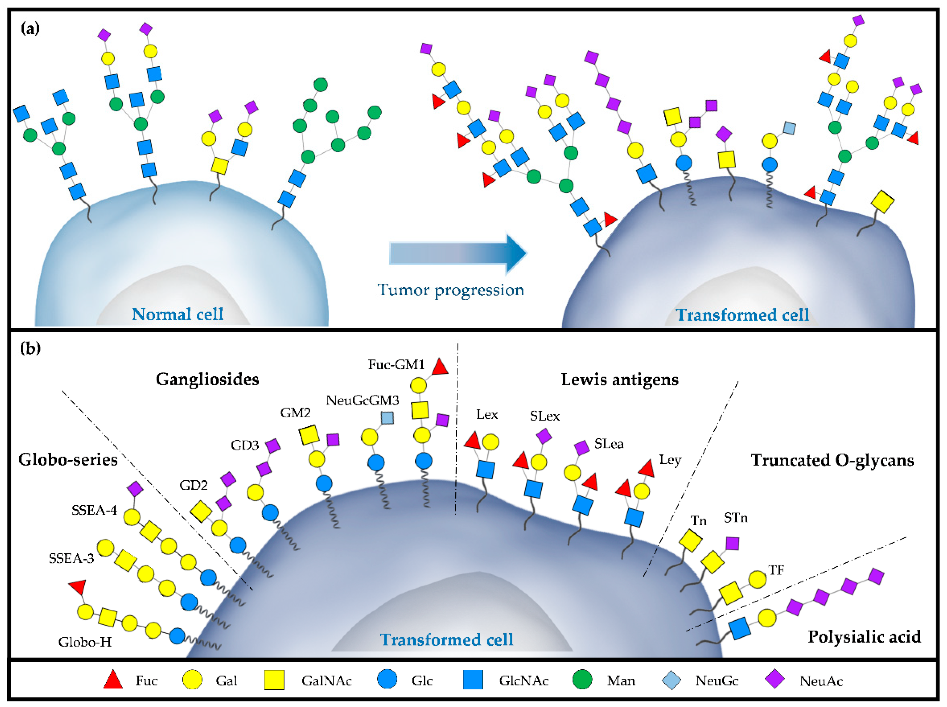
2. TACAs as Cancer Targets

According to their structural similarities, TACAs can be classified into five groups: (1) the globo-series glycosphingolipids, including Globo-H, Stage-specific embryonic antigen-3 (SSEA-3) and Stage-specific embryonic antigen-4 (SSEA-4); (2) the gangliosides, including GD2, GD3, GM2, NeuGcGM3, and Fuc-GM1; (3) the blood groups or Lewis antigens, including Lewis X (Lex), Lewis Y (Ley), sialyl Lewis X (SLex), and sialyl Lewis A (SLea); (4) the truncated O-glycans, including Thomsennouveau (Tn), sialyl-Tn (STn), and Thomsen−Friendreich (TF); and (5) the polysialic acid (Figure 1b).
Glycosphingolipids share the characteristic of being composed of complex glycans covalently linked to a ceramide backbone. Globo-H ceramide is thought to be the most prevalent cancer-associated glycosphingolipid since its overexpression was detected by immunohistochemistry in a wide variety of epithelial cancers such as breast, uterus, ovary, prostate, lung, liver, and colon cancers [23,24]. Considering its expression is restricted to undifferentiated embryonic stem cells and the luminal surface of glandular tissues, some publications propose Globo-H as a potential target for cancer immunotherapy [25]. SSEA-3 and SSEA-4 were originally identified as human embryonic stem cell-specific markers, but subsequent research demonstrated their association with malignant behaviour in tumour cells [26]. The SSEA-4 expression is positively related to breast [27], glioblastoma [28], and prostate [29] cancer aggressiveness. Since its expression is related to the loss of cell-to-cell interaction and the cell migratory phenotype, SSEA-4 has been associated with epithelial-mesenchymal transition [30]. In breast cancer samples, a high percentage of positivity was reported for SSEA-3 (77.5%), which may correlate with tumorigenicity and multidrug resistance [31,32]. Similarly, in non-small cell lung cancer (NSCLC), SSEA-3 expression increases in samples resistant to multiple anticancer drugs [33]. Moreover, SSEA-3 was postulated as a novel cell surface marker in human colorectal cancer since the SSEA-3-expressing cell subpopulation has a greater proliferative capacity in vitro and a higher tumorigenicity in vivo than SSEA-3-negative cells [34].
Gangliosides are the second TACA group with a glycosphingolipid nature that are composed of at least one sialic acid in the carbohydrate structure [35,36]. The disialylganglioside GD2 is only expressed in a few normal tissues, like the brain, peripheral sensory nerve fibres, and in particular melanocytes from the skin. Nonetheless, this ganglioside is overexpressed in neuroblastomas, melanomas, retinoblastomas, and breast cancer [37,38,39,40]. It was established that GD2 is the most important complex ganglioside present in neuroblastoma, whose expression correlates with tumour progression and poor patient outcomes [41,42]. Another ganglioside widely described in association with malignant transformation is GD3, which is expressed normally in embryonic cells and in some tissues under disease conditions, such as the nervous system in neurodegenerative disorders and tumours of neuroectodermal origin [43]. Interestingly, this TACA is detected in 60% of primary melanoma and 75% of metastatic melanoma, even though it is not expressed in normal melanocytes [44]. In this indication, the upregulation of GD3 on the cell surface is associated with tumour progression, brain metastasis, and poor clinical outcome [45]. GM3 is a monosialoganglioside that can contain a N-acetylneuraminic acid (NeuAc), which is abundant in normal tissues, or a N-glycolylneuraminic acid (NeuGc), which is considered a ‘non-human’ sialic acid due to a mutation in the gene that encodes the enzyme capable of catalysing the conversion of NeuAc to NeuGc [46]. Whereas NeuGcGM3 is rarely detected in normal tissue, it is found in multiple types of tumours such as melanoma [47], neuroblastoma [48], retinoblastoma [49], colon [50], bladder [51] and lung cancer [52], among others. Most literature ascribes the presence of this sialic acid in malignant cells to the metabolic assimilation of NeuGc, obtained principally from red meat, under hypoxic conditions [53,54,55]. On the other side, Fuc-GM1 is a fucose-containing glycolipid that is naturally expressed in a subset of peripheral sensory neurons and dorsal root ganglia [56,57]. However, multiple reports have demonstrated enhanced levels of this antigen in small cell lung cancer (SCLC), even though only 20% and 10% of squamous epithelial and large cell lung cancer specimens were positive, respectively. In addition, no Fuc-GM1 was detected in lung adenocarcinoma, bronchial carcinoma, or normal lung or bronchus, so it could be considered a hallmark of SCLC and useful for sample stratification as well as for targeted immunotherapies [58,59].
Lewis antigens correspond to a family of terminal glycan structures characterised by the presence of one or two fucoses linked to a disaccharide of N-acetylglucosamine and galactose that can also be sialylated. Lex antigen, also known as Stage-Specific Embryonic Antigen-1 (SSEA-1) or CD15, is mainly found in epithelial tissues like the stomach, colon, salivary glands, kidneys, bladder, uterus, cervix, and medulla [60]. Nevertheless, the overexpression of Lex can be found on the surface of various types of cancer cells, which usually correlates with tumour progression and patient survival [61]. In bladder, hepatic, and breast cancer, this molecule is strongly associated with systemic dissemination, possibly related to its role in the adhesion of cancer cells to blood vessel walls [62,63,64]. Particularly in glioblastoma multiforme, this epitope has been proposed as a cancer stem cell marker, and it could be a potential marker of malignant glioma progression [65,66]. Its sialylated version, the terminal glycan SLex, is usually detected on the surface of lymphocytes, neutrophils, and a subset of T lymphocytes [67]. However, expression of this well-known sialofucosylated motif was also reported in high-grade gliomas [68] and gastrointestinal cancers [69]. SLex binds to selectins, mainly E-selectin, expressed on the surface of endothelial cells, facilitating leukocyte extravasation and inflammatory processes [70]. In accordance, in vivo studies have shown that Slex-positive tumour cells exhibit increased metastatic potential by arresting tumour cells in the vasculature of target organs through adhesive interactions [71,72,73]. Another member of this family is SLea, also called cancer antigen 19-9 (CA19-9), which is the most widely used and best validated marker for pancreatic cancers. It is also present in other malignancies such as gastric, endometrial, and colorectal cancers. Initially found in glycolipids, SLea is now known to also be present on glycoproteins and proteoglycans [74]. Although to date it could be considered an indicator for predicting patient prognosis, further studies are needed to fully understand its involvement in cancer biology [75,76,77]. The last TACA of this family, Ley antigen, is a prognostic factor associated with cell dedifferentiation and proliferation in NSCLC and hepatocellular carcinoma [78,79]. It has been demonstrated that the in vitro blockade of Ley significantly reduced the invasion and metastasis of ovarian cancer cells [80]. In addition, numerous publications have shown that overexpression of Ley on these tumour cells is associated with chemotherapy resistance [81,82,83].
Altered O-glycan expression is observed in several tumour indications [84,85,86]. Truncated glycans refer to a set of molecules composed of a small number of carbohydrates present as a consequence of related O-glycan core expression. In this regard, Tn and STn antigens show little or no expression in normal adult cells and tissues. Many studies have shown that Tn antigen is detected in various human carcinomas, such as those of the cervix, ovary, breast, bladder, prostate, gastric, and colon [87,88,89]. Indeed, in many cases, its expression has been associated with tumour progression, dissemination, and patient prognosis [90,91,92]. Tn and STn are frequently co-expressed in tumour cells, possibly because they share synthetic pathways. STn detection is observed in more than 80% of human carcinomas, including gastric, endometrial, and bladder tumours, and in all cases, STn expression is associated with an adverse outcome and decreased overall survival for the patients [93,94,95,96]. Although the mechanism by which STn participates in the interaction between tumour cells and the microenvironment is not fully understood, it has been reported to promote the separation of individual cells from the primary tumour mass by disrupting the interaction of galectins with terminal galactose residues. In this scenario, STn antigen could promote the escape of cancer cells participating in the migration and invasion of the surrounding tissue [97]. TF is a truncated O-glycan highly expressed in approximately 70–90% of carcinomas like ovarian, prostate, breast, colon, stomach, and bladder malignant tissues, although the percentage of positive cases varies among the carcinoma types [98]. Aberrant expression of TF antigen is usually related to the dissipation of the pH gradient in the Golgi apparatus of tumour cells, which can relocate certain glycosyltransferases, leading to an abnormal synthesis of mucin-type O-glycans [99,100]. This TACA plays a crucial role in the interaction between cancer cells and galectin-3, a carbohydrate-binding protein of the endothelium, promoting tumour aggressiveness and metastasis [101,102,103].
Tumours are not only characterised by having a high expression of monosialylated structures such as SSEA-4 or Slex, among others, but also of polysialylated glycans. These TACAs present a limited expression in normal tissue and are associated with particular proteins and cell types, especially those linked to neuronal cells. However, in clinical samples from patients with NSCLC, breast cancer, and glioblastoma, it was shown that the expression of polysialic acids and the glycosyltransferase responsible for its synthesis correlate with cell migration, invasion, and poor prognosis [34,104].
TACAs are promising targets for the development of novel therapeutic strategies due to their active participation in tumour biology (Table 1). Successful development of active immunotherapeutic approaches requires an understanding of how glycans interact with the immune system and the identification of strategies to trigger appropriate immune responses against them.

3. Immune Response against Glycans

Besides TACAs are considered suitable targets for developing specific therapeutic strategies against cancer, there are several glycan-targeted active immunotherapies that are currently being evaluated in clinical trials or even registered. However, none of these therapeutic alternatives has been approved by the US Food and Drug Administration (FDA) so far. One of the limitations to their success is the fact that they were unable to improve patient survival associated with the lack of potent T cell-mediated immunity. In general, TACAs are poorly immunogenic since they are unable to trigger a proper immune response when administered alone. In order to induce an effective immune response, dendritic cells (DC) process and present T cell-dependent or thymus-dependent (TD) antigens to T cells as complexes with MHC molecules, which represents the first signal needed for T cell activation. In addition, co-stimulatory signals are provided through the interaction of CD80 and CD86 molecules expressed on mature DC with CD28 on T cells and the production of chemokines and cytokines that polarise the T cell response [105]. When TD antigens are presented on MHC class I (MHC I) molecules, naïve CD8+ T cells are primed into cytotoxic T cells capable of killing pathogen-infected and tumour cells [106,107]. Conversely, TD antigen presentation on MHC class II (MHC II) primes CD4+ T cells into different subsets, including T helper (TH) 1, TH2, TH9, TH17, and TH22, follicular helper T (TFH), or regulatory T cells (Treg), depending on which cytokine DC secrete as co-stimulatory signals during priming [108,109] (Figure 2a,b). In particular, TFH cells mediate germinal centre formation and especially induce B cell differentiation into memory or antibody-secreting plasma cells, enabling the generation of high-affinity, class-switched antibodies and eventually long-lived antibody-mediated protection [110]. In the absence of additional TFH help, B cells secrete low titers of low-affinity IgM since the generation of IgG requires both the antigen-specific signal provided by the cross-linking of B cell receptors and the cytokine signal delivered by T cells [111].
While peptides are classified as TD antigens, TACAs are considered T cell-independent or thymus-independent (TI) antigens that, even though they do not directly bind to MHC molecules and cannot activate T cells by themselves, are able to cross-link B cell receptors and weakly activate B cells in the absence of T cell help [112]. In TI responses, DC present the antigen to the B cell, providing the required co-stimulatory signals through transmembrane activator and CAML interactor (TACI), B lymphocyte stimulator (BLyS), and APRIL ligand [113,114]. Therefore, the immune response to TACAs is a primary response characterised by being less robust and, in general, short-lived, in which B cells mainly produce low-affinity IgM antibodies with no affinity maturation and isotype switching [115]. Although an IgG response is preferred, it is important to consider that an IgM reaction is also a key response since it can induce complement-dependent cytotoxicity (CDC) [116]. Moreover, since it is possible that long-lasting IgM-producing plasma cells and IgM memory cells will have developed [117], IgM induced by a TI response could not be considered an unsuccessful immune response. In this regard, clinical trials have not only demonstrated that gangliosides (and TACAs in general)-specific IgM antibodies have antitumor activity based on the CDC but also that high levels of these IgM positively correlate with survival in metastatic melanoma patients [118,119]. Moreover, endogenous anti-ganglioside IgM has been suggested to eliminate immunosuppressive gangliosides shed by tumour cells to restore host immune competence [120]. Additionally, using a preclinical transgenic model with a high IgM titer against GD2, it was demonstrated that mice had prolonged survival after being injected with GD2-expressing EL4 or B16 cells, and an increased number of NK cells mediated this antitumor activity [121]. All these findings highlight the relevance TACAs have in humoral immunotherapy and provide supporting evidence for the role that specific IgM responses have in mobilising innate mechanisms of tumour surveillance [101,122,123].
Besides MHC I and MHC II molecules, there is an additional subset of antigen-presenting molecules called the CD1 family, composed of CD1a-Cd1d [124]. Among TACAs, gangliosides and glycolipids in general are presented via the CD1d receptor expressed by antigen-presenting cells [125,126] (Figure 2c). This family of proteins binds and presents glycolipids to the T cell receptors on NK cells, activating the invariant natural killer T (iNKT) cell subpopulation. iNKT cells are a subtype of T cells known to express an αβ-T cell receptor that underwent somatic rearrangement [127], show typical NK surface markers like CD56 and CD16, and show granzyme production [128]. Activated iNKT cells secrete several cytokines, which will further initiate a selective TD response with no participation of CD4+ T cells, resulting in the necessary class switching to produce an IgG response against the specific glycolipid antigen [128,129].
Another limitation associated with the poor outcome of glycans-directed active immunotherapies relies on the difficulties of overcoming the immunosuppression and immunotolerance of TACAs. Even in small amounts, most TACAs are also present on normal cells and are therefore recognized as self-antigens by the host immune system, inducing immunosuppression as well as tolerance [101,130]. How to break immunotolerance is one of the main challenges TACAs-based active immunotherapies have to face. Techniques to overcome these problems include: (1) TACA conjugation with a peptide or protein carrier, turning them into TD antigens able to bind to MHC molecules; (2) use of chemically modified TACAs that would be recognized as foreign antigens; and (3) use of peptides or proteins that mimic the structure of the TACA. Within this last group, mimetic peptides and anti-Id antibodies are both strategies based on mimicking the three-dimensional structure of the nominal antigen in order to trigger a strong and long-lasting immune response. For anti-Id antibodies, the mechanism relies on the activation of the Id network, while mimetic peptides can be processed and presented by DC as a standard TD antigen. These strategies will be discussed in subsequent sections.
In most active immunotherapies, TACAs are covalently conjugated to an immunological protein or peptide carrier containing T-cell epitopes to convert them into TD antigens. As mentioned before, the induction of a T-cell response will further lead to the production of high-affinity antibodies that will recognize the tumour, making this strategy an attractive alternative to improving the immunological character of TACAs. Even though TACAs like Lewis structures, O-glycans, and glycolipids, including Globo-series and gangliosides, have been successfully conjugated to carriers such as keyhole limpet hemocyanin (KLH), MUC1 peptides, diphtheria or tetanus toxoids, bovine serum albumin, and ovalbumin [131,132,133], only six TACAs-targeted immunotherapies have reached phase III clinical trials to date, with one of them being Dinutuximab, a passive immunotherapy directed to the GD2 ganglioside, already approved to treat high-risk neuroblastoma [134]. The other five are GM2-KLH (NCT00005052); Racotumomab (NCT01460472) and BEC2 (NCT00037713), both anti-Id antibodies directed to NeuGc-containing glycoconjugates and GD3 respectively; Theratope® (NCT00003638) and OPT-822 (NCT01516307), directed to STn and Globo-H respectively, and also conjugated to KLH. Unfortunately, in the intention-to-treat analysis, neither GM2-KLH nor OPT-822 improved progression-free survival (PFS) for stage II melanoma [135] or metastatic breast cancer [136]. However, for OPT-822 active immunotherapy, both PFS and interim overall survival were significantly improved in patients who developed IgG antibodies against Globo-H. Although a successful IgG antibody response was developed against STn in Theratope®-treated metastatic breast cancer patients, no overall benefit in time to progression or survival was observed [137].
In recent publications, Hossain et al. and Mettu et al. have extensively reviewed different approaches related to the design of active immunotherapies directed at TACAs, including the conjugation of alternative protein carriers and different chemical modifications made to carbohydrates [133,138]. Additionally, in the last few years, many reviews have been published describing recent advances in carbohydrates-based active immunotherapies, which are mainly focused on summarising methods to improve the immunological properties of TACAs [139,140,141]. This review will refer to the last strategy mentioned in a previous paragraph and will specifically discuss mimetic peptides and anti-Id antibodies as approaches to overcome TACAs-associated immunotolerance and immunosuppression.

4. TACAs Mimetic Peptides

Mimetic peptides, or peptidomimetics, are small compounds of a peptidic nature that resemble the steric and chemical properties of other molecules such as bioactive peptides, carbohydrates, or glycoproteins. Most mimetic peptide developments are focused on biologically relevant protein segments. Vagner et al. describe this type of mimetics as “compounds whose essential elements (pharmacophore) mimic a natural peptide or protein in three-dimensional space and retain the ability to interact with the biological target and produce the same biological effect” [142]. However, TACAs mimetic peptides are developed for immunotherapeutic approaches with the aim of triggering an immune response against the nominal TACA based on their conformational similarity in three-dimensional space. This well-known immunological mechanism is called antigenic mimicry [143]. In this scenario, an induction of a cross-reactive antibody response against the target TACA or the activation of T cells upon recognition of MHC-I-associated glycopeptides is expected.
Mimetic peptides are proposed as good tools to trigger an immune response against carbohydrates, mainly because of the advantage conferred by their chemical nature [144]. First, mimetic peptides can be presented by MHC molecules on antigen-presenting cells and are thus potentially capable of generating long-lasting memory responses in the organism. In addition, they can break the body’s natural immune tolerance to carbohydrates, contributing to immunosurveillance and boosting the antitumor activity of TACAs [101]. Finally, the existence of easier and more standardised productivity strategies to generate and modify peptides in comparison with those available for the synthesis of glycans makes them a desirable tool for the development of therapeutic strategies.
Mimicry of glycans using peptides has been fully demonstrated by several research groups, either because the selected mimetic peptides are able to be recognized by specific antibodies or lectins and/or because they develop a specific immune response against the target glycan. The mechanisms by which a sequence of a short peptide is able to trigger an immune response against a TACA not only appear to be due to a three-dimensional structural similarity but also to a condition known as functional mimicry [144]. Meanwhile, while structural mimicry refers to the capacity of the peptide to imitate the contacts made by the carbohydrate by using a similar three-dimensional arrangement of functional groups and benefiting from its high degree of flexibility, functional mimicry is attributed to its ability to fit in the antibody Id through hydrophobic interactions [145,146,147,148,149]. There are two main strategies to develop mimetic peptides so far. The least chosen option used for the mimetic peptide development of TACAs is known as synthetic or rational design, which uses bioinformatics tools combined with crystallographic structure analysis to rationally design a peptide sequence. Using specific antibodies and its ability to develop an anti-glycan immune response, the peptide will be further evaluated for structural mimicry [150]. On the other hand, the most common option involves the use of a phage display combinatorial library of short peptides consisting of a large number of bacteriophages, each of which exposes a different random polypeptide. Phages carrying mimetic peptides of interest are first selected using specific antibodies or lectins, then isolated and finally expanded to identify the peptide sequence [151,152]. Commercial phage libraries including millions of different peptides with an arbitrary combination of a particular number of amino acids are available, and they have been effectively used to look for both protein epitopes and glycan mimetic peptides [153].
The identification of peptides that effectively mimic TACAs leads to the development of vaccination strategies that have not only been demonstrated to induce an effective immune response against the nominal glycan when administered in vivo but have also provided protection against tumours that express the target antigen. In this regard, several mimetic peptides were developed against the disialoganglioside GD2, a known overexpressed glycolipid in neuroectodermal cancers such as neuroblastoma, melanoma, and SCLC. GRL and DGG, two circularised decamers conjugated each one to KLH, were selected from a phage display using the ch14.18 antibody, with proven capacity to develop an IgG anti-GD2 response in a preclinical setting [154]. In addition, a DNA vaccine containing a GD2 mimotope, so-called 47-LDA, applied in combination with IL-15 and IL-21 in mice, was able to elicit a GD2 cross-reactive IgG antibody response as well as MHC-I-restricted CD8+ T cells reactive to neuroblastoma cells [155]. Also, Horwacik et al. published a set of five GD2 mimetic peptides that were able to bind in vitro to an anti-GD2 antibody when presented in a cyclic conformation via an internal disulfide bridge between positions 2 and 11 [156].
GD3P4 is a GD3 mimetic peptide identified from a phage display library using the 4F6 antibody. This mimetic peptide was able to trigger a humoral anti-GD3 response. In an exquisite way to show antigen presentation and mimicry, Popa et al. were able to develop an anti-GD3 hybridoma from GD3P4 immunised mice, demonstrating that the mimetic peptide was processed and presented by antigen-presenting cells [157].
TF, as a core for the extension of O-glycans, is a cryptic antigen in normal tissue. However, it is found on the vast majority of human carcinomas since truncated forms of O-glycans are normally present in aberrant glycosylation. Heimburg-Molinaro et al. developed a set of mimetic peptides capable of being recognized by the TF-specific peanut agglutinin lectin in vitro. Among them, the one designated as D2 was the most promising since it induced an anti-TF humoral response when administered in a presentation of eight 15-mer peptides forming a single molecule [158].
Kieber-Emmons and colleagues have been studying carbohydrate mimetic peptides and their use as therapies for several years [159]. In the field of carbohydrate mimotopes, their contributions have strengthened the concept of multiple antigen mimotopes (MAM) [160]. This concept supports the idea that a peptide selected for its recognition by an antibody can elicit an immune response against a related antigen [144]. Using a reverse engineering strategy on a pre-selected mimetic peptide presented on a phage display library, they developed a mimetic peptide called P10s that induced an immune response to various TACAs, including Ley and selected gangliosides such as GD2 [150,161,162]. P10s was conjugated to the pan T cell carrier PADRE using MONTANIDE™ ISA 51 as an adjuvant and was the first TACA-mimetic peptide to be tested in a phase I clinical trial on advanced breast cancer patients [131]. Immunisation was well tolerated and triggered a specific antibody response against LeY and GD2 in all tested patients. Humoral antibodies showed caspase3-dependent apoptotic activity against Ley or GD2-positive breast cancer cell lines [163]. In a recent clinical trial, the objective was to examine the feasibility, safety, and immunogenicity of adding P10s-PADRE to standard-of-care chemotherapy in HR+/HER2− early-stage breast cancer patients. Results showed that combination therapy induced an antibody response, increased the NK cell activation markers NKp46 and CD94, and was associated with the infiltration of T cells in the tumour microenvironment [164]. In Table 2, we summarised the main mimetic peptides developed and evaluated to date.

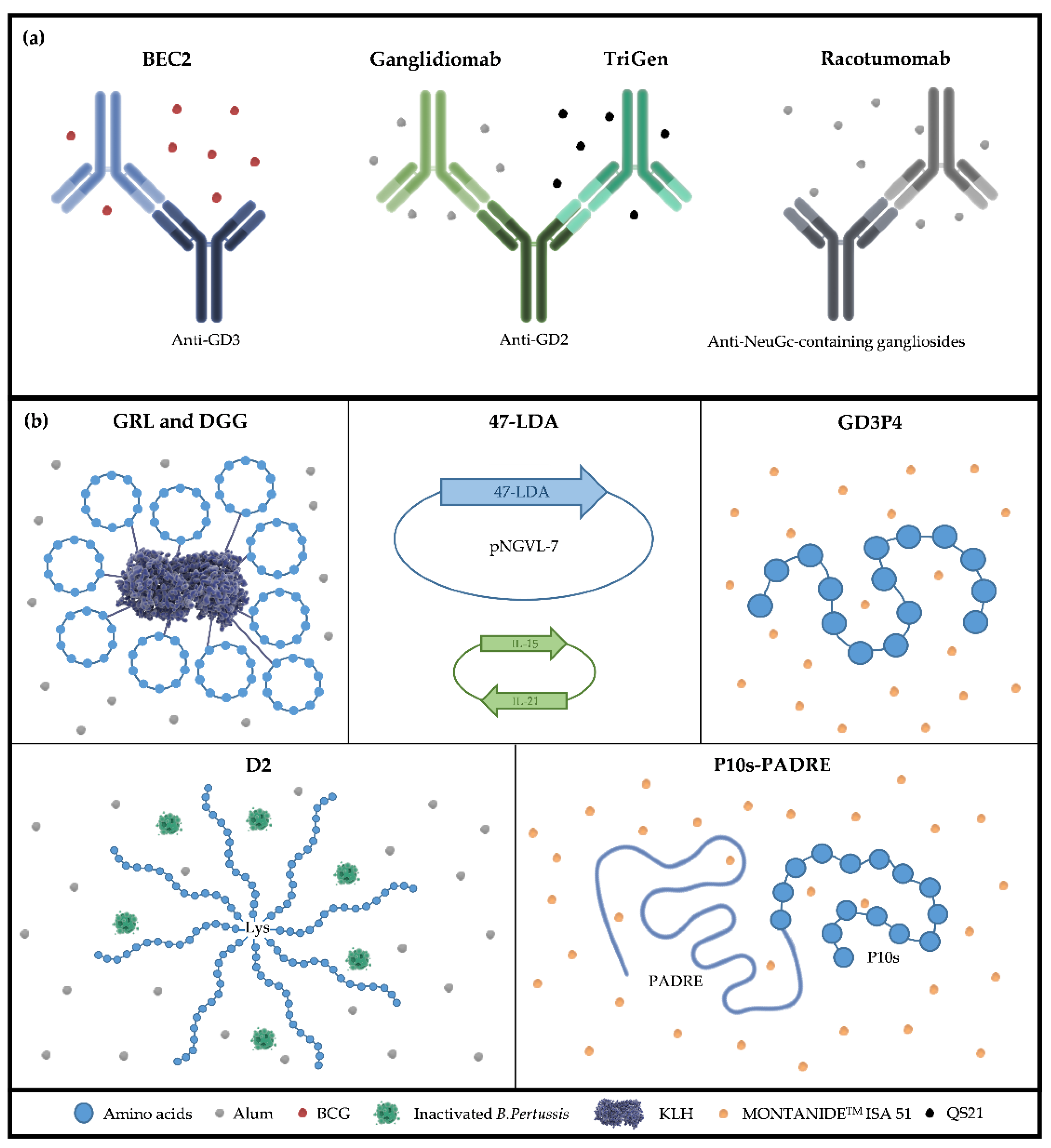
5. Anti-Idiotype Antibodies as TACA Surrogates

The idiotypic network theory published by Jerne in 1974 postulates that the immune system works as a network of antibodies (with their antigen-specific Ids) and their anti-Ids antibodies, in which B lymphocyte clones are stimulated and regulated by antibodies produced by different clones within the network [168]. An interesting point behind Jerne’s theory is that each antibody can recognize an antigen as well as be recognized by a different antibody that binds to its paratope. According to this theory, immunisation with an antigen will produce specific antibodies termed Ab1 that can further generate anti-Id antibodies, also known as Ab2, against the Id of the Ab1. The particular pool of anti-Id Ab2 that recognizes the antigen-binding site of Ab1 is able to induce a specific humoral response against the nominal antigen when used for subsequent immunisation (Ab3). As a consequence of the idiotypic network, anti-Id antibodies could be used to mimic the structure of a particular antigen to induce a specific Ab3 response that will recognize the immunising Id of the Ab2 as well as the antigen that induced Ab1 production in the first place. Since this work focused on therapeutic strategies based on TACA mimicry as surrogates of nominal TACAs against which a high affinity and long-lasting immune response is sought, anti-Id antibodies can be considered a particular kind of mimetic peptides, suitable for the development of active immunotherapies directed to TACAs-expressing cancer cells.
The main advantage of anti-Id antibodies over the use of anti-TACAs Ab1 as passive immunotherapies is that the former will ideally induce both cellular and humoral responses, with the consequent activation of a long-lasting immunity. Most Ab1 antibodies, when used clinically as single-agent therapy, induce tumour destruction via either CDC or ADCC. Interestingly, some Ab1 used in therapeutic settings also contribute to antitumor effects by triggering the idiotypic cascade and inducing an antigen-specific immune response [169,170,171]. An interesting report shows a prolonged survival of stage 4 neuroblastoma children at the time of remission treated with the anti-GD2 antibody 3F8 (Ab1) associated with the induction of the Ab3 anti-GD2 response, indicating the relevance the idiotypic network has in controlling tumour progression [171].
In 2009, the National Cancer Institute Translational Research Working Group made a priority-ranked list of cancer antigens in order to contribute to the development of therapeutically effective active immunotherapies [172]. Among the 75 antigens ranked, 9 were TACAs, including 4 gangliosides (GD2, GD3, Fuc-GM1, and GM3), Globo-H, Lewis glycans (Slea), truncated O-glycans (Tn and STn), and polysialic acid. Considering the criteria established by Cheever et al. a few years later, Gomez et al. evaluated NeuGc-containing gangliosides, showing that NeuGcGM3 in particular matched all criteria at least in some proportion, ranking within the top 15th cancer antigens selected [173]. Anti-Id-based active immunotherapies directed to the three gangliosides of the ranked TACAs have been developed over the past decades, with results from different clinical trials demonstrating they are able to induce an antigen-specific humoral response when administered in the presence of a proper adjuvant.
BEC2 is an anti-Id antibody directed to the anti-GD3 R24. Preclinical studies in rabbits demonstrated that BEC2 mimics GD3 ganglioside and can induce a specific Ab3 response of IgG and IgM isotypes, able to inhibit BEC2 binding to R24 [174]. Clinical trials in melanoma patients who underwent surgery but were at high risk of recurrence and in patients with limited-disease SCLC showed that BEC2 is immunogenic when administered with BCG as an adjuvant, but it only induces detectable anti-GD3 antibodies in approximately 20% to 33% of patients [175,176,177,178]. Results from the phase III trial carried out in 515 responding patients after combined therapy for limited-disease SCLC confirmed that one third of patients developed a humoral response against GD3. Even though none of the major endpoints of the study were achieved and patients treated with BEC2/BCG showed no improvement in their survival, those who responded and developed anti-GD3 Ab3 had an improvement in survival, although this did not reach statistical significance [179].
Besides BEC2, a second anti-Id antibody has been developed as active immunotherapy against a ganglioside. TriGem is an anti-Id mimicking GD2, originally named 1A7, formulated with the adjuvant QS21. It was first developed in mice and evaluated in preclinical and clinical studies. Active immunisation of mice, rabbits, and monkeys with TriGem induced polyclonal IgG GD2-specific responses, able to lyse cells with ganglioside expression by an antibody-dependent cell-mediated cytotoxicity (ADCC) mechanism as well as stimulate a specific T cell response [180,181]. Results from the first phase I trial conducted in advanced melanoma patients demonstrated that TriGem induced an Ab3 response that specifically recognized purified GD2 when evaluated by ELISA in 100% of treated patients [182]. A second study confirmed the previous result, since 40 of 47 patients showed an Ab3 response [183]. When humoral responses were evaluated, both trials exhibited responses predominantly of the IgG isotype, with all IgG subclasses represented, and ADCC activity in some patients [182,183]. Even though median survival was not reached, it was observed that 1 patient had a complete response after 24 months, and 12 patients were stable for a median of 18 months (14 to 37 months) [183]. In a phase II trial, TriGem was formulated with either QS21, granulocyte macrophage colony-stimulating factor, or aluminium hydroxide (alum) as adjuvants. All stage III melanoma patients included in the study developed a robust IgG response against GD2, with no differences in the antibody titer between the evaluated adjuvants. One third of treated patients also received high-dose interferon alpha in combination with TriGem, showing a similar humoral response to patients that were only treated with the anti-Id. After a median follow-up of two years, more than 80% of patients were alive [184].
Lode et al. generated Ganglidiomab, another anti-Id directed to GD2. Because ganglidiomab was proved to inhibit the binding of antibodies 14G2a, ch14.18/CHO, and hu14.18 to GD2 as well as competitively inhibit the GD2-specific lysis of neuroblastoma cells by modified-NK effector cells, this anti-Id antibody was proposed as a GD2 surrogate. When mice were treated with ganglidiomab formulated with alum, it was demonstrated that the induction of an antigen-specific humoral response was able to mediate an ADCC and a complement-dependent cytotoxicity response against GD2-expressing neuroblastoma cells [185]. Recently, results from the first-in-man use of ganglidiomab active immunotherapy were published. Seven patients with high-risk neuroblastoma who achieved complete remission after frontline or salvage therapy were treated with the anti-Id antibody adsorbed to alum. Anti-GD2 humoral response was detected in three patients, and none of the treated ones experienced relapse during the 56 months of follow-up [186]. These results are encouraging and provide important information to evaluate ganglidiomab in prospective clinical trials.
In collaboration with the Centre of Molecular Immunology (Habana, Cuba), our group has contributed to the development of racotumomab, a highly immunogenic anti-Id antibody that has been approved in Latin American countries as active immunotherapy for the treatment of patients with advanced-stage NSCLC after progression on first-line therapy. Racotumomab is an Ab2, formerly named 1E10, able to block the binding of the Ab1 P3 to NeuGc-containing gangliosides and sulfated glycolipids (Ab3, Id+) [187]. Racotumomab also induces an Ab3 response that recognizes the nominal target when administered in preclinical animal models [188,189,190] and even in cancer patients [191,192,193,194,195], suggesting that this Ab2 behaves as a NeuGc-glycoconjugates surrogate (Ab3, Ag+). Initial studies to evaluate racotumomab immunogenicity were carried out using mice as a preclinical model. It was first formulated in Freund’s adjuvant using KLH as a protein carrier, showing antitumor effect in the subcutaneous tumour growth as well as in the spontaneous dissemination to the lungs of the F3II mammary carcinoma [196]. Afterward, racotumomab was conjugated with alum as an adjuvant (racotumomab-alum) and evaluated using the Lewis lung carcinoma 3LL, demonstrating a significant reduction in the number of spontaneously-disseminated lung nodules, an increase in tumour infiltrated CD4+ and CD8+ T cells [197], and the induction of tumour apoptosis in combination with cyclophosphamide [198]. A chemo-immunotherapy combination using racotumomab-alum plus pemetrexed was also evaluated, with a significant anti-metastatic effect shown over Lewis lung carcinoma. Moreover, it was also demonstrated that racotumomab-alum exhibited antitumor activity in a highly aggressive, clinically relevant model using NeuGc-pre-incubated 3LL cells [189].
Several clinical trials have proved racotumomab-alum safety and immunogenicity in patients with neuroblastoma [199], melanoma [200], breast [192], and lung cancer [201] (extensively reviewed by Gabri et al.) [202]. In particular, the results of the phase II/III randomised, placebo-controlled study carried out in 176 advanced NSCLC patients confirmed that switch maintenance with racotumomab-alum is a well-tolerated treatment in which the most common adverse effects were classified as mild or moderate. Additionally, the study demonstrated the effectiveness of the treatment with this active immunotherapy since a significant survival improvement was observed when compared with those patients who received a placebo. Because racotumomab is an anti-Id antibody, as demonstrated in previous phase I trials, it is able to induce an Ab3 response against the TACA NeuGcGM3, and this study also evaluated whether induced antibodies after treatment had the same specificity as the Ab1. Not only did racotumomab-alum elicit an antibody response of both IgM and IgG isotypes against NeuGcGM3 ganglioside in almost all treated patients (95.8%), but also those induced antibodies were able to kill antigen-expressing cells [194]. Using serum samples from patients included in the phase III clinical trial (NCT01460472), we further demonstrated that antibodies induced by racotumomab-alum were also able to mediate the lysis of cells expressing NeuGc-glycoconjugates by an ADCC mechanism, and this response was antigen-dependent [195].

6. Discussion

Glycosylation is a post-translational modification that affects more than 50% of cellular proteins, making it a major player in several processes of tumour biology since it is involved in the interaction between different cells and between cells and the microenvironment. Glycosylation also participates in epithelial-mesenchymal transition, tumour proliferation, invasion, metastasis, and angiogenesis. Because of the altered glycosylation that occurs concomitantly with the process of malignant transformation, TACAs have emerged in recent decades as suitable therapeutic targets for the development of promising immunotherapies. To date, a significant amount of research has described the involvement of TACAs in tumour biology and the development of immunotherapeutic strategies directed at them aimed at combating tumour growth. In this manuscript, we have compiled and described immunotherapeutic strategies based on TACA mimicry. In this regard, mimetic peptides and anti-Id antibodies are the two main strategies that best fit this scenario.
The main objective of active immunotherapy is to achieve, through an appropriate immunisation, a long-lasting immune response capable of persistently limiting tumour growth and effectively controlling possible relapses. All anti-Id antibodies evaluated in clinical trials have demonstrated the ability to generate a humoral IgG antibody response against the nominal TACA that correlated with a better clinical outcome. Furthermore, racotumomab is the only anti-Id antibody registered so far. It also triggers an immunological response based on a humoral IgG response that recognizes NeuGc-containing glycoconjugates and participates in ADCC response. Until now, the only mimetic peptide-based active immunotherapy that has reached clinical trials is P10s-PADRE, which showed significant anti-GD2 and Ley IgG production. Although evidence from clinical trials reporting clinical benefit in patients treated with TACAs-based therapies is scarce, the results shown so far are promising since they demonstrated the induction of a specific immune response.
Figure 2 shows the different active immunotherapies based on anti-Id antibodies and mimetic peptides published to date and specifically indicates which adjuvants or protein carrier therapeutic agents were formulated or conjugated, respectively. As summarised in Figure 3a, anti-Id-based active immunotherapies are composed of the antibody and an immunological adjuvant. Even though the use of protein carriers such as KLH attached to the antibody has been evaluated in preclinical and clinical settings, results demonstrated that unconjugated anti-Id induces an equal immune response when compared to KLH-conjugated, while avoiding its potentially toxic effect [126,178,191,203]. The choice of a proper adjuvant is an essential step in the formulation of active immunotherapies and should be optimised for each strategy in order to break immune tolerance, trigger a strong immune response, and finally, maximise clinical efficacy. Mimetic peptides and anti-Id antibodies discussed in this review have been formulated with a limited set of adjuvants. However, currently approved adjuvants for human use were very elegantly discussed in the work recently published by Cuzzubbo, showing how classical immunostimulatory and depot adjuvants face suppressive tumour microenvironment and immunosenescence in aged patients [204]. It is therefore worth analysing if these therapeutic strategies could be adjuvanted in a more suitable compound with a proven ability to induce humoral and cellular responses that lead to an effective antitumor immune response. Conversely, active immunotherapies based on mimetic peptides show a variety of alternatives, as mentioned in a previous section (Figure 3b).
In March 2011, the FDA approved the use of Ipilimumab (Yerboy©, Bristol-Myers Squibb), a human monoclonal antibody directed to cytotoxic T lymphocyte-associated antigen 4 (CTLA4), for the treatment of unresectable or metastatic melanoma [205,206]. This event initiated a new era in which the field of immunotherapy was largely conquered by immune checkpoint inhibitors (ICIs). Most of the scientific community working in the immunotherapy field focused its efforts on understanding and evaluating the therapeutic potential of these promising drugs in various types of cancer, in different clinical settings, and in combination with other therapeutic strategies, leading so far to the approval of eight ICIs for the treatment of more than 15 different tumour indications [207,208,209]. ICIs are still being evaluated in clinical trials to seek out more effective combinatorial schemes that will benefit patients with different cancer types and indications. In this regard, we evaluated the combination of racotumomab with anti-PD1 blockade in preclinical models of NSCLC using two experimental approaches based on concomitant or sequential administration of both treatments, with promising results that will soon be submitted for publication.
It is noteworthy that most of the publications focused on active immunotherapies using the mimetic strategies of TACAs were published before 2010. Only a few vaccines have reached the clinical phases of drug development with results that demonstrate their effectiveness [131,165,194]. Could the successful breakthrough of ICIs be the cause of the delay in research and development of active immunotherapies based on glycan mimetics? Although this discussion is not the subject of this review, we consider that the evaluation of TACAs-mimicking active immunotherapies has not yielded the unsatisfactory clinical responses that would allow us to rule them out.
As described in a previous section, four anti-Id antibodies for cancer treatment have been developed against TACAs. Despite the few options assessed, one of them has successfully completed clinical evaluation and is currently being used as a therapeutic alternative for advanced NSCLC treatment. Results obtained in the phase II/III randomised, placebo-controlled study demonstrated that racotumomab-alum significantly improved the intention-to-treat OS as well as PFS in patients with stage IIIB and IV NSCLC. In addition, the 1- and 2-year survival rates were 40.2% and 18.4% for the racotumomab-alum-treated group, while those for the placebo group were 22.5% and 6.7%, respectively. Racotumomab-alum was proven to be immunogenic since all immunised patients developed a high antibody response of the IgG isotype against the anti-Id. Furthermore, it was also able to induce a humoral response of both IgM and IgG isotypes against NeuGcGM3, as shown by ELISA and flow cytometry against TACA-expressing tumour cells [194]. In serum samples from patients included in the phase III clinical trial (NCT01460472), we further demonstrated the induction of an antigen-dependent ADCC response against cells expressing NeuGc-glycoconjugates [184]. A phase II study for high-risk neuroblastoma paediatric patients is currently being evaluated, and results are expected to be published this year (NCT02998983).
Although strategies based on TACAs mimetic peptides have not made into routine clinical treatment yet, the antitumor and immunological responses published by different authors allow us to be hopeful about their potential use in the fight against cancer. All published reports, both preclinical and clinical, regarding the immune response of mimetic peptide-based active immunotherapies show their ability to develop an antibody response against the target peptide and the nominal TACA. Among those that only reached preclinical phases, the DNA vaccine containing the 47-LDA sequence was shown to be able to trigger a strong antitumor response, accompanied by the development of anti-GD3 antibody titers capable of mediating ADCC and CDC in immunised mice [155]. In clinical settings, P10s-PADRE has been evaluated in two published clinical trials conducted in breast cancer patients so far. The first results were obtained in a group of six metastatic breast cancer patients in a phase I dose-escalation trial. No toxicities were observed in this trial. Additionally, immunisation developed a significant anti-peptide and anti-TACA humoral response in post-immunised serum [163]. Subsequently, a phase I trial was published in HR+/HER2− early-stage breast cancer patients, examining the feasibility, safety, and immunogenicity of adding P10s-PADRE to standard-of-care chemotherapy. Antibody titers against P10s were 48-fold higher when compared between post- and pre-immunised serum [164].
Although therapeutic developments based on glycan mimicry strategies are still limited in number, the results obtained so far are promising and suggest active immunotherapies are a suitable strategy for cancer treatment. For mimetic peptides in particular, it seems that effective formulation is still under exploration since several approaches have demonstrated similar results, although some of them are not formulated according to the requirements needed to be tested in clinical settings. In addition, clinical therapeutic scenarios that explore alternative multi-target formulations, immune adjuvants, protein carriers, and drug combinations will result in a new era for active immunotherapy against TACAs.

7. Conclusions

Modulation of the immune system has proven to be a successful strategy for cancer treatment. It could include different therapeutic approaches such as the use of cytokines, antibodies directed at TAAs, ICIs, and anti-Id antibodies. All of them are strategies that have nowadays become part of the therapeutic arsenal against the disease in routine clinical practice. Active immunotherapy appears to be a not-fully-explored approach so far, which could be a good opportunity for novel drugs development. In this regard, glycan-based therapeutic strategies need to be thoroughly evaluated to understand and exploit their potential. Following this statement, developments based on glycan mimicry have enormous potential that is worth considering.

8. Future Directions

New therapies based on mimetic peptides of TACAs are still under development. Besides analysing the expression of a specific TACA in tumours clinical samples, its role in that particular tumour type, including whether it is involved in immunosuppressive mechanisms, still needs to be addressed in order to identify more relevant therapeutic targets that could further lead to novel clinical developments. Glycan-mimetic peptides formulated with different adjuvants and conjugated with different protein carriers should be evaluated to identify the best therapeutic outcome. Finally, a better clinical response is expected when these strategies are evaluated in combination with immunomodulatory drugs such as ICIs, which modify the immunosuppressive environment of the tumour.

Author Contributions

Conceptualization, V.I.S. and M.R.G.; investigation, V.I.S., G.M.F., S.R. and M.R.G.; resources, V.I.S. and M.R.G.; writing—original draft preparation, V.I.S., G.M.F., S.R. and M.R.G.; writing—review and editing, V.I.S., G.M.F., C.A.G. and M.R.G.; visualization, V.I.S., G.M.F., S.R., A.C.N., J.O.C., C.A.G. and M.R.G.; supervision, V.I.S. and M.R.G.; project administration, M.R.G.; funding acquisition, V.I.S. and M.R.G. All authors have read and agreed to the published version of the manuscript.

Funding

This work was funded by Universidad Nacional de Quilmes (program grant number 1297/19) and the Agencia Nacional de Promoción de la Investigación, el Desarrollo Tecnológico y la Innovación, Argentina (grant number PICT 2017-0272).

Institutional Review Board Statement

Not applicable.

Informed Consent Statement

Not applicable.

Data Availability Statement

Not applicable.

Acknowledgments

G.M.F and S.R. are research fellows from CONICET.

Conflicts of Interest

V.I.S. and M.R.G. have served as scientific advisors for ELEA-Phoenix.

References

  1. Garraway, L.A.; Lander, E.S. Lessons from the cancer genome. Cell 2013, 153, 17–37. [Google Scholar] [CrossRef] [Green Version]
  2. Peterson, C.; Denlinger, N.; Yang, Y. Recent Advances and Challenges in Cancer Immunotherapy. Cancers 2022, 14, 3972. [Google Scholar] [CrossRef] [PubMed]
  3. Galluzzi, L.; Clayton, A.; Colombo, M.P.; Coussens, L.; Dhodapkar, M.V.; Eggermont, A.M.; Fearon, D.T.; Fridman, W.H.; Fučíková, J.; Ghiringhelli, D.I.G.F.; et al. Available online: http://www.impactjournals.com/oncotarget/ (accessed on 30 December 2014).
  4. Gupta, M.; Wahi, A.; Sharma, P.; Nagpal, R.; Raina, N.; Kaurav, M.; Bhattacharya, J.; Oliveira, S.M.R.; Dolma, K.G.; Paul, A.K.; et al. Recent Advances in Cancer Vaccines: Challenges, Achievements, and Futuristic Prospects. Vaccines 2022, 10, 2011. [Google Scholar] [CrossRef] [PubMed]
  5. Lin, M.J.; Svensson-Arvelund, J.; Lubitz, G.S.; Marabelle, A.; Melero, I.; Brown, B.D.; Brody, J.D. Cancer vaccines: The next immunotherapy frontier. Nat. Cancer 2022, 3, 911–926. [Google Scholar] [CrossRef] [PubMed]
  6. Zhao, Y.; Baldin, A.V.; Isayev, O.; Werner, J.; Zamyatnin, A.A.; Bazhin, A.V. Cancer vaccines: Antigen selection strategy. Vaccines 2021, 9, 85. [Google Scholar] [CrossRef] [PubMed]
  7. Haen, S.P.; Rammensee, H.G. The repertoire of human tumor-associated epitopes—Identification and selection of antigens and their application in clinical trials. Curr. Opin. Immunol. 2013, 25, 277–283. [Google Scholar] [CrossRef]
  8. Bright, R.K.; Bright, J.D.; Byrne, J.A. Overexpressed oncogenic tumor-self antigens. Hum. Vaccines Immunother. 2014, 10, 3297–3305. [Google Scholar] [CrossRef] [Green Version]
  9. Connerotte, T.; Pel, A.V.; Godelaine, D.; Tartour, E.; Schuler-Thurner, B.; Lucas, S.; Thielemans, K.; Schuler, G.; Coulie, P.G. Functions of anti-MAGE T-cells induced in melanoma patients under different vaccination modalities. Cancer Res. 2008, 68, 3931–3940. [Google Scholar] [CrossRef] [Green Version]
  10. Li, M.; Shi, H.; Mu, Y.; Luo, Z.; Zhang, H.; Wan, Y.; Zhang, D.; Lu, L.; Men, K.; Tian, Y.; et al. Effective inhibition of melanoma tumorigenesis and growth via a new complex vaccine based on NY-ESO-1-alum-polysaccharide-HH2. Mol. Cancer 2014, 13, 179. [Google Scholar] [CrossRef] [Green Version]
  11. Al-Khadairi, G.; Decock, J. Cancer testis antigens and immunotherapy: Where do we stand in the targeting of PRAME? Cancers 2019, 11, 984. [Google Scholar] [CrossRef] [Green Version]
  12. Yarchoan, M.; Lutz, E.R.; Laheru, D.A.; Elizabeth, M. Targeting neoantigens to augment antitumour immunity. Nat. Rev. Cancer 2018, 17, 209–222. [Google Scholar] [CrossRef] [PubMed]
  13. Pinho, S.S.; Reis, C.A. Glycosylation in cancer: Mechanisms and clinical implications. Nat. Rev. Cancer 2015, 15, 540–555. [Google Scholar] [CrossRef] [PubMed]
  14. Munkley, J. Aberrant Sialylation in Cancer: Therapeutic Opportunities. Cancers 2022, 14, 4248. [Google Scholar] [CrossRef]
  15. Varki, A. Biological roles of glycans. Glycobiology 2017, 27, 3–49. [Google Scholar] [CrossRef] [PubMed] [Green Version]
  16. Anderluh, M.; Berti, F.; Bzducha-Wróbel, A.; Chiodo, F.; Colombo, C.; Compostella, F.; Durlik, K.; Ferhati, X.; Holmdahl, R.; Jovanovic, D.; et al. Recent advances on smart glycoconjugate vaccines in infections and cancer. FEBS J. 2022, 289, 4251–4303. [Google Scholar] [CrossRef]
  17. Boligan, K.F.; Mesa, C.; Fernandez, L.E.; von Gunten, S. Cancer intelligence acquired (CIA): Tumor glycosylation and sialylation codes dismantling antitumor defense. Cell. Mol. Life Sci. 2015, 72, 1231–1248. [Google Scholar] [CrossRef] [PubMed]
  18. Martinez, P.; Denys, A.; Delos, M.; Sikora, A.-S.; Carpentier, M.; Julien, S.; Pestel, J.; Allain, F. Macrophage polarization alters the expression and sulfation pattern of glycosaminoglycans. Glycobiology 2015, 25, 502–513. [Google Scholar] [CrossRef] [Green Version]
  19. Mantuano, N.R.; Natoli, M.; Zippelius, A.; Läubli, H. Tumor-associated carbohydrates and immunomodulatory lectins as targets for cancer immunotherapy. J. ImmunoTherapy Cancer 2020, 8, e001222. [Google Scholar] [CrossRef]
  20. da Costa Santos, M.A.R.; Dos Reis, J.S.; do Nascimento Santos, C.A.; da Costa, K.M.; Barcelos, P.M.; de Oliveira Francisco, K.Q.; Barbosa, P.A.G.N.; da Silva, E.D.S.; Freire-de-Lima, C.G.; Morrot, A.; et al. Expression of O-glycosylated oncofetal fibronectin in alternatively activated human macrophages. Immunol. Res. 2023, 71, 92–104. [Google Scholar] [CrossRef]
  21. Stanczak, M.A.; Rodrigues Mantuano, N.; Kirchhammer, N.; Sanin, D.E.; Jacob, F.; Coelho, R.; Everest-Dass, A.V.; Wang, J.; Trefny, M.P.; Monaco, G.; et al. Targeting cancer glycosylation repolarizes tumor-associated macrophages allowing effective immune checkpoint blockade. Sci. Transl. Med. 2022, 14, eabj1270. [Google Scholar] [CrossRef]
  22. Freeze, H.H.; Chong, J.X.; Bamshad, M.J.; Ng, B.G. Solving glycosylation disorders: Fundamental approaches reveal complicated pathways. Am. J. Hum. Genet. 2014, 94, 161–175. [Google Scholar] [CrossRef] [PubMed] [Green Version]
  23. Astronomo, R.D.; Burton, D.R. Carbohydrate vaccines: Developing sweet solutions to sticky situations? Nat. Rev. Drug Discov. 2010, 9, 308–324. [Google Scholar] [CrossRef] [PubMed] [Green Version]
  24. Zhang, S.; Cordon-Cardo, C.; Zhang, H.S.; Reuter, V.E.; Adluri, S.; Hamilton, W.B.; Lloyd, K.O.; Livingston, P.O. Selection of Tumor Antigens as Targets for Immune Attack Using immunohistochemistry: I. focus on Gangliosides. J. Cancer 1997, 73, 42–49. [Google Scholar] [CrossRef]
  25. Yu, A.L.; Hung, J.T.; Ho, M.Y.; Yu, J. Alterations of Glycosphingolipids in Embryonic Stem Cell Differentiation and Development of Glycan-Targeting Cancer Immunotherapy. Stem Cells Dev. 2016, 25, 1532–1548. [Google Scholar] [CrossRef]
  26. Ho, M.Y.; Yu, A.L.; Yu, J. Glycosphingolipid dynamics in human embryonic stem cell and cancer: Their characterization and biomedical implications. Glycoconj. J. 2017, 34, 765–777. [Google Scholar] [CrossRef]
  27. Aloia, A.; Petrova, E.; Tomiuk, S.; Bissels, U.; Déas, O.; Saini, M.; Zickgraf, F.M.; Wagner, S.; Spaich, S.; Sütterlin, M.; et al. The sialyl-glycolipid stage-specific embryonic antigen 4 marks a subpopulation of chemotherapy-resistant breast cancer cells with mesenchymal features. Breast Cancer Res. 2015, 17, 1–17. [Google Scholar] [CrossRef] [PubMed] [Green Version]
  28. Lou, Y.W.; Wang, P.Y.; Yeh, S.C.; Chuang, P.K.; Li, S.T.; Wu, C.Y.; Khoo, K.H.; Hsiao, M.; Hsu, T.L.; Wong, C.H. Stage-specific embryonic antigen-4 as a potential therapeutic target in glioblastoma multiforme and other cancers. Proc. Natl. Acad. Sci. USA 2014, 111, 2482–2487. [Google Scholar] [CrossRef] [PubMed] [Green Version]
  29. Nakamura, Y.; Miyata, Y.; Matsuo, T.; Shida, Y.; Hakariya, T.; Ohba, K.; Taima, T.; Ito, A.; Suda, T.; itiroh Hakomori, S.; et al. Stage-specific embryonic antigen-4 is a histological marker reflecting the malignant behavior of prostate cancer. Glycoconj. J. 2019, 36, 409–418. [Google Scholar] [CrossRef] [Green Version]
  30. Sigal, D.S.; Hermel, D.J.; Hsu, P.; Pearce, T. The role of Globo H and SSEA-4 in the development and progression of cancer, and their potential as therapeutic targets. Future Oncol. 2022, 18, 117–134. [Google Scholar] [CrossRef]
  31. Chang, W.W.; Chien, H.L.; Lee, P.; Lin, J.; Hsu, C.W.; Hung, J.T.; Lin, J.J.; Yu, J.C.; Shao, L.E.; Yu, J.; et al. Expression of Globo H and SSEA3 in breast cancer stem cells and the involvement of fucosyl transferases 1 and 2 in Globo H synthesis. Proc. Natl. Acad. Sci. USA 2008, 105, 11667–11672. [Google Scholar] [CrossRef] [Green Version]
  32. Chuang, P.K.; Hsiao, M.; Hsu, T.L.; Chang, C.F.; Wu, C.Y.; Chen, B.R.; Huang, H.W.; Liao, K.S.; Chen, C.C.; Chen, C.L.; et al. Signaling pathway of globo-series glycosphingolipids and β1,3-galactosyltransferase V (β3GalT5) in breast cancer. Proc. Natl. Acad. Sci. USA 2019, 116, 3518–3523. [Google Scholar] [CrossRef] [PubMed] [Green Version]
  33. Levina, V.; Marrangoni, A.M.; DeMarco, R.; Gorelik, E.; Lokshin, A.E. Drug-Selected Human Lung Cancer Stem Cells: Cytokine Network, Tumorigenic and Metastatic Properties. PLoS ONE 2008, 3, e3077. [Google Scholar] [CrossRef] [Green Version]
  34. Suzuki, Y.; Haraguchi, N.; Takahashi, H.; Uemura, M.; Nishimura, J.; Hata, T.; Takemasa, I.; Mizushima, T.; Ishii, H.; Doki, Y.; et al. SSEA-3 as a novel amplifying cancer cell surface marker in colorectal cancers. Int. J. Oncol. 2013, 42, 161–167. [Google Scholar] [CrossRef] [PubMed] [Green Version]
  35. Krengel, U.; Bousquet, P.A. Molecular recognition of gangliosides and their potential for cancer immunotherapies. Front. Immunol. 2014, 5, 325. [Google Scholar] [CrossRef] [PubMed] [Green Version]
  36. Yu, R.K.; Tsai, Y.T.; Ariga, T.; Yanagisawa, M. Structures, biosynthesis, and functions of gangliosides–an overview. J. Oleo Sci. 2011, 60, 537–544. [Google Scholar] [CrossRef] [Green Version]
  37. Ohmi, Y.; Kambe, M.; Ohkawa, Y.; Hamamura, K.; Tajima, O.; Takeuchi, R.; Furukawa, K.; Furukawa, K. Differential roles of gangliosides in malignant properties of melanomas. PLoS ONE 2018, 13, e0206881. [Google Scholar] [CrossRef] [Green Version]
  38. Portoukalian, J.; David, M.-J.; Richard, M.; Gain, P. Shedding of GD2 ganglioside in patients with retinoblastoma. Int. J. Cancer 1993, 53, 948–951. [Google Scholar] [CrossRef]
  39. Schnaar, R.L. Gangliosides of the vertebrate nervous system. J. Mol. Biol. 2016, 428, 3325. [Google Scholar] [CrossRef] [Green Version]
  40. Shao, C.; Anand, V.; Andreeff, M.; Battula, V.L. Ganglioside GD2: A novel therapeutic target in triple-negative breast cancer. Ann. N. Y. Acad. Sci. 2022, 1508, 35–53. [Google Scholar] [CrossRef]
  41. Sariola, H.; Terava, H.; Rapola, J.; Saarinen, U.M. Cell-surface ganglioside GD2 in the immunohistochemical detection and differential diagnosis of neuroblastoma. Am. J. Clin. Pathol. 1991, 96, 248–252. [Google Scholar] [CrossRef] [Green Version]
  42. Wu, Z.-L.; Ladisch, S.; Feig, S.; Ulsh, L.; Schwartz, E.; Floutsis, G.; Wiley, F.; Lenarsky, C.; Seeger, R. Shedding of GD2 ganglioside by human neuroblastoma. Int. J. Cancer 1987, 39, 73–76. [Google Scholar] [CrossRef]
  43. Cavdarli, S.; Groux-Degroote, S.; Delannoy, P. Gangliosides: The Double-Edge Sword of Neuro-Ectodermal Derived Tumors. Biomolecules 2019, 9, 311. [Google Scholar] [CrossRef] [PubMed] [Green Version]
  44. Hersey, P.; Jamal, O. Expression of the gangliosides GD3 and GD2 on lymphocytes in tissue sections of melanoma. Pathology 1989, 21, 51–58. [Google Scholar] [CrossRef] [PubMed]
  45. Ramos, R.I.; Bustos, M.A.; Wu, J.; Jones, P.; Chang, S.C.; Kiyohara, E.; Tran, K.; Zhang, X.; Stern, S.L.; Izraely, S.; et al. Upregulation of cell surface GD3 ganglioside phenotype is associated with human melanoma brain metastasis. Mol. Oncol. 2020, 14, 1760–1778. [Google Scholar] [CrossRef] [PubMed]
  46. Varki, A. N-glycolylneuraminic acid deficiency in humans. Biochimie 2001, 83, 615–622. [Google Scholar] [CrossRef] [PubMed]
  47. Carr, A.; Mullet, A.; Mazorra, Z.; Vázquez, A.M.; Alfonso, M.; Mesa, C.; Rengifo, E.; Pérez, R.; Fernández, L.E. A Mouse IgG1 Monoclonal Antibody Specific for N-Glycolyl GM3 Ganglioside Recognized Breast and Melanoma Tumors. Hybridoma 2004, 19, 241–247. [Google Scholar] [CrossRef] [PubMed]
  48. Scursoni, A.M.; Galluzzo, L.; Camarero, S.; Lopez, J.; Lubieniecki, F.; Sampor, C.; Segatori, V.I.; Gabri, M.R.; Alonso, D.F.; Chantada, G.; et al. Detection of N-glycolyl GM3 ganglioside in neuroectodermal tumors by immunohistochemistry: An attractive vaccine target for aggressive pediatric cancer. Clin. Dev. Immunol. 2011, 2011, 245181. [Google Scholar] [CrossRef] [Green Version]
  49. Torbidoni, A.V.; Scursoni, A.; Camarero, S.; Segatori, V.; Gabri, M.; Alonso, D.; Chantada, G.; Dávila, M.T.G.D. Immunoreactivity of the 14F7 Mab raised against N-Glycolyl GM3 Ganglioside in retinoblastoma tumours. Acta Ophthalmol. 2015, 93, e294–e300. [Google Scholar] [CrossRef]
  50. Lahera, T.; Calvo, A.; Torres, G.; Rengifo, C.E.; Quintero, S.; Arango, M.d.C.; Danta, D.; Vazquez, J.; Escobar, X.; Carr, A. Prognostic Role of 14F7 Mab Immunoreactivity against N-Glycolyl GM3 Ganglioside in Colon Cancer. J. Oncol. 2014, 2014, 482301. [Google Scholar] [CrossRef] [Green Version]
  51. Albertó, M.; Cuello, H.A.; Gulino, C.A.; Pifano, M.; Belgorosky, D.; Gabri, M.R.; Eiján, A.N.A.M.; Segatori, V.I. Expression of bladder cancer—Associated glycans in murine tumor cell lines. Oncol. Lett. 2019, 17, 3141–3150. [Google Scholar] [CrossRef] [Green Version]
  52. Blanco, R.; Domínguez, E.; Morales, O.; Blanco, D.; Martínez, D.; Rengifo, C.E.; Viada, C.; Cedeño, M.; Rengifo, E.; Carr, A. Prognostic Significance of N-Glycolyl GM3 Ganglioside Expression in Non-Small Cell Lung Carcinoma Patients: New Evidences. Patholog. Res. Int. 2015, 2015, 132326. [Google Scholar] [CrossRef] [Green Version]
  53. Bardor, M.; Nguyen, D.H.; Diaz, S.; Varki, A. Mechanism of uptake and incorporation of the non-human sialic acid N-glycolylneuraminic acid into human cells. J. Biol. Chem. 2005, 280, 4228–4237. [Google Scholar] [CrossRef] [Green Version]
  54. Wang, J.; Shewell, L.K.; Day, C.J.; Jennings, M.P. N-glycolylneuraminic acid as a carbohydrate cancer biomarker. Transl. Oncol. 2023, 31, 101643. [Google Scholar] [CrossRef]
  55. Yin, J.; Hashimoto, A.; Izawa, M.; Miyazaki, K.; Chen, G.; Takematsu, H.; Kozutsumi, Y.; Suzuki, A.; Furuhata, K.; Cheng, F.; et al. Hypoxic Culture Induces Expression of Sialin, a Sialic Acid Transporter, and Cancer-Associated Gangliosides Containing Non-Human Sialic Acid on Human Cancer Cells. Cancer Res. 2006, 66, 2937–2946. [Google Scholar] [CrossRef] [PubMed] [Green Version]
  56. Kusunoki, S.; Inoue, K.; Iwamori, M.; Nagai, Y.; Mannen, T. Fucosylated glycoconjugates in human dorsal root ganglion cells with unmyelinated axons. Neurosci. Lett. 1991, 126, 159–162. [Google Scholar] [CrossRef]
  57. Kusunoki, S.; Inoue, K.; Iwamori, M.; Nagai, Y.; Mannen, T.; Kanazawa, I. Developmental changes of fucosylated glycoconjugates in rabbit dorsal root ganglia. Neurosci. Res. 1992, 15, 74–80. [Google Scholar] [CrossRef] [PubMed]
  58. Brezicka, F.; Olling, S.; Nilsson, O.; Bergh, J.; Holmgren, J.; Sörenson, S.; Yngvason, F.; Lindholm, L. Immunohistological detection of fucosyl-GM1 ganglioside in human lung cancer and normal tissues with monoclonal antibodies. Cancer Res. 1989, 49, 1300–1305. [Google Scholar] [PubMed]
  59. Livingston, P.O.; Hood, C.; Krug, L.M.; Warren, N.; Kris, M.G.; Brezicka, T.; Ragupathi, G. Selection of GM2, fucosyl GM1, globo H and polysialic acid as targets on small cell lung cancers for antibody mediated immunotherapy. Cancer Immunol. Immunother. 2005, 54, 1018–1025. [Google Scholar] [CrossRef] [PubMed]
  60. Blanas, A.; Sahasrabudhe, N.M.; Rodríguez, E.; van Kooyk, Y.; van Vliet, S.J. Fucosylated Antigens in Cancer: An Alliance toward Tumor Progression, Metastasis, and Resistance to Chemotherapy. Front. Oncol. 2018, 8, 39. [Google Scholar] [CrossRef] [PubMed]
  61. Kadota, A.; Masutani, M.; Takei, M.; Horie, T. Evaluation of expression of CD15 and sCD15 in non-small cell lung cancer. Int. J. Oncol. 1999, 15, 1081–1089. [Google Scholar] [CrossRef]
  62. Koh, Y.W.; Lee, H.J.; Ahn, J.H.; Lee, J.W.; Gong, G. Expression of Lewis X Is Associated With Poor Prognosis in Triple-Negative Breast Cancer. Am. J. Clin. Pathol. 2013, 139, 746–753. [Google Scholar] [CrossRef] [PubMed] [Green Version]
  63. Konety, B.R.; Ballou, B.; Jaffe, R.; Singh, J.; Reiland, J.; Hakala, T.R. Expression of SSEA-1 (Lewisx) on Transitional Cell Carcinoma of the Bladder. Urol. Int. 1997, 58, 69–74. [Google Scholar] [CrossRef] [PubMed]
  64. Torii, A.; Nakayama, A.; Harada, A.; Nakao, A.; Nonami, T.; Sakamoto, J.; Watanabe, T.; Lto, M.; Takagi, H. Expression of the C D I 5 Antigen in Hepatocellular Carcinoma. Br. J. Cancer 2015, 112, 1911–1920. [Google Scholar] [CrossRef]
  65. Son, M.J.; Woolard, K.; Nam, D.H.; Lee, J.; Fine, H.A. SSEA-1 is an enrichment marker for tumor-initiating cells in human glioblastoma. Cell Stem Cell 2009, 4, 440–452. [Google Scholar] [CrossRef] [Green Version]
  66. Wang, P.; Gong, S.; Liao, B.; Pan, J.; Wang, J.; Zou, D.; Zhao, L.; Xiong, S.; Deng, Y.; Yan, Q.; et al. HIF1α/HIF2α induces glioma cell dedifferentiation into cancer stem cells through Sox2 under hypoxic conditions. J. Cancer 2022, 13, 1–14. [Google Scholar] [CrossRef]
  67. Jin, F.; Wang, F. The physiological and pathological roles and applications of sialyl Lewis x, a common carbohydrate ligand of the three selectins. Glycoconj. J. 2020, 37, 277–291. [Google Scholar] [CrossRef] [PubMed]
  68. Cuello, H.A.; Ferreira, G.M.; Gulino, C.A.; Toledo, A.G.; Segatori, V.I.; Gabri, M.R. Terminally sialylated and fucosylated complex N-glycans are involved in the malignant behavior of high-grade glioma. Oncotarget 2021, 11, 4822–4835. [Google Scholar] [CrossRef] [PubMed]
  69. Trinchera, M.; Aronica, A.; Dall’Olio, F. Selectin Ligands Sialyl-Lewis a and Sialyl-Lewis x in Gastrointestinal Cancers. Biology 2017, 6, 16. [Google Scholar] [CrossRef] [PubMed] [Green Version]
  70. Munro, J.M.; Lo, S.K.; Corless, C.; Robertson, M.J.; Lee, N.C.; Barnhill, R.L.; Weinberg, D.S.; Bevilacqua, M.P. Expression of sialyl-Lewis X, an E-selectin ligand, in inflammation, immune processes, and lymphoid tissues. Am. J. Pathol. 1992, 141, 1397. [Google Scholar]
  71. Baldus, S.E.; Zirbes, T.K.; Mönig, S.P.; Engel, S.; Monaca, E.; Rafiqpoor, K.; Hanisch, F.G.; Hanski, C.; Thiele, J.; Pichlmaier, H.; et al. Histopathological subtypes and prognosis of gastric cancer are correlated with the expression of mucin-associated sialylated antigens: Sialosyl-Lewis(a), Sialosyl-Lewis(x) and sialosyl-Tn. Tumour Biol. 1998, 19, 445–453. [Google Scholar] [CrossRef]
  72. Esposito, M.; Mondal, N.; Greco, T.M.; Wei, Y.; Spadazzi, C.; Lin, S.C.; Zheng, H.; Cheung, C.; Magnani, J.L.; Lin, S.H.; et al. Bone vascular niche E-selectin induces mesenchymal-epithelial transition and Wnt activation in cancer cells to promote bone metastasis. Nat. Cell Biol. 2019, 21, 627–639. [Google Scholar] [CrossRef] [PubMed]
  73. Nakamori, T.I.S.; Kameyama, M.; Imaoka, S.; Furukawa, H.; Ishikawa, O.; Sasaki, Y.; Kabuto, T.; Iwanaga, T.; Matsushita, Y. Increased expression of sialyl Lewisx antigen correlates with poor survival in patients with colorectal carcinoma: Clinicopathological and immunohistochemical study. Cancer Res. 1993, 53, 3632–3637. Available online: https://pubmed.ncbi.nlm.nih.gov/8101764/ (accessed on 1 August 1993). [PubMed]
  74. Goonetilleke, K.S.; Siriwardena, A.K. Systematic review of carbohydrate antigen (CA 19-9) as a biochemical marker in the diagnosis of pancreatic cancer. Eur. J. Surg. Oncol. 2007, 33, 266–270. [Google Scholar] [CrossRef] [PubMed]
  75. Kolben, T.; Müller, L.; Meister, S.; Keilmann, L.; Buschmann, C.; Trillsch, F.; Burges, A.; Czogalla, B.; Mitter, S.; Schmoeckel, E.; et al. Blood group antigens SLeX, SLeA, and LeY as prognostic markers in endometrial cancer. J. Cancer Res. Clin. Oncol. 2022, 148, 3323–3335. [Google Scholar] [CrossRef] [PubMed]
  76. Moriyama, J.; Oshima, Y.; Nanami, T.; Suzuki, T.; Yajima, S.; Shiratori, F.; Funahashi, K.; Shimada, H. Prognostic impact of CEA/CA19-9 at the time of recurrence in patients with gastric cancer. Surg. Today 2021, 51, 1638–1648. [Google Scholar] [CrossRef] [PubMed]
  77. Rao, H.; Wu, H.; Huang, Q.; Yu, Z.; Zhong, Z. Clinical Value of Serum CEA, CA24-2 and CA19-9 in Patients with Colorectal Cancer. Clin. Lab. 2021, 67, 1079–1089. [Google Scholar] [CrossRef]
  78. Tanaka, F.; Miyahara, R.; Ohtake, Y.; Yanagihara, K.; Fukuse, T.; Hitomi, S.; Wada, H. Lewis Y antigen expression and postoperative survival in non-small cell lung cancer. Ann. Thorac. Surg. 1998, 66, 1745–1750. [Google Scholar] [CrossRef]
  79. Wakabayashi, M.; Shiro, T.; Seki, T.; Nakagawa, T.; Lmamura, M.; Shiozaki, Y.; Lnoue, K.; Okamura, A. Lewis Y Antigen Expression in Hepatocellular Carcinoma An Immunohistochemical Study. Cancer 1995, 75, 2827–2835. [Google Scholar] [CrossRef]
  80. Zhuang, H.; Hu, Z.; Tan, M.; Zhu, L.; Liu, J.; Liu, D.; Yan, L.; Lin, B. Overexpression of Lewis y antigen promotes human epididymis protein 4-mediated invasion and metastasis of ovarian cancer cells. Biochimie 2014, 105, 91–98. [Google Scholar] [CrossRef]
  81. Gao, J.; Zhu, L.; Zhuang, H.; Lin, B. Human Epididymis Protein 4 and Lewis y Enhance Chemotherapeutic Resistance in Epithelial Ovarian Cancer Through the p38 MAPK Pathway. Adv. Ther. 2022, 39, 360–378. [Google Scholar] [CrossRef]
  82. Liu, J.; Zheng, M.; Qi, Y.; Wang, H.; Liu, M.; Liu, Q.; Lin, B. Lewis(y) antigen-mediated positive feedback loop induces and promotes chemotherapeutic resistance in ovarian cancer. Int. J. Oncol. 2018, 53, 1774–1786. [Google Scholar] [CrossRef] [PubMed]
  83. Zhang, D.; Gao, J.; Zhu, L.; Hu, Z.; Hou, R.; Liu, S.; Tan, M.; Liu, J.; Lin, B. Chemoresistance is associated with MUC1 and Lewis y antigen expression in ovarian epithelial cancers. Int. J. Mol. Sci. 2013, 14, 11024–11033. [Google Scholar] [CrossRef] [PubMed] [Green Version]
  84. Dalziel, M.; Whitehouse, C.; McFarlane, I.; Brockhausen, I.; Gschmeissner, S.; Schwientek, T.; Clausen, H.; Burchell, J.M.; Taylor-Papadimitriou, J. The Relative Activities of the C2GnT1 and ST3Gal-I Glycosyltransferases Determine O -Glycan Structure and Expression of a Tumor-associated Epitope on MUC1. J. Biol. Chem. 2001, 276, 11007–11015. [Google Scholar] [CrossRef] [PubMed] [Green Version]
  85. Ju, T.; Aryal, R.P.; Kudelka, M.R.; Wang, Y.; Cummings, R.D. The Cosmc connection to the Tn antigen in cancer. Cancer Biomark. 2014, 14, 63–81. [Google Scholar] [CrossRef] [Green Version]
  86. Cuello, H.A.; Segatori, V.I.; Albertó, M.; Gulino, C.A.; Aschero, R.; Camarero, S.; Mutti, L.G.; Madauss, K.; Alonso, D.F.; Lubieniecki, F.; et al. Aberrant O-glycosylation modulates aggressiveness in neuroblastoma. Oncotarget 2018, 9, 34176–34188. [Google Scholar] [CrossRef] [Green Version]
  87. Desai, P.R. Immunoreactive T and Tn antigens in malignancy: Role in carcinoma diagnosis, prognosis, and immunotherapy. Transfus. Med. Rev. 2000, 14, 312–325. [Google Scholar] [CrossRef]
  88. Springer, G.F. T and Tn, general carcinoma autoantigens. Science 1984, 224, 1198–1206. [Google Scholar] [CrossRef]
  89. Rømer, T.B.; Aasted, M.K.M.; Dabelsteen, S.; Groen, A.; Schnabel, J.; Tan, E.; Pedersen, J.W.; Haue, A.D.; Wandall, H.H. Mapping of truncated O-glycans in cancers of epithelial and non-epithelial origin. Br. J. Cancer 2021, 125, 1239. [Google Scholar] [CrossRef]
  90. Festari, M.F.; Da Costa, V.; Rodríguez-Zraquia, S.A.; Costa, M.; Landeira, M.; Lores, P.; Solari-Saquieres, P.; Kramer, M.G.; Freire, T. The tumor-associated Tn antigen fosters lung metastasis and recruitment of regulatory T cells in triple negative breast cancer. Glycobiology 2022, 32, 366–379. [Google Scholar] [CrossRef]
  91. Hamada, S.-i.; Furumoto, H.; Kamada, M.; Hirao, T.; Aono, T. High expresion rate of Tn antigen in metastatic lesions of uterine cervical cancers. Cancer Lett. 1993, 74, 167–173. [Google Scholar] [CrossRef]
  92. Konno, A.; Hoshino, Y.; Terashima, S.; Motoki, R.; Kawaguchi, T. Carbohydrate expression profile of colorectal cancer cells is relevant to metastatic pattern and prognosis. Clin. Exp. Metastasis 2002, 19, 61–70. [Google Scholar] [CrossRef] [PubMed]
  93. Ferreira, J.A.; Videira, P.A.; Lima, L.; Pereira, S.; Silva, M.; Carrascal, M.; Severino, P.F.; Fernandes, E.; Almeida, A.; Costa, C.; et al. Overexpression of tumour-ssociated carbohydrate antigen sialyl-Tn in advanced bladder tumours. Mol. Oncol. 2013, 7, 719–731. [Google Scholar] [CrossRef] [Green Version]
  94. Hashiguchi, Y.; Kasai, M.; Fukuda, T.; Ichimura, T.; Yasui, T.; Sumi, T. Serum Sialyl-Tn (STN) as a Tumor Marker in Patients with Endometrial Cancer. Pathol. Oncol. Res. 2016, 22, 501–504. [Google Scholar] [CrossRef] [PubMed]
  95. Itzkowitz, S.; Kjeldsen, T.; Friera, A.; Hakomori, S.; Yang, U.S.; Kim, Y.S. Expression of Tn, sialosyl Tn, and T antigens in human pancreas. Gastroenterology 1991, 100, 1691–1700. [Google Scholar] [CrossRef] [PubMed]
  96. Julien, S.; Videira, P.A.; Delannoy, P. Sialyl-tn in cancer: (How) did we miss the target? Biomolecules 2012, 2, 435–466. [Google Scholar] [CrossRef] [PubMed] [Green Version]
  97. Fu, C.; Zhao, H.; Wang, Y.; Cai, H.; Xiao, Y.; Zeng, Y.; Chen, H. Tumor-associated antigens: Tn antigen, sTn antigen, and T antigen. HLA 2016, 88, 275–286. [Google Scholar] [CrossRef]
  98. Karsten, U.; Goletz, S. What controls the expression of the core-1 (Thomsen-Friedenreich) glycotope on tumor cells? Biochemistry 2015, 80, 801–807. [Google Scholar] [CrossRef]
  99. Rivinoja, A.; Kokkonen, N.; Kellokumpu, I.; Kellokumpu, S. Elevated Golgi pH in breast and colorectal cancer cells correlates with the expression of oncofetal carbohydrate T-antigen. J. Cell. Physiol. 2006, 208, 167–174. [Google Scholar] [CrossRef]
  100. Thorens, B.; Vassalli, P. Chloroquine and ammonium chloride prevent terminal glycosylation of immunoglobulins in plasma cells without affecting secretion. Nature 1986, 321, 618–620. [Google Scholar] [CrossRef]
  101. Monzavi-Karbassi, B.; Pashov, A.; Kieber-Emmons, T. Tumor-Associated Glycans and Immune Surveillance. Vaccines 2013, 1, 174–203. [Google Scholar] [CrossRef] [Green Version]
  102. Takenaka, Y.; Fukumori, T.; Raz, A. Galectin-3 and metastasis. Glycoconj. J. 2002, 19, 543–549. [Google Scholar] [CrossRef]
  103. Yu, L.G. The oncofetal Thomsen-Friedenreich carbohydrate antigen in cancer progression. Glycoconj. J. 2007, 24, 411–420. [Google Scholar] [CrossRef] [PubMed]
  104. Soukhtehzari, S.; Berish, R.B.; Fazli, L.; Watson, P.H.; Williams, K.C. The different prognostic significance of polysialic acid and CD56 expression in tumor cells and lymphocytes identified in breast cancer. NPJ Breast Cancer 2022, 8, 78. [Google Scholar] [CrossRef] [PubMed]
  105. Kapsenberg, M.L. Dendritic-cell control of pathogen-driven T-cell polarization. Nat. Rev. Immunol. 2003, 3, 984–993. [Google Scholar] [CrossRef] [PubMed]
  106. Albert, M.L.; Sauter, B.; Bhardwaj, N. Dendritic cells acquire antigen from apoptotic cells and induce class I-restricted CTLs. Nature 1998, 392, 86–89. [Google Scholar] [CrossRef] [PubMed]
  107. Liu, K. Dendritic Cells. Encycl. Cell Biol. 2016, 3, 741–749. [Google Scholar] [CrossRef]
  108. Zhu, J.; Yamane, H.; Paul, W.E. Differentiation of effector CD4 T cell populations (*). Annu. Rev. Immunol. 2010, 28, 445–489. [Google Scholar] [CrossRef] [Green Version]
  109. Caza, T.; Landas, S. Functional and Phenotypic Plasticity of CD4 (+) T Cell Subsets. Biomed Res. Int. 2015, 2015, 521957. [Google Scholar] [CrossRef] [Green Version]
  110. Crotty, S. T Follicular Helper Cell Biology: A Decade of Discovery and Diseases. Immunity 2019, 50, 1132–1148. [Google Scholar] [CrossRef]
  111. Kennedy, R.; Celis, E. Multiple roles for CD4+ T cells in anti-tumor immune responses. Immunol. Rev. 2008, 222, 129–144. [Google Scholar] [CrossRef]
  112. Zimmermann, S.; Lepenies, B. Glycans as vaccine Antigens and Adjuvants: Immunological considerations. Methods Mol. Biol. 2015, 1331, 11–26. [Google Scholar] [CrossRef]
  113. Yoshida, T.; Mei, H.; Dörner, T.; Hiepe, F.; Radbruch, A.; Fillatreau, S.; Hoyer, B.F. Memory B and memory plasma cells. Immunol. Rev. 2010, 237, 117–139. [Google Scholar] [CrossRef]
  114. von Bülow, G.-U.; van Deursen, J.M.; Bram, R.J. Regulation of the T-Independent Humoral Response by TACI. Immunity 2001, 14, 573–582. [Google Scholar] [CrossRef] [Green Version]
  115. Heimburg-Molinaro, J.; Lum, M.; Vijay, G.; Jain, M.; Almogren, A.; Rittenhouse-Olson, K. Cancer Vaccines and Carbohydrate Epitopes. Vaccine 2011, 29, 8802. [Google Scholar] [CrossRef] [PubMed] [Green Version]
  116. Sharp, T.H.; Boyle, A.L.; Diebolder, C.A.; Kros, A.; Koster, A.J.; Gros, P. Insights into IgM-mediated complement activation based on in situ structures of IgM-C1-C4b. Proc. Natl. Acad. Sci. USA 2019, 116, 11900–11905. [Google Scholar] [CrossRef] [Green Version]
  117. Bortnick, A.; Chernova, I.; Quinn, W.J.; Mugnier, M.; Cancro, M.P.; Allman, D. Long-lived bone marrow plasma cells are induced early in response to T cell-independent or T cell-dependent antigens. J. Immunol. 2012, 188, 5389–5396. [Google Scholar] [CrossRef] [PubMed] [Green Version]
  118. Takahashi, T.; Johnson, T.D.; Nishinaka, Y.; Morton, D.L.; Irie, R.F. IgM Anti-Ganglioside Antibodies Induced by Melanoma Cell Vaccine Correlate with Survival of Melanoma Patients. J. Investig. Dermatol. 1999, 112, 205–209. [Google Scholar] [CrossRef] [PubMed] [Green Version]
  119. Hsueh, E.C.; Gupta, R.K.; Qi, K.; Morton, D.L. Correlation of specific immune responses with survival in melanoma patients with distant metastases receiving polyvalent melanoma cell vaccine. J. Clin. Oncol. 1998, 16, 2913–2920. [Google Scholar] [CrossRef]
  120. Ravindranath, M.H.; Muthugounder, S.; Presser, N.; Ye, X.; Brosman, S.; Morton, D.L. Endogenous immune response to gangliosides in patients with confined prostate cancer. Int. J. Cancer 2005, 116, 368–377. [Google Scholar] [CrossRef]
  121. Kawashima, A.; Tsugawa, S.; Boku, A.; Kobayashi, M.; Minamoto, T.; Nakanishi, I.; Oda, Y. Expression of alphav integrin family in gastric carcinomas: Increased alphavbeta6 is associated with lymph node metastasis. Pathol. Res. Pract. 2003, 199, 57–64. [Google Scholar] [CrossRef]
  122. Pashov, A.; Monzavi-Karbassi, B.; Chow, M.; Cannon, M.; Kieber-Emmons, T. Immune surveillance as a rationale for immunotherapy? Hum. Vaccines 2007, 3, 224–228. [Google Scholar] [CrossRef] [PubMed]
  123. Pashov, A.; Monzavi-Karbassi, B.; Kieber-Emmons, T. Immune surveillance and immunotherapy: Lessons from carbohydrate mimotopes. Vaccine 2009, 27, 3405–3415. [Google Scholar] [CrossRef]
  124. Zajonc, D.M. The CD1 family: Serving lipid antigens to T cells since the Mesozoic era. Immunogenetics 2016, 68, 561–576. [Google Scholar] [CrossRef] [Green Version]
  125. Gentilini, M.V.; Pérez, M.E.; Fernández, P.M.; Fainboim, L.; Arana, E. The tumor antigen N-glycolyl-GM3 is a human CD1d ligand capable of mediating B cell and natural killer T cell interaction. Cancer Immunol. Immunother. 2016, 65, 551–562. [Google Scholar] [CrossRef] [PubMed]
  126. Wu, D.Y.; Segal, N.H.; Sidobre, S.; Kronenberg, M.; Chapman, P.B. Cross-presentation of disialoganglioside GD3 to natural killer T cells. J. ExMed. 2003, 198, 173–181. [Google Scholar] [CrossRef] [PubMed] [Green Version]
  127. Salio, M.; Silk, J.D.; Jones, E.Y.; Cerundolo, V. Biology of CD1- and MR1-Restricted T Cells. Annu. Rev. Immunol. 2014, 32, 323–366. [Google Scholar] [CrossRef]
  128. Krovi, S.H.; Gapin, L. Invariant Natural Killer T Cell Subsets—More Than Just Developmental Intermediates. Front. Immunol. 2018, 9, 1393. [Google Scholar] [CrossRef] [Green Version]
  129. Dellabona, P.; Abrignani, S.; Casorati, G. iNKT-cell help to B cells: A cooperative job between innate and adaptive immune responses. Eur. J. Immunol. 2014, 44, 2230–2237. [Google Scholar] [CrossRef]
  130. Doherty, D.G.; Melo, A.M.; Moreno-Olivera, A.; Solomos, A.C. Activation and regulation of B cell responses by invariant natural killer T cells. Front. Immunol. 2018, 9, 1360. [Google Scholar] [CrossRef] [Green Version]
  131. Makhoul, I.; Hutchins, L.; Emanuel, P.D.; Pennisi, A.; Siegel, E.; Jousheghany, F.; Karbassi, B.M.-; Kieber-Emmons, T. Moving a carbohydrate mimetic peptide into the clinic clinical response of a breast cancer patient after mimotope-based immunotherapy. Hum. Vaccines Immunother. 2015, 11, 37–44. [Google Scholar] [CrossRef] [Green Version]
  132. Buskas, T.; Thompson, P.; Boons, G.-J. Immunotherapy for cancer: Synthetic carbohydrate-based vaccines. Chem. Commun. 2009, 36, 5335. [Google Scholar] [CrossRef] [PubMed] [Green Version]
  133. Mettu, R.; Chen, C.Y.; Wu, C.Y. Synthetic carbohydrate-based vaccines: Challenges and opportunities. J. Biomed. Sci. 2020, 27, 9. [Google Scholar] [CrossRef] [Green Version]
  134. Dhillon, S. Dinutuximab: First Global Approval. Drugs 2015, 75, 923–927. [Google Scholar] [CrossRef] [PubMed]
  135. Eggermont, A.M.M.; Suciu, S.; Rutkowski, P.; Marsden, J.; Santinami, M.; Corrie, P.; Aamdal, S.; Ascierto, P.A.; Patel, P.M.; Kruit, W.H.; et al. Adjuvant Ganglioside GM2-KLH/QS-21 Vaccination Versus Observation After Resection of Primary Tumor > 1.5 mm in Patients With Stage II Melanoma: Results of the EORTC 18961 Randomized Phase III Trial. J. Clin. Oncol. 2013, 31, 3831–3837. [Google Scholar] [CrossRef] [PubMed]
  136. Huang, C.-S.; Yu, A.L.; Tseng, L.-M.; Chow, L.W.C.; Hou, M.-F.; Hurvitz, S.A.; Schwab, R.B.; Wong, C.-H.; Murray, J.L.; Chang, H.-K.; et al. Randomized phase II/III trial of active immunotherapy with OPT-822/OPT-821 in patients with metastatic breast cancer. J. Clin. Oncol. 2016, 34, 1003. [Google Scholar] [CrossRef]
  137. Miles, D.; Roché, H.; Martin, M.; Perren, T.J.; Cameron, D.A.; Glaspy, J.; Dodwell, D.; Parker, J.; Mayordomo, J.; Tres, A.; et al. Phase III Multicenter Clinical Trial of the Sialyl-TN (STn)-Keyhole Limpet Hemocyanin (KLH) Vaccine for Metastatic Breast Cancer. Oncologist 2011, 16, 1092–1100. [Google Scholar] [CrossRef] [Green Version]
  138. Hossain, F.; Andreana, P.R. Developments in Carbohydrate-Based Cancer Therapeutics. Pharmaceuticals 2019, 12, 84. [Google Scholar] [CrossRef] [Green Version]
  139. Jin, K.T.; Lan, H.R.; Chen, X.Y.; Wang, S.B.; Ying, X.J.; Lin, Y.; Mou, X.Z. Recent advances in carbohydrate-based cancer vaccines. Biotechnol. Lett. 2019, 41, 641–650. [Google Scholar] [CrossRef]
  140. Shivatare, S.S.; Shivatare, V.S.; Wong, C.H. Glycoconjugates: Synthesis, Functional Studies, and Therapeutic Developments. Chem. Rev. 2022, 122, 15603–15671. [Google Scholar] [CrossRef]
  141. Berois, N.; Pittini, A.; Osinaga, E. Targeting Tumor Glycans for Cancer Therapy: Successes, Limitations, and Perspectives. Cancers 2022, 14, 645. [Google Scholar] [CrossRef]
  142. Vagner, J.; Qu, H.; Hruby, V.J. Peptidomimetics, a synthetic tool of drug discovery. Curr. Opin. Chem. Biol. 2008, 12, 292–296. [Google Scholar] [CrossRef] [Green Version]
  143. Cohen, I.R. Antigenic Mimicry, Clonal Selection and Autoimmunity. J. Autoimmun. 2001, 16, 337–340. [Google Scholar] [CrossRef] [PubMed] [Green Version]
  144. Pashov, A.; Perry, M.; Dyar, M.; Chow, M.; Kieber-Emmons, T. Carbohydrate Mimotopes in the Rational Design of Cancer Vaccines. Curr. Top. Med. Chem. 2005, 5, 1171–1185. [Google Scholar] [CrossRef] [PubMed]
  145. Vyas, N.K.; Vyas, M.N.; Chervenak, M.C.; Bundle, D.R.; Pinto, B.M.; Quiocho, F.A. Structural basis of peptide-carbohydrate mimicry in an antibody-combining site. Proc. Natl. Acad. Sci. USA 2003, 100, 15023–15028. [Google Scholar] [CrossRef] [PubMed] [Green Version]
  146. Collis, A.V.J.; Brouwer, A.P.; Martin, A.C.R. Analysis of the antigen combining site: Correlations between length and sequence composition of the hypervariable loops and the nature of the antigen. J. Mol. Biol. 2003, 325, 337–354. [Google Scholar] [CrossRef]
  147. Sawada, R.; Sun, S.M.; Wu, X.; Hong, F.; Ragupathi, G.; Livingston, P.O.; Scholz, W.W. Human monoclonal antibodies to sialyl-Lewisa (CA19.9) with potent CDC, ADCC, and antitumor activity. Clin. Cancer Res. 2011, 17, 1024–1032. [Google Scholar] [CrossRef] [Green Version]
  148. Luo, P.; Canziani, G.; Cunto-Amesty, G.; Kieber-Emmons, T. A molecular basis for functional peptide mimicry of a carbohydrate antigen. J. Biol. Chem. 2000, 275, 16146–16154. [Google Scholar] [CrossRef] [Green Version]
  149. Horwacik, I.; Golik, P.; Grudnik, P.; Kolinski, M.; Zdzalik, M.; Rokita, H.; Dubin, G. Structural basis of GD2 ganglioside and mimetic peptide recognition by 14G2a antibody. Mol. Cell. Proteom. 2015, 14, 2577–2590. [Google Scholar] [CrossRef] [Green Version]
  150. Liebert, M.A.; Qiu, J.; Luo, P.; Wasmund, K.; Steplewski, Z.; Kieber-emmons, T. Towards the Development of Peptide Mimotopes of Carbohydrate Antigens as Cancer Vaccines. Hybridoma 1999, 18, 103–112. [Google Scholar]
  151. Rahbarnia, L.; Farajnia, S.; Babaei, H.; Majidi, J.; Veisi, K.; Ahmadzadeh, V.; Akbari, B. Evolution of phage display technology: From discovery to application. J. Drug Target. 2017, 25, 216–224. [Google Scholar] [CrossRef]
  152. Jaroszewicz, W.; Morcinek-Orłowska, J.; Pierzynowska, K.; Gaffke, L.; Wȩgrzyn, G. Phage display and other peptide display technologies. FEMS Microbiol. Rev. 2022, 46, fuab052. [Google Scholar] [CrossRef] [PubMed]
  153. Goracci, M.; Pignochino, Y.; Marchiò, S. Phage display-based nanotechnology applications in cancer immunotherapy. Molecules 2020, 25, 843. [Google Scholar] [CrossRef] [PubMed] [Green Version]
  154. Riemer, A.B.; Förster-Waldl, E.; Brämswig, K.H.; Pollak, A.; Zielinski, C.C.; Pehamberger, H.; Lode, H.N.; Scheiner, O.; Jensen-Jarolim, E. Induction of IgG antibodies against the GD2 carbohydrate tumor antigen by vaccination with peptide mimotopes. Eur. J. Immunol. 2006, 36, 1267–1274. [Google Scholar] [CrossRef]
  155. Kozbor, D. Cancer vaccine with mimotopes of tumor-associated carbohydrate antigens. Immunol. Res. 2010, 46, 23–31. [Google Scholar] [CrossRef] [PubMed] [Green Version]
  156. Horwacik, I.; Czaplicki, D.; Talarek, K.; Kowalczyk, A.; Bolesta, E.; Kozbor, D.; Rokita, H. Selection of novel peptide mimics of the GD2 ganglioside from a constrained phage-displayed peptide library. Int. J. Mol. Med. 2007, 19, 829–839. [Google Scholar] [CrossRef] [PubMed] [Green Version]
  157. Popa, I.; Ishikawa, D.; Tanaka, M.; Ogino, K.; Portoukalian, J.; Taki, T. GD3-replica peptides selected from a phage peptide library induce a GD3 ganglioside antibody response. FEBS Lett. 2006, 580, 1398–1404. [Google Scholar] [CrossRef] [Green Version]
  158. Heimburg-Molinaro, J.; Almogren, A.; Morey, S.; Glinskii, O.V.; Roy, R.; Wilding, G.E.; Cheng, R.P.; Glinsky, V.V.; Rittenhouse-Olson, K. Development, characterization, and immunotherapeutic use of peptide mimics of the Thomsen-Friedenreich carbohydrate antigen. Neoplasia 2009, 11, 780–792. [Google Scholar] [CrossRef] [PubMed] [Green Version]
  159. Kieber-Emmons, T.; Murali, R.; Greene, M.I. Therapeutic peptides and peptidomimetics. Curr. Opin. Biotechnol. 1997, 8, 435–441. [Google Scholar] [CrossRef]
  160. Hoffmüller, U.; Knaute, T.; Hahn, M.; Höhne, W.; Schneider-Mergener, J.; Kramer, A. Evolutionary transition pathways for changing peptide ligand specificity and structure. EMBO J. 2000, 19, 4866–4874. [Google Scholar] [CrossRef] [Green Version]
  161. onzavi-Karbassi, B.; Hennings, L.J.; Artaud, C.; Liu, T.; Jousheghany, F.; Pashov, A.; Murali, R.; Hutchins, L.F.; Kieber-Emmons, T. Preclinical studies of carbohydrate mimetic peptide vaccines for breast cancer and melanoma. Vaccine 2007, 25, 3022–3031. [Google Scholar] [CrossRef]
  162. Hennings, L.; Artaud, C.; Jousheghany, F.; Monzavi-Karbassi, B.; Pashov, A.; Kieber-Emmons, T. Carbohydrate mimetic peptides augment carbohydrate-reactive immune responses in the absence of immune pathology. Cancers 2011, 3, 4151–4169. [Google Scholar] [CrossRef] [PubMed] [Green Version]
  163. Hutchins, L.F.; Makhoul, I.; Emanuel, P.D.; Pennisi, A.; Siegel, E.R.; Jousheghany, F.; Guo, X.; Pashov, A.D.; Monzavi-Karbassi, B.; Kieber-Emmons, T. Targeting tumor-associated carbohydrate antigens: A phase I study of a carbohydrate mimetic-peptide vaccine in stage IV breast cancer subjects. Oncotarget 2017, 8, 99161–99178. [Google Scholar] [CrossRef] [PubMed] [Green Version]
  164. Makhoul, I.; Ibrahim, S.M.; Abu-Rmaileh, M.; Jousheghany, F.; Siegel, E.R.; Rogers, L.J.; Lee, J.J.; Pina-Oviedo, S.; Post, G.R.; Beck, J.T.; et al. P10s-PADRE vaccine combined with neoadjuvant chemotherapy in ER-positive breast cancer patients induces humoral and cellular immune responses. Oncotarget 2021, 12, 2252–2265. [Google Scholar] [CrossRef] [PubMed]
  165. Kowalczyk, A.; Wierzbicki, A.; Gil, M.; Bambach, B.; Kaneko, Y.; Rokita, H.; Repasky, E.; Fenstermaker, R.; Brecher, M.; Ciesielski, M.; et al. Induction of protective immune responses against NXS2 neuroblastoma challenge in mice by immunotherapy with GD2 mimotope vaccine and IL-15 and IL-21 gene delivery. Cancer Immunol. Immunother. 2007, 56, 1443–1458. [Google Scholar] [CrossRef] [PubMed]
  166. Wierzbicki, A.; Gil, M.; Ciesielski, M.; Fenstermaker, R.A.; Kaneko, Y.; Rokita, H.; Lau, J.T.; Kozbor, D. Immunization with a Mimotope of GD2 Ganglioside Induces CD8 + T Cells That Recognize Cell Adhesion Molecules on Tumor Cells 1. J. Immunol. 2008, 181, 6644–6653. [Google Scholar] [CrossRef] [Green Version]
  167. Gil, M.; Bieniasz, M.; Wierzbicki, A.; Bambach, B.J.; Rokita, H.; Kozbor, D. Targeting a Mimotope Vaccine to Activating Fcγ Receptors Empowers Dendritic Cells to Prime Specific CD8+ T Cell Responses in Tumor-Bearing Mice. J. Immunol. 2009, 183, 6808–6818. [Google Scholar] [CrossRef] [Green Version]
  168. Jerne, N.K. Towards a network theory of the immune system. Ann. Immunol. 1974, 125C, 373–389. [Google Scholar]
  169. Cheung, N.K.; Cheung, I.Y.; Canete, A.; Yeh, S.J.; Kushner, B.; Bonilla, M.A.; Heller, G.; Larson, S.M. Antibody response to murine anti-GD2 monoclonal antibodies: Correlation with patient survival. Cancer Res. 1994, 54, 2228–2233. [Google Scholar]
  170. Schultes, B.C.; Baum, R.P.; Niesen, A.; Noujaim, A.A.; Madiyalakan, R. Anti-idiotype induction therapy: Anti-CA125 antibodies (Ab3) mediated tumor killing in patients treated with Ovarex mAb B43.13 (Ab1). Cancer Immunol. Immunother. 1998, 46, 201–212. [Google Scholar] [CrossRef]
  171. Cheung, N.K.; Guo, H.F.; Heller, G.; Cheung, I.Y. Induction of Ab3 and Ab3′ antibody was associated with long-term survival after anti-G(D2) antibody therapy of stage 4 neuroblastoma. Clin. Cancer Res. 2000, 6, 2653–2660. [Google Scholar]
  172. Cheever, M.A.; Allison, J.P.; Ferris, A.S.; Finn, O.J.; Hastings, B.M.; Hecht, T.T.; Mellman, I.; Prindiville, S.A.; Viner, J.L.; Weiner, L.M.; et al. The prioritization of cancer antigens: A National Cancer Institute pilot project for the acceleration of translational research. Clin. Cancer Res. 2009, 15, 5323–5337. [Google Scholar] [CrossRef] [Green Version]
  173. Gomez, D.E.; Vázquez, A.M.; Alonso, D.F. Cancer Antigen Prioritization: A Road Map to Work in Defining Vaccines Against Specific Targets. A Point of View. Front. Oncol. 2012, 2, 66. [Google Scholar] [CrossRef] [PubMed] [Green Version]
  174. Chapman, P.B.; Houghton, A.N. Induction of IgG antibodies against GD3 ganglioside in rabbits by an anti-idiotypic monoclonal antibody. J. Clin. Investig. 1991, 88, 186–192. [Google Scholar] [CrossRef] [PubMed] [Green Version]
  175. McCaffery, M.; Yao, T.J.; Williams, L.; Livingston, P.O.; Houghton, A.N.; Chapman, P.B. Immunization of melanoma patients with BEC2 anti-idiotypic monoclonal antibody that mimics GD3 ganglioside: Enhanced immunogenicity when combined with adjuvant. Clin. Cancer Res. 1996, 2, 679–686. [Google Scholar] [PubMed]
  176. Yao, T.J.; Meyers, M.; Livingston, P.O.; Houghton, A.N.; Chapman, P.B. Immunization of melanoma patients with BEC2-keyhole limpet hemocyanin plus BCG intradermally followed by intravenous booster immunizations with BEC2 to induce anti-GD3 ganglioside antibodies. Clin. Cancer Res. 1999, 5, 77–81. [Google Scholar]
  177. Grant, S.C.; Kris, M.G.; Houghton, A.N.; Chapman, P.B. Long survival of patients with small cell lung cancer after adjuvant treatment with the anti-idiotypic antibody BEC2 plus Bacillus Calmette-Guérin. Clin. Cancer Res. 1999, 5, 1319–1323. [Google Scholar]
  178. Chapman, P.B.; Williams, L.; Salibi, N.; Hwu, W.-J.; Krown, S.E.; Livingston, P.O. A phase II trial comparing five dose levels of BEC2 anti-idiotypic monoclonal antibody vaccine that mimics GD3 ganglioside. Vaccine 2004, 22, 2904–2909. [Google Scholar] [CrossRef]
  179. Giaccone, G.; Debruyne, C.; Felip, E.; Chapman, P.B.; Grant, S.C.; Millward, M.; Thiberville, L.; D’addario, G.; Coens, C.; Rome, L.S.; et al. Phase III Study of Adjuvant Vaccination With Bec2/Bacille Calmette-Guerin in Responding Patients With Limited-Disease Small-Cell Lung Cancer (European Organisation for Research and Treatment of Cancer 08971-08971B; Silva Study). J. Clin. Oncol. 2005, 23, 6854–6864. [Google Scholar] [CrossRef]
  180. Sen, G.; Chakraborty, M.; Foon, K.A.; Reisfeld, R.A.; Bhattacharya-Chatterjee, M. Induction of IgG Antibodies by an Anti-Idiotype Antibody Mimicking Disialoganglioside GD2. J. Immunother. 1998, 21, 75–83. [Google Scholar] [CrossRef]
  181. Sen, G.; Chakraborty, M.; Foon, K.A.; Reisfeld, R.A.; Bhattacharya-Chatterjee, M. Preclinical evaluation in nonhuman primates of murine monoclonal anti-idiotype antibody that mimics the disialoganglioside GD2. Clin. Cancer Res. 1997, 3, 1969–1976. [Google Scholar]
  182. Foon, K.A.; Sen, G.; Hutchins, L.; Kashala, O.L.; Baral, R.; Banerjee, M.; Chakraborty, M.; Garrison, J.; Reisfeld, R.A.; Bhattacharya-Chatterjee, M. Antibody responses in melanoma patients immunized with an anti-idiotype antibody mimicking disialoganglioside GD2. Clin. Cancer Res. 1998, 4, 1117–1124. [Google Scholar] [PubMed]
  183. Foon, K.A.; Lutzky, J.; Baral, R.N.; Yannelli, J.R.; Hutchins, L.; Teitelbaum, A.; Kashala, O.L.; Das, R.; Garrison, J.; Reisfeld, R.A.; et al. Clinical and Immune Responses in Advanced Melanoma Patients Immunized With an Anti-Idiotype Antibody Mimicking Disialoganglioside GD2. J. Clin. Oncol. 2000, 18, 376–384. [Google Scholar] [CrossRef] [PubMed]
  184. Lutzky, J.; Gonzalez-Angulo, A.M.; Orzano, J.A. Antibody-based vaccines for the treatment of melanoma. Semin. Oncol. 2002, 29, 462–470. [Google Scholar] [CrossRef] [PubMed]
  185. Lode, H.N.; Schmidt, M.; Seidel, D.; Huebener, N.; Brackrock, D.; Bleeke, M.; Reker, D.; Brandt, S.; Mueller, H.-P.; Helm, C.; et al. Vaccination with anti-idiotype antibody ganglidiomab mediates a GD2-specific anti-neuroblastoma immune response. Cancer Immunol. Immunother. 2013, 62, 999–1010. [Google Scholar] [CrossRef]
  186. Klingel, L.; Siebert, N.; Troschke-Meurer, S.; Zumpe, M.; Ehlert, K.; Huber, S.; Loibner, H.; Mutschlechner, O.; Lode, H.N. Immune Response and Outcome of High-Risk Neuroblastoma Patients Immunized with Anti-Idiotypic Antibody Ganglidiomab: Results from Compassionate-Use Treatments. Cancers 2022, 14, 5802. [Google Scholar] [CrossRef]
  187. Vázquez, A.M.; Pérez, A.; Hernández, A.M.; Macías, A.; Alfonso, M.; Bombino, G.; Pérez, R. Syngeneic Anti-Idiotypic Monoclonal Antibodies to an Anti-NeuGc-Containing Ganglioside Monoclonal Antibody. Hybridoma 1998, 17, 527–534. [Google Scholar] [CrossRef]
  188. Hernández, A.M.; Rodríguez, M.; López-Requena, A.; Beausoleil, I.; Pérez, R.; Vázquez, A.M. Generation of anti-Neu-glycolyl-ganglioside antibodies by immunization with an anti-idiotype monoclonal antibody: A self versus non-self-matter. Immunobiology 2005, 210, 11–21. [Google Scholar] [CrossRef]
  189. Segatori, V.I.; Vazquez, A.M.; Gomez, D.E.; Gabri, M.R.; Alonso, D.F. Preclinical evaluation of racotumomab, an anti-idiotype monoclonal antibody to N-glycolyl-containing gangliosides, with or without chemotherapy in a mouse model of non-small cell lung cancer. Front. Oncol. 2012, 2, 160. [Google Scholar] [CrossRef] [Green Version]
  190. Guthmann, M.D.; Venier, C.; Toledo, D.; Segatori, V.I.; Alonso, D.F.; Fainboim, L.; Vázquez, A.M.; Ostrowski, H. Anti-ganglioside antibodies induced in chickens by an alum-adsorbed anti-idiotype antibody targeting NeuGcGM3. Front. Immunol. 2012, 3, 422. [Google Scholar] [CrossRef] [Green Version]
  191. Alfonso, M.; Díaz, A.; Hernández, A.M.; Pérez, A.; Rodríguez, E.; Bitton, R.; Pérez, R.; Vázquez, A.M. An Anti-Idiotype Vaccine Elicits a Specific Response to N -Glycolyl Sialic Acid Residues of Glycoconjugates in Melanoma Patients. J. Immunol. 2002, 168, 2523–2529. [Google Scholar] [CrossRef] [Green Version]
  192. Díaz, A.; Alfonso, M.; Alonso, R.; Saurez, G.; Troche, M.; Catalá, M.; Díaz, R.M.; Pérez, R.; Vázquez, A.M. Immune responses in breast cancer patients immunized with an anti-idiotype antibody mimicking NeuGc-containing gangliosides. Clin. Immunol. 2003, 107, 80–89. [Google Scholar] [CrossRef] [PubMed]
  193. Hernández, A.M.; Rodríguez, N.; González, J.E.; Reyes, E.; Rondón, T.; Griñán, T.; Macías, A.; Alfonso, S.; Vázquez, A.M.; Pérez, R. Anti-NeuGcGM3 Antibodies, Actively Elicited by Idiotypic Vaccination in Nonsmall Cell Lung Cancer Patients, Induce Tumor Cell Death by an Oncosis-Like Mechanism. J. Immunol. 2011, 186, 3735–3744. [Google Scholar] [CrossRef] [PubMed] [Green Version]
  194. Alfonso, S.; Valdés-Zayas, A.; Santiesteban, E.R.; Flores, Y.I.; Areces, F.; Hernández, M.; Viada, C.E.; Mendoza, I.C.; Guerra, P.P.; García, E.; et al. A Randomized, Multicenter, Placebo-Controlled Clinical Trial of Racotumomab-Alum Vaccine as Switch Maintenance Therapy in Advanced Non–Small Cell Lung Cancer Patients. Clin. Cancer Res. 2014, 20, 3660–3671. [Google Scholar] [CrossRef] [PubMed] [Green Version]
  195. Segatori, V.I.; Cuello, H.A.; Gulino, C.A.; Albertó, M.; Venier, C.; Guthmann, M.D.; Demarco, I.A.; Alonso, D.F.; Gabri, M.R. Antibody-dependent cell-mediated cytotoxicity induced by active immunotherapy based on racotumomab in non-small cell lung cancer patients. Cancer Immunol. Immunother. 2018, 67, 1285–1296. [Google Scholar] [CrossRef] [PubMed]
  196. Vázquez, A.M.; Gabri, M.R.; Hernández, A.M.; Alonso, D.F.; Beausoleil, I.; Gomez, D.E.; Pérez, R. Antitumor properties of an anti-idiotypic monoclonal antibody in relation to N-glycolyl-containing gangliosides. Oncol. Rep. 2000, 7, 751–756. [Google Scholar] [CrossRef] [Green Version]
  197. Diaz, Y.; Gonzalez, A.; Lopez, A.; Perez, R.; Vazquez, A.M.; Montero, E. Anti-ganglioside anti-idiotypic monoclonal antibody-based cancer vaccine induces apoptosis and antiangiogenic effect in a metastatic lung carcinoma. Cancer Immunol. Immunother. 2009, 58, 1117–1128. [Google Scholar] [CrossRef]
  198. Fuentes, D.; Avellanet, J.; Garcia, A.; Iglesias, N.; Gabri, M.R.; Alonso, D.F.; Vazquez, A.M.; Perez, R.; Montero, E. Combined therapeutic effect of a monoclonal anti-idiotype tumor vaccine against NeuGc-containing gangliosides with chemotherapy in a breast carcinoma model. Breast Cancer Res. Treat. 2010, 120, 379–389. [Google Scholar] [CrossRef]
  199. Cacciavillano, W.; Sampor, C.; Venier, C.; Gabri, M.R.; de Dávila, M.T.G.; Galluzzo, M.L.; Guthmann, M.D.; Fainboim, L.; Alonso, D.F.; Chantada, G.L. A Phase I Study of the Anti-Idiotype Vaccine Racotumomab in Neuroblastoma and Other Pediatric Refractory Malignancies. Pediatr. Blood Cancer 2015, 62, 2120–2124. [Google Scholar] [CrossRef]
  200. Guthmann, M.D.; Bitton, R.J.; Carnero, A.J.L.; Gabri, M.R.; Cinat, G.; Koliren, L.; Lewi, D.; Fernandez, L.E.; Alonso, D.F.; Gómez, D.E.; et al. Active Specific Immunotherapy of Melanoma with a GM3 Ganglioside-Based Vaccine. J. Immunother. 2004, 27, 442–451. [Google Scholar] [CrossRef]
  201. Neninger, E.; Diaz, R.M.; de la Torre, A.; Rives, R.; Diaz, A.; Saurez, G.; Gabri, M.R.; Alonso, D.F.; Wilkinson, B.; Alfonso, A.M.; et al. Active immunotherapy with 1E10 anti-idiotype vaccine in patients with small cell lung cancer: Report of a phase I trial. Cancer Biol. Ther. 2007, 6, 145–150. [Google Scholar] [CrossRef] [Green Version]
  202. Gabri, M.R.; Cacciavillano, W.; Chantada, G.L.; Alonso, D.F. Racotumomab for treating lung cancer and pediatric refractory malignancies. Expert Opin. Biol. Ther. 2016, 16, 573–578. [Google Scholar] [CrossRef] [PubMed]
  203. Bitton, R.J.; Guthmann, M.D.; Gabri, M.R.; Carnero, A.J.L.; Alonso, D.F.; Fainboim, L.; Gomez, D.E. Cancer vaccines: An update with special focus on ganglioside antigens. Oncol. Rep. 2002, 9, 267–276. Available online: http://www.ncbi.nlm.nih.gov/pubmed/11836591 (accessed on 4 April 2002). [CrossRef] [PubMed]
  204. Cuzzubbo, S.; Mangsbo, S.; Nagarajan, D.; Habra, K.; Pockley, A.G.; Mcardle, S.E.B.; Klinman, D. Cancer Vaccines: Adjuvant Potency, Importance of Age, Lifestyle, and Treatments. Front. Immunol. 2021, 11, 3850. [Google Scholar] [CrossRef]
  205. Hodi, F.S.; O’Day, S.J.; McDermott, D.F.; Weber, R.W.; Sosman, J.A.; Haanen, J.B.; Gonzalez, R.; Robert, C.; Schadendorf, D.; Hassel, J.C.; et al. Improved Survival with Ipilimumab in Patients with Metastatic Melanoma. N. Engl. J. Med. 2010, 363, 711–723. [Google Scholar] [CrossRef] [PubMed]
  206. Sondak, V.K.; Smalley, K.S.M.; Kudchadkar, R.; Grippon, S.; Kirkpatrick, P. Ipilimumab. Nat. Rev. Drug Discov. 2011, 10, 411–412. [Google Scholar] [CrossRef]
  207. Shiravand, Y.; Khodadadi, F.; Mohammad, S.; Kashani, A.; Hosseini-fard, S.R. Immune Checkpoint Inhibitors in Cancer Therapy. Curr. Oncol. 2022, 29, 3044–3060. [Google Scholar] [CrossRef]
  208. Lee, J.B.; Kim, H.R. Immune Checkpoint Inhibitors in 10 Years: Contribution of Basic Research and Clinical Application in Cancer Immunotherapy. Immune Netw. 2022, 22, e2. [Google Scholar] [CrossRef]
  209. Ascierto, P.A.; Matamala, L.; Gutiérrez, E.C.; Rutkowski, P.; Gogas, H.J.; Lao, C.D.; Abaskharoun, M.; Pharm, D.; Hamilton, M.; Keidel, S.; et al. Relatlimab and Nivolumab versus Nivolumab in Untreated Advanced Melanoma. N. Engl. J. Med. 2022, 386, 24–34. [Google Scholar] [CrossRef]
Figure 1. Schematic representation of the altered glycan expression that occurs during tumour progression. (a) Aberrant glycosylation in malignant transformation. Tumour cells show high expression of sialylated and fucosylated glycans, overexpress glycosphingolipids, and demonstrate increased expression of immature truncated O-glycans, which are almost absent in normal cells. (b) Tumour-associated carbohydrate antigens (TACAs) can be divided into five groups according to their structural similarities: (1) The globo-series family includes Globo-H, SSEA-3, and SSEA-4. (2) Gangliosides are glycosphingolipids with at least one sialic acid as a terminal glycan. The main family members are GD2, GD3, GM2, NeuGcGM3, and FucGM1. (3) Lewis antigen structures comprise terminal Lex, SLex, Slea, and Ley. (4) Truncated O-glycans include Tn, STn, and TF antigens. (5) Polysialic acid glycans are also expressed in tumour cells.
Immuno 03 00009 g001
Figure 2. Cytotoxic cellular responses are induced after protein and glycan presentation on antigen-presenting cells. To induce an effective cytotoxic response directed at tumour cells, antigen-presenting cells process and present antigens to T and/or invariant natural killer T (iNKT) cells. T cell-dependent or thymus-dependent antigens are processed and presented on (a) major histocompatibility complex class I (MHC I) or (b) class II (MHC II) molecules to CD8+ or CD4+ T cells, respectively, depending on whether they are endogenous or extracellular proteins. CD8+ T cells will mediate the lysis of tumour or pathogen-infected cells, while the CD4+ T cell subpopulation will induce B cell differentiation into memory or antibody-secreting plasma cells, enabling the generation of antibodies and eventually a long-lasting immune response. (c) Conversely, gangliosides and glycolipids in general are presented via the CD1d receptor expressed by antigen-presenting cells. CD1d antigen presentation will further activate the iNKT cell subpopulation.
Immuno 03 00009 g002
Figure 3. Schematic representation of the active immunotherapies directed at TACAs that have reached preclinical or clinical stages so far. (a) Therapeutic approaches based on anti-idiotype (Id) antibodies. Ab1, which recognises GD3, GD2, and NeuGc-containing glycoconjugates was used to develop BEC2, ganglidiomab, TriGem, and racotumomab anti-Id antibodies. When administered in patients formulated with their specific adjuvants, all of these anti-Id antibodies demonstrated the induction of specific Ab3 able to recognize the nominal TACA. (b) Mimetic peptide-based therapeutic strategies. GRL and DGG are circularised decamers that mimic GD2 ganglioside. Each decamer is conjugated to KLH and finally adjuvanted in aluminium hydroxide (alum). The administration of the DNA vaccine 47-LDA induces the synthesis of a GD2 mimetic peptide when combined with an additional plasmid containing IL-15 and IL-21 sequences as immunostimulators. The mimetic peptide GD3P4 resembles the GD3 ganglioside structure. When formulated in MONTANIDETM ISA 51, is able to induce an anti-GD3 humoral response. Regarding immature truncated O-glycans, the so-called D2 strategy is based on 15-mer peptides in a single molecule, which are recognized by a TF-specific lectin. Its final formulation with alum and inactivated B. pertussis induces anti-TF antibodies. Finally, the mimetic peptide P10s conjugated with the pan-T cell carrier PADRE and MONTANIDETM ISA 51 as an adjuvant induces an immune response mainly against GD2 and Ley when administered to patients. P10s-PADRE is the first mimetic peptide to reach clinical trials and show promising results since it also induces a cellular immune response.
Immuno 03 00009 g003
Table 1. Main tumour-associated carbohydrate antigens (TACAs).
NameStructureClassification of GlycanType of Cancer
Globo HFucα1-2Galβ1-3GalNAcβ1-3Galα1-4Galβ1-4GlcβGlobo SeriesBreast, Uterus, Ovary, Prostate, Lung, Liver, Colon
SSEA-3Galβ1-3GalNAcβ1-3Galα1-4Galβ1Globo SeriesBreast, NSCLC, Colon
SSEA-4Neu5Acα1-3Galβ1-3GalNAcβ1-3Galα1-4Galβ1-4GlcβGlobo SeriesBreast, Glioblastoma, Prostate
GD2GalNAcβ1,4(Neu5Acα2,
8Neu5Acα2,3)Galβ1, 4Glcβ1Cer
GangliosidesNeuroblastoma, Melanoma, Retinoblastoma, Breast
GD3Neu5Acα2,8Neu5Acα2,3Galβ1, 4Glcβ1CerGangliosidesNeuroectodermal, Melanoma
NeuGcGM3Neu5Gcα2-3Galβ1-4GlcβCerGangliosidesMelanoma, Neuroblastoma, Retinoblastoma, Colon, Bladder, NSCLC
Fuc-GM1Fucα1-2Galβ1-3GalNAcβ1-4(Neu5Acα2-3)Galβ1-4GlcβCerGangliosidesSCLC
LexGalβ1-4(Fucα1-3)GlcNAc-Lewis antigensBladder, Hepatic, Breast, Glioblastoma
SLexNeu5Acα2-3Galβ1-4(Fucα1-3)GlcNAcLewis antigensGlioma, Gastrointestinal
SLeaNeu5Acα2-3Galβ1-3(Fucα1-4)GlcNAcLewis antigensPancreatic, Gastric, Endometrial, Colon
LeyFucα1-2Galβ1-4(Fucα1-3)GlcNAc-Lewis antigensNSCLC, Hepatocellular, Ovarian
TnGalNAcαSer/ThrO-glycansCervix, Ovarian, Breast, Prostate, Colon
STnNeu5Acα2-6GalNAcαSer/ThrO-glycansGastric, Endometrial, Bladder
TFGalβ1-3GalNAcαSer/ThrO-glycansOvarian, Prostate, Breast, Colon, Stomach and Bladder
Polysialic acidα2,8-/α2,9 NeuAcPolysialylatedNSCLC, Breast, Glioblastoma
Table 2. Mimetic peptides developed against TACAs.
NamePeptide Size (Feature)Status ReachedCarbohydrate-
Reactive Immune
Response
References
47-LDA10-merPreclinicalGD2Kowalczyk, A. et al., 2007 [165]
Wierzbicki, A. et al., 2008 [166]
Gil, M. et al., 2009 [167]
Set of five peptides12-mer (internal disulfide bridge)PreclinicalGD2Horwacik, I. et al., 2006 [156]
GD3P415-merPreclinicalGD3Popa, I et al., 2006 [157]
D215-mer (eight peptides in a single molecule)PreclinicalTFHeimburg-Molinaro, J. et al., 2009 [158]
P10s15-merPhase IGD2 and LeyHutchins, L. F. et al., 2017 [163]
Makhoul, I et al., 2021 [164]
Disclaimer/Publisher’s Note: The statements, opinions and data contained in all publications are solely those of the individual author(s) and contributor(s) and not of MDPI and/or the editor(s). MDPI and/or the editor(s) disclaim responsibility for any injury to people or property resulting from any ideas, methods, instructions or products referred to in the content.

