Review

The RadScopal Technique as an Immune Adjuvant to Treat Cancer

Department of Radiation Oncology, The University of Texas MD Anderson Cancer Center, Houston, TX 77030, USA
*
Author to whom correspondence should be addressed.
These authors contributed equally to the work.
Immuno 2023, 3(1), 74-85; https://doi.org/10.3390/immuno3010006
Submission received: 18 January 2023 / Revised: 14 February 2023 / Accepted: 16 February 2023 / Published: 18 February 2023
(This article belongs to the Section Cancer Immunology and Immunotherapy)

Abstract

:
Since the momentous discovery of X-rays, high-dose radiotherapy (H-XRT) has been a cornerstone for combating cancer. The high-energy electromagnetic waves induce direct damage to tumor-cells’ DNA, thereby halting cell growth and proliferation, and eventually leading to tumor eradication. Furthermore, recent evidence suggests that H-XRT may have immunomodulatory properties which arise from its ability to induce the release of neoantigens, which in turn prime T-cells and contribute to T-cell repertoire diversity. Throughout the years, there have been different treatment modalities introduced as complements to H-XRT that have yielded greater results than monotherapy alone. In this review, we will discuss preclinical and clinical data related to the recently introduced low-dose radiotherapy (L-XRT) modality. We will also explore the justification for combining L-XRT and H-XRT, which became known as the “RadScopal Technique”, as a novel immune adjuvant to treat cancer. In this analysis, we detail and dissect the physiological mechanisms of action of each modality and describe the synergistic amalgamation effect observed on primary and metastatic tumors. Finally, we will explore the impetus for further studies to investigate combinations of the “RadScopal Technique” with various immune-oncology drug candidates.

1. Introduction

The word “Light” is often used synonymously with what is referred to scientifically as “Visible light”, but this understanding is distorted. “Visible light” is but only one narrow category on the spectrum of “Light” otherwise known as the electromagnetic spectrum. The spectrum is a distribution of electromagnetic waves based on their wavelengths. Towards the decreasing wavelengths and increasing frequency end of the spectrum are X-rays and Gamma rays, which possess high photon energy that can be harnessed to kill cancer cells. The progression in our understanding of the physical nature of electromagnetic waves ultimately led to a cross-pollination of physics and medicine in what became known as Radiation Therapy. The use of high-intensity radiation has been common for over a century to treat cancer.
X-rays were discovered by Wilhelm Conrad Röntgen in 1895; one year later, Emil Herman Grubbe treated a breast cancer patient with X-rays. The mechanism as we have come to understand it is that X-ray photons contain sufficient energy to disrupt molecular bonds and ionize atoms, consequently leading to breaks in the DNA structure and ultimately resulting in tumor cell death. This was trail-blazing in the field of medicine and a major milestone in Radiation Oncology. Due to radiation’s poor tissue penetration with techniques and machines available back then, skin malignancies were the most frequently treated cancers. In the 1910s, William Coolidge revolutionized the fields of radiology and radiation oncology with his invention of the X-ray tube which utilized a heated filament as the source of electrons. The X-ray tube enabled the treatment of deeper and more advanced malignancies. However, the high-intensity X-rays also resulted in greater side effects [1]. In an effort to minimize adverse effects, radiation fractioning was proposed as a way of administering a high total radiation dose divided over numerous fractions. Classically, radiation fractionation incrementally harms tumor cells with each fraction, reaching a point where the cumulative damage is irreversible, and the cancer cells halt their growth and division and eventually die. In this context, radiation has been used as monotherapy, or more recently in conjunction with various immunotherapies [1], which we will further discuss in this review. İn the 1920s, another technique was put forward to more accurately ablate primary and oligometastatic tumors called stereotactic body radiation therapy (SBRT). SBRT was first proposed in an attempt to treat intracranial tumors which required very high precision in order to spare healthy surrounding neural tissue. The technique divides the radiation dose into several beams that are delivered from different angles, but all converging toward one isocenter. Hypofractionated SBRT as we know it today delivers doses of 6 Gy–15 Gy per fraction, up to five fractions total. SBRT has demonstrated outstanding outcomes when used to treat early-stage non-small cell lung cancer in individuals who cannot undergo surgery. Moreover, it was proven efficacious in other solid tumor types such as pancreatic, head and neck, liver, kidney, prostate, and renal systems [2].
In addition to inflicting direct damage to cancer cells, H-XRT has also been shown to have various immunomodulatory effects. One of those effects arises from its ability to elicit the release of neoantigens, which in turn prime T-cells and contribute to T-cell repertoire diversity [3]. This is pivotal for establishing adaptive immunity to cancer cells and promoting a systemic antitumor response. The immunomodulation effects of H-XRT constitute the underlying mechanisms that initiate the abscopal response. Essentially, secondary unirradiated lesions benefit from the immune-mediated response that is induced by H-XRT directed to primary lesions [4]. The concept of the abscopal response was further buttressed when various immunotherapy agents complementing H-XRT produced synergistic effects [5,6].
More recently, there has been increasing interest in low-dose radiation (L-XRT) as a complement to H-XRT, immunotherapy, and/or cell therapy, as it has been observed to enhance systemic anti-tumor effects. A primary mechanism of L-XRT’s action is its ability to modulate the inhibitory tumor stroma, which primarily facilitates the infiltration of effector immune cells [7,8]. Low-dose radiation is able to counteract the disadvantages of high-dose radiation such as reducing TGF-β inhibitory cytokine. Since low-dose radiation acts as a countermeasure to high-dose radiation, they can be paired together to attain better results [8]. The novel strategy of combining H-XRT directed at the primary tumor and L-XRT directed at secondary metastatic tumors is coined “RadScopal Technique” [7,8] (Figure 1). We will next dissect the benefits of using the RadScopal strategy in combination with currently available immunotherapies, as well as propose new immune-oncology candidates with promising synergistic potential.

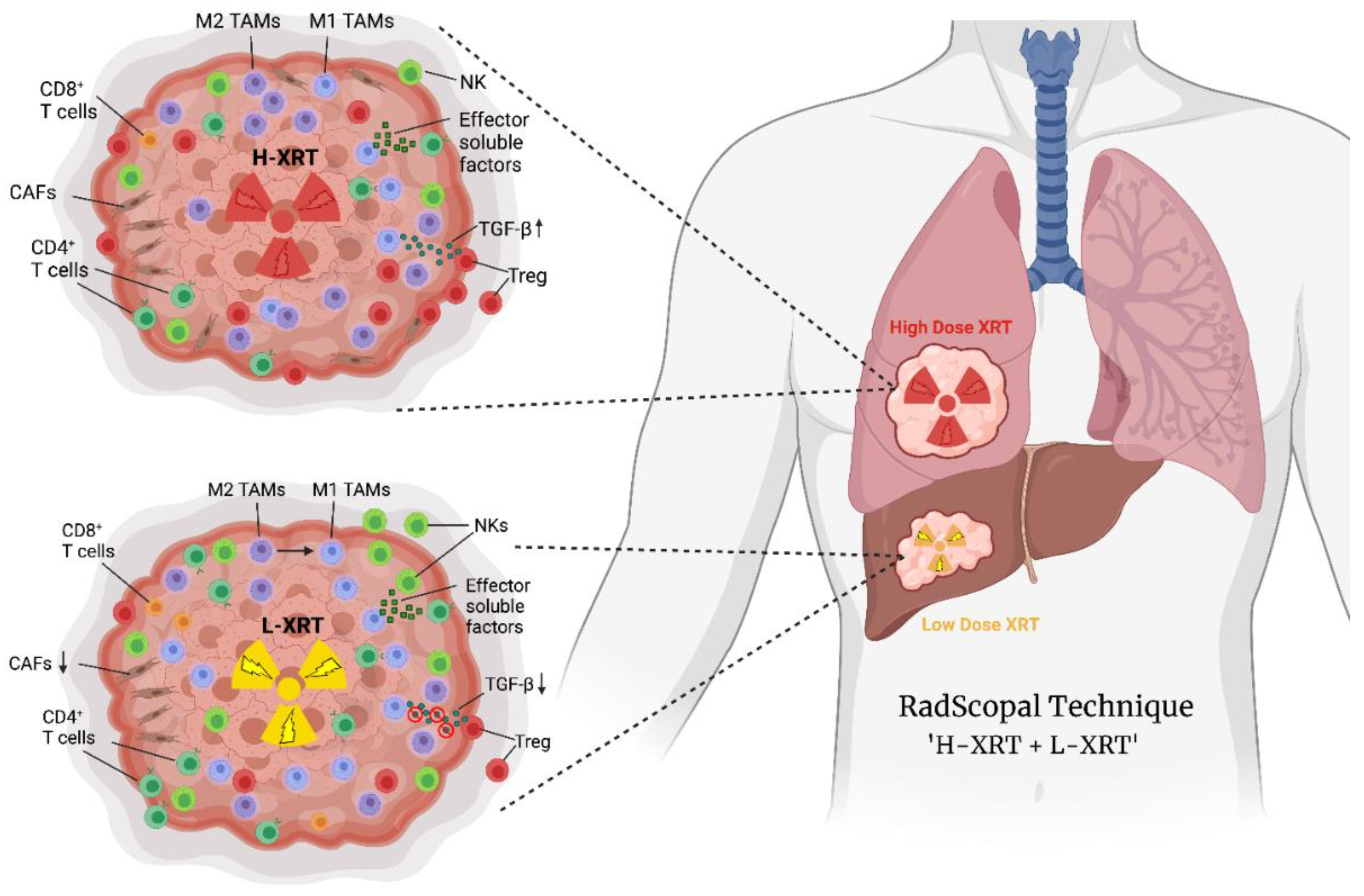
2. High-Dose Radiation (H-XRT) in Immune Priming and Abscopal Responses

Hypofractionated high-dose radiation provides numerous benefits. For example, it increases MHC class I expression in the tumor microenvironment (TME) and enhances antigen release [9]. This is especially favorable towards patients who are anti-PD1-resistant as they tend to present with decreased MHC class I expression. This suggests that H-XRT may in part serve as a method for reversing resistance to anti-PD1 [9]. In that context, it is also critical to consider the sequence and schedule of anti-PD1 administration relative to H-XRT, as others have shown that the sequence aspect can alter the potency of induced abscopal responses [10]. Meanwhile, the release of tumor neoantigens in response to H-XRT assists in T-cell priming and T-cell receptor (TCR) repertoire diversification. Determining optimal radiation dosage is often a challenge as some dose ranges can cause undesirable effects. For example, administering radiation doses above 12–18 Gray (Gy) per fraction induced the activation of DNA exonuclease Trex1. This resulted in the degradation of cytosolic DNA, preventing the activation of the cGAS-STING pathway [11]. The activation of the cGAS-STING pathway in dendritic cells and macrophages triggers the synthesis of type-I interferons (IFNs) which help prime T-cells and drive adaptive immunity. Furthermore, H-XRT turns immunologically “cold” tumors into “hot” by inducing the death of cancer cells, resulting in the release of proinflammatory mediators, such as damage-associated molecular patterns (DAMPs), calreticulins, ATP, and high-mobility group box 1 protein (HMGB1), which stimulate Toll-like receptor 4 (TLR4). Together, these signals produce a robust inflammatory cytokine response that encourages dendritic cell maturation, the elevation of costimulatory signals that aid in the cross-priming of cytotoxic CD8+ T-cells, and the upregulation of chemokine receptors. Effector-adaptive immune cells will eventually be attracted to and activated by this chain of events to produce antitumor effects [12,13].
On the other hand, H-XRT may have certain side effects including TGF-B and Tregs upregulation [14]. Priming T-cells with H-XRT may also accelerate T-cell exhaustion. Therefore, it is imperative that high-dose radiation be coupled with immune checkpoint inhibitors (ICIs) such as anti-CTLA-4 and anti-PD1 to block Tregs and mitigate exhaustion respectively. Checkpoint inhibitors can thus be used wisely to reduce fatigue and partially restore antitumor functions. In high-tumor-burden models, however, checkpoint blocking fails to sustain optimum effectiveness, necessitating further therapies to overcome the suppressive tumor stroma. Radiotherapy is one such technique for resetting the tumor microenvironment and optimizing systemic anticancer outcomes [15]. This opened the door for a variety of studies to promote abscopal responses and harness the benefits of radiation locally as well as halt tumor growth at metastatic sites [16].
In one study using a mouse model of anti-PD1 resistant lung adenocarcinoma, the authors examined the effectiveness of an OX40 agonist as a costimulatory molecule in conjunction with H-XRT [17]. Secondary tumors were not treated in order to study the effects of abscopal outcomes, whereas primary tumors received 12 Gy x 3 dose followed by intratumoral injection of the anti-OX40 monoclonal antibody. The combination of radiotherapy and OX40 activation successfully reduced lung metastases, decreased local and systemic anticancer growth, and significantly prolonged survival. The expansion of CD4+ and CD8+ T-cells was aided by this treatment plan. OX40 expression on T-cells in tumors and spleens was upregulated by radiotherapy, and the proportion of splenic CD103+ dendritic cells was increased [17].
Similarly, in our work with the glucocorticoid-induced tumor necrosis factor (TNF)–related protein (GITR) agonist, we treated the same anti-PD1 resistant mouse model with a combination of H-XRT, anti-GITR, and anti-PD1. Our results showed that H-XRT in conjunction with either anti-PD1 or anti-GITR alone showed no dramatic improvements. However, with the triple combination therapy of H-XRT, anti-GITR, and anti-PD1, there were significant results, where secondary tumors completely flatlined in 4 out of 11 treated mice. In addition, long-term effector memory was generated that was detected by flow cytometry phenotyping of spleens and secondary tumor-draining lymph nodes, proving that this triple therapy helped to combat recurrent cancer [14].
On the clinical level, a phase II trial of Ipilimumab with stereotactic radiation showed favorable results in terms of abscopal response and toxicity. Patients were treated with Ipilimumab (Ipi) for four cycles every three weeks and stereotactic radiation on the day of the first Ipi cycle (concurrent treatment) or the week after the second Ipi cycle (sequential treatment) was administered. Patients were given H-XRT in either 50 Gy in 4 fractions or 60 Gy in 10 fractions to the liver or the lung. Ipi is a monoclonal antibody that targets the checkpoint inhibitor CTLA-4 expressed by Tregs, hence liberating effector immunity from suppression. The combination of H-XRT + anti-CTLA-4 produced an abscopal response, where nonirradiated tumor volume had a clinical benefit rating of 26%. In terms of toxicity, adverse effects of diarrhea, ALT/AST elevation, and skin rash were the most common [18].
Another technique that we studied was pulsed radiotherapy, which treats two to four tumor lesions with two to three irradiation cycles, separated by about a one-month gap between each cycle. The idea was to reinitiate the cancer-immunity loop with each cycle, which starts with the production of cancer-associated neoantigens due to irradiation damage and continues via the activation of innate and adaptive immune responses, culminating in further tumor-cell death. In other words, with each irradiation cycle comes the opportunity for neoantigens to be generated which once presented to T-Cell Receptors lead to T-cell priming and clonal expansion. Consequently, the TCR repertoire of the tumor-specific T-cells grows wider and more diverse with each cycle, leading to increasingly more effective and robust immune responses [19]. In addition to its effect on TCR repertoire diversification, pulsed radiotherapy can also improve immunological memory and mold the tumor-directed T-cell populations, especially when combined with immunotherapy [19]. The notion of pulsed radiotherapy, comprising multiple radiotherapy cycles, relies on creating tumor-associated antigens with each treatment cycle to build cellular and humoral memory, similar to traditional vaccines that may require booster shots to bring about long-lasting memory [12].

3. Low-Dose Radiation (L-XRT) to Overcome the Tumor Stroma

Traditionally, high-dose radiation has been used to treat cancer patients; however, now scientists are beginning to investigate the benefits of low-dose radiation and how it can be used to promote an enhanced antitumor response and prolong survival. Low-dose radiation is defined as 0.5–2 Gy per fraction for up to 1–10 Gy total [8]. In our preclinical studies using murine models, we found that 1 Gy x 2 fractions was able to modulate the TME and address the surrounding inhibitory stroma. In the first 24 h after L-XRT, analysis of the tumor-infiltrating lymphocytes (TILs) showed a significant decrease in Transforming Growth Factor β (TGF-β) inhibitory cytokine, which is correlated with M2 tumor-associated macrophages (TAMs), compared to non-treated control tumors. Then, 2–3 days after receiving L-XRT, there was an increase in the infiltration of effector T-cells as well as NK cells, helping elicit a better antitumor response. Furthermore, L-XRT polarized the TAMs from the protumor M2 phenotype to the antitumor M1 phenotype [7]. Upon looking at the draining lymph nodes, we also detected an upregulation of activated CD4+ and CD8+ T-cells expressing the CD44 activation marker. Additionally, in a single-tumor 344SQ lung adenocarcinoma model, 1 Gy x 2 dose alone was capable to control tumor growth and extend survival. Moreover, the efficacy of α-PD1 and α-CTLA-4 checkpoints was significantly improved when combined with L-XRT [7]. In the same context of tumor stroma, L-XRT decreased cancer-associated fibroblasts (CAFs), identified by FAP and S100A4 histological stains, and allowed for increased infiltration of immune cells [20].
In our review regarding low-dose radiation to liver metastasis, we once again showed that L-XRT to the whole liver (1.4 Gy x 4 fractions) can reprogram the TME from immune suppressive to immune active in a patient with multi-organ metastases and refractory to prior T-cell therapy. Ipilimumab + Nivolumab immunotherapy was maintained as a treatment backbone and the patient presented with a complete response in the liver upon 19-month follow-up, but with no response in lesions that did not receive L-XRT [8]. One of the major advantages of L-XRT is that it does not cause DNA damage like H-XRT and can be administered safely to or in the proximity of critical organs. In fact, in a phase II clinical trial, low-dose radiation combined with high-dose radiation dramatically improved antitumor responses in α-PD1/α-PDL1 refractory patients [21]. This combination is powerful because low-dose radiation is able to mitigate some of the adverse effects caused by high-dose radiation such as the upregulation of TGF-β. In the same study, we assessed the infiltration of CD4 T-cells, CD8 T-cells, and NK cells in a head and neck cancer case treated with L-XRT. The fold increases in these populations were 4.3, 2.3, and 187 folds respectively [21]. In the context of the preclinical and clinical data above, L-XRT can be used to enhance cell-based therapies, such as CAR-T and CAR-NK, by improving their tumor infiltration rate and persistence.
The tumor microenvironment is characterized by a network of tumor, stromal, and tolerogenic immune cells that promote immunosuppressive effects [22]. For example, the TME promotes the infiltration of myeloid-derived suppressor cells (MDSCs) into tumors. MDSCs express high levels of arginase and Indoleamine 2,3-dioxygenase (IDO-1) that inhibit T-cell responses; they also contribute to Treg recruitment into the TME via the secretion of chemokines such as CCL3, CCL4, and CCL5 [22]. Metabolites such as reactive nitrogen species (RNS) and reactive oxygen species (ROS) can reduce T-cell immune responses when present in excess but can also activate and expand effector T-cells at metronomic doses [23]. L-XRT may indeed help to shift the balance towards desired antitumor outcomes by reprogramming the TME. For example, in a murine ID8 ovarian model, 1 Gy treatment resulted in lymphocyte, NK cell, macrophage, and DCs inflow with high infiltration of CD8+ and CD4+ T-cells [24]. Furthermore, weekly administration of 1 Gy treatment maintained immune cell recruitment [24]. Another study with a murine B16F10 melanoma model showed that 1.25Gy total body irradiation treatment reduced Tregs and increased effector memory T-cells leading to reduced tumor burden and improved survival [25].
Tumor metabolism also plays a vital role in the TME; it can adapt to support tumor growth, angiogenesis, and metastasis. Intracellular ROS can have different effects on tumor cells depending on dose and exposure time. Tumor cells can undergo metabolic reprogramming to optimize intracellular ROS levels in the TME to drive malignant progression and promote survival and metastasis [26]. For example, aberrant expression of the KRAS oncogene can transform normal cells into malignant phenotypes and induce the production of ROS at a level that benefits malignant cells [27]. L-XRT (0.1Gy) was found to suppress the transformation by inducing antioxidants and reducing ROS [27]. However, once tumors are formed, L-XRT is shown to overproduce ROS within tumor cells by inducing mitochondrial stress, leading to tumor instability and apoptosis [28]. In a cohort study of 32 patients with solid metastatic tumors, lesions were treated with hypofractionated L-XRT of 4 Gy x 3 fractions given on days 1, 3, and 5, along with a single shot of anti-PD1 administered on day 2. The combination treatment resulted in a complete response in 2 patients, partial response in 12 patients, and stable disease in 18 patients with improved quality of life [29]. In conclusion, L-XRT seems to have differential effects on different cell types in the context of ROS production and it is critical to understand its unique impact on immune cells vs tumor cells as elaborated above.

4. RadScopal as an Immunotherapy Booster

RadScopal is a unique technique where H-XRT is applied to a primary tumor and L-XRT is applied to secondary tumor(s) in patients undergoing or who have progressed on immunotherapy. This method allows a phenomenon we call the prime pull method to occur. The combination of H-XRT, which primes the T-cells, and L-XRT, which pulls in and activates the T-cells takes place. By taking both routes of high and low-dose radiation, we also incorporate both the advantages and disadvantages of each method. In essence, when L-XRT is added to H-XRT it helps mitigate a lot of disadvantages of H-XRT. Radscopal is also evidently different from the traditional abscopal response where distant sites are not exposed to radiation [18].
RadScopal immunoradiation involves both high-dose radiation in the primary tumor for immune priming and low-dose radiation in the secondary tumor for facilitating immune cell infiltration and tumor killing. Therefore, strategies that can improve either immune priming or attracting immune cells or both may enhance the treatment efficacy of RadScopal immunoradiation. NBTXR3, a hafnium oxide nanoparticle, was initially designed to increase radiation energy deposition in adjacent tumor cells [30]. It allows effective tumor destruction at a relatively low dose of radiation. In recent years, an increasing body of data has demonstrated that NBTXR3 is able to enhance antitumor immune activation in various models [31,32]. The potential of the immune modulation effect of NBTXR3 was discovered through the finding that it increases cGAS-STING pathway response [33]. Activation of the cGAS-STING pathway can lead to enhanced activation of dendritic cells, elevated expression of type I interferon, and subsequent CD8 T-cell activation [34]. Our data demonstrates that NBTXR3 in combination with localized photon therapy resulted in a significant increase in tumor apoptosis in the irradiated tumors and higher CD8 T-cell infiltration in the unirradiated tumors [32]. Subsequent studies showed that the combination of NBTXR3 with RadScopal immunoradiation led to remarkable treatment efficacy in 344SQR anti-PD1 resistant metastatic lung cancer [35]. This combination therapy not only effectively contained the growth of the primary tumors that received high-dose radiation, but also eradicated secondary tumors treated with low-dose radiation. In addition, NBTXR3-mediated RadScopal immunoradiation significantly reduced the number of lung metastases. The mice treated with this combination therapy developed potent and long-term memory immune responses. Analysis of the immune landscape of the low-dose radiation-treated tumors indicates that this combination therapy reshapes T-cell repertoire, which may enhance tumor antigen recognition and targeting. Moreover, major immune pathways in tumors related to adaptive response, innate response, T-cell function, NK cell function, etc. were markedly upregulated by NBTXR3-RadScopal radiotherapy. We also observed increased infiltration of CD8 T-cells and decreased infiltration of Tregs in the low-dose radiation-treated tumors. Collectively, these findings demonstrate that NBTXR3 in combination with RadScopal radiotherapy is able to favorably modulate the tumor immune microenvironment and maximize tumor killing at the tumors that received either high-dose radiation or low-dose radiation.
Spatially fractionated radiotherapy, also known as GRID, can overcome radiation dose limitations while still preserving normal tissue function. A matrix with several pencil-like openings is used at the tumor site. Part of the tumor site is shielded by the matrix while radiation beams are delivered through the openings. From an immunological perspective, this also allows the partial sparing of immune cells in the TME that are needed for antigen presentation and priming T-cells. A clinical study of 71 patients with advanced large tumors demonstrated that single doses of 10–20 Gy had effective tumor control with no damage to epidermal and subcutaneous tissue [36]. The mass effect response was 72.5% including CR/PR, with head and neck patients benefiting the most from the utilized GRID approach. The latter can also be used at the microscopic level, which allows the delivery of greater radiation doses. Doses of up to 4000 Gy can be administered via 25 µm microbeam arrays without permanently compromising vasculature and tissue framework [37]. This could resolve lung fibrosis caused when delivering conventional therapies.
The micro-slit uses parallel beams approximately 25 µm to 75 µm wide. This provides tissue-sparing effects while allowing the administration of higher radiation doses. A preclinical murine EMT-6 carcinoma model compared cross-planar beams (410 Gy, 520 Gy, 650 Gy), vertical beams (410 Gy, 520 Gy, 650 Gy), and broad beam (30 Gy, 38 Gy, 45 Gy) treatments in murine hind legs administered as single fractions [38]. They found that toxicity and leg dysfunction were significantly lower in vertical and cross-planar beams compared to broad beams [38]. These findings suggest that micro-slit single fraction treatments have robust antitumor efficacy. Another preclinical model delivered micro-slit radiation treatment as an alternative to whole-brain radiation, as the latter has the potential for irreversible encephalon damage [39]. Doses of 96 Gy to 960 Gy for micro-slit beams and 24 Gy to 120 Gy for broad beams were used. The broad beams resulted in demyelination and hemorrhaging, whereas the micro-slit beams preserved tissue structure and function at higher doses. Currently, GRID and micro-slit radiation therapies must be further explored to determine their efficacy in the context of the RadScopal technique and their ability to spare immune cells to mount proper antitumor responses. For instance, The H-XRT portion of the RadScopal regimen can be delivered to primary tumors in form of a GRID or Lattice radiation to maintain the release of tumor-associated antigens while saving antigen-presenting cells and T-cells in surrounding tissue. On a single lesion level, GRID radiation may also be interpreted as “local RadScopal”, where high and low-dose radiation spots are created beneath the GRID matrix, resembling RadScopal on a miniature level.

5. Future Directions and Immune Oncology Drug Candidates to Combine with RadScopal Therapy

In order to fine tune the RadScopal technique as well as predict its effect, it is critical to understand the different factors that are involved in generating the response. Considering all the factors involved allows us to explore ways of modulating them in order to increase antitumor efficacy. The first element to consider is the selection of lesion(s) to be irradiated. Coupled with this variable is the determination of radiation dosage and fractionation which are important parameters in influencing antitumor responses [40]. In an effort to consider different radiation schemas, some mathematical models have been constructed in order to compare different radiotherapy protocols. This can allow us to determine the optimal radiation doses required to yield a robust systemic antitumor immunity [40]. Other factors that predict responses are the availability of systemic T-cells, their trafficking to tumor sites and their ability to infiltrate the TME [41]. The trafficking and infiltration of T-cells are largely aided by the L-XRT of the RadScopal technique [7]. However, other variables such as T-cell exhaustion as well as their metabolic profile within the TME can also have a serious impact on the immune response. Lastly, target lesion volume and its location play a huge role in determining treatment plans. Lesions located near critical organs may present a challenge to H-XRT, and L-XRT may be considered as an alternative to spare surrounding tissue. Given the unique biology of the RadScopal technique described above, it may provide and receive reciprocal benefits from immune agents currently under development involved in priming (signal 1), costimulation (signal 2), and activation/expansion by soluble factors (signal 3). All three signals are necessary to drive a potent T-cell activation and develop long-term memory [42].
To start with signal 1, It is previously described that H-XRT helps initiate the priming step by upregulating MHC-I and producing IFNs [9]. This step can be enhanced by combining with non-fucosylated anti-CTLA-4, a second-generation anti-CTLA-4 antibody with enhanced Treg depletion capacity, which in turn enables better priming of T-effectors (NCT04785287). A similar outcome may be achieved by using anti-CCR8 monoclonal antibody. It has been shown that tumor resident Tregs express high levels of CCR8 that can be specifically depleted by anti-CCR8 without depleting systemic Tregs [43]. This approach reduces systemic toxicity and focuses efficacy on primary as well as secondary lesions, which renders it highly suitable to combine with RadScopal. Another new approach to facilitate priming is to block CD73 expressed by stromal cells and a subset of T-cells, hence improving the availability of ATP in the TME, which serves as an energy reservoir for dendritic cells and macrophages to improve antigen presentation, a critical step in the priming process [44].
Proceeding to signal 2 and costimulation, there are several agonists tested as monotherapy or with checkpoint inhibitors, but not yet fully utilized with radiation at least on the clinical level. The importance of costimulation stems from its ability to complement the initial TCR signal and sustain the immune activation cascade, otherwise, T-cells become anergic and eventually die. Classical signal 2 agents include agonists to CD28, OX40, 4-1BB, and CD40. The RadScopal technique orchestrates both innate and adaptive immunity to systemically treat solid tumors. The innate response can be amplified by using anti-CD40 for instance, to polarize macrophages from M2 to M1 and produce a set of antitumor proinflammatory cytokines [45]. The adaptive arm on the other hand can be boosted through 4-1BB for example, to induce T-cell trafficking and generate effector and central memory [46]. Other small molecules are also under development to maintain CD28 costimulation and prolong the survival and function of T-cells and NK cells to achieve better outcomes. One such molecule is CBL-B inhibitor (CBL-Bi) that has high potential to be combined with the RadScopal approach, given the molecule’s ability to liberate PI3K and AKT, and eventually activate NF-kB in T-cells and NK cells [47].
Moving forward to signal 3 to attain a full-fledged immune activation, The RadScopal technique can benefit from a wide variety of soluble factors to achieve the culmination of its potential. These include next-generation cytokine therapies, such as pegylated IL-2 (PEG-IL-2) for slow and prolonged release of IL-2, augmenting the expansion of newly primed T-cells post radiation; engineered IL-2 that binds to β and γ chains of the receptor but does not engage with the high-affinity α chain, which preferentially expands T-effectors but not Tregs [48]; engineered IL-12 cytokine that shifts the balance towards Th1 responses and polarizes macrophages to antitumor M1 phenotype [49]; and IL-15 which does not only activate and expand NK cells, but also augment with T-cell memory generation on the long run [50,51].
Finally, it is critical to address activation-induced exhaustion, especially after strong combinatorial regimens with RadScopal + immune agents. To overcome this hurdle, there are checkpoint inhibitors that can be used with or without anti-PD1, to target TIM-3, LAG-3, and TIGIT on exhausted T-cells and NK cells [52], or to block intracellular downstream targets such as SHP-2 [53]. However, there are instances where the cells are terminally exhausted and hard to rejuvenate, leading to acquired resistance in conjunction with other immune evasion mechanisms deployed by tumors. In such cases, we seek to deliver a second pulse of RadScopal treatment to reset the TME of primary and secondary tumors, restart the immune cycle, and initiate a second wave of priming, costimulation, activation/proliferation, trafficking, infiltration, and tumor killing in that particular order.

Author Contributions

All authors contributed to writing, editing, and reviewing the manuscript. All authors have read and agreed to the published version of the manuscript.

Funding

This research received no external funding.

Institutional Review Board Statement

Not applicable.

Informed Consent Statement

Not applicable.

Data Availability Statement

Not applicable.

Conflicts of Interest

J.W.W. reports research support from GlaxoSmithKline, Bristol Meyers Squibb, Merck, Nanobiotix, RefleXion, Alkermes, Artidis, Mavu Pharma, Takeda, Varian, Checkmate Pharmaceuticals, and HotSpot Therapeutics. J.W.W. serves/served on the scientific advisory board for Legion Healthcare Partners, RefleXion Medical, MolecularMatch, Merck, AstraZeneca, Aileron Therapeutics, OncoResponse, Checkmate Pharmaceuticals, Mavu Pharma, Alpine Immune Sciences, Ventana Medical Systems, Nanobiotix, China Medical Tribune, GI Innovation, Genentech and Nanorobotix. J.W.W. serves as a consultant for Lifescience Dynamics Limited. J.W.W. has/had Speaking Engagements for Ventana Medical Systems, US Oncology, Alkermes, Boehringer Ingelheim, Accuray, and RSS. J.W.W. holds/held stock or ownership in Alpine Immune Sciences, Checkmate Pharmaceuticals, Healios, Mavu Pharma, Legion Healthcare Partners, MolecularMatch, Nanorobotix, OncoResponse, and RefleXion. J.W.W. has accepted honoraria in the form of travel costs from Nanobiotix, RefleXion, Varian, Shandong University, The Korea Society of Radiology, Aileron Therapeutics, and Ventana. J.W.W. has the following patents: MP470 (amuvatinib), MRX34 regulation of PDL1, XRT technique to overcome immune resistance, Radiotherapies and uses thereof. MD Anderson Cancer Center has a trademark for RadScopalTM. HBB has the following patent: Radiotherapies and uses thereof. The rest of the authors disclosed no competing interests.

References

  1. Gianfaldoni, S.; Gianfaldoni, R.; Wollina, U.; Lotti, J.; Tchernev, G.; Lotti, T. An Overview on Radiotherapy: From Its History to Its Current Applications in Dermatology. Open Access Maced. J. Med. Sci. 2017, 5, 521–525. [Google Scholar] [CrossRef] [PubMed] [Green Version]
  2. Baskar, R.; Lee, K.A.; Yeo, R.; Yeoh, K.W. Cancer and radiation therapy: Current advances and future directions. Int. J. Med. Sci. 2012, 9, 193–199. [Google Scholar] [CrossRef] [PubMed] [Green Version]
  3. Rudqvist, N.P.; Pilones, K.A.; Lhuillier, C.; Wennerberg, E.; Sidhom, J.W.; Emerson, R.O.; Robins, H.S.; Schneck, J.; Formenti, S.C.; Demaria, S. Radiotherapy and CTLA-4 Blockade Shape the TCR Repertoire of Tumor-Infiltrating T Cells. Cancer Immunol. Res. 2018, 6, 139–150. [Google Scholar] [CrossRef] [PubMed] [Green Version]
  4. Rodriguez-Ruiz, M.E.; Vanpouille-Box, C.; Melero, I.; Formenti, S.C.; Demaria, S. Immunological Mechanisms Responsible for Radiation-Induced Abscopal Effect. Trends Immunol. 2018, 39, 644–655. [Google Scholar] [CrossRef]
  5. Golden, E.B.; Chhabra, A.; Chachoua, A.; Adams, S.; Donach, M.; Fenton-Kerimian, M.; Friedman, K.; Ponzo, F.; Babb, J.S.; Goldberg, J.; et al. Local radiotherapy and granulocyte-macrophage colony-stimulating factor to generate abscopal responses in patients with metastatic solid tumours: A proof-of-principle trial. Lancet Oncol. 2015, 16, 795–803. [Google Scholar] [CrossRef]
  6. Vanpouille-Box, C.; Pilones, K.A.; Wennerberg, E.; Formenti, S.C.; Demaria, S. In situ vaccination by radiotherapy to improve responses to anti-CTLA-4 treatment. Vaccine 2015, 33, 7415–7422. [Google Scholar] [CrossRef] [Green Version]
  7. Barsoumian, H.B.; Ramapriyan, R.; Younes, A.I.; Caetano, M.S.; Menon, H.; Comeaux, N.I.; Cushman, T.R.; Schoenhals, J.E.; Cadena, A.P.; Reilly, T.P.; et al. Low-dose radiation treatment enhances systemic antitumor immune responses by overcoming the inhibitory stroma. J. Immunother. Cancer 2020, 8, e000537. [Google Scholar] [CrossRef]
  8. He, K.; Barsoumian, H.B.; Bertolet, G.; Verma, V.; Leuschner, C.; Koay, E.J.; Ludmir, E.B.; Hsu, E.; Pisipati, E.; Voss, T.A.; et al. Novel Use of Low-Dose Radiotherapy to Modulate the Tumor Microenvironment of Liver Metastases. Front. Immunol. 2021, 12, 812210. [Google Scholar] [CrossRef]
  9. Wang, X.; Schoenhals, J.E.; Li, A.; Valdecanas, D.R.; Ye, H.; Zang, F.; Tang, C.; Tang, M.; Liu, C.G.; Liu, X.; et al. Suppression of Type I IFN Signaling in Tumors Mediates Resistance to Anti-PD-1 Treatment That Can Be Overcome by Radiotherapy. Cancer Res. 2017, 77, 839–850. [Google Scholar] [CrossRef] [Green Version]
  10. Wei, J.; Montalvo-Ortiz, W.; Yu, L.; Krasco, A.; Ebstein, S.; Cortez, C.; Lowy, I.; Murphy, A.J.; Sleeman, M.A.; Skokos, D. Sequence of alphaPD-1 relative to local tumor irradiation determines the induction of abscopal antitumor immune responses. Sci. Immunol. 2021, 6, eabg0117. [Google Scholar] [CrossRef]
  11. Vanpouille-Box, C.; Alard, A.; Aryankalayil, M.J.; Sarfraz, Y.; Diamond, J.M.; Schneider, R.J.; Inghirami, G.; Coleman, C.N.; Formenti, S.C.; Demaria, S. DNA exonuclease Trex1 regulates radiotherapy-induced tumour immunogenicity. Nat. Commun. 2017, 8, 15618. [Google Scholar] [CrossRef] [PubMed] [Green Version]
  12. He, K.; Barsoumian, H.B.; Sezen, D.; Puebla-Osorio, N.; Hsu, E.Y.; Verma, V.; Abana, C.O.; Chen, D.; Patel, R.R.; Gu, M.; et al. Pulsed Radiation Therapy to Improve Systemic Control of Metastatic Cancer. Front. Oncol. 2021, 11, 737425. [Google Scholar] [CrossRef] [PubMed]
  13. Caetano, M.S.; Younes, A.I.; Barsoumian, H.B.; Quigley, M.; Menon, H.; Gao, C.; Spires, T.; Reilly, T.P.; Cadena, A.P.; Cushman, T.R.; et al. Triple Therapy with MerTK and PD1 Inhibition Plus Radiotherapy Promotes Abscopal Antitumor Immune Responses. Clin. Cancer Res. 2019, 25, 7576–7584. [Google Scholar] [CrossRef] [Green Version]
  14. Schoenhals, J.E.; Cushman, T.R.; Barsoumian, H.B.; Li, A.; Cadena, A.P.; Niknam, S.; Younes, A.I.; Caetano, M.D.S.; Cortez, M.A.; Welsh, J.W. Anti-glucocorticoid-induced Tumor Necrosis Factor-Related Protein (GITR) Therapy Overcomes Radiation-Induced Treg Immunosuppression and Drives Abscopal Effects. Front. Immunol. 2018, 9, 2170. [Google Scholar] [CrossRef] [Green Version]
  15. Barsoumian, H.B.; Sezen, D.; Menon, H.; Younes, A.I.; Hu, Y.; He, K.; Puebla-Osorio, N.; Wasley, M.; Hsu, E.; Patel, R.R.; et al. High Plus Low Dose Radiation Strategy in Combination with TIGIT and PD1 Blockade to Promote Systemic Antitumor Responses. Cancers 2022, 14, 221. [Google Scholar] [CrossRef] [PubMed]
  16. Liu, Y.; Dong, Y.; Kong, L.; Shi, F.; Zhu, H.; Yu, J. Abscopal effect of radiotherapy combined with immune checkpoint inhibitors. J. Hematol. Oncol. 2018, 11, 104. [Google Scholar] [CrossRef] [Green Version]
  17. Niknam, S.; Barsoumian, H.B.; Schoenhals, J.E.; Jackson, H.L.; Yanamandra, N.; Caetano, M.S.; Li, A.; Younes, A.I.; Cadena, A.; Cushman, T.R.; et al. Radiation Followed by OX40 Stimulation Drives Local and Abscopal Antitumor Effects in an Anti-PD1-Resistant Lung Tumor Model. Clin. Cancer Res. 2018, 24, 5735–5743. [Google Scholar] [CrossRef] [Green Version]
  18. Welsh, J.W.; Tang, C.; de Groot, P.; Naing, A.; Hess, K.R.; Heymach, J.V.; Papadimitrakopoulou, V.A.; Cushman, T.R.; Subbiah, V.; Chang, J.Y.; et al. Phase II Trial of Ipilimumab with Stereotactic Radiation Therapy for Metastatic Disease: Outcomes, Toxicities, and Low-Dose Radiation-Related Abscopal Responses. Cancer Immunol. Res. 2019, 7, 1903–1909. [Google Scholar] [CrossRef] [Green Version]
  19. Sezen, D.; Barsoumian, H.B.; He, K.; Hu, Y.; Wang, Q.; Abana, C.O.; Puebla-Osorio, N.; Hsu, E.Y.; Wasley, M.; Masrorpour, F.; et al. Pulsed radiotherapy to mitigate high tumor burden and generate immune memory. Front. Immunol. 2022, 13, 984318. [Google Scholar] [CrossRef]
  20. Patel, R.R.; Barsoumian, H.; Verma, V.; Cortez, M.A.; Welsh, J.W. Low-Dose Radiation Decreases Cancer-Associated Fibroblasts and May Increase T-Cell Trafficking into Tumors. Int. J. Radiat. Oncol. Biol. Phys. 2020, 108, e530–e531. [Google Scholar] [CrossRef]
  21. Patel, R.R.; He, K.; Barsoumian, H.B.; Chang, J.Y.; Tang, C.; Verma, V.; Comeaux, N.; Chun, S.G.; Gandhi, S.; Truong, M.T.; et al. High-dose irradiation in combination with non-ablative low-dose radiation to treat metastatic disease after progression on immunotherapy: Results of a phase II trial. Radiother. Oncol. 2021, 162, 60–67. [Google Scholar] [CrossRef] [PubMed]
  22. Shi, R.; Tang, Y.Q.; Miao, H. Metabolism in tumor microenvironment: Implications for cancer immunotherapy. MedComm 2020, 1, 47–68. [Google Scholar] [CrossRef] [PubMed]
  23. Chen, X.; Song, M.; Zhang, B.; Zhang, Y. Reactive Oxygen Species Regulate T Cell Immune Response in the Tumor Microenvironment. Oxid. Med. Cell. Longev. 2016, 2016, 1580967. [Google Scholar] [CrossRef] [Green Version]
  24. Herrera, F.G.; Ronet, C.; Ochoa de Olza, M.; Barras, D.; Crespo, I.; Andreatta, M.; Corria-Osorio, J.; Spill, A.; Benedetti, F.; Genolet, R.; et al. Low-Dose Radiotherapy Reverses Tumor Immune Desertification and Resistance to Immunotherapy. Cancer Discov. 2022, 12, 108–133. [Google Scholar] [CrossRef] [PubMed]
  25. Liu, R.; Xiong, S.; Zhang, L.; Chu, Y. Enhancement of antitumor immunity by low-dose total body irradiationis associated with selectively decreasing the proportion and number of T regulatory cells. Cell. Mol. Immunol. 2010, 7, 157–162. [Google Scholar] [CrossRef] [Green Version]
  26. Rodic, S.; Vincent, M.D. Reactive oxygen species (ROS) are a key determinant of cancer’s metabolic phenotype. Int. J. Cancer 2018, 142, 440–448. [Google Scholar] [CrossRef] [Green Version]
  27. Kim, R.K.; Kim, M.J.; Seong, K.M.; Kaushik, N.; Suh, Y.; Yoo, K.C.; Cui, Y.H.; Jin, Y.W.; Nam, S.Y.; Lee, S.J. Beneficial effects of low dose radiation in response to the oncogenic KRAS induced cellular transformation. Sci. Rep. 2015, 5, 15809. [Google Scholar] [CrossRef] [Green Version]
  28. Averbeck, D.; Rodriguez-Lafrasse, C. Role of Mitochondria in Radiation Responses: Epigenetic, Metabolic, and Signaling Impacts. Int. J. Mol. Sci. 2021, 22, 11047. [Google Scholar] [CrossRef]
  29. Li, D.; Zhu, W.; Zhou, J.; Peng, M.; Geng, Q.; Pu, X.; Wang, M.; Jiang, H. Hypofractionated Low-Dose Radiotherapy Combined with Immune Checkpoint Inhibition in Metastatic Solid Tumors. OncoTargets Ther. 2021, 14, 773–783. [Google Scholar] [CrossRef]
  30. Marill, J.; Anesary, N.M.; Zhang, P.; Vivet, S.; Borghi, E.; Levy, L.; Pottier, A. Hafnium oxide nanoparticles: Toward an in vitro predictive biological effect? Radiat. Oncol. 2014, 9, 150. [Google Scholar] [CrossRef] [Green Version]
  31. Zhang, P.; Darmon, A.; Marill, J.; Mohamed Anesary, N.; Paris, S. Radiotherapy-Activated Hafnium Oxide Nanoparticles Produce Abscopal Effect in a Mouse Colorectal Cancer Model. Int. J. Nanomed. 2020, 15, 3843–3850. [Google Scholar] [CrossRef] [PubMed]
  32. Hu, Y.; Paris, S.; Barsoumian, H.; Abana, C.O.; He, K.; Wasley, M.; Younes, A.I.; Masrorpour, F.; Chen, D.; Yang, L.; et al. Radiation Therapy Enhanced by NBTXR3 Nanoparticles Overcomes Anti-PD1 Resistance and Evokes Abscopal Effects. Int. J. Radiat. Oncol. Biol. Phys. 2021, 111, 647–657. [Google Scholar] [CrossRef]
  33. Marill, J.; Mohamed Anesary, N.; Paris, S. DNA damage enhancement by radiotherapy-activated hafnium oxide nanoparticles improves cGAS-STING pathway activation in human colorectal cancer cells. Radiother. Oncol. 2019, 141, 262–266. [Google Scholar] [CrossRef] [PubMed]
  34. Li, A.; Yi, M.; Qin, S.; Song, Y.; Chu, Q.; Wu, K. Activating cGAS-STING pathway for the optimal effect of cancer immunotherapy. J. Hematol. Oncol. 2019, 12, 35. [Google Scholar] [CrossRef] [Green Version]
  35. Hu, Y.; Paris, S.; Barsoumian, H.; Abana, C.O.; He, K.; Sezen, D.; Wasley, M.; Masrorpour, F.; Chen, D.; Yang, L.; et al. A radioenhancing nanoparticle mediated immunoradiation improves survival and generates long-term antitumor immune memory in an anti-PD1-resistant murine lung cancer model. J. Nanobiotechnol. 2021, 19, 416. [Google Scholar] [CrossRef] [PubMed]
  36. Mohiuddin, M.; Fujita, M.; Regine, W.F.; Megooni, A.S.; Ibbott, G.S.; Ahmed, M.M. High-dose spatially-fractionated radiation (GRID): A new paradigm in the management of advanced cancers. Int. J. Radiat. Oncol. Biol. Phys. 1999, 45, 721–727. [Google Scholar] [CrossRef]
  37. Schultke, E.; Balosso, J.; Breslin, T.; Cavaletti, G.; Djonov, V.; Esteve, F.; Grotzer, M.; Hildebrandt, G.; Valdman, A.; Laissue, J. Microbeam radiation therapy—Grid therapy and beyond: A clinical perspective. Br. J. Radiol. 2017, 90, 20170073. [Google Scholar] [CrossRef]
  38. Dilmanian, F.A.; Morris, G.M.; Zhong, N.; Bacarian, T.; Hainfeld, J.F.; Kalef-Ezra, J.; Brewington, L.J.; Tammam, J.; Rosen, E.M. Murine EMT-6 carcinoma: High therapeutic efficacy of microbeam radiation therapy. Radiat. Res. 2003, 159, 632–641. [Google Scholar] [CrossRef]
  39. Mukumoto, N.; Nakayama, M.; Akasaka, H.; Shimizu, Y.; Osuga, S.; Miyawaki, D.; Yoshida, K.; Ejima, Y.; Miura, Y.; Umetani, K.; et al. Sparing of tissue by using micro-slit-beam radiation therapy reduces neurotoxicity compared with broad-beam radiation therapy. J. Radiat. Res. 2017, 58, 17–23. [Google Scholar] [CrossRef] [Green Version]
  40. Poleszczuk, J.; Enderling, H. The Optimal Radiation Dose to Induce Robust Systemic Anti-Tumor Immunity. Int. J. Mol. Sci. 2018, 19, 3377. [Google Scholar] [CrossRef] [Green Version]
  41. Poleszczuk, J.T.; Luddy, K.A.; Prokopiou, S.; Robertson-Tessi, M.; Moros, E.G.; Fishman, M.; Djeu, J.Y.; Finkelstein, S.E.; Enderling, H. Abscopal Benefits of Localized Radiotherapy Depend on Activated T-cell Trafficking and Distribution between Metastatic Lesions. Cancer Res. 2016, 76, 1009–1018. [Google Scholar] [CrossRef] [Green Version]
  42. Sckisel, G.D.; Bouchlaka, M.N.; Monjazeb, A.M.; Crittenden, M.; Curti, B.D.; Wilkins, D.E.; Alderson, K.A.; Sungur, C.M.; Ames, E.; Mirsoian, A.; et al. Out-of-Sequence Signal 3 Paralyzes Primary CD4(+) T-Cell-Dependent Immunity. Immunity 2015, 43, 240–250. [Google Scholar] [CrossRef] [PubMed] [Green Version]
  43. Kidani, Y.; Nogami, W.; Yasumizu, Y.; Kawashima, A.; Tanaka, A.; Sonoda, Y.; Tona, Y.; Nashiki, K.; Matsumoto, R.; Hagiwara, M.; et al. CCR8-targeted specific depletion of clonally expanded Treg cells in tumor tissues evokes potent tumor immunity with long-lasting memory. Proc. Natl. Acad. Sci. USA 2022, 119, e2114282119. [Google Scholar] [CrossRef]
  44. Perrot, I.; Michaud, H.A.; Giraudon-Paoli, M.; Augier, S.; Docquier, A.; Gros, L.; Courtois, R.; Dejou, C.; Jecko, D.; Becquart, O.; et al. Blocking Antibodies Targeting the CD39/CD73 Immunosuppressive Pathway Unleash Immune Responses in Combination Cancer Therapies. Cell Rep. 2019, 27, 2411–2425.e2419. [Google Scholar] [CrossRef] [PubMed] [Green Version]
  45. Yasmin-Karim, S.; Bruck, P.T.; Moreau, M.; Kunjachan, S.; Chen, G.Z.; Kumar, R.; Grabow, S.; Dougan, S.K.; Ngwa, W. Radiation and Local Anti-CD40 Generate an Effective in situ Vaccine in Preclinical Models of Pancreatic Cancer. Front. Immunol. 2018, 9, 2030. [Google Scholar] [CrossRef]
  46. Barsoumian, H.B.; Batra, L.; Shrestha, P.; Bowen, W.S.; Zhao, H.; Egilmez, N.K.; Gomez-Gutierrez, J.G.; Yolcu, E.S.; Shirwan, H. A Novel Form of 4-1BBL Prevents Cancer Development via Nonspecific Activation of CD4(+) T and Natural Killer Cells. Cancer Res. 2019, 79, 783–794. [Google Scholar] [CrossRef] [PubMed] [Green Version]
  47. Zhou, X.; Sun, S.C. Targeting ubiquitin signaling for cancer immunotherapy. Signal Transduct. Target Ther. 2021, 6, 16. [Google Scholar] [CrossRef] [PubMed]
  48. Lopes, J.E.; Sun, L.; Flick, H.L.; Murphy, E.A.; Losey, H.C. Pharmacokinetics and Pharmacodynamic Effects of Nemvaleukin Alfa, a Selective Agonist of the Intermediate-Affinity IL-2 Receptor, in Cynomolgus Monkeys. J. Pharmacol. Exp. Ther. 2021, 379, 203–210. [Google Scholar] [CrossRef]
  49. Mirlekar, B.; Pylayeva-Gupta, Y. IL-12 Family Cytokines in Cancer and Immunotherapy. Cancers 2021, 13, 167. [Google Scholar] [CrossRef]
  50. Berard, M.; Brandt, K.; Bulfone-Paus, S.; Tough, D.F. IL-15 promotes the survival of naive and memory phenotype CD8+ T cells. J. Immunol. 2003, 170, 5018–5026. [Google Scholar] [CrossRef] [Green Version]
  51. Richer, M.J.; Pewe, L.L.; Hancox, L.S.; Hartwig, S.M.; Varga, S.M.; Harty, J.T. Inflammatory IL-15 is required for optimal memory T cell responses. J. Clin. Investig. 2015, 125, 3477–3490. [Google Scholar] [CrossRef] [PubMed] [Green Version]
  52. Anderson, A.C.; Joller, N.; Kuchroo, V.K. Lag-3, Tim-3, and TIGIT: Co-inhibitory Receptors with Specialized Functions in Immune Regulation. Immunity 2016, 44, 989–1004. [Google Scholar] [CrossRef] [PubMed] [Green Version]
  53. Chen, D.; Barsoumian, H.B.; Yang, L.; Younes, A.I.; Verma, V.; Hu, Y.; Menon, H.; Wasley, M.; Masropour, F.; Mosaffa, S.; et al. SHP-2 and PD-L1 Inhibition Combined with Radiotherapy Enhances Systemic Antitumor Effects in an Anti-PD-1-Resistant Model of Non-Small Cell Lung Cancer. Cancer Immunol. Res. 2020, 8, 883–894. [Google Scholar] [CrossRef] [PubMed] [Green Version]
Figure 1. Schematic summary of the RadScopal technique combining high-dose (H-XRT) and low-dose radiation (L-XRT) to elicit local and systemic antitumor responses. In brief, at the primary tumor site, H-XRT releases antigens and danger signals, upregulates MHC-I, activates the STING pathway, and helps prime T-cells in the TME and draining lymph nodes. TGF-β, Tregs, and fibrosis signals are also upregulated at the primary site; however, the immune benefits of hypofractionated stereotactic H-XRT outweigh the side effects. Next, the primed T-cells traffic systemically to secondary tumor sites where L-XRT plays its critical role in overcoming the inhibitory stroma and generating a cytokine/chemokine gradient to pull in the effector T-cells and NK cells. L-XRT also reprograms the TME by reducing cancer-associated fibroblasts (CAFs), reducing TGF-β, and polarizing TAMs into an M1 phenotype. Altogether, the RadScopal technique can benefit from and elevate the efficacy of currently available immune-oncology agents in a reciprocal fashion.
Immuno 03 00006 g001
Disclaimer/Publisher’s Note: The statements, opinions and data contained in all publications are solely those of the individual author(s) and contributor(s) and not of MDPI and/or the editor(s). MDPI and/or the editor(s) disclaim responsibility for any injury to people or property resulting from any ideas, methods, instructions or products referred to in the content.

