Article

The p16 Antagonist Gankyrin Is Overexpressed in Melanocytic Neoplasms

Department of Pathology and Laboratory Medicine, Hartford Hospital, 80 Seymour Street, Hartford, CT 06102, USA
*
Author to whom correspondence should be addressed.
Current address: Memorial Sloan Kettering Cancer Center, Department of Pathology and Laboratory Medicine, 1275 York Avenue, New York, NY 10065, USA.
J. Mol. Pathol. 2022, 3(4), 319-328; https://doi.org/10.3390/jmp3040027
Submission received: 8 October 2022 / Revised: 9 November 2022 / Accepted: 16 November 2022 / Published: 21 November 2022

Abstract

:
Gankyrin has a household function in essentially all cells by acting as a chaperone in the assembly of the 26S proteasome, but also functions as a tumor-promoting protein by antagonizing the tumor suppressors retinoblastoma protein, p16, and p53. While gankyrin is overexpressed in many neoplasms outside the skin, its expression in normal skin and cutaneous neoplasms has not been reported previously. We studied the expression of gankyrin in archival human formalin-fixed tissues of cutaneous neoplasms by immunohistochemistry with a monoclonal antibody, and found gankyrin to be overexpressed in 3 of 20 squamous cell carcinomas, none of 10 basal cell carcinomas, 13 of 18 melanocytic nevi, and 7 of 10 melanomas, in many cases with a predominantly nuclear location. Normal epidermal melanocytes expressed gankyrin to a lesser extent than neoplastic melanocytes. The overexpression in the in situ stage of squamous cell carcinoma and in melanocytic nevi suggests that gankyrin acts as a tumor-promoting protein in the early stages of the transition from normal to neoplastic cells. The frequent overexpression of gankyrin in melanocytic neoplasms is significant because it antagonizes the tumor suppressor, p16, which is strongly expressed in melanocytic nevi and some melanomas.

1. Introduction

The ankyrin-repeat protein gankyrin was originally identified as a subunit of the regulatory complex of the 26S proteasome [1], but later investigations suggested that it acts as a chaperone for proteasome assembly and it only transiently associates with the proteasome [2,3]. In addition, gankyrin has independently been identified as a tumor-promoting protein and is overexpressed in hepatocellular carcinoma [4] and many other malignancies, including carcinomas of the breasts, lungs, the alimentary tract and reproductive organs, liposarcoma, glioma, and testicular germ cell tumors, amongst others. [5,6]. It functions as a tumor-promoting protein by antagonizing the expression or function of the tumor suppressors retinoblastoma protein (pRB), p53, and p16 [4,5,6,7,8,9,10]. On a molecular level, probably the best-studied oncogenic function of gankyrin is its antagonism of p16. The pRB-phosphorylating enzymatic activity of CDK4 and CDK6 (collectively referred to as CDK4/6) is inhibited by p16, resulting in a halt of the cell cycle. Gankyrin binds to CDK4 at the same site as p16 [11], but in contrast to p16, it does not inhibit its enzymatic activity [12,13]. Thus, gankyrin is a competitive inhibitor of p16 and abrogates its inhibitory effect on the cell cycle, resulting in an oncogenic function. Small molecule inhibitors of gankyrin are in development [14,15,16] but have not yet led to clinically employed anti-cancer medications.
Germline mutations of p16 and CDK4 cause familial melanoma [17], and these proteins are generally assumed to play an important role in the initiation of familial and likely also sporadic melanomas. In melanocytic nevi, p16 is strongly expressed as a consequence of oncogene-induced senescence [18], and this is generally considered a high barrier that needs to be overcome for melanoma to develop from a nevus [19]. It has not been definitively determined how this barrier is cleared, because in sporadic melanomas, the loss of the p16 protein usually does not happen in early stages but is observed mostly in late stages of melanoma progression [20]. We reasoned that if gankyrin were expressed in melanocytic neoplasms, its antagonism of p16 would be expected to lower this barrier and promote malignant transformation. As we are unaware of reports on the expression of gankyrin in cutaneous neoplasms, we investigated its expression in melanocytic nevi and melanomas in comparison to squamous cell and basal cell carcinomas.

2. Materials and Methods

2.1. Case Material

Archival human formalin-fixed, paraffin-embedded tissue of cases of squamous cell carcinomas, basal cell carcinomas, and melanocytic neoplasms were retrieved from the files of our dermatopathology service. Of the six invasive melanomas, four were of the superficial spreading type and two of the nodular type. The thickness ranged from 1.0 to 5.2 mm.

2.2. Immunohistochemical Stains

Sections were deparaffinized in xylene and descending concentrations of ethanol. Antigen retrieval was performed in Tris buffer pH 9 (Vector Laboratories, Burlingame, CA, USA, catalog number H-3301) at 95 °C for 30 min for all antibodies. The immunohistochemical staining was performed using a commercial kit based on the avidin-biotin complex method (Vector Laboratories, catalog number PK-7200). AEC (3-amino-9-ethylcarbazole, Vector Laboratories, catalog number SK-4200) was used as a peroxidase substrate in most of the experiments. DAB (diaminobenzidine, Vector Laboratories, catalog number SK-4100) and AMEC Red (Vector Laboratories, catalog number SK-4285) were used as a substrate in initial optimization experiments. After immunohistochemical staining, the slides were counterstained with hematoxylin (Vector Laboratories, catalog number H-3404) and mounted in an aqueous medium (Vector Laboratories, catalog number H-5501). The monoclonal antibody for gankyrin (clone OTI3F6, catalog number NBP2-02199) was purchased from Novus Biologicals, Centennial, CO, USA, and the monoclonal antibody for SOX-10 (clone SP275, catalog number ab227684) was purchased from Abcam, Cambridge, UK. Both antibodies were used at 1:200 dilution.
Sequential dual immunohistochemical staining with two antibodies was performed as follows. Staining with the first antibody (gankyrin) was performed as described above, after which the slides were mounted with distilled water, and photomicrographs of areas of interest were taken. The AEC reaction product was then removed by incubation in 100% ethanol for 15 min, after which a second antigen retrieval step, staining with the second antibody (SOX-10), and aqueous mounting were performed under the conditions described above. Photomicrographs were then taken of the same areas of interest.

2.3. Statistics

Statistical analyses were done by Fisher’s exact test using SPSS v. 26 (IBM, Armonk, NY, USA).

3. Results

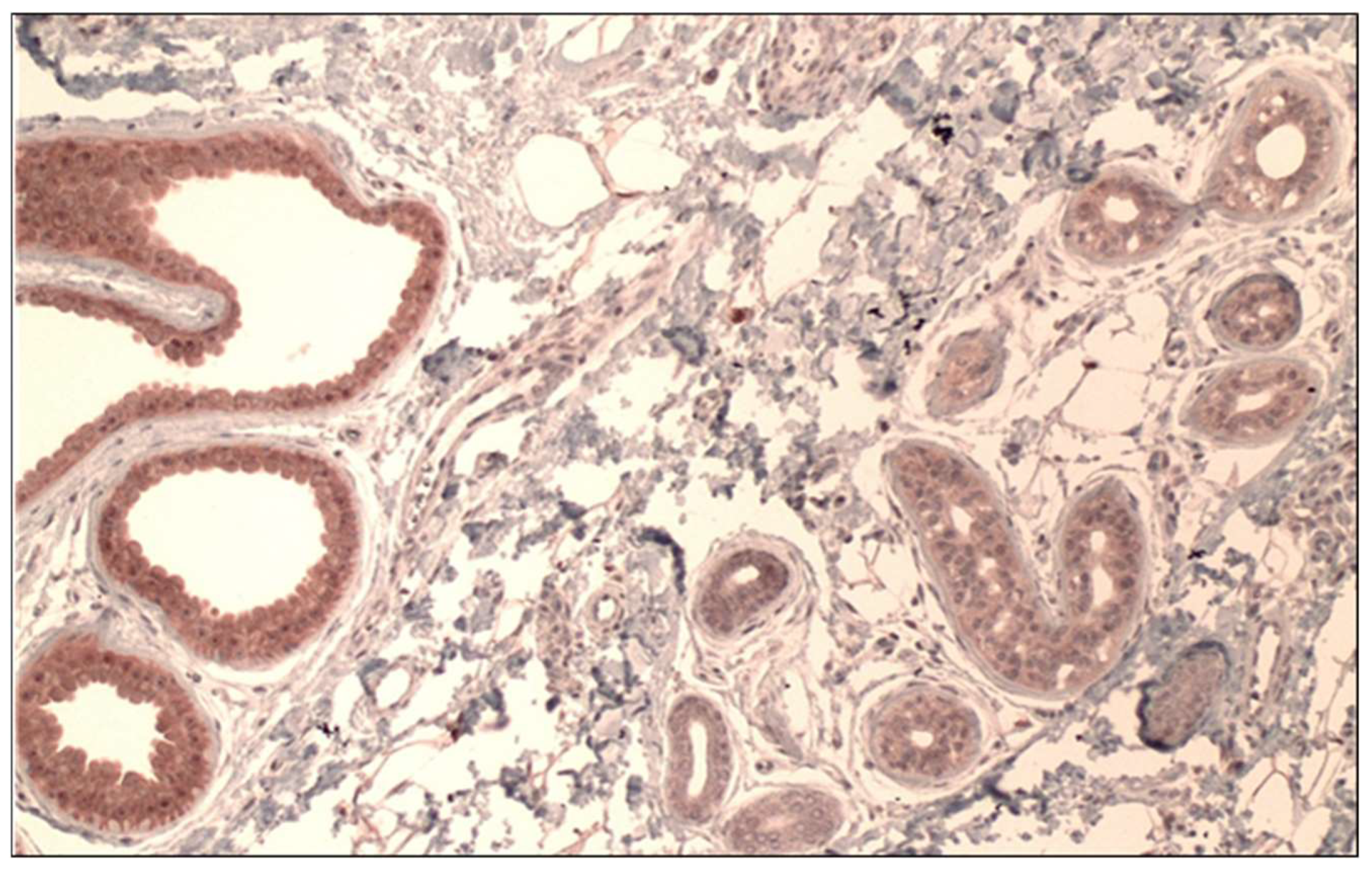
3.1. Optimization of Immunohistochemical Gankyrin Staining in Normal Skin

Initial studies with normal skin showed that gankyrin is expressed at a relatively low level in virtually all cells, including epithelial cells of epidermis and adnexae, as well as mesenchymal cells. This ubiquitous staining is expected because gankyrin is an assembly chaperone of the 26S proteasome and is present in all cells. The only cutaneous structures that we found stained considerably stronger than the ubiquitous background were the epithelial cells of apocrine glands, which showed strong diffuse cytoplasmic and nuclear labeling in all of 10 axillary skin samples (Figure 1). Thus, we used apocrine glands in axillary skin as positive controls. Based on these results, staining intensities were defined as follows: 1+ for intensity similar to the background intensity, 3+ for intensity similar to apocrine glands, and 2+ for an intermediate intensity. In the evaluation of the neoplasms, only staining intensities clearly above background (i.e., with intensity of 2+ or 3+) were scored as positive. Due to the ubiquitous background staining, an intensity of zero was essentially not encountered.
During these optimization studies, it turned out that the choice of the peroxidase substrate was an important factor. Comparing DAB (diaminobenzidine), AMEC Red, and AEC (3-amino-9-ethylcarbazole) as substrates, we found best results with AEC, for which an antibody dilution of 1:200 yielded strong staining of apocrine glands and mild background staining. DAB and AMEC Red were much more effective substrates, meaning that much higher antibody dilutions (in the range of 1:2000 to 1:4000) were necessary to keep the background staining low. At these high dilutions, the reactions tended to suffer from artefactual regional variation of staining intensity, which we attributed to effects of local antibody depletion. Therefore, AEC was used as the routine peroxidase substrate in this study.
With AEC as the peroxidase substrate, we were initially surprised that it did not result in the usual brick red reaction product but in a reddish brown product. This appears to be a peculiarity of the avidin-biotin complex (ABC) method that was employed in this study. Consultation of the product sheet and communication with the vendor (Craig Pow, Vector Laboratories, personal communication) confirmed that a reddish brown product is the expected result. A possible explanation is a metachromasia effect that was described by Koretz et al. and that is brought about by a second oxidation step of AEC under high antibody/peroxidase concentrations to yield a metachromatic reaction product [21].

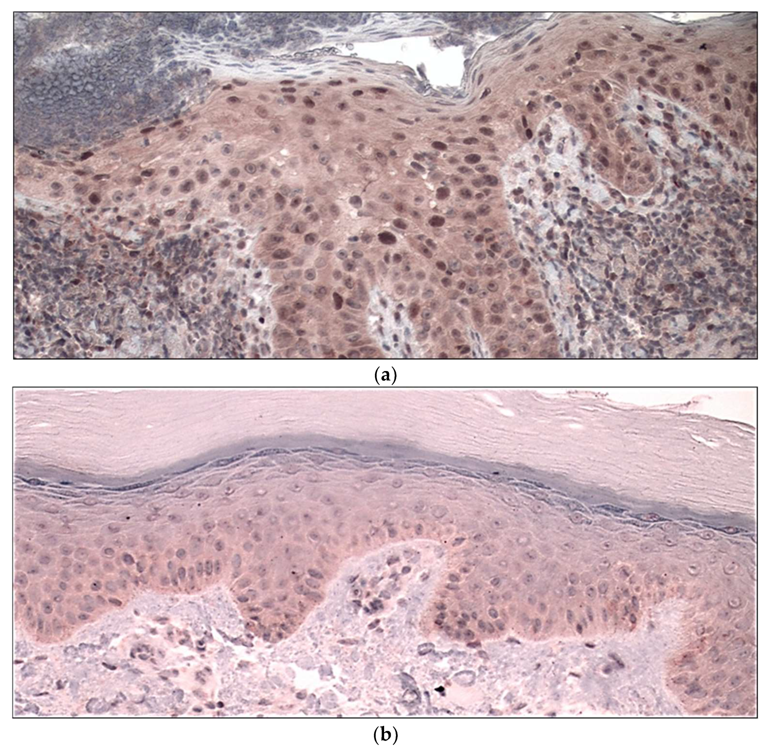
3.2. Gankyrin Staining in Squamous Cell Carcinomas and Basal Cell Carcinomas

In total, 3 of the 20 squamous cell carcinomas showed staining that was considerably stronger than background intensity (Figure 2, Table 1). All of these three cases were in situ carcinomas. The staining was predominantly nuclear in all 3 cases. None of the 13 invasive squamous cell carcinomas and none of 10 basal cell carcinomas demonstrated gankyrin overexpression. When inflammatory cells were present, they stained with a moderate intensity (Figure 2a).

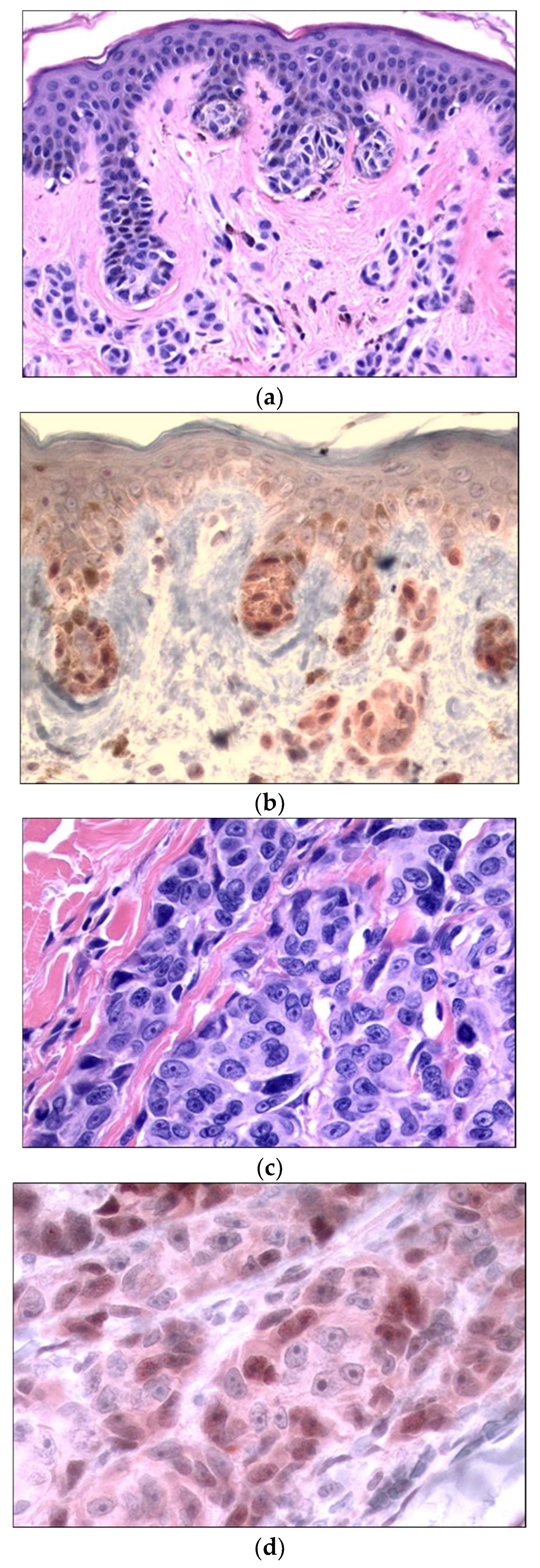
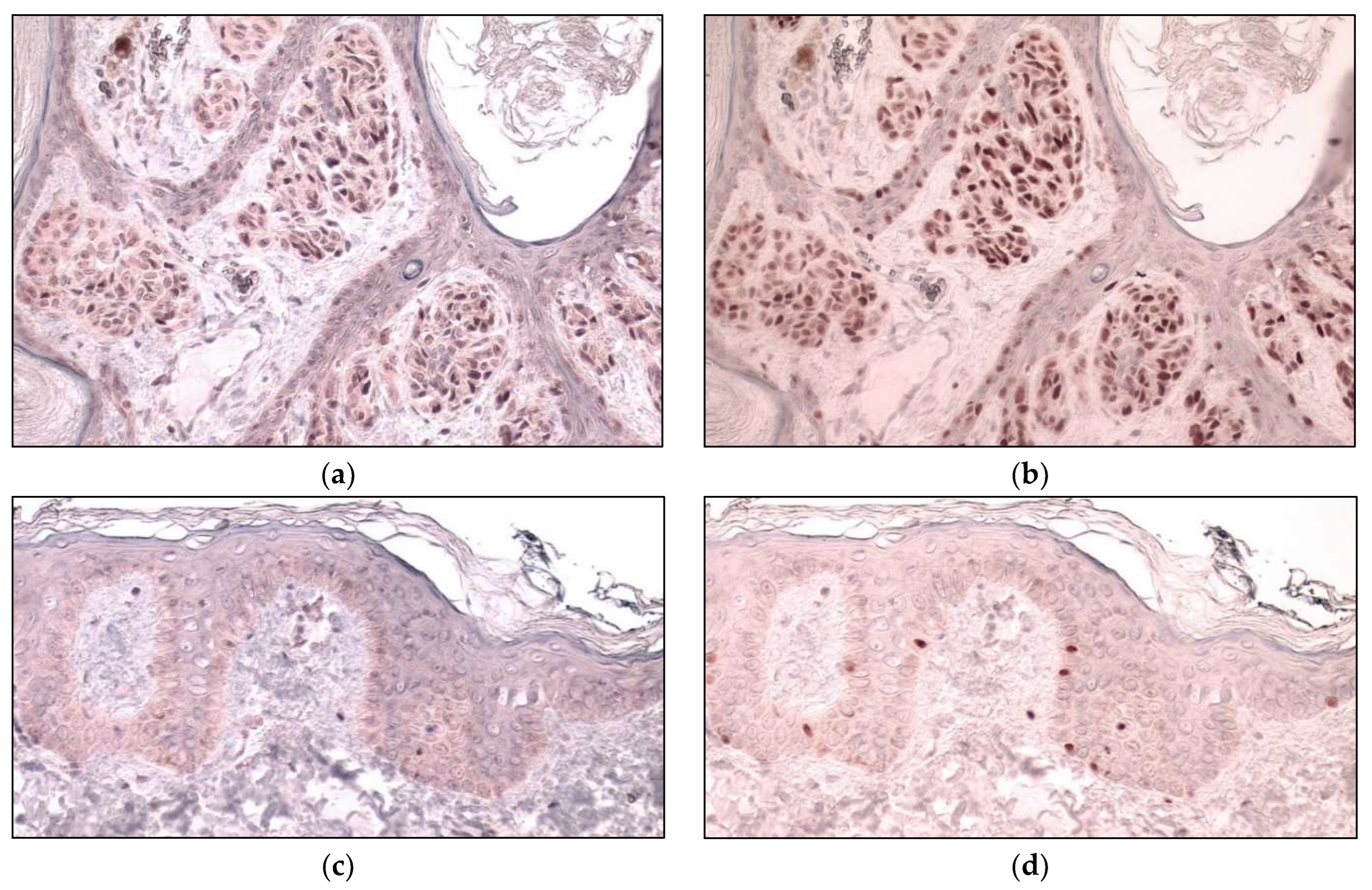
3.3. Gankyrin Staining in Melanocytic Neoplasms

Thirteen of 18 melanocytic nevi and 7 of 10 melanomas showed expression of gankyrin that was clearly stronger than the background staining of the surrounding epidermal keratinocytes (Figure 3, Table 1). In most nevi and melanomas, the staining intensity showed regional variation, and strongly positive areas alternating with weakly stained or negative areas could often be seen. The distribution of reactivity between nuclei and cytoplasm was variable. Predominantly nuclear staining was seen in 6 nevi and 5 melanomas, predominantly cytoplasmic staining in 4 nevi and 1 melanoma, and equal nuclear and cytoplasmic staining in 3 nevi and 1 melanoma. Both epidermal and dermal components were stained in nevi and melanomas, with a tendency for the epidermal component to show stronger reactivity than the dermal component. Normal melanocytes in epidermis uninvolved by nevus or melanoma expressed gankyrin to a much lesser degree than the neoplastic melanocytes. We confirmed the presence of normal melanocytes in uninvolved epidermis by an immunohistochemical stain for melanocytic marker SOX-10, and in parallel slides stained for gankyrin, we found only mildly stained melanocytes. To further demonstrate this point more directly, in two cases of melanocytic nevi, we performed sequential immunohistochemical stains in which the same slide was stained first for gankyrin, subsequently destained and then stained for SOX-10, permitting direct evaluation of gankyrin expression in melanocytes identified by SOX-10 labeling. The result of such a sequential staining for a melanocytic nevus and adjacent uninvolved skin is shown in Figure 4. It demonstrates that the SOX-10-positive neoplastic melanocytes in the nevus (4b) stain strongly for gankyrin (4a), whereas melanocytes in the adjacent epidermis identified by a SOX-10 stain (4d) show much weaker staining for gankyrin (4c).

4. Discussion

An innate complication of studying the expression of gankyrin is that it is expected to be present in all cells, corresponding to its household function as an assembly chaperone for the 26S proteasome, which is a ubiquitous cell component. Accordingly, we found that in normal skin, most cells, epithelial and mesenchymal, show a low level of gankyrin expression that causes an undesirable background staining, precluding the detection of low levels of overexpression. In this study, we only regarded staining as overexpression if it was clearly stronger than background, and we disregarded staining with an intensity equal to or slightly above background. Although, with this policy we may have missed subtle expression in some neoplasms, we nevertheless adhered to it to avoid over-interpretation of our results.
In normal skin, we found that apocrine glands were the only structures that showed strong expression of gankyrin significantly exceeding background intensity (Figure 1), and, therefore, apocrine glands were used as a convenient positive control. To the best of our knowledge, this finding has not been reported before and appears unique as we are not aware of any other organ or organelle where similarly strong expression of gankyrin has been reported in the literature. Expression of gankyrin has been found in spermatocytes of human testes [22], but in our experience, this testicular expression is not as strong as the expression in apocrine glands (results not shown). The physiological significance of the prominent gankyrin expression in apocrine glands is unknown. Since gankyrin is a chaperone for assembly of the proteasome, we were interested in whether the expression of gankyrin would be paralleled by strong proteasome expression. Using an antibody against the proteasome subunit rpn1, we did not find significant overexpression (own unpublished results).
The main objective of this study was to determine the expression of gankyrin in cutaneous melanocytic neoplasms. We found that gankyrin was frequently overexpressed in melanocytic neoplasms, but only rarely in squamous cell carcinoma (3 out of 20 cases) and in none of the 10 basal cell carcinomas (Table 1).
Of the squamous cell carcinomas, the 3 positive cases were all in situ carcinomas, which raises the possibility that in some cases gankyrin may function as a tumor-promoting protein in the early stages of squamous malignancies. As p53 has a prominent role in the initiation of cutaneous squamous cell carcinomas [23], our results are consistent with the assumption that gankyrin promotes squamous neoplasms by its capacity to antagonize p53.
We found gankyrin to be frequently expressed in melanocytic neoplasms (Table 1). Our initial hypothesis had been that gankyrin would be upregulated in melanomas relative to nevi, which would have supported a direct role in malignant transformation. Rather, we found overexpression of gankyrin not only in melanomas but also in nevi (Figure 3, Table 1), whereas the expression in normal melanocytes was much weaker than in neoplastic melanocytes (Figure 4). This supports a role of gankyrin in formation of nevi, similar to what has been proposed for the oncogene B-RAF, which is mutated both in nevi and melanomas [24]. This does not mean that gankyrin is not involved in the formation of melanomas, but its effect in this respect would likely be an indirect one by facilitating the action of other effector molecules. An obvious candidate for such an effector molecule is CDK4/6, which is expressed more strongly in melanomas than in nevi according to some authors [25,26] but not others [27]. The high expression of p16 in most nevi poses a high barrier towards transformation into melanoma, and to overcome this barrier would require a considerable overexpression of CDK4/6. However, in the presence of gankyrin, this barrier would be expected to be much lower, and to overcome it would require a relatively small increase in nuclear CDK4/6 (e.g., by overexpression or by cytoplasmic-to-nuclear import) to initiate the cell cycle and to facilitate the transformation into early melanoma.
We found a frequent nuclear location of gankyrin in squamous cell carcinoma in situ, nevi and melanomas (Figure 2a and Figure 3b). This location is consistent with its proposed role as a tumor-promoting protein. As the anti-proliferative effect of pRB, p53, and p16 is primarily dependent on their function in the nucleus [28,29], nuclear gankyrin would be in a suitable location to antagonize these tumor suppressors. In addition to its nuclear location, we also found gankyrin in a cytoplasmic location (Figure 3d). Although cytoplasmic gankyrin could represent a sequestered inactive pool, it may have additional functions by antagonizing cytoplasmic p53 and CDK4. The p53 protein, when located in the cytoplasm, can directly participate in apoptosis, autophagy, membrane trafficking, and cell metabolism (reviewed in [28]), and it is possible that cytoplasmic gankyrin antagonizes these functions. Cytoplasmic CDK4 participates in assembly of the CDK4-cyclin D-p21/p27 complex, which is assembled in the cytoplasm and then transported to the nucleus guided by the nuclear localization sequence that is present in p21 and p27 (but not in CDK4 or cyclin D) [29]. While cytoplasmic p16 disrupts this complex and thus blocks the translocation to the nucleus [29], gankyrin can form a complex with Cyclin D2 and CDK4 [12] (but not with cyclin D1 or cyclin D3 [30]). This complex would likely also contain p21 or p27 and thus be transported to the nucleus. If this is true, one would expect that overexpression of cytoplasmic gankyrin would result in nuclear accumulation of cyclin D2. To the best of our knowledge, this has not been tested experimentally.
The expression of gankyrin in melanocytic neoplasms raises the possibility but does not prove that it promotes the neoplastic transformation of melanocytes. To date, gankyrin’s tumor-promoting activity has only been demonstrated in non-melanocytic cultured cell lines, where its forced overexpression promotes cell proliferation and results in increased degradation of p53 and hyperphosphorylation of pRB, with reduction in expression of p53 transactivation targets p21 and mdm2 [4,9]. It would be important to determine whether gankyrin has the same effects in cultured melanocytes, and this could also be addressed by studies on archival pathologic material of melanocytic neoplasms by determining whether expression of gankyrin correlates with expression of p53, p21, and mdm2, and with the phosphorylation status of pRB. Furthermore, competition of gankyrin with p16 for binding to CDK4 has been demonstrated in vitro [11,12,13], but whether this mechanism is operative in vivo in the formation of melanocytic neoplasms is unclear. In this respect, it would be important to study co-expression of gankyrin and p16 because co-expression in the same cell would be a prerequisite for competition. In melanocytic nevi, p16 typically assumes a mosaic expression pattern [18], and in some cases we observed a similar mosaic pattern for gankyrin expression (Figure 3d). It would therefore be of interest to investigate whether or not these patterns coincide. Furthermore, expression of p16 is frequently lost with progression of melanoma [20], and if competition with p16 were an important mechanism for the tumor-promoting activity of gankyrin, loss of p16 expression would—assuming a Darwinian tumor evolution model—result in loss of selection pressure to keep gankyrin expressed. Therefore, if loss of p16 expression were paralleled by a loss of gankyrin expression, this would strengthen the interpretation that gankyrin has a tumor-promoting role. Finally, it would also be of interest to study the co-expression of gankyrin and MAGE-A4, because the latter has been shown to interact with gankyrin, and this is correlated with the suppression of anchorage-independent growth [31].
Competition with gankyrin for CDK4 has been only reported for p16 [12,13] but not for the other members of a wider group of similar CDK-inhibitors that also includes p15, p18, and p19 (INK4 proteins). As all four INK4 proteins have a similar gankyrin repeat structure [32], it is likely that they will bind to the same site on CDK4 as p16 and gankyrin, implying that gankyrin can be expected to antagonize the function of all four INK4 proteins. In this context it is interesting to note that p15 is expressed in melanocytic nevi and in melanomas (less frequently in the latter) [33,34], raising the possibility that gankyrin competes with p15 for binding to CDK4, similar to the proposed competition with p16. However, because p15 is infrequently mutated in sporadic and familial melanomas, it is more difficult to implicate p15 in the process of melanoma formation.
In summary, our study raises the possibility that gankyrin may be involved in the formation of melanocytic nevi and may modulate the molecular landscape of melanoma formation. However, additional studies need to be done before such a role can be firmly accepted.

Author Contributions

Conceptualization, T.E.; methodology, writing: S.M. and T.E. All authors have read and agreed to the published version of the manuscript.

Funding

This work was supported by Hartford Hospital Medical Staff Grant HHC-2021-003.

Institutional Review Board Statement

Reviewed and approved by Hartford Health Care IRB; approval #HHC-2021-003.

Informed Consent Statement

Not applicable.

Data Availability Statement

Not applicable.

Conflicts of Interest

The authors declare no conflict of interest.

References

  1. Hori, T.; Kato, S.; Saeki, M. cDNA cloning and functional analysis of p28 (Nas6p) and p40.5 (Nas7p), two novel regulatory subunits of the 26S proteasome. Gene 1998, 216, 113–122. [Google Scholar] [CrossRef] [PubMed]
  2. Tomko, R.J.; Hochstrasser, M. Order of proteasomal ATPases and eukaryotic proteasome assembly. Cell Biochem. Biophys. 2011, 60, 13–20. [Google Scholar] [CrossRef] [PubMed] [Green Version]
  3. Kaneko, T.; Hamazaki, J.; Iemura, S.I. Assembly pathway of the mammalian proteasome base subcomplex is mediated by multiple specific chaperones. Cell 2009, 137, 914–925. [Google Scholar] [CrossRef] [Green Version]
  4. Higashitsuji, H.; Itoh, K.; Nagao, Y. Reduced stability of retinoblastoma protein by gankyrin, an oncogenic ankyrin-repeat protein overexpressed in hepatomas. Nat. Med. 2000, 6, 96–99. [Google Scholar] [CrossRef]
  5. Li, H.; Zhang, J.; Zhen, C. Gankyrin as a potential target for tumor therapy: Evidence and perspectives. Am. J. Transl. Res. 2018, 10, 1949–1960. [Google Scholar] [PubMed]
  6. Kashyap, D.; Varshney, N.; Parmar, H.S. Gankyrin: At the crossroads of cancer diagnosis, disease prognosis, and development of efficient cancer therapeutics. Adv. Cancer Biol. —Metastasis 2022, 4, 100023. [Google Scholar] [CrossRef]
  7. Dawson, S.; Higashitsuji, H.; Wilkinson, A.J. Gankyrin: A new oncoprotein and regulator of pRb and p53. Trends Cell Biol. 2006, 16, 229–233. [Google Scholar] [CrossRef] [PubMed]
  8. Mayer, R.J.; Fujita, J. Gankyrin, the 26S proteasome, the cell cycle and cancer. Biochem. Soc. Trans. 2006, 34, 746–748. [Google Scholar] [CrossRef]
  9. Higashitsuji, H.; Higashitsuji, H.; Itoh, K. The onkoprotein gankyrin binds to MDM2/HDM2, enhancing ubiquitylation and degradation of p53. Cancer Cell 2005, 8, 75–87. [Google Scholar] [CrossRef] [Green Version]
  10. Higashitsuji, H.; Liu, Y.; Mayer, R.J. The oncoprotein gankyrin negatively regulates both p53 and RB by enhancing proteosomal degradation. Cell Cycle 2005, 4, 1335–1337. [Google Scholar] [CrossRef]
  11. Krzywda, S.; Brzozowski, A.M.; Higashitsuji, H. The crystal structure of gankyrin, an oncoprotein found in complexes with cyclin-dependent kinase 4, a 19S proteosomal ATPase regulator, and the tumor suppressors Rb and p53. J. Biol. Chem. 2004, 279, 1541–1545. [Google Scholar] [CrossRef] [PubMed] [Green Version]
  12. Li, J.; Tsai, M.D. Novel insights into the INK4-CDK4/6-Rb pathway: Counter action of gankyrin against INK4 proteins regulates the CDK4-mediated phosphorylation of Rb. Biochemistry 2002, 41, 3977–3983. [Google Scholar] [CrossRef] [PubMed]
  13. Mahajan, A.; Guo, Y.; Yuan, C. Dissection of protein-protein interaction and CDK4 inhibition in the oncogenic versus tumor suppressing functions of gankyrin and p16. J. Mol. Biol. 2007, 373, 990–1005. [Google Scholar] [CrossRef] [Green Version]
  14. D’Souza, A.M.; Cast, A.; Kumbaji, M. Small molecule cjoc42 improves chemo-sensitivity and increases levels of tumor suppressor proteins in hepatoblastoma cells and in mice by inhibiting oncogene gankyrin. Front. Pharmacol. 2021, 12, 580722. [Google Scholar] [CrossRef]
  15. Kanabar, D.; Goyal, M.; Kane, E.I. Small-molecule gangkyrin inhibition as a therapeutic strategy for breast and lung cancer. J. Med. Chem. 2022, 65, 8975–8997. [Google Scholar] [CrossRef]
  16. Sudharsan, M.; Chikhale, R.; Nanaware, P.P. A druggable pocket on PSMD10/gankyrin that can accommodate an interface peptide and doxorubicin. Eur. J. Pharmacol. 2022, 915, 174718. [Google Scholar]
  17. Aoude, L.G.; Wadt, K.A.; Pritchard, A.L. Genetics of familial melanoma: 20 years after CDKN2A. Pigment. Cell Melanoma Res. 2015, 28, 148–160. [Google Scholar] [CrossRef]
  18. Michaloglou, C.; Vredeveld, L.C.W.; Soengas, M.S. BRAFE600-associated senescence-like cell cycle arrest of human naevi. Nature 2005, 436, 720–724. [Google Scholar] [CrossRef] [Green Version]
  19. Bennett, D.C. Genetics of melanoma progression: The rise and fall of cell senescence. Pigment Cell Mel. Res. 2016, 29, 122–140. [Google Scholar] [CrossRef] [Green Version]
  20. Ming, Z.; Lim, S.J.; Rizos, H. Genetic alterations in the INK4A/ARF locus: Effects on melanoma development and progression. Biomolecules 2020, 10, 1447. [Google Scholar] [CrossRef]
  21. Koretz, K.; Leman, J.; Brandt, I. Metachromasia of 3-amino-9-ethylcarbazole (AEC) and its prevention in immunoperoxidase techniques. Histochemistry 1987, 86, 471–478. [Google Scholar] [CrossRef] [PubMed]
  22. Ando, S.; Matsuoka, T.; Kawai, K. Expression of the oncoprotein gankyrin and phosphorylated retinoblastoma protein in human testis and testicular germ cell tumor. Int. J. Urol. 2014, 21, 992–998. [Google Scholar] [CrossRef] [Green Version]
  23. Piipponen, M.; Riihila, P.; Nissinen, L. The role of p53 in progression of cutaneous squamous cell carcinoma. Cancers 2021, 13, 4507. [Google Scholar] [CrossRef] [PubMed]
  24. Newton-Bishop, J.A.; Bishop, D.T.; Harland, M. Melanoma Genomics. Acta Derm. Venereol. 2020, 100, adv00138. [Google Scholar] [CrossRef]
  25. Wang, Y.L.; Uhara, H.; Yamazaki, Y. Immunohistochemical detection of CDK4 and p16INK4 proteins in cutaneous malignant melanoma. Br. J. Dermatol. 1996, 134, 269–275. [Google Scholar] [CrossRef]
  26. Lee, W.J.; Skalamera, D.; Dahmer-Heath, M. Genome-wide overexpression screen identified genes able to bypass p16-mediated senescence in melanoma. SLAS Discov. 2017, 22, 298–308. [Google Scholar] [CrossRef] [Green Version]
  27. Georgieva, J.; Sinha, P.; Schadendorf, D. Expression of cyclins and cyclin dependent kinases in human benign and malignant melanocytic lesions. J. Clin. Pathol. 2001, 54, 229–235. [Google Scholar] [CrossRef] [Green Version]
  28. Comel, A.; Sorrentino, G.; Capaci, V. The cytoplasmic side of p53’s oncosuppressive activities. FEBS Lett. 2014, 588, 2600–2609. [Google Scholar] [CrossRef] [Green Version]
  29. Sherr, C.J.; Roberts, J.M. CDK inhibitors: Positive and negative regulators of G1-phase progression. Genes Dev. 1999, 13, 1501–1512. [Google Scholar] [CrossRef] [Green Version]
  30. Dawson, S.; Apcher, S.; Mee, M. Gankyrin is an ankyrin-repeat oncoprotein that interacts with CDK4 kinase and the S6 ATPase of the 26 S proteasome. J. Biol. Chem. 2002, 277, 10893–10902. [Google Scholar] [CrossRef] [Green Version]
  31. Nagao, T.; Higashitsuji, H.; Nonoguchi, K. MAGE-A4 interacts with the liver oncoprotein gankyrin and suppresses its tumorigenic activity. J. Biol. Chem. 2003, 278, 10668–10674. [Google Scholar] [CrossRef] [PubMed]
  32. Canepa, E.T.; Scassa, M.E.; Ceruti, J.M. INK4 proteins, a family of mammalian CDK inhibitors with novel biological functions. IUBMB Life 2007, 59, 419–426. [Google Scholar] [CrossRef] [PubMed]
  33. Taylor, L.A.; O’Day, C.; Dentchev, T. p15 expression differentiates nevus from melanoma. Am. J. Pathol. 2016, 186, 3094–3099. [Google Scholar] [CrossRef] [Green Version]
  34. Ma, S.A.; O’Day, C.; Dentchev, T. Expression of p15 in a spectrum of spitzoid melanocytic neoplasms. J. Cutan. Pathol. 2019, 46, 310–316. [Google Scholar] [CrossRef] [PubMed]
Figure 1. Expression of gankyrin in axillary skin. Apocrine glands (on the left) exhibit strong expression whereas eccrine glands (on the right) show background intensity. Original magnification 100×.
Jmp 03 00027 g001
Figure 2. Expression of gankyrin in a positive squamous cell carcinoma in situ (a) compared to normal epidermis (b). The gankyrin expression in the carcinoma is predominantly nuclear. The normal epidermis demonstrates weak background reactivity for gankyrin. Some of the inflammatory cells in the dermis stain with moderate intensity (a). Original magnifications 200×.
Jmp 03 00027 g002
Figure 3. H&E (a,c) and gankyrin expression (b,d) in a melanocytic nevus (a,b) and in a nodular melanoma (c,d). The localization of gankyrin is predominantly nuclear in the nevus, and both nuclear as well as cytoplasmic in the melanoma. Original magnifications 200× (a,b), 400× (c,d).
Jmp 03 00027 g003
Figure 4. Expression of gankyrin in normal melanocytes versus neoplastic melanocytes. Sequential dual immunohistochemical staining was performed on the same slide for gankyrin (a,c) and SOX-10 (b,d) in a nevus (a,b) and in the adjacent uninvolved skin (c,d). Melanocytes identified by SOX-10 expression on average stain more strongly for gankyrin in the nevus than in normal skin. (a,b) 200×; (c,d) 400×.
Jmp 03 00027 g004
Table 1. Gankyrin immunoreactivity in cutaneous neoplasms.
Number
of Cases
NegativePositive
1+2+3+Total(%)
Melanocytic nevus18510313 a,b(72)
Banal compound nevus70437
Spitz nevus42202
Congenital nevus32101
Dysplastic nevus41303
Melanoma103527 c,d(70)
In situ42112
Invasive61415
Basal cell carcinoma1010000 a,d(0)
Squamous cell carcinoma2017303 b,c(15)
In situ74303
Invasive1313000
An intensity of 1+ corresponds to the ubiquitous background reaction present in every cell and was therefore considered as negative. Data of negative and positive pairs designated by superscripts a, b, c, and d are significantly different from each other (p < 0.01).
Publisher’s Note: MDPI stays neutral with regard to jurisdictional claims in published maps and institutional affiliations.

