Proceeding Paper

Potential Hazard to Human and Animal Health from Bacterial and Fungal Contaminants in Small Freshwater Reservoirs †

1
Department of Biology and Environment, University of Trás-os-Montes and Alto Douro (UTAD), Quinta de Prados, 5000-801 Vila Real, Portugal
2
Centre for the Research and Technology of Agro-Environmental and Biological Sciences (CITAB), UTAD, Quinta de Prados, 5000-801 Vila Real, Portugal
*
Author to whom correspondence should be addressed.
Presented at the 4th International Electronic Conference on Environmental Research and Public Health—Climate Change and Health in a Broad Perspective, 15–30 October 2022; Available online: https://ecerph-4.sciforum.net/.
Environ. Sci. Proc. 2022, 24(1), 7; https://doi.org/10.3390/ECERPH-4-13071
Published: 17 October 2022

Abstract

:
In general, the assessment of microbiological quality in aquatic systems focuses on the presence of some bacterial groups or species. Although quantification of fungi presence is not a mandatory parameter, recently the WHO advises its detection/quantification. Its concentration and diversity varies greatly among the various types of aquatic systems. Fungi are mesophilic, dependent on organic matter to grow and their presence can be associated with pollution. Depending on their concentration and diversity, fungi may pose a risk to human and animal health. The objective of the present work was to evaluate the presence of some bacterial indicators (Escherichia coli, fecal enterococci, among others) and fungi (total yeasts and molds) in freshwater reservoirs (water tanks) with different sources, sun exposures and anthropogenic and animal influences. Additionally, it was intended to assess the diversity of molds. For this, filamentous colonies were isolated, purified, and morphologically identified (whenever possible to the genus). The three tanks differed in bacterial (presence of Escherichia coli, fecal enterococci, Proteus sp. and Staphylococcus aureus) and fungal (total and mold) presence. Regarding molds, 16 different taxa were identified and, depending on the water tank, the Penicillium, Aspergillus and Fusarium genera and the Chytridiomycota phylum were the most representative. Some of the taxa isolated may pose a risk to human and animal health (Trichophyton, Aspergillus fumigatus and some dematiaceous). The water reservoirs presented different fungal communities. Although preliminary, the results show that freshwater tanks can be a source of potentially pathogenic bacteria and fungi to humans and animals that use them.

1. Introduction

Fresh water has high microbial diversity, as microorganisms play a fundamental role in the nutrient cycle and in the purification of aquatic ecosystems. However, they can also cause disorders and be pathogenic for humans, animals and other organisms. The classic parameters used are fecal indicator organisms (FIO) associated to pollution of fecal origin are fecal coliforms from the Enterobacteriaceae family, fecal enterococci and clostridia, although there are other organisms such as helminths or protozoa can also pose health risks to humans [1,2]. Among these indicators, fecal coliforms are widely used, being associated with a high number of human intestinal infections, and involved or being participants in pathologies such as meningitis, urinary tract infections and nosocomial pneumonias [3]. However, recent studies pointed out that these organisms may not be sufficient as indicator organisms, and it may be necessary to assess the presence of others such as filamentous fungi and yeasts. It is therefore important to know the presence of these organisms in their counts, distribution, diversity and behavior, in different aquatic environments [4,5]. Fungi are heterotrophic eukaryotes widely distributed in nature. They are present in soil, air, organic matter and water, especially in untreated water, reservoirs and distribution systems. Several authors have reported the presence of yeasts and molds in the aquatic environment, the latter being found in greater numbers [6,7]. Among many different taxa of fungi found in aquatic environments, several species are opportunistic or pathogenic, produce toxins and are allergenic. Most of these species belong to the phyla Ascomycota, Zygomycota and Chytridiomycota. The fungi with the most significant presence are Aspergillus, Penicillium, Fusarium, Clasdosporium and Curvularia, and some of them increase the risk of diseases in humans and animals [7,8]. Although most of the time fungal infections or mycoses do not result in the death of patients, they can be a public health problem. The most common mycoses are caused by dermatophytes that affect the skin, hair and nails, and are very contagious [9,10]. Hyaline, dematiaceous and dimorphic fungi can affect various tissues and organs causing severe mycoses [11,12]. Systemic mycoses are difficult to treat and have an unpredictable prognosis, especially in immuno-incompetent populations.
The main objective of the present work was to evaluate the presence of FIO (E. coli, fecal enterococci), other bacterial species associated to humans and animals and fungi (mainly molds) in freshwater reservoirs from three different sources, sun exposures, and anthropogenic and animal influences.

2. Materials and Methods

2.1. Localization of Water Tanks

The present study was carried out between April and June 2022. All the tanks are located on the Campus of the University of Trás-os-Montes e Alto Douro (UTAD), in Vila Real (Latitude: 41.2885° N; Longitude: 7.7391° W; Altitude: 462 m), and are close to each other (115 to 270 m apart). Tanks 1 and 2 are mostly fed by natural founts and are nearby foot paths and pastures, while tank 3 is fed by rainwater, and in an inner courtyard with restricted access (Figure 1).
Tank 1 water was cloudy with cherry blossoms, and although surrounded by vegetation, it had the cleanest water compared to the other tanks. In tank 2, the water was covered with macroalgae, filamentous microalgae and aquatic plants such as Lemna sp. Tank 3 had very turbid water, green in color due to the massive growth of microalgae, it was surrounded by vegetation and did not get direct sunlight.

2.2. Culture Media

In the evaluation of the water samples microbiology the following culture media were used: (a) for bacteria: Slanetz & Bartley Agar (Slanetz, Oxoid, Hants, UK) for fecal enterococci, Chromogenic Coliform Agar (Chromo, Oxoid) for Escherichia coli and other fecal coliforms; Cysteine Lactose Electrolyte-Deficient Agar with Andrade indicator (C.L.E.D., Liofilchem, Roseto, Italy) for bacteria that cause urinary infections (S. aureus, E. coli and Proteus vulgaris); (b) for fungi: Yeast Malt agar (YMA, Liofilchem), Yeast Glucose Chloramphenicol Agar (YGCA, Himedia, Mumbai, India), Mycosel Agar (Mycosel, Liofilchem) for dermatophytes; and Potato Dextrose Agar (PDA, Liofilchem), for molds isolation and maintenance. All media were prepared according to the manufacturers’ specifications.

2.3. Water Sampling and Microbiological Analyses

For each tank, three independent water samples were collected using 500 mL sterilized plastic bottles. Each bottle was quickly submerged approximately 30 cm deep, except for tank 3 which was shallower. The samples were taken on 20 April (tanks 1 and 2, air temperature 12 °C, water temperature 8 °C) and 9 May (tank 3, air temperature 21 °C; water temperature 10 °C), between 10:30 and 11:00 a.m. For the detection of bacteria, the membrane filtration technique was used, filtering 100 mL of water per filter (0.45 µm pore). For the quantification of fungi, in addition to the membrane filtration technique, the spread of a small volume (100 or 200 µL per plate) of the sample on a Petri dish surface was used. The media were incubated at 37 °C (bacteria and fungi) and 25 °C (fungi). After 24–48 h of incubation (bacteria) or 2, 5 and 7 days (fungi) the colony forming units (UFC) were counted and expressed as UFC/100 mL or UFC/mL, respectively, for the membrane and the spread techniques. In the case of fungi, all morphologically distinct colonies were quantified, isolated, purified and maintained (in PDA) until their identification.

3. Results and Discussion

3.1. Water Samples Bacterial Load

In the Chromo medium, large numbers of total coliforms (uncountable, (>300 CFU/100 mL), E. coli, and other colonies of yellow and white color that probably correspond to Actinobacteria and yeasts were detected. In Slanetz medium, only red colonies were detected, which indicate the presence of fecal enterococci. The maximum concentration of E. coli and fecal enterococci was obtained in tank 3, with respectively, 191 and 282 CFU/100 mL of water (Table 1), values above the recommended guideline for recreational water (the European Union threshold 100 CFU/mL for E. coli and the WHO guideline 200 CFU/100 mL for fecal enterococci [10]). The simultaneous presence of E. coli and fecal enterococci may indicate recent fecal contamination, as fecal enterococci are able to survive in water longer than E. coli [13,14]. In C.L.E.D. medium used in the clinic, the presence of S. aureus, Proteus spp., yeasts and Actinobacteria was detected.
It would be expected, due to their locations and easy access for people and animals, that tanks 1 and 2 would present higher values of FIO than tank 3, located in an interior courtyard. Additionally, tanks 1 and 2 collect water from founts and rain, unlike tank 3 fed exclusively by rainwater. However, the results point out a higher bacterial load in tank 3 than can be explained by the many bird droppings observed on site, a lower volume of water and a lower rate of water renewal (high retention time).
S. aureus indicates human presence (the inner courtyard is surround by offices, a museum and a laboratory). In all the three tanks, Proteus was the most represented bacteria. This Enterobacteriaceae is a saprophytic mostly associated with animal organic matter, and is present in the mammalian gastrointestinal tract. Additionally, it is often associated or responsible for infections in the urinary tract [15].

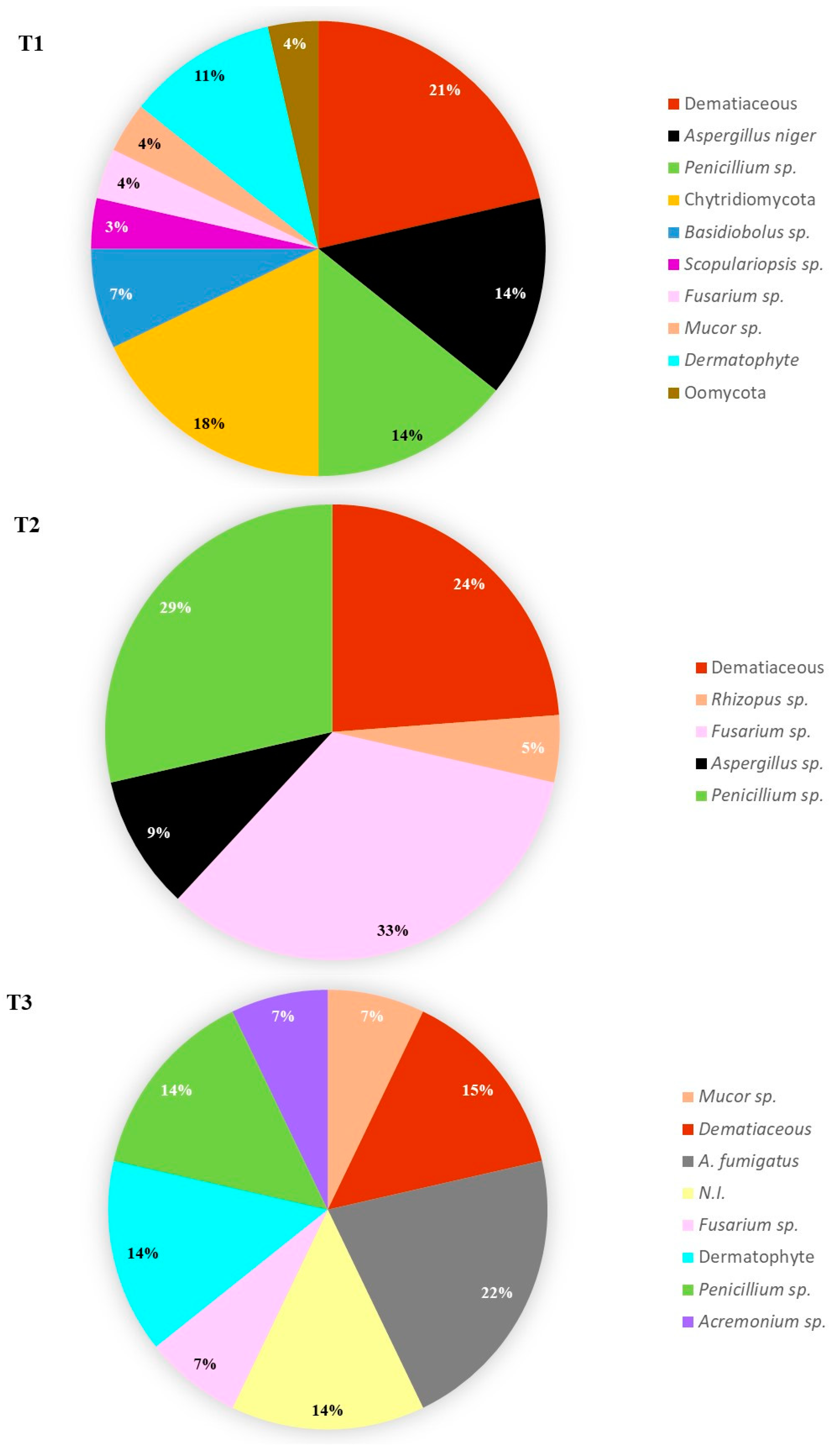
3.2. Water Samples Fungal Load

Overall, the total fungi concentrations were low [0–100 UFC/100 mL] and [0–100 UFC/mL], depending on the incubation temperature and the quantification method, and highly variable within the sampling replicas. In general, the highest fungal loads were obtained by the inoculum spreading method, compared to the membrane filtration method. Furthermore, and on average, at 25 °C more colonies of yeasts and molds grew than at 37 °C. In addition, with YGCA, more fungi were recovered, both at 25 °C and 37 °C. At 37 °C, there was no growth of yeasts in any of the tanks and there was only growth of molds in tanks 1 and 3. In Mycosel, it showed more growth at 25 °C, while at 37 °C there was only growth of molds in tank 3.
In all media molds dominate over yeasts. Yeasts were not recovered from tank 3. This pattern is generally observed in waters from different sources [16,17].
The analysis of the frequency of molds found in the ponds is shown in Figure 2. Clearly, the diversity of fungi and the prevalence of taxa varied among the three water reservoirs. Tank 1 presented higher diversity than tank 3 and tank 2. Among the identified genera, the ones common to the three reservoirs were also the most frequently observed: Penicillium (14.3–28.6%), Aspergillus (9.5–21.4%) and Fusarium (3.6–33.3%). These results are in agreement with previous works in fresh water [10,18]. Aspergillus, the second most abundant genus in this work, was reported as the most frequent in other studies [19].
The genera Penicillium and Aspergillus were particularly isolated and include species that can be allergenic or cause human infections. These two genera can be found in environmental samples (soil, water, rhizosphere and air) and produce large amounts of spores [20].
The dematiaceous group had an expressive prevalence in water tanks (21, 24 and 15%, respectively for tanks 1, 2 and 3) with several genera identified: Phialophora, Fonsecaea, Rhinocladiella, Ulocladium and Stachybotrys. Some taxa were isolated only in one of the tanks: Chytridiomycota, Basidiobolus sp., Scopulariopsis sp. and Oomycota (tank 1), Rhizopus sp. (tank 2) and Acremonium sp. (tank 3). Dermatophytes were isolated only from tanks 1 (11%) and 3 (14%).
It is important to note that several of the identified taxa are of clinical interest: Basidiobolus sp. and several dermatophyte species are pathogens [21], and although Aspergillus spp. are opportunistic fungi, A. fumigatus is responsible for approximately 90% of diagnosed invasive aspergillosis [22].

4. Conclusions

Small freshwater constructions such as tanks are very frequent in green spaces managed by man, due to their useful, recreational, pleasant and aesthetic values. However, their microbiology is poorly understood. Our results point to the presence of potential hazard microorganisms in freshwater tanks. The relationship between FIO/other bacteria and bacteria/fungi should be further studied to better understand their significance and potential risks in these largely ignored constructions.

Author Contributions

Conceptualization, resources, writing—review and editing, supervision, project administration and funding acquisition, A.S.; methodology, software, data curation, formal analysis and writing—original draft preparation, A.V.M. and A.S.; investigation, A.V.M. All authors have read and agreed to the published version of the manuscript.

Funding

This work was funded by the Foundation for Science and Technology (FCT, Portugal) and FEDER under Programe PT2020 for financial support to CITAB (UID/AGR/04033/2020).

Institutional Review Board Statement

Not applicable.

Informed Consent Statement

Not applicable.

Data Availability Statement

The raw data are available upon request. Please contact the corresponding author.

Conflicts of Interest

The authors declare no conflict of interest.

References

  1. Taylor, H. Surface waters. In Handbook of Water and Wastewater Microbiology; Mara, D., Horan, N., Eds.; Academic Press: Cambridge, MA, USA, 2003; pp. 611–626. [Google Scholar]
  2. Ashbolt, N.J.; Grabow, W.O.; Snozzi, M. Indicators of microbial water quality. In Water Quality: Guidelines, Standards and Health: Assessment of Risk and Risk Management for Water-Related Infectious Disease; Fewtrell, L., Bartram, J., Eds.; IWA Publishing: London, UK, 2001; pp. 289–316. [Google Scholar]
  3. Kator, H.; Rhodes, M. 8—Detection, enumeration, and identification of environmental microorganisms of public health significance. In Handbook of Water and Wastewater Microbiology; Mara, D., Horan, N., Eds.; Academic Press: Cambridge, MA, USA, 2003; pp. 113–144. [Google Scholar]
  4. Hageskal, G.; Knutsen, A.K.; Gaustad, P.; de Hoog, G.S.; Skaar, I. Diversity and significance of mold species in Norwegian drinking water. Appl. Environ. Microbiol. 2006, 72, 7586–7593. [Google Scholar] [CrossRef] [PubMed] [Green Version]
  5. Pereira, V.J.; Basílio, M.C.; Fernandes, D.; Domingues, M.; Paiva, J.M.; Benoliel, M.J.; San Romão, M.V. Occurrence of filamentous fungi and yeasts in three different drinking water sources. Water Res. 2009, 43, 3813–3819. [Google Scholar] [CrossRef] [PubMed]
  6. Hoffman, J.J.; Burton, M.J.; Leck, A. Mycotic keratitis—A global threat from the filamentous fungi. J. Fungi 2021, 7, 273. [Google Scholar] [CrossRef] [PubMed]
  7. Brandão, J.; Gangneux, J.P.; Arikan-Akdagli, S.E.V.T.A.P.; Barac, A.; Bostanaru, A.C.; Brito, S.; Segal, E. Mycosands: Fungal diversity and abundance in beach sand and recreational waters—Relevance to human health. Sci. Total Environ. 2021, 781, 146598. [Google Scholar] [CrossRef] [PubMed]
  8. Shearer, C.A.; Descals, E.; Kohlmeyer, B.; Kohlmeyer, J.; Marvanová, L.; Padgett, D.; Voglymayr, H. Fungal biodiversity in aquatic habitats. Biodivers. Conserv. 2007, 16, 49–67. [Google Scholar] [CrossRef]
  9. Chakrabarti, A.; Shivaprakash, M.R. Microbiology of systemic fungal infections. J. Postgrad Med. 2005, 51, 16. [Google Scholar]
  10. WHO. Guidelines on Recreational Water Quality. Volume 1: Coastal and Fresh Waters; World Health Organization: Geneva, Switzerland, 2021; Licence: CC BY-NC-SA 3.0 IGO. [Google Scholar]
  11. Ameen, M.; Arenas, R. Developments in the management of mycetomas. Clin. Exp. Dermatol. 2009, 34, 1–7. [Google Scholar] [CrossRef] [PubMed]
  12. Revankar, S.G.; Sutton, D.A. Melanized Fungi in Human Disease. Clin. Microbiol. Rev. 2012, 25, 720. [Google Scholar] [CrossRef] [Green Version]
  13. Boehm, A.B.; Sassoubre, L.M. Enterococci as Indicators of Environmental Fecal Contamination. In Enterococci: From Commensals to Leading Causes of Drug Resistant Infection; Gilmore, M.S., Clewell, D.B., Ike, Y., Shankar, N., Eds.; Massachusetts Eye and Ear Infirmary: Boston, MA, USA, 2014; pp. 73–90. [Google Scholar]
  14. WHO. Guidelines for Drinking-Water Quality: Fourth Edition Incorporating the First and Second Addenda; World Health Organization: Geneva, Switzerland, 2022; Licence: CC BY-NC-SA 3.0 IGO. [Google Scholar]
  15. O’Hara, C.M.; Brenner, F.W.; Miller, J.M. Classification, identification, and clinical significance of Proteus, Providencia, and Morganella. Clin. Microbiol. Rev. 2000, 13, 534–546. [Google Scholar] [CrossRef] [PubMed]
  16. Arvanitidou, M.; Kanellou, K.; Vagiona, D.G. Diversity of Salmonella spp. and fungi in northern Greek rivers and their correlation to fecal pollution indicators. Environ. Res. 2005, 99, 278–284. [Google Scholar] [CrossRef] [PubMed]
  17. Hageskal, G.; Lima, N.; Skaar, I. The study of fungi in drinking water. Mycol. Res. 2009, 113, 165–172. [Google Scholar] [CrossRef] [PubMed]
  18. Gonçalves, A.B.; Paterson, R.R.M.; Lima, N. Survey and significance of filamentous fungi from tap water. Int. J. Hyg. Environ. Health 2006, 209, 257–264. [Google Scholar] [CrossRef] [PubMed]
  19. Kelley, J.; Paterson, R.; Kinsey, G.; Pitchers, R.; Rossmoore, H. Identification, significance and control of fungi in water distribution systems. In Proceedings of the Water Technology Conference, Denver, CO, USA, 9–12 November 1997. [Google Scholar]
  20. Webster, J.; Weber, R. Introduction to Fungi; Cambridge University Press: Cambridge, UK, 2007. [Google Scholar]
  21. Raugi, G.; Nguyen, T.U. 22—Superficial Dermatophyte Infections of the Skin. In Netter’s Infectious Diseases; Jong, E.C., Stevens, D.L., Eds.; Saunders: Philadelphia, PA, USA, 2012; pp. 102–109. [Google Scholar]
  22. Araújo, R.; Pina-Vaz, C.; Rodrigues, A.G. Surveillance of airborne Aspergillus in a Portuguese University Hospital. Mycoses 2005, 48, 45. [Google Scholar]
Figure 1. Visual aspect of the three tanks in the University Campus UTAD. Tanks 1 and 3 are oriented to the north and northeast, and tank 2 to the east.
Environsciproc 24 00007 g001
Figure 2. Fungal taxa frequency (%) in tanks (T) 1, 2 and 3. N.I.—Not identified.
Environsciproc 24 00007 g002
Table 1. Presumptive bacteria counting, at 37 °C, by membrane filtration (UFC/100 mL) on the differential media Slanetz and Chromo media, or by spread techniques (UFC/100 mL) on C.L.E.D. medium. Mean values (n = 3) ± standard deviation).
Presumptive BacteriaTank 1Tank 2Tank 3
E. coli (UFC/100 mL) 14 ± 243 ± 6133 ± 58
Fecal coliforms (UFC/100 mL) 2>300>300>300
Fecal enterococci (UFC/100 mL) 18 ± 33 ± 1167 ± 115
S. aureus (UFC/mL)23 ± 153 ± 677 ± 45
Proteus sp. (UFC/mL)2690 ± 279260 ± 46740 ± 426
Other (UFC/mL) 30027 ± 25
1 fecal indicator organisms (FIO). 2 Other than E. coli. 3 such as Actinobacteria and yeasts.
Publisher’s Note: MDPI stays neutral with regard to jurisdictional claims in published maps and institutional affiliations.

