Proceeding Paper

Lyotropic Liquid Crystal Precursor as an Innovative Herpes Simplex Virus Vector for Melanoma Therapy †

1
School of Pharmaceutical Sciences, Sun Yat-Sen University, Guangzhou 510006, China
2
School of Medical and Pharmacy, Ocean University of China, Qingdao 266003, China
3
Shanghai Ghost Consulting Co., Ltd., Shanghai 200241, China
4
College of Pharmacy, Jinan University, Guangzhou 510632, China
*
Authors to whom correspondence should be addressed.
Presented at the 3rd International Electronic Conference on Applied Sciences, 1–15 December 2022; Available online: https://asec2022.sciforum.net/.
These authors contributed equally to this work.
Eng. Proc. 2023, 31(1), 45; https://doi.org/10.3390/ASEC2022-13754
Published: 1 December 2022
(This article belongs to the Proceedings of The 3rd International Electronic Conference on Applied Sciences)

Abstract

:
To overcome the low-efficiency toxic side effects and high recurrence of traditional therapy for malignant melanoma, an in situ gel system, HSV-LLCP, was developed as a local treatment for malignant melanoma in this study. This system was based on a lyotropic liquid crystal precursor (LLCP) loading with oncolytic virus herpes simplex virus-1 (HSV-1). With the unique lattice structure, HSV-LLCP, which could enhance the stability of HSV-1 and arrest HSV-1 at the injection site. The performance of LLCP as a virus vector was evaluated comprehensively. The HSV-LLCP showed a rapid gelling property (within 2 s) and the shear viscosity ranged from 5 to 9 mPa·s. The result also revealed the outstanding stability of HSV-LLCP. The release behavior showed a triphasic sustained-release pattern during the experiment period. In addition, HSV-LLCP exhibited a superior oncolytic activity compared to the HSV-1 solution in murine melanoma B16 cells. This study showed that HSV-LLCP would become an alternative and promising HSV-1 vector with high safety and stability for melanoma treatment in the clinic.

1. Introduction

Melanoma is a life-threatening skin cancer, imposing significant burdens to healthcare systems globally [1]. The primary treatment for melanoma is surgical resection supplemented by chemotherapy [2]. However, surgical resection could lead to unbearable pain in patients and the risk of recurrence cancers if an incomplete resection of the tumor tissue occurs. In addition, chemotherapy could lead to unexpected toxic side effects and tumor resistance [3]. Currently, nonsurgical treatments are increasingly being employed for melanoma therapy, including nonspecific immune adjuvants, cancer-specific vaccines, monoclonal antibodies and specific immunostimulants. Oncolytic virotherapy has become the frontier of biological therapy for tumor treatment in recent years. With The U.S. Food and Drug Administration (FDA) approval of the only oncolytic immunotherapy approach, Imlydic (talimogene laherparepvec, T-VEC), for the treatment of melanoma, a genetically modified herpes simplex type 1 virus, oncolytic virotherapy using Herpes Simplex Virus-1 (HSV-1) has been successfully applied for the treatment of melanoma [4,5,6]. The main reasons include: (1) HSV-1 can exert increased antitumor activity via triggering a tumor-specific cytotoxicity T cells response [7]; (2) The gene editing of HSV-1 by some mutants may reduce the invasiveness towards nontargeted systems [8]; (3) Undesired viral replication of HSV-1 can be effectively controlled by antiherpetic agents such as acyclovir [9].
However, current commercial formulations using HSV-1, such as Imlydic, require strict storage conditions (−80 °C) to cope with its unstable characteristics. In addition, direct injection of HSV-1 may lead to virus migration to nontumor tissues or cause unexpected immune response representing a therapeutic risk, such as chronic granulomatous dermatitis [10]. Because of these concerns, we developed a HSV-loaded lyotropic liquid crystal precursor (LLCP) system with in situ gelation properties as the vector for HSV-1, improving storage conditions (−4 °C) and enhancing the storage stability by protecting HSV-1 with its unique crystal lattice [11]. When the injection spot comes in contact with water, LLCP transforms into a solid-state lyotropic liquid crystal gel (LLCG), and then it is locked by a crystal lattice, which can prevent the migration of HSV-1 to nontumor sites [12] and prohibit their rapid clearance from the blood circulation owning to their blinding to plasma protein and the host system defense involving the mononuclear macrophage system [13].
In this study, an in situ gel system HSV-LLCP was developed. The preparation design is shown in Figure 1. Preparation characterizations and in vitro efficacy analyses were performed to evaluate the feasibility of LLCP as an HSV-1 vector. The results show that this system has good stability, a low leakage tendency and is a promising candidate for the treatment of melanoma.

2. Materials and Methods

HSV-LLCP was fabricated by HSV-1, inositol- and sorbitol-containing phosphate-buffered solutions (PBS), glyceryl monooleate and Pluronic F127, according to the proprietary procedure (CN201611247953.3). The HSV-1-loaded LLCP (HSV-LLCP), HSV-1-loaded LLCG (HSV-LLCG) and the HSV-1-containing solution (HSV-Sol) were used in subsequent experiments.
Characterizations of preparation: The gelation time of HSV-LLCP in PBS at 37 °C was recorded by a timer. The phase behaviors of HSV-LLCP and HSV-LLCG were examined using a polarized optical microscopy (POM, Micro-shot Technology Co., Ltd., Guangzhou, China) at ambient temperature. The shearing viscosity of HSV-LLCP was measured using a Kinexus Lab+ rotational rheometer (Malvern Instruments Ltd., Worcestershire, UK) from 10−1 to 102 s−1 at 37 °C. The stability of the virus titer of HSV-LLCP, HSV-LLCG and HSV-Sol after 28 day of storage at 4 °C was investigated using plaque assays.
In vitro efficacy profiles: The release profiles of HSV-1 from HSV-LLCG and HSV-Sol were examined in PBS media for 2880 min, and model fitting of the release curves was conducted. The replication of HSV-1 in B16 cells (2 × 10⁵ cells/dish) was investigated using an MTT assay at multiplicities of infection (MOI) from 0.003 to 0.3 PFU/cell [14]. The MTT assay was also employed to test the cytotoxicity of HSV-LLCP, HSV-LLCG and HSV-Sol in B16 cells at an MOI level of 0.003 PFU/cell.
Statistical analysis: The statistical significance of the differences between test groups was analyzed by Student’s t-test. When p value was less than 0.05, the difference was considered significant.

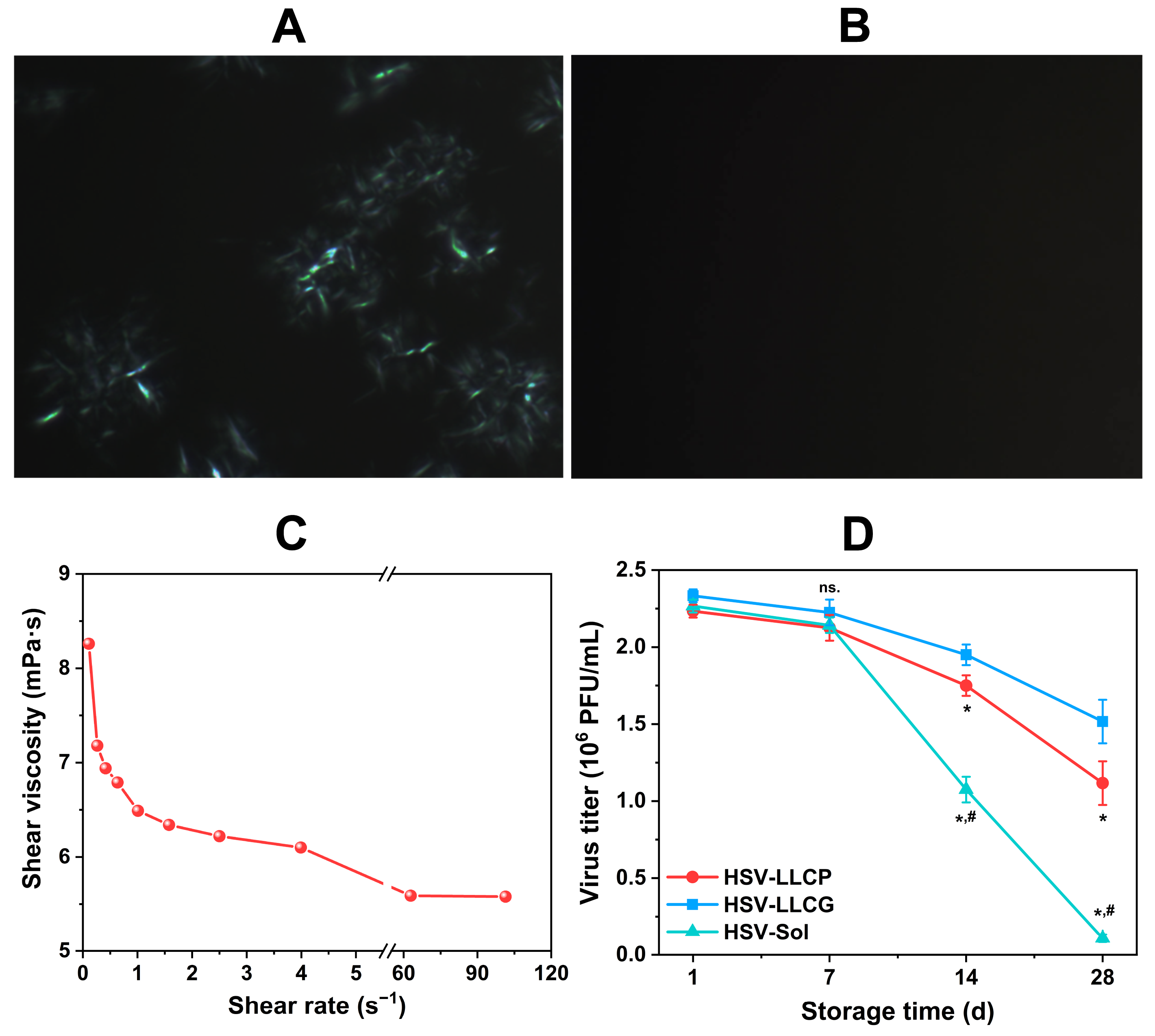
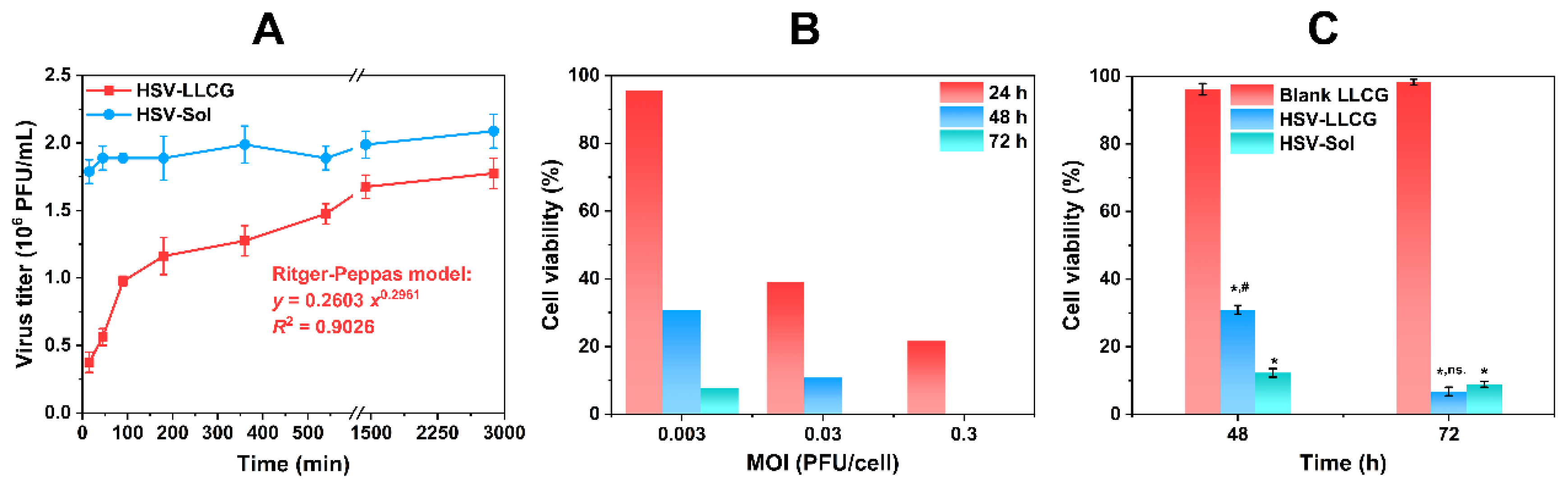
3. Results and Discussion

The gelation time of HSV-LLCP was 1.34 s. The rapid gelation in aqueous environment allows the transfer of HSV-LLCP into HSV-LLCG after injection, which can further serve as a depot for HSV-1. The POM images of HSV-LLCP and HSV-LLCG exhibited cruciate flower texture and dark site, suggesting their lamellar phase and cubic phase, respectively (Figure 2A,B) [15]. The shear viscosity ranged from 5 to 9 mPa·s (Figure 2C), which is suitable for an injection system [12]. All groups revealed good stability within seven days, while the virus titer of HSV-1 subsequently dropped significantly. On Day 14 and 28, the virus titer of HSV-LLCP and HSV-LLCG was significantly higher than that of HSV-Sol (p < 0.05). Figure 2D shows that the virus titer of HSV-LLCP was lower than that of HSV-LLCG (p < 0.05), indicating that the gel state in the cubic phase can provide stronger protective effects. HSV-LLCP could be stored at 4 °C, which can be easily achieved. These characteristics of HSV-LLCP facilitate the application as a stable and convenient vector for oncolytic virus delivery.
Upon encountering water, HSV-LLCP spontaneously transformed into HSV-LLCG. Therefore, we evaluated the in vitro efficacy of HSV-LLCG. The release profile of HSV-1 exhibited a triphasic sustained-release pattern (Figure 3A): (I) 0~90 min, rapid release; (II) 90~540 min, slow release; (III) 1440~2880 min, plateau phase. An XXXX simulation using Origin 2018 suggested that Ritger–Peppas model was the best fitted model with a correlation coefficient R2 = 0.9026. When the kinetic exponent was n ≤ 0.43, the drug release mechanism was Fickian diffusion depending on the Ritger–Peppas model. When n = 0.85 and 0.45 < n < 0.85, a Case II transport and intermediate transport mechanisms dominated the release process, respectively. The obtained exponent was n = 0.2961 < 0.43, implying that HSV-1 was released in a Fickian diffusion manner (concentration-dependent) [16]. In contrast, the release of HSV-Sol was instant, which could cause unexpected side effects due to the high virus concentration, and could even move to nontumor sites. The replication of HSV-1 in B16 cells was demonstrated by the viability of cells infected by different MOI levels (Figure 3B). With the increases in the MOI level and incubation time, the viability of B16 cells substantially decreased, suggesting that the cytotoxicity of HSV-1 was induced by viral replication. Furthermore, the cytotoxicity of blank LLCG, HSV-LLCG, and HSV-Sol was shown in Figure 3C. MTT assays showed that HSV-LLCG and HSV-Sol had higher cytotoxicity compared to blank LLCG (p < 0.05), indicating their strong oncolytic activity. HSV-Sol showed higher toxicity than HSV-LLCG with 48 h of incubation (p < 0.05), whereas at 72 h, their cytotoxicity showed no significant difference (p > 0.05). This could be attributed to the sustained-release of HSV-1 from HSV-LLCG. Taken together, HSV-LLCG exerted a sustained-release profile and an acceptable oncolytic activity in vitro.
The unique lattice structure of cubic liquid crystals transformed from lamellar liquid crystals after encountering the aqueous condition that could endow HSV-1 with a stable and confined environment, which isolated HSV-1 from the external environment and endued HSV-1 with a sustained-release pattern (Figure 4). This confined environment shielded the clearance of HSV-1 from circulation and contributed to the realization of the “viremic threshold”, which was pivotal for the spread of therapeutic viruses [17].

4. Conclusions

In this study, a novel in situ HSV-LLCP system was developed as a local treatment for malignant melanoma; its feasibility as a HSV-1 vector was investigated. The transition from LLCP to LLCG prevented HSV-1 from inactivating and penetrating surrounding tissues, providing high stability and a low leakage tendency when applying HSV-1. Moreover, HSV-LLCP could be stored at 4 °C, an easier storage condition. HSV-LLCG also exhibited a moderate in vitro cytotoxicity and replication in murine melanoma cells, and possessed a sustained-release profile. Our results demonstrate that the HSV-LLCP system is a promising vector for oncolytic therapy and could be shifted to clinical use with great potential. Before proceeding further with more applications, the pharmacokinetics and antitumor mechanisms of HSV-LLCP will be investigated in our lab.

Author Contributions

Conceptualization, F.F.; Data curation, W.W.; Investigation, F.F. and Y.G.; Methodology, W.W.; Project administration, Z.H., Y.H., C.W. and X.P.; Supervision, C.W.; Writing—original draft, F.F.; Writing—review and editing, Z.H. and Y.H. All authors have read and agreed to the published version of the manuscript.

Funding

This research did not receive any specific grant from funding agencies in the public, commercial, or not-for-profit sectors. This work was supported by the National Natural Science Foundation of China (No. 81173002).

Institutional Review Board Statement

Not applicable.

Informed Consent Statement

Not applicable.

Data Availability Statement

Not applicable.

Acknowledgments

We thank Jiayuan Huang for his critical reading of this manuscript.

Conflicts of Interest

The authors declare no conflict of interest.

References

  1. Gershenwald, J.E.; Scolyer, R.A.; Hess, K.R.; Sondak, V.K.; Long, G.V.; Ross, M.I.; Lazar, A.J.; Faries, M.B.; Kirkwood, J.M.; McArthur, G.A.; et al. Melanoma staging: Evidence-based changes in the American Joint Committee on Cancer eighth edition cancer staging manual. CA Cancer J. Clin. 2017, 67, 472–492. [Google Scholar] [CrossRef] [PubMed] [Green Version]
  2. Valentín-Nogueras, S.M.; Brodland, D.G.; Zitelli, J.A.; González-Sepúlveda, L.; Nazario, C.M. Mohs Micrographic Surgery Using MART-1 Immunostain in the Treatment of Invasive Melanoma and Melanoma In Situ. Dermatol. Surg. 2016, 42, 733–744. [Google Scholar] [CrossRef] [PubMed] [Green Version]
  3. Ho, H.; Aruri, J.; Kapadia, R.; Mehr, H.; White, M.A.; Ganesan, A.K. RhoJ Regulates Melanoma Chemoresistance by Suppressing Pathways That Sense DNA Damage. Cancer Res. 2012, 72, 5516–5528. [Google Scholar] [CrossRef] [PubMed] [Green Version]
  4. Thomas, S.; Kuncheria, L.; Roulstone, V.; Kyula, J.N.; Mansfield, D.; Bommareddy, P.K.; Smith, H.; Kaufman, H.L.; Harrington, K.J.; Coffin, R.S. Development of a new fusion-enhanced oncolytic immunotherapy platform based on herpes simplex virus type 1. J. Immunother. Cancer 2019, 7, 214. [Google Scholar] [CrossRef] [PubMed] [Green Version]
  5. Hua, L.; Wakimoto, H. Oncolytic herpes simplex virus therapy for malignant glioma: Current approaches to successful clinical application. Expert Opin. Biol. Ther. 2019, 19, 845–854. [Google Scholar] [CrossRef] [PubMed]
  6. Aghi, M.; Martuza, R.L. Oncolytic viral therapies–the clinical experience. Oncogene 2005, 24, 7802–7816. [Google Scholar] [CrossRef] [PubMed] [Green Version]
  7. Todo, T.; Martuza, R.L.; Rabkin, S.D.; Johnson, P.A. Oncolytic herpes simplex virus vector with enhanced MHC class I presentation and tumor cell killing. Proc. Natl. Acad. Sci. USA 2011, 98, 6396–6401. [Google Scholar] [CrossRef] [PubMed] [Green Version]
  8. Koshizuka, T.; Kawaguchi, Y.; Nishiyama, Y. Herpes simplex virus type 2 membrane protein UL56 associates with the kinesin motor protein KIF1A. J. Gen. Virol. 2005, 86, 527–533. [Google Scholar] [CrossRef] [PubMed]
  9. Nawa, A.; Nozawa, N.; Goshima, F.; Nagasaka, T.; Kikkawa, F.; Niwa, Y.; Nakanishi, T.; Kuzuya, K.; Nishiyama, Y. Oncolytic viral therapy for human ovarian cancer using a novel replication-competent herpes simplex virus type I mutant in a mouse model. Gynecol. Oncol. 2003, 91, 81–88. [Google Scholar] [CrossRef] [PubMed]
  10. Everett, A.S.; Pavlidakey, P.G.; Contreras, C.M.; Santos, J.F.D.L.; Kim, J.Y.; McKee, S.B.; Kaufman, H.L.; Conry, R.M. Chronic granulomatous dermatitis induced by talimogene laherparepvec therapy of melanoma metastases. J. Cutan. Pathol. 2018, 45, 48–53. [Google Scholar] [CrossRef] [PubMed]
  11. Cardoso, L.N.B.; Depieri, L.V.; Diniz, H.; Calzzani, R.A.J.; Fantini, M.C.D.A.; Iyomasa, M.M.; Vicentini, F.; Bentley, M.V.L.B. Self-assembling gelling formulation based on a crystalline-phase liquid as a non-viral vector for siRNA delivery. Eur. J. Pharm. Sci. 2014, 58, 72–82. [Google Scholar] [CrossRef]
  12. Mei, L.; Xie, Y.; Huang, Y.; Wang, B.; Chen, J.; Quan, G.; Pan, X.; Liu, H.; Wang, L.; Liu, X.; et al. Injectable in situ forming gel based on lyotropic liquid crystal for persistent postoperative analgesia. Acta Biomater. 2018, 67, 99–110. [Google Scholar] [CrossRef] [PubMed]
  13. Jazowiecka-Rakus, J.; Sochanik, A.; Rusin, A.; Hadryś, A.; Fidyk, W.; Villa, N.; Rahman, M.M.; Chmielik, E.; Franco, L.S.; McFadden, G. Myxoma Virus-Loaded Mesenchymal Stem Cells in Experimental Oncolytic Therapy of Murine Pulmonary Melanoma. Mol. Ther. Oncolytics 2020, 18, 335–350. [Google Scholar] [CrossRef] [PubMed]
  14. Rekha, S.; Anila, E. In vitro cytotoxicity studies of surface modified CaS nanoparticles on L929 cell lines using MTT assay. Mater. Lett. 2019, 236, 637–639. [Google Scholar] [CrossRef]
  15. Zheng, T.; Huang, X.; Chen, J.; Feng, D.; Mei, L.; Huang, Y.; Quan, G.; Zhu, C.; Singh, V.; Ran, H.; et al. A liquid crystalline precursor incorporating chlorhexidine acetate and silver nanoparticles for root canal disinfection. Biomater. Sci. 2018, 6, 596–603. [Google Scholar] [CrossRef] [PubMed]
  16. Spizzirri, U.G.; Hampel, S.; Cirillo, G.; Nicoletta, F.P.; Hassan, A.; Vittorio, O.; Picci, N.; Iemma, F. Spherical gelatin/CNTs hybrid microgels as electro-responsive drug delivery systems. Int. J. Pharm. 2013, 448, 115–122. [Google Scholar] [CrossRef] [PubMed]
  17. Russell, S.J.; Peng, K.-W.; Bell, J.C. Oncolytic virotherapy. Nat. Biotechnol. 2012, 30, 658–670. [Google Scholar] [CrossRef] [PubMed] [Green Version]
Figure 1. Schematic illustration of the preparation design in this work.
Engproc 31 00045 g001
Figure 2. Preparation characterizations: (A) POM image of HSV-LLCP; (B) POM image of HSV-LLCG; (C) Shear viscosity of HSV-LLCP; (D) Stability of virus titer of HSV-LLCP, HSV-LLCG and HSV-Sol (n = 3). ns.: not significantly lower than Day 1 (p > 0.05); *: significantly lower than HSV-LLCG on the same day (p < 0.05); #: significantly lower than HSV-LLCP on the same day (p < 0.05).
Engproc 31 00045 g002
Figure 3. Results of in vitro efficacy evaluations. (A) Release studies of HSV-LLCG and HSV-Sol (n = 3); (B) In vitro replication of HSV-1 in B16 cells, expressed as the cell viability compared to uninfected cells (mean of three replicates); (C) Cytotoxicity of blank LLCG, HSV-LLCG and HSV-Sol in B16 cells (n = 3). *: significantly lower than blank LLCG at the same time (p < 0.05); #: significantly higher than HSV-Sol at the same time (p < 0.05); ns.: no significant difference with HSV-Sol at the same time (p > 0.05).
Engproc 31 00045 g003
Figure 4. The diagrammatic sketch of HSV-LLCP system after administration.
Engproc 31 00045 g004
Disclaimer/Publisher’s Note: The statements, opinions and data contained in all publications are solely those of the individual author(s) and contributor(s) and not of MDPI and/or the editor(s). MDPI and/or the editor(s) disclaim responsibility for any injury to people or property resulting from any ideas, methods, instructions or products referred to in the content.

