Proceeding Paper

A New Fluorogenic Substrate for Granzyme B Based on Fluorescence Resonance Energy Transfer †

by
Cátia D. F. Martins
,
M. Manuela M. Raposo
and
Susana P. G. Costa
*
Centre of Chemistry, University of Minho, Campus de Gualtar, 4710-057 Braga, Portugal
*
Author to whom correspondence should be addressed.
Presented at the 24th International Electronic Conference on Synthetic Organic Chemistry, 15 November 2020–15 December 2020. Available online: https://ecsoc-24.sciforum.net/.
Chem. Proc. 2021, 3(1), 6; https://doi.org/10.3390/ecsoc-24-08311
Published: 14 November 2020

Abstract

:
The synthesis and characterization of a new fluorogenic substrate for granzyme B (GzmB) is reported. The substrate design was based on the fluorescence resonance energy transfer (FRET) principle using 5-(2′-aminoethyl)aminonaphthalene sulfonic acid (Edans) and 4-[[4′-(N,N-dimethylamino)phenyl]diazenyl]benzoic acid (Dabcyl) as a donor–acceptor pair, linked to a specific sequence for GzmB (AAD), with an additional amino acid as the anchoring point (K). The tetrapeptide was synthesized by microwave-assisted solid-phase peptide synthesis (MW-SPPS) and coupled to Dabcyl and Edans at its N- and C-termini, respectively. The obtained probe was purified by semi-preparative HPLC and characterized by NMR, UV/Vis absorption and fluorescence spectroscopy and mass spectrometry.

1. Introduction

The serine protease granzyme B (GzmB) is a potent inducer of apoptosis in target cells when released by cytotoxic T lymphocytes (CTL) or natural killer (NK) cells, representing one of the two dominant mechanisms by which T cells mediate cancer cell death [1,2]. Considering GzmB’s preference for cleaving after aspartic acid, several substrates containing IEPD and IETD sequences coupled to chromogenic or fluorescent leaving groups have been successfully used for GzmB detection [3,4,5]. However, these two sequences lack specificity as they can also be cleaved by some caspases, a family of cysteine proteases that are also activated during apoptosis [6,7]. Alternatively, recent studies have revealed a specific substrate for GzmB, Boc-AAD-SBzl, which cannot be cleaved by any caspase [4].
The use of a fluorescence resonance energy transfer (FRET) strategy is widely used to design probes for the detection of enzyme activity. In these probes, a fluorescent donor and an acceptor group are attached to either end of a cleavable enzyme substrate. Upon enzymatic cleavage of the substrate, the donor and acceptor are separated and emission from the fluorophore is restored, resulting in an increase in fluorescence intensity proportional to enzyme activity [8,9,10]. Among the many possible combinations of donor–acceptor, the pair 5-(2′-aminoethyl)aminonaphthalene sulfonic acid (Edans) and 4-[[4′-(N,N-dimethylamino)phenyl]diazenyl]benzoic acid (Dabcyl) is largely used to monitor serine proteases activity, with Edans acting as the acceptor and Dabcyl as the donor. In fact, this donor–acceptor pair has been successfully applied to fluorescence-based assays of SARS coronavirus 3C-like protease [11], human immunodeficiency virus protease [12], human neutrophil elastase [13], human cytomegalovirus protease [14] and hepatitis C virus protease [15], among others.
Having these facts in mind and considering our interest in using FRET-based techniques to monitor GzmB activity in a controlled release system of immunostimulating drugs applied in the treatment of colorectal cancer [16], we report, herein, the synthesis and characterization of a new fluorogenic GzmB substrate. For this, our substrate design was based on the FRET principle using Edans/Dabcyl as a donor–acceptor pair, linked to a specific sequence for GzmB (AAD), with an additional amino acid as the anchoring point (K).

2. Experimental Section

2.1. Reagents and Solvents

All Fmoc-amino acids and 2-chlorotrityl chloride resin (with a degree of functionalization of 1.3 mmol/g) were purchased from AAPPTec (Louisville, KY, USA). The coupling reagents and additives ethyl-2-cyano-2-(hydroxyimino) acetate (Oxyma) and N-ethyl-N′-(3-dimethylaminopropyl) carbodiimide hydrochloride (EDC) were obtained from AAPPTec; N-hydroxysuccinimide (NHS) and N,N′-diisopropylcarbodiimide (DIC) were purchased from Acros Organics (Geel, Belgium). Additional reagents and solvents were purchased from Merck KGaA (Darmstadt, Germany) and Acros Organics and used as received. The fluorophore Edans was acquired from Sigma-Aldrich (St. Louis, MO, USA) and the chromophore/quencher Dabcyl was synthesized according to standard azo coupling reactions [17].

2.2. Instrumentation

TLC analyses were carried out on 0.25-mm-thick silica plates coated with fluorescent indicator F254 (Merck KGaA, Darmstadt, Germany) and spots were visualized in a CN15 viewing cabinet under UV lamp at 365 nm (Vilber Lourmat, Marne-la-Vallée, France). NMR spectra were obtained on a Bruker Avance III 400 at an operating frequency of 400 MHz for 1H and 100.6 MHz for 13C using the solvent peak as an internal reference. The solvents are indicated in parentheses before the chemical shift values (δ relative to tetramethylsilane (TMS) and given in ppm). Assignments were supported by two-dimensional heteronuclear correlation techniques. Mass spectrometry analyses were performed at the “C.A.C.T.I. Unidad de Espectrometria de Masas” at the University of Vigo, Spain. Microwave-assisted solid-phase peptide synthesis was carried out on a CEM Discover SPS instrument (CEM Corp., Matthews, GA, USA). Probe purification was performed by semi-preparative HPLC on a Shimadzu LC-8A, UV/Vis JASCO 875-UV detector and a Shimadzu C-RGA Chromatopac register (Shimadzu Europa GmbH, Duisburg, Germany) on a Europa Peptide 120 C18 (5 mm) column (Teknokroma, Barcelona, Spain) using ACN/water (3:1, v/v) (λdet = 432 nm). UV/Vis absorption spectra were obtained using a Shimadzu UV/2501PC spectrophotometer (Shimadzu Europa GmbH, Duisburg, Germany) and fluorescence spectra were collected using a FluoroMax-4 spectrofluorometer (HORIBA Europe GmbH, Darmstadt, Germany) in standard quartz cuvettes.

2.3. Methods

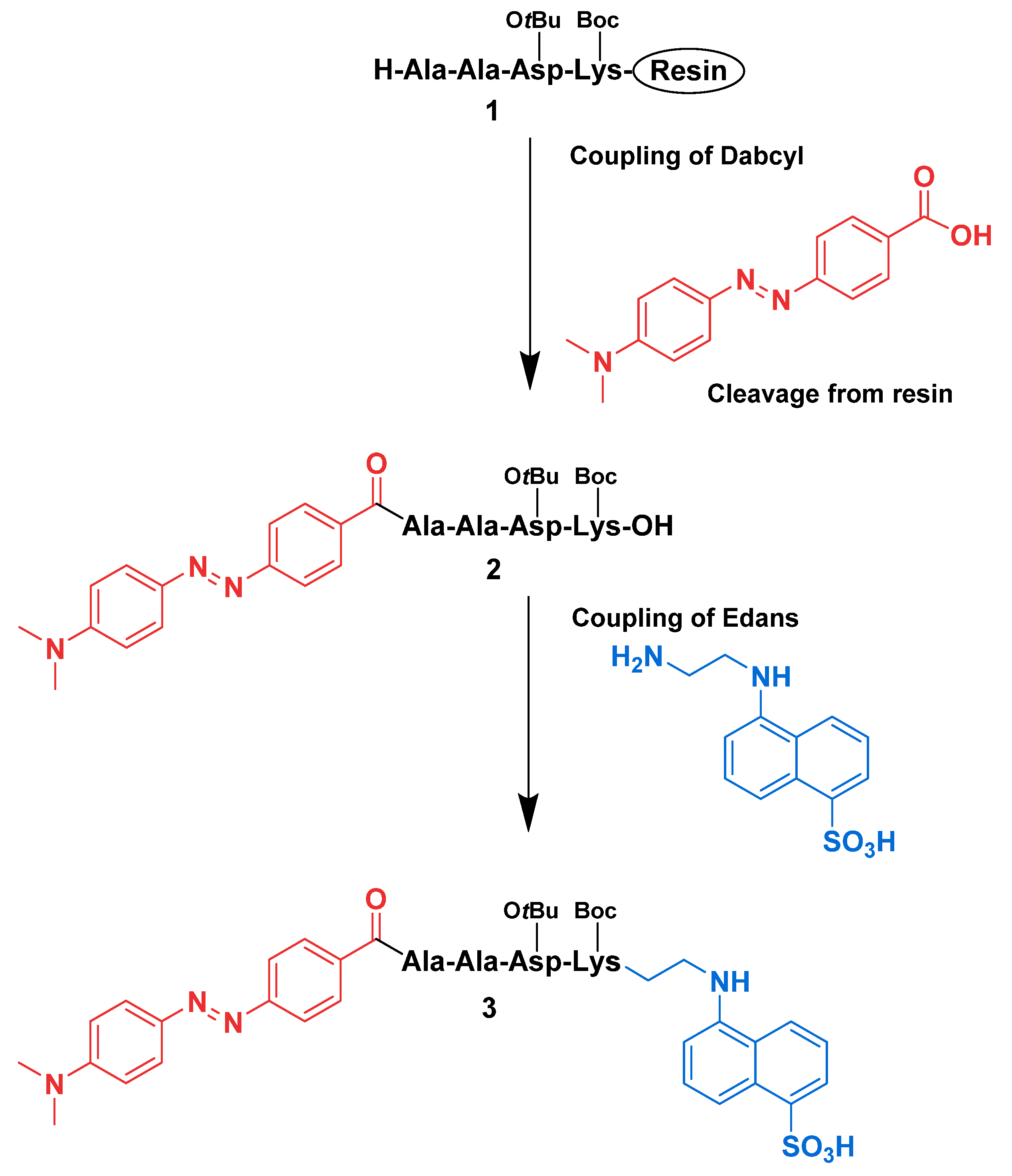
2.3.1. Synthesis of H-Ala-Ala-Asp(OtBu)-Lys(Boc)-OH on Resin (1)

The peptide was synthesized by a microwave-assisted solid-phase peptide synthesis protocol on a manual microwave-assisted peptide synthesizer using the 9-fluorenylmethoxycarbonyl (Fmoc) strategy.
Attachment of the first amino acid to the resin. The 2-chlorotrityl chloride resin (1 g) was swollen in DCM for 15 min and filtered. The protected C-terminal amino acid residue, Fmoc-Lys(Boc)-OH (2 equiv on the degree of functionalization of the resin), was attached to the resin in the presence of 4 equiv of N,N-diisopropylethylamine (DIPEA) (based on the amino acid) under anhydrous conditions in DCM solution (10 mL). The mixture was stirred for 5 h at room temperature. The resin was filtered and washed sequentially with a mixture of DCM/MeOH/DIPEA (25.5:3.0:1.5, v/v; 3 × 10 mL), DCM (3 × 10 mL), DMF (3 × 10 mL), MeOH (3 × 10 mL) and diethyl ether (2 × 10 mL). After, the resin was dried in vacuo. A standard protocol was applied to determine the loading of the resin, resulting in a loading of 0.4872 mmol/g. The peptide chain was elongated in consecutive cycles of deprotection and coupling.
Deprotection of Fmoc-amino acids. Deprotection was performed with 20% piperidine in DMF (5 mL/g resin) for 30 s, under microwave irradiation (power = 50 W, temperature = 75 °C). The resin was filtered, then a fresh solution of 20% piperidine in DMF (7 mL/g resin) was added and allowed to react for 3 min under microwave irradiation (power = 50 W, temperature = 75 °C). The resin was washed with DMF (3 × 10 mL) and MeOH (3 × 10 mL) and this washing cycle was repeated four times.
Coupling of Fmoc-amino acids. The Fmoc-amino acid (5 equiv on the loading) was dissolved in dry DMF (6 mL/g resin) and DIC (5 equiv) and Oxyma (5 equiv) dissolved in dry DMF (2 mL/g) were added. The reaction mixture was allowed to stir at room temperature for 5 min before being added to the resin. The suspension was then subjected to two microwave irradiation cycles for 5 min (power = 25 W, temperature = 75 °C). The resin was washed with DMF (3 × 10 mL) followed by MeOH (3 × 10 mL) and this washing cycle was repeated tree times. Part of the resin was subjected to cleavage to afford the free peptide (see details below) for characterization by NMR.
1H NMR (400 MHz, DMSO-d6): δ = 1.16–1.38 (28H, m, 2 × CH3 Ala, γ-CH2 Lys, δ-CH2 Lys, 3 × CH3 Boc and 3 × CH3 OtBu), 1.45–1.58 (2H, m, β-CH2 Lys), 2.39–2.48 (1H, m, β-CH2 Asp), 2.69 (1H, dd, J = 5.6 and 17.0 Hz, β-CH2 Asp), 2.81–2.88 (2H, m, ε-CH2 Lys), 3.82–3.90 (2H, m, α-CH Lys and α-CH Ala), 4.28 (1H, s, α-CH Ala), 4.51–4.56 (1H, m, α-CH Asp), 6.65 (1H, s, NH Boc), 7.50 (1H, d, J = 6.8 Hz, NH Lys), 8.35 (1H, d, J = 7.2 Hz, NH Asp or NH Ala 2), 8.43 (1H, s, NH Asp or NH Ala 2) ppm.
13C NMR (100.6 MHz, DMSO-d6): δ = 18.36 (CH3 Ala), 18.50 (CH3 Ala), 22.26 (γ-CH2 Lys), 27.68 (CH3 Boc or CH3 OtBu), 28.29 (CH3 Boc or CH3 OtBu), 29.40 (δ-CH2 Lys), 31.54 (β-CH2 Lys), 37.38 (β-CH2 Asp), 39.50 (ε-CH2 Lys), 48.55 (α-CH Ala 1 or α-CH Ala 2), 48.84 (α-CH Ala 1 or α-CH Ala 2), 49.65 (α-CH Asp), 53.33 (α-CH Lys), 77.34 (C(CH3) Boc or C(CH3) OtBu), 80.15 (C(CH3) Boc or C(CH3) OtBu), 155.55 (C=O Boc), 169.44 (C=O Asp), 171.45 (C=O Ala 1), 171.92 (C=O Ala 2), 172.14 (C=O OtBu), 173.30 (C=O Lys) ppm.

2.3.2. Synthesis of Dabcyl-Ala-Ala-Asp(OtBu)-Lys(Boc)-OH (2)

After the last Fmoc group had been removed from the peptide-resin 1 with 20% piperidine in DMF, Dabcyl (5 equiv on the loading) was coupled to the N-terminal alanine of the peptide-resin 1 with the aid of DIC (5 equiv) and Oxyma (5 equiv) in dry DMF. The mixture was shaken at room temperature for 15 min before being added to the resin. The suspension was subjected to four microwave irradiation cycles for 5 min (power = 25 W, temperature = 75 °C). The resin was washed with DMF (3 × 10 mL) followed by MeOH (3 × 10 mL) and this washing cycle was repeated six times. The resin with Dabcyl-peptide was stirred for 2 h in a mixture of acetic acid (AcOH)/2,2,2-trifluoroethanol (TFE)/DCM (1:2:7, v/v; 10 mL) at room temperature. This mixture was left overnight at room temperature. The solution was filtered, and the resin was washed with TFE/DCM (2:8, v/v; 2 × 10 mL). The solvent was removed under reduced pressure in a rotary evaporator and the product was precipitated with diethyl ether and stored at 4 °C overnight. The product was collected by centrifugation, three times suspended in diethyl ether, collected by centrifugation and dried in vacuo. Dabcyl-Ala-Ala-Asp(OtBu)-Lys(Boc)-OH 2 (0.082 g, yield 30%) was obtained as an orange solid.
1H NMR (400 MHz, DMSO-d6): δ = 1.05–1.29 (28H, m, 2 × CH3 Ala, γ-CH2 Lys, δ-CH2 Lys, 3 × CH3 Boc and 3 × CH3 OtBu), 1.49–1.59 (1H, m, β-CH2 Lys), 1.62–1.70 (1H, m, β-CH2 Lys), 2.39–2.50 (2H, m, β-CH2 Asp), 2.78–2.89 (2H, m, ε-CH2 Lys), 3.06 (6H, s, 2 × CH3 Dabcyl), 3.92–3.97 (1H, m, α-CH Lys), 4.24–4.31 (1H, m, α-CH Ala 2), 4.45–4.59 (2H, m, α-CH Ala 1 and α-CH Asp), 6.69 (1H, t, J = 5.2 Hz, NH Boc), 6.84 (2H, d, J = 9.2 Hz, H-3′ and H-5′), 7.55 (1H, d, J = 7.2 Hz, NH Lys), 7.81 (4H, dd, J = 2.8 and 9.2 Hz, H-3, H-5, H-2′ and H-6′), 8.03 (2H, d, J = 8.8 Hz, H-2 and H-6), 8.12 (1H, d, J = 7.2 Hz, NH Ala 2 or NH Asp), 8.23 (1H, d, J = 8.4 Hz, NH Ala 2 or NH Asp), 8.64 (1H, d, J = 6.8 Hz, NH Ala 1) ppm.
13C NMR (100.6 MHz, DMSO-d6): δ = 17.79 (CH3 Ala), 18.31 (CH3 Ala), 22.34 (γ-CH2 Lys), 27.64 (CH3 Boc or CH3 OtBu), 28.29 (CH3 Boc or CH3 OtBu), 29.33 (δ-CH2 Lys), 31.40 (β-CH2 Lys), 37.37 (β-CH2 Asp), 39.55 (ε-CH2 Lys), 48.29 (α-CH Ala 2), 49.26 (α-CH Ala 1 or α-CH Asp), 49.46 (α-CH Ala 1 or α-CH Asp), 52.93 (α-CH Lys), 64.95 (2 × CH3 Dabcyl), 77.36 (C(CH3) Boc or C(CH3) OtBu), 80.18 (C(CH3) Boc or C(CH3) OtBu), 111.61 (CH-3′ and CH-5′), 121.44 (CH-3 and CH-5), 125.15 (CH-2′ and CH-6′), 128.73 (CH-2 and CH-6), 134.27(C4), 142.68 (C4′), 152.90 (C1′), 154.12 (C1), 155.57 (C=O Boc), 165.73 (C=O Asp), 169.36 (C=O Ala 1), 169.42 (C=O Ala 2), 172.23 (C=O OtBu), 173.56 (C=O Lys) ppm.
MS m/z (ESI, %): 811 ([M + H]+, 19), 356 (19), 145 (20), 134 (34), 329 (39), 132 (74), 328 (100). HRMS: m/z (ESI) calc. for C40H59N8O10 811.4349; found 811.4338.

2.3.3. Synthesis of Dabcyl-Ala-Ala-Asp(OtBu)-Lys(Boc)-Edans (3)

The attachment of Edans to the peptide C-terminal was performed using a solution method with EDC/NHS [18]. For this, Dabcyl-peptide 2 (0.078 g, 0.096 mmol) was dissolved in dry DMF (5 mL) and placed in an ice bath. After stirring for 5 min, NHS (0.074 g, 0.643 mmol) was added, followed by EDC (0.085 g, 0.443 mmol) and Et3N (62 µL). The reaction mixture was stirred for 1 h, then Edans (0.052 g, 0.197 mmol) and Et3N (27 µL) were added. The mixture was stirred at room temperature for 5 h. The solvent was removed under reduced pressure in a rotary evaporator. The crude product was purified by semi-preparative HPLC using an ACN/water mixture (3:1) as eluent and a flow of 1 mL/min. The desired product eluted from the column with a retention time of 16 min. The probe 3 (0.005 g, yield 5%) was obtained as an orange solid.
1H NMR (400 MHz, DMSO-d6): δ = 1.22–1.35 (28H, m, 2 × CH3 Ala, γ-CH2 Lys, δ-CH2 Lys, 3 × CH3 Boc and 3 × CH3 OtBu), 1.47–1.65 (1H, m, β-CH2 Lys), 1.66–1.68 (1H, m, β-CH2 Lys), 2.44–2.54 (2H, m, β-CH2 Asp), 2.58–2.71 (2H, m, ε-CH2 Lys), 3.07 (6H, s, 2 × CH3 Dabcyl), 3.33–3.43 (4H, m, CH2-1′ and CH2-2′), 4.15–4.19 (1H, m, α-CH Lys), 4.26 (1H, t, J = 7.2 Hz, α-CH Ala 2), 4.46–4.56 (2H, m, α-CH Ala 1 and α-CH Asp), 6.63–6.68 (2H, t, J = 5.2 Hz, NH Boc and 2′-NH Edans), 6.84 (2H, d, J = 9.2 Hz, H-3′ and H-5′ Dabcyl), 7.17 (1H, d, J = 8.0 Hz, H-4 Edans), 7.27–7.34 (2H, m, H-3 and H-7 Edans), 7.57 (1H, d, J = 8.0 Hz, NH Lys), 7.61–7.83 (6H, m, H-3, H-5, H-2′, H-6′ Dabcyl and H-6, H-8 Edans), 8,02 (2H, dd, J = 2.0 and 6.8 Hz, H-2 and H-6 Dabcyl), 8.10–8.14 (2H, m, NH Ala 2 or NH Asp and H-2 Edans), 8.17 (1H, d, J = 8.0 Hz, NH Ala 2 or NH Asp), 8.24 (1H, s, 5-NH Edans), 8.59 (1H, d, J = 7.2 Hz, NH Ala 1) ppm.
MS m/z (ESI, %): 1059 ([M + H]+, 7), 132 (8), 445 (8), 513 (10), 142 (14), 542 (17), 481 (19), 453 (20), 541 (28), 481 (48), 453 (20), 541 (28), 481 (48), 453 (52), 480 (83), 452 (100). HRMS: m/z (ESI) calc. for C52H71N10O12S 1059.4968; found 1059.4965.

2.3.4. UV/Vis Absorption and Fluorescence Spectroscopy of Peptides 2–3

The spectroscopic characterization of peptides 2–3 was carried out by UV/Vis absorption spectroscopy (300–700 nm). Solutions of the compounds (1 × 10⁻⁵ M) were made in phosphate buffer at pH 7.5. Fluorescence spectra of probe 3 and Edans were obtained by excitation at the wavelength of maximum absorption, with a 5 nm slit.

3. Results and Discussion

3.1. Synthesis of the Fluorogenic Substrate for Granzyme B

Considering our interest in using FRET-based techniques to monitor GzmB activity in a controlled release system of immunostimulating drugs applied in the treatment of colorectal cancer, we synthesized and fully characterized a new fluorogenic GzmB substrate. For this, our substrate design was based on two criteria: (1) the specific sequence for GzmB (Ala-Ala-Asp, AAD) should be attached to an additional amino acid as the anchoring point (Lys, K); and (2) upon enzymatic cleavage of this substrate, the fluorophore had to be linked to Lys residue. Taking into account the above-mentioned aspects and considering GzmB’s preference for cleaving after aspartic acid at the P1 position (nomenclature according to Schechter and Berger) [19], in our substrate, the fluorophore was coupled to the C-terminus of the tetrapeptide (AADK) and the quencher was attached to the N-terminus of this peptide. GzmB would cleave the substrate between residues Asp (P1) and Lys (P1′), -AADK- (the scissile Asp-Lys bond is underlined).
Therefore, the synthesis of the tetrapeptide and the introduction of Dabcyl were obtained by solid phase, while the incorporation of Edans was performed in solution synthesis (Scheme 1). The side chain amino and carboxyl groups of lysine and aspartic acid protected with Boc and OtBu groups, respectively, were maintained during the inclusion of the FRET pair on the peptide to avoid cross-reactions.
The required tetrapeptide AADK in its protected form (H-Ala-Ala-Asp(OtBu)-Lys(Boc)-OH 1) was synthesized by microwave-assisted solid-phase peptide synthesis (MW-SPPS) applying the Fmoc strategy. The protected derivative of the C-terminal amino acid residue, Fmoc-Lys(Boc)-OH, was attached to the 2-chorotrityl chloride resin through a nucleophilic substitution reaction using an excess of Fmoc-amino acid and a base, DIPEA, resulting in a loading of 0.487 mmol/g. The peptide chain was elongated in consecutive cycles of deprotection and coupling of the required amino acids.
The free N-terminal of peptide 1, still attached to the resin, was then coupled to the quencher Dabcyl by MW-SPPS using DIC/Oxyma in dry DMF. The labeled peptide, Dabcyl-Ala-Ala-Asp(OtBu)-Lys(Boc)-OH 2, was cleaved from the solid support by treatment with AcOH/TFE/DCM (1:2:7, v/v). The desired product was obtained with a global yield of 30% and characterized by 1H and 13C NMR spectroscopy and high-resolution mass spectroscopy. The obtained data were in agreement with the expected structure.
Finally, the fluorophore Edans was coupled in a solution reaction to the C-terminal lysine using EDC/NHS in dry DMF according to the method reported by Krafft et al. [18]. The obtained product was carefully purified by semi-preparative HPLC to ensure proper separation from free Edans which could disturb the subsequent fluorescence measurements. Thus, the desired probe Dabcyl-Ala-Ala-Asp(OtBu)-Lys(Boc)-Edans 3 was obtained in 5% yield and characterized by 1H NMR spectroscopy and high-resolution mass spectroscopy. The obtained data were in agreement with the expected structure.

3.2. UV/Vis Absorption and Fluorescence Spectroscopy

To evaluate the photophysical properties, the absorption and fluorescence spectra of labeled peptide 2 (with the quencher attached) and peptide probe 3 (with both the quencher and fluorophore attached) were measured in phosphate buffer at pH 7.5. As depicted in Figure 1a, Dabcyl-AADK 2 and Dabcyl-AADK-Edans 3 showed absorption bands with similar wavelengths of maximum absorption (470 nm and 462 nm, respectively), related to the Dabcyl chromophore. As expected, the fluorescence spectrum of probe 3 exhibited a very low intensity emission band at 494 nm, upon excitation at 342 nm, thus confirming the existence of intramolecular quenching between Edans (acceptor) and Dabcyl (donor) in probe 3. To further confirm this observation, a fluorescence spectrum of free Edans obtained the same conditions. Comparison of the fluorescence spectrum of probe 3 with that of free Edans (Figure 1b) clearly showed that the acceptor Dabcyl efficiently quenches the donor Edans’ fluorescence in probe 3, as predicted.

4. Conclusions

In summary, based on the FRET principle, a new fluorogenic substrate for granzyme B was designed, synthesized and fully characterized. We demonstrated that microwave-assisted solid-phase synthesis accompanied by solution synthesis can be effectively used to obtain the desired fluorogenic substrate bearing a donor–acceptor pair such as Edans/Dabcyl. Moreover, future work will be developed to optimize the synthesis method to increase the overall synthesis yield.
By comparison of the fluorescence spectra of the synthesized probe and free Edans, the existence of intramolecular fluorescence quenching between Edans and Dabcyl in the probe was confirmed, thus enabling it to be a very promising candidate as a probe for the subsequent activity assays with GzmB.

Acknowledgments

The authors acknowledge Fundação para a Ciência e Tecnologia—FCT (Portugal), for funding through CQUM (UIDB/00686/2020) and project PTDC/QUI-COL/28052/2017. The NMR spectrometer Bruker Avance III 400 is part of the National NMR Network and was purchased within the framework of the National Program for Scientific Re-equipment, contract REDE/1517/RMN/2005 with funds from POCI 2010 (FEDER) and FCT.

Conflicts of Interest

The authors declare no conflict of interest.

References

  1. Larimer, B.M.; Wehrenberg-Klee, E.; Dubois, F.; Mehta, A.; Kalomeris, T.; Flaherty, K.; Boland, G.; Mahmood, U. Granzyme B PET Imaging as a Predictive Biomarker of Immunotherapy Response. Cancer Res. 2017, 77, 2318–2327. [Google Scholar] [CrossRef] [PubMed]
  2. Ida, H.; Utz, P.J.; Anderson, P.; Eguchi, K. Granzyme B and Natural Killer (NK) Cell Death. Mod. Rheumatol. 2005, 15, 315–322. [Google Scholar] [CrossRef] [PubMed]
  3. Waugh, S.M.; Craik, C.S.; Harris, J.L.; Fletterick, R. The Structure of the Pro-Apoptotic Protease Granzyme B Reveals the Molecular Determinants of Its Specificity. Nat. Struct. Biol. 2000, 7, 762–765. [Google Scholar] [CrossRef] [PubMed]
  4. Hagn, M.; Sutton, V.R.; Trapani, J.A. A Colorimetric Assay That Specifically Measures Granzyme B Proteolytic Activity: Hydrolysis of Boc-Ala-Ala-Asp-S-Bzl. J. Vis. Exp. 2014, 93, e52419. [Google Scholar] [CrossRef] [PubMed]
  5. Nie, Z.; Phenix, B.N.; Lum, J.J.; Alam, A.; Lynch, D.H.; Beckett, B.; Krammer, P.H.; Sekaly, R.P.; Badley, A.D. HIV-1 Protease Processes Procaspase 8 to Cause Mitochondrial Release of Cytochrome c, Caspase Cleavage and Nuclear Fragmentation. Cell Death Differ. 2002, 9, 1172–1184. [Google Scholar] [CrossRef] [PubMed]
  6. Estébanez-Perpiñá, E.; Fuentes-Prior, P.; Belorgey, D.; Braun, M.; Kiefersauer, R.; Maskos, K.; Huber, R.; Rubin, H.; Bode, W. Crystal Structure of the Caspase Activator Human Granzyme B, a Proteinase Highly Specific for an Asp-P1 Residue. Biol. Chem. 2000, 381, 1203–1214. [Google Scholar] [CrossRef] [PubMed]
  7. Harris, J.L.; Peterson, E.P.; Hudig, D.; Thornberry, N.A.; Craik, C.S. Definition and Redesign of the Extended Substrate Specificity of Granzyme B. J. Biol. Chem. 1998, 273, 27364–27373. [Google Scholar] [CrossRef] [PubMed]
  8. Drake, C.R.; Miller, D.C.; Jones, E.F. Activatable Optical Probes for the Detection of Enzymes. Curr. Org. Synth. 2011, 8, 498–520. [Google Scholar] [CrossRef] [PubMed]
  9. Johansson, M.K.; Cook, R.M. Intramolecular Dimers: A New Design Strategy for Fluorescence-Quenched Probes. Chem. Eur. J. 2003, 9, 3466–3471. [Google Scholar] [CrossRef] [PubMed]
  10. Grahn, S.; Ullmann, D.; Jakubke, H.-D. Design and Synthesis of Fluorogenic Trypsin Peptide Substrates Based on Resonance Energy Transfer. Anal. Biochem. 1998, 265, 225–231. [Google Scholar] [CrossRef] [PubMed]
  11. Chen, S.; Chen, L.L.; Luo, H.B.; Sun, T.; Chen, J.; Ye, F.; Cai, J.; Shen, J.; Shen, X.; Jiang, H.L. Enzymatic Activity Characterization of SARS Coronavirus 3C-like Protease by Fluorescence Resonance Energy Transfer Technique. Acta Pharmacol. Sin. 2005, 26, 99–106. [Google Scholar] [CrossRef] [PubMed]
  12. Windsor, I.W.; Raines, R.T. Fluorogenic Assay for Inhibitors of HIV-1 Protease with Sub-Picomolar Affinity. Sci. Rep. 2015, 5, 1–7. [Google Scholar] [CrossRef] [PubMed]
  13. Ferreira, A.V.; Perelshtein, I.; Perkas, N.; Gedanken, A.; Cunha, J.; Cavaco-Paulo, A. Detection of Human Neutrophil Elastase (HNE) on Wound Dressings as Marker of Inflammation. Appl. Microbiol. Biotechnol. 2017, 101, 1443–1454. [Google Scholar] [CrossRef] [PubMed]
  14. Holskin, B.P.; Bukhtiyarova, M.; Dunn, B.M.; Baur, P.; Dechastonay, J.; Pennington, M.W. A Continuous Fluorescence-Based Assay of Human Cytomegalovirus Protease Using a Peptide Substrate. Anal. Biochem. 1995, 227, 148–155. [Google Scholar] [CrossRef] [PubMed]
  15. Konstantinidis, A.K.; Richardson, P.L.; Kurtz, K.A.; Tripathi, R.; Chen, C.M.; Huang, P.; Randolph, J.; Towne, D.; Donnelly, J.; Warrior, U.; et al. Longer Wavelength Fluorescence Resonance Energy Transfer Depsipeptide Substrates for Hepatitis C Virus NS3 Protease. Anal. Biochem. 2007, 368, 156–167. [Google Scholar] [CrossRef] [PubMed]
  16. Martins, C.D.F.; Raposo, M.M.M.; Costa, S.P.G. Intermolecular Quenching of Edans/Dabcyl Donor–Acceptor FRET Pair. Proceedings 2019, 41, 34. [Google Scholar] [CrossRef]
  17. Hunger, K.; Mischke, P.; Rieper, W.; Raue, R.; Kunde, K.; Engel, A. Azo Dyes. In Ullmann’s Encyclopedia of Industrial Chemistry; Wiley-VCH: Weinheim, Germany, 2000. [Google Scholar] [CrossRef]
  18. Wang, G.T.; Matayoshi, E.; Jan Huffaker, H.; Krafft, G.A. Design and Synthesis of New Fluorogenic HIV Protease Substrates Based on Resonance Energy Transfer. Tetrahedron Lett. 1990, 31, 6493–6496. [Google Scholar] [CrossRef]
  19. Schechter, I.; Berger, A. On the Size of the Active Site in Proteases. I. Papain. Biochem. Biophys. Res. Commun. 1967, 27, 157–162. [Google Scholar] [CrossRef]
Scheme 1. Synthesis of Dabcyl-Ala-Ala-Asp(OtBu)-Lys(Boc)-Edans (3).
Chemproc 03 00006 sch001
Figure 1. UV/Vis absorption and fluorescence spectroscopy: (a) normalized UV/Vis spectra of peptides 2–3 (1.0 × 10⁻⁵ M) at pH 7.5 in phosphate buffer; (b) fluorescence spectra of free Edans and peptide probe 3 (1.0 × 10⁻⁵ M) at pH 7.5 in phosphate buffer, upon excitation at 342 nm.
Chemproc 03 00006 g001
Publisher’s Note: MDPI stays neutral with regard to jurisdictional claims in published maps and institutional affiliations.

