Proceeding Paper

New 2,6-Bis(5-phenyloxazolyl)pyridine Ligands for Luminescent LnIII Complexes †

by
André Carvalho
*,
Paula M. T. Ferreira
and
José A. Martins
Centre of Chemistry (CQUM), University of Minho—Campus de Gualtar, 4710-057 Braga, Portugal
*
Author to whom correspondence should be addressed.
Presented at the 26th International Electronic Conference on Synthetic Organic Chemistry, 15–30 November 2022; Available online: https://sciforum.net/event/ecsoc-26.
Chem. Proc. 2022, 12(1), 56; https://doi.org/10.3390/ecsoc-26-13714
Published: 18 November 2022

Abstract

:
Lanthanide (Ln(III)) luminescent complexes have been attracting interest for technological applications and molecular imaging. The luminescence of Ln(III)) ions is weak and depends on the use of light absorbing coordination ligands which sensitizes the lanthanide ion. A large variety of coordination ligands has been screened such as dipicolinates, oligo-pyridines, cyclen and crown ether derivatives, porphyrins, cryptands or calixarenes. In our research group we have developed an expeditious methodology to prepare bis(oxazolyl)pyridine ligands for LnIII from threonine and 2,6-pyridinedicarbonyl dichloride. In this work, two new pyridine-bis-oxazolyl ligands with an aromatic ring in position 5 of the oxazole ring were prepared from phenyl-serine and 2,6-pyridinedicarbonyl dichloride. The photophysical properties of compounds 1 and 2 were studied in acetonitrile and in Tris-HCl buffer (0.1 M, pH 7.1). These compounds were used for complexation with Eu(III) and/or Tb(III) ions and the photophysical properties of the complexes studied. Luminescence titrations with anhydrous EuCl3 and TbCl3 allowed the determination of the stoichiometry of the complexes and of the stability constants.

1. Introduction

Lanthanide chemistry is dominated by the oxidation state (III) Lanthanide(III) ions-Ln3+, although oxidation states (II) and (IV) are also energetically accessible. The characteristic luminescence (fluorescence/phosphorescence) and magnetism (para-magnetism) properties of complexes of Lanthanide(III) (Ln3+) make the complexes useful in diagnostic (Magnetic Resonance Imaging, MRI, and optical imaging) and therapeutic applications [1]. Direct excitation of Ln3+ ions is inefficient due to the forbidden nature of f–f electronic transitions (low molar absorptivity coefficients). However, Ln3+ ions in complexes with organic ligands with high molar absorptivity can be excited indirectly—the antenna effect. The excitation energy of the ligand can be transferred to the Ln3+ ions and result in emission centered on the Ln3+ ions, producing larger Stokes shift, brighter visible emission and longer lifetime [2,3]. Antenna ligands are usually planar conjugated aromatic organic molecules that efficiently absorb energy in the UV-Vis region of the electromagnetic spectrum. In recent decades, several derivatives of the pyridine moiety have been used as sensitizers for different metals of the LnIII group [4,5,6]. Throughout the reported pyridine derivatives, the remarkably high efficiency of energy transfer from the ligand to the metal is highlighted. In this work two new potential ligands for Ln3+ ions were prepared and their complexes with Eu(III) and Tb (III) studied.

2. Materials and Methods

The 1H-NMR spectra were obtained using the Bruker Avance III 400 (400 MHz) equipment, using a solvent peak as an internal reference. The coupling constant (J) was obtained in Hertz and the chemical displacement (δ) in parts per million (ppm). The deuterated solvents used were dimethyl sulfoxide (DMSO-d6) and chloroform (CDCl3-d1).
Thin layer chromatography (TLC) was performed on Merck-Kieselgel 60 F254 plates and were developed in the ultraviolet (ν = 50 Hz) in a CN-6 ultraviolet light chamber. The petroleum ether used refers to the fraction with a boiling point of 40–60 °C. The organic phases were dried using anhydrous magnesium sulfate (Riedel) and anhydrous potassium carbonate (Merck).
The solutions were prepared using solvents with an HPLC grade. All measurements were performed at room temperature.
Absorbance measurements were performed on a Shimadzu UV-3101PC UV/Vis/NIR spectrophotometer. The fluorescence measurements were performed on a Spex Fluorolog 2 spectrofluorometer equipped with double monochromators in excitation and emission. The fluorescence spectra were corrected for the instrumental response to the system.

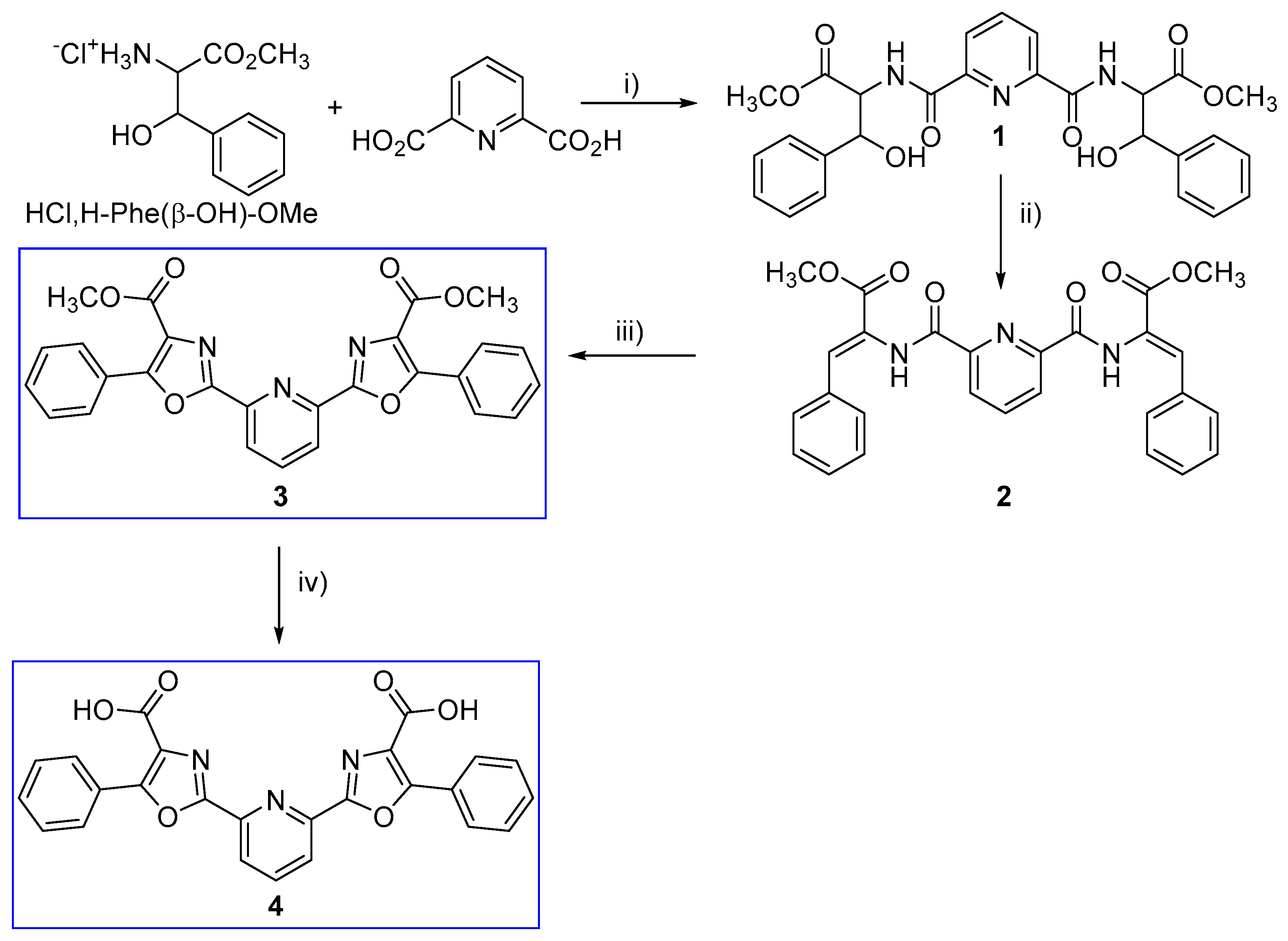
2.1. Synthesis of Compounds

2.1.1. Compound 1

Di-picolinic acid (0.514 g, 3.08 mmol) and the phenyl serine methyl ester (2.2 eq., 1.57 g) were added to acetonitrile (50 mL). The flask was placed in an ice bath and Et3N (1.5 eq., 1.4 mL), HOBt and DCC (2.2 eq., 1.4 g) were added. The reaction was left stirring at room temperature for 18 h. The reaction mixture was filtered and solvent removed under reduced pressure. The residue was recovered in acetone (50 mL) and the solution was left in the freezer for 18 h. The precipitated urea was filtered off and the solvent was removed. The residue was recovered in ethyl acetate (150 mL) and washed with sodium bicarbonate (3 × 50 mL) and with saturated sodium chloride solution (2 × 50 mL). The organic phase was dried with anhydrous magnesium sulfate. After removal of the solvent, compound 1 (1.88 g, 2.88 mmol) was obtained in 93% yield.
1H-NMR (400 MHz, CDCl3 δ): (mixture of diastereomers) 3.71 and 3.72 (2s, 6H, OCH3); 5.00–5.09 (m, 4H, αH + OH); 5.45 (broad s, 2H, βH); 7.19–7.45 (m, 10H, ArH); 7.70–8.10 (m, 3H, ArH); 8.87–8.94 (m, 2H, NH) ppm.

2.1.2. Compound 2

Compound 1 (2.88 mmol, 1.50 g) was dissolved in dry acetonitrile (25 mL) and Boc₂O (2.2 eq. 1.38 g) and DMAP (0.1 eq., 71 mg) were added and left stirring. The reaction was followed by 1H NMR. After checking the disappearance of compound 1, TMG (4% v/v) was added. The solvent was removed and the residue was recovered in ethyl acetate (50 mL), washed with 1M NaHCO₃ (3 × 15 mL) and saturated sodium chloride (3 × 15 mL). The organic phase was dried with anhydrous magnesium sulfate and the solvent removed under reduced pressure. The compound 2 was purified by “dry-flash” using ethyl acetate/petroleum ether 40–60 as eluent. Compound 2 was obtained in 33% yield (0.95 mmol, 0.46 g).
1H NMR (400 MHz, CDCl3 δ): 3.87 (s, 6H, OCH3); 7.28–7.37 (m, 6H, ArH); 7.49 (s, 2H, βCH); 7.50–7.52 (m, 4H, ArH); 8.09 (t, J = 8.0 Hz, 1H, ArH); 8.40 (d, J = 8.0 Hz, 2H, ArH); 9.40 (s, 2H, NH) ppm.

2.1.3. Compound 3

1,2-dichloroethane was treated with 100 mL of 10% H₂SO₄. To 5 mL of the 1,2-dichloroethane solution, compound 2 (142 mg, 0.292 mmol) was added with BF₃O(CH₂CH₃)₂ (4 equivalents, 1.17 mmol, 150 μL). The mixture was refluxed with heating, then PIDA (244 mg, 0.760 mmol, 2.6 equivalents) was added and the mixture dissolved in 2 mL of CH₂Cl₂. A yield of approximately 26% (0.076 mmol, 37 mg) was obtained.
1H NMR (400 MHz, CDCl3 δ): 4.01 (s, 6H, 2 OCH3); 7.53–7.57 (m, 6H, ArH); 8.04 (t, J = 7.6 Hz, 1H, ArH); 8.24–8.26 (m, 4H, ArH); 8.41 (d, J = 7.6 Hz, 2H, ArH) ppm.

2.1.4. Compound 4

All compound 3 (0.076 mmol, 37 mg) obtained previously was dissolved in dioxane (7 mL) and NaOH 1 (M) was added. The reaction was followed by TLC. Then the solvent was evaporated. Water was added, acidified to pH 2–3 and the aqueous phase was extracted. The organic phase was washed with H2O (3 × 15 mL) and saturated NaCl solution (3 × 15 mL). The organic phase was dried with anhydrous MgSO4 and the solvent was removed. The reaction yield was 80% (0.061 mmol, 28 mg).
1H NMR (400 MHz, D2O δ): 7.42–7.48 (m, 6H, 4H ArH); 7.94–7.96 (m, 4H, ArH); 8.05–8.12 (m, 3H, ArH) ppm.

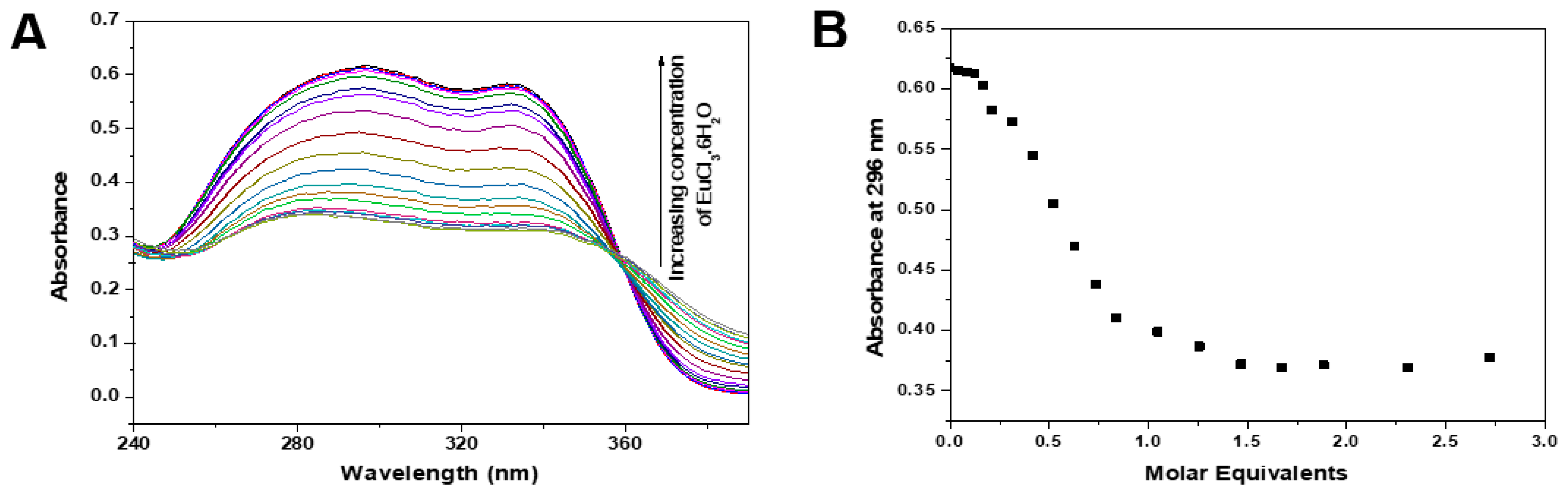
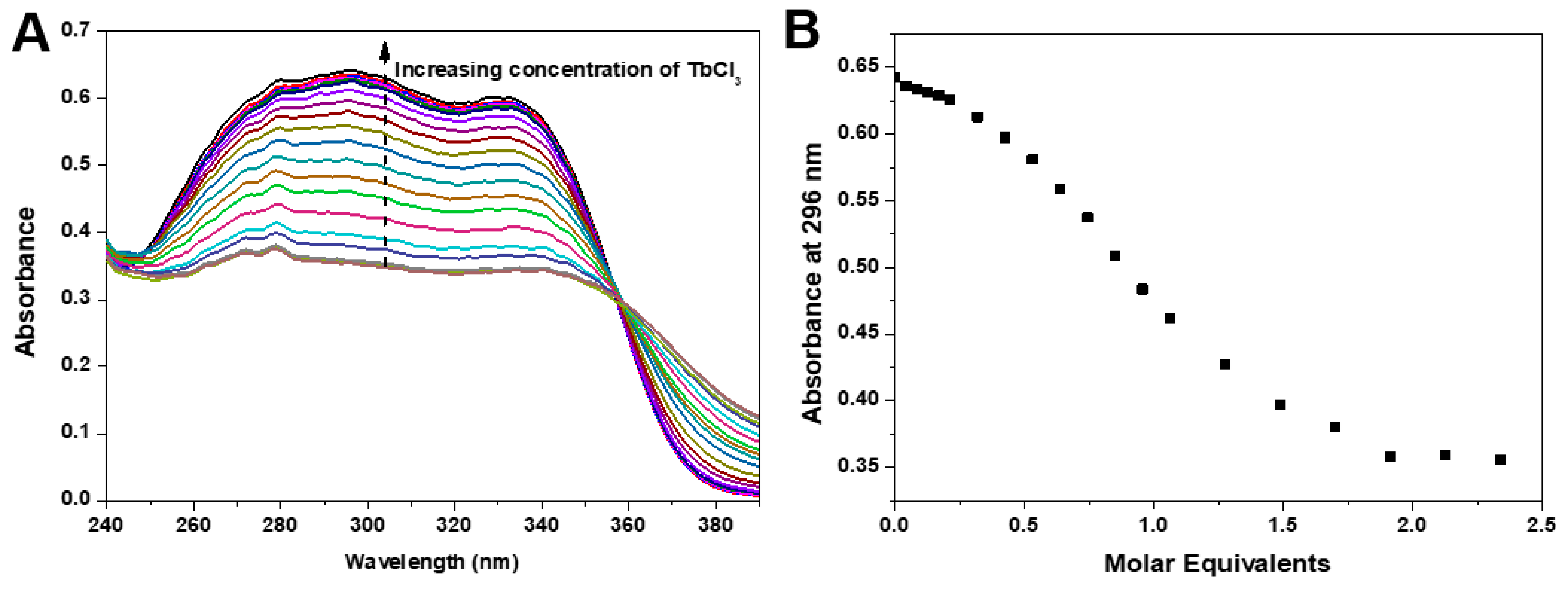
3. Results and Discussion

3.1. Synthesis

Two oxazolyl-pyridine derivatives were prepared from the methyl ester of β-hydroxy-phenylalanine and di-picolinic acid (Figure 1). The synthetic strategy to obtain compound 3 involved a coupling reaction, followed by a dehydration and a cyclization. The cyclization of compound 2 was carried out by treatment with a hypervalent iodine reagent ((diacetoxyiodo)benzene, PIDA). The basic hydrolysis of compound 3 afforded compound 4.

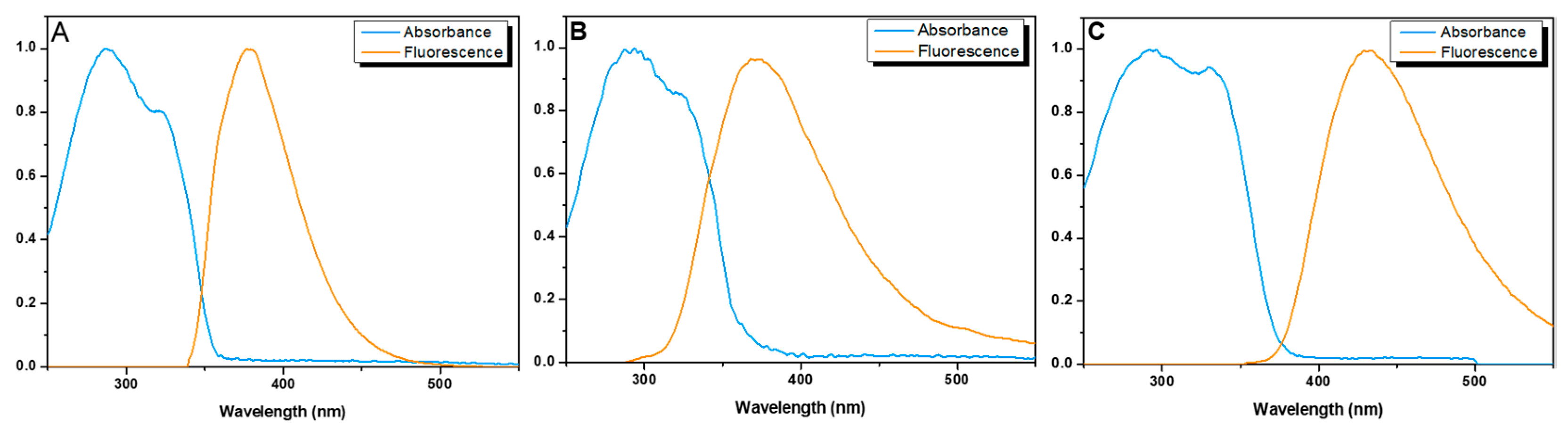
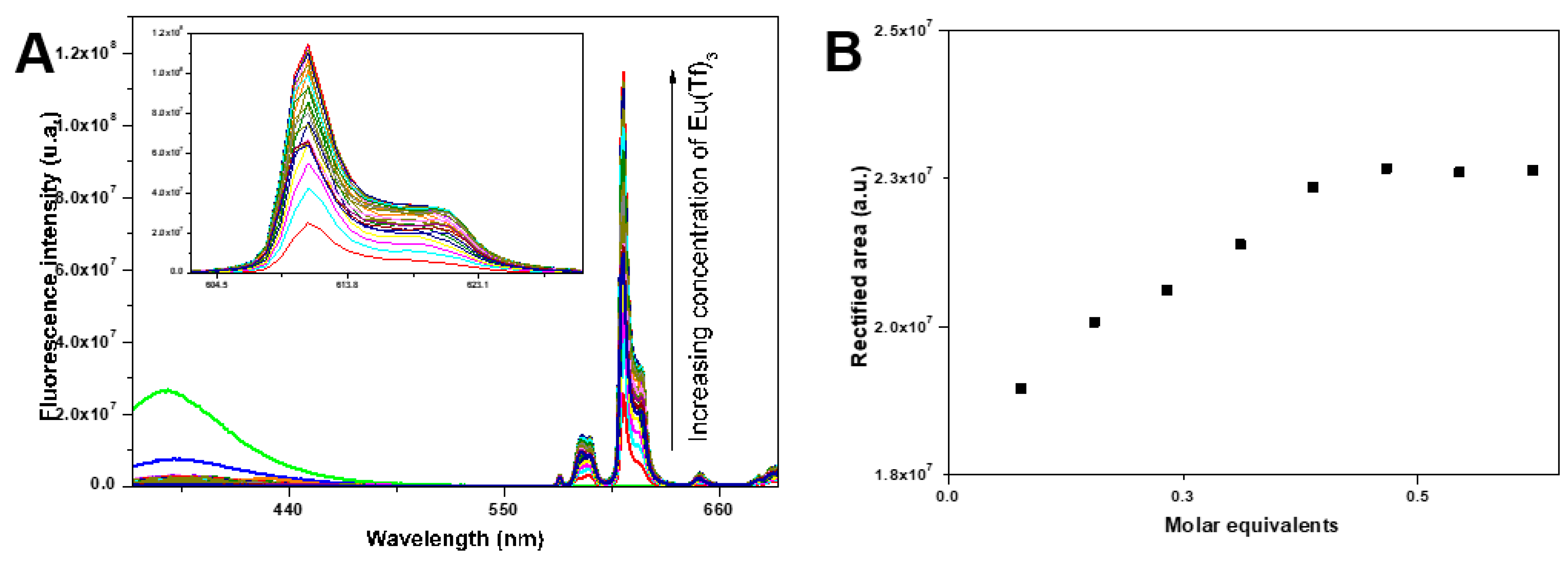
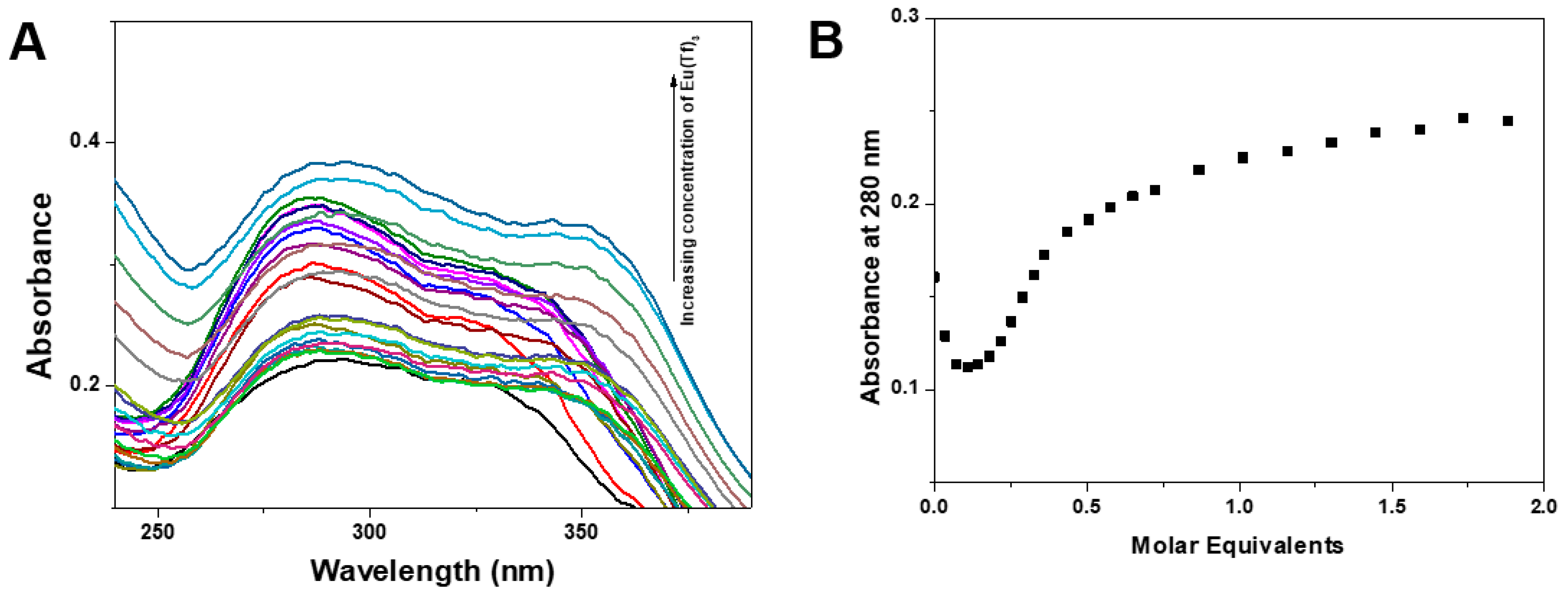
3.2. Fluorescence Properties of Ligands

The photophysical properties of compounds 3 and 4 were studied in acetonitrile and Tris-HCl buffer (0.1 M, pH 7.1). Figure 1 shows the normalized absorption and emission spectra of these compounds. The maximum absorption (λabs) and emission wavelengths (λem), molar absorption coefficients (ε) and fluorescence quantum yields (ΦF) are presented in Table 1. Both compounds 3 and 4 absorb strongly in UV, with maxima at λ = 280 nm (Figure 2). These bis-oxazoles are also fluorescent, with emission quantum yields of around 50% for compound 3 and 30% for compound 4. The fluorescence quantum yields of ligands 3 and 4 are significantly lower than fluorescence quantum yields of analogous ligands with a methyl group at the 5-position of the oxazole ring [7] A red-shift in emission is observed for compound 4 relative to compound 3 (Table 1) which could be attributed, not only to solvent effects, but also to the additional intramolecular charge transfer (ICT) character of the excited state of bis-oxazole 4. This was also observed for other 5-substituted bis-oxazolyl-pyridine derivatives [7].
Fluorescence titration of the ligand 4 in acetonitrile with a solution of Eu(III) trifluoro-methane-sulfonate (Eu(Tf)3) in acetonitrile (Figure 3A) was carried out to determine the stoichiometry of the complex.
As can be seen in Figure 3A, the fluorescence emission band of the ligand 4 at 364 nm (green curve) undergoes a progressive decrease of intensity with the addition of Eu(Tf)3. Simultaneously, new bands appear at 578, 592, 614, and 652 nm, which undergo progressive intensification with the addition of Eu(Tf)3. This phenomenon indicates the formation of a Eu complex with ligand 4 and the sensitization of the Eu3+ ion fluorescence by ligand 4. The bands at 578, 592, 614, and 652 nm have a characteristic shape, dependent on the symmetry of the complex and can be attributed to transitions 5D0-7FJ (J = 0, 1, 2, 3, 4. The progressive intensification of the more intense band at 614 nm (hypersensitive transition) with the addition of Eu(Tf)3 allows estimation of the stoichiometry of the complex.
The stabilization of the curve after the addition of approximately 0.5 molar equivalents of Eu(Tf)3 suggests the formation of a 2:1 complex. The formation of a complex is also likely to cause significant changes in the absorption spectrum of the ligand in the UV-Vis region of the electromagnetic spectrum.
Addition of Eu(Tf)3 causes significant changes in the absorption spectrum of ligand 4 indicating the formation of a complex (Figure 4A).
There is initially a decrease in the band intensity at 280 nm, followed by an increase in the intensity with tendency to stabilize after addition of approximately one molar equivalent of Eu3+ ions (Figure 4B). The decrease in the slope of the curve after the addition of approximately 0.5 molar equivalents of Eu(Tf)3 also suggests the formation of a complex with stoichiometry of 1:2 Eu(4)2 which may continue to react more Eu(Tf)3 to give another complex, possibly with M:L (1:1) Eu(4) stoichiometry.
Ligand 4 is not very soluble in acetonitrile but soluble in aqueous medium. The formation of a water-soluble Eu(4)x luminescent complex would be of great interest for biological studies as a cell marker, sensor or molecular imaging.
Fluorescence titrations (λexc = 280 nm) of aqueous solutions (0.1 M Tris-HCl buffer, pH = 7.1) of ligand 4 with EuCl3.6H2O revealed no change in the emission spectrum of the ligand nor the appearance of bands attributed to electronic transitions of Eu3+ ion. However, the spectrophotometric titration reveals that there is interaction between the ligand 4 and the Eu3+ ion in aqueous solution (Figure 5A).
The dependence of the absorption band intensity at 296 nm on the number of Eu3+ molar equivalents clearly indicates the formation in aqueous solution of an Eu(4) complex with M:L (1:1) stoichiometry (Figure 5B).
While ligand 4 forms a fluorescent complex with Eu3+ in acetonitrile, the respective complex with Tb3+ ions is non-fluorescent in acetonitrile. To investigate the complexation of ligand 4 with Tb3+ ions in aqueous medium a spectrophotometric titration was performed (Figure 6A).
The dependence of the absorption band intensity at 296 nm on the number of molar equivalents of Tb3+ also suggests the formation in aqueous solution of a Tb(4) complex with M:L stoichiometry (1:1) (Figure 6B).

4. Conclusions

In this work, two new ligands of the 2–6-bis(oxazolyl)-pyridine type were synthesized and characterized. The photophysical properties of these ligands were studied in acetonitrile and in aqueous medium (Tris-HCl buffer). The complexation of Ln3+ ions by ligands 3 and 4 was studied in acetonitrile and in aqueous medium through fluorescence titrations and spectrophotometric titrations (UV-Vis). Ligand 4 forms a weakly fluorescent Eu(4)2 complex in acetonitrile with M:L stoichiometry of 1:2. In aqueous medium, ligand 4 forms a non-fluorescent complex with M:L stoichiometry of 1: 1. These results suggest that the ligand structure, due to geometric or stereo restrictions, prevents the formation of a complex with M:L stoichiometry of 1:3. It also appears that in the M:L complexes of 1:2 and 1:1 coordination occurs through the N3 tridentate motif of the pyridine-bis-oxazole nucleus leaving free positions in the metal coordination sphere, which may be occupied by molecules of solvent, with fluorescence quenching. The phenyl group at position 5 of the oxazole ring appears to have a negative effect on the quantum fluorescence yield, the coordinating capacity of the ligands for Ln3+ ions and the efficiency of the Ln3+ ion fluorescence sensitization process.

Author Contributions

Conceptualization, P.M.T.F. and J.A.M.; methodology, P.M.T.F. and J.A.M.; investigation, A.C.; writing—original draft preparation, A.C.; writing—review and editing, P.M.T.F. and J.A.M.; supervision, P.M.T.F. and J.A.M.; funding acquisition, P.M.T.F. and J.A.M. All authors have read and agreed to the published version of the manuscript.

Funding

This work was supported by the Portuguese Foundation for Science and Technology (FCT) in the framework of the Strategic Funding of CQUM (UID/QUI/00686/2019). The NMR spectrometers are part of the National NMR Network (PTNMR) and are partially supported by Infrastructure Project No 022161 (co-financed by FEDER through COMPETE 2020, POCI and PORL and FCT through PIDDAC).

Acknowledgments

André Carvalho acknowledges FCT for a Ph.D. grant (2020.07743.BD).

Conflicts of Interest

The authors declare no conflict of interest.

References

  1. Rocha, J.; Carlos, L.D.; Paz, F.A.A.; Ananias, D. Luminescent Multifunctional Lanthanides-Based Metal–Organic Frameworks. Chem. Soc. Rev. 2011, 40, 926–940. [Google Scholar] [CrossRef] [PubMed]
  2. Weissman, S.I. Intramolecular Energy Transfer The Fluorescence of Complexes of Europium. J. Chem. Phys. 1942, 10, 214–217. [Google Scholar] [CrossRef]
  3. Su, Y.; Yu, J.; Li, Y.; Phua, S.F.Z.; Liu, G.; Lim, W.Q.; Yang, X.; Ganguly, R.; Dang, C.; Yang, C.; et al. Versatile Bimetallic Lanthanide Metal-Organic Frameworks for Tunable Emission and Efficient Fluorescence Sensing. Commun. Chem. 2018, 1, 12. [Google Scholar] [CrossRef]
  4. De Bettencourt-Dias, A.; Barber, P.S.; Viswanathan, S.; de Lill, D.T.; Rollett, A.; Ling, G.; Altun, S. Para-Derivatized Pybox Ligands As Sensitizers in Highly Luminescent Ln(III) Complexes. Inorg. Chem. 2010, 49, 8848–8861. [Google Scholar] [CrossRef] [PubMed]
  5. De Bettencourt-Dias, A.; Rossini, J.S.K.; Sobrinho, J.A. Effect of the Aromatic Substituent on the Para-Position of Pyridine-Bis(Oxazoline) Sensitizers on the Emission Efficiency of Their EuIII and TbIII Complexes. Dalt. Trans. 2020, 49, 17699–17708. [Google Scholar] [CrossRef] [PubMed]
  6. Arrico, L.; Benetti, C.; Di Bari, L. Combining Lanthanides with PyBox Ligands: A Simple Route to Circularly Polarized Light Emitters. ChemPhotoChem 2021, 5, 815–821. [Google Scholar] [CrossRef]
  7. Pereira, G.; Ferreira, M.F.; Castanheira, E.M.S.; Martins, J.A.; Ferreira, P.M.T. Synthesis of 2,6-Bis(Oxazolyl)Pyridine Ligands for Luminescent LnIII Complexes. Eur. J. Org. Chem. 2012, 2012, 3905–3910. [Google Scholar] [CrossRef]
  8. Chen, R.F. Fluorescence Quantum Yields of Tryptophan and Tyrosine. Anal. Lett. 1967, 1, 35–42. [Google Scholar] [CrossRef]
Figure 1. Synthesis of bis-(phenyl-oxazolyl)piridine derivatives 3 and 4. (i) DCC/HOBT; (ii) 1. Boc2O/DMAP, 2. TMG (2%); (iii) PIDA, BF3; (iv) NaOH 1M.
Chemproc 12 00056 g001
Figure 2. Normalized absorption and fluorescence spectra (λexc = 280 nm) of compounds 3 and 4. (A) Ligand 3 in MeCN; (B) Ligand 4 in MeCN; (C) Ligand 4 in Tris-HCl Buffer (0.1 M; pH = 7.1).
Chemproc 12 00056 g002
Figure 3. (A) Fluorescence titration (λexc = 280 nm) of a solution of ligand 4 (1.24 × 10⁻⁵ M in acetonitrile, 2 mL) with a solution of Eu(Tf)3 (9.68 × 10⁻⁴ M, in acetonitrile) (B) Representation of the rectified area of the emission band of the Eu(4)x complex at 614 nm (λexc = 280 nm) as a function of the number of molar equivalents of Eu(Tf)3 added.
Chemproc 12 00056 g003
Figure 4. (A) Spectrophotometric titration of a solution of ligand 4 (1.34 × 10⁴ M in acetonitrile, 1 mL) with Eu (Tf)3 (9.68 × 10⁴ M, in acetonitrile) (B) Dependence of the intensity of the absorption band at 280 nm of a solution of ligand 4 (1.34 × 10⁴ M, in acetonitrile) with the number of molar equivalents of Eu(Tf)3 added.
Chemproc 12 00056 g004
Figure 5. (A) Spectrophotometric titration of a solution of ligand 4 (8.29 × 10⁵ M, in 0.1 M Tris-HCl buffer, pH = 7.1) with EuCl3.6H2O (1.74 × 10³ M, in 0.1 M Tris-HCl buffer), pH = 7.1) (B) Dependence of the intensity of the absorption band at 296 nm on the number of Eu3+ molar equivalents in the titration of an aqueous solution (0.1 M Tris-HCl buffer, pH = 7.1) of ligand 4 (8.29 × 10⁵ M) with an aqueous solution of EuCl3.6H2O (1.74 × 10³ M).
Chemproc 12 00056 g005
Figure 6. (A) Spectrophotometric titration of a solution of ligand 4 (8.29 × 10⁵ M, in 0.1 M Tris-HCl buffer, pH = 7.1) with TbCl3.6H2O (1.54 × 10³ M, in 0.1 M Tris-HCl buffer, pH = 7.1) (B) Dependence of the intensity of the absorption band at 296 nm on the number of Tb3+ molar equivalents in the titration of an aqueous solution (0.1 M Tris-HCl buffer, pH = 7.1) of ligand 4 (8.29 × 10⁵ M) with an aqueous solution of TbCl3.6H2O (1.54 × 10³).
Chemproc 12 00056 g006
Table 1. Maximum absorption (λabs) and emission wavelengths (λem), molar absorption coefficients (ε) and fluorescence quantum yields (ΦF) for compounds 3 and 4.
LigandSolventλabs (nm)
(ε M−1cm−1)
λem (nm) *
(Stokes Shift, nm)
ΦF **
3Acetonitrile 280   ( 3.47 × 10 4 )376 (96)0.49
4Acetonitrile 280   ( 488.7 )364 (84)0.32
4Tris-HCl buffer 0.1 M, pH = 7.1 280   ( 7.28 × 10 3 )425 (145)0.35
* λexc = 280 nm. ** using tryptophan as standard, ΦF = 0.13 [8].
Disclaimer/Publisher’s Note: The statements, opinions and data contained in all publications are solely those of the individual author(s) and contributor(s) and not of MDPI and/or the editor(s). MDPI and/or the editor(s) disclaim responsibility for any injury to people or property resulting from any ideas, methods, instructions or products referred to in the content.

