Proceeding Paper

Can the Antimicrobial Peptide Ctx(Ile21)-Ha-Ahx-Cys Grafted onto Nanochitosan Sensitize Extensively Drug-Resistant Mycobacterium tuberculosis? †

by
Laura Maria Duran Gleriani Primo
1,
Cesar Augusto Roque-Borda
1,*,
Eduardo Festozo Vicente
2,
Paula Aboud Barbugli
3 and
Fernando Rogério Pavan
1,*
1
Tuberculosis Research Laboratory, School of Pharmaceutical Sciences, São Paulo State University (UNESP) Araraquara, São Paulo 14801-903, Brazil
2
School of Sciences and Engineering, São Paulo State University (UNESP), Tupã, São Paulo 17602-496, Brazil
3
Department of Dental Materials and Prosthodontics, School of Dentistry, São Paulo State University (UNESP), Araraquara, São Paulo 14801-903, Brazil
*
Authors to whom correspondence should be addressed.
Presented at the 26th International Electronic Conference on Synthetic Organic Chemistry, 15–30 November 2022; Available online: https://sciforum.net/event/ecsoc-26.
Chem. Proc. 2022, 12(1), 51; https://doi.org/10.3390/ecsoc-26-13700
Published: 18 November 2022

Abstract

:
The infectious agent Mycobacterium tuberculosis (MTB) has several defense and resistance mechanisms that must be eliminated. The treatment is prolonged, which in many cases generates susceptibility to microbial resistance. This research aimed to study whether the antimicrobial peptide Ctx(Ile21)-Ha-Ahx-Cys (Ctx-SH) functionalized in nanochitosan matrices could eliminate resistant MTB. For this, a nanosystem was developed with chitosan matrices previously modified with N-acetylcysteine functionalized to Ctx-SH. Modified chitosan nanoparticles (NPQ) were obtained by ionic gelation using sodium tripolyphosphate and loaded with rifampicin. Both chitosan and NPQ modifications were analyzed for physicochemical parameters by Fourier/Raman transform infrared spectroscopy and Zeta potential. Antimicrobial activity was performed in a level 3 biosafety laboratory with strains H37Rv (standard) and CF169 (extensively drug-resistant, XDR) incubated in 7H9 broth supplemented with oleic acid, albumin, dextrose, and catalase at 37 °C and 5% CO2 and read using fluorescence with 0.01% resazurin after 7 days. Insertion and mapping of NPQ into macrophages were assessed using a confocal microscope after 24 h with NPQ conjugated to fluorescein isothiocyanate. Preliminary results show that the spectroscopies corroborate the hypothesis of the functionalization of the Ctx-SH peptide to the chitosan-N-acetylcysteine system because, when comparing the three spectroscopies, a gradual increase in the intensity of several bands and the formation of captive disulfide are observed, and the Zeta potential (+30 mV) confirmed high application stability. Bacterial inhibition studies revealed that rifampicin-loaded antimicrobial peptide-conjugated chitosan nanoparticles have better activity than rifampicin alone against CF169, with a minimum inhibitory concentration of <0.977 µg/mL, similar to the standard strain. In addition, it was shown that NPQ would be able to enter the macrophage without causing toxicity and thus take better advantage of the activity of rifampicin. Finally, it is possible to verify that the nanobioconjugation of the Ctx-SH-N-acetylcysteine-chitosan compound is capable of enhancing the activity of obsolete drugs and/or sensitizing XDR bacteria.

1. Introduction

Tuberculosis (TB) has as its etiologic agent the bacillus alcohol-acid resistant Mycobacterium tuberculosis (MTB), and it is reported as one of the world’s leading infectious killers by the World Health Organization (WHO) [1,2]. The pathology can be briefly described as: an infection by the air pathway, a phagocytosis of the bacteria by the lung´s resident macrophages, an adaptation of the mycobacteria to the phagolysosome environment, and an inhibition of MTB death [1,3]. The disease can be characterized by a chronic inflammation of the air pathway and pulmonary cavitation [4]. The current world situation is critical, especially with the increase of cases of drug-resistant TB, which hinders the use of first-line anti-MTB drugs (such as isoniazid, rifampicin, and ethambutol) [5]. Antimicrobial peptides (AMPs) are macromolecules that have a fast bactericidal effect, a low minimal inhibitory concentration (MIC), and a low possibility of bacterial resistance [6,7]. They are also compounds of the innate immune system, and as such, they can modulate the immune response [8]. The AMP Ctx(Ile21)-Ha is a peptide with 21 residues of amino acids, a cationic feature, and action against Gram-negative and Gram-positive bacteria [9,10,11]. The mechanism of action of Ctx(Ile21) is the barrel-stave model, which means it forms pores in the bacterial membrane [9]. This AMP has a potentially biologic application when inserted into polymeric superficies, such as chitosan [12]. Chitosan is a polysaccharide with great biocompatibility, low toxicity, and antibacterial activity [13,14]. Its mechanism of action is encompassing the bacterial cell and carries out a cationic multi-attack [15]. Because TB is an intracellular facultative pathogen of macrophages, it is necessary to develop strategies of drug delivery to these cells [7,12]. One of these strategies is to use nanotechnology as target delivery in a way to improve the pharmacology proprieties of molecules and to enable the controlled release of drugs [11,16]. Ctx(Ile21)-Ha AMP grafted onto nanochitosan by the bisulfate bond demonstrated lower MICs to S. aureus and P. aeruginosa [12]. It can also add the strategy of modifying chitosan with N-acetylcysteine to improve pharmacology proprieties and half-time [17]. It was demonstrated that N-acetylcysteine has anti-biofilm action and is an important inhibitor of air pathway infection [18]. The aim of this work was to analyze the application of Ctx (Ile21)-Ha-Ahx-Cys grafted onto nanochitosan to sensitize extremely resistant MTB strains.

2. Material and Methods

2.1. Chemical Reagents

Chitosan, N-acetylcysteine, sodium tripolyphosphate, resin, Fmoc-aminoacids, and other reagents for peptide synthesis were purchased from Sigma-Aldrich; dimethylformamide (DMF) was purchased from Neon Comercial (São Paulo, Brazil); and dichloromethane (DCM) was purchased from Anidrol Laboratory (São Paulo, Brazil).

2.2. Synthesis of Antimicrobial Peptide

The synthesis of AMP Ctx(Ile21) was assembled by standard solid-phase peptide syntheses methodologies by Fmoc/tBu. Rick amide MBHA resin was used as the solid support. It started with 1:1 v/v DMF/DCM to wash the resin, and then a solution of 20% 4-methylpiperidine in DMF was added in order to remove the Fmoc group from it. Then it was time to add the amino acid Fmoc- Ile (1.2 eq, molar equivalents) with 1.2/1.2 eq of hydroxybenzotriazole/N,N′-diisopropylcarbodiimide and 1:1 v/v DMF/DCM for 2 h at 40 °C. The Kaiser test was used to confirm the couplings, with a blue color for negative results. With a positive result, it was time to add the solution of 20% 4-methylpiperidine in DMF and the next amino acid in the sequence. With the primary sequence coupling complete, the resin was removed using a solution of trifluoroacetic acid, triisopropylsilane, and water 95:2.5:2.5 v/v/v for 2 h at room temperature. After that, the peptide was precipitated with cold ethyl ether and centrifuged three times (at 6500× g for 5 min). The supernatant was dried and then the acetic acid 30% solvent was added after the centrifuge and freeze-dried [7].
The purification and characterization of the Ctx(Ile21)-Ha peptide was carried out using liquid chromatography/mass spectroscopy and the HPLC (Shimadzu, with DGU-20A5R membrane degasser, CTO-20A column oven, sampler automatic SIL-10AF, fraction collector FRC-10A, UV detector SPD-20A, and LC-20AT of double pump), respectively. The mobile phase was an aqueous solution with TFA and an acetonitrile solution with TFA. The method had a 1 mL min−1 flow and a dual mode with wavelengths at 220 nm and 280 nm. The products of the separation were collected by an automatic fraction collector and analyzed by comparing the chromatographic profile [7].

2.3. Purification of Commercial Chitosan

According to the method of purification described by Costa et al. [19], the chitosan was solubilized with acetic acid at 1% and precipitated with sodium hydroxide at 1 M. After that, it was washed with distillate water, centrifuged, and filtrated with a 0.2-nm membrane filter. Later, it was lyophilized.

2.4. Modification of Chitosan with N-Acetylcysteine

The modification of chitosan with N-acetylcysteine was carried out by the method of carbodiimide [20]. The previously purificated chitosan was solubilized with 1% acetic acid and added to a conjugation solution (50 mM of N-ethylcarbodiimide hydrochloride and 50 mM of N-hydroxysuccinimide). N-acetylcysteine 1:1:1 v/v/v was added to this solution at room temperature with agitation for 24 h.

2.5. Functionalization Ctx(Ile21)-Ha onto N-Acetylcysteine-Chitosan

Disulfide bonds were spontaneously formed to graft the Ctx(Ile21)-Ha onto the surface of N-acetylcysteine-chitosan. N-acetylcysteine-chitosan compound was solubilized with 1% acetic acid, and after 12 h of stirring, it was precipitated with 0.1 M sodium hydroxide. Finally, it was filtered and lyophilized [21].

2.6. Nanoparticles Formation

In order to obtain the nanoparticles of N-acetylcystein-chitosan with Ctx(Ile21), the method of ionic gelation was used [7]. The compound N-acetylcystein-chitosan with Ctx(Ile21) was solubilized with acetic acid at 1% for 2 h, and then it was added to rifampicin and Tween 80. It was homogenized and 5 mg/mL of sodium tripolyphosphate was added in a controlled agitation.

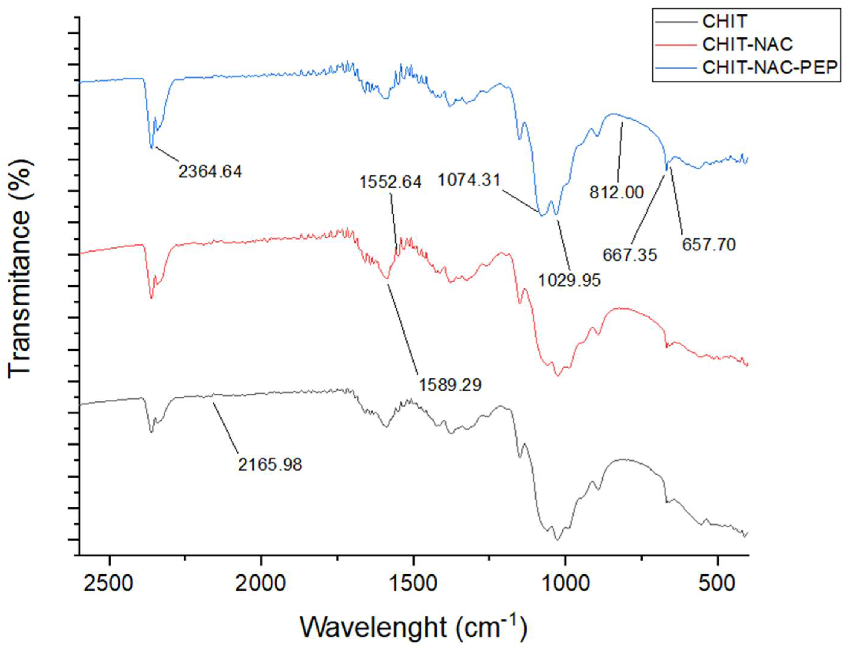
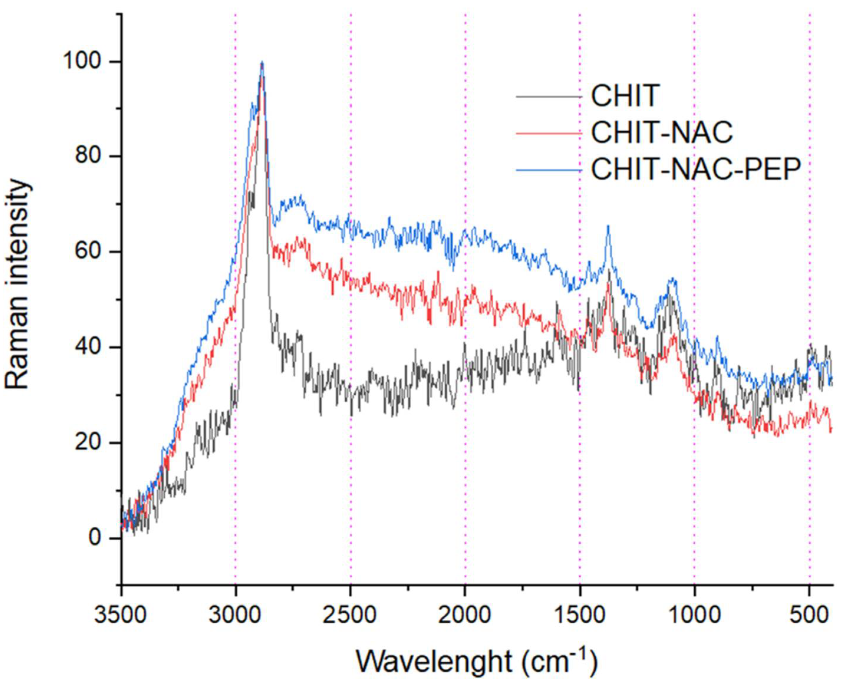
2.7. Physicalchemical Characterization

Infrared spectroscopy (Perkin-Elmer, Frontier model, Waltham, MA, USA) was used with an attenuated total reflectance fixture (Bruker Vertex 70 FTIR) and a resolution of 4 cm. Raman spectroscopy (Burker Ram II Raman spectrophotometer) was also used. Both spectra were analyzed by Origin Pro 2019 software. It was also used by a Zetasizer to determine the Zeta potential.

2.8. Activity against Mycobacterium tuberculosis

Accordingly to Palomino et al. [22], antimicrobial activity against MTB was carried out using the Resazurin Microtiter Assay (REMA) method. Briefly, a nanoparticle solution was added to Middlebrook 7H9 Broth (Difco®), which was supplied with oleic acid, bovine albumin, sodium chloride, dextrose, and catalase. Rifampicin and isoniazid (25 µg/mL) were used as controls. Nanoparticles loaded and controls were tested against MTB strains H37Rv (standard) and CF169 (clinically isolated and extremely resistant). The samples were incubated for seven days at 37 °C and 5% CO2 and read with 30 µL of resazurin at 0.001% by fluorescence.

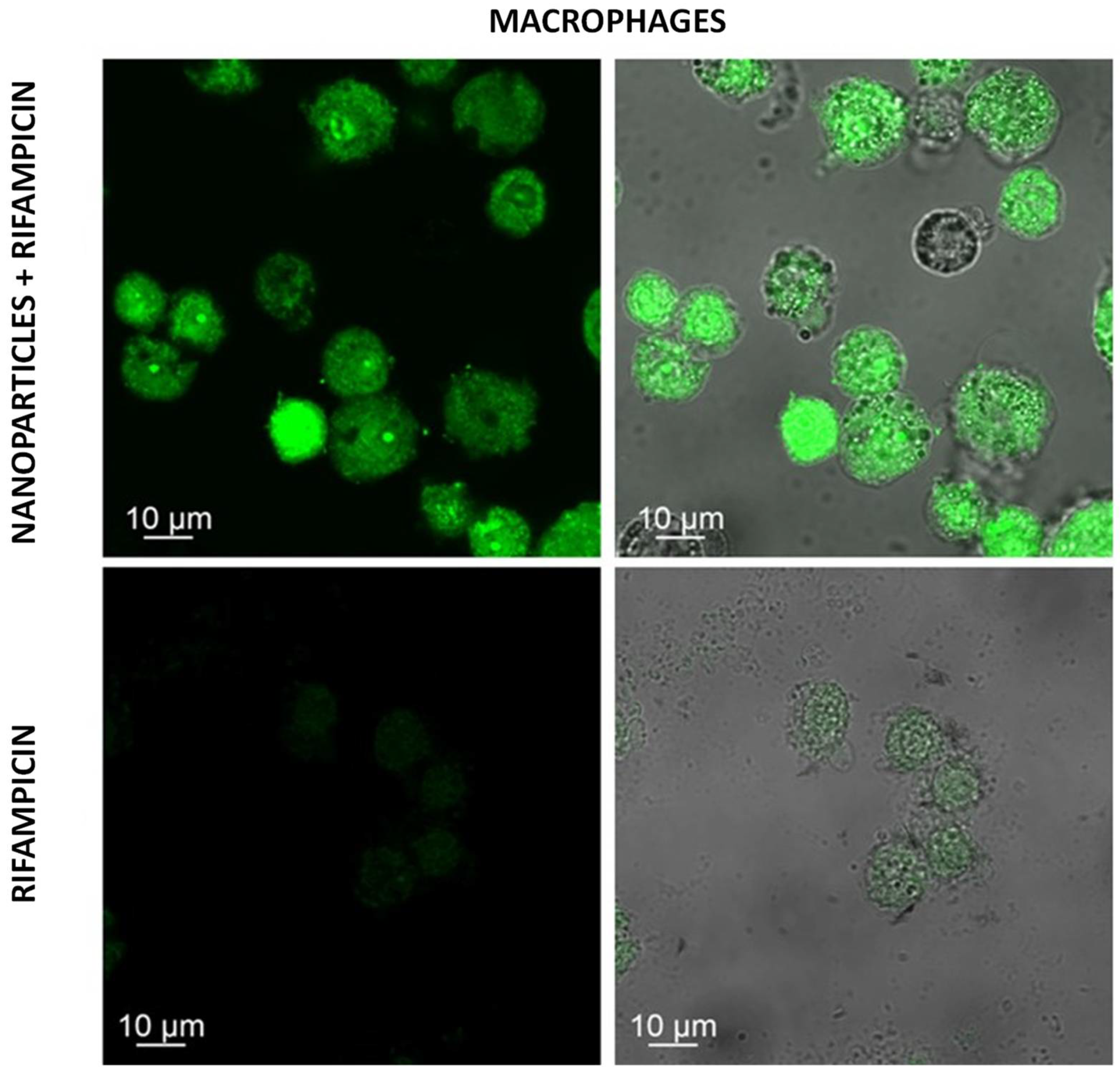
2.9. Confocal Microscopy

The polymers before their encapsulation were conjugated with fluorescein isothiocyanate, precipitated with NaOH, and washed with distilled water until pH 7 was obtained.
Nanoparticles were then formed and brought to a confocal microscope 24 h after incubation in murine macrophages at a concentration of 1 mg/mL.

3. Results and Discussion

The spectroscopies confirm the modification and functionalization of the nanocomposites with Ctx(Ile21) Ha-Ahx-Cys. Infrared spectroscopy (FTIR) was used in three samples of nanoparticles: chitosan purification, chitosan modified with N-acetylcysteine, and chitosan modified with N-acetylcysteine and functionalized with Ctx(Ile21)Ha-Ahx-Cys. The intensity of the band 2364 cm−1, corresponding to nitrile groups, was higher in the nanoparticles with modifications, and the intensity of the bands 1029 cm−1 and 1074 cm−1, corresponding to free amine groups, was lower in the same samples [23]. That confirmed that the modification of chitosan with N-acetylcysteine was effective. It was also possible to observe in the spectroscopy with modification and functionalized a band in 1457cm−1 (Figure 1), corresponding to CH2 and CH3 of aromatics structures of the secondary structure of Ctx(Ile21)-Ha-Ahx-Cys, which shows that the functionalization of the peptide was successful [7].
The method of RAMAN spectroscopy was used with the same samples. It confirmed the functionalization of Ctx(Ile21)-Ha-Ahx-Cys onto a modified polymer. This is because it has shown bands as 885 cm−1, 1004 cm−1, and 1333 cm−1, corresponding to aromatics amino acids in the peptide (Figure 2). A band in 1230 cm−1 was also observed, a characteristic of the secondary structure of Ctx(Ile21)-Ha-Ahx-Cys [7].
The anti-Mycobacterium tuberculosis activity was evaluated with the Minimal Inhibitory Concentration (MIC) of the nanoparticles of chitosan- N- acetylcysteine and was functionalized with Ctx(Ile21)-Ha-Ahx-Cys and loaded with rifampicin. The MIC of the compound against CF169, a clinically isolated multi-resistant strain of tuberculosis, was 0.977 µg/mL. The MIC value of this compound against the H37Rv MTB strain was lower than 0.977 µg/mL. Considering that the MIC of rifampicin, the control compound against CF169 is 25 µg/mL [24], and the similarity of the values of the MIC (Table 1), it can be concluded that the nanoparticles were able to sensitize this clinically isolated multi-resistance to drugs.
The images obtained with confocal microscopy (Figure 1) showed that the nanoparticles of chitosan-N-acetylcysteine functionalized with Ctx(Ile21)-Ha-Ahx-Cys and loaded with rifampicin were able to go through the plasmatic membrane of macrophages. It can be observed on the Figure 3 that the fluorescence intensity in the image of the compound is higher than in the image of the control, rifampicin isolated. Considering that macrophages are the most common cells infected with MTB [25], the ability of the compound to enter the intracellular environment can indicate a select transport of drug propriety. It is also possible to conclude that the compound is not cytotoxic to this cell.

4. Conclusions

It can be concluded that the antimicrobial peptide Ctx(Ile21)-Ha-Ahx-Cys grafted onto nanochitosan was able to sensitize a strain of extremely resistant Mycobacterium tuberculosis and intensify the effect of rifampicin, drug obsolete against CF169.

Author Contributions

Conceptualization, L.M.D.G.P. and C.A.R.-B.; investigation, L.M.D.G.P., C.A.R.-B., F.R.P., P.A.B. and E.F.V.; writing—original draft preparation, L.M.D.G.P.; writing—review and editing, L.M.D.G.P., C.A.R.-B. and F.R.P.; supervision, F.R.P.; funding acquisition, F.R.P. All authors have read and agreed to the published version of the manuscript.

Funding

This research was funded by Fundação de Amparo à Pesquisa do Estado de São Paulo, grant number 20/13497-4; 2020/16573-3; 2021/14603-5 and by Conselho Nacional de Desenvolvimento Científico e Tecnológico, grant number 429139/2018-7; 404181/2019-8; 303603/2018-6.

Institutional Review Board Statement

Not applicable.

Informed Consent Statement

Not applicable.

Data Availability Statement

Not applicable.

Conflicts of Interest

The authors declare no conflict of interest.

References

  1. Bell, L.C.K.; Noursadeghi, M. Pathogenesis of HIV-1 and Mycobacterium tuberculosis co-infection. Nat. Rev. Genet. 2017, 16, 80–90. [Google Scholar] [CrossRef] [PubMed]
  2. Khawbung, J.L.; Nath, D.; Chakraborty, S. Drug resistant Tuberculosis: A review. Comp. Immunol. Microbiol. Infect. Dis. 2021, 74, 101574. [Google Scholar] [CrossRef] [PubMed]
  3. Simmons, J.; Stein, C.; Seshadri, C.; Campo, M.; Alter, G.; Fortune, S.; Schurr, E.; Wallis, R.S.; Churchyard, G.; Mayanja-Kizza, H.; et al. Immunological mechanisms of human resistance to persistent Mycobacterium tuberculosis infection. Nat. Rev. Immunol. 2018, 18, 575–589. [Google Scholar] [CrossRef]
  4. Sakamoto, K. The Pathology of Mycobacterium tuberculosis Infection. Veter Pathol. 2012, 49, 423–439. [Google Scholar] [CrossRef]
  5. Laws, M.; Jin, P.; Rahman, K.M. Efflux pumps in Mycobacterium tuberculosis and their inhibition to tackle antimicrobial resistance. Trends Microbiol. 2021, 30, 57–68. [Google Scholar] [CrossRef]
  6. Bahar, A.A.; Ren, D. Antimicrobial Peptides. Pharmaceuticals 2013, 6, 1543–1575. [Google Scholar] [CrossRef]
  7. Roque-Borda, C.A.; Silva, H.R.L.; Junior, E.C.; Serafim, J.A.; Meneguin, A.B.; Chorilli, M.; Macedo, W.C.; Teixeira, S.R.; Guastalli, E.A.L.; Soares, N.M.; et al. Alginate-based microparticles coated with HPMCP/AS cellulose-derivatives enable the Ctx(Ile21)-Ha antimicrobial peptide application as a feed additive. Int. J. Biol. Macromol. 2021, 183, 1236–1247. [Google Scholar] [CrossRef]
  8. Magana, M.; Pushpanathan, M.; Santos, A.L.; Leanse, L.; Fernandez, M.; Ioannidis, A.; Giulianotti, M.A.; Apidianakis, Y.; Bradfute, S.; Ferguson, A.L.; et al. The value of antimicrobial peptides in the age of resistance. Lancet Infect. Dis. 2020, 20, e216–e230. [Google Scholar] [CrossRef]
  9. Lorenzón, E.N.; Cespedes, G.F.; Vicente, E.F.; Nogueira, L.G.; Bauab, T.M.; Castro, M.S.; Cilli, E.M. Effects of Dimerization on the Structure and Biological Activity of Antimicrobial Peptide Ctx-Ha. Antimicrob. Agents Chemother. 2012, 56, 3004–3010. [Google Scholar] [CrossRef]
  10. Cespedes, G.F.; Lorenzon, E.N.; Vicente, E.F.; Mendes-Giannini, M.J.S.; Fontes, W.; Castro, M.D.S.; Cilli, E.M. Mechanism of Action and Relationship Between Structure and Biological Activity of Ctx-Ha: A New Ceratotoxin-like Peptide from Hypsiboas albopunctatus. Protein Pept. Lett. 2012, 19, 596–603. [Google Scholar] [CrossRef]
  11. Roque-Borda, C.A.; da Silva, P.B.; Rodrigues, M.C.; Azevedo, R.B.; Di Filippo, L.; Duarte, J.L.; Chorilli, M.; Festozo Vicente, E.; Pavan, F.R. Challenge in the Discovery of New Drugs: Antimicrobial Peptides against WHO-List of Critical and High-Priority Bacteria. Pharmaceutics 2021, 13, 773. [Google Scholar] [CrossRef] [PubMed]
  12. Roque-Borda, C.A.; Antunes, B.F.; Borgues, A.B.T.; de Pontes, J.T.C.; Meneguin, A.B.; Chorilli, M.; Trovatti, E.; Teixeira, S.R.; Pavan, F.R.; Vicente, E.F. Conjugation of Ctx(Ile21)-Ha Antimicrobial Peptides to Chitosan Ultrathin Films by N-Acetylcysteine Improves Peptide Physicochemical Properties and Enhances Biological Activity. ACS Omega 2022, 7, 28238–28247. [Google Scholar] [CrossRef] [PubMed]
  13. Aranaz, I.; Alcántara, A.R.; Civera, M.C.; Arias, C.; Elorza, B.; Caballero, A.H.; Acosta, N. Chitosan: An Overview of Its Properties and Applications. Polymers 2021, 13, 3256. [Google Scholar] [CrossRef] [PubMed]
  14. Ke, C.-L.; Deng, F.-S.; Chuang, C.-Y.; Lin, C.-H. Antimicrobial Actions and Applications of Chitosan. Polymers 2021, 13, 904. [Google Scholar] [CrossRef] [PubMed]
  15. Rashki, S.; Asgarpour, K.; Tarrahimofrad, H.; Hashemipour, M.; Ebrahimi, M.S.; Fathizadeh, H.; Khorshidi, A.; Khan, H.; Marzhoseyni, Z.; Salavati-Niasari, M.; et al. Chitosan-based nanoparticles against bacterial infections. Carbohydr. Polym. 2021, 251, 117108. [Google Scholar] [CrossRef]
  16. Nasiruddin, M.; Neyaz, K.; Das, S. Nanotechnology-Based Approach in Tuberculosis Treatment. Tuberc. Res. Treat. 2017, 2017, 1–12. [Google Scholar] [CrossRef]
  17. Costa, F.; Sousa, D.M.; Parreira, P.; Lamghari, M.; Gomes, P.; Martins, M.C.L. N-acetylcysteine-functionalized coating avoids bacterial adhesion and biofilm formation. Sci. Rep. 2017, 7, 1–13. [Google Scholar] [CrossRef]
  18. Blasi, F.; Page, C.; Rossolini, G.M.; Pallecchi, L.; Matera, M.G.; Rogliani, P.; Cazzola, M. The effect of N -acetylcysteine on biofilms: Implications for the treatment of respiratory tract infections. Respir. Med. 2016, 117, 190–197. [Google Scholar] [CrossRef]
  19. Costa, F.M.; Maia, S.R.; Gomes, P.A.; Martins, M.C.L. Dhvar5 antimicrobial peptide (AMP) chemoselective covalent immobilization results on higher antiadherence effect than simple physical adsorption. Biomaterials 2015, 52, 531–538. [Google Scholar] [CrossRef]
  20. Du, Z.; Liu, J.; Zhai, J.; Huang, H.; Wei, S.; Zhang, T.; Ding, L.; Liu, B. Fabrication of N-acetyl-l-cysteine and l-cysteine functionalized chitosan-casein nanohydrogels for entrapment of hydrophilic and hydrophobic bioactive compounds. Food Hydrocoll. 2019, 96, 377–384. [Google Scholar] [CrossRef]
  21. Barbosa, M.; Costa, F.; Monteiro, C.; Duarte, F.; Martins, M.C.L.; Gomes, P. Antimicrobial coatings prepared from Dhvar-5-click-grafted chitosan powders. Acta Biomater. 2019, 84, 242–256. [Google Scholar] [CrossRef] [PubMed]
  22. Palomino, J.-C.; Martin, A.; Camacho, M.; Guerra, H.; Swings, J.; Portaels, F. Resazurin Microtiter Assay Plate: Simple and Inexpensive Method for Detection of Drug Resistance in Mycobacterium tuberculosis. Antimicrob. Agents Chemother. 2002, 46, 2720–2722. [Google Scholar] [CrossRef] [PubMed]
  23. Anand, M.; Sathyapriya, P.; Maruthupandy, M.; Beevi, A.H. Synthesis of chitosan nanoparticles by TPP and their potential mosquito larvicidal application. Front. Lab. Med. 2018, 2, 72–78. [Google Scholar] [CrossRef]
  24. Rivas, F.; Medeiros, A.; Arce, E.R.; Comini, M.; Ribeiro, C.M.; Pavan, F.R.; Gambino, D. New heterobimetallic ferrocenyl derivatives: Evaluation of their potential as prospective agents against trypanosomatid parasites and Mycobacterium tuberculosis. J. Inorg. Biochem. 2018, 187, 73–84. [Google Scholar] [CrossRef] [PubMed]
  25. Santucci, P.; Greenwood, D.J.; Fearns, A.; Chen, K.; Jiang, H.; Gutierrez, M.G. Intracellular localisation of Mycobacterium tuberculosis affects efficacy of the antibiotic pyrazinamide. Nat. Commun. 2021, 12, 3816. [Google Scholar] [CrossRef]
Figure 1. Infrared spectroscopy of the samples.
Chemproc 12 00051 g001
Figure 2. RAMAN spectroscopy of the samples.
Chemproc 12 00051 g002
Figure 3. Images of the confocal microscopy of the nanoparticles of chitosan-N-acetylcysteine functionalized with Ctx(Ile21)-Ha-Ahx-Cys loaded with rifampicin and the control, rifampicin isolated. The first and second were corresponding to the nanoparticles, and the third and fourth were corresponding to rifampicin.
Chemproc 12 00051 g003
Table 1. This table shows the results of minimal inhibitory concentration values from chitosan modified nanoparticles loaded with rifampicin and the control compound (rifampicin) against the strains H37Rv and CF169.
CompoundMinimal Inhibitory Concentration (MIC) against CF169 (µg/mL)Minimal Inhibitory Concentration (MIC) against H37Rv (µg/mL)
Nanoparticles loaded with rifampicin 0.977<0.977
Rifampicin 250.977
Disclaimer/Publisher’s Note: The statements, opinions and data contained in all publications are solely those of the individual author(s) and contributor(s) and not of MDPI and/or the editor(s). MDPI and/or the editor(s) disclaim responsibility for any injury to people or property resulting from any ideas, methods, instructions or products referred to in the content.

