Article

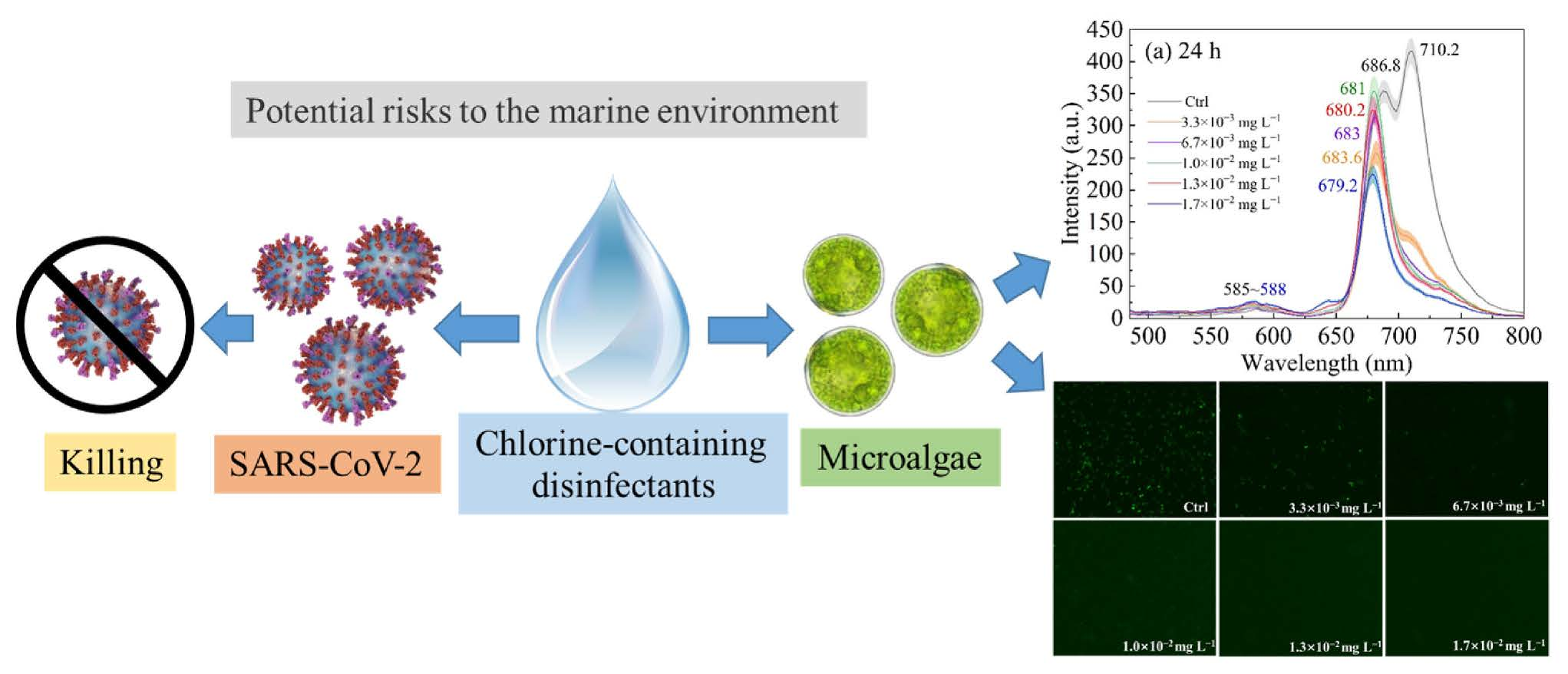
Impacts of Chlorine on the Change of Chlorophyll Fluorescence Spectrum to Phaeodactylum tricornutum

1
College of Marine Science and Environmental Engineering, Dalian Ocean University, Dalian 116023, China
2
Department of Marine Engineering, Dalian Maritime University, Dalian 116026, China
*
Author to whom correspondence should be addressed.
Analytica 2023, 4(2), 102-112; https://doi.org/10.3390/analytica4020009
Submission received: 26 March 2023 / Revised: 7 April 2023 / Accepted: 7 April 2023 / Published: 11 April 2023

Abstract

:
Chlorine-containing disinfectants have been widely used all over the world to prevent COVID-19. However, little is known about the potential risk of chlorine-containing disinfectants in the marine environment. Phaeodactylum tricornutum (P. tricornutum) is a typical marine economic diatom, often used as an effective biomarker in ecotoxicology research. Here, the present study has investigated the effect of different effective chlorine concentrations on photosynthesis of P. tricornutum by chlorophyll fluorescence spectroscopy. Results have demonstrated that chlorine exposure promoted the chlorophyll fluorescence intensity at initial stage (24 h), suggesting that a large amount of energy is emitted in the form of fluorescence. However, the chlorophyll fluorescence intensity could not be detected under the high effective chlorine concentrations (6.7 × 10⁻³, 1.0 × 10⁻², 1.3 × 10⁻² and 1.7 × 10⁻² mg L−1) after 48 h, indicating that the chlorine had high toxicity leading to the death of microalgae. In addition, the emission spectra of P. tricornutum were determined to contain two distinct fluorescence peaks representing the core antenna of photosystem II (685 nm) and the photosystem I complexes (710 nm) in the control group. The fluorescence emission peak value at 685 nm is significantly lower than the peak value at 710 nm in the control group, whereas chlorine treatments were opposite. It can be concluded that microalgae can regulate the distribution of excitation energy between the two photosystems to ensure that algae can utilize light energy. The result also found that the peak position of fluorescence emission spectra has a blue shift in all of NaClO treatments. The fluorescence intensity of microalgae excited at 467 nm was lower than that at 439 nm in chlorine treatments, illustrating chlorophyll b antenna was more easily damaged than chlorophyll a antenna. Our findings are providing new insights into the changing mechanism of chlorophyll fluorescence on P. tricornutum under chlorine stress and valuable data for risk assessment of marine environments.

Graphical Abstract

1. Introduction

The emergence of the Coronavirus Disease-2019 (COVID-19) pandemic is having an extremely serious impact on human health, global economy and environment. The family of coronaviruses contains Severe Acute Respiratory Syndrome Coronavirus 2 (SARS-CoV-2), which is the cause of COVID-19 [1]. The SARS-CoV-2 has spread worldwide, causing a substantial amount of human infections or even deaths [2]. According to the World Health Organization, COVID-19 has infected more than 700 million people and killed 6.8 million people by 2023. It has been reported that the chlorine-containing disinfectants have positive efficacy in killing SARS-CoV-2, so chlorine-containing disinfectants have been mainly used for sanitary disinfection during the COVID-19 pandemic in the People’s Republic of China (P.R.C) [3]. The concentrations of residual chlorine were found to be as high as 0.4 mg L−1 in some lakes of Wuhan (China) due to the excessive use of disinfectants [4]. However, even at concentrations below 0.1 mg L−1, the residual chlorine is toxic to sensitive aquatic species and may pose potential ecological risks [5,6].
The chlorine-containing disinfectants can be resolved into hypochlorous acid (HClO), hypochlorite ions (HClO) and chloride (Cl) in water, which are able to persist in the aquatic environment for a long time [7]. By the time these chlorine-containing compounds enter the ocean, they are causing a range of toxicological effects on marine organisms. Previous studies have shown that sodium hypochlorite (NaClO) can significantly damage cell structures and inhibit the growth and photosynthesis of algae [8,9]. As primary producers in the marine ecosystem, microalgae can convert CO2 into organic carbon through photosynthesis [10]. The process can increase marine carbon sink and maintain ecosystem homeostasis. Microalgae contribute to carbon emission reduction, which has important implications for global carbon neutralization. However, the potential threat of chlorine-containing disinfectants to microalgae photosynthesis has not attracted sufficient attention.
Chlorophyll, as a photosynthetic pigment existing in microalgae, is an extremely important foundation of photosynthesis. The light energy absorbed by chlorophyll is dissipated through photochemical reactions, including heat and fluorescence, respectively [11]. The three ways of transmission compete with each other for light energy, in which the fluorescence generally only accounts for 1–3% of the total energy. In a different perspective, almost all changes in the photosynthetic process can be reflected by chlorophyll fluorescence. Chlorophyll fluorescence can be used as a natural probe to investigate the changes of photosynthesis. In addition, the chlorophyll fluorescence spectrum technology has become an emerging detection technology. The chlorophyll fluorescence spectrum technology can reflect a large number of important physiological information of microalgae, such as growth status, the activities of photosystem II (PS II) and photosystem I (PS I), light energy absorptions and transformations.
Phaeodactylum tricornutum (P. tricornutum) is a typical marine economic diatom, often used as an effective biomarker in ecotoxicology research. Therefore, the purpose of this study is to assess the effects of different NaClO concentrations on the fluorescence characteristic of P. tricornutum by investigating the chlorophyll fluorescence spectra within 96 h. The objectives of the present study were to: (1) investigate the influence of NaClO on energy absorption and transformation between chlorophyll a and b; (2) reveal the effects of NaClO on the energy transfer process of microalgae photosystem; (3) explore the variation mechanism of NaClO on the spectral signature of microalgae. To the best of our knowledge, the results gained in this paper might explore the change mechanism of chlorophyll fluorescence on P. tricornutum and might be useful for ecological risk assessment of chlorine-containing disinfectants in the oceanographic investigations.

2. Materials and Methods

2.1. Microalgae Culture

P. tricornutum were provided by the Shanghai Guangyu Biological Technology Co., Ltd. (Shanghai, China). They were cultured in a 150 mL conical flask with f/2 culture medium (Table 1). The microalgae were maintained in a constant temperature light incubator with an irradiance of 60 μmol of photons m−2·s under a 12 h:12 h (light/dark) photoperiod. The initial pH and salinity in the f/2 medium were adjusted to 8.0 ± 0.06 and 32 ± 0.5, respectively. The temperature was set to 20 ± 1 °C. These flasks were manually agitated three times a day to avoid cell sedimentation. Before the sodium hypochlorite (NaClO) exposure test, the cell density of P. tricornutum was approximately 4 × 10⁵ cell mL−1.

2.2. Experimental Design

To examine the effects of NaClO on chlorophyll fluorescence of P. tricornutum, microalgae were exposed to different concentrations of effective chlorine. P. tricornutum were treated with effective chlorine concentrations of 0 (control group), 3.3 × 10⁻³, 6.7 × 10⁻³, 1.0 × 10⁻², 1.3 × 10⁻² and 1.7 × 10⁻² mg L−1 in a 100 mL conical flask. The sampling time points of microalgae were 24 h, 48 h, 72 h and 96 h after NaClO exposure. Every treatment was conducted in triplicate.

2.3. Measurement of Chlorophyll Fluorescence Spectroscopy

The chlorophyll fluorescence spectra of microalgae were measured by a fluorescence spectrophotometer (F4600, Hitachi, Ltd., Tokyo, Japan). The excitation (Ex) and emission (Em) wavelength range were 485–800 nm and 200–660 nm at 10 nm intervals using a slit width of 10 nm, respectively. The scanning speed and photomultiplier (PMT) voltage were 1200 nm min−1 and 700 V, respectively. An amount of 2 mL microalgae liquid was placed in a 1 cm path-length cuvettes for fluorescence spectrum scanning. The pre-experiments showed that the fluorescence maximum Ex wavelengths of chlorophyll a and chlorophyll b in P. tricornutum were 439 and 467 nm, respectively. The maximum Em wavelength of chlorophyll was around 685 nm. Therefore, the Ex wavelengths were determined to be 437 and 468 nm, and the Em wavelength was 685 nm.
In addition, the fluorescence effect of P. tricornutum was observed by the CCD camera (DS-Qi1Mc, Nikon, Ltd., Tokyo, Japan) of an inverted optical microscope imaging system (Ti-E, Nikon, Japan). The fluorescence excitation and emission filter module of the microscope was set as EX: 527–553 nm, DM: 565 nm and RA: 577–633 nm.

2.4. Statistical Analysis

All data were expressed as mean ± standard deviation (SD) of three replicates (n = 3). The spectral curves are smoothed through the Origin Pro 2021 software. In order to extract more accurate spectral information, the fluorescence spectrum was analyzed by Gaussian peak analysis [12]. The equation used for fitting has the form of Equations (1) and (2):
f ( x ) = a e ( x b ) 2 2 e 2
y = y 0 + A 0 ω 0 π 2 e ( x μ 0 ) 2 ω 0 2
where x is the wavelength (nm); f(x) is the relative intensity of fluorescence; y0 is the slope (baseline); A0 is an area of the peak; μ0 is the center of the peak (nm); ω0 is peak half-width (nm).

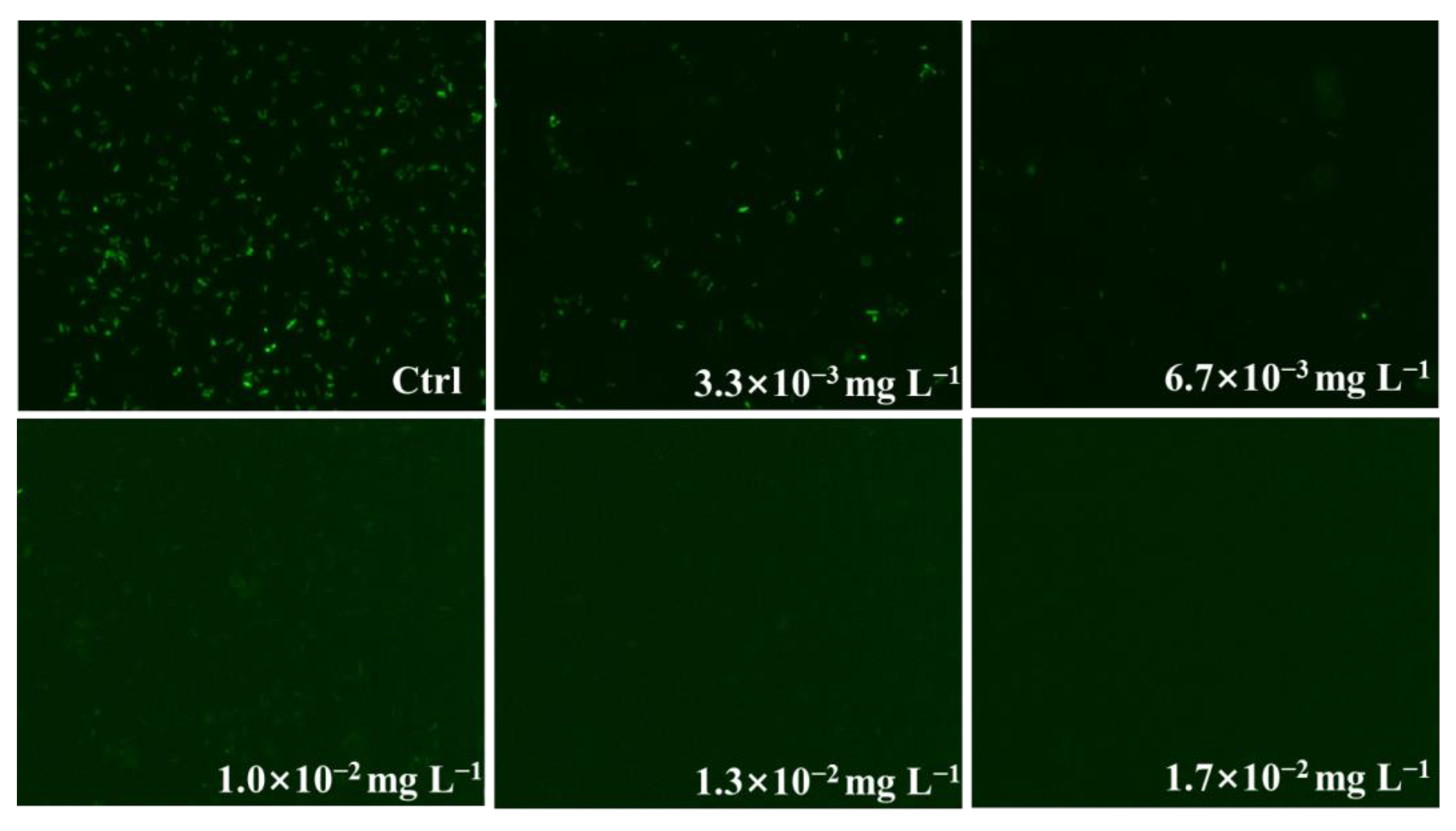
3. Results and Discussion

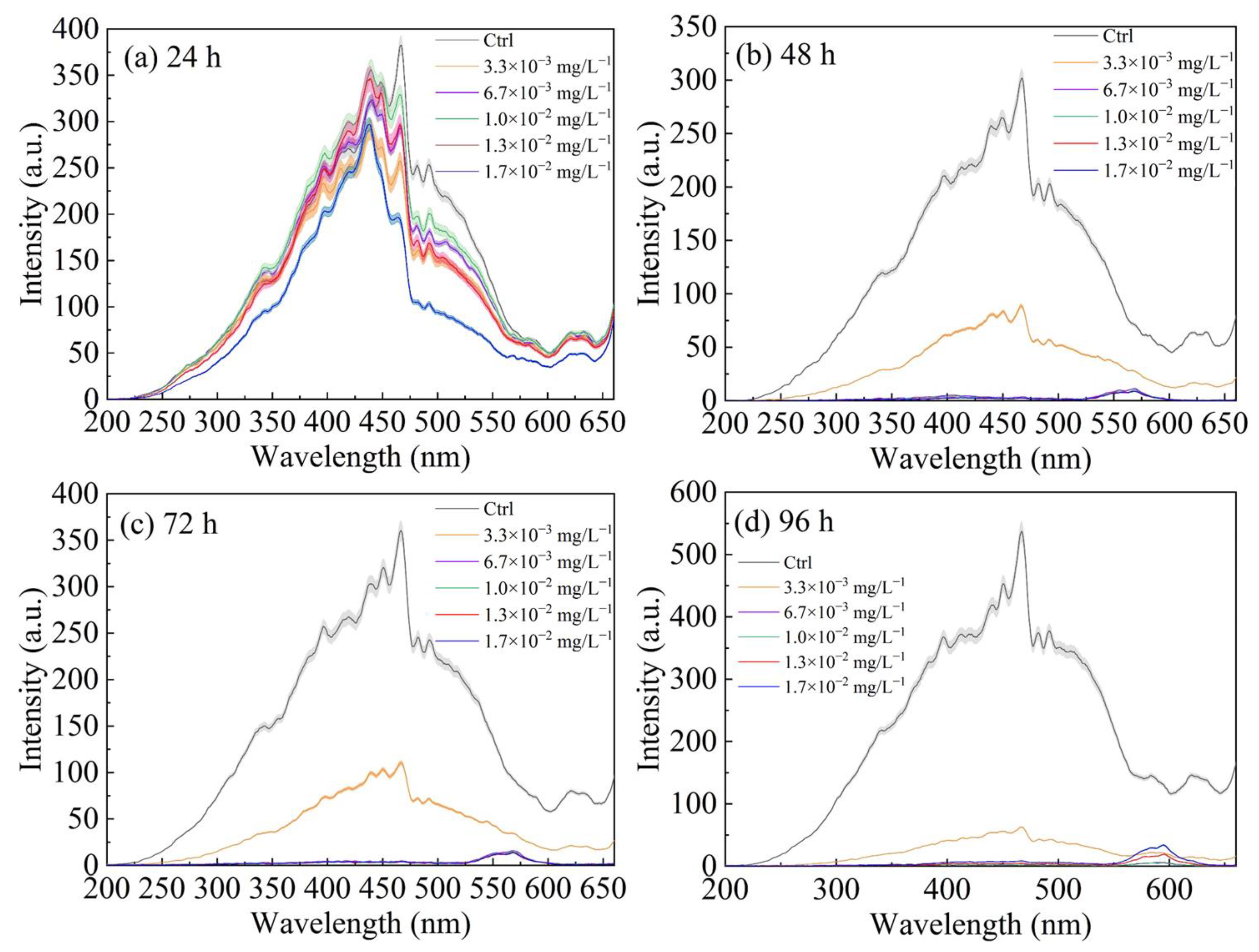
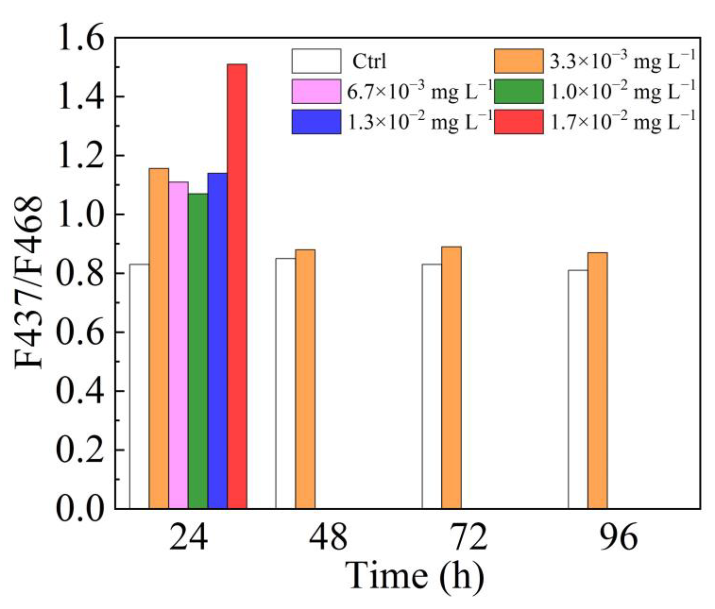
3.1. Fluorescence Excitation Spectra

Photosynthesis is a necessary activity for microalgae growth, which synthesize organic substances from inorganic materials and stores energy. The chlorophyll fluorescence is a sensitive probe for studying photosynthesis. Therefore, the fluorescence excitation spectra were determined to investigate the potential influence of NaClO on the chlorophyll fluorescence system of P. tricornutum. When the Em wavelength was 685 nm, the fluorescence Ex peaks were located near 437 nm and 468 nm of P. tricornutum (Figure 1). This finding was consistent with a previous study [13], which confirmed that the fluorescence Ex peaks at 437 nm (F437) and 468 nm (F468) were chlorophyll a and chlorophyll b antenna, respectively.
At the initial stage (24 h), the relative chlorophyll fluorescence intensity of P. tricornutum increased significantly at effective chlorine concentrations in 6.7 × 10⁻³ and 1.3 × 10⁻² mg L−1, compared to the control group (Figure 1a). Superficially, the low concentrations of NaClO can promote the increase in chlorophyll fluorescence of algae. It is known that NaClO decomposes in water to form chloric acid and hypochlorite (NaClO + H2O = HClO + NaOH). NaClO is a neutral molecule, which can rapidly diffuse to the surface of negatively charged microalgae. The entry of NaClO into cells mainly depends on the active transport process. It disrupts microalgae metabolic processes, such as carbohydrate metabolism, protein metabolism, respiration produces and photosynthesis. Therefore, the “stimulatory effect” is due to NaClO potentially disturbing the photosynthetic electron transport chain. Ultimately, a large amount of energy is emitted in the form of fluorescence.
The chlorophyll fluorescence intensity of P. tricornutum significantly increased during 96 h at the culture group. However, the intensity of chlorophyll fluorescence decreased with prolonged exposure time under the NaClO. As shown in Figure 1b–d, it is clear that the relative intensity of chlorophyll fluorescence decreased with the increase in culture time under low effective chlorine concentration (3.3 × 10⁻³ mg L−1). Under the high concentrations (6.7 × 10⁻³, 1.0 × 10⁻², 1.3 × 10⁻² and 1.7 × 10⁻² mg L−1) stress, the relative fluorescence intensity of microalgae was almost undetectable after 48 h. It has previously been reported that the chlorophyll auto-fluorescence intensity of Prorocentrum minimum under low biocide chlorine concentrations stress (0.1, 0.5 and 1.0 mg L−1) decreased by 68%, compared with the control group, whereas the fluorescence of algae could not be detected under higher concentrations (2.0 and 3.0 mg L−1) [14]. Moreover, the relative chlorophyll fluorescence intensity of P. tricornutum decreased with the increase in the effective chlorine concentrations at the same culture time (Figure 1). The chlorophyll fluorescence intensity showed a dose-dependent and time-dependent decrease exposure to NaClO during 96 h. Similarly, NaClO at 0.1–3 mg L−1 caused a significant decrease in the chlorophyll auto-fluorescence intensity of Closterium ehrenbergii [15]. The content of chlorophyll and carotene were significantly inhibited at chlorine treatments [16], indicating that the photosynthesis capability was reduced of microalgae. The damage to photosynthetic activity directly impacts the light-trapping function of the antenna protein in chlorophyll. In addition, the value of maximum quantum yield of PS II (Fv/Fm) also decreased [6], illustrating that the presence of NaClO disrupting the normal electron transfer process. Therefore, these results confirmed that NaClO caused serious damage to the photosystem of microalgae [17].
The fluorescence of PS II light-harvesting complex (LHC II) is provided by chlorophyll a and b together. The absorbed light energy was transferred between chlorophyll a and b until stable equilibrium was reached. Therefore, the chlorophyll a/b fluorescence intensity (F437/F468) can be used to reflect the proportion of excitation energy in LHC II. As shown in Figure 2, the average value of F437/F468 in the control group remained at 0.83 ± 0.02 during 24–96 h, indicating that the light energy conversion process of microalgae is normal and stable. Moreover, the value of F437/F468 is less than 1, illustrating that the excitation energy of chlorophyll b in LHC II was higher than that of chlorophyll a. This phenomenon was most probably due to the fact that light energy transferred from chlorophyll b to chlorophyll a. However, during exposure of different NaClO concentrations, the value of F437/F468 was significantly higher than the control group during the initial stage (within the first 24 h). The results of this study demonstrated that NaClO could inhibit the absorption of light energy by chlorophyll b and interfere with electron transport. To protect the ability of LHC II to capture light under NaClO exposure, chlorophyll a absorbs a large quantity of excitation energy for photosynthesis. Moreover, the value of F437/F468 of microalgae decreased from 1.16 to 0.87 within 24–96 h at low effective chlorine concentration (3.3 × 10⁻³ mg L−1), indicating that the inhibitory effect of NaClO on photosynthesis of microalgae gradually weakened. Reduction in F437/F468 value over exposure time was related to self-recovery ability of the photosynthetic efficiency. Under the high effective chlorine concentrations (6.7 × 10⁻³, 1.0 × 10⁻², 1.3 × 10⁻² and 1.7 × 10⁻² mg L−1), the value of F437/F468 was below the detection line after 48 h. Previous studies have found that the acute toxicity of NaClO to microalgae was significant and could cause microalgae death [18].

3.2. Fluorescence Emission Spectra

The fluorescence Em spectra of P. tricornutum was induced with 467 nm irradiance in the presence of different effective chlorine concentrations as a function of exposure times (Figure 3). In the control group, the Em spectra of P. tricornutum contained two prominent fluorescence peaks located near 687 nm and 710 nm, respectively. The main peak of chlorophyll fluorescence in most microalgae was between 680 nm and 690 nm [19]. This can be interpreted as the fluorescence Em peak of P. tricornutum at 687 nm representing the core antenna of PS II. The reaction center pigments of PS II are called P680, which are able to absorb light with the wavelength of 680 nm (Figure 4). In addition, The PS I core complex consists of reaction center pigments P700, of which the maximum absorption wavelength is 700 nm. Previous studies found that the fluorescence emission peak of PS I in P. tricornutum is located at 717 nm [20]. The PS I complex of P. tricornutum showed fluorescence Em peaks at 690 nm and 716 nm at 77 K [21]. The shoulder peak of chlorophyll fluorescence Em of Codium chloroplasts also isolated at 712 nm [22]. Therefore, the 687 nm band is associated with the core antenna of PS II and the broad band at 710 nm is mainly associated with PS I complexes.
Em peak value at 685 nm is significantly lower than the peak value at 710 nm in the control group during 96 h (Figure 3). The PS I and PS II are both photosynthesis units in the thylakoid membrane of microalgae. LHC II and LHC I (PS I light-harvesting complex) can absorb and transfer the light energy, which in turn drive photosynthetic electron transport and energy conversion process (Figure 4). The algae cells distribute energy between the two photosystems by regulating the light-harvesting complexes. Hence, the excitation levels of PS I and PS II are in a relatively balanced state. In the control group, the excitation energy could efficiently be transferred from PS II to PS I of P. tricornutum. However, the peak value at 710 nm of algae cultured in low effective chlorine concentration was significantly lower than the peak value at 685 nm. It is worth noting that the photosynthesis of microalgae performs under the synergistic action of PS I and PS II [23]. The PS II of microalgae may be overstimulated under low NaClO concentration stress. A portion of LHC II is phosphorylated in this process, resulting in the formation of the super-complexes PS I-LHC I-LHC II [24]. The super-complexes PS I-LHC I-LHC II can transfer energy to PS I, achieving a balanced distribution of excitation energy between PS II and PS I (Figure 4). Therefore, the microalgae can regulate the distribution of excitation energy between the two photosystems to ensure that algae can utilize light energy. In addition, responding to low effective chlorine concentration stress, a decrease in relative chlorophyll fluorescence intensity was observed in P. tricornutum. The Em peak value at 710 nm of algae was always lower than the peak value at 685 nm during 96 h under the NaClO stress. The phenomenon demonstrated that the photosynthetic capacity of microalgae decreased after NaClO stress. Microalgae can generate stress protection mechanisms to maintain cell metabolism. Both PS II and PS I are prone to oxidative damage under stress. The difference is that the repair process of PS I is very slow, which is obviously different from PS II. Therefore, the NaClO stress can result in irreversible PS I damage of P. tricornutum.
As shown in Figure 3b–d, the fluorescence Em peak near 685 nm and 710 nm was not detected under the high effective chlorine concentrations, suggesting that the light energy transfer process suffered serious damage under the NaClO treatments. However, the peak of fluorescence Em spectrum appeared near 585 nm. The peak value at 585 nm increases with the increase in effective chlorine concentrations. Furthermore, the fluorescence effect was observed under the optical microscope to check the viability of P. tricornutum (Figure 5). It is clear that the microalgae could emit fluorescence under control group and 3.3 × 10⁻³ mg L−1 effective chlorine concentration, whereas microalgae barely emit fluorescence in the other treatments at 48 h. The cell debris contain un-degraded fluorescent groups. Therefore, the fluorescence Em peak near 585 nm may be caused by a large number of dead microalgae and colored dissolved organic matter (CDOM).
The changes in the relative fluorescence intensity near 685 nm of P. tricornutum in the presence of NaClO over a period (24 h, 48 h, 72 h and 96 h) are presented in Figure 3. The relative fluorescence intensity of algae cultured in all of the NaClO treatments increased at 24 h (Figure 3a). In a short time, the addition of NaClO could stimulate the intracellular stress response and increase the chlorophyll content [25]. The capacity of reaction center pigments in chloroplast to absorb and transfer light energy was enhanced, which increased the fluorescence intensity of the microalgae. After 48 h, the fluorescence intensity of microalgae decreased significantly with increasing effective chlorine concentrations (Figure 3b–d). Specifically, the relative fluorescence intensity of algae was not detected under the high effective chlorine concentrations. Previous studies found that the effective reagent of chlorine can kill algae [26,27,28], which confirmed that 90% of algae were destroyed within 6 min under chlorine concentration of 10 mg L−1 [9]. Meanwhile, microalgae death probably results from chlorine attacking the cell membrane, obstructing photosynthesis electron transfer, damaging the intracellular structure and inhibiting synthetic molecules [29,30].
The peak position of fluorescence Em spectra of P. tricornutum are shown in Table 2. The fluorescence EM spectra were induced by 439 nm and 467 nm irradiance, respectively. The peak position of P. tricornutum was 686.9 ± 0.5 nm in the control group. The main peak of chlorophyll fluorescence near 685 nm is mainly provided by the antenna pigment system of PS II at normal temperature. However, the peak position of fluorescence Em spectra has a blue shift in all of NaClO treatments. For instance, when exciting at wavelengths of 439 nm and 467 nm, the fluorescence Em peak shifts 12 nm and 8 nm at 24 h under effective chlorine concentrations of 1.7 × 10⁻² mg L−1. The maximum peak of C. vulgaris and P. helgolandica shifted from 685.2 nm to 681.9nm and 685.2 nm to 680.5 within 6 h under the NaClO stress, respectively [17]. Moreover, similar trends were observed for other experimental results [31,32]. It can be found that the NaClO treatments are expected to destroy the internal structure of chloroplast [33,34], resulting in the change of light absorption pigment structure in PS II antenna.
The Ex wavelength of the experiment was set at 439 nm (chlorophyll a antenna) or 467 nm (chlorophyll b antenna) to measure the fluorescence Em spectra. The results show that the peak position of fluorescence Em spectra in P. tricornutum was consistent under different Ex wavelengths, indicating that the peak position was independent of the Ex wavelengths (Table 2). However, the fluorescence intensity of microalgae excited at 467 nm was higher than that at 439 nm in the control group. For example, the relative fluorescence intensity of algae was 250.6 ± 12.2 a.u. and 352.2 ± 17.4 a.u., when excited at 439 nm and 467 nm at 24 h, respectively. On the contrast, the algae exposed to NaClO treatments observed an opposite trend at 24 h. The peak value of 439 nm irradiance was 7.2%, 5.3%, 3.4%, 18.0% and 34.8% higher than that of 467 nm irradiance under effective chlorine concentrations of 3.3 × 10⁻³, 6.7 × 10⁻³, 1.0 × 10⁻², 1.3 × 10⁻² and 1.7 × 10⁻² mg L−1, respectively. Thus, it could be speculated that chlorophyll b antenna was more easily damaged than chlorophyll a antenna under NaClO treatments. During 48–96 h, the fluorescence intensity near 685 nm excited at 467 nm was higher than that at 439 nm under the low effective chlorine concentration (3.3 × 10⁻³ mg L−1). The phenomenon was similar to the trend in the control group. The chlorophyll a antenna are the main photosynthetic pigments, while chlorophyll b antenna are the accessory pigments. The chlorophyll b antenna collects energy and transfers it to the chlorophyll a antenna. Under the low effective chlorine concentration, the light-absorbing capacity of chlorophyll a antenna was lower than that of chlorophyll b antenna over the short term (within 24 h), whereas the trend was reversed after 48 h. In other words, the photosynthetic capacity of chlorophyll b gradually recovered with the extension of culture time. Therefore, this phenomenon indicated that microalgae have a relatively complete repair mechanism, which are able to repair or reconstruct damaged parts of the photosystem [35].

4. Conclusions

This study was conducted to evaluate the acute toxicity of different concentrations of NaClO on P. tricornutum at increasing time intervals. During the initial stage (24 h), NaClO exposure promoted the relative intensity of chlorophyll fluorescence, indicating that an abundance of energy is emitted in the form of fluorescence. The value of F439/F467 exposure to NaClO was significantly higher than the control group, suggesting that NaClO could inhibit the absorption of light energy by chlorophyll b and interfere with electron transport. However, when microalgae were exposed to the high effective chlorine concentrations, the relative intensity of chlorophyll fluorescence could not be detected after 48 h. According to fluorescence emission spectra, the peak value at 710 nm of P. tricornutum significantly decreased compared to the peak value at 685 nm at 3.3 × 10⁻³ mg L−1 effective chlorine concentration. Moreover, the peak position of fluorescence EM spectra has a blue shift in all of the NaClO treatments. The present study might be helpful to understand the potential damages of chlorine-containing disinfectants to microalgae photosynthesis and provide theoretical supports for protecting marine ecological environments.

Author Contributions

Conceptualization, N.L.; validation, J.S. and Y.S.; formal analysis, N.L.; investigation, S.C.; data curation, J.Y.; writing—review and editing, N.L.; project administration, Y.S.; funding acquisition, Y.S. All authors have read and agreed to the published version of the manuscript.

Funding

This work was financially supported by the National Nature Science Foundation Program of China (grant number 51979019) to Yongxin Song, the Dalian Science and Technology Talent Innovation Support Project (grant number 2022RQ009) to Shimeng Chen.

Institutional Review Board Statement

Not applicable.

Informed Consent Statement

Not applicable.

Data Availability Statement

The data presented in this study are available on request from the corresponding author.

Conflicts of Interest

The authors declare no conflict of interest.

References

  1. Facciolà, A.; Laganà, P.; Caruso, G. The COVID-19 pandemic and its implications on the environment. Environ. Res. 2021, 201, 111648. [Google Scholar] [CrossRef]
  2. García-Ávila, F.; Valdiviezo-Gonzales, L.; Cadme-Galabay, M.; Gutiérrez-Ortega, H.; Altamirano-Cárdenas, L.; Arévalo, C.Z.; Flores del Pino, L. Considerations on water quality and the use of chlorine in times of SARS-CoV-2 (COVID-19) pandemic in the community. Case Stud. Chem. Environ. Eng. 2020, 2, 100049. [Google Scholar] [CrossRef]
  3. Wang, J.; Zhang, B.; Duan, H.; Liang, C.; Sun, H.; Zhang, J.; Shen, J.; Zhang, L. Key Points of the Program for Disinfection Technology in Special Places during the Coronavirus Disease-2019 (COVID-19) Outbreak. China CDC Wkly. 2020, 2, 140–142. [Google Scholar] [CrossRef] [PubMed]
  4. Chu, W.; Fang, C.; Deng, Y.; Xu, Z. Intensified Disinfection Amid COVID-19 Pandemic Poses Potential Risks to Water Quality and Safety. Environ. Sci. Technol. 2021, 55, 4084–4086. [Google Scholar] [CrossRef] [PubMed]
  5. Vannoni, M.; Creach, V.; Barry, J.; Sheahan, D. Chlorine toxicity to Navicula pelliculosa and Achnanthes spp. in a flow-through system: The use of immobilised microalgae and variable chlorophyll fluorescence. Aquat. Toxicol. 2018, 202, 80–89. [Google Scholar] [CrossRef] [PubMed]
  6. Cao, S.; Zhang, D.; Teng, F.; Liao, R.; Cai, Z.; Tao, Y.; Hu, H. Inhibitory effects of ultralow-dose sodium hypochlorite on Microcystis aeruginosa and Chlorella vulgaris: Differences in sensitivity and physiology. Sci. Total Environ. 2021, 774, 145638. [Google Scholar] [CrossRef]
  7. Zhang, Z.; Zhang, Q.; Lu, T.; Zhang, J.; Sun, L.; Hu, B.; Hu, J.; Peñuelas, J.; Zhu, L.; Qian, H. Residual chlorine disrupts the microbial communities and spreads antibiotic resistance in freshwater. J. Hazard. Mater. 2022, 423, 127152. [Google Scholar] [CrossRef]
  8. Ma, Z.; Gao, K.; Li, W.; Xu, Z.; Lin, H.; Zheng, Y. Impacts of chlorination and heat shocks on growth, pigments and photosynthesis of Phaeodactylum tricornutum (Bacillariophyceae). J. Exp. Mar. Biol. Ecol. 2011, 397, 214–219. [Google Scholar] [CrossRef]
  9. Garoma, T.; Yazdi, R.E. Investigation of the disruption of algal biomass with chlorine. BMC Plant Biol. 2019, 19, 18. [Google Scholar] [CrossRef]
  10. Li, N.; Wang, P.; Wang, S.; Wang, C.; Zhou, H.; Kapur, S.; Zhang, J.; Song, Y. Electrostatic charges on microalgae surface: Mechanism and applications. J. Environ. Chem. Eng. 2022, 10, 107516. [Google Scholar] [CrossRef]
  11. Gan, T.; Zhao, N.; Yin, G.; Chen, M.; Wang, X.; Liu, J.; Liu, W. Optimal chlorophyll fluorescence parameter selection for rapid and sensitive detection of lead toxicity to marine microalgae Nitzschia closterium based on chlorophyll fluorescence technology. J. Photochem. Photobiol. B Biol. 2019, 197, 111551. [Google Scholar] [CrossRef] [PubMed]
  12. Küpper, H.; Seibert, S.; Parameswaran, A. Fast, Sensitive, and Inexpensive Alternative to Analytical Pigment HPLC: Quantification of Chlorophylls and Carotenoids in Crude Extracts by Fitting with Gauss Peak Spectra. Anal. Chem. 2007, 79, 7611–7627. [Google Scholar] [CrossRef]
  13. Gruszecki, W.I.; Krupa, Z. Changes of Excitation Spectra of in vivo Chlorophyll Fluorescence during Induction of Photosynthesis. Z. Für Nat. C 1993, 48, 46–51. [Google Scholar] [CrossRef]
  14. Ebenezer, V.; Ki, J.-S. Physiological and biochemical responses of the marine dinoflagellate Prorocentrum minimum exposed to the oxidizing biocide chlorine. Ecotoxicol. Environ. Saf. 2013, 92, 129–134. [Google Scholar] [CrossRef] [PubMed]
  15. Sathasivam, R.; Ebenezer, V.; Guo, R.; Ki, J.-S. Physiological and biochemical responses of the freshwater green algae Closterium ehrenbergii to the common disinfectant chlorine. Ecotoxicol. Environ. Saf. 2016, 133, 501–508. [Google Scholar] [CrossRef]
  16. Patil, J.S.; Jagadeesan, V. Effect of chlorination on the development of marine biofilms dominated by diatoms. Biofouling 2011, 27, 241–254. [Google Scholar] [CrossRef]
  17. Li, N.; Liu, Z.; Wang, P.; Suman, K.; Zhang, J.; Song, Y. Effects of sodium hypochlorite treatment on the chlorophyll fluorescence in photosystem II of microalgae. Sci. Total Environ. 2022, 833, 155192. [Google Scholar] [CrossRef]
  18. López-Galindo, C.; Garrido, M.C.; Casanueva, J.F.; Nebot, E. Degradation models and ecotoxicity in marine waters of two antifouling compounds: Sodium hypochlorite and an alkylamine surfactant. Sci. Total Environ. 2010, 408, 1779–1785. [Google Scholar] [CrossRef]
  19. Lichtenthaler, H.K.; Buschmann, C.; Knapp, M. How to correctly determine the different chlorophyll fluorescence parameters and the chlorophyll fluorescence decrease ratio RFd of leaves with the PAM fluorometer. Photosynthetica 2005, 43, 379–393. [Google Scholar] [CrossRef]
  20. Berkaloff, C.; Caron, L.; Rousseau, B. Subunit organization of PSI particles from brown algae and diatoms: Polypeptide and pigment analysis. Photosynth. Res. 1990, 23, 181–193. [Google Scholar] [CrossRef]
  21. Caron, L.; Berkaloff, C. Chlorophyll-Carotenoid Protein Complexes from the Diatom, Phaeodactylum tricornutum: Spectrophotometric, Pigment and Polypeptide Analyses. Plant Cell Physiol. 1987, 28, 775–785. [Google Scholar] [CrossRef]
  22. Anderson, J.M. Chlorophyll-protein complexes of a Codium species, including a light-harvesting siphonaxanthin-Chlorophylla ab-protein complex, an evolutionary relic of some Chlorophyta. Biochim. Biophys. Acta (BBA) Bioenerg. 1983, 724, 370–380. [Google Scholar] [CrossRef]
  23. Gao, S.; Chi, Z.; Chen, H.; Zheng, Z.; Weng, Y.; Wang, G. A Supercomplex, of Approximately 720 kDa and Composed of Both Photosystem Reaction Centers, Dissipates Excess Energy by PSI in Green Macroalgae Under Salt Stress. Plant Cell Physiol. 2019, 60, 166–175. [Google Scholar] [CrossRef]
  24. Pan, X.; Tokutsu, R.; Li, A.; Takizawa, K.; Song, C.; Murata, K.; Yamasaki, T.; Liu, Z.; Minagawa, J.; Li, M. Structural basis of LhcbM5-mediated state transitions in green algae. Nat. Plants 2021, 7, 1119–1131. [Google Scholar] [CrossRef] [PubMed]
  25. da Silva Ferreira, V.; Sant’Anna, C. Impact of culture conditions on the chlorophyll content of microalgae for biotechnological applications. World J. Microbiol. Biotechnol. 2016, 33, 20. [Google Scholar] [CrossRef]
  26. Aasco, N.C.; Koyama, J.; Imai, S.; Nakamura, K. Toxicity of Residual Chlorines from Hypochlorite-treated Seawater to Marine Amphipod Hyale barbicornis and Estuarine Fish Oryzias javanicus. Water Air Soil Pollut. 2008, 195, 129. [Google Scholar] [CrossRef]
  27. Wang, J.; Wang, G.; Chen, M.; Wang, Y.; Ding, G.; Zhang, Y.; Kang, Y.; Pan, X. An integrated microfluidic chip for treatment and detection of microalgae cells. Algal Res. 2019, 42, 101593. [Google Scholar] [CrossRef]
  28. Song, Y.; Li, Z.; Feng, A.; Zhang, J.; Liu, Z.; Li, D. Electrokinetic detection and separation of living algae in a microfluidic chip: Implication for ship’s ballast water analysis. Environ. Sci. Pollut. Res. 2021, 28, 22853–22863. [Google Scholar] [CrossRef] [PubMed]
  29. Fukuzaki, S. Mechanisms of actions of sodium hypochlorite in cleaning and disinfection processes. Biocontrol Sci. 2006, 11, 147–157. [Google Scholar] [CrossRef]
  30. Small, D.A.; Chang, W.; Toghrol, F.; Bentley, W.E. Toxicogenomic analysis of sodium hypochlorite antimicrobial mechanisms in Pseudomonas aeruginosa. Appl. Microbiol. Biotechnol. 2007, 74, 176–185. [Google Scholar] [CrossRef]
  31. Peter, C.; Thoms, S.; Koch, F.; Sartoris, F.J.; Bickmeyer, U. Sponge-derived Ageladine A affects the in vivo fluorescence emission spectra of microalgae. PLoS ONE 2020, 15, e0242464. [Google Scholar] [CrossRef]
  32. Seder-Colomina, M.; Burgos, A.; Maldonado, J.; Solé, A.; Esteve, I. The effect of copper on different phototrophic microorganisms determined in vivo and at cellular level by confocal laser microscopy. Ecotoxicology 2013, 22, 199–205. [Google Scholar] [CrossRef] [PubMed]
  33. Zheng, H.; Sun, C.; Hou, X.; Wu, M.; Yao, Y.; Li, F. Pyrolysis of Arundo donax L. to produce pyrolytic vinegar and its effect on the growth of dinoflagellate Karenia brevis. Bioresour. Technol. 2018, 247, 273–281. [Google Scholar] [CrossRef] [PubMed]
  34. Li, H.; Yao, J.; Duran, R.; Liu, J.; Min, N.; Chen, Z.; Zhu, X.; Zhao, C.; Ma, B.; Pang, W.; et al. Toxic response of the freshwater green algae Chlorella pyrenoidosa to combined effect of flotation reagent butyl xanthate and nickel. Environ. Pollut. 2021, 286, 117285. [Google Scholar] [CrossRef]
  35. Blatchley, E.R.; John, C.; Brian, P.; Keith, B.; Nicholas, W. The Biological Basis for Ballast Water Performance Standards: “Viable/Non-Viable” or “Live/Dead”? Environ. Sci. Technol. 2018, 52, 8075–8086. [Google Scholar] [CrossRef] [PubMed]
Figure 1. The fluorescence excitation spectra of P. tricornutum exposed to the different effective chlorine concentrations, 24 h (a), 48 h (b), 72 h (c) and 96 h (d). The different colors of shadows indicate the standard deviation of each group (mean ± SD, n = 3).
Analytica 04 00009 g001
Figure 2. The chlorophyll fluorescence ratio F437/F468 of P. tricornutum under the different effective chlorine concentrations.
Analytica 04 00009 g002
Figure 3. The fluorescence emission spectra of P. tricornutum exposed to the different effective chlorine concentrations, 24 h (a), 48 h (b), 72 h (c) and 96 h (d). The different colors of shadows indicate the standard deviation of each group (mean ± SD, n = 3).
Analytica 04 00009 g003
Figure 4. Schematic diagram of energy transfer process in microalgae photosystem. Cyt b6f, cytochrome b6f; Pc, plastocyanin; Fd, ferredoxin; FNR, Ferredoxin-NADP PQ, Oxidized plastoquinone; PQH2, Reduced plastoquinone; P680, Reaction center pigments of PS II; P700, Reaction center pigments of PS I.
Analytica 04 00009 g004
Figure 5. The chlorophyll fluorescence of P. tricornutum under different the effective chlorine concentrations at 48 h.
Analytica 04 00009 g005
Table 1. The nutrient formula of f/2.
f/2 MediumChemical MedicineDosage (g)
A1NaNO37.5
A2NaH2PO4·2H2O0.562
BNa2SiO3·9H2O3
CNa2EDTA0.436
FeCl3·6H2O0.315
DCuSO4·5H2O0.098
ZnSO4·7H2O0.22
CoCl2·6H2O0.1
MnCl2·6H2O1.8
Vitamins
Vitamin B125 mg
Vitamin H5 mg
Vitamin B110 mg
Ultrapure water100 mL
Table 2. The peak position and peak value of emission fluorescence of P. tricornutum exposed to different effective chlorine concentrations at 24 h, 48 h, 72 h and 96 h (mean ± SD, n = 3).
Culture Time24 h48 h
439 nm467 nm439 nm467 nm
Peak PositionPeak ValuePeak PositionPeak ValuePeak PositionPeak ValuePeak PositionPeak Value
Control686.2250.6 ± 12.2686.8352.2 ± 17.4686.4283.7 ± 9.3687341.9 ± 11.1
3.3 × 10⁻³ mg L−1683.4292 ± 22.7683.6271.7 ± 19.3684.288.36 ± 4.368499.71 ± 4.6
6.7 × 10⁻³ mg L−1683.2335.8 ± 12.2683317.9 ± 9.1684.24.92 ± 0.046844.36 ± 0.02
1.0 × 10⁻² mg L−1681.6383.3 ± 28.3681370.2 ± 24.2681.24.37 ± 0.02683.64.22 ± 0.04
1.3 × 10⁻² mg L−1680411.2 ± 30.1680.2337.2 ± 21.4680.62.75 ± 0.076803.07 ± 0.06
1.7 × 10⁻² mg L−1679.2355.2 ± 25.6679.2231.6 ± 13.66802.45 ± 0.096802.33 ± 0.1
Culture time72 h96 h
439 nm467 nm439 nm467 nm
Peak positionPeak valuePeak positionPeak valuePeak positionPeak valuePeak positionPeak value
Control687.4333.9 ± 8.8687401.4 ± 10.5686.4375.3 ± 15.0687.6476.1 ± 19.0
3.3 × 10⁻³ mg L−1684.4109.8 ± 3.6683.8128.4 ± 4.2684.696.34 ± 1.8683.6113.3 ± 2.2
6.7 × 10⁻³ mg L−1683.25.51 ± 0.16846.03 ± 0.16844.38 ± 0.3684.24.36 ± 0.2
1.0 × 10⁻² mg L−1683.84.39 ± 0.1683.25.13 ± 0.1683.24.23 ± 0.056843.81 ± 0.02
1.3 × 10⁻² mg L−1681.84.20 ± 0.046815.07 ± 0.03681.84.02 ± 0.03682.42.86 ± 0.2
1.7 × 10⁻² mg L−1680.63.77 ± 0.05680.65.10 ± 0.06681.82.38 ± 0.01682.42.07 ± 0.09
Disclaimer/Publisher’s Note: The statements, opinions and data contained in all publications are solely those of the individual author(s) and contributor(s) and not of MDPI and/or the editor(s). MDPI and/or the editor(s) disclaim responsibility for any injury to people or property resulting from any ideas, methods, instructions or products referred to in the content.

