Next Article in Journal
Voltammetric Determination of Trimethoprim Using a Glassy Carbon Electrode Modified with Printex(6L) Carbon and Gold Nanoparticles
Previous Article in Journal
Modified Cassava Starches’ Identification through Mid-Infrared Spectroscopy and Exploratory Analysis
 
 
analytica-logo
Article Menu
Article

Ximenia americana L.: Chemical Characterization and Gastroprotective Effect

by
Renata Torres Pessoa
1,
Isabel Sousa Alcântara
1,
Lucas Yure Santos da Silva
1,
Roger Henrique Souza da Costa
1,
Tarcísio Mendes Silva
1,
Cícera Datiane de Morais Oliveira-Tintino
2,
Andreza Guedes Barbosa Ramos
1,
Maria Rayane Correia de Oliveira
1,3,
Anita Oliveira Brito Pereira Bezerra Martins
1,
Bruna Caroline Gonçalves Vasconcelos de Lacerda
4,
Edlane Martins de Andrade
4,
Jaime Ribeiro-Filho
5,
Clara Mariana Gonçalves Lima
6,*,
Henrique Douglas Melo Coutinho
2,* and
Irwin Rose Alencar de Menezes
1,*
1
Laboratory of Pharmacology and Molecular Chemistry, Department of Biological Chemistry, Regional University of Cariri, Rua Coronel Antônio Luis 1161, Crato 63105-000, CE, Brazil
2
Laboratory of Microbiology and Molecular Biology, Department of Biological Chemistry, Regional University of Cariri, Rua Coronel Antônio Luis 1161, Crato 63105-000, CE, Brazil
3
Graduate Program in Biotechnology-Northeast Biotechnology Network (RENORBIO), State University of Ceará (UECE), Av. Dr. Silas Munguba, 1700, Fortaleza 60741-000, CE, Brazil
4
CECAPE College, Av. Padre Cícero, 3917, Juazeiro do Norte 63024-015, CE, Brazil
5
Department of Biotechnology, Oswaldo Cruz Foundation, Fiocruz Ceará, Eusébio, Fortaleza 60180-190, CE, Brazil
6
Department of Food Science, Federal University of Lavras, Lavras 37203-202, MG, Brazil
*
Authors to whom correspondence should be addressed.
Analytica 2023, 4(2), 141-158; https://doi.org/10.3390/analytica4020012
Submission received: 10 March 2023 / Revised: 19 April 2023 / Accepted: 26 April 2023 / Published: 30 April 2023

Abstract

:
Ximenia americana L., popularly known in Brazil as “ameixa do-mato, ameixa-brava, and ameixa-do-sertão,” is widely used in folk medicine to treat several intestinal disorders. The present study assessed the potential mechanisms of action underlying the gastroprotective activity of the hydroethanolic extract of Ximenia americana L. (EHXA) stem bark. The acute toxicity of EHXA was estimated, and later, the gastroprotective effect in mice was assessed through acute models of gastric lesions induced by acidified or absolute ethanol and indomethacin, where the following mechanisms were pharmacologically analyzed: the involvement of prostaglandins (PG), histamine (H2) receptors, ATP-dependent potassium channels, sulfhydryl groups (SH), α2 adrenergic receptors, nitric oxide (NO), myeloperoxidase (MPO), gastric mucus production, and acetylcholine-mediated intestinal motility. Regarding toxicity, EHXA did not cause deaths or signs of toxicity (LD50 greater than or equal to 2000 mg/kg/p.o.). When the gastroprotective effect was assessed, EHXA (50, 100, and 200 mg/kg/p.o.) reduced the rate of lesions induced by acidified ethanol by 65.63; 53.66, and 58.02% in absolute ethanol at 88.91, 78.82, and 74.68%, respectively, when compared to the negative control group. In the indomethacin-induced gastric injury model, the reductions were 84.69, 55.99, 55.99, and 42.50%, respectively. The study revealed that EHXA might stimulate mucus production and reduce intestinal motility through SH groups, NO production, and activation of α2 adrenergic receptors. The results indicated that EHXA had significant gastroprotective activity in the evaluated models. However, further investigation is required to elucidate the cellular and molecular events underlying the action of EHXA components and to correlate them with the modulation of the signaling pathways, as demonstrated by the current pharmacological approach. Therefore, the results demonstrated in the present study, as well as previously reported findings, support the recommendation of using this species in traditional communities in Brazil.

1. Introduction

The pathogenesis of gastric diseases is a complex and multifactorial process that involves genetic, environmental, and lifestyle factors. One of the most well-known gastrointestinal diseases is gastric inflammation, characterized by increased acid secretion, oxidative stress, and immune system dysfunction [1]. Peptic ulcers are a health problem affecting about 10% of the world’s population, classified as a gastric ulcer (wound in the stomach wall) or a duodenal ulcer (wound in the duodenum wall). Such condition results mainly from an imbalance between protective and aggressive factors, including deficiency of essential nutrients for homeostasis, excessive alcohol consumption, infection by Helicobacter pylori, and the prolonged use of non-steroidal anti-inflammatory drugs [2,3,4,5,6].
Despite the existence of anti-ulcerogenic drugs capable of treating ulcers, the currently available drugs can cause several adverse effects, primarily when used for an extended period, which limits their use and can discourage adherence to conventional treatment. This scenario stimulates the search for alternative treatments, including natural products such as medicinal plants and their secondary metabolites [7,8,9,10].
The family Olacaceae has a pantropical distribution, including 27 genera with about 200 species. Its representatives are characterized as woody plants, trees, or shrubs. In Brazil, about 13 genera and approximately 60 species have been described, with the genera Heisteria, Liriosma, Schoepfia, and Ximenia having the largest number of species having several therapeutic activities reported in the literature [11].
Ximenia americana is a medicinal plant found in tropical regions. This species is known for its various pharmacological properties, which include anti-inflammatory, analgesic, gastroprotective, anti-diarrheal, antipyretic, and antioxidant [12,13,14]. The chemical composition of Ximenia americana is diverse and includes flavonoids, triterpenoids, sterols, and fatty acids. Some significant components identified in this species are lupeol, betulinic acid, β-sitosterol, stigmasterol, campesterol, procyanidin, and quercetin [15,16,17]. Lupeol is a triterpenoid reported to have anti-inflammatory and analgesic effects [18]. Literature data have indicated that betulinic acid presents anti-inflammatory, antioxidant, and antitumor properties [19]. At the same time, β-sitosterol, stigmasterol, and campesterol are phytosterols reported with anti-inflammatory and analgesic effects [20,21] rocyanidin is a polyphenolic polymer with gastroprotective, antioxidant, anticancer, antitumor, anti-inflammatory, and immunosuppressive [14,15,16,17,18,19,20,21,22] and quercetin presents anti-inflammatory, antioxidant, and antimicrobial properties [23]. Evidence has shown that the biological activity of these compounds, especially flavonoids and triterpenoids, may be due to their ability to inhibit pro-inflammatory cytokines and prostaglandins [24,25] protecting cells from oxidative damage and reducing inflammation [14].
Ximenia americana L. is a plant species native to the Brazilian Northeast, especially found in the state of Ceará, where it is popularly known as “ameixa do-mato, ameixa-brava ou ameixa-do-sertão” [26,27]. Ethnopharmacological studies revealed that local communities use the bark of this plant to treat diarrhea, pain, fever, virus infections, wounds [28,29], leprotic ulcerations [30] inflammation of the uterus and ovaries, and stomach ulcer [31].
Considering that previous research demonstrated the gastroprotective potential of aqueous and methanolic extracts of the stem bark of Ximenia americana [13,14], the present research characterized the pharmacological mechanisms underlying the gastroprotective effect of the hydroethanolic extract obtained from the stem bark of Ximenia americana L. extract (EHXA), analyzing the involvement of crucially involved mediators/pathways such as Prostaglandin E2, histamine (H2 receptors), ATP-dependent potassium channels, and α2 adrenergic and nitric oxide (NO).

2. Results

The chemical composition of the ethanolic extract of X. americana bark (EHXA) was determined using qualitative and quantitative analyses. Qualitative analysis revealed the presence of various metabolite classes, including phenols, anthocyanins, anthocyanidins, flavonols, chalcones, aurones, leucoanthocyanidin, catechins, and steroids [15,31]. In previous research, our group reported the quantitative chemical profile of polyphenols in EHXA using the HPLC-DAD method, which identified significant compounds such as caffeic acid and quercitrin, as well as other phenolic derivatives, including gallic acid, chlorogenic acid, ellagic acid, and flavonoids such as catechin, rutin, quercetin, and kaempferol [31,32].

2.1. Acute Toxicity and Screening for the Gastroprotective Activity of EHXA

2.1.1. Non-Clinical Acute Oral Toxicity—LD50

The treatment with EHXA (2000 mg/kg) caused no deaths or signs of toxicity in orally treated animals, indicating that the extract has no significant toxicity in vivo.

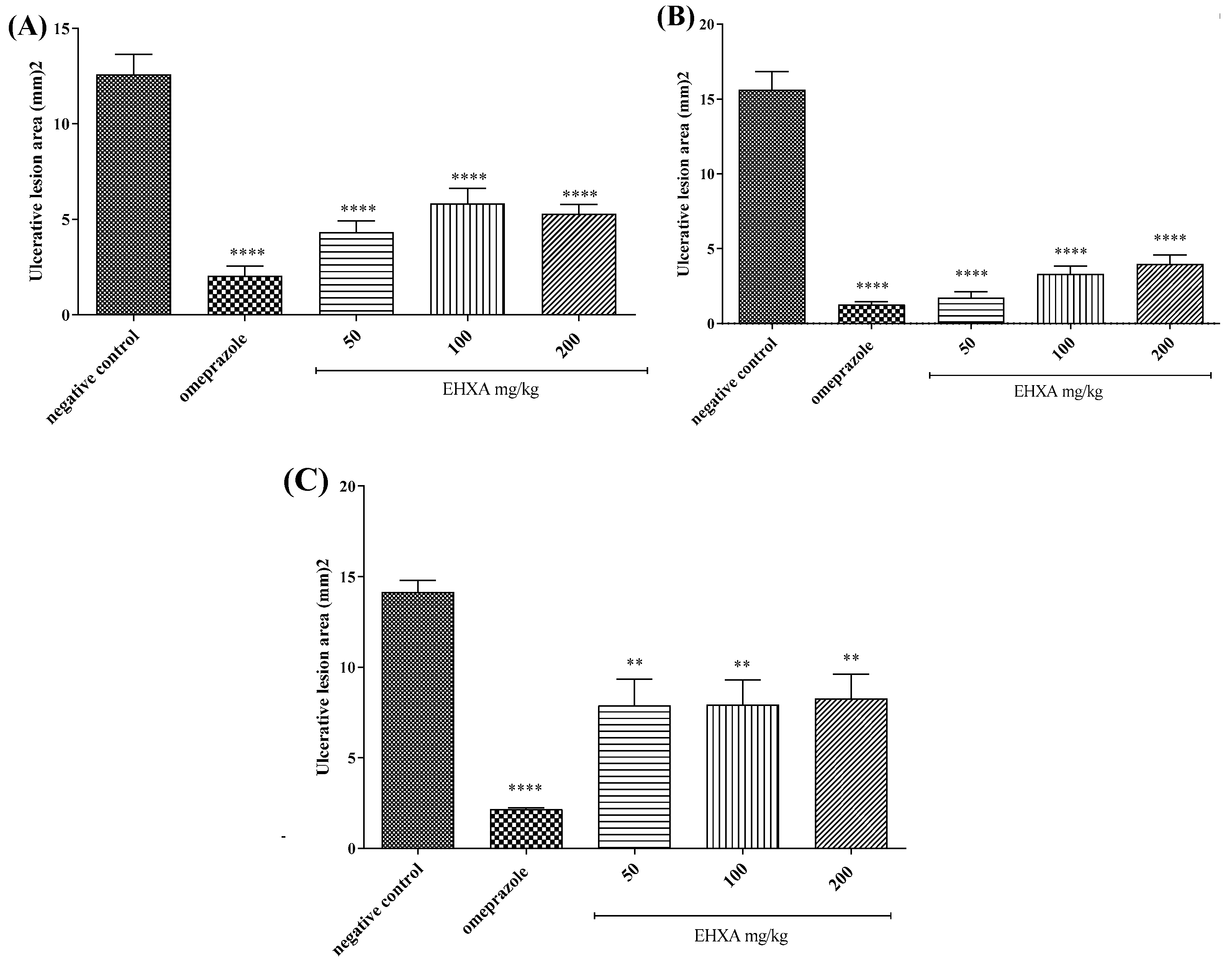
2.1.2. Acute Gastric Injuries Induced by Acidified Ethanol

The groups treated with EHXA (50, 100, and 200 mg/kg) and omeprazole (30 mg/kg) showed significantly reduced development of ulcerative lesions induced by acidified ethanol (by 65.63%, 53.66%, 58.02%, and 83.82%, respectively) when compared to the negative control group (Figure 1A), demonstrating the anti-ulcerative potential of the extract.

2.1.3. Absolute Ethanol-Induced Acute Gastric Injuries

Likewise, the groups of mice treated with EHXA (50, 100, and 200 mg/kg) and omeprazole (30 mg/kg) had the ulcerative lesions reduced by 88.91%, 78.82%, 74.68%, and 91.91%, respectively, in comparison with the negative control group (Figure 1B).

2.1.4. Indomethacin-Induced Acute Gastric Lesions

Considering the importance of non-steroidal anti-inflammatory drugs (NSAIDs) in the development of gastric lesions, we evaluated the effects of EHXA on indomethacin-induced acute gastric lesions. The treatment with EHXA (50, 100, and 200 mg/kg) and omeprazole (30 mg/kg) significantly inhibited the development of ulcerative lesions (by 55.99%, 55.99%, 42.50%, and 84.69%, respectively) when compared to the negative control group (Figure 1C), confirming the gastroprotective activity of the extract in different models.

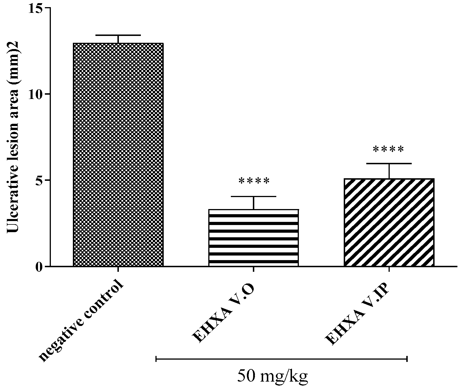
2.2. The Physical Barrier Test

The oral and intraperitoneal administration of EHXA (50 mg/kg) significantly reduced the area of ulcerative lesions of the stomachs by 74.35% and 60.68%, respectively, compared to the negative control group. These results demonstrate that the gastroprotective effect of EHXA occurs both due to the modulation of gastroprotective signaling pathways and through the formation of a physical barrier (Figure 2).

2.3. Involvement of Gastroprotective Signaling Pathways in the Mechanism of Action of EHXA

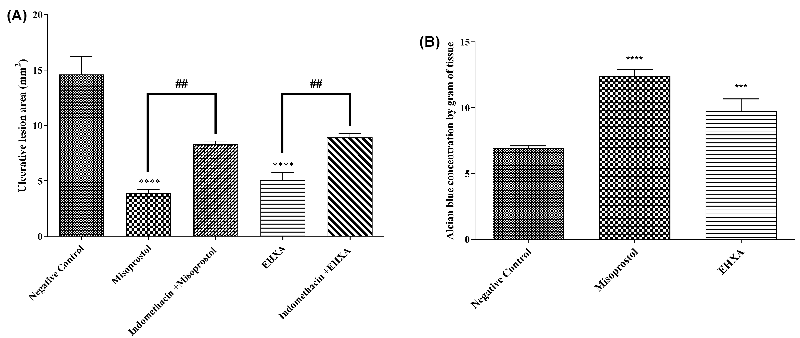
2.3.1. Involvement of Prostaglandins E2 (PGE2)

After administering EHXA (50 mg/kg) and misoprostol (0.016 mg/kg), a PGE2 analog, the area of ulcerative lesions decreased by 65.28% and 73.42%, respectively, compared to the negative control group. However, with the administration of indomethacin (a specific inhibitor of the PGE2 synthesis pathway), the gastroprotective effects of EHXA and misoprostol were reversed, indicating that the gastroprotective effect of EHXA depends, at least partially, on the synthesis of PGE2 (Figure 3A).
Since PGE2-mediated gastroprotection significantly involves mucus production by the gastric mucosa, we analyzed the effects of EHXA (50 mg/kg) and misoprostol (0.016 mg/kg) on mucus production. These treatments significantly increased the production of adherent mucus by 79.92% and 40.40%, respectively, compared to the negative control group, corroborating the evidence that EHXA might be acting through PGE2-dependent mechanisms (Figure 3B).

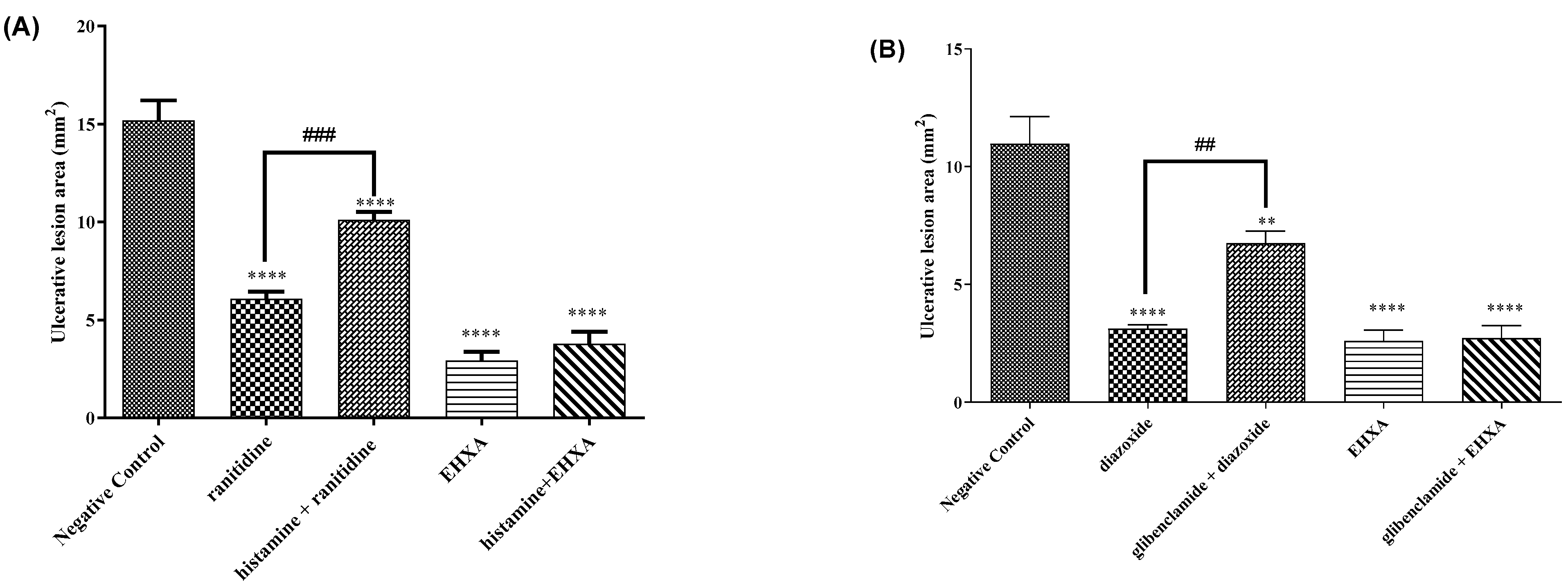
2.3.2. Involvement of H2 Receptors

The administration of EHXA (50 mg/kg) and ranitidine (40 mg/kg), an H2 receptor antagonist, significantly decreased the occurrence of ulcerative lesions in the stomach by 80.71% and 59.92%, respectively, when compared with the negative control group. However, the pretreatment with histamine (H2 receptor agonist) only reversed the protective effect of ranitidine, indicating that the gastroprotective effect of EHXA does not occur via the inhibition of H2 receptors (Figure 4A).

2.3.3. Involvement of ATP-Dependent Potassium Channels

The treatment of animals with EHXA (50 mg/kg) and diazoxide (3 mg/kg), a potassium channel opener, led to a significant reduction in the occurrence of ulcerative lesions in the stomach, with percentual reductions of 76.28% and 71.54%, respectively, compared to the negative control group. However, the pretreatment with glibenclamide, a specific blocker of ATP-dependent potassium channels, had no significant impact on the therapeutic effect of EHXA, indicating that this effect does not result from an interference with potassium-dependent channels (Figure 4B).

2.3.4. Involvement of Sulfhydryl Groups (-SH Groups)

The treatment with EHXA (50 mg/kg) and carbenoxolone (100 mg/kg) significantly reduced ulcerative lesions, with percentual inhibitions of 79.57% and 73%, respectively, compared with the negative control group. However, when the animals in the EHXA group were pretreated with N-ethylmaleimide (NEM), the gastroprotective effect of EHXA and carbenoxolone was reversed, indicating the participation of sulfhydryl groups (Figure 5A) in the gastroprotective action of EHXA (Figure 5A).

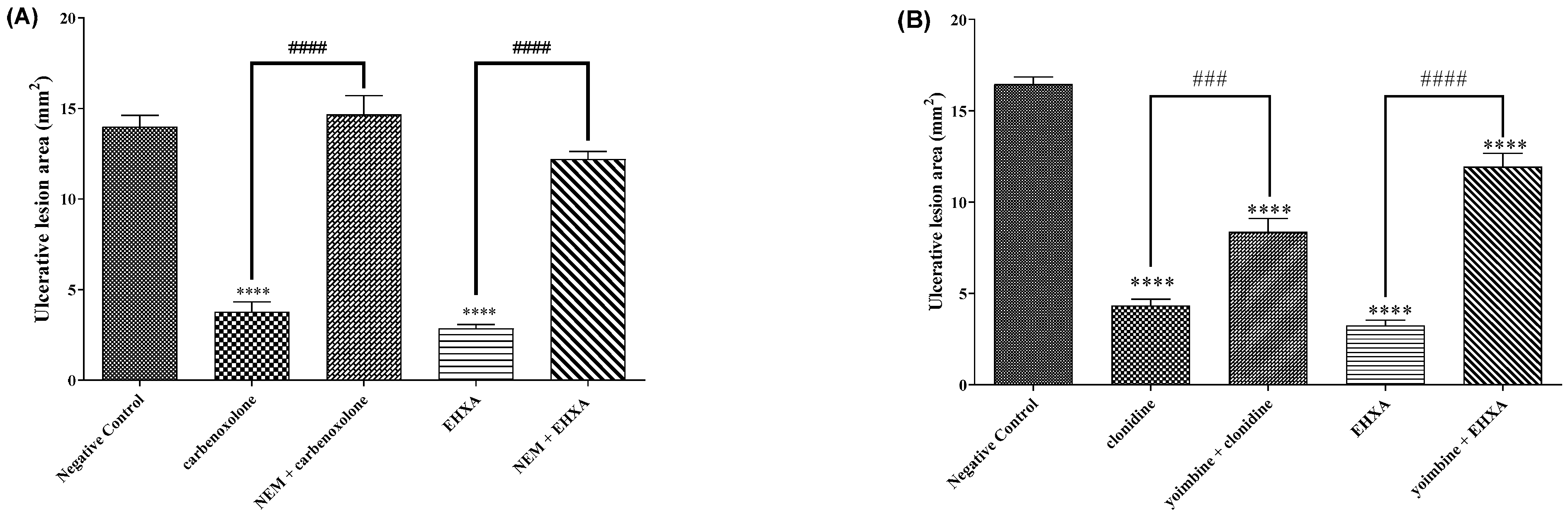
2.3.5. Involvement of α2 Adrenergic Receptors

The treatments with EHXA (50 mg/kg) and clonidine (0.05 mg/kg), an α2 adrenergic receptor agonist, significantly decreased the occurrence of ulcerative lesions in the stomachs of ethanol-challenged animals (inhibitions of 79.76% and 73.68%, respectively) compared to the negative control group. However, in the animals pretreated with yohimbine, a specific inhibitor of α2 adrenergic receptors, the gastroprotective effect of EHXA (50 mg/kg) was reversed, suggesting that α2 adrenergic receptors can, at least partially, mediate the gastroprotective effect of EHXA (Figure 5B).

2.3.6. Involvement of the Nitric Oxide (NO) Pathway

The treatment with EHXA (50 mg/kg) and L-arginine (600 mg/kg) significantly reduced the generation of ulcerative lesions by 73.61% and 71.60%, respectively, compared to the negative control group. However, when the animals in the EHXA group received pretreatment with L-NAME (nonspecific inhibitor of nitric oxide synthase enzymes), a reversal of the gastroprotective effect of EHXA and L-arginine was observed, thus suggesting the potential involvement of NO-dependent mechanisms (Figure 6A).

2.3.7. Nitrate/Nitrite Quantification

The treatment with EHXA (50 mg/kg) and L-arginine (600 mg/kg) significantly increased nitrate and nitrite production by 61.62% and 75.32%, respectively, in tissue homogenates when compared to the negative control group. This result corroborates the previous result of the nitric oxide test (Figure 6B).

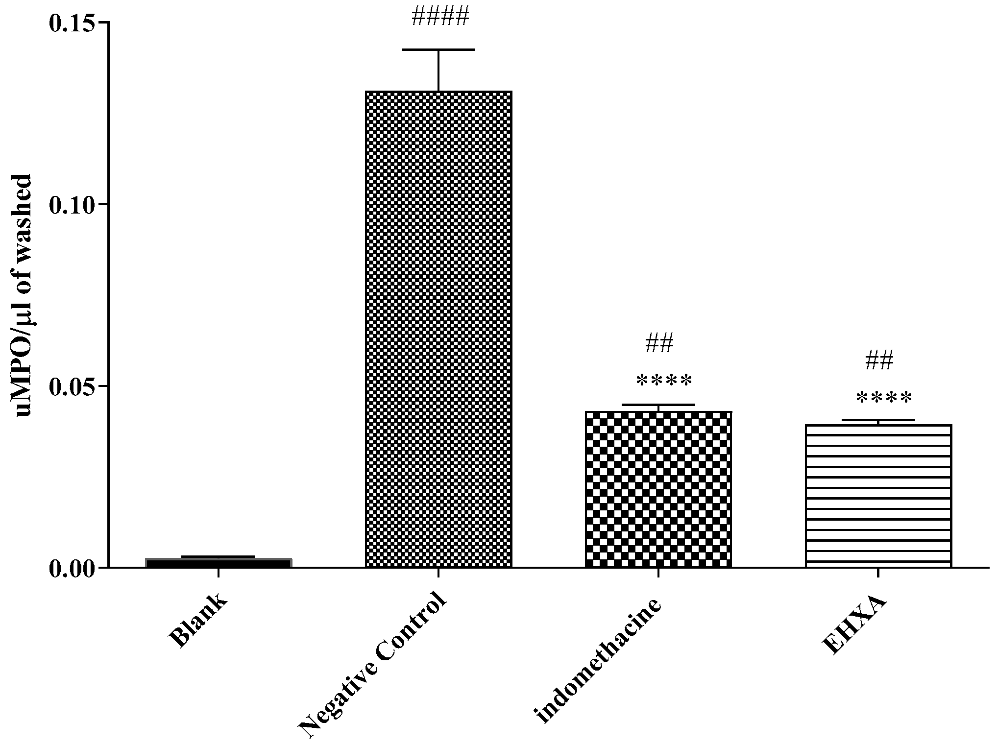
2.3.8. Myeloperoxidase (MPO) Activity

The administration of EHXA (50 mg/kg) and indomethacin (10 mg/kg) resulted in a significant reduction in the myeloperoxidase (MPO) activity produced in the inflamed tissue of animals challenged with acidified ethanol, with inhibitions of 69.93% and 65.07%, respectively, compared to the negative control group (Figure 7), indicating that the activity of EHXA is associated with a decrease in the inflammatory response in the gastric mucosa.

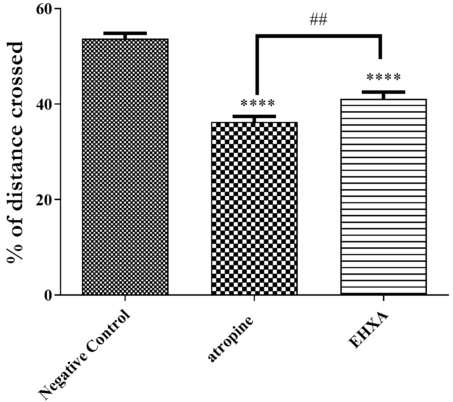
2.3.9. Inhibition of Gastrointestinal Motility by EHXA and ATROPINE

The treatment with EHXA (50 mg/kg) and the muscarinic receptor antagonist atropine (0.01 mg/kg) partially inhibited the intestinal motility of animals by 23.47% and 32.50%, respectively, when compared to the negative control group. This finding suggests that EHXA may inhibit gastrointestinal motility by interfering with the cholinergic system (Figure 8).

3. Discussion

The present study reports, for the first time, the potential of the hydroethanolic extract obtained from the stem bark of Ximenia americana L. in inhibiting the development of gastric lesions in different acute experimental models in mice. The studies carried out by Silva et al. (2018) and Costa et al. (2019) characterized the chromatographic profile (by high-performance liquid chromatography—HPLC) of the same extract, revealing the presence of the compounds quercitrin and caffeic acid as major components, in addition to several others, such as quercetin, rutin, gallic acid, catechin, chlorogenic acid, ellagic acid, and kaempferol [31,32]. A similar chemical composition was observed in other studies, indicating low chemical variation among the samples [14,15].
According to a previously published study, the HPLC analysis of Ximenia americana fruit pulp extract identified various phenolic compounds, including rutin, gallic acid, catechin, epicatechin, p-coumaric acid, and ferulic acid [29]. The ethanolic extract obtained from the stems of Ximenia americana was found to contain steroids such as stigmasterol and sitosterol, as well as triterpenoids such as betulinic acid, oleanolic acid, 28-O-(-D-glucopyranosyl) oleanolic acid, 3-oxo-oleanolic acid, 3β–hydroxycicloart-24(E)-ene-26-oic acid, and furanoic and widdrane sesquiterpenoids [15,33,34]. Another study also reported the identification of several flavonoids, such as quercetin, kaempferol, and isorhamnetin, in the leaves and fruits. Analysis of the fatty acid profile of this species by HPLC identified the presence of palmitic acid, stearic acid, oleic acid, linoleic acid, and alpha-linolenic acid [35].
Notably, compounds present in EHXA have already been proven to present significant gastroprotective effects. Quercitrin has a broad spectrum of bioactivities, including antioxidant, anti-inflammatory, antimicrobial, immunomodulatory, analgesic, wound-healing, vasorelaxant, and gastroprotective [36]. This compound effectively reduced acute colitis in a study by Sanches et al. (2002) [37]. In addition, the gastroprotective activity of gallic acid, which is also present in EHXA, has been evidenced in several in vivo models [38]. Evidence suggests the involvement of the NF-κb pathway in its mechanism of action [39]. Caffeic acid also demonstrated biological activities related to our study, such as antimicrobial, anticancer [40], antioxidant [41], anti-inflammatory, and antinociceptive [42]. Romana-Souza et al., in 2018, showed that caffeic acid administration promotes wound healing by inhibiting the activation of the NO/NOS2 pathway via NF-κB and preventing oxidative stress through reducing peroxynitrite production and nitrite synthesis, thus corroborating our results [43]. A study by Kolgazi et al. (2021) also demonstrated that the reduction in macroscopic and microscopic lesions in the ethanol model was associated with decreased MPO activity and modulation of the NO pathway [44].
It is worth mentioning that the gastroprotective potential of the EHXA constituent epicatechin has been associated with [45] adrenergic receptor activation and modulation of SH, NO, SOD, and HSP-70. Given the proven biological activities of the constituents present in EHXA, we can suggest that its gastroprotective and anti-ulcerative activity may be attributed to these bioactive compounds in the extract.
Regarding the assessment of acute toxicity, the present study demonstrated that EHXA did not cause mortality nor visible signs of toxicity after treatment with the extract, corroborating the result obtained by [14], which also describes the low toxicity of Ximenia americana L.
Regarding the screening models assessed in the present study, treatments with EHXA (50, 100, and 200 mg/kg) reduced gastric lesions in the acidified ethanol, absolute ethanol, and indomethacin models, thus demonstrating their effectiveness in gastric protection.
The gastric ulcer model induced by acidified ethanol is widely used to assess new substances with gastroprotective potential in mice. Acidified ethanol acts by promoting gastric lesions with areas of intense hemorrhage, reductions in the concentrations of glutathione and antioxidant enzymes, increased vascular permeability and infiltration of neutrophils, the subsequent release of reactive oxygen species, and the loss of gastric epithelial cells [14,46,47,48].
Regarding the administration of absolute ethanol, it generates severe lesions in the gastric mucosa, reducing gastric blood flow and causing infiltration of inflammatory cells, tissue destruction, extensive hemorrhages, and production of free radicals [49,50,51]. On the other hand, gastric ulcers induced by indomethacin cause an increase in the levels of inflammatory cytokines such as IL-1β and IL-6. It decreases the mucosa’s anti-inflammatory cytokines, such as IL-4 and TGF-β [52].
Based on the relevant data supporting the gastroprotective effects of EHXA, we evaluated whether the extract acts by modulating systemic pathways or by forming a protective barrier. In the present study, EHXA significantly reduced the lesions when the animals were treated both orally and intraperitoneally, demonstrating that EHXA was acting both at the protective barrier level and on the signaling pathways of the gastroprotective response. It is worth mentioning that the stomach’s first line of defense against aggressive agents is the physical barrier formed by mucus, surfactant phospholipids, and bicarbonates that cover the surface layer and increase blood flow and cell renewal, in addition to maintaining the structural integrity of the mucosa [53].
To better characterize the gastroprotective effect observed in the screening test, we investigated the involvement of signaling pathways that could contribute to the protective mechanisms exhibited by the EHXA.
Prostaglandins, especially PGE2, have a significant cytoprotective effect, stimulating the secretion of sodium bicarbonate in the stomach and inhibiting the release of histamine, TNF-α, platelet-activating factor (PAF), IL-1, IL-8, and LTB4. This mediator also contributes to gastroprotection by inhibiting mast cell activation, gastric motility, gastric acid secretion, and leukocyte adherence to the vascular endothelium [54,55]. Importantly, evidence indicates that these effects involve the activation of EP1, EP3, and EP4 receptors [56,57]. Using pharmacological tools, we demonstrated that PGE2 was able to significantly mediate the effects of EHXA concerning gastric ulcer generation. This finding was corroborated by evidence that EHXA significantly stimulates mucus production in the gastric mucosa. Ethanol promotes damage to the gastric mucosa by destroying protective factors, such as mucus and the non-protein sulfhydryl group, causing an increase in lipid peroxidation and reactive oxygen species production [51,55,58] and a decrease in glutathione levels [59]. It is worth mentioning that in the study described by Mizui, T.& Doteuchi, M 1983 [60], the crude extract, quercetin, and the isolated compound epicatechin were tested and their antioxidant effects were demonstrated in in vitro tests, thus corroborating our findings as well as the evidence that the antioxidant activity is related to the gastroprotective activity of several substances.
Sulfhydryl groups (SH) play an important role in gastric cytoprotective mechanisms [61]. They are found in high concentrations in the stomach, where they are linked to free radicals formed after injury [62]. When adhering to the mucous layer, they form a barrier that supports the surface neutralization of acid [63]. The formation of a complex between glutathione and acetaldehyde, due to ethanol oxidation by the enzyme alcohol dehydrogenase, increases lipid peroxidation and can also cause oxidative stress [64,65]. In this context, this study showed that SH groups participate in the mechanism of action of EHXA. Corroborating the present study [14] showed that an aqueous extract of the species Ximenia americana L. reduced gastric ulcer lesions through sulfhydryl groups, nitric oxide, and antisecretory activity.
Regarding the involvement of α2 adrenergic receptors, these proteins are formed from deduced amino acid sequences to form the three genes of the α2 receptor and its subunits α2A, α2B, and α2C. α2 receptors can bind with different G protein subunits and stimulate the Go protein, inhibiting the Ca2+ channel. The α2 adrenergic receptor inhibits adenyl cyclase, causing the dissociation of the Gi protein and its subunits. That is, the α subunit is loaded with guanosine triphosphate (GTP) and another βγ unit when these α2 adrenergic receptors are stimulated in the stomach, reducing gastric motility [66].
Nitric oxide is involved in maintaining the integrity of the gastric epithelium. According to Cho (2001), nitric oxide exerts either protective or harmful effects, depending on the type of nitric oxide synthetase [67]. NO also contributes to the regulation of gastric blood flow and directly stimulates gastric mucus secretion through the stimulation of guanylate cyclase in epithelial cells. Obstruction of NO production leads to vascular impairment and subsequent alkaline flow into the lumen [67].
To better understand the role of NO in the effect of EHXA in ethanol-induced ulcers, the concentrations of nitrite (NO2) and nitrate (NO3) in the gastric mucosa were assessed as an estimate of NO production [68]. It was possible to verify that nitric oxide is produced at the lesion site and that it is potentially associated with the protective role of EHXA.
Myeloperoxidase (MPO) is an enzyme found mainly in neutrophils, which release it in the inflammatory site in response to several stimuli [69]. In the present study, EHXA reduced MPO levels in the gastric mucosa, suggesting a reduction in neutrophil recruitment and inhibition of the inflammatory response in the injured area. Studies carried out by Pantoja et al., 2018 [70] with total polysaccharides of the extract (TPL-Xa) or tea (Tea-Xa) of Ximenia americana L. showed that its gastroprotective effect is due to the reduction in neutrophil infiltration and, consequently, the reduction in MPO activity.
Regarding gastrointestinal motility, EHXA and the muscarine receptor antagonist atropine presented significant inhibitory effects. Motility is regulated via the cholinergic system in the circular muscular layer of the gastrointestinal tract through M1 and M3 receptors [71]. The study carried out in [72] with caffeic acid, one of the main constituents of EHXA, demonstrated its effective potential in muscle relaxation, corroborating our results.
Finally, this study contributes to the list of Brazilian medicinal plants with the potential to be used therapeutically due to their gastroprotective activity, such as Caryocar coriaceum Wittm. [73], Croton rhamnifolioides [74], Spondias mombin L. [75], and Ziziphus joazeiro Mart. [76].

4. Materials and Methods

4.1. Legal Requirements: Ethical Aspects of the Research

These in vivo assays were conducted using Swiss mice (Mus musculus) of both sexes, with body masses between 20–30 g. The animals were monitored at the Experimental Animal House at URCA and housed in polypropylene cages under controlled conditions, at a temperature of 24 ± 2 °C under a 12 h light/dark cycle. They had free access to a specific food for rodents (Labina, Presence®, São Paulo, Brazil) and drinking water, except for during an 8–12 h fasting period before testing. The research followed the animal use rules and was approved by the Commission for Experimentation and Use of Animals of the Regional University of Cariri (CEUA/URCA) under number 00157/2021.2.

4.2. Hydroethanolic Extract from the Bark of Ximenia americana L. (EHXA) and Chemical Profile

The collection of botanical materials, the extract preparation, and the phytochemical profile were described by Silva et al., 2018 [31].

4.3. In Vivo Assays

In the screening tests of gastric injury induced by acidified ethanol, absolute ethanol, and indomethacin, the animals (n = 6) were divided into groups and treated with either H2O (0.1 mL/10 g/p.o.) or different doses of EHXA (50, 100, and 200 mg/kg/p.o.). The lowest effective dose of EHXA (50 mg/kg/p.o.) identified in the screening tests was used for the other tests and the assessment of the signaling pathways of the gastroprotective response. The investigated mechanisms included the determination of gastric mucosal adherent mucus; the involvement of H2 receptors; ATP-dependent potassium channels; sulfhydryl groups (SH); the cholinergic pathway; α2 adrenergic, nitric oxide, and nitrite/nitrate production; and myeloperoxidase activity. All procedures followed the rules for the use of animals, and the study was approved and registered under number 00157/2021.2.

Non-Clinical Acute Oral Toxicity—LD50

The non-clinical oral acute toxicity evaluation followed the Organization for Economic Cooperation and Development [77], with some modifications, and used the Malone and Robichaud table [78]. The animals were divided into two groups (n = 3). The control group received water for injection (0.01 mL/g/p.o.), and the group treated with EHXA received a single dose of 2000 mg/kg/p.o. The animals were observed at 30, 60, 120, 180, and 360 min post-treatment, and then subsequently every 24 h for 14 days.

4.4. Screening of the Gastroprotective Assessment of EHXA

4.4.1. Acidified Ethanol-Induced Gastric Injury

Six mice were randomly assigned to five groups for the study. The groups received the following treatments: water for injection (0.1 mL/10 g/p.o.), omeprazole (30 mg/kg/p.o.), and EHXA (50, 100, and 200 mg/kg/p.o.). One hour after administering the treatments, the mice were challenged with acidified ethanol (0.2 mL of 0.3 M HCL solution in 60% ethanol/animal, p.o.). After 30 min, the mice were euthanized by cervical dislocation. The greater curvatures of the stomachs were opened, and the stomachs were washed with 0.9% saline before being scanned for quantification and evaluation of gastric lesions. The ulcerated area was determined by planimetry using ImageJ software (Bethesda, MD, USA). Results were expressed as the total area of ulcerative lesions (mm2) [61].

4.4.2. Absolute Ethanol-Induced Gastric Injury

Mice (n = 6) were divided into 5 groups: water for injection (0.1 mL/10 g/p.o.), omeprazole (30 mg/kg/p.o.), and EHXA (50, 100, and 200 mg/kg/p.o.). After 1 h, lesions were induced using ethanol (0.2 mL/animal, p.o.) [79]. After 30 min, the animals were euthanized by cervical dislocation. The stomachs were removed and opened by the greater curvatures, washed with 0.9% saline, and prepared for evaluation and quantification of gastric lesions as described in Section 4.4.1.

4.4.3. Indomethacin-Induced Gastric Injury

Six mice were randomly assigned into five groups: water for injection (0.1 mL/10 g/p.o.), omeprazole (30 mg/kg/p.o.), and EHXA (50, 100, and 200 mg/kg/p.o.). After 1 h, indomethacin (10 mg/kg/p.o.) was administered orally to induce lesions. The treatments were repeated 3 h after the challenge. After six hours of indomethacin administration, the mice were euthanized by cervical dislocation, and their stomachs were removed and opened along the greater curvatures [80]. The stomachs were then washed with 0.9% saline solution and evaluated and quantified for gastric lesions using the scoring method. The scoring was performed by quantifying the gastric lesions as follows: 0 (no lesions), 1 (slight edema), 2 (edema and hemorrhage), 1-2 (punctual lesions), 1-3 (extensive lesions), 5 (several punctual lesions), and 6 (extensive lesions visible throughout the mucosa) [81].

4.5. The Physical Barrier Test

Mice (n = 6) were divided into 3 groups, receiving either water for injection (0.1 mL/10 g/p.o.), EHXA (50 mg/kg/p.o.), and EHXA (50 mg/kg, i.p.). After 30 min of i.p. or 1 h after administering the p.o., 96% ethanol (0.2 mL/animal) was administered. After 30 min, the animals were euthanized by cervical dislocation. The stomachs were removed and opened along the greater curvature, washed with 0.9% saline, and prepared for evaluation and quantification of gastric lesions as described previously.

4.6. Characterization of EHXA Gastroprotective Mechanism

4.6.1. Involvement of the Prostaglandin E2 Pathway

Mice (n = 6) were divided into groups as follows: water for injection (0.1 mL/10 g/p.o.), misoprostol (0.016 mg/kg/p.o.), and EHXA (50 mg/kg/p.o.). The involvement of prostaglandins in the gastroprotective effect of EHXA was evidenced by the previous administration of indomethacin (10 mg/kg/p.o.), a COX inhibitor, 2 h before the administration of EHXA (50 mg/kg/p.o.) or misoprostol (0.016 mg/kg/p.o.). After 1 h of administration of EHXA or misoprostol, the animals received 96% ethanol (0.2 mL/animal; p.o.) [82]. After 30 min, the animals were euthanized, and the stomachs were removed, washed, and prepared for evaluation and quantification of gastric lesions as described previously.

Determination of Gastric Mucosal Mucus

Six mice were divided into three groups: negative control (water for injection 0.1 mL/10 g/p.o.), misoprostol (0.016 mg/kg/p.o.), and EHXA (50 mg/kg/p.o.). After 1 h, gastric lesions were induced by administering 96% ethanol (0.2 mL/animal, p.o.). Thirty minutes after the ethanol administration, the mice were euthanized, and their stomachs were removed and washed. Subsequently, the gastric mucosa was taken out, weighed, and incubated in 10 mL of 0.1% Alcian blue solution for two hours. The dye, complexed with mucus, was extracted using magnesium chloride by stirring it for 1 min every 30 min for 2 h. After this, the homogenate was centrifuged at 3600 rpm for 10 min. The concentration of Alcian blue was measured using spectrophotometry at 598 nm and expressed as μg Alcian blue/mL/g tissue [83].

4.6.2. Involvement of H2 Histamine Receptors

Mice (n = 6) were divided into groups as follows: water for injection (0.1 mL/10 g/p.o.), ranitidine (40 mg/kg/p.o.), and EHXA (50 mg/kg/p.o.). The participation of histamine H2 receptors in the gastroprotective effect of EHXA was evidenced by the previous administration of histamine (3 mg/kg, s.c.) 30 min before the administration of EHXA (50 mg/kg/p.o.) or ranitidine (40 mg/kg/p.o.) [84,85]. After 30 min of ranitidine administration and 1 h after administration of EHXA and negative control, the animals received 96% ethanol (0.2 mL/animal; p.o.). After 30 min, the animals were euthanized and their stomachs were removed, washed, and prepared for evaluation and quantification of gastric lesions as previously described.

4.6.3. Involvement of ATP-Dependent Potassium Channels

Six mice were assigned into five groups, with the first three groups receiving water for injection (0.1 mL/10 g/p.o.), diazoxide (3 mg/kg, i.p.), and EHXA (50 mg/kg/p.o.). The involvement of potassium channels in EHXA’s gastroprotective effect was demonstrated by administering glibenclamide (5 mg/kg, i.p.) 30 min before administering EHXA (50 mg/kg/p.o.) or diazoxide (3 mg/kg, i.p.) [80]. After one hour of oral treatments or 30 min of intraperitoneal treatment, the animals were given 96% ethanol (0.2 mL/animal, p.o). Thirty minutes later, the mice were euthanized, and their stomachs were removed, cleaned, and analyzed for gastric lesions, as explained in Section 4.4.1.

4.6.4. Involvement of Sulfhydryl (SH) Groups

Six mice were assigned into five groups, with the initial three groups receiving water for injection (0.1 mL/10 g/p.o.), carbenoxolone (100 mg/kg/p.o.), or EHXA (50 mg/kg/p.o.). The involvement of sulfhydryl groups in EHXA’s gastroprotective effect was demonstrated by administering N-ethylmaleimide (NEM) (10 mg/kg, i.p.) 30 min before administering carbenoxolone (100 mg/kg) or EHXA (50 mg/kg/p.o.). After one hour of oral treatments or 30 min of intraperitoneal treatment, the animals were given 96% ethanol (0.2 mL/animal, p.o). Thirty minutes later, the mice were euthanized, and their stomachs were removed, cleaned, and analyzed for gastric lesions, as explained in Section 4.4.1.

4.6.5. Involvement of α2 Adrenergic Receptors

Six mice were assigned into five groups, with the initial three groups receiving water for injection (0.1 mL/10 g/p.o.), clonidine (0.05 mg/kg/p.o.), and EHXA (50 mg/kg/p.o.), respectively. The involvement of α2 adrenergic receptors in EHXA’s gastroprotective effect was evaluated by administering yohimbine (2 mg/kg, i.p.) 20 min before administering EHXA (50 mg/kg/p.o.) or clonidine (0.05 mg/kg/p.o.). After 1 h of oral treatments or 30 min of intraperitoneal treatment, the animals were given 96% ethanol (0.2 mL/animal, p.o). Thirty minutes later, the mice were euthanized, and their stomachs were removed, cleaned, and analyzed for gastric lesions as previously described.

4.6.6. Involvement of the Nitric Oxide Pathway

A group of animals was divided into five subgroups of six mice, with the first three groups receiving water for injection (0.1 mL/10 g/p.o.), L-arginine (600 mg/kg, i.p.), and EHXA (50 mg/kg/p.o.), respectively. This study aimed to investigate the role of nitric oxide (NO) in the gastroprotective effect of EHXA. For this, the inhibitor L-NAME (20 mg/kg, i.p.) was administered 30 min prior to the administration of EHXA (50 mg/kg) or L-arginine (600 mg/kg). After 1 h (p.o) or 30 min (i.p) of treatment, 96% ethanol (0.2 mL/animal, p.o) was administered [82]. After 30 min, the mice were euthanized, and their stomachs were removed, washed, and prepared for evaluation and quantification of gastric lesions.

Nitrate/Nitrite Quantification

This method was based on the diazotization reaction and Griess reagent coupling, which reveals the presence of nitrite in the injured tissue sample. The absorbance reading was performed by a spectrophotometer with a 560 nm filter [86].

4.6.7. Myeloperoxidase (MPO) Activity

Six mice were randomly assigned to three groups: negative control (water for injection 0.1 mL/10 g/p.o.), indomethacin (10 mg/kg/p.o.), and EHXA (50 mg/kg/p.o.). The treatments were administered 1 h before inducing lesions with acidified ethanol (0.2 mL of 0.3 M HCl solution in 60% ethanol/animal, p.o.). After 1 h, the mice were euthanized by cervical dislocation, and their stomachs were removed and opened along the greater curvature. The stomachs were then washed with 0.9% saline solution and prepared for the evaluation of myeloperoxidase enzyme activity. MPO levels were determined by measuring the optical density of the mixture of the samples and o-dianisidine solution using a spectrophotometer at 600 nm, with hydrogen peroxide as a substrate for MPO. The results were expressed as UMPO/μL of wash [87].

4.7. Assessment of Gastrointestinal Motility

A total of 3 groups of mice (n = 6) were formed: negative control (received H2O, 0.1 mL/10 g/p.o.), positive control (atropine—0.01 mg/kg), and EHXA (50 mg/kg/p.o.). After one hour, a semi-solid-colored marker (0.05% phenol red in 1.5% carboxymethylcellulose) was orally administered, and the mice were euthanized 30 min later. The small intestines were removed and measured to determine the total length. The percentage of the distance traveled by the marker was calculated and used as a marker of motility [88].

4.8. Statistical Analysis

The mean ± standard error of the mean (S.E.M) was used to express the results. To analyze the differences between means, one-way and two-way analyses of variance (ANOVA) were employed, followed by Tukey’s multiple comparison test. Statistical analysis was carried out using GraphPad Prism 6.1, with a significance level of 5% (p < 0.05) set for rejecting the null hypothesis.

5. Conclusions

In conclusion, EHXA showed a significant gastroprotective effect that might be associated with the inhibition of MPO activity and modulation of α2 adrenergic receptors, NO synthesis, SH groups, stimulation of mucus secretion mediated by PGE2, and reduction in intestinal motility.
Based on the obtained data, we can attest to the preclinical efficacy of EHXA and suggest its potential mechanisms of action. However, there is a need for further studies to prove its effectiveness in the treatment of gastric ulcers and to elucidate the molecular mechanisms of action of its constituents.

Author Contributions

Conceptualization, I.R.A.d.M. and R.T.P.; methodology, R.T.P., I.S.A. and L.Y.S.d.S.; software, T.M.S.; validation, R.T.P. and I.S.A.; formal analysis, M.R.C.d.O.; investigation, L.Y.S.d.S.; resources, R.H.S.d.C. and B.C.G.V.d.L.; data curation, A.G.B.R.; writing—original draft preparation, R.T.P.; writing—review and editing, J.R.-F., C.D.d.M.O.-T. and C.M.G.L.; supervision, A.O.B.P.B.M., H.D.M.C., C.M.G.L. and I.R.A.d.M.; project administration, E.M.d.A.; funding acquisition, J.R.-F. and I.R.A.d.M. All authors have read and agreed to the published version of the manuscript.

Funding

The authors would like to thank the financial support provided of support of the Brazilian agencies CAPES, FUNCAP, CNPq, and FINEP. This article is a contribution of the Nacional Institute of Science and Technology—Ethnobiology, Bioprospecting and Nature Conservation/CNPq/FACEPE.

Institutional Review Board Statement

The animal study protocol was approved by the Commission for Experimentation and Use of Animals of the Regional University of Cariri (CEUA/URCA), registered under number 00157/2021.2.

Data Availability Statement

The data supporting reported results can be solicited from the corresponding author.

Acknowledgments

Regional University of Cariri (URCA).

Conflicts of Interest

The authors declare no conflict of interest.

References

  1. De Filippis, A.; Ullah, H.; Baldi, A.; Dacrema, M.; Esposito, C.; Garzarella, E.U.; Santarcangelo, C.; Tantipongpiradet, A.; Daglia, M. Gastrointestinal Disorders and Metabolic Syndrome: Dysbiosis as a Key Link and Common Bioactive Dietary Components Useful for Their Treatment. Int. J. Mol. Sci. 2020, 21, 4929. [Google Scholar] [CrossRef]
  2. Belaiche, J.; Burette, A.; De Vos, M.; Louis, E.; Huybrechts, M.; Deltenre, M. Observational Survey of NSAID-Related Upper Gastrointestinal Adverse Events in Belgium. Acta Gastroenterol. Belg. 2002, 65, 65–73. [Google Scholar]
  3. Zapata-Colindres, J.C.; Zepeda-Gómez, S.; Montaño-Loza, A.; Vásquez-Ballesteros, E.; de Jesús Villalobos, J.; Valdovinos-Andraca, F. The Association of Helicobacter Pylori Infection and Nonsteroidal Anti-Inflammatory Drugs in Peptic Ulcer Disease. Can. J. Gastroenterol. 2006, 20, 277–280. [Google Scholar] [CrossRef] [Green Version]
  4. da Silva, M.S.; de Almeida, A.C.A.; de Faria, F.M.; Luiz-Ferreira, A.; da Silva, M.A.; Vilegas, W.; Pellizzon, C.H.; Brito, A.R.M.S. Abarema Cochliacarpos: Gastroprotective and Ulcer-Healing Activities. J. Ethnopharmacol. 2010, 132, 134–142. [Google Scholar] [CrossRef] [PubMed]
  5. Klein, L.C.; Gandolfi, R.B.; Santin, J.R.; Lemos, M.; Filho, V.C.; De Andrade, S.F. Antiulcerogenic Activity of Extract, Fractions, and Some Compounds Obtained from Polygala cyparissias St. Hillaire & Moquin (Polygalaceae). Naunyn-Schmiedeberg’s Arch. Pharmacol. 2010, 381, 121–126. [Google Scholar] [CrossRef]
  6. Porth, C.M.; Matfin, G. Fisiopatologia, 8th ed.; Editora Guanabara Koogan: Rio de Janeiro, Brazil, 2010. [Google Scholar]
  7. Rates, S.M.K. Plants as Source of Drugs. Toxicon 2001, 39, 603–613. [Google Scholar] [CrossRef] [PubMed]
  8. Chan, F.K.L.; Leung, W.K. Peptic-Ulcer Disease. Seminar 2002, 360, 933–941. [Google Scholar]
  9. Schmeda-Hirschmann, G.; Yesilada, E. Traditional Medicine and Gastroprotective Crude Drugs. J. Ethnopharmacol. 2005, 100, 61–66. [Google Scholar] [CrossRef]
  10. Falcão, H.S.; Mariath, I.R.; Diniz, M.F.F.M.; Batista, L.M.; Barbosa-Filho, J.M. Plants of the American Continent with Antiulcer Activity. Phytomedicine 2008, 15, 132–146. [Google Scholar] [CrossRef]
  11. Brasileiro, M.T. Padronização, Atividade Biológica e Desenvolvimento de Formas Farmacêuticas Semi-Sólidas à Base de Ximenia americana L. Z. Phytother. 2008, 29, 150–153. [Google Scholar] [CrossRef]
  12. Shettar, A.K.; Kotresha, K.; Kaliwal, B.B.; Vedamurthy, A.B. Evaluation of in Vitro Antioxidant and Anti-Inflammatory Activities of Ximenia americana Extracts. Asian Pac. J. Trop. Dis. 2015, 5, 918–923. [Google Scholar] [CrossRef]
  13. Agyigra, A.I.; Ejiofor, J.I.; Magaji, M.G.; Yakubu, Y. Evaluation of Methanol Stem-Bark Extract of Ximenia americana Linn (Olacaceae) for Phytoconstituents and Gastroprotection in Rats. Afr. J. Pharmacol. Ther. 2017, 6, 161–165. [Google Scholar]
  14. Aragão, T.P.; dos Prazeres, L.D.K.T.; Brito, S.A.; Neto, P.J.R.; Rolim, L.A.; da Silva Almeida, J.R.G.; Caldas, G.F.R.; Wanderley, A.G. Contribution of Secondary Metabolites to the Gastroprotective Effect of Aqueous Extract of Ximenia americana L. (Olacaceae) Stem Bark in Rats. Molecules 2018, 23, 112. [Google Scholar] [CrossRef] [PubMed] [Green Version]
  15. De Araújo, M.R.S.; Da Costa Assunção, J.C.; Dantas, I.N.F.; Costa-Lotufo, L.V.; Monte, F.J.Q. Chemical Constituents of Ximenia americana. Nat. Prod. Commun. 2008, 3, 857–860. [Google Scholar] [CrossRef] [Green Version]
  16. Sharief, T.M.; Bashier, R.S.M.; Haroon, M.I. Phytochemical Evaluation and Uses of Ximenia americana L. in Central Darfur. Int. J. Curr. Microbiol. Appl. Sci. 2022, 11, 353–360. [Google Scholar] [CrossRef]
  17. Le, N.H.T.; Malterud, K.E.; Diallo, D.; Paulsen, B.S.; Nergård, C.S.; Wangensteen, H. Bioactive Polyphenols in Ximenia americana and the Traditional Use among Malian Healers. J. Ethnopharmacol. 2012, 139, 858–862. [Google Scholar] [CrossRef]
  18. Liu, K.; Zhang, X.; Xie, L.; Deng, M.; Chen, H.; Song, J.; Long, J.; Li, X.; Luo, J. Lupeol and Its Derivatives as Anticancer and Anti-Inflammatory Agents: Molecular Mechanisms and Therapeutic Efficacy. Pharmacol. Res. 2021, 164, 105373. [Google Scholar] [CrossRef] [PubMed]
  19. Lou, H.; Li, H.; Zhang, S.; Lu, H.; Chen, Q.; Zhang, X.; Wang, Y. A Review on Preparation of Betulinic Acid and Its Biological Activities. Molecules 2021, 26, 5583. [Google Scholar] [CrossRef]
  20. Nattagh-Eshtivani, E.; Barghchi, H.; Pahlavani, N.; Barati, M.; Amiri, Y.; Fadel, A.; Khosravi, M.; Talebi, S.; Arzhang, P.; Ziaei, R.; et al. Biological and Pharmacological Effects and Nutritional Impact of Phytosterols: A Comprehensive Review. Phytother. Res. 2022, 36, 299–322. [Google Scholar] [CrossRef]
  21. Babu, S.; Jayaraman, S. An Update on β-Sitosterol: A Potential Herbal Nutraceutical for Diabetic Management. Biomed. Pharmacother. 2020, 131, 110702. [Google Scholar] [CrossRef]
  22. Dasiman, R.; Md Nor, N.; Eshak, Z.; Syairah, S.; Mutalip, M.; Suwandi, N.R.; Bidin, H. A Review of Procyanidin: Updates on Current Bioactivities and Potential Health Benefits. Biointerface Res. Appl. Chem. 2022, 12, 5918–5940. [Google Scholar] [CrossRef]
  23. Costa, A.C.F.; de Sousa, L.M.; Dos Santos Alves, J.M.; Goes, P.; Pereira, K.M.A.; Alves, A.P.N.N.; Vale, M.L.; Gondim, D.V. Anti-Inflammatory and Hepatoprotective Effects of Quercetin in an Experimental Model of Rheumatoid Arthritis. Inflammation 2021, 44, 2033–2043. [Google Scholar] [CrossRef]
  24. Leyva-López, N.; Gutierrez-Grijalva, E.P.; Ambriz-Perez, D.L.; Basilio Heredia, J.; Segura-Carretero, A.; Maria, A.; Caravaca, G. Flavonoids as Cytokine Modulators: A Possible Therapy for Inflammation-Related Diseases. Int. J. Mol. Sci. 2016, 17, 921. [Google Scholar] [CrossRef] [Green Version]
  25. Ribeiro, D.; Freitas, M.; Tomé, S.M.; Silva, A.M.; Laufer, S.; Lima, J.L.; Fernandes, E. Flavonoids Inhibit COX-1 and COX-2 Enzymes and Cytokine/Chemokine Production in Human Whole Blood. Inflammation 2014, 38, 858–870. [Google Scholar] [CrossRef]
  26. Da Silva, G.G.; De Souza, P.A.; De Morais, P.L.D.; Dos Santos, E.C.; Moura, R.D.; Menezes, J.B. Caracterização Do Fruto de Ameixa Silvestre (Ximenia americana L.). Rev. Bras. Frutic. 2008, 30, 311–314. [Google Scholar] [CrossRef] [Green Version]
  27. Oliveira, F.C.S.; Barros, R.F.M.; Moita Neto, J.M. Plantas Medicinais Utilizadas Em Comunidades Rurais de Oeiras, Semiárido Piauiense. Rev. Bras. Plantas Med. 2010, 12, 282–301. [Google Scholar] [CrossRef]
  28. Chaves, E.M.F.; Chaves, E.d.B.F.; Coelho-de-Souza, G.; Figueiredo, L.S.; de Barros, R.F.M.; Kubo, R. Um Olhar Sobre Ximenia americana L. e Suas Potencialidades. Acta Tecnol. 2014, 9, 70–77. [Google Scholar] [CrossRef]
  29. Queiroz Monte, F.J.; de Lemos, T.L.G.; de Arajo, M.R.S.; de Sousa Gomes, E. Ximenia americana: Chemistry, Pharmacology and Biological Properties, a Review. In Phytochemicals—A Global Perspective of Their Role in Nutrition and Health; IntechOpen: London, UK, 2012. [Google Scholar] [CrossRef] [Green Version]
  30. Ogunleye, D.S.; Ibitoye, S.F. Studies of Antimicrobial Activity and Chemical Constituents of Ximenia americana. Trop. J. Pharm. Res. 2003, 2, 239–241. [Google Scholar]
  31. da Silva, B.A.F.; da Costa, R.H.S.; Fernandes, C.N.; Leite, L.H.I.; Ribeiro-Filho, J.; Garcia, T.R.; Coutinho, H.D.M.; Wanderley, A.G.; de Menezes, I.R.A. HPLC Profile and Antiedematogenic Activity of Ximenia americana L. (Olacaceae) in Mice Models of Skin Inflammation. Food Chem. Toxicol. 2018, 119, 199–205. [Google Scholar] [CrossRef]
  32. de Menezes, I.R.A.; da Costa, R.H.S.; Augusti Boligon, A.; Rolón, M.; Coronel, C.; Vega, C.; Melo Coutinho, H.D.; da Costa, M.S.; Tintino, S.R.; Silva Pereira, R.L.; et al. Ximenia americana L. Enhances the Antibiotic Activity and Inhibits the Development of Kinetoplastid Parasites. Comp. Immunol. Microbiol. Infect. Dis. 2019, 64, 40–46. [Google Scholar] [CrossRef]
  33. Okhale, S.E.; Nnachor, A.C.; Bassey, U.E. Evaluation of HPLC-UV-DAD and Antiproliferative Characteristics of the Leaf Infusion of Ximenia americana Linn. MicroMedicine 2017, 5, 45–52. [Google Scholar] [CrossRef]
  34. Almeida, M.L.B.; Freitas, W.E.D.S.; De Morais, P.L.D.; Sarmento, J.D.A.; Alves, R.E. Bioactive Compounds and Antioxidant Potential Fruit of Ximenia americana L. Food Chem. 2016, 192, 1078–1082. [Google Scholar] [CrossRef] [PubMed]
  35. Bazezew, A.M.; Emire, S.A.; Sisay, M.T. Bioactive Composition, Free Radical Scavenging and Fatty Acid Profile of Ximenia americana Grown in Ethiopia. Heliyon 2021, 7, e07187. [Google Scholar] [CrossRef] [PubMed]
  36. Chen, J.; Li, G.; Sun, C.; Peng, F.; Yu, L.; Chen, Y.; Tan, Y.; Cao, X.; Tang, Y.; Xie, X.; et al. Chemistry, Pharmacokinetics, Pharmacological Activities, and Toxicity of Quercitrin. Phytother. Res. 2022, 36, 1545–1575. [Google Scholar] [CrossRef]
  37. Sánchez De Medina, F.; Vera, B.; Gálvez, J.; Zarzuelo, A. Effect of Quercitrin on the Early Stages of Hapten Induced Colonic Inflammation in the Rat. Life Sci. 2002, 70, 3097–3108. [Google Scholar] [CrossRef] [Green Version]
  38. Zhou, D.; Yang, Q.; Tian, T.; Chang, Y.; Li, Y.; Duan, L.R.; Li, H.; Wang, S.W. Gastroprotective Effect of Gallic Acid against Ethanol-Induced Gastric Ulcer in Rats: Involvement of the Nrf2/HO-1 Signaling and Anti-Apoptosis Role. Biomed. Pharmacother. 2020, 126, 110075. [Google Scholar] [CrossRef]
  39. Zhu, L.; Gu, P.; Shen, H. Gallic Acid Improved Inflammation via NF-κB Pathway in TNBS-Induced Ulcerative Colitis. Int. Immunopharmacol. 2019, 67, 129–137. [Google Scholar] [CrossRef]
  40. Matejczyk, M.; Świsłocka, R.; Golonko, A.; Lewandowski, W.; Hawrylik, E. Cytotoxic, Genotoxic and Antimicrobial Activity of Caffeic and Rosmarinic Acids and Their Lithium, Sodium and Potassium Salts as Potential Anticancer Compounds. Adv. Med. Sci. 2018, 63, 14–21. [Google Scholar] [CrossRef]
  41. Amorati, R.; Pedulli, G.F.; Cabrini, L.; Zambonin, L.; Landi, L. Solvent and PH Effects on the Antioxidant Activity of Caffeic and Other Phenolic Acids. J. Agric. Food Chem. 2006, 54, 2932–2937. [Google Scholar] [CrossRef] [PubMed]
  42. Gamaro, G.D.; Suyenaga, E.; Borsoi, M.; Lermen, J.; Pereira, P.; Ardenghi, P. Effect of Rosmarinic and Caffeic Acids on Inflammatory and Nociception Process in Rats. ISRN Pharmacol. 2011, 2011, 451682. [Google Scholar] [CrossRef] [PubMed] [Green Version]
  43. Romana-Souza, B.; dos Santos, J.S.; Monte-Alto-Costa, A. Caffeic Acid Phenethyl Ester Promotes Wound Healing of Mice Pressure Ulcers Affecting NF-ΚB, NOS2 and NRF2 Expression. Life Sci. 2018, 207, 158–165. [Google Scholar] [CrossRef] [PubMed]
  44. Kolgazi, M.; Cilingir, S.; Yilmaz, O.; Gemici, M.; Yazar, H.; Ozer, S.; Acikel-Elmas, M.; Arbak, S.; Suyen, G.G. Caffeic Acid Attenuates Gastric Mucosal Damage Induced by Ethanol in Rats via Nitric Oxide Modulation. Chem. Biol. Interact. 2021, 334, 109351. [Google Scholar] [CrossRef] [PubMed]
  45. Rozza, A.L.; Hiruma-Lima, C.A.; Tanimoto, A.; Pellizzon, C.H. Morphologic and Pharmacological Investigations in the Epicatechin Gastroprotective Effect. Evid.-Based Complement. Altern. Med. 2012, 2012, 708156. [Google Scholar] [CrossRef] [PubMed] [Green Version]
  46. Adeyemi, O.O.; Yemitan, O.K.; Taiwo, A.E. Neurosedative and Muscle-Relaxant Activities of Ethyl Acetate Extract of Baphia nitida AFZEL. J. Ethnopharmacol. 2006, 106, 312–316. [Google Scholar] [CrossRef] [PubMed]
  47. Repetto, M.G.; Llesuy, S.F. Antioxidant Properties of Natural Compounds Used in Popular Medicine for Gastric Ulcers. Braz. J. Med. Biol. Res. 2002, 35, 523–534. [Google Scholar] [CrossRef] [Green Version]
  48. Ribeiro, A.R.S.; Diniz, P.B.F.; Pinheiro, M.S.; Albuquerque-Júnior, R.L.C.; Thomazzi, S.M. Gastroprotective Effects of Thymol on Acute and Chronic Ulcers in Rats: The Role of Prostaglandins, ATP-Sensitive K+ Channels, and Gastric Mucus Secretion. Chem. Biol. Interact. 2016, 244, 121–128. [Google Scholar] [CrossRef] [PubMed]
  49. Guslandi, M. Effects of Ethanol on the Gastric Mucosa. Dig. Dis. 1987, 5, 21–32. [Google Scholar] [CrossRef]
  50. Lee, J.S.; Oh, T.Y.; Kim, Y.K.; Baik, J.H.; So, S.; Hahm, K.B.; Surh, Y.J. Protective Effects of Green Tea Polyphenol Extracts against Ethanol-Induced Gastric Mucosal Damages in Rats: Stress-Responsive Transcription Factors and MAP Kinases as Potential Targets. Mutat. Res.—Fundam. Mol. Mech. Mutagen. 2005, 579, 214–224. [Google Scholar] [CrossRef] [PubMed]
  51. Szabo, S.; Trier, J.S.; Brown, A.; Schnoor, J. Early Vascular Injury and Increased Vascular Permeability in Gastric Mucosal Injury Caused by Ethanol in the Rat. Gastroenterology 1985, 88, 228–236. [Google Scholar] [CrossRef]
  52. Yadav, S.K.; Adhikary, B.; Maity, B.; Bandyopadhyay, S.K.; Chattopadhyay, S. The Gastric Ulcer-Healing Action of Allylpyrocatechol Is Mediated by Modulation of Arginase Metabolism and Shift of Cytokine Balance. Eur. J. Pharmacol. 2009, 614, 106–113. [Google Scholar] [CrossRef] [PubMed]
  53. DeFoneska, A.; Kaunitz, J.D. Gastroduodenal Mucosal Defense. Curr. Opin. Gastroenterol. 2010, 26, 604–610. [Google Scholar] [CrossRef] [PubMed]
  54. Atay, S.; Tarnawski, A.S.; Dubois, A. Eicosanoids and the Stomach. Prostaglandins Other Lipid Mediat. 2000, 61, 105–124. [Google Scholar] [CrossRef] [PubMed]
  55. Wallace, J.L. Prostaglandins, NSAIDs, and Gastric Mucosal Protection: Why Doesn’t the Stomach Digest Itself? Physiol. Rev. 2008, 88, 1547–1565. [Google Scholar] [CrossRef]
  56. Aoi, M.; Aihara, E.; Nakashima, M.; Takeuchi, K. Participation of Prostaglandin E Receptor EP4 Subtype in Duodenal Bicarbonate Secretion in Rats. Am. J. Physiol. Gastrointest. Liver Physiol. 2004, 287, 96–103. [Google Scholar] [CrossRef]
  57. Nakashima, M.; Aoi, M.; Aihara, E.; Takeuchi, K. No Role for Prostacyclin IP Receptors in Duodenal HCO3-secretion Induced by Mucosal Acidification in Mice—Comparison with Capsaicin-Induced Response. Digestion 2004, 70, 16–25. [Google Scholar] [CrossRef]
  58. Mizui, T.; Sato, H.; Hirose, F.; Doteuchi, M. Effect of Antiperoxidative Drugs on Gastric Damage Induced by Ethanol in Rats. Life Sci. 1987, 41, 755–763. [Google Scholar] [CrossRef]
  59. Ignatowicz, E.; Wozniak, A.; Kulza, M.; Señczuk-Przybylowska, M.; Cimino, F.; Piekoszewski, W.; Chuchracki, M.; Florek, E. Exposure to Alcohol and Tobacco Smoke Causes Oxidative Stress in Rats. Pharmacol. Rep. 2013, 65, 906–913. [Google Scholar] [CrossRef] [Green Version]
  60. Uchôa, V.T.; Sousa, C.M.M.; Carvalho, A.A.; Sant’Ana, A.E.G.; Chaves, M.H. Free Radical Scavenging Ability of Ximenia americana L. Stem Bark and Leaf Extracts. J. Appl. Pharm. Sci. 2016, 6, 91–96. [Google Scholar] [CrossRef] [Green Version]
  61. Mizui, T.; Doteuchi, M. Effect of Polyamines on Acidified Ethanol-Induced Gastric Lesions in Rats. Jpn. J. Pharmacol. 1983, 33, 939–945. [Google Scholar] [CrossRef] [PubMed]
  62. Avila, J.R.; Alarcón De La Lastra, C.; Martín, M.J.; Motilva, V.; Luque, I.; Delgado, D.; Esteban, J.; Herrerias, J. Role of Endogenous Sulphydryls and Neutrophil Infiltration in the Pathogenesis of Gastric Mucosal Injury Induced by Piroxicam in Rats. Inflamm. Res. 1996, 45, 83–88. [Google Scholar] [CrossRef]
  63. Allen, A.; Flemström, G. Gastroduodenal Mucus Bicarbonate Barrier: Protection against Acid and Pepsin. Am. J. Physiol. Cell Physiol. 2005, 288, C1–C19. [Google Scholar] [CrossRef] [Green Version]
  64. Chen, H.; Liao, H.; Liu, Y.; Zheng, Y.; Wu, X.; Su, Z.; Zhang, X.; Lai, Z.; Lai, X.; Lin, Z.X.; et al. Protective Effects of Pogostone from Pogostemonis Herba against Ethanol-Induced Gastric Ulcer in Rats. Fitoterapia 2015, 100, 110–117. [Google Scholar] [CrossRef] [PubMed]
  65. Jordão, A.A., Jr.; Chiarello, P.G.; Bernardes, M.S.M.; Vannucchi, H. Peroxidação Lipídica e Etanol: Papel da Glutationa Reduzida e da Vitamina e Lipid Peroxidation and Ethanol: Role of Vitamin-E and Glutathione. Med. Ribeirão Preto 1998, 31, 434–449. [Google Scholar] [CrossRef] [Green Version]
  66. Yokotani, K.; Okuma, Y.; Nakamura, K.; Osumi, Y. Release of Endogenous Acetylcholine from a Vascularly Perfused Rat Stomach in Vitro; Inhibition by M3 Muscarinic Autoreceptors and Alpha-2 Adrenoceptors. J. Pharmacol. Exp. Ther. 1993, 266, 1190–1195. [Google Scholar] [CrossRef]
  67. Cho, C.H. Current Roles of Nitric Oxide in Gastrointestinal Disorders. J. Physiol. Paris 2001, 95, 253–256. [Google Scholar] [CrossRef] [PubMed]
  68. Silva, R.O.; Lucetti, L.T.; Wong, D.V.T.; Aragão, K.S.; Junior, E.M.A.; Soares, P.M.G.; Barbosa, A.L.R.; Ribeiro, R.A.; Souza, M.H.L.P.; Medeiros, J.V.R. Alendronate Induces Gastric Damage by Reducing Nitric Oxide Synthase Expression and NO/CGMP/KATP Signaling Pathway. Nitric Oxide 2014, 40, 22–30. [Google Scholar] [CrossRef]
  69. Bayir, H. Reactive Oxygen Species. Crit. Care Med. 2005, 33, S498–S501. [Google Scholar] [CrossRef]
  70. da Silva Pantoja, P.; Assreuy, A.M.S.; Silva, R.O.; Damasceno, S.R.B.; Mendonça, V.A.; Mendes, T.S.; Morais, J.A.V.; de Almeida, S.L.; Teixeira, A.É.E.A.; de Souza, M.H.L.P.; et al. The Polysaccharide-Rich Tea of Ximenia americana Barks Prevents Indomethacin-Induced Gastrointestinal Damage via Neutrophil Inhibition. J. Ethnopharmacol. 2018, 224, 195–201. [Google Scholar] [CrossRef]
  71. Hansen, M.B. Neurohumoral Control of Gastrointestinal Motility. Physiol. Res. 2003, 52, 1–30. [Google Scholar]
  72. de Alencar Silva, A.; Pereira-de-Morais, L.; Rodrigues da Silva, R.E.; de Menezes Dantas, D.; Brito Milfont, C.G.; Gomes, M.F.; Araújo, I.M.; Kerntopf, M.R.; Alencar de Menezes, I.R.; Barbosa, R. Pharmacological Screening of the Phenolic Compound Caffeic Acid Using Rat Aorta, Uterus and Ileum Smooth Muscle. Chem. Biol. Interact. 2020, 332, 109269. [Google Scholar] [CrossRef]
  73. de Lacerda Neto, L.J.; Ramos, A.G.B.; Santos Sales, V.; de Souza, S.D.G.; dos Santos, A.T.L.; de Oliveira, L.R.; Kerntopf, M.R.; de Albuquerque, T.R.; Coutinho, H.D.M.; Quintans-Júnior, L.J.; et al. Gastroprotective and Ulcer Healing Effects of Hydroethanolic Extract of Leaves of Caryocar coriaceum: Mechanisms Involved in the Gastroprotective Activity. Chem. Biol. Interact. 2017, 261, 56–62. [Google Scholar] [CrossRef] [PubMed]
  74. Vidal, C.S.; Oliveira Brito Pereira Bezerra Martins, A.; de Alencar Silva, A.; de Oliveira, M.R.C.; Ribeiro-Filho, J.; de Albuquerque, T.R.; Coutinho, H.D.M.; da Silva Almeida, J.R.G.; Quintans, L.J.; de Menezes, I.R.A. Gastroprotective Effect and Mechanism of Action of Croton rhamnifolioides Essential Oil in Mice. Biomed. Pharmacother. 2017, 89, 47–55. [Google Scholar] [CrossRef]
  75. Brito, S.A.; Barbosa, I.S.; de Almeida, C.L.F.; de Medeiros, J.W.; Silva Neto, J.C.; Rolim, L.A.; da Silva, T.G.; Ximenes, R.M.; de Menezes, I.R.A.; Caldas, G.F.R.; et al. Evaluation of Gastroprotective and Ulcer Healing Activities of Yellow Mombin Juice from Spondias mombin L. PLoS ONE 2018, 13, e0201561. [Google Scholar] [CrossRef]
  76. Brito, S.M.O.; Martins, A.O.B.P.B.; de Oliveira, M.R.C.; Vidal, C.S.; de Lacerda Neto, L.J.; Ramos, A.G.B.; da Cruz, L.P.; Nascimento, E.A.; da Costa, J.G.M.; Coutinho, H.D.M.; et al. Gastroprotective and Cicatrizing Activity of the Ziziphus joazeiro Mart. Leaf Hydroalcoholic Extract. J. Physiol. Pharmacol. 2020, 71, 429–436. [Google Scholar] [CrossRef]
  77. Organisation for Economic Co-Operation and Development (OECD). Guidelines for Testing of Chemicals, Acute Oral Toxicity-Fixed Dose Procedure; Organisation for Economic Co-Operation and Development (OECD): Paris, France, 2001.
  78. Malone, M.H.; Robichaud, R.C. A Hippocratic Screen for Pure or Crude Drug Materials. Lloydia 1962, 25, 320–332. [Google Scholar]
  79. Morimoto, Y.; Shimohara, K.; Oshima, S.; Sukamoto, T. Effects of the New Anti-Ulcer Agent KB-5492 on Experimental Gastric Mucosal Lesions and Gastric Mucosal Defensive Factors, as Compared to Those of Teprenone and Cimetidine. Jpn. J. Pharmacol. 1991, 57, 495–505. [Google Scholar] [CrossRef] [PubMed]
  80. Rahgozar, M.; Pazokitoroudi, H.; Bakhtiarian, A.; Djahanguiri, B. Diazoxide, a KATP Opener, Accelerates Restitution of Ethanol or Indomethacin-Induced Gastric Ulceration in Rats Independent of Polyamines. J. Gastroenterol. Hepatol. 2001, 16, 290–296. [Google Scholar] [CrossRef]
  81. Zinkievich, J.M.; George, S.; Jha, S.; Nandi, J.; Levine, R.A. Gastric Acid Is the Key Modulator in the Pathogenesis of Non-Steroidal Anti-Inflammatory Drug-Induced Ulceration in Rats. Clin. Exp. Pharmacol. Physiol. 2010, 37, 654–661. [Google Scholar] [CrossRef]
  82. Matsuda, H.; Li, Y.; Yoshikawa, M. Roles of Capsaicin-Sensitive Sensory Nerves, Endogenous Nitric Oxide, Sulfhydryls, and Prostaglandins in Gastroprotection by Momordin Ic, an Oleanolic Acid Oligoglycoside, on Ethanol-Induced Gastric Mucosal Lesions in Rats. Life Sci. 1999, 65, PL27–PL32. [Google Scholar] [CrossRef]
  83. Rafatullah, S.; Tariq, M.; Al-Yahya, M.A.; Mossa, J.S.; Ageel, A.M. Evaluation of Turmeric (Curcuma longa) for Gastric and Duodenal Antiulcer Activity in Rats. J. Ethnopharmacol. 1990, 29, 25–34. [Google Scholar] [CrossRef]
  84. Alphin, R.S.; Ward, J.W. An Investigation of Antihistaminic Activity and Gastric Ulceration. Eur. J. Pharmacol. 1969, 6, 61–66. [Google Scholar] [CrossRef] [PubMed]
  85. Bodhankar, S.; Jain, B.; Ahire, B.; Daude, R.; Shitole, P. The Effect of Rabeprazole and Its Isomers on Aspirin and Histamine-Induced Ulcers in Rats. Indian J. Pharmacol. 2006, 38, 357. [Google Scholar] [CrossRef]
  86. Green, L.C.; Tannenbaum, S.R.; Goldman, P. Nitrate Synthesis in the Germfree and Conventional Rat. Science 1981, 212, 56–58. [Google Scholar] [CrossRef]
  87. Bradley, P.P.; Christensen, R.D.; Rothstein, G. Cellular and Extracellular Myeloperoxidase in Pyogenic Inflammation. Blood 1982, 60, 618–622. [Google Scholar] [CrossRef] [PubMed] [Green Version]
  88. Stickney, J.C.; Northup, D.W. Effect of Gastric Emptying upon Propulsive Motility of Small Intestine of Rats. Exp. Biol. Med. 1959, 101, 582–583. [Google Scholar] [CrossRef] [PubMed]
Figure 1. Gastroprotective effect of EHXA (50, 100, and 200 mg/kg) in acute lesions induced by acidified ethanol (A), absolute ethanol (B), and indomethacin (C). The values are the means ± S.E.M (standard error of the mean); n = 6 animals/group. One-way (ANOVA) was used, followed by Tukey’s test. ** p < 0.01, **** p < 0.0001, compared to the negative control group.
Analytica 04 00012 g001
Figure 2. Gastroprotective effect of EHXA (50 mg/kg) in acute injuries induced by absolute ethanol in the physical barrier test. The values are the means ± S.E.M (standard error of the mean); n = 6 animals/group. One-way (ANOVA) was used, followed by Tukey’s test (**** p < 0.0001, when compared to the negative control group).
Analytica 04 00012 g002
Figure 3. Involvement of PGE2 in the gastroprotective activity of EHXA (50 mg/kg) in acute lesions induced by absolute ethanol. (A) Gastric lesion area; (B) mucus production by Alcian blue staining. The values are the means ± S.E.M (standard error of the mean); n = 6 animals/group. One-way (ANOVA) was used, followed by Tukey’s test (*** p < 0.001, **** p < 0.0001, when compared to the negative control group). ## p < 0.01, when comparing antagonist + agonist vs. agonist; EHXA alone vs. antagonist + EHXA.
Analytica 04 00012 g003
Figure 4. Involvement of H2 receptor (A) and ATP-dependent potassium channel (B) signaling pathways involved in the gastroprotective activity of EHXA (50 mg/kg) in acute lesions induced by absolute ethanol. The values are the means ± S.E.M (standard error of the mean); n = 6 animals/group. One-way (ANOVA) was used, followed by Tukey’s test (** p < 0.01 and **** p < 0.0001 when compared to the negative control group). ### p < 0.001 when compared to the ranitidine group and ## p < 0.01 when compared to the diazoxide group.
Analytica 04 00012 g004
Figure 5. Involvement of sulfhydryl (-SH) groups (A) and α2 adrenergic receptors (B) in the gastroprotective activity of EHXA (50 mg/kg) in acute lesions induced by absolute ethanol. The values are the means ± S.E.M (standard error of the mean); n = 6 animals/group. One-way (ANOVA) was used, followed by Tukey’s test (**** p < 0.0001, when compared to the negative control group). #### p < 0.0001, when comparing antagonist + agonist vs. agonist; ### p < 0.001, when comparing antagonist + agonist vs. agonist; EHXA alone vs. antagonist + EHXA.
Analytica 04 00012 g005
Figure 6. Involvement of NO pathway in the gastroprotective activity of EHXA (50 mg/kg) in acute lesions induced by absolute ethanol. (A) Ulcerative lesion area; (B) percentage of nitrite/nitrate levels. The values are the means ± S.E.M (standard error of the mean); n = 6 animals/group. These values are the means ± S.E.M (standard error of the mean). One-way (ANOVA) was used, followed by Tukey’s test (**** p < 0.0001, when compared to the negative control group). #### p < 0.0001, when comparing antagonist + agonist vs. agonist; EHXA alone vs. antagonist + EHXA.
Analytica 04 00012 g006
Figure 7. Analysis of myeloperoxidase activity in the gastric tissue of animals treated with EHXA (50 mg/kg) and challenged with absolute ethanol. The values are the means ± S.E.M (standard error of the mean); n = 6 animals/group. These values are the means ± S.E.M (standard error of the mean). One-way (ANOVA) was used, followed by Tukey’s test (**** p < 0.0001, when compared to the negative control group). #### p < 0.0001 and ## p < 0.01, when comparing blank group.
Analytica 04 00012 g007
Figure 8. Effects of EHXA (50 mg/kg) and atropine on the gastrointestinal motility of mice. These values are the means ± S.E.M (standard error of the mean); n = 6 animals/group. One-way (ANOVA) was used, followed by Tukey’s test (**** p < 0.0001, when compared to the negative control group). ## p < 0.01, when comparing atropine vs. EHXA group.
Analytica 04 00012 g008
Disclaimer/Publisher’s Note: The statements, opinions and data contained in all publications are solely those of the individual author(s) and contributor(s) and not of MDPI and/or the editor(s). MDPI and/or the editor(s) disclaim responsibility for any injury to people or property resulting from any ideas, methods, instructions or products referred to in the content.

