Article

Anthocyanin-Rich Jamun (Syzygium cumini L.) Pulp Transported on Protein-Coated Ionic Gelation Microparticles of Calcium Alginate: Production and Morphological Characteristics

by
Vitor Gonçalves da Silva
1,
Gislaine Ferreira Nogueira
2,*,
Cyntia Trevisan Soares
1 and
Rafael Augustus de Oliveira
1
1
School of Agricultural Engineering, University of Campinas, Campinas 13083-875, SP, Brazil
2
Academic Unit of Passos, Minas Gerais State University, Passos 37900-106, MG, Brazil
*
Author to whom correspondence should be addressed.
Polysaccharides 2023, 4(1), 33-50; https://doi.org/10.3390/polysaccharides4010002
Submission received: 29 October 2022 / Revised: 15 December 2022 / Accepted: 4 January 2023 / Published: 11 January 2023

Abstract

:
Jamun (Syzygium cumini L.) is a fruit rich in anthocyanins, an important group of natural pigments, with color ranging from red to blue, soluble in water, highly antioxidant. Despite its great potential for use as a natural dye, its application is a challenge, due to the instability of these compounds in the environmental conditions of processing and storage commonly used by the food industry. Therefore, this study evaluated the microencapsulation of anthocyanin-rich jamun pulp by ionic gelation (IG) and its protein-coating by electrostatic interaction (PC). The effect of the ratio of sodium alginate solids and jamun pulp (1:0.40 to 1:2, w/w) and the concentration of gelatin coating solution (0% to 10%, w/w) on the morphology, water and total protein content and anthocyanins content in the microparticles were evaluated. Visually, the IG particles showed color tones ranging from reddish to purplish, which became less intense and opaque after being submitted to the gelatin coating process. Microscopic images demonstrated that microparticles formed had an irregular and heterogeneous shape with disorganized gel network formation is due to the presence of solid structures of jamun pulp, observed within the microparticles. The greater the concentration of gelatin in the coating solution, the greater the protein adsorption for the formation of the protective layer, ranging from 21.82 ± 0.72% (T1) to 55.87 ± 4.23% (T6). Protein adsorption on the GI resulted in a decrease in moisture content (ranging from 87.04 ± 0.22 to 97.06 ± 0.12%) and anthocyanins contents (ranging from 5.84 ± 0.62 to 0.78 ± 0.14%) in the PC microparticles.

1. Introduction

Recently, several studies have focused on researching natural antioxidant compounds for food industry application [1,2,3]. Jamun (Syzygium cumini L.) shows great potential in this context, as it is a rich source of bioactive compounds such as phenolic compounds, anthocyanins and ascorbic acid, which have antioxidant activity and are valuable components in nutraceuticals [4,5].
Jamun is a tropical fruit species, part of the Myrtaceae family, has a dark blue color, is fleshy with a single seed and is edible; when ripe has slightly sweet and astringent taste. The various parts of Jamun fruit have beneficial and nutritional effects, such as hypoglycemic, antimicrobial, hypotensive, diuretic, cardiotonic, astringent, anti-inflammatory, antiemetic, central nervous system stimulant, antipyretic, anticonvulsant, antihemorrhagic, carminative and antiscorbutic functions [6]. These characteristics make it a potential source to be exploited in human food, due to easy cultivation and its desirable characteristics regarding flavor, nutritional value and biological activities.
Anthocyanins play an extremely important role in the prevention and reduction of various diseases, such as cardiovascular ones, as they have antidiabetic, anticancer, anti-inflammatory, antimicrobial and anti-obesity effects [7]. Ghosh et al. [8] found 195.58 ± 6.15 mg of anthocyanins in 100 g of jamun pulp, a higher amount than that found in blackberry pulp (109.26 ± 1.75 mg 100 g−1 of blackberry solids) [9], grape pomace extract (159.27 ± 2.73 mg 100 g−1 of GPE solids) [10], Pera orange (from 1.3 ± 0.7 mg L−1 to 60.0 ± 9.4 mg L−1) [11] and sour cherry juice (11.77 ± 0.45 mg L−1) [12]. Due to their attractive color, which varies from red to bluish, and their non-toxicity and high antioxidant activity, anthocyanins are among the most attractive water-soluble natural dyes for applications in the food industry [13]. Unlike other anthocyanins-rich fruits, jamun has no relevant economic value as a crop in Brazil and is still very unexplored in the industry, which highlights its promising potential.
However, according to Codevilla et al. [14], the use of antioxidant effects from bioactive fruit compounds may be limited due to its compounds instability when exposed to high temperatures, light and oxygen. One way to overcome this barrier and increase stability of its compounds is by microencapsulation, method by which a wall material encapsulates an active material in order to protect it against the adverse conditions of surrounding environment, stabilizing and increasing material shelf life [15]. Furthermore, microencapsulation can also be used to mask the unpleasant taste and odor of encapsulated materials, such as bioactive plant extracts, which often also have a strong taste and odor [16].
The use of natural polymers, such as polysaccharides and proteins, in particle production has been growing in the last few years, given their compatibility, stability and degradability, as well as being considered safe for human consumption [17,18]. Within this context, alginate has been highlighted for being an anionic polysaccharide, made up of β-D-mannuronic (M) and α-L-guluronic (G) acids, linearly joined by α-1,4 glycosidic bonds [19]. It is negatively charged above pH value corresponding to its pKa (pH 3.65–3.80) and undissociated in pH below this value [20].
Alginate undergoes ionic gelation when divalent cations, such as calcium, interact ionically with guluronic acid residues blocks, resulting in the formation of insoluble beads which provides encapsulation of active material [21]. It is reported by Ćujić et al. [22] when Chokeberry extract was microencapsulated within calcium alginate and calcium alginate/inulin matrices. In the same way, Kurtulbaş et al. [23] used chitosan/alginate to encapsulate Moringa oleifera leaf extract and alginate beads with gum arabic, guar gum, pectin and xanthan gum obtained by calcium gelation were produced by Sharma et al. [24] to encapsulate lyophilized jamun extract.
Moreover, not only is the preparation of alginate microparticles by ionic gelation is a simple, easy to perform and low-cost process, it is also environmentally friendly, as it does not use organic solvents and often does not consume energy, which makes it a sustainable method. Particles produced by this technique present high porous gel matrices, which can accelerate the permeation of oxygen through the matrix leading to the oxidation and degradation of anthocyanins during their storage period [25]. The presence of these pores in the gel matrices also tends to facilitate the release of the bioactive compound inserted in the gel before reaching the target release site, which limits its functionality. Microparticles produced by ionic gelation containing a mixture of jabuticaba extract and propolis extract (2:1, v/v) released about 43.1% of total anthocyanins in simulated gastric condition after 240 min [26]. Particles prepared with cooking oil emulsion and hibiscus extract by ionic gelation using drip extrusion and atomization showed release of 47% and 51% of phenolic compounds and capsulated into the simulated gastric medium after 120 min, respectively [27].
In order to overcome this hurdle and increase stability, ionic gelation particles can be coated with biopolymers by electrostatic interaction [28,29]. The electrostatic interaction is carried out by the interaction between charged biopolymers [30]. Direct electrostatic interactions between anionic carboxylic groups of alginate and anthocyanins (flavylium cation) and cationic 3-deoxyanthocyanins were described by Liudvinaviciute et al. [13] and Herrman et al. [31], respectively.
During ionic gelation, not all alginate carboxylic groups interact with calcium ions, remaining negative free charges on the particle surface, allowing their interaction with polyelectrolytes charged with opposite charge, generally protein [26,32]. This interaction leads to the formation of a protective layer on particle surface, decreasing its porosity and increasing its functional barrier and protective properties [25,26,32]. Beraldo et al. [33], studied the adsorption of different proteins, gelatin and their hydrolysates Collagel® (>10 kDa) and Fortigel® (3 kDa) in calcium alginate microparticles to reduce their porosity and protective effect during gastrointestinal assay. Among the several protein materials being researched as coating agents, gelatin stands out for being a linear protein, the unfolded protein structure of which, according to Beraldo et al. [33], allows it to form a maximum number of contacts with a charged polysaccharide chain, covering more efficiently when produced by ionic gelation.
In addition to the gelatin being a positively charged polyelectrolyte [33], it has excellent membrane formation capacity, biocompatibility and non-toxicity. The gelatin-alginate complexation for particles formation is a topic of special interest, since it may capture functional compounds in a vehicle and promote protection against oxidation or degradation during storage, extending bioactive compounds’ shelf life period [25]. The microencapsulation may be used to modulate functional compounds release in bioactive food products or even in drugs, thus allowing their controlled release when ingested [25].
Alginate microparticles containing jussara (Euterpe edulis Martius) extract released 76% of anthocyanins in the simulated gastric phase, while microparticles coated with chitosan, whey protein concentrate and gelatin released 73%, 71% and 70%, respectively [25]. In this work, the calcium alginate particles were previously produced by ionic gelation and then the anthocyanins from the jussara extract (Euterpe edulis Martius) were loaded onto the granules by the adsorption technique for subsequent coating with proteins by electrostatic interaction. According to the authors, this particle production method was used because the dripping of the mixture of alginate and extract in calcium chloride formed fragile particles and caused a great loss of anthocyanins for the calcium chloride solution given its hydrophilicity [25].
Differing from the method proposed by Carvalho et al. [25], our research group proposed the preparation of microparticles with jamun pulp directly by ionic gelation. As the jamun pulp was used instead of an extract, it is believed that because it contains more solids, this may favor its entrapment in the gel network and consequently lead to a higher anthocyanins content, in addition to generating a greater stability of the pigment.
In this sense, this work investigated the production of alginate microparticles containing anthocyanin-rich jamun (Syzygium cumini L.) pulp by ionic gelation and coated with gelatin by electrostatic interaction, aiming for its use as a natural additive. The solid proportions of sodium alginate and jamun (1:0.40 to 1:2, w/w) and the concentration of the gelatin coating solution (0% to 10%, w/w) were considered to be the independent variables of the process. The microparticles produced were evaluated for the total anthocyanins content, total protein content and morphology. From an industrial point of view, these microparticles have a high potential for use as antioxidant compounds in supplements for athletes or as nutritional fortifiers in processed foods, such as beverages, yogurt, cheese, ice cream and pasta. In addition, the microparticles can also be incorporated into other foods as food additives. In particular as natural dyes with the purpose of conferring or intensifying the color, or even preventing food discoloration, as well as antioxidants aimed at delaying the appearance of oxidative changes in food.

2. Materials and Methods

2.1. Materials

Sodium alginate (Protanal® SF 120 RB Alginate, FMC BioPolymer, Campinas, SP, Brazil), with viscosity of 410 mPa s, as reported by the producer, was used to constitute matrix-forming of ionic gelation microparticles, followed by the use of type A edible gelatin of bovine origin (Gelita, Cotia, SP, Brazil), 240 bloom 30 mesh, as reported by the producer, for protein-coating. Calcium chloride (Dinâmica, batch 44034, SP, Brazil) aqueous solution (2%, w/v) was used as the ionic solution for microencapsulation process. Distilled and deionized water were used, and all other reagents were of analytical grade.
Jamun pulp was used as core material, produced from jamun fruits harvested in Campinas, São Paulo, Brazil. The peel and pulp of ripe fruits were processed in a blender (450 W, MAGD 19108 model, Arno, São Paulo, Brazil), divided in 400 g-portions in polypropylene bottles frozen and stored at –10 ± 1 °C, away from the light, until their use. The physicochemical composition of the jamun pulp showed 92.00 ± 0.10% of water, 0.70 ± 0.01% of protein, 0.60 ± 0.01% of fat, 3.36 ± 0.04% of ash and 3.34 ± 0.04% of carbohydrate contents, through AOAC methods [34], and 72.03 ± 1.99 mg 100 g−1 of anthocyanins content through the Sims and Gamon method [35]. Pulp soluble solids content was 9°Brix (AR200 model, Reichert, Depew, NY, USA). Pulp presented total solid content of 9.4 g 100 g−1 and pH of 4.04 ± 0.01, measured by pH meter (pH 300 M model, ANALYSER®, São Paulo, Brazil).

2.2. Methods

2.2.1. Zeta Potential of Biopolymer Solutions

The zeta potentials of alginate, gelatin and jamun pulp solutions were determined by using Zetasizer (Nano-Z model, Malvern Instruments, Worcestershire, UK). Solutions were prepared in 0.2% w/w concentration, with water dispersion of the materials. Alginate and gelatin solutions were kept under constant stirring for 1 h and 50 °C until full solubilization, whereas jamun pulp solution was kept at room temperature (25 ± 1 °C). The charge measurement of alginate and jamun pulp solutions were made at their natural pH, 6 and 4, respectively. In the case of gelatin solution, its pH was manually adjusted to 4 by using chloride acid (HCl, 0.1 N) and sodium hydroxide (NaOH, 0.1 N) and measured by pH meter (Digimed, DM20, São Paulo, Brazil) before solution charge measurement. All measures were run in independent duplicates for each solution.

2.2.2. Experimental Design

The experiment was performed by producing alginate-jamun microparticles by ionic gelation in a spray system with atomizer and pressurized air, which ones were subsequently protein-coated by electrostatic interaction. The experimental design was a 22 central composite, in which are included four experimental runs as axial points, four experimental runs as factorial points and three central point replications, totaling eleven independent runs (Table 1), designed to study the effects of (i) the solids proportion of sodium alginate and jamun pulp (1:0.40 to 1:2, w/w) and (ii) the concentration of gelatin coating solution (0% to 10%, w/w) on the total anthocyanins content, total protein content and the morphology of the microparticles. The biopolymers concentration ranges evaluated in microparticles production were defined on the basis of the reported by references studies [32,36,37] and by preliminary testing. The production parameters that were able to create particles coated with gelatin and with the highest anthocyanins content were chosen as ideal.

2.2.3. Ionic Gelation Microparticles (IG) Production

Sodium alginate solutions were produced by material water dispersion and constant stirring for 12 h at 50 °C. Subsequently, jamun pulp was mixed in different concentrations, as also outlined in the experimental design (Table 1), and homogenized by Ultra-Turrax disperser (Dispersor Extratur, QUIMIS, Diadema, São Paulo, Brazil), at room temperature (25 ± 1 °C) for 5 min at 14,000 rpm rotation speed. The solution was sprayed in a calcium chloride solution (2% w/v, as used by Nogueira et al. [32]) adjusted to pH 4.0, under constant magnetic stirring and at room temperature (25 ± 1 °C), keeping a 12 cm distance between the atomizer lower extremity and chloride calcium solution surface. We used a double fluid atomizer with a 0.7 mm diameter hole for spraying, fed with solution to be encapsulated throughout a peristaltic pump (Masterflex Easy-Load 7518-10 model, Cole-Parmer, Vernon Hills, IL, USA), adjusted to 35% of its maximum rotation speed, working under constant compressed air flow of 0.6 m3 h−1, with a solution flow rate of 3 L h−1. The system was adapted by Nogueira et al. [32].
After spraying, microparticles were kept in the calcium chloride solution for 30 min under constant stirring, allowing the calcium diffusion to into the newly formed particle and strengthening internal ionic bonds. Then, they were removed by a sieve and immersed in acidified deionized water at pH 4.0 with HCl 0.1 N for 5 min, followed by a new wash in acidified deionized water (pH 4.0) in order to remove the excess of calcium on particle surface, at the end of this process obtaining ionic gelation microparticles, called IG. A 50-g-portion of IG was subject to coating process by electrostatic interaction, and the remainder was stored in airtight tube, away from the light, until its characterization.

2.2.4. Protein-Coating (PC) by Electrostatic Interaction

Once microparticles were produced, a 50-g-portion of IG was coated in the protein solutions, adjusted to pH 4.0 (as proposed by Beraldo et al. [33]), in the concentrations shown in the experimental design (Table 1). Gelatin solutions were produced by hydration for 1 h in deionized water, with a subsequent heating at 50 °C under constant stirring for 1 h, until full solubilization. Microparticles were kept in protein solutions for 30 min, under constant stirring and at room temperature (25 ± 1 °C) and then filtered and washed with acidified deionized water at pH 4.0 with HCl 0.1 N to remove non-adsorbed proteins. Afterwards, they were stored in airtight tubes, away from the light, until their characterization.

2.3. Microparticles Characterization

Ionic gelation microparticles (IG) and themselves after protein-coating (PC) were characterized, in triplicate, based on their appearance and morphology, as well as moisture, protein and anthocyanins contents.

2.3.1. Visual Aspect and Morphology

The appearance of the microparticles were captured in images by an 18:9 (8.7 MP) smartphone camera (Q6 PLUS model, LG, Seoul, South Korea), in order to evaluate their visible characteristics to the unaided eye. In terms of morphology, we utilized a binocular microscope (DMLS model, Leica, Wetzlar, Germany), using a 10× objective and a 10× ocular lenses and a 100× zoom factor. Images were captured by a digital camera (HDCE-X5 model, Ningbo Ioce, Nongbo, China) through ScopeImage 9.0 software (v9.3.7.574, Nanjing Jiangnan NOVEL Optical, Nanjing, Jiangsu, China).

2.3.2. Moisture and Protein Contents

Moisture contents, on a wet basis, were determined from 1 g samples using the forced-air oven method, at 105 °C for 24 h [38]. Protein contents were determined by the Kjeldahl method [34], using 6.25 as the nitrogen conversion factor.

2.3.3. Total Anthocyanins Content

The total anthocyanins content was determined using an adapted version of the method described by Sims and Gamon [35]. Pigments from 1 g samples of microparticles were extracted in 3 mL of cold acetone/Tris buffer solution (80:20 v:v, pH = 7.8, 0.2M), homogenized by a vortex mixer and centrifuged for 5 min at 820× g. Subsequently, supernatants were collected and their absorbances were measured at 537 nm (anthocyanins), 647 nm (chlorophyll b) and 663 nm (chlorophyll a), using a UV–visible spectrophotometer (Q798U2M model, QUIMIS, Diadema, Brazil). The absorbance values were converted to mg 100 g−1 of jamun solids.

2.4. Statistical Analysis

The average results of the different tests were evaluated in Statistica 9.0 software (StatSoft, Tulsa, OK, USA) and subjected to statistical treatment by analysis of variance (ANOVA), based on the assessment of the determination coefficient (R2) and on F-test, checking significance at 95% confidence (p ≤ 0.05) and prediction of the models of experimental data, plotting contour curves corresponding to the significant models as a function of the studied variables. Optimal production parameters were determined from the critical analysis of combined responses, based on adsorbed protein content and anthocyanins maintenance capacity. Significant differences between mean results were evaluated by analysis of variance (ANOVA) and Tukey’s test at a 5% level of significance.

3. Results and Discussion

3.1. Evaluation of Zeta Potential of Biopolymer Solutions

Beraldo et al. [33] determined the zeta potential (ZP) of sodium alginate and type A gelatin solutions, diluted at 0.1% (w/w), with pH ranging from 3 to 7. The sodium algi-nate solution showed negative ZP, while gelatin showed a positive charge throughout the pH range studied, suggesting that electrostatic interaction between the materials may occur in a pH range of 3 to 7. Based on this, the present study chose to adjust the pH of the gelatin solution to 4 to favor the interaction between the biopolymers, considering that its positivity was higher than at higher pHs.
For biopolymers to interact with each other by electrostatic interaction, they must present opposite charges [39]. In most cases, the biopolymer systems include a protein molecule as a positive polyelectrolyte and a polysaccharide molecule as a negative polyelectrolyte [40]. As shown in Table 2, the analysis of the zeta potential allowed to identify sampled differences in the charge of the materials used in the production of microparticles and their coating, indicating an electrostatic interaction between the cationic gelatin with the alginate and jamun pulp, of anionic character.
The proteins have a net negative charge above the pH corresponding to their isoelectric point (IEP) and a net positive charge below it [41]. This behavior was observed by You et al. [42], when the authors determined the zeta potential of gelatin under different pH conditions, finding the isoelectric point between pH 4 and 5 and negative charges at higher values. In this work, the gelatin solution showed positive zeta potential (8.96 ± 1.15) at pH 4, allowing the interaction of their positively charged amino groups with the negatively charged carboxylic groups, the free remnants of the alginate microparticles with jamun pulp and consequent adsorption on their surface [32].
The alginate solution had a negative zeta potential at pH 6 (−74.40 ± 7.35 mV), close to the values reported by Nogueira et al. [32], around −70 mV. A study by Carneiro-da-Cunha et al. [43] evaluating the zeta potential of alginate solutions at 0.2% (w/v) found values of −59.3 mV and −63.9 mV for pH 5 and 7, respectively. The net negative charge of alginate comes from the negatively charged carboxyl groups (COO) along the D-mannuronic and L-guluronic blocks [21], for solutions with a pH above its pKa, as this pH increases, the greater is its negative charge [41].
Therefore, the ideal would be to quantify the amount of charges remaining on the alginate microparticles after ionic gelation, since it is expected that the adsorption of proteins on their surface is driven by electrostatic interaction. However, due to the large size of the microparticles, it was not possible to perform this quantification due to their rapid sedimentation in the measurement cells. This is because, according to the manufacturer, Zeta Nano-Z Electrophoretic is only capable of measuring the electrical charge of particles in suspension with sizes smaller than 10 µm. The literature has reported that after the ionic gelation process, the amount of negative charges present in the alginate microparticle tends to decrease (−0.68 ± 0.08 mV at pH 4.0) compared to alginate solution (−36.10 ± 5.8 mV at pH 3 and −74.50 ± 4.20 mV at pH 7) due to its interaction with calcium ions [37].
However, during this process, it is believed that not all alginate carboxylic groups interact with calcium ions, leaving free charges remaining on the surface of the particle, since the jamun pulp, used as the material to be encapsulated, also has negative charges. The presence of negatively charged carboxyl or hydroxyl groups present in the jamun pulp allow for its electrostatic interaction with cations, specifically calcium ions, during ionic gelation and then the remaining charges with the gelatin in the coating step, leading to the formation of insoluble complexes. Jamun pulp had negative zeta potential (−17.29 mV), which is corroborated by Canto and Kumon [44] who also found negative zeta potential of −2.70 mV at pH 4 for cashew juice.
It has also been suggested that although the zeta potential of jamun pulp shows a predominance of negative charges, it is also possible that compounds naturally present in its composition, such as anthocyanins, may have residual positive charges. Pasukamonset et al. [45] produced insoluble complexes with sodium alginate and anthocyanins extracted from Vaccinium myrtilus, exploring electrostatic interaction between sodium alginate carboxylate (ALG) groups and anthocyanins flavylium cations. It was found that in anthocyanins solutions with pH values ranging from 2 to 4, both the flavylium cation (red color) and blue quinoidal base were present [46]. Herrman et al. [31] also reported the existence of direct electrostatic interaction between anionic alginate carboxylic groups with cationic 3-deoxyanthocyanines.
The interaction of alginate with anthocyanins has been used as a strategy to promote pigment stability in aqueous media. It is known that the electrical potential of jamun pulp was also able to influence the precipitation of fruit particles in the dispersion medium, limiting its potential application as a dye.
Silva Carvalho et al. [25] chose to maintain the pH value of the jussara (Euterpe edulis Martius) extract at 3.0 during its encapsulation by ionic gelation for the better stability of anthocyanins. Benítez et al. [47] investigated the effect of pH on the stability of particles in apple juice and observed that increasing the pH from 2 to 4.5 caused a decrease in the zeta potential and, consequently, an increase in the repulsion forces of the particles. In pH regions close to the isoelectric point, particles precipitate rapidly.
Thus, the zeta potential allowed the determination of the amount of the net charge in jamun pulp, a solution of polysaccharide and proteins, thus indicating whether the pH used for production and electrostatic interaction satisfied the pKa < pH < isoelectric point (IEP) conditions for the gelatin-coating of the alginate microparticles to take place [33].

3.2. Microparticles Characterization

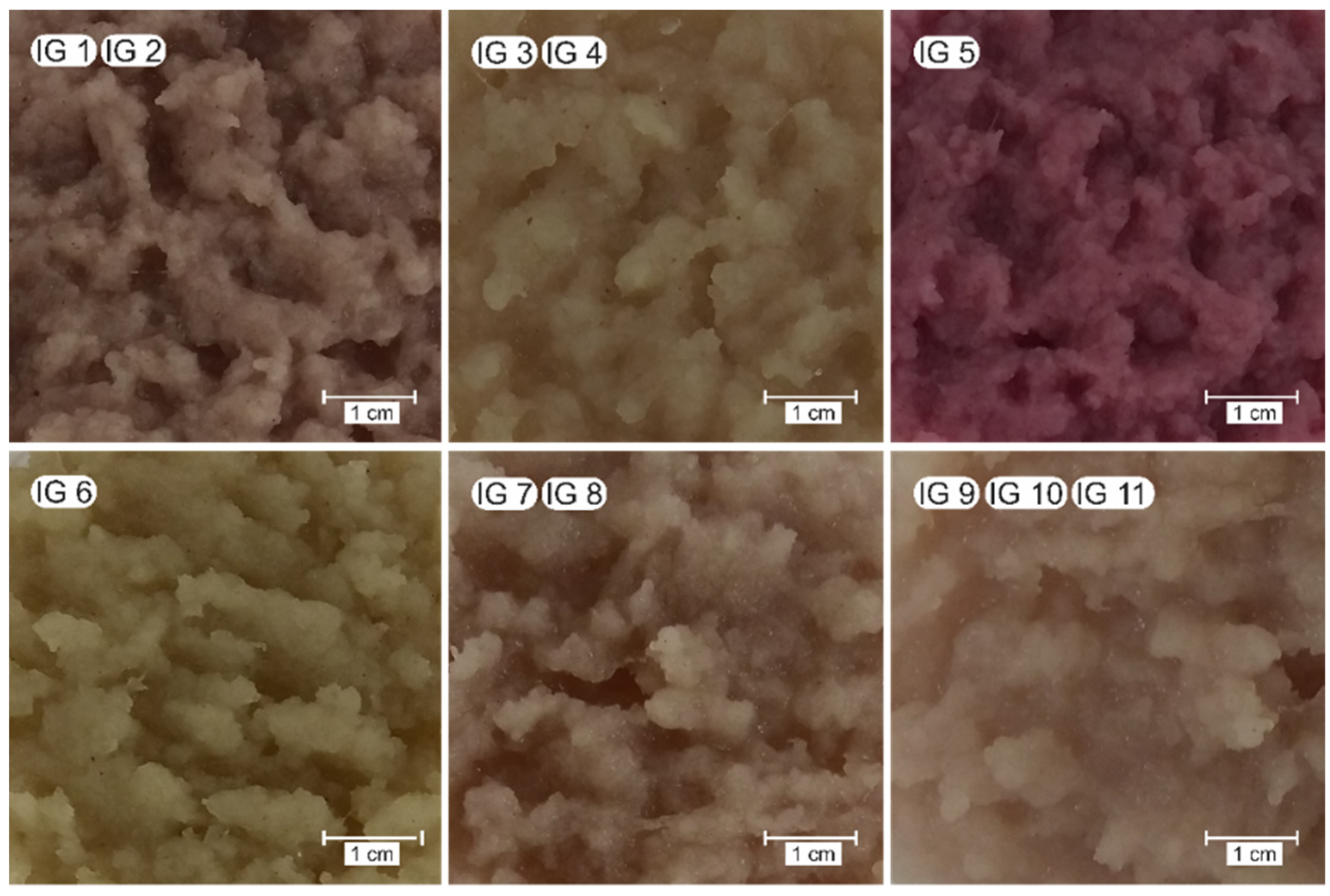
3.2.1. Visual Aspect
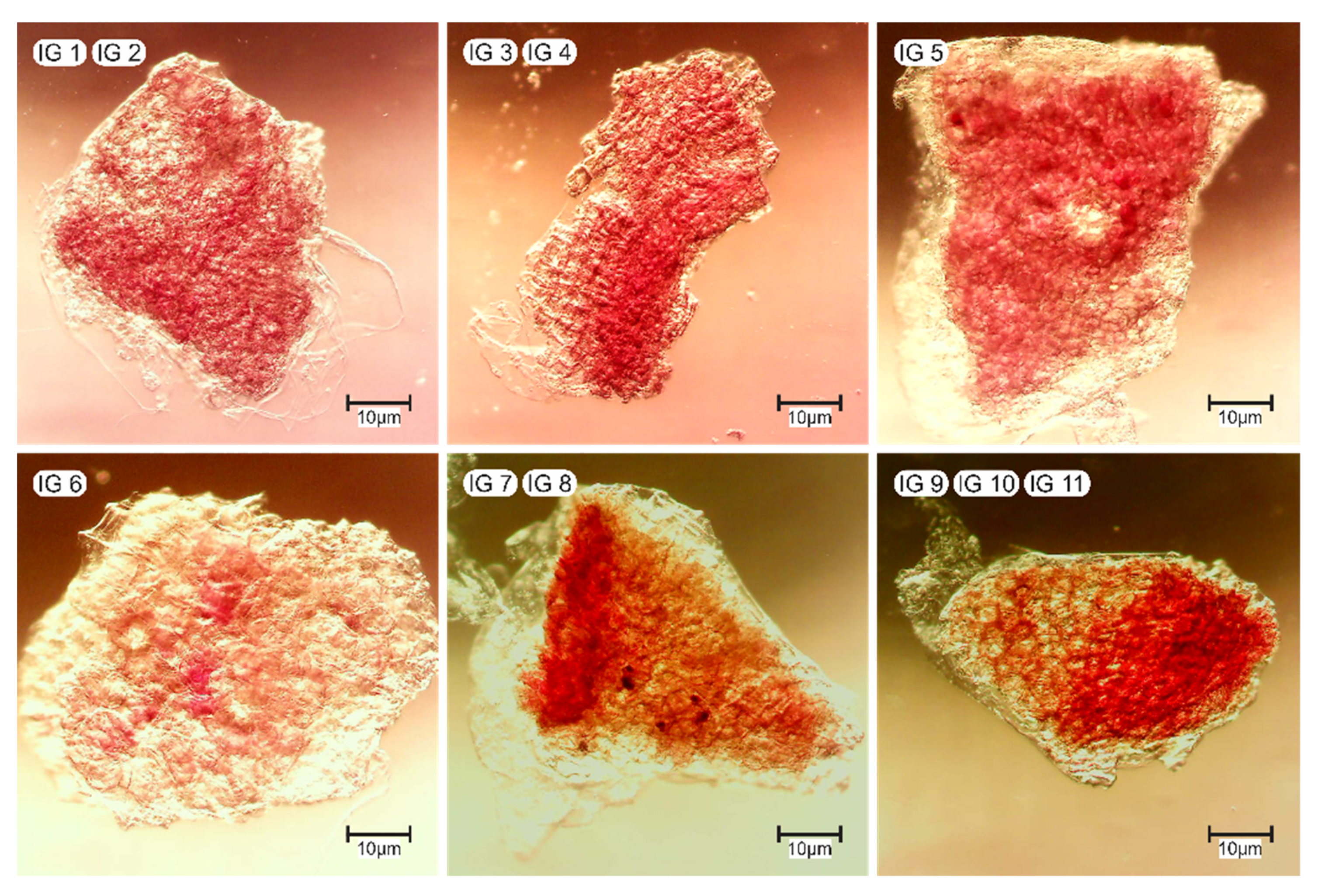
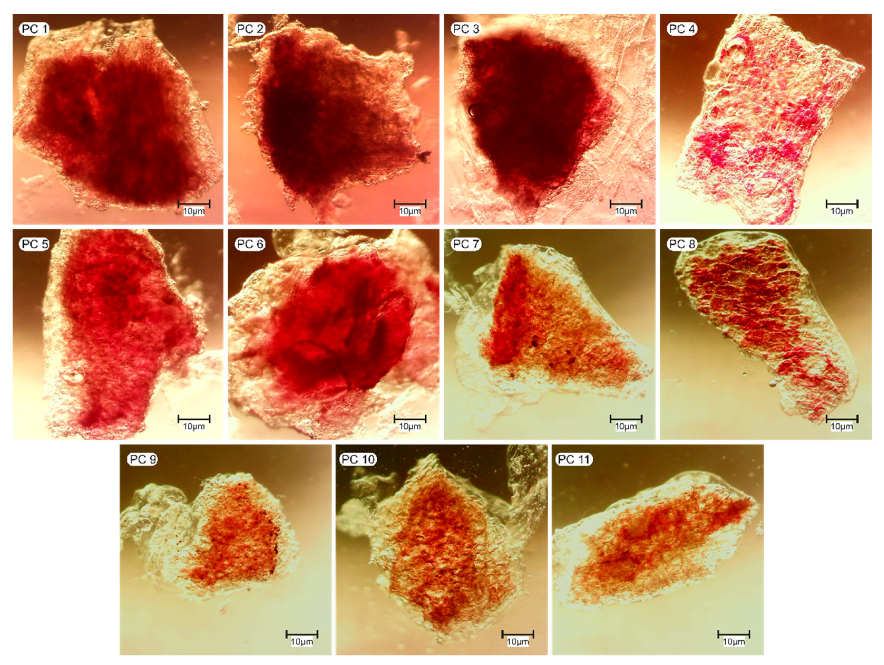
The IG particles produced had a smooth texture, were moist and wet to touch. Visually, IG particles showed a color with shades ranging from reddish to purplish, typical of jamun pulp (Figure 1). This color is due to the anthocyanins pigment, responsible for the purple color of jamun. In a larger proportion of jamun solids in relation to alginate, the color was more visually remarkable. However, after being subjected to the electrostatic interaction process in a gelatin protein solution, the particles showed less intense coloration, visibly clear and opaque (Figure 2). Such behavior can be clearly observed in the IG microparticles after coating with protein in the PC 7 and PC 8 runs, which used the minimum (0%) and maximum (10% w/w) concentration of tested protein, respectively.
The alginate microparticles presented differences in coloration due to the coating process using chitosan, whey protein concentrate or gelatin. Samples coated with chitosan showed a bright intense red color, similar to the microparticles containing extract only, whey protein concentrate provided an opaque red color and the microparticles coated with gelatin presented a pink color. The authors attributed the color changes in the hydrogel microparticles is related to the loss of anthocyanins into the hydrophilic medium during the coating process [25].
In addition, the visual appearance of the ionic gelation microparticles of calcium alginate and jamun (Syzygium cumini L.) pulp (IG) (Figure 1) and after coating with gelatin (PC) (Figure 2) shows aggregated structures. According to Lengyel et al. [48], the surface charge plays a key role in the aggregation of microparticles. Peng et al. [49] observed that the mixtures of gliadin nanoparticles with a fibril-β-lactoglobulin peptide mixture, at a pH of 3.0 to 6.0, formed aggregates. In addition, the closer the zeta potential of the mixture was to zero, the denser the aggregates became, which is due to the lower electrostatic repulsion between the materials, causing them to approach and associate through non-covalent interactions, such as bonds hydrophobic and hydrogen bonds [49]. Xi et al. [50] observed that at pH 4, whey protein isolate and propylene glycol alginate began to interact locally and particles in the solution began to aggregate. For the authors, the hydrogen bond was also one of the interaction forces besides the electrostatic interactions between the complex. The aggregates of complexes formed between zein and propylene glycol alginate were irregular and had rough branch-like microstructures, with many spherical zein particles adsorbed on their surfaces and were also observed by atomic force microscopy [51].

3.2.2. Morphology

Figure 3 shows the microstructure of the ionic gelation microparticles (IG) obtained with jamun pulp, alginate and CaCl2 from the 11 experimental runs, and Figure 4 shows the microstructure of these particles after their protein-coating with gelatin (PC). In the microscopic images presented in Figure 3, it can be observed that the formed microparticles had an irregular and heterogeneous shape. This disorganized gel network formation is due to the presence of solid structures of jamun pulp, observed within the microparticles, represented by the darker areas. The presence of structures in jamun pulp is due to its non-filtration, since the intention was to use most of the solid material from jamun, rich in nutritional compounds, such as fibers, lipids, proteins, phosphorus, vitamin C, phenolic acids, flavonoids and anthocyanins [52]. During the ionic gelation process, the cooperative bonds of calcium ions with alginate carboxylic groups resulted in the exchange of the sodium ions of guluronic acids and mannuronic acid for calcium, bridging the gap between the guluronic acids chains, and promoting the formation of a three-dimensional network that traps the jamun pulp structures, generally described by the egg box model [53]. The properties of the gel were directly related to the number of interaction sites between alginate carboxylic groups and calcium ions. It is believed that during this interaction, jamun pulp may have disturbed the system, due to its own interaction with calcium ions, as it also presented negative charges, which may have favored the formation of an even more disorganized gel network and of varying shape and size. It is also possible that the positively charged flavylium cation of anthocyanins present in jamun pulp also formed directly interacting negatively charged carboxyl groups in the alginate.
The microparticles of extract from hibiscus, rapeseed oil and pectin by ionic gelation using dripping-extrusion and atomization showed an irregular spherical shape and emulsion droplets with considerable size variation, respectively [27]. As in the work by Moura et al. [27], it is possible to observe in Figure 3 that the structures of the jamun pulp were also quite irregular and presented considerable variations in size, resulting from the process of crushing the fruit to obtain the jamun pulp. Furthermore, a tendency of the core material (alginate/CaCl2; jamun pulp/CaCl2complex) to wrap the structures of the jamun pulp following its shape is visible. With the protein coating, the IG microparticles became denser and less transparent, indicating the formation of a protective layer on its surface, as can be seen in Figure 4.

3.2.3. Moisture Content

The IG particles had high water contents, above 97% with no significant variations between the different tests (Table 3). These values are higher than those obtained by [27]. The authors reported values of 83.07 ± 0.01% and 77.03 ± 0.01% of water content for microparticles of hibiscus extract, rapeseed oil and pectin by ionic gelation by drip-extrusion and atomization, respectively, probably due to a different composition of microparticles.
In the present work, this characteristic is a consequence of the hydrophilic nature of the material and its high moisture content (>95%) [54,55,56]. Polysaccharides, such as alginate, have a high hydration capacity, as the carboxylic groups present in their structure interact with water by hydrogen bonds, causing their retention [57]. However, it can be seen in Table 3 and in the response surface that the adsorption of protein on the particle led to a significant reduction in its moisture, being more expressive in runs in which the concentration of the coating protein solution was higher, as also observed by Nogueira et al. [32].
The moisture content in the protein-coated particles (PC) ranged from 87.04 ± 0.22 to 96.64 ± 0.07%. Souza et al. [36] found similar values for moisture content, ranging from 85.8% to 91.1% and from 96.3% to 97.1% for pectin particles obtained by ionic gelation and coated with a whey protein solution without and with heat treatment, respectively. The reduction in moisture content is associated with an increase in protein on the surface of the particle. This is because the electrostatic interaction process between protein, polysaccharide and jamun pulp promotes the migration of free water from ionic gelation microparticles to the medium [32,57].

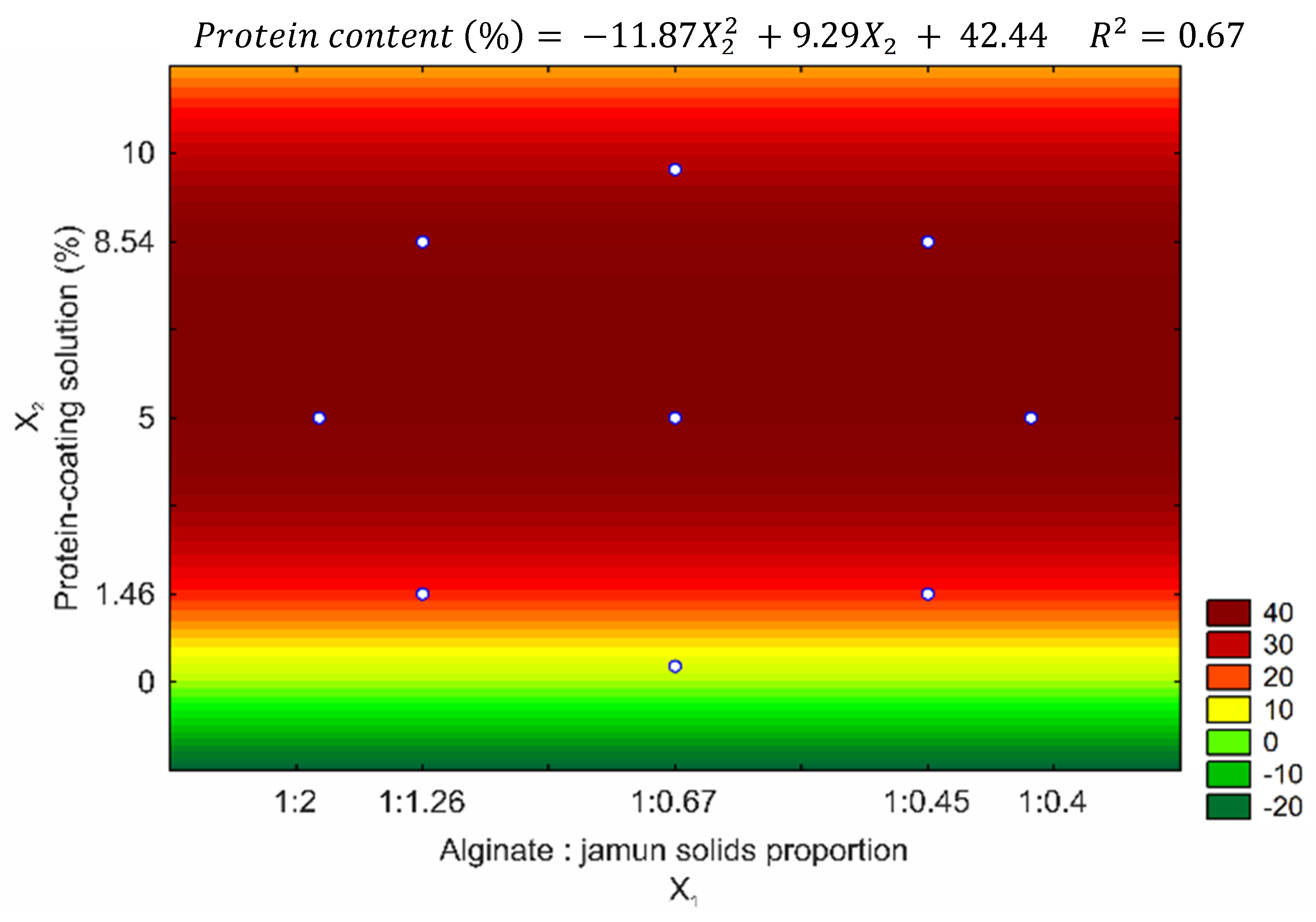
3.2.4. Protein Content

The protein content detected in GI after its immersion in protein solution (Table 3) indicates that interactions occurred between the alginate carboxyl groups and the positively charged amino groups of gelatin, confirming the visual and microstructure observations of the microparticles presented in the Figure 1, Figure 2, Figure 3 and Figure 4.
The protein content of the microparticles after coating suffered statistically significant effects from the linear and quadratic factors of the coating variable, with no significant effect being observed for the variable alginate: jamun ratio and for the interaction. The adjusted model showed that the linear gelatin concentration factor had a positive effect on protein content, that is, the higher the gelatin concentration in the coating protein solution, the greater the protein adsorption for the formation of the protective layer (Figure 5), reaffirming that such behavior has already been observed in the literature in other studies with gelatin [33], egg white [37] and whey protein concentrate [32,36].
According to the F Test, the model was significant and predictive, inferring that it is adequate, with significant regression, small residual values, the absence of a lack of adjustment and satisfactory coefficients of determination. The coefficient of determination (R2) for the adjusted model was 0.677, indicating that this model explains 67.7% of the variation in the observed data. It also shows a significant regression and indicates that there is no lack of adjustment with 95% confidence, given the F test.
As shown in Table 4, most tests had low relative deviations, with the exception of R7 run whose DR is undeterminable with the value 0 in the denominator of the equation, totaling a mean relative deviation of 19.22%, reaffirming the adequacy of the model to the production process of protein-coated ionic gelation microparticles, regarding the adsorption of proteins in the formation of a protective layer.
The amount of protein adsorbed on alginate microparticles and jamun pulp ranged from 21.82 ± 0.72% to 55.87 ± 4.23%. Souza et al. [36] studied the adsorption of whey proteins on pectin-based microparticles produced by gelation and obtained 50% of protein adsorption (w/w dry basis) when working with a high concentration of protein in solution (12%). For Tello et al. [37], the 8% concentration of egg white and whey proteins in solution led to 62% and 60% of protein adsorption on pectin microparticles, respectively. Nogueira et al. [32] reported adsorption of 32.5 ± 2.7% (w/w dry basis) of proteins onto sodium alginate microparticles when using whey protein solution at 4% (w/w) concentration.
In the present study, the maximum protein adsorption occurred in the R9 run, corresponding to a ratio of 1:0.67 (w/w) of alginate/jamun pulp and gelatin solution concentration of 5% (w/w). A lower protein adsorption was observed in the R1 run (Table 3), which corresponds to the lower protein concentration (1.46%, w/w) used in the experimental design, indicating that the amount of adsorbed protein was limited by the positive charges available for interactions with the alginate charges.
Although a predominance of interactions between alginate and gelatin is expected, it is also likely that jamun pulp participated in the electrostatic interaction with gelatin, because they have negative charges. It is believed that this complexation between alginate and gelatin and gelatin and jamun pulp are capable of promoting their greater retention in the particle network, thus favoring the anthocyanins protection against oxidation, resulting in improvements in its conformal stability. For Sharma et al. [24], the results of the Fourier transform infrared spectroscopy (FTIR) analysis demonstrated the existence of interactions between the molecules of phytochemicals from jamun pulp in the alginate gel matrix and guar gum, confirming the encapsulation efficiency. Higher amounts of polyphenols were found in particles with chokeberry extract obtained by electrostatic extrusion process using an alginate/inulin system compared to particles without inulin. For the authors, the explanation for these results may be due to the additional interaction between inulin and polyphenols, as confirmed by FTIR [22]. The encapsulation of blueberry extract and purple corn extract in pectin-alginate hydrogel particles was able to significantly reduce the photodegradation of anthocyanins after exposure to fluorescent light [58]. Additional studies with alginate microparticles obtained by gelation with jamun pulp and coated with gelatin by electrostatic interaction would be necessary to establish their anthocyanins stabilization properties under various conditions encountered in food processing and handling.
However, it is important to point out that although electrostatic interactions are the most important factors to determine the total protein adsorbed, it is known that protein adsorption is a phenomenon that involves the chemical properties of the protein molecule and its different surface forces, including van der Waals forces and hydrogen bonds, in addition to electrostatic and hydrophobic interactions [33,59,60]. This is because, as observed by Beraldo and Tello, even when stabilization of zeta potential values (zero charge due to saturation of bonds) in the microparticle is detected, the amount of protein adsorbed on the particle increases with the increase in the concentration of the protein solution, regardless of the type of protein, demonstrates that other types of interactions may be influencing this adsorption.
In another study [61], the formation of complexes between carrageenan and soy protein was observed at low and high pH values. According to the authors, at low pH values, the interaction between the materials is due to electrostatic interaction, while at high pH values, hydrophobic interactions were the dominant ones for the formation of complexes.

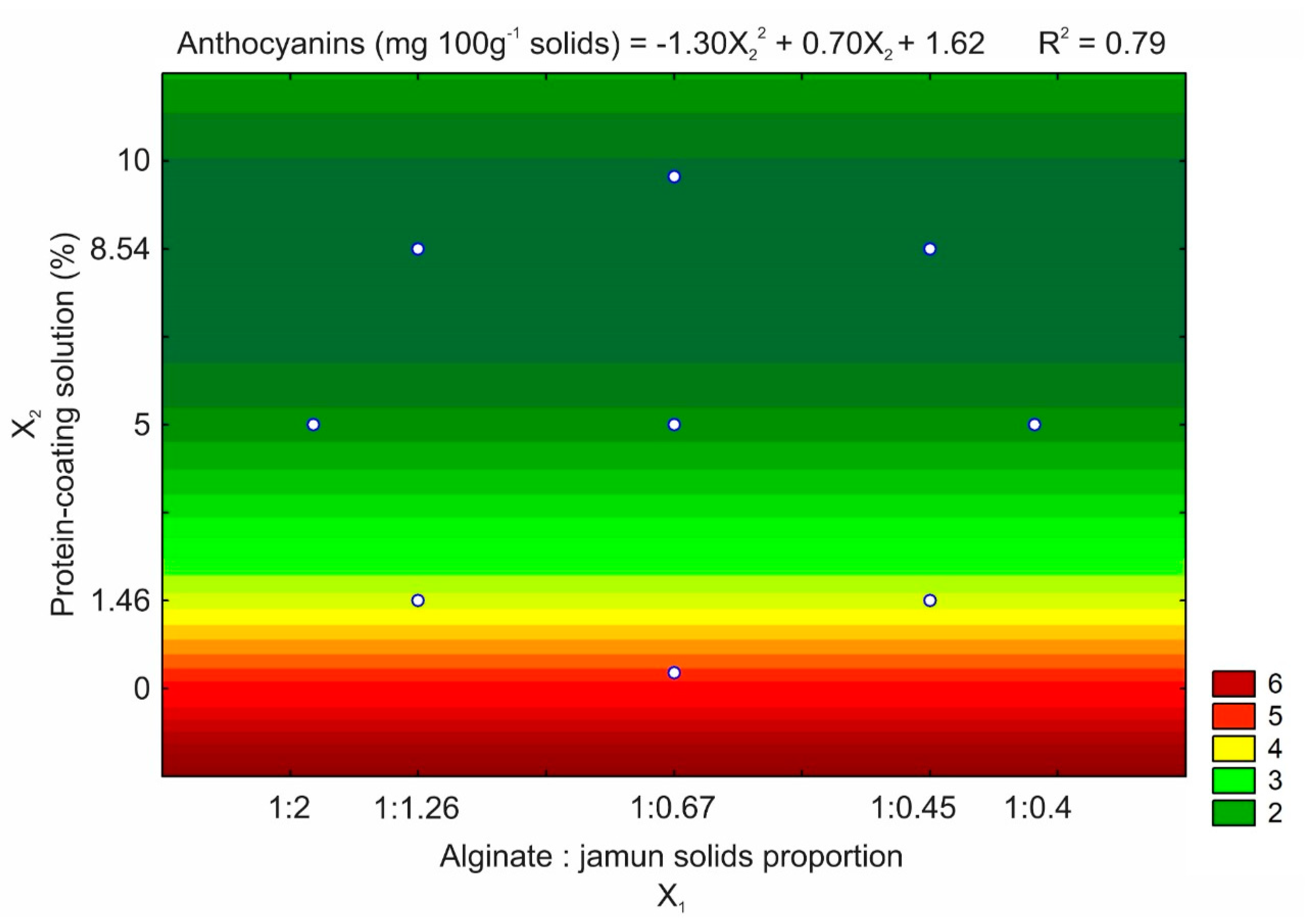
3.2.5. Anthocyanins Content

Da Silva Carvalho et al. [25] reported difficulties when trying to encapsulate anthocyanins from jussara (Euterpe edulis Martius) extract by ionic gelation. According to the authors, the dripping of a mixture of sodium alginate and jussara extract (2 g of alginate in 100 g of extract) in 2% of calcium chloride (2 g 100 mL−1) formed fragile particles, with no definition of format and with a large loss of anthocyanins to the calcium chloride solution, due to the process of diffusion of the hydrophilic compounds to the surrounding medium.
In the present study, it was observed that the ionic gelation microparticles formed with the highest proportions of jamun solids to alginate, i.e., R1 and R5 at 1:1.26 and 1:2 in the Alginate:Jamun ratio (w/w), were those with the highest anthocyanins contents 6.61 ± 0.87 and 6.59 ± 1.53 mg 100 g−1 solids, respectively (Table 3). The more jamun solids incorporated, the higher the content of anthocyanins in the ionic gelation microparticle produced.
The presence of solid structures in the jamun pulp probably reduced the pore dimensions of the gel particle. During the formation of the three-dimensional biopolymeric network, the structures may have filled the space between the macromolecules, in addition to interacting electrostatically with calcium ions, creating a barrier against the diffusion of anthocyanins through the particle to the calcium chloride, contributing to its retention and being evidenced by the reddish in the central part of the gel particles formed (Figure 4).
However, there was a significant decrease in anthocyanins content in the ionic gelation particles containing jamun when compared to jamun pulp. Fresh jamun pulp presented anthocyanins content of 72.03 ± 1.99 mg 100 g−1. Anthocyanins are bioactive compounds that are highly susceptible to degradation when exposed to high levels of temperature, light and oxygen, as well as changes in pH. Consequently, anthocyanins are highly susceptible to oxidation when exposed to environmental factors during particle production.
Another decrease in their anthocyanins content was also observed after microparticles protein-coating, for all experimental runs. One of the hypotheses suggested for this decrease in the anthocyanins contents of the ionic gelation particles after their protein-coating is the addition of total solids. With the increase of protein by adsorption in the formation of the protective layer, the solids content in the microparticle increased, which changes the relationship between the anthocyanins mass and the solid mass, since its content is given by milligrams of anthocyanins per 100 g of solids. Ferrari et al. [62] also observed this same effect when they added maltodextrin as an encapsulating agent for blackberry pulp. The increase in maltodextrin concentration provided a decrease in the total content of anthocyanins in blackberry powders obtained by spray drying. According to the authors, maltodextrin caused a dilution effect of anthocyanins, pigments present in the sample, through the increase in the total solids content. The same behavior was observed by Tonon et al. [63] when they evaluated the drying of açaí by spray dryer in different concentrations of maltodextrin.
In addition, it is possible to observe in Table 3 that the strong electrostatic interaction between the remaining groups with negative electrical charge of the alginate and the positively charged amino groups of the gelatin may have intensified the interchain and intrachain interaction biopolymer. This consequently tightened the three-dimensional network of the IG microparticle, expelling part of the water carrying the anthocyanins, which, given its hydrophilicity, resulted in a decrease in the water and anthocyanins content in the coated particle at the end of the process [32].
The anthocyanins content in the PC microparticles ranged from 5.84 ± 0.62 mg 100 g−1 (T7 test, 1:0.67 (w/w) ratio of alginate/jamun pulp and protein solution concentration of 0%, w/w) to 0.78 ± 0.14 mg 100 g−1 (T8 test, 1:0.67 (w/w) ratio of alginate/jamun pulp and gelatin solution concentration of 10%, w/w), corresponding to the minimum and maximum concentrations of protein in solution tested in the protein coating solutions, respectively.
Thus, for the anthocyanins content, only the protein-coating variable had a significant effect on the PC microparticles, both their quadratic and linear factors, with negative and positive coefficients, respectively (Figure 6). It was noted that the anthocyanins content was higher for the assays in which the coating protein solution had lower concentrations, an effect explained by the increase in solids in the particle composition without the modification of the anthocyanins content intrinsic to the microencapsulated jamun pulp. Although significant, the adjusted model was not predictive according to the F test, presenting a regression coefficient R2 of 0.79.
Although the anthocyanins content in the microparticles was not significantly influenced by the proportion of alginate and jamun solids, probably due to this behavior of increasing solids in the particle composition by protein adsorption, it should be noted that the concentration of solids from pulp of jamun added is an important factor that influences the amount of anthocyanins encapsulated in the IG microparticles.

4. Conclusions

Calcium alginate and jamun pulp microparticles produced by ionic gel and coated with protein with gelatin, according to conditions of proportion of solids of sodium alginate and jamun pulp (1: 0.40 to 1: 2, w/w) and the concentration of gelatin-coating solution (0% to 10%, w/w), were able to promote interactions between biopolymers. Calcium alginate and jamun pulp microparticles (IG) produced by ionic gelation presented irregular and heterogeneous morphology, with a disorganized gel network due to the presence of solid jamun particles inside the particles. The IG microparticles had a flaccid texture on physical contact, were wet and showed intense and visible color, ranging from reddish to purplish. After protein-coating (PC), its color intensity decreased and water content was reduced.
The interaction between gelatin’s positive charge and the remaining negative charge of alginate microparticles promoted its adsorption. Higher protein adsorption was observed at the highest concentration of protein in the solution and the amount adsorbed was inversely proportional to the content of anthocyanins in the microparticles. The adsorption of proteins in the GI resulted in a decrease in their moisture and anthocyanins content, and the strong electrostatic interaction between the biopolymers probably led to the length of their gel networks, which consequently expelled part of the water and anthocyanins, given their hydrophilicity. Furthermore, the decrease in the anthocyanins contents of the GI particles after their PC protein coating also occurred due to the addition of total solids.
It can be concluded that the combination of the encapsulation process by ionic gelation and electrostatic interaction led to the formation of microparticles coated with proteins containing jamun pulp, with an intense reddish color due to the presence of anthocyanins. These results suggest that these particles can serve as a transport vehicle for natural pigments; however, in future studies, the protective effect of the protein layer during the shelf life of the microparticles should be evaluated.

Author Contributions

V.G.d.S.: conceptualization, data collection, analysis, interpretation, writing—original draft preparation, writing—review and editing, visualization. G.F.N.: Conceptualization, methodology, formal analysis, investigation, writing—original draft preparation, writing—review and editing, visualization, supervision. C.T.S.: methodology, software, validation, formal analysis, investigation, writing—original draft preparation. R.A.d.O.: Conceptualization, resources, writing—review and editing, visualization, supervision, project administration, funding acquisition. All authors have read and agreed to the published version of the manuscript.

Funding

This research was funded by the National Council for Scientific and Technological Development (PIBIC/CNPq, Brazil).

Institutional Review Board Statement

Not applicable.

Informed Consent Statement

Not applicable.

Data Availability Statement

Data sharing not applicable.

Acknowledgments

The authors are grateful to the to the to the National Council for Scientific and Technological Development (PIBIC/CNPq, Brazil) and to the School of Agricultural Engineering—University of Campinas for their financial support.

Conflicts of Interest

The authors declare no conflict of interest. The funders had no role in the design of the study; in the collection, analyses, or interpretation of data; in the writing of the manuscript; or in the decision to publish the results.

References

  1. Llorent-Martínez, E.J.; Ortega-Barrales, P.; Zengin, G.; Mocan, A.; Simirgiotis, M.J.; Ceylan, R.; Uysal, S.; Aktumsek, A. Evaluation of Antioxidant Potential, Enzyme Inhibition Activity and Phenolic Profile of Lathyrus Cicera and Lathyrus Digitatus: Potential Sources of Bioactive Compounds for the Food Industry. Food Chem. Toxicol. 2017, 107, 609–619. [Google Scholar] [CrossRef]
  2. Caleja, C.; Ribeiro, A.; Barreiro, M.F.; Ferreira, I.C.F.R. Phenolic Compounds as Nutraceuticals or Functional Food Ingredients. Curr. Pharm. Des. 2017, 23, 2787–2806. [Google Scholar] [CrossRef] [PubMed]
  3. Dimou, C.; Karantonis, H.C.; Skalkos, D.; Koutelidakis, A.E. Valorization of Fruits By-Products to Unconventional Sources of Additives, Oil, Biomolecules and Innovative Functional Foods. Curr. Pharm. Biotechnol. 2019, 20, 776–786. [Google Scholar] [CrossRef]
  4. Chaudhary, A.; Bag, S.; Banerjee, P.; Chatterjee, J. Wound Healing Efficacy of Jamun Honey in Diabetic Mice Model through Reepithelialization, Collagen Deposition and Angiogenesis. J. Tradit. Complement. Med. 2020, 10, 529–543. [Google Scholar] [CrossRef]
  5. Kumar, M.; Hasan, M.; Lorenzo, J.M.; Dhumal, S.; Nishad, J.; Rais, N.; Verma, A.; Changan, S.; Barbhai, M.D.; Radha; et al. Jamun (Syzygium cumini (L.) Skeels) Seed Bioactives and Its Biological Activities: A Review. Food Biosci. 2022, 50, 102109. [Google Scholar] [CrossRef]
  6. Migliato, K.F.; Moreira, R.R.D.; Mello, J.C.P.; Sacramento, L.V.S.; Corrêa, M.A.; Salgado, H.R.N. Controle Da Qualidade Do Fruto de Syzygium cumini (L.) Skeels. Braz. J. Pharmacogn. 2007, 17, 94–101. [Google Scholar] [CrossRef] [Green Version]
  7. Chhikara, N.; Kaur, R.; Jaglan, S.; Sharma, P.; Gat, Y.; Panghal, A. Bioactive Compounds and Pharmacological and Food Applications of: Syzygium cumini—A Review. Food Funct. 2018, 9, 6096–6115. [Google Scholar] [CrossRef] [PubMed]
  8. Ghosh, P.; Pradhan, R.C.; Mishra, S.; Patel, A.S.; Kar, A. Physicochemical and Nutritional Characterization of Jamun (Syzygium cuminii). Curr. Res. Nutr. Food Sci. 2017, 5, 25–35. [Google Scholar] [CrossRef]
  9. Nogueira, G.F.; Soares, C.T.; Cavasini, R.; Fakhouri, F.M.; de Oliveira, R.A. Bioactive Films of Arrowroot Starch and Blackberry Pulp: Physical, Mechanical and Barrier Properties and Stability to PH and Sterilization. Food Chem. 2019, 275, 417–425. [Google Scholar] [CrossRef] [PubMed]
  10. Nogueira, G.F.; Soares, I.H.B.T.; Soares, C.T.; Fakhouri, F.M.; de Oliveira, R.A. Development and Characterization of Arrowroot Starch Films Incorporated with Grape Pomace Extract. Polysaccharides 2022, 3, 250–263. [Google Scholar] [CrossRef]
  11. Carmona, L.; Sulli, M.; Diretto, G.; Alquézar, B.; Alves, M.; Peña, L. Improvement of Antioxidant Properties in Fruit from Two Blood and Blond Orange Cultivars by Postharvest Storage at Low Temperature. Antioxidants 2022, 11, 547. [Google Scholar] [CrossRef] [PubMed]
  12. Yıldız, D.; Gürel, D.B.; Çağındı, Ö.; Kayaardı, S. Heat Treatment and Microwave Applications on Homemade Sour Cherry Juice: The Effect on Anthocyanin Content and Some Physicochemical Properties. Curr. Plant Biol. 2022, 29, 100242. [Google Scholar] [CrossRef]
  13. Liudvinaviciute, D.; Rutkaite, R.; Bendoraitiene, J.; Klimaviciute, R.; Dagys, L. Formation and Characteristics of Alginate and Anthocyanin Complexes. Int. J. Biol. Macromol. 2020, 164, 726–734. [Google Scholar] [CrossRef]
  14. Codevilla, C.F.; Bazana, M.T.; Da Silva, C.; Barin, J.; Ragagnin, C. Nanostructures Containing Bioactive Compounds Extracted from Plants. Ciência Nat. 2015, 37, 142–151. [Google Scholar] [CrossRef]
  15. Ballesteros, L.F.; Ramirez, M.J.; Orrego, C.E.; Teixeira, J.A.; Mussatto, S.I. Encapsulation of Antioxidant Phenolic Compounds Extracted from Spent Coffee Grounds by Freeze-Drying and Spray-Drying Using Different Coating Materials. Food Chem. 2017, 237, 623–631. [Google Scholar] [CrossRef] [Green Version]
  16. Gómez-Guillén, M.C.; Giménez, B.; López-Caballero, M.E.; Montero, M.P. Functional and Bioactive Properties of Collagen and Gelatin from Alternative Sources: A Review. Food Hydrocoll. 2011, 25, 1813–1827. [Google Scholar] [CrossRef] [Green Version]
  17. Wang, R.; Tian, Z.; Chen, L. A Novel Process for Microencapsulation of Fish Oil with Barley Protein. Food Res. Int. 2011, 44, 2735–2741. [Google Scholar] [CrossRef]
  18. Li, Y.; Liu, J.; Ma, S.; Yang, M.; Zhang, H.; Zhang, T.; Yu, Y.; Du, Z. Co-Assembly of Egg White-Derived Peptides and Protein-Polysaccharide Complexes for Curcumin Encapsulation: The Enhancement of Stability, Redispersibility, and Bioactivity. Food Chem. 2022, 394, 133496. [Google Scholar] [CrossRef]
  19. Leong, J.-Y.; Lam, W.-H.; Ho, K.-W.; Voo, W.-P.; Lee, M.F.-X.; Lim, H.-P.; Lim, S.-L.; Tey, B.-T.; Poncelet, D.; Chan, E.-S. Advances in Fabricating Spherical Alginate Hydrogels with Controlled Particle Designs by Ionotropic Gelation as Encapsulation Systems. Particuology 2016, 24, 44–60. [Google Scholar] [CrossRef]
  20. Simsek-Ege, F.A.; Bond, G.M.; Stringer, J. Polyelectrolye Complex Formation between Alginate and Chitosan as a Function of PH. J. Appl. Polym. Sci. 2003, 88, 346–351. [Google Scholar] [CrossRef]
  21. Bajpai, S.K.; Tankhiwale, R. Investigation of Water Uptake Behavior and Stability of Calcium Alginate/Chitosan Bi-Polymeric Beads: Part-1. React. Funct. Polym. 2006, 66, 645–658. [Google Scholar] [CrossRef]
  22. Ćujić, N.; Trifković, K.; Bugarski, B.; Ibrić, S.; Pljevljakušić, D.; Šavikin, K. Chokeberry (Aronia melanocarpa L.) Extract Loaded in Alginate and Alginate/Inulin System. Ind. Crops Prod. 2016, 86, 120–131. [Google Scholar] [CrossRef]
  23. Kurtulbaş, E.; Albarri, R.; Torun, M.; Şahin, S. Encapsulation of Moringa Oleifera Leaf Extract in Chitosan-Coated Alginate Microbeads Produced by Ionic Gelation. Food Biosci. 2022, 50, 102158. [Google Scholar] [CrossRef]
  24. Sharma, M.; Dash, K.K.; Badwaik, L.S. Physicochemical and Release Behaviour of Phytochemical Compounds Based on Black Jamun Pulp Extracts-Filled Alginate Hydrogel Beads through Vibration Dripping Extrusion. Int. J. Biol. Macromol. 2022, 194, 715–725. [Google Scholar] [CrossRef]
  25. da Silva Carvalho, A.G.; da Costa Machado, M.T.; de Freitas Queiroz Barros, H.D.; Cazarin, C.B.B.; Maróstica Junior, M.R.; Hubinger, M.D. Anthocyanins from Jussara (Euterpe edulis Martius) Extract Carried by Calcium Alginate Beads Pre-Prepared Using Ionic Gelation. Powder Technol. 2019, 345, 283–291. [Google Scholar] [CrossRef]
  26. Dallabona, I.D.; de Lima, G.G.; Cestaro, B.I.; Tasso, I.D.S.; Paiva, T.S.; Laureanti, E.J.G.; Jorge, L.M.d.M.; da Silva, B.J.G.; Helm, C.V.; Mathias, A.L.; et al. Development of Alginate Beads with Encapsulated Jabuticaba Peel and Propolis Extracts to Achieve a New Natural Colorant Antioxidant Additive. Int. J. Biol. Macromol. 2020, 163, 1421–1432. [Google Scholar] [CrossRef] [PubMed]
  27. de Moura, S.C.S.R.; Berling, C.L.; Garcia, A.O.; Queiroz, M.B.; Alvim, I.D.; Hubinger, M.D. Release of Anthocyanins from the Hibiscus Extract Encapsulated by Ionic Gelation and Application of Microparticles in Jelly Candy. Food Res. Int. 2019, 121, 542–552. [Google Scholar] [CrossRef]
  28. Iurciuc-Tincu, C.E.; Atanase, L.I.; Ochiuz, L.; Jérôme, C.; Sol, V.; Martin, P.; Popa, M. Curcumin-Loaded Polysaccharides-Based Complex Particles Obtained by Polyelectrolyte Complexation and Ionic Gelation. I-Particles Obtaining and Characterization. Int. J. Biol. Macromol. 2020, 147, 629–642. [Google Scholar] [CrossRef]
  29. Ma, J.; Liu, C.; Dong, Y.; Fan, Q.; Bao, Y.; Yan, H. Waterborne Polyurethane/Silica Nanocomposites Based on Electrostatic Interaction: Interfacial Interactions and Properties. Prog. Org. Coat. 2022, 171, 107052. [Google Scholar] [CrossRef]
  30. Wang, W.; Narain, R.; Zeng, H. Hydrogels. Polym. Sci. Nanotechnol. Fundam. Appl. 2020, 10, 203–244. [Google Scholar] [CrossRef]
  31. Herrman, D.A.; Brantsen, J.F.; Awika, J.M. Interactions of 3-Deoxyanthocyanins with Gum Arabic and Sodium Alginate Contributing to Improved Pigment Aqueous Stability. Food Chem. 2022, 372, 131233. [Google Scholar] [CrossRef]
  32. Nogueira, G.F.; Prata, A.S.; Grosso, C.R.F. Alginate and Whey Protein Based-Multilayered Particles: Production, Characterisation and Resistance to PH, Ionic Strength and Artificial Gastric/Intestinal Fluid. J. Microencapsul. 2017, 34, 151–161. [Google Scholar] [CrossRef] [PubMed]
  33. Beraldo, J.C.; Nogueira, G.F.; Prata, A.S.; Grosso, C.R.F. Effect of Molar Weight of Gelatin in the Coating of Alginate Microparticles. Polimeros 2021, 31, 1–9. [Google Scholar] [CrossRef]
  34. AOAC—Association of Official Analytical Chemists. Official Methods of Analysis of AOAC International, 18th ed.; AOAC: Gaithersburgs, MD, USA, 2006. [Google Scholar]
  35. Sims, D.A.; Gamon, J.A. Relationships between Leaf Pigment Content and Spectral Reflectance across a Wide Range of Species, Leaf Structures and Developmental Stages. Remote Sens. Environ. 2002, 81, 337–354. [Google Scholar] [CrossRef]
  36. Souza, F.N.; Gebara, C.; Ribeiro, M.C.E.; Chaves, K.S.; Gigante, M.L.; Grosso, C.R.F. Production and Characterization of Microparticles Containing Pectin and Whey Proteins. Food Res. Int. 2012, 49, 560–566. [Google Scholar] [CrossRef] [Green Version]
  37. Tello, F.; Falfan-Cortés, R.N.; Martinez-Bustos, F.; Martins da Silva, V.; Hubinger, M.D.; Grosso, C. Alginate and Pectin-Based Particles Coated with Globular Proteins: Production, Characterization and Anti-Oxidative Properties. Food Hydrocoll. 2015, 43, 670–678. [Google Scholar] [CrossRef]
  38. AOAC—Association of Official Analytical Chemists. Official Methods of Analysis of AOAC International, 16th ed.; AOAC: Arlington, TX, USA, 1998. [Google Scholar]
  39. Philipp, B.; Dautzenberg, H.; Linow, K.-J.; Kötz, J.; Dawydoff, W. Polyelectrolyte Complexes—Recent Developments and Open Problems. Prog. Polym. Sci. 1989, 14, 91–172. [Google Scholar] [CrossRef]
  40. Jun-xia, X.; Hai-yan, Y.; Jian, Y. Microencapsulation of Sweet Orange Oil by Complex Coacervation with Soybean Protein Isolate/Gum Arabic. Food Chem. 2011, 125, 1267–1272. [Google Scholar] [CrossRef]
  41. Jones, O.G.; Decker, E.A.; McClements, D.J. Comparison of Protein–Polysaccharide Nanoparticle Fabrication Methods: Impact of Biopolymer Complexation before or after Particle Formation. J. Colloid Interface Sci. 2010, 344, 21–29. [Google Scholar] [CrossRef]
  42. You, G.; Niu, G.; Long, H.; Zhang, C.; Liu, X. Elucidation of Interactions between Gelatin Aggregates and Hsian-Tsao Gum in Aqueous Solutions. Food Chem. 2020, 319, 126532. [Google Scholar] [CrossRef]
  43. Carneiro-da-Cunha, M.G.; Cerqueira, M.A.; Souza, B.W.S.; Teixeira, J.A.; Vicente, A.A. Influence of Concentration, Ionic Strength and PH on Zeta Potential and Mean Hydrodynamic Diameter of Edible Polysaccharide Solutions Envisaged for Multinanolayered Films Production. Carbohydr. Polym. 2011, 85, 522–528. [Google Scholar] [CrossRef] [Green Version]
  44. Canto, A.R.; Kumon, T. Efeito Do PH e Da Homogeneização a Ultra Alta Pressão Sobre a Estabilidade Física Do Suco de Caju (Anacardium occidentale). Acta Tecnológica 2013, 8, 44–49. [Google Scholar] [CrossRef]
  45. Pasukamonset, P.; Kwon, O.; Adisakwattana, S. Alginate-Based Encapsulation of Polyphenols from Clitoria ternatea Petal Flower Extract Enhances Stability and Biological Activity under Simulated Gastrointestinal Conditions. Food Hydrocoll. 2016, 61, 772–779. [Google Scholar] [CrossRef]
  46. Castañeda-Ovando, A.; Pacheco-Hernández, M.D.L.; Páez-Hernández, M.E.; Rodríguez, J.A.; Galán-Vidal, C.A. Chemical Studies of Anthocyanins: A Review. Food Chem. 2009, 113, 859–871. [Google Scholar] [CrossRef]
  47. Benítez, E.I.; Genovese, D.B.; Lozano, J.E. Effect of PH and Ionic Strength on Apple Juice Turbidity: Application of the Extended DLVO Theory. Food Hydrocoll. 2007, 21, 100–109. [Google Scholar] [CrossRef]
  48. Lengyel, M.; Kállai-Szabó, N.; Antal, V.; Laki, A.J.; Antal, I. Microparticles, Microspheres, and Microcapsules for Advanced Drug Delivery. Sci. Pharm. 2019, 87, 20. [Google Scholar] [CrossRef] [Green Version]
  49. Peng, D.; Jin, W.; Sagis, L.M.C.; Li, B. Adsorption of Microgel Aggregates Formed by Assembly of Gliadin Nanoparticles and a β-Lactoglobulin Fibril-Peptide Mixture at the Air/Water Interface: Surface Morphology and Foaming Behavior. Food Hydrocoll. 2022, 122, 107039. [Google Scholar] [CrossRef]
  50. Xi, C.; Sun, Z.; Chen, X.; Ding, X.; Zhang, T. Characterization of Coacervation Behavior between Whey Protein Isolate and Propylene Glycol Alginate: A Morphology, Spectroscopy, and Thermodynamics Study. Food Chem. X 2022, 15, 100402. [Google Scholar] [CrossRef]
  51. Sun, C.; Dai, L.; Gao, Y. Interaction and Formation Mechanism of Binary Complex between Zein and Propylene Glycol Alginate. Carbohydr. Polym. 2017, 157, 1638–1649. [Google Scholar] [CrossRef]
  52. Lago, E.S.; Gomes, E.; Da Silva, R. Produção de Geléia de Jambolão (Syzygium cumini Lamarck): Processamento, Parâmetros Físico—Químicos e Avaliação Sensorial. Cienc. Tecnol. Aliment. 2006, 26, 847–852. [Google Scholar] [CrossRef]
  53. Cuadros, T.R.; Skurtys, O.; Aguilera, J.M. Mechanical Properties of Calcium Alginate Fibers Produced with a Microfluidic Device. Carbohydr. Polym. 2012, 89, 1198–1206. [Google Scholar] [CrossRef] [PubMed]
  54. Burey, P.; Bhandari, B.R.; Howes, T.; Gidley, M.J. Hydrocolloid Gel Particles: Formation, Characterization, and Application. Crit. Rev. Food Sci. Nutr. 2008, 48, 361–377. [Google Scholar] [CrossRef] [PubMed]
  55. De Vos, P.; De Haan, B.J.; Kamps, J.A.A.M.; Faas, M.M.; Kitano, T. Zeta-Potentials of Alginate-PLL Capsules: A Predictive Measure for Biocompatibility? J. Biomed. Mater. Res.—Part A 2007, 80, 813–819. [Google Scholar] [CrossRef]
  56. Hamidi, M.; Azadi, A.; Rafiei, P. Hydrogel Nanoparticles in Drug Delivery. Adv. Drug Deliv. Rev. 2008, 60, 1638–1649. [Google Scholar] [CrossRef] [PubMed]
  57. Crouzier, T.; Boudou, T.; Picart, C. Polysaccharide-Based Polyelectrolyte Multilayers. Curr. Opin. Colloid Interface Sci. 2010, 15, 417–426. [Google Scholar] [CrossRef]
  58. Guo, J.; Giusti, M.M.; Kaletunç, G. Encapsulation of Purple Corn and Blueberry Extracts in Alginate-Pectin Hydrogel Particles: Impact of Processing and Storage Parameters on Encapsulation Efficiency. Food Res. Int. 2018, 107, 414–422. [Google Scholar] [CrossRef]
  59. Haynes, C.A.; Norde, W. Globular Proteins at Solid/Liquid Interfaces. Colloids Surfaces B Biointerfaces 1994, 2, 517–566. [Google Scholar] [CrossRef]
  60. Silverio, G.B.; Sakanaka, L.S.; Alvim, I.D.; Shirai, M.A.; Grosso, C.R.F. Production and Characterization of Alginate Microparticles Obtained by Ionic Gelation and Electrostatic Adsorption of Concentrated Soy Protein. Cienc. Rural 2018, 48. [Google Scholar] [CrossRef] [Green Version]
  61. Molina Ortiz, S.E.; Puppo, M.C.; Wagner, J.R. Relationship between Structural Changes and Functional Properties of Soy Protein Isolates-Carrageenan Systems. Food Hydrocoll. 2004, 18, 1045–1053. [Google Scholar] [CrossRef]
  62. Ferrari, C.C.; Ribeiro, C.P.; Aguirre, J.M. de Secagem Por Atomização de Polpa de Amora-Preta Usando Maltodextrina Como Agente Carreador. Braz. J. Food Technol. 2012, 15, 157–165. [Google Scholar] [CrossRef]
  63. Tonon, R.V.; Brabet, C.; Hubinger, M.D. Influence of Drying Air Temperature and Carrier Agent Concentration on the Physicochemical Properties of Açai Juice Powder. Cienc. Tecnol. Aliment. 2009, 29, 444–450. [Google Scholar] [CrossRef]
Figure 1. Visual appearance of calcium alginate microparticles and jamun (Syzygium cumini L.) pulp obtained by ionic gelation (IG) in the 11 runs of the experimental design.
Polysaccharides 04 00002 g001
Figure 2. Visual appearance of ionic gelation microparticles of calcium alginate and jamun (Syzygium cumini L.) pulp (IG) after coating with gelatin (PC) in the 11 runs of the experimental design.
Polysaccharides 04 00002 g002
Figure 3. Digital images obtained using a binocular microscope (10× objective, 10× ocular) of calcium alginate microparticles and jamun (Syzygium cumini L.) pulp obtained by ionic gelation (IG) from the 11 runs of the experiment.
Polysaccharides 04 00002 g003
Figure 4. Digital image obtained using a binocular microscope (10× objective, 10× ocular) of gelation microparticles of calcium alginate and jamun (Syzygium cumini L.) pulp (IG) after protein-coating with gelatin (PC) from the 11 runs of the experiment.
Polysaccharides 04 00002 g004
Figure 5. Surface contour curves of protein content responses (%) as a function of the proportion of sodium alginate solids and jamun pulp and the concentration of coating solution (%) for ionic gelation particles after protein-coating.
Polysaccharides 04 00002 g005
Figure 6. Surface contour curves of anthocyanins content responses (%) as a function of the proportion of sodium alginate solids and jamun pulp and the concentration of coating solution (%) for ionic gelation particles after protein-coating.
Polysaccharides 04 00002 g006
Table 1. Experimental design used for microencapsulation of jamun (Syzygium cumini L.) pulp by ionic gelation with alginate (IG) and its coating by electrostatic interaction with gelatin (PC).
TestExperimental DesignIG SolutionPC Solution
Alginate:Jamun Ratio 1 (w/w)Coating 1
(%)
Alginate
(g Solids 100 g−1)
Jamun 2
(g Solids 100 g−1)
Gelatin
(g Solids 100 g−1)
11:1.26 (−1)1.46 (−1)2.002.521.46
21:1.26 (−1)8.54 (1)2.002.528.54
31:0.45 (1)1.46 (−1)2.000.911.46
41:0.45 (1)8.54 (1)2.000.918.54
51:2.00 (−1.41)5.00 (0)2.004.005.00
61:0.40 (1.41)5.00 (0)2.000.805.00
71:0.67 (0)0.00 (−1.41)2.001.330.00
81:0.67 (0)10.00 (1.41)2.001.3310.00
91:0.67 (0)5.00 (0)2.001.335.00
101:0.67 (0)5.00 (0)2.001.335.00
111:0.67 (0)5.00 (0)2.001.335.00
1 The independent variables correspond to the real values. Values in parentheses correspond to the coded values. 2 To determine the composition of the microencapsulation solutions, the water content of 86.68 ± 0.05% was considered in the jamun pulp for correcting the basis in solids.
Table 2. Characterization of sodium alginate, gelatin and jamun pulp solutions in relation to zeta potential.
Solutions (0.2% w/w)pHZP (mV)
Alginate6.0−74.40 ± 7.35
Gelatin4.08.96 ± 1.15
Jamun pulp4.0−17.20 ± 1.84
Table 3. Moisture content (%), protein content (%) and anthocyanins content (mg 100 g−1 solids) of ionic gelation microparticles of calcium alginate and jamun (Syzygium cumini L.) pulp (IG) and after protein-coating with gelatin (PC) from the 11 runs of the experiment.
RunMoisture Content
(%)
Protein Content 1
(%)
Anthocyanins Content
(mg 100 g−1)
IGPCPCIGPC
197.29 ± 0.6495.53 ± 0.1221.82 ± 0.726.61 ± 0.871.96 ± 0.47
297.29 ± 0.6488.24 ± 0.2937.89 ± 5.886.61 ± 0.871.24 ± 0.20
397.22 ± 0.1396.64 ± 0.0729.33 ± 1.744.51 ± 0.643.64 ± 0.73
497.22 ± 0.1387.04 ± 0.2236.62 ± 4.334.51 ± 0.641.15 ± 0.09
596.40 ± 0.0591.41 ± 0.0823.75 ± 0.236.59 ± 1.531.23 ± 0.04
697.02 ± 0.0591.36 ± 0.0636.87 ± 0.812.26 ± 0.161.89 ± 0.45
797.06 ± 0.1297.06 ± 0.120.00 ± 0.005.84 ± 0.625.84 ± 0.62
897.06 ± 0.1293.50 ± 0.0835.99 ± 3.065.84 ± 0.620.78 ± 0.14
997.06 ± 0.1294.36 ± 0.0555.87 ± 4.235.82 ± 0.301.88 ± 0.28
1097.06 ± 0.1294.38 ± 0.0445.01 ± 1.035.82 ± 0.301.86 ± 0.08
1197.06 ± 0.1294.37 ± 0.0549.00 ± 1.405.82 ± 0.301.84 ± 0.19
1 Protein contents of ionic gelation microparticles (IG) are not shown once they were insignificant.
Table 4. Experimental, predicted and relative deviation values for protein content for microparticles after protein coating (PC) for the different runs of the experimental design.
RunProtein Content (%)Relative Deviation
(%)
Experimental ValuePredicted Value
121.8221.282.48
237.8939.865.20
329.3321.2827.45
436.6239.868.85
523.7542.4478.68
636.8742.4415.10
70.005.74-
835.9931.9511.24
955.8742.4424.05
1045.0142.445.72
1149.0042.4413.40
Mean relative deviation (%)19.22
Disclaimer/Publisher’s Note: The statements, opinions and data contained in all publications are solely those of the individual author(s) and contributor(s) and not of MDPI and/or the editor(s). MDPI and/or the editor(s) disclaim responsibility for any injury to people or property resulting from any ideas, methods, instructions or products referred to in the content.

