Article

Taxonomic Diversity of the Microbial Biofilms Collected along the Thermal Streams on Kunashir Island

by
Alexandra Malygina
1,
Alexander Balkin
1,2,
Elena Polyakova
1,3,
Sergey Stefanov
4,
Alexey Potekhin
1,5 and
Natalia Gogoleva
1,3,5,*
1
Institute of Fundamental Medicine and Biology, Kazan Federal University, 420111 Kazan, Russia
2
Institute for Cellular and Intracellular Symbiosis, Ural Division, Russian Academy of Sciences, 460000 Orenburg, Russia
3
Kazan Institute of Biochemistry and Biophysics, Kazan Science Centre, Russian Academy of Sciences, 420111 Kazan, Russia
4
Kurilskii State Nature Reserve, 694500 Yuzhno-Kurilsk, Russia
5
Research Department for Limnology, University of Innsbruck, 5310 Mondsee, Austria
*
Author to whom correspondence should be addressed.
Ecologies 2023, 4(1), 106-123; https://doi.org/10.3390/ecologies4010009
Submission received: 31 December 2022 / Revised: 28 January 2023 / Accepted: 6 February 2023 / Published: 10 February 2023
(This article belongs to the Special Issue Feature Papers of Ecologies 2022)

Abstract

:
Hot springs are known as highly adverse extreme environments where thermophilic and hyperthermophilic microorganisms can survive. We describe taxonomic diversity of several microbial biofilms collected along water temperature gradient in hot streams in the aquatic system of the Stolbovskie hot springs on Kunashir Island, Kurils, Russia. The taxonomic composition of the studied microbial communities was assessed by the 16S rRNA gene metabarcoding for bacteria and archaea, and by the 18S rRNA gene metabarcoding for protists. Richness and diversity of bacteria in the geothermal microbial communities decreased with the increase of temperature, while for archaea, the tendency was the opposite. Ciliophora was the most represented taxon of protists. The biofilms of various kinds that we found in a very local area of the geothermal system were different from each other by taxonomic composition, and the level of their taxonomic diversity was significantly influenced by water temperature.

1. Introduction

Kunashir Island is the southernmost island in the Greater Kuril Chain, which is a part of the Pacific Ring of Fire. It is an area of high tectonic activity around the rim of the Pacific Ocean, where volcanic eruptions and earthquakes frequently occur. There are four active volcanoes, Tyatya, Rurui, Golovnin, and Mendeleev [1], several acidic lakes, and numerous hydrothermal systems [2] on the island. There are two types of geothermal springs. The first type is represented by neutral or alkaline thermal waters, for example the Stolbovskie hot springs, the second is represented by sulfurous waters with acidic or semi-neutral pH, characteristic for numerous springs and rills in the Golovnin volcano area [3]. Extremophilic microorganisms can survive also in such adverse conditions, making Kunashir a hot spot of their diversity. Thermal biotopes with extreme conditions can serve as a source of novel temperature-resistant microbial species, and of heat-stable enzymes with a biotechnological potential. Thermophiles are a natural resource of extracellular polymeric substances that form biofilms and are promising for applications in pharmaceutical, food and other industries [4,5,6]. While earlier studies mainly focused on the thermophilic cyanobacteria and some other microorganisms [7,8,9] were mostly describing their morphological diversity in geothermal environments, a comprehensive analysis of the microbial diversity in such biotopes became possible when culture-independent approaches such as metagenomics, and 16S rRNA gene and 18S rRNA gene metabarcoding came into use [10,11,12]. Thanks to modern sequencing methods, it became possible to study the genomes and metabolism of uncultured forms of bacteria and archaea. Bioinformatic analysis of high-quality genomes of thermophilic microorganisms, including those assembled from metagenomes and single-amplified genomes make a huge contribution to understanding the origin and evolution of life, changes in the biogeochemistry of our planet and microbial ecology [13,14]. It is worth mentioning that while prokaryotic communities of the geothermal springs were a subject of numerous studies, the diversity of protists present in these extreme niches remains much less known [15]. In general, thermophilic protists are rather rare, and no hyperthermophiles were found among them [16].
Microbial communities of the Kunashir hot springs are poorly known; there were only a few studies of the methanotrophic [17], thermal and acidophilic [18,19] communities on the island. The purpose of the present work was to discover the taxonomic composition and diversity of the spectacular biofilms (Figure 1) that we found in the aquatic system of the Stolbovskie hot springs on Kunashir Island.

2. Materials and Methods

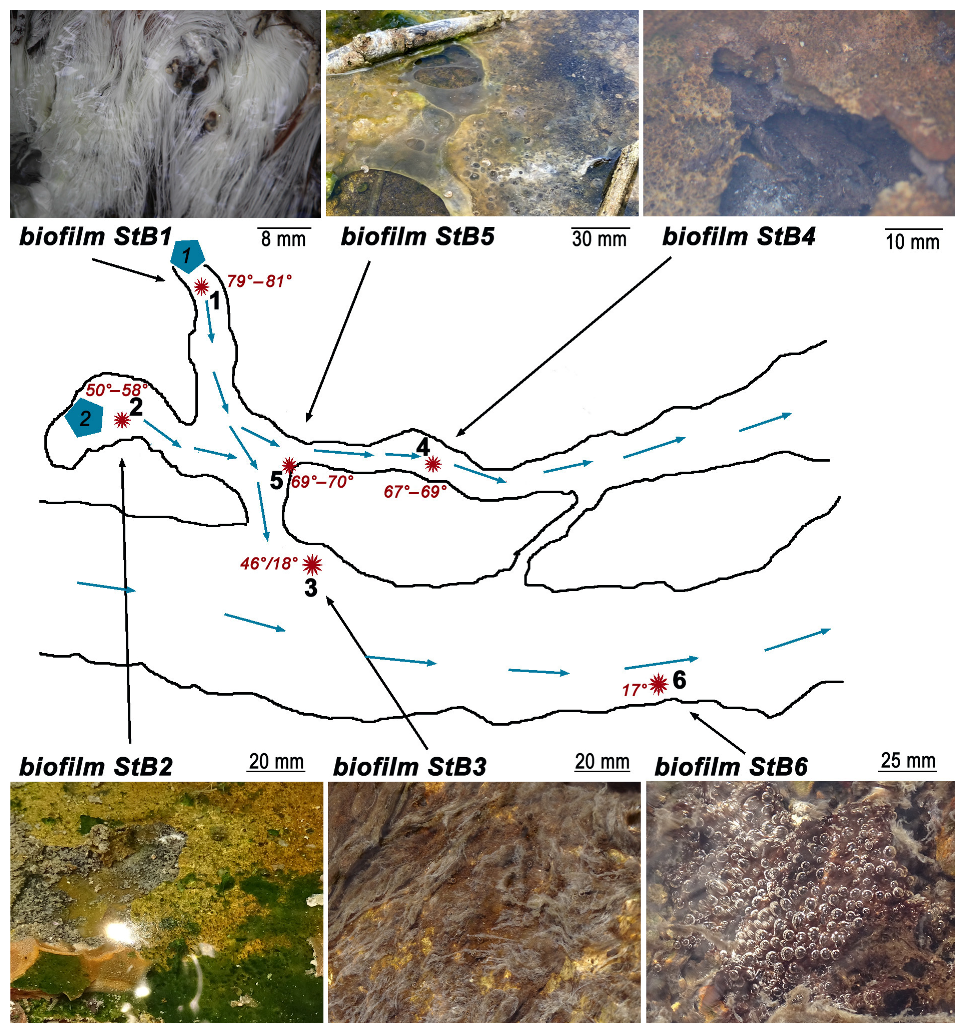
2.1. Description of the Sample Collection Sites

The sampling was performed at the Stolbovskie hot springs, Kunashir Island in September 2021 (Table 1). The Stolbovskie hot springs were categorized as Na-Cl type springs with semi-neutral pH (6.09–6.9) originating from the Mendeleev volcano top area [2,20]. The springs give origin to the hot streams and rills that fell into the big Zmeiny stream running to the Sea of Okhotsk. The characteristics of the chemical composition of water and dissolved gases for the hot spring at sampling site 1 were extracted from [20] and are provided in Supplementary Table S1. Unfortunately, there was no possibility to perform a detailed chemical analysis of water from the sampling sites, thus we could not take into account any possible changes of water chemistry that may occur along the hot streams. The temperature at the sampling sites ranged between 17 °C and 81.5 °C, and the circumneutral pH-values of water were in agreement with the data reported earlier [2,20]. The scheme of the sampling sites and the photographs of the studied biofilms are shown on Figure 1.
The main stream of the Stolbovskie hot springs system has rapid current and is characterized by chilly water. Two hot springs with different water temperatures were situated 8–10 m aside from the main stream and at 10 m from each other (Figure 1). Both springs supplied with water two small rills that entered the main stream in different points. Spring 1 with water temperature reaching 79.5–81.5 °C gushed out of the ground 0.5 m higher than the entire system of streams was located. Its water flowed downhill, and was mixed with water of the main stream at the distance of 8.5 m forming a peculiar thermocline at the mixing zone. Very characteristic white biofilm with thick filamentous structure (StB1) was collected 15–30 cm away from the point where hot water was coming to the surface. Water of Spring 1 was slightly acidified (pH = 6.6) compared to other sampling points where pH was about 7.5. Spring 2 was weaker, and its opening was located somewhere in a shallow hot water pool 4 m away from the rill running from the spring 1. Biofilm StB2 grew in the pool of Spring 2, where the water temperature was in range of 50.0–58.5 °C. This biofilm had a layered structure of dark green, brown and orange brick color. Spring 2 gave birth to a small stream that ran in parallel with the main stream and was also partly fed with the waters from Spring 1. Two biofilms, StB4 and StB5, were collected along this stream. Both biofilms had layered structure. Biofilm StB5 was formed close to the point where the waters of both hot springs mixed, and water from Spring 1 heated its environment up to 69.0–70.2 °C. It was almost transparent and resembled a rigid and dense jelly. Biofilm StB4 was located 4 m downstream StB5, and water temperature there was 67.0–69.0 °C. The biofilm StB4 structure and color was visually similar to that of StB2. We also sampled two biofilms from the main stream, aiming to compare them with the biofilms from the hot streams. One such biofilm (StB3) was abundantly growing at the thermocline zone at the mouth of the hot spring, thus being disposed to two contrasting temperatures (46.0 °C and 18.0 °C) simultaneously. It had filamentous structure, but the relatively thin brownish filaments were not similar to the thick “hairy” structure of white biofilm StB1. Biofilm StB6 that developed several meters downstream in the main stream (17.0 °C) was amorphous, brown-black, and covered with gas bubbles.

2.2. DNA Extraction, Purification, Amplification, and Sequencing

The samples of the biofilms were collected from sites in four replicates using sterile scalpels, pooled into sterile tubes and immediately filled with DNA/RNA Shield reagent (Zymo Research, Irvine, CA, USA). Fixed samples were transported to the laboratory, where they were stored at −80 °C until DNA purification and further analysis.
Total genomic DNA was extracted from the microbial biofilm samples using the Power Biofilm Kit (Qiagen, Germantown, MD, USA) according to the manufacturer’s instructions. Cells were disrupted using the PowerLyzer (MP Biomedicals, Irvine, CA, USA). V3-V4 regions of the 16S rRNA genes were amplified by PCR using primers for bacteria S-D-Bact-0341-b-S-17 (CCTACGGGNGGCWGCAG) and S-D-Bact-0785-a-A-21 (GACTACHVGGGTATCTAATCC) [21]; for archaea Arch349F (5′-GYGCASCAGKCGMGAAW-3′) and Arch806R (5′-GGACTACVSGGGTATCTAAT-3′) [22]. V4 region of the 18S SSU rRNA gene was targeted using primers TAReuk454FWD1 (CCAGCASCYGCGGTAATTCC) and TAReukREV3 (ACTTTCGTTCTTGATYRA) [23]. We used Q5 High-Fidelity 2X Master Mix (New England Biolabs, UK). The PCR protocol suggested by the Illumina 16S Metagenomic Sequencing Library Preparation protocol ((https://support.illumina.com/documents/documentation/chemistry_documentation/16s/16s-metagenomic-library-prep-guide-15044223-b.pdf, accessed on 27 November 2013) was applied. After 25 cycles of PCR, its results were checked by electrophoresis in 1% agarose gel stained with ethidium bromide. Amplicon libraries were prepared following the Illumina 16S Metagenomic Sequencing Library Preparation protocol. Qubit® 3.0 Fluorometer (Thermo Fisher Scientific, Waltham, MA, USA) with a dsDNA High Sensitivity Assay Kit (Thermo Fisher Scientific, Waltham, MA, USA) was used to measure DNA concentrations. The libraries were pooled by equal molarity. Denaturation and sample loading were performed according to the Illumina Sample Preparation Guide using a MiSeq Reagent Kit v3, 600 cycles (Illumina, San Diego, CA, USA). The sequencing of the libraries was performed on the Illumina MiSeq platform (Illumina, San Diego, CA, USA).

2.3. Data Analysis

The sequencing data quality was checked using FastQC (https://www.bioinformatics.babraham.ac.uk/projects/fastqc/, accessed on 1 December 2022). Cutadapt [24] was used for primer sequences removal from the reads. The DADA2 workflow [25] was used for further sequence analysis, including quality filtering, reads merging, chimera removal, and amplicon sequence variants (ASVs) generation. The ASVs-based approach was used, as it is more precise, sensitive, reproducible, and comprehensive than the OTUs-based approach [26]. The amplicon sequence variants (ASVs) obtained were taxonomically classified using a pre-trained naive Bayes classifier, which was trained on SILVA 138 SSU database [27]. All statistical analyses and visualization were performed in the R environment using phyloseq [28], ggplot2 [29], MicrobiotaProcess [30], microbiome [31] packages. For community composition analysis and visualization of specific taxonomy levels, we merged ASVs related to one genus with tax_glom function and took ASVs that represent at least 1% of reads in at least one sample for bacteria and archaea, or 5% of reads for protists. Alpha diversity was calculated using the alpha() function from microbiome package in R. Bray–Curtis dissimilarity was used for beta diversity comparison in get_pcoa function. Then, we computed the Pearson’s rank correlation coefficient to test if there was any dependence between alpha diversity and temperature.

3. Results

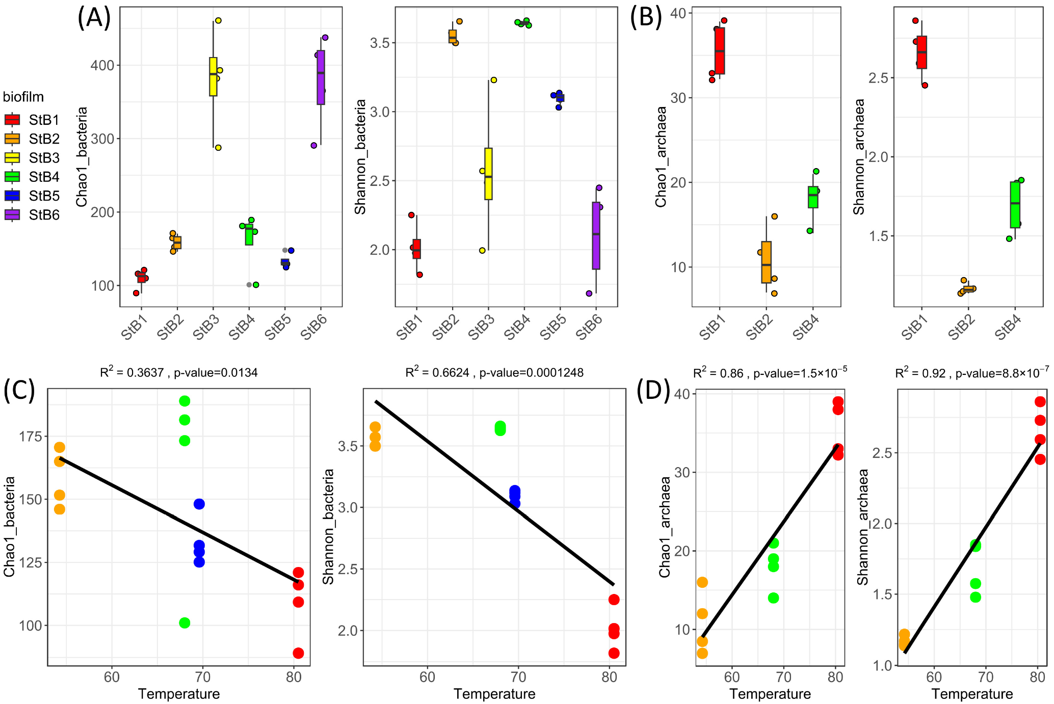
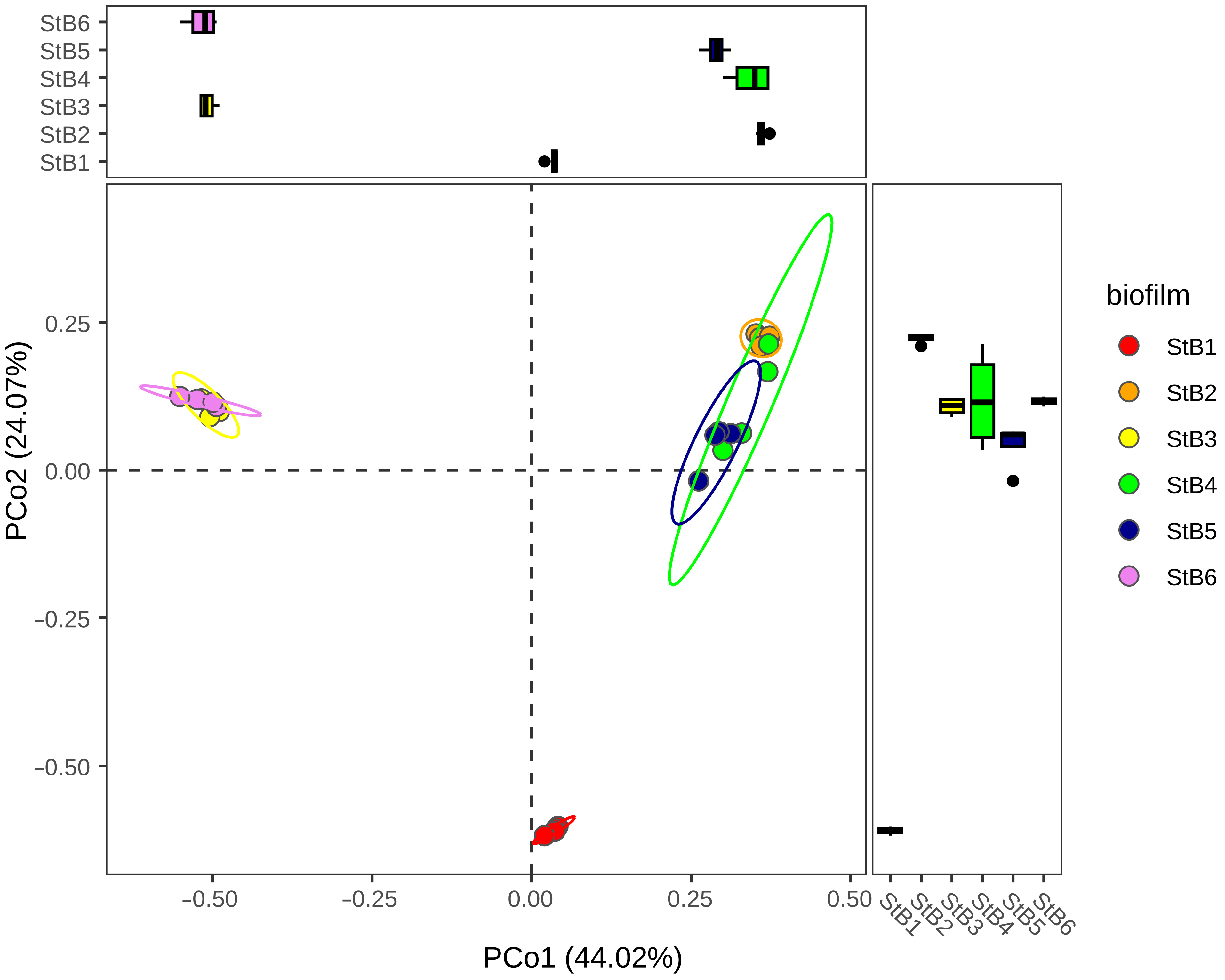
3.1. Estimation of Richness and Diversity of the Biofilm-Forming Microbial Communities

For the bacterial community, after excluding occasional archaeal and chloroplast sequences, the total number of reads amounted to 449,694 which were compiled into 1628 ASVs with an average sequence length of 417 bp. For archaea, we excluded StB3, StB5, and StB6 samples as archaeal sequences there were underrepresented, in three remaining samples (StB1, StB2, and StB4) the final number of archaeal reads was 17,342, which yielded 87 ASVs with an average sequence length of 339 bp. As we also aimed to assess the diversity of protists in the microbial communities analyzed, we got rid of eukaryotic non-protist ASVs. After quality control, 171,722 reads were classified into 562 ASVs with an average sequence length of 382 bp (Supplementary Table S2). After ASVs assembly, we obtained the rarefaction curves for all samples, and each of those reached a plateau, indicating good representation of the microbial communities (Supplementary Figure S1).
We calculated two alpha diversity indexes, Chao1 and Shannon, for bacterial and archaeal components of the communities (Figure 2A,B). Chao1 is a richness index, evaluating the total number of taxa in the sample; hence, it allows to estimate how many of the taxa may be missing in the results of analysis. Shannon is a diversity index, the higher it is, the more diverse the community is.
According to Chao1 index, bacterial communities of StB3 and StB6 biofilms were characterized by the greatest richness among all studied samples (Figure 2A). At the same time, StB2, StB4, and StB5 were the most diverse biofilms ((Figure 2A).
We found that for bacteria there was a strong inverse correlation between diversity indexes and temperature (R = −0.81 for Shannon and R= −0.6 for Chao1, Figure 2C). Our results based on linear regression (explanatory power: R2  =  0.6624, p value = 0.0001248 for Shannon diversity and R2 = 0.3637, p value = 0.0134 for Chao1) indicated that the richness and diversity of bacteria in the geothermal microbial communities decreased with the increase of temperature. The archaeal component of the communities demonstrated reverse dependence (Figure 2D). The richest and the most diverse archaeal component (Figure 2B) was observed in StB1 biofilm growing at the highest temperature. The correlation between diversity indexes and temperature was R = 0.96 for Shannon and R = 0.93 for Chao1. Explanatory power of linear regression model was R2  =  0.92, p value = 8.8 × 10⁻⁷ for Shannon diversity and R2 = 0.86, p value = 1.5 × 10⁻⁵ for Chao1.
A beta diversity analysis was performed to compare the bacterial community structure in different biofilms. It revealed that six studied samples clustered into three distinct groups (Figure 3) according to the habitat: one group was formed by biofilms StB3 and StB6 growing in the main stream with the lower water temperature (17–20 °C), the second included communities StB2, StB4, and StB5 that developed in moderately hot (50–70 °C) environments, and biofilm StB1 picked directly from the hot spring (79–81 °C) stood alone on the plot.

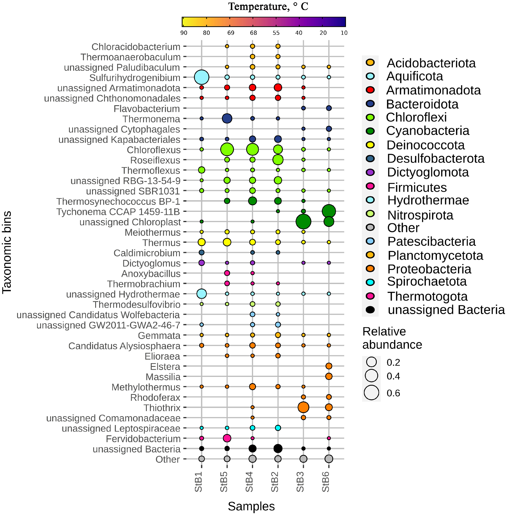
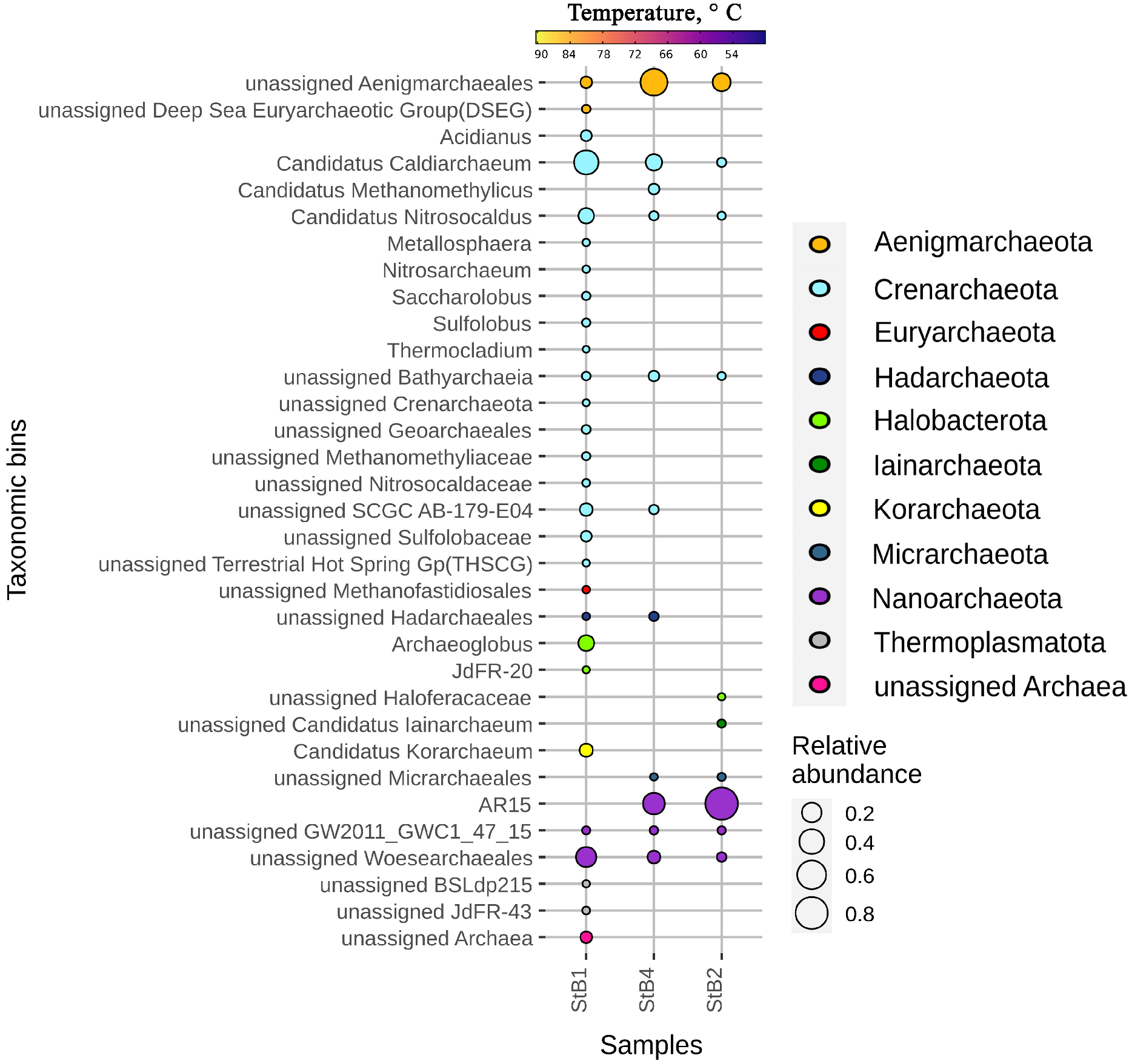
3.2. The Taxonomic Diversity of Bacteria and Archaea in the Biofilm-Forming Communities

Within the domain bacteria, more than 96.5% of the assigned sequences were classified at the phylum level. In total, 25, 26, 25, 34, 23, and 29 bacterial phyla were detected in StB1, StB2, StB3, StB4, StB5, and StB6 samples, respectively.
The most abundant bacterial phylum in biofilm StB1 (Figure 4) from the hot spring was Aquificota (61% of ASVs) followed by Hydrothermae (19%), Deinococcota (7%), Chloroflexi (5.5%), and Dictyoglomota (3%). At the genus level, Sulfurihydrogenibium was the absolute dominant (60% of the total number of ASVs), almost exclusively representing the phylum Aquificota. All ASVs attributed to Hydrothermae phylum could not be classified to the genus level. ASVs affiliated with Chloroflexi phylum mostly belonged to the genus Thermoflexus. We also recovered Thermus sequences from Deinococcota phylum and Dictyoglomus ASVs from Dictyoglomota.
The second group of ‘moderate temperature samples’ included biofilms StB2, StB4, and StB5. They had relatively similar taxonomic composition (Figure 3 and Figure 4). Bacteria belonging to phylum Chloroflexi were the most abundant, and they ranged from 46 to 50% of the community in these biofilms. Among representatives of this phylum Chloroflexus (35% and 43% of total number of ASVs, respectively in StB4 and StB5), RBG-13-54-9, which was classified only down to order level (4% in both biofilms) and Roseiflexus (2% and 1%) were the dominants in biofilms StB4 and StB5, while in biofilm StB2, Roseiflexus (23%) was the major dominant, and the Chloroflexus share was 15%, RBG-13-54-9 was 8%. Among other bacteria, we revealed the dominance of Bacteroidota and Armatimonadota, followed by Cyanobacteria, Deinococcota, and Proteobacteria (Figure 4). Majority of the members of Armatimonadota phylum were not classified to the genus level, and Bacteroidota was classified to Thermonema only in biofilm StB5. An unusual finding was a significant number of cyanobacterial taxa related to Thermosynechococcus BP-1, this taxon had the highest representation in biofilm StB4 (11%). Some sequences were well-represented only in biofilm StB5 among the moderate temperature group, such as Thermus (9% of total number of ASVs) and obligately anaerobic genus Fervidobacterium (9%) [32].
Biofilms StB3 and StB6 sampled from the main stream with water of ambient temperature appeared relatively similar in terms of bacterial diversity (Figure 3 and Figure 4). The filamentous bacteria from Thiothrix genus (Gammaproteobacteria) were the major dominant in StB3 biofilm, as 73% of all ASVs found there belonged to it. The representatives of Cyanobacteria, Chloroflexi and several other phyla were detected at a fairly low relative abundance. The situation was opposite in biofilm StB6, where the filamentous Tychonema_CCAP_1459_11B was the most prevalent cyanobacteria (60%) [33], while abundance of Thiothrix was just 9%.
Regarding archaea (Figure 5), in biofilm StB1 the dominant phylum was Crenarchaeota (57%) consisting of Candidatus Caldiarchaeum (37.5% from the whole community reads), Candidatus Nitrosocaldus (9%), and thermoacidophilic genus Acidianus (2%) [34]. Less abundant ASVs belonging to Nanoarchaeota phylum (22%) represented insufficiently explored order Woesearchaeales. Furthermore, significant fractions were also formed by Halobacterota (10%), Korarchaeota (5%), and Aenigmarchaeota (3%).
The taxonomic composition of archaea in biofilms StB2 and StB4 differed from that of biofilm StB1 but also from each other. The dominant phyla were Nanoarchaeota (83% and 32% for StB2 and StB4, respectively) with prevailing genus AR15, and Aenigmarchaeota (15% and 51%, respectively). In biofilm StB4, phylum Crenarchaeota was also well represented (16.5%) compared to biofilm StB2, and 11% of the StB4 community consisted of archaea belonging to Candidatus Caldiarchaeum.

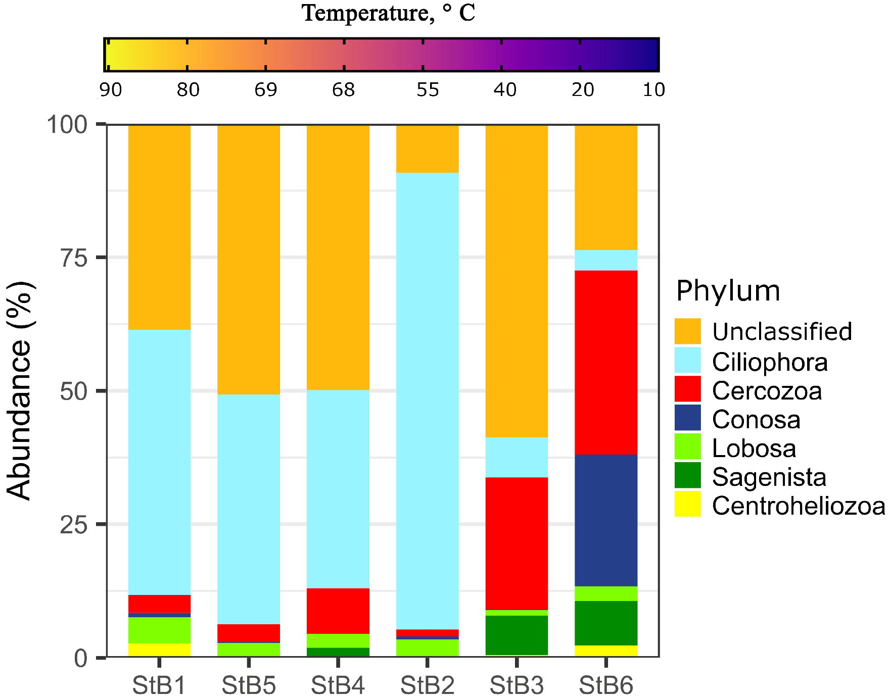
3.3. Taxonomic Diversity of Protists in the Biofilm-Forming Microbial Communities

Protists were also represented in the studied microbial communities (Figure 6). Many eukaryotic sequences (9–59% in different samples) did not belong to the known protists and possibly resulted from the hot spring contamination with plant and animal remnants, so we did not analyze them further on. In all biofilms, Ciliophora sequences were the most numerous among those ASVs which could be attributed to protists. Mainly anaerobic ciliates from the genus Trimyema [35] were present, followed by Oxytricha and some scuticociliates related to Cinetochilum. The representatives of the macrotaxon Cercozoa were characteristic for all biofilms, but only in biofilms StB3 and StB6 could they be considered dominant, reaching 24.5% and 34%, respectively. Furthermore, subphylum Sagenista was well presented in these two biofilms too, with a small number of reads also in biofilm StB4. Amoebae of Conosa subphylum were characteristic only for biofilm StB6, where they were one of the dominant taxa (24%). ASVs of lobose amoebae were present in minor quantities in all six studied biofilms.

4. Discussion

4.1. Prokaryotic Community Changes Correlate with the Temperature Gradient

Metagenomic studies of the microbial communities in the extreme biotopes are numerous and address not only the taxonomic diversity of such communities but also the influence of the environmental factors on the community composition. Microbial communities of the hot springs in different parts of the world have been studied, and in several works the correlation between temperature and diversity was found and proved [36,37,38]. It has also been reported that pH of water may have a strong effect upon the aquatic microbial communities’ structure [39]. However, the relation between the composition of microbial communities and environmental factors shaping them is very complex, and many factors should be taken into account even when similar biotopes from different localities are compared [12,40]. The studied biofilms StB1, StB2, StB4, and StB5 developed in the same geothermal aquatic system that was formed by the Stolbovskie hot springs fed with underground waters of the Mendeleev volcano top area [20]. We assumed that the chemical composition of water at the sampling sites could be considered almost identical, as two water sources were closely neighboring, and all sampling sites were just several meters apart from each other. However, the water temperature varied significantly along the hot streams even at short distances (Figure 1). It has been shown that differences in alpha diversity of the microbial samples taken from the hot springs were higher if sampling points were located close to each other but differed in water temperature than if the distance was larger but the temperature was the same [41]. We found that water temperature inversely correlated with the number of bacterial taxa detected as ASVs in the studied biofilms, and with their phylogenetic assemblies (Figure 2), thus, they were obviously influencing and shaping the microbial communities. A similar trend can also be observed in other studies [39,41]. Biofilm StB1 developed at 79 °C, i.e., at the lowest temperature limit of hyperthermophilic growth, and, respectively, was characterized by unique taxonomic composition, where the representatives of the phyla Aquificae and Hydrothermae dominated. Almost all ASVs of Aquificae in biofilm StB1 belonged to bacteria of the genus Sulfurihydrogenibium, which constituted about 60% of total bacterial abundance there (Figure 4). This genus includes anaerobic or microaerobic, facultatively heterotrophic or chemolithoautotrophic hydrogen-, or sulfur-, or thiosulphate-oxidizing bacteria which preferentially live at a neutral pH in high-temperature conditions [39,42]. Sulfurihydrogenibium representatives are able to consume oxygen from the environment even in minor concentrations, thus creating and maintaining anaerobic conditions for other community participants [43]. Sulfurihydrogenibium is a dominant genus in many biofilms growing in geothermal aquatic biotopes in Yellowstone caldera [44], Kamchatka peninsula hot springs [45], Japan [46], New Zealand [47], and Azores [48], though its species are different. The appearance and structure of ‘hairy’ biofilm StB1 from the Stolbovskie hot springs strongly resembles the ‘fettucini-like’ biofilms from Mammoth Hot Springs in Yellowstone National Park [49], though besides Sulfurihydrogenibium dominance, these biofilms are not identical by taxonomic composition.
Biofilms StB2, StB4, and StB5growing at the temperature interval 50–70 °C, almost in the range considered optimal for moderate (55–60 °C) and extreme (70–75 °C) thermophiles, were relatively similar to each other by taxonomic composition (and also by layered morphology and general appearance) and clustered together at PCoA (Figure 3). At this temperature, the hyperthermophiles from biofilm StB1 were substituted by photosynthetic representatives of Chloroflexi and Cyanobacteria, which dominated in thermophilic biofilms StB2, StB4, and StB5 (Figure 4). Thus, we observe a transition from the chemotrophic community in the hyperthermophilic zone to the photosynthetic community along the temperature gradient, which has also been shown by other authors [41,50,51,52,53,54]. Detected phototrophic genera such as Chloroflexus, Roseiflexus, and Thermosynechococcus are common for phototrophic biofilms at the temperature interval 50–70 °C [41,51,52,55,56]. Biofilms StB2 and StB4 were similar in appearance to the multilayer biofilms known from the other hot springs, including those in Yellowstone National Park [57,58,59]. Such biofilms usually consist of a green surface layer of Synechococcus cyanobacteria and a red lower layer dominated by Chloroflexi [52,60,61]. The layering of such mats is explained by different photosynthetic capabilities of Cyanobacteria and Chloroflexi at high oxygen levels, as well as by different resistance to sulfides. The richness of phototrophic cyanobacteria decreases with increasing temperature, leading to decrease in the oxygenic photosynthesis abilities, whereas the richness of Chloroflexi and the rate of anoxygenic photosynthesis are not influenced by increasing temperature [62]. However, we did not detect bacteria of the genus Synechococcus in the Kunashir biofilms. While in biofilm StB5 there was no green layer at all, we suppose that thin surface green layer of biofilms StB2 and StB4 was formed by Thermosynechococcus BP-1 (Figure 4). The T. elongatus strain BP-1 was originally isolated as a thermophilic cyanobacterium from the hot spring in Beppu, a town in the southern part of Japan [63]. It was initially identified as Synechococcus elongatus strain BP-14, which was later described as a new genus Thermosynechococcus [64]. Interestingly, according to the 16S rRNA gene sequence phylogenetic analysis, T. elongatus branches very close to the origin of cyanobacteria, while seven major lineages arose rather recently as “crown” groups [13,65].
The genus Chloroflexus dominated in biofilms StB4 and StB5 at temperatures close to 70 °C degrees, but when temperatures dropped to 60 °C and below, the genus Roseiflexus became dominant in the community. Similar results were previously obtained for Mushroom Spring (Yellowstone National Park, USA), where Chloroflexus was more abundant at 65 °C while Roseiflexus was more abundant already at 60 °C [66]. However, in the other Yellowstone hot spring study, it was shown that Roseiflexus was more abundant at higher temperatures [59]. Apparently, temperature plays a role in the occurrence of specific phototrophic Chloroflexi. However, in addition to temperature, morphology, geochemistry, and metabolic abilities are also not less important factors [62]. To conclude, biofilms StB2, StB4, and StB5 were taxonomically different from the morphologically similar mats found under similar conditions in the Yellowstone National Park hot springs [57,58,59].
Biofilms StB3 and StB6 grew in the main stream with an ambient water temperature, even if biofilm StB3 was also influenced by a hot water current. Anoxygenic phototrophs almost disappeared (Figure 4). Mixotrophic bacteria (chemoorganotrophic and chemolithoautotrophic that use inorganic sulfur) of genus Thiotrix (Gammaproteobacteria) dominated in biofilm StB3. Thiotrix spp. have been described as a major component of biofilms growing on various surfaces in flowing water that contained sulfides [67]. The biofilms edified by these bacteria were white and had a thin-thread structure, which is consistent with our observations. Biofilm StB6 was obviously a photosynthetic community where filamentous cyanobacteria Tychonema_CCAP_1459_11 likely played a major role. Tychonema belongs to the benthic cyanobacteria and can produce potent neurotoxins. Pieces of benthic biofilms can float to the surface, thus there is a certain risk of poisoning [68], which should be considered when the tourists take baths in Zmeiny downstream the Stolbovskie hot springs. Similar tendency for succession of anoxygenic phototrophs by oxygenic phototrophs, first of all trichome cyanobacteria, with temperature decrease is observed also in the communities of the other hot springs [61,69]. A large number of chloroplasts ASVs may also indicate algae as primary producers in biofilm StB3, though algae usually dominate in the biofilms of acidified habitats with pH < 5 [19,70].
For archaeal taxa the water temperature had a positive effect on richness and taxonomic diversity (Figure 2). The opposite effect of temperature on the occurrence and diversity for archaea and bacteria may be explained by generally better adaptation of archaea to the extreme environments [71]. Archaea are the main group of living organisms that thrive in biotopes with extreme conditions, and, in particular, hyperthermophilic species are present almost in all lineages of archaea [72]. Some archaea are able to survive at temperatures exceeding 100 °C, and many can tolerate 70–80 °C, thus there is no surprise that archaeal abundance, richness, and diversity in the hot springs is higher than bacterial [44]. Previously, it has been shown that the highest taxonomic diversity of Crenarchaeota was observed in temperature range 59–77 °C [73,74]. At lower or higher temperatures, prevalence and abundance of different crenarchaea in the hot springs decreased [74]. Thus, Crenarchaeota and Korarchaeota include predominantly thermophilic organisms, and representatives of these phyla appeared to be the most diverse in biofilm StB1 growing at 79 °C, though being less present in biofilms StB2 and StB4 (Figure 5). Candidatus Caldiarchaeum subterraneum was the best represented in biofilm StB1. Candidatus C. subterraneous is an uncultivated archaeal lineage that was first described from the metagenomic assembly of microbial mat from a geothermal water stream in a subsurface gold mine [75]. Candidatus C. subterraneum was also found in the other hot springs that were characterized by similar temperature and pH [52,76,77]. Analysis of the metabolic potential suggested a predominantly aerobic nature of Candidatus C. subterraneum, that was a member of subsurface thermophilic microbial mats with a heterotrophic lifestyle [78]. Although in SILVA database (SILVA 138 release) this archaeon is indicated (and referred to in our work) as a member of the phylum Crenarchaeota, it should be noted that in the recent publication this lineage has been reassigned to the order Candidatus Caldarchaeales within the phylum Thermoproteota, class Nitrososphaeria [79]. The other characteristic representative of hyperthermophiles in biofilm StB1 growing at 79 °C was Archaeoglobus. Members of the class Archaeoglobi (phylum Halobacterota) are known as members of subsurface microbial communities, and are capable of growing by reducing sulfite and thiosulfate [14,80,81].

4.2. The Evidence of Presence of Ciliates in the Hot Springs

There is much less data on eukaryotic occurrence and diversity than on bacteria and archaea in hot springs. It is believed that most of thermophilic protists and fungi can survive at temperatures up to 60 °C [82], while at higher temperatures, eukaryotic cells require too much of oxygen which may be depleted, and also eukaryotic cell membranes have different lipid composition not favorable to withstand heat [16]. The comprehensive report on diversity of protists in the hydrothermal systems of Lassen Volcanic National Park (Northern California) suggested that some protists might survive at temperatures up to 68–72 °C [83]. Presence of diatoms (Bacillariophyceae), some chrysophytes and chlamydomonadales algae, fungi (Basidiomycota and Chytridiomycota), amoebae, cercozoans, and ciliates was molecularly confirmed in those extreme conditions. We found an unexpectedly high number of ASVs belonging to different protist lineages in the biofilms sampled from the Stolbovskie hot springs, which was inconsistent with the observation that species richness of thermophilic protists is usually very low in such habitats [15]. Metabarcoding of the 18S rRNA genes revealed significant diversity of protists, first of all cercozoans, amoebae of two subphyla (one of those includes Echinamoeba thermarum, the thermophilic lobose amoeba found in hot springs all over the world [84]), and heterokonts from subphylum Sagenista. At the same time, protists were not abundant. The dominant group in the studied biofilms were ciliates (Ciliophora), which ASVs constituted on average 57% of all protistan ASVs. Similar results were obtained at screening for protists in the New Zealand hot springs [12], though the authors suspected a certain bias in their data. The highest temperatures at which living ciliates have been ever isolated from the thermal springs was 68 °C (see references in [85]). However, in more recent reports the surviving cysts of ciliates, namely Dexiotricha colpidiopsis (Oligohymenophorea) and Oxytricha granulifera (Spirotrichea) were collected in a hot spring in Iceland at temperatures up to 75 °C [86,87]. No ciliates were detected at temperatures above 80 °C [15]. Interestingly, some thermophilic ciliates can be maintained preferentially at much lower temperature, as was shown for a ‘eurythermic’ hymenostomatid Cyclidium citrullus (Oligohymenophorea) isolated from a hot spring in Israel but able to grow in the laboratory at temperature 10 °C lower than in the habitat [85]. Representatives of another ciliate genus Trimyema (Plagiopylea) frequent in the hot springs were found at temperatures up to 65 °C [12,35]. In our samples the number of ASVs witnessed that Trimyema was the dominant genus, though the temperatures of biofilm StB5 and especially biofilm StB1 growth were higher than permissive for ciliates temperatures reported in the literature. Interestingly, Trimyema minutum isolated from the hydrothermal vent sediments with temperatures up to 70 °C was further maintained at the temperature interval 28–52 °C being unable to survive already at 55 °C [35]. The authors supposed that this discrepancy was due to temperature micro-inhomogeneities in the habitat allowing ciliates to escape from too harsh conditions. This hypothesis can also explain our finding of Trimyema sp. in the biofilms, at least multilayered ones, which may offer certain compartmentalization. In addition to temperature, an important factor for all microorganisms is the presence or absence of oxygen. Indeed, Trimyema is known as an anaerobic ciliate, and appropriate anoxic conditions could be achieved in the hot spring due to the scavenging of dissolved oxygen by Sulfurihydrogenibium [88], the major genus of the biofilms growing in the hottest sampling sites. Two other ciliates documented by us among the dominants, Oxytricha (Spirotrichea) and some scuticociliate (Oligohymenophorea) related to a marine ciliate Cinetochilum, still were significantly less present. Oxytricha is known to be able to survive in adverse conditions such as acidic environments [28,57], and its resting cysts were also found in the hot spring in Iceland [87] and dominated among ciliates in majority of the hot springs in New Zealand [12], and it seems likely that its sequence was detected as anonymous Spirotrichea sp. at 68 °C in thermal springs of Lassen Volcanic National Park [83]. Many scuticociliates are presumably anaerobic and contain methanogenic archaea as symbionts [89], and at least one of them, Cyclidium, has been shown to be thermophilic [85]. We did not detect any representative of ciliates belonging to class Colpodea, such as Cyrtolophosididae which were among the dominants in some hot springs in New Zealand [12] and are also able to encyst. However, in general our data support the hypothesis that several protist lineages include the majority of thermophilic taxa, thus revealing certain adaptations to the temperature extremities [12].
The direct proof of presence of protists in the hot spring biofilms could be obtained by thorough microscopy of the sample, but such facility was absent at the collection site, and no eukaryotes could survive transportation to the equipped laboratory. At least some protists revealed by metabarcoding, such as Oxytricha, are able to form surviving cysts, but, again, tiny cysts are very difficult to find in a sample.
Thus, the biofilms found at the Stolbovskie hot springs on Kunashir Island represent complex microbial communities formed by bacteria, archaea, and protists. All participants of these communities are likely characterized by certain interconnections, as it was shown that protistan community composition was moderately correlated with bacterial and archaeal assemblies in the hot springs in New Zealand [12]. The biofilms of six kinds that we found in a local area along the hot streams of the geothermal system were different from each other by taxonomic composition, and the level of their similarity or diversity was significantly influenced by water temperature.

Supplementary Materials

The following supporting information can be downloaded at: https://www.mdpi.com/article/10.3390/ecologies4010009/s1, Figure S1: Rarefaction curves built for bacterial (A), archaeal (B) and protists (C) reads; Table S1: The chemical composition of thermal waters of the studied hot spring in the Stolbovskie springs.; Table S2: Statistical data of the analysis of amplicons.

Author Contributions

Conceptualization, A.P. and N.G.; methodology, N.G.; validation and formal analysis, A.M., A.B. and E.P.; investigation, A.M., S.S., A.P. and N.G.; data curation, A.B.; writing—original draft preparation, A.M. and E.P.; writing—review and editing, A.P. and N.G.; visualization, A.M., A.B. and A.P. All authors have read and agreed to the published version of the manuscript.

Funding

This work was supported by the Kazan Federal University Strategic Academic Leadership Program (PRIORITY-2030).

Institutional Review Board Statement

Not applicable.

Informed Consent Statement

Not applicable.

Data Availability Statement

Sequencing data generated during this study are available in the NCBI SRA under project PRJNA922438.

Acknowledgments

The authors are grateful to the administration and staff of the Kurilskii State Nature Reserve for providing facilities to collect the samples. E.P. and N.G. are grateful to the Ministry of Science and Higher Education of the Russian Federation for their administrative support within the framework of the government assignment for FRC Kazan Scientific Center of the RAS.

Conflicts of Interest

The authors declare no conflict of interest.

References

  1. Zharkov, R.V. Active volcanoes and thermal springs of Kunashir Island (Russia). IOP Conf. Ser. Earth Environ. Sci. 2020, 459, 022039. [Google Scholar] [CrossRef]
  2. Kalacheva, E.G.; Taran, Y.A. Processes responsible for variations in the isotopic composition (ΔD and Δ18O) of thermal waters of the Kuril Islands arc. Volcanolog. Seismol. 2019, 4, 3–17. [Google Scholar] [CrossRef]
  3. Markhinin, E.K.; Stratula, D.S. Hydrothermal Systems of Kuril Islands; Nauka: Moscow, Russia, 1977; p. 212. (In Russian) [Google Scholar]
  4. Straub, C.T.; Counts, J.A.; Nguyen, D.M.N.; Wu, C.H.; Zeldes, B.M.; Crosby, J.R.; Conway, J.M.; Otten, J.K.; Lipscomb, G.L.; Schut, G.J.; et al. Biotechnology of extremely thermophilic archaea. FEMS Microbiol. Rev. 2018, 42, 543–578. [Google Scholar] [CrossRef] [PubMed]
  5. Sani, R.; Rathinam, N. Extremophilic Microbial Processing of Lignocellulosic Feedstocks to Biofuels, Value-Added Products, and Usable Power; Springer: Berlin/Heidelberg, Germany, 2018; Chapter 8; ISBN 978-3-319-74457-5. [Google Scholar]
  6. Khaire, K.C.; Patel, S.; Gavande, P.V.; Moholkar, V.A.S.; Goyal, A. Extremophilic biofilms: Exploring the prospects. In Introduction to Biofilm Engineering; ACS Symposium Series; American Chemical Society: Washington, DC, USA, 2019; Volume 1323, pp. 141–157. ISBN 978-0-8412-3473-4. [Google Scholar]
  7. Castenholz, R.W. Thermophilic blue-green algae and the thermal environment. Bacteriol. Rev. 1969, 33, 476–504. [Google Scholar] [CrossRef] [PubMed]
  8. Brock, T.D. Thermophilic Microorganisms and Life at High Temperatures; Springer Series in Microbiology; Springer: New York, NY, USA, 1978; ISBN 978-1-4612-6286-2. [Google Scholar]
  9. Kublanov, I.V.; Perevalova, A.A.; Slobodkina, G.B.; Lebedinsky, A.V.; Bidzhieva, S.K.; Kolganova, T.V.; Kaliberda, E.N.; Rumsh, L.D.; Haertlé, T.; Bonch-Osmolovskaya, E.A. Biodiversity of thermophilic prokaryotes with hydrolytic activities in hot springs of Uzon Caldera, Kamchatka (Russia). Appl. Environ. Microbiol. 2009, 75, 286–291. [Google Scholar] [CrossRef] [PubMed]
  10. DeCastro, M.-E.; Rodríguez-Belmonte, E.; González-Siso, M.-I. Metagenomics of thermophiles with a focus on discovery of novel thermozymes. Front. Microbiol. 2016, 7, 1521. [Google Scholar] [CrossRef]
  11. López-López, O.; Cerdán, M.E.; González-Siso, M.-I. Hot spring metagenomics. Life 2013, 3, 308–320. [Google Scholar] [CrossRef]
  12. Oliverio, A.M.; Power, J.F.; Washburne, A.; Cary, S.C.; Stott, M.B.; Fierer, N. The ecology and diversity of microbial eukaryotes in geothermal springs. ISME J. 2018, 12, 1918–1928. [Google Scholar] [CrossRef]
  13. Nakamura, Y.; Kaneko, T.; Sato, S.; Ikeuchi, M.; Katoh, H.; Sasamoto, S.; Watanabe, A.; Iriguchi, M.; Kawashima, K.; Ki-mura, T.; et al. Complete genome structure of the thermophilic cyanobacterium Thermosynechococcus elongatus BP-1. DNA Res. 2002, 9, 123–130. [Google Scholar] [CrossRef]
  14. Slobodkina, G.; Allioux, M.; Merkel, A.; Cambon-Bonavita, M.A.; Alain, K.; Jebbar, M.; Slobodkin, A. Physiological and genomic characterization of a hyperthermophilic archaeon Archaeoglobus neptunius sp. nov. isolated from a deep-sea hydrothermal vent warrants the reclassification of the genus Archaeoglobus. Front. Microbiol. 2021, 16, 679245. [Google Scholar] [CrossRef]
  15. Hu, X. Ciliates in extreme environments. J. Eukaryot. Microbiol. 2014, 61, 410–418. [Google Scholar] [CrossRef]
  16. Hickey, D.A.; Singer, G.A. Genomic and proteomic adaptations to growth at high temperature. Genome Biol. 2004, 5, 117. [Google Scholar] [CrossRef]
  17. Kizilova, A.; Sukhacheva, M.; Pimenov, N.; Yurkov, A.; Kravchenko, I. Methane oxidation activity and diversity of aerobic methanotrophs in pH-neutral and semi-neutral thermal springs of the Kunashir Island, Russian Far East. Extremophiles 2014, 18, 207–218. [Google Scholar] [CrossRef]
  18. Rozanov, A.S.; Korzhuk, A.V.; Shlyakhtun, V.N.; Shipova, A.A.; Peltek, S.E. Metagenomic data on prokaryotic diversity of Kunashir Island geothermal spring. Data Brief 2020, 32, 106244. [Google Scholar] [CrossRef] [PubMed]
  19. Rozanov, A.S.; Korzhuk, A.V.; Shekhovtsov, S.V.; Vasiliev, G.V.; Peltek, S.E. Microorganisms of two thermal pools on Kunashir Island, Russia. Biology 2021, 10, 924. [Google Scholar] [CrossRef] [PubMed]
  20. Kalacheva, E.G.; Taran, Y.A.; Kotenko, T.A.; Inguaggiato, S.; Voloshina, E.V. The hydrothermal system of Mendeleev Volcano, Kunashir Island, Kuril Islands: The geochemistry and the transport of magmatic components. J. Volcanolog. Seismol. 2017, 11, 335–352. [Google Scholar] [CrossRef]
  21. Klindworth, A.; Pruesse, E.; Schweer, T.; Peplies, J.; Quast, C.; Horn, M.; Glöckner, F.O. Evaluation of general 16S ribosomal RNA gene PCR primers for classical and next-generation sequencing-based diversity studies. Nucleic Acids Res. 2013, 41, e1. [Google Scholar] [CrossRef] [PubMed]
  22. Takai, K.; Horikoshi, K. Rapid detection and quantification of members of the archaeal community by quantitative PCR using fluorogenic probes. Appl. Environ. Microbiol. 2000, 66, 5066–5072. [Google Scholar] [CrossRef]
  23. Stoeck, T.; Bass, D.; Nebel, M.; Christen, R.; Jones, M.D.M.; Breiner, H.-W.; Richards, T.A. Multiple marker parallel tag environmental DNA sequencing reveals a highly complex eukaryotic community in marine anoxic water. Mol. Ecol. 2010, 19 (Suppl. 1), 21–31. [Google Scholar] [CrossRef]
  24. Martin, M. Cutadapt removes adapter sequences from high-throughput sequencing reads. EMBnet J. 2011, 17, 10–12. [Google Scholar] [CrossRef]
  25. Callahan, B.J.; McMurdie, P.J.; Rosen, M.J.; Han, A.W.; Johnson, A.J.A.; Holmes, S.P. DADA2: High-resolution sample inference from Illumina amplicon data. Nat. Methods 2016, 13, 581–583. [Google Scholar] [CrossRef]
  26. Callahan, B.; McMurdie, P.; Holmes, S. Exact sequence variants should replace operational taxonomic units in marker-gene data analysis. ISME J. 2017, 11, 2639–2643. [Google Scholar] [CrossRef]
  27. Quast, C.; Pruesse, E.; Yilmaz, P.; Gerken, J.; Schweer, T.; Yarza, P.; Peplies, J.; Glöckner, F.O. The SILVA ribosomal RNA gene database project: Improved data processing and web-based tools. Nucleic Acids Res. 2013, 41, D590–D596. [Google Scholar] [CrossRef]
  28. McMurdie, P.J.; Holmes, S. Phyloseq: An R package for reproducible interactive analysis and graphics of microbiome census data. PLoS ONE 2013, 8, e61217. [Google Scholar] [CrossRef]
  29. Wickham, H.; Chang, W.; Henry, L.; Pedersen, T.; Takahashi, K.; Wilke, C.; Woo, K.; Yutani, H.; Dunnington, D. RStudio. ggplot2: Elegant Graphics for Data Analysis; Springer: New York, NY, USA, 2016. [Google Scholar]
  30. Xu, S.; Yu, G. Microbiota Process: A Comprehensive R Package for Managing and Analyzing Microbiome and Other Ecological Data within the Tidy Framework. R Package Version 1.8.1. Available online: https://github.com/YuLab-SMU/MicrobiotaProcess (accessed on 29 December 2022).
  31. Lahti, L.; Shetty, S. Tools for Microbiome Analysis in R. R PackageVersion 1.19.1. Available online: http://microbiome.github.com/microbiome (accessed on 29 December 2022).
  32. Huber, R.; Stetter, K.O. Fervidobacterium. In Bergey’s Manual of Systematics of Archaea and Bacteria; John Wiley & Sons, Ltd.: Hoboken, NJ, USA, 2015; pp. 1–5. ISBN 978-1-118-96060-8. [Google Scholar]
  33. Suda, S.; Watanabe, M.M.; Otsuka, S.; Mahakahant, A.; Yongmanitchai, W.; Nopartnaraporn, N.; Liu, Y.; Day, J.G. Taxonomic revision of water-bloom-forming species of oscillatorioid cyanobacteria. Int. J. Syst. Evol. Microbiol. 2002, 52 Pt 5, 1577–1595. [Google Scholar] [CrossRef] [PubMed]
  34. Kato, S.; Itoh, T.; Yamagishi, A. Archaeal diversity in a terrestrial acidic spring field revealed by a novel PCR primer targeting archaeal 16S rRNA genes. FEMS Microbiol. Lett. 2014, 319, 34–43. [Google Scholar] [CrossRef] [PubMed]
  35. Baumgartner, M.; Stetter, K.O.; Foissner, W. Morphological, small subunit rRNA, and physiological characterization of Trimyema minutum (Kahl, 1931), an anaerobic ciliate from submarine hydrothermal vents growing from 28 °C to 52 °C. J. Eukaryot. Microbiol. 2002, 49, 227–238. [Google Scholar] [CrossRef] [PubMed]
  36. Sharp, C.E.; Brady, A.L.; Sharp, G.H.; Grasby, S.E.; Stott, M.B.; Dunfield, P.F. Humboldt’s Spa: Microbial diversity Is controlled by temperature in geothermal environments. ISME J. 2014, 8, 1166–1174. [Google Scholar] [CrossRef]
  37. Chiriac, C.M.; Szekeres, E.; Rudi, K.; Baricz, A.; Hegedus, A.; Dragoş, N.; Coman, C. Differences in temperature and water chemistry shape distinct diversity patterns in thermophilic microbial communities. Appl. Environ. Microbiol. 2017, 83, e01363-17. [Google Scholar] [CrossRef]
  38. Chan, C.S.; Chan, K.-G.; Ee, R.; Hong, K.-W.; Urbieta, M.S.; Donati, E.R.; Shamsir, M.S.; Goh, K.M. Effects of physiochemical factors on prokaryotic biodiversity in Malaysian circumneutral hot springs. Front. Microbiol. 2017, 8, 1252. [Google Scholar] [CrossRef] [Green Version]
  39. Power, J.F.; Carere, C.R.; Lee, C.K.; Wakerley, G.L.J.; Evans, D.W.; Button, M.; White, D.; Climo, M.D.; Hinze, A.M.; Morgan, X.C.; et al. Microbial biogeography of 925 geothermal springs in New Zealand. Nat. Commun. 2018, 9, 2876. [Google Scholar] [CrossRef]
  40. Massello, F.L.; Chan, C.S.; Chan, K.-G.; Goh, K.M.; Donati, E.; Urbieta, M.S. Meta-analysis of microbial communities in hot springs: Recurrent taxa and complex shaping factors beyond pH and temperature. Microorganisms 2020, 8, 906. [Google Scholar] [CrossRef]
  41. Podar, P.T.; Yang, Z.; Björnsdóttir, S.H.; Podar, M. Comparative Analysis of microbial diversity across temperature gradients in hot springs from Yellowstone and Iceland. Front. Microbiol. 2020, 11, 1625. [Google Scholar] [CrossRef]
  42. Escuder-Rodríguez, J.-J.; DeCastro, M.-E.; Saavedra-Bouza, A.; Becerra, M.; González-Siso, M.-I. Insights on microbial communities inhabiting non-volcanic hot springs. Int. J. Mol. Sci. 2022, 23, 12241. [Google Scholar] [CrossRef] [PubMed]
  43. Kubo, K.; Knittel, K.; Amann, R.; Fukui, M.; Matsuura, K. Sulfur-metabolizing bacterial populations in microbial mats of the Nakabusa hot spring, Japan. Syst. Appl. Microbiol. 2011, 34, 293–302. [Google Scholar] [CrossRef]
  44. Inskeep, W.P.; Rusch, D.B.; Jay, Z.J.; Herrgard, M.J.; Kozubal, M.A.; Richardson, T.H.; Macur, R.E.; Hamamura, N.; Jennings, R.D.; Fouke, B.W. Metagenomes from high-temperature chemotrophic systems reveal geochemical controls on microbial community structure and function. PLoS ONE 2010, 5, e9773. [Google Scholar] [CrossRef] [PubMed]
  45. Merkel, A.; Pimenov, N.; Rusanov, I.; Slobodkin, A.; Slobodkina, G.; Tarnovetckii, I.; Frolov, E.; Dubin, A.; Perevalova, A.; Bonch-Osmolovskaya, E. Microbial diversity and autotrophic activity in Kamchatka hot springs. Extremophiles 2017, 21, 307–317. [Google Scholar] [CrossRef] [PubMed]
  46. Nakagawa, T.; Fukui, M. Molecular characterization of community structures and sulfur metabolism within microbial streamers in Japanese hot springs. Appl. Environ. Microbiol. 2003, 69, 7044–7057. [Google Scholar] [CrossRef]
  47. Hetzer, A.; Morgan, H.; Mcdonald, I.; Daughney, C. Microbial life in champagne pool, a geothermal spring in Waiotapu, New Zealand. Extremophiles 2007, 11, 605–614. [Google Scholar] [CrossRef]
  48. Sahm, K.; John, P.; Nacke, H.; Wemheuer, B.; Grote, R.; Daniel, R.; Antranikian, G. High abundance of heterotrophic prokaryotes in hydrothermal springs of the Azores as revealed by a network of 16S rRNA gene-based methods. Extremophiles 2013, 17, 649–662. [Google Scholar] [CrossRef]
  49. Dong, Y.; Sanford, R.A.; Inskeep, W.P.; Srivastava, V.; Bulone, V.; Fields, C.J.; Yau, P.M.; Sivaguru, M.; Ahrén, D.; Fouke, K.W.; et al. Physiology, metabolism, and fossilization of hot-spring filamentous microbial mats. Astrobiology 2019, 19, 1442–1458. [Google Scholar] [CrossRef] [PubMed]
  50. Bowen De León, K.; Gerlach, R.; Peyton, B.M.; Fields, M.W. Archaeal and bacterial communities in three alkaline hot springs in Heart Lake geyser basin, Yellowstone National Park. Front. Microbiol. 2013, 4, 330. [Google Scholar] [CrossRef] [PubMed]
  51. Wang, S.; Hou, W.; Dong, H.; Jiang, H.; Huang, L.; Wu, G.; Zhang, C.; Song, Z.; Zhang, Y.; Ren, H.; et al. Control of temperature on microbial community structure in hot springs of the Tibetan Plateau. PLoS ONE 2013, 8, e62901. [Google Scholar] [CrossRef]
  52. Thiel, V.; Wood, J.M.; Olsen, M.T.; Tank, M.; Klatt, C.G.; Ward, D.M.; Bryant, D.A. The dark side of the Mushroom Spring microbial Mat: Life in the Shadow of Chlorophototrophs. I. Microbial diversity based on 16S rRNA gene amplicons and metagenomic sequencing. Front. Microbiol. 2016, 17, 919. [Google Scholar] [CrossRef] [PubMed]
  53. Rozanov, A.S.; Bryanskaya, A.V.; Ivanisenko, T.V.; Malup, T.K.; Peltek, S.E. Biodiversity of the microbial mat of the Garga hot spring. BMC Evol. Biol. 2017, 17, 254. [Google Scholar] [CrossRef] [PubMed]
  54. Shu, W.-S.; Huang, L.-N. Microbial diversity in extreme environments. Nat. Rev. Microbiol. 2022, 20, 219–235. [Google Scholar] [CrossRef]
  55. Klatt, C.G.; Inskeep, W.P.; Herrgard, M.J.; Jay, Z.J.; Rusch, D.B.; Tringe, S.G.; Niki Parenteau, M.; Ward, D.M.; Boomer, S.M.; Bryant, D.A.; et al. Community structure and function of high-temperature chlorophototrophic microbial mats inhabiting diverse geothermal environments. Front. Microbiol. 2013, 4, 106. [Google Scholar] [CrossRef]
  56. Darima, B.; Buryukhaev, S.; Dambaev, V.; Tsyrenova, D.; Lavrentieva, E. Taxonomical and functional diversity of microbial communities in two hot springs of the Baikal rift zone. IOP Conf. Ser. Earth Environ. Sci. 2021, 908, 012001. [Google Scholar] [CrossRef]
  57. Boomer, S.M.; Noll, K.L.; Geesey, G.G.; Dutton, B.E. Formation of multilayered photosynthetic biofilms in an alkaline thermal spring in Yellowstone National Park, Wyoming. Appl. Environ. Microbiol. 2009, 75, 2464–2475. [Google Scholar] [CrossRef]
  58. Nübel, U.; Bateson, M.; Vandieken, V.; Wieland, A.; Kühl, M.; Ward, D. Microscopic examination of distribution and phenotypic properties of phylogenetically diverse Chloroflexaceae-related bacteria in hot spring microbial matst. Appl. Environ. Microbiol. 2002, 68, 4593–4603. [Google Scholar] [CrossRef] [Green Version]
  59. Bennett, A.C.; Murugapiran, S.K.; Hamilton, T.L. Temperature impacts community structure and function of phototrophic Chloroflexi and Cyanobacteria in two alkaline hot springs in Yellowstone National Park. Environ. Microbiol. Rep. 2020, 12, 503–513. [Google Scholar] [CrossRef] [PubMed]
  60. Tank, M.; Thiel, V.; Ward, D.; Bryant, D. A Panoply of Phototrophs: An Overview of the Thermophilic Chlorophototrophs of the Microbial Mats of Alkaline Siliceous Hot Springs in Yellowstone National Park, WY, USA; Springer International Publishing: Berlin/Heidelberg, Germany, 2017; pp. 87–137. [Google Scholar] [CrossRef]
  61. Hamilton, T.; Havig, J. Meet me in the middle: Median temperatures impact Cyanobacteria and photoautotrophy in eruptive Yellowstone hot springs. mSystems 2022, 7, e01450-21. [Google Scholar] [CrossRef] [PubMed]
  62. Mammola, S.; Meierhofer, M.B.; Borges, P.A.V.; Colado, R.; Culver, D.C.; Deharveng, L.; Delić, T.; Di Lorenzo, T.; Dražina, T.; Ferreira, R.L.; et al. Towards evidence-based conservation of subterranean ecosystems. Biol. Rev. Camb. Philos. Soc. 2022, 97, 1476–1510. [Google Scholar] [CrossRef] [PubMed]
  63. Yamaoka, T.; Satoh, K.; Katoh, S. Photosynthetic activities of a thermophilic blue-green alga. Plant Cell Physiol. 1978, 19, 943–954. [Google Scholar] [CrossRef]
  64. Katoh, H.; Itoh, S.; Shen, J.R.; Ikeuchi, M. Functional analysis of psbV and a novel c-type cytochrome gene psbV2 of the thermophilic cyanobacterium Thermosynechococcus elongatus strain BP-1. Plant Cell Physiol. 2001, 42, 599–607. [Google Scholar] [CrossRef]
  65. Honda, D.; Yokota, A.; Sugiyama, J. Detection of seven major evolutionary lineages in cyanobacteria based on the 16S rRNA gene sequence analysis with new sequences of five marine Synechococcus strains. J. Mol. Evol. 1999, 48, 723–739. [Google Scholar] [CrossRef]
  66. Klatt, C.G.; Liu, Z.; Ludwig, M.; Kühl, M.; Jensen, S.I.; Bryant, D.A.; Ward, D.M. Temporal metatranscriptomic patterning in phototrophic Chloroflexi inhabiting a microbial mat in a geothermal spring. ISME J. 2013, 7, 1775–1789. [Google Scholar] [CrossRef]
  67. Brigmon, R.L.; Furlong, M.; Whitman, W.B. Identification of Thiothrix unzii in two distinct ecosystems. Lett. Appl. Microbiol. 2003, 36, 88–91. [Google Scholar] [CrossRef]
  68. Bauer, F.; Stix, M.; Bartha-Dima, B.; Geist, J.; Raeder, U. Spatio-temporal monitoring of benthic anatoxin-a-producing Tychonema sp. in the River Lech, Germany. Toxins 2022, 14, 357. [Google Scholar] [CrossRef]
  69. Alcamán-Arias, M.; Pedrós-Alió, C.; Tamames, J.; Fernández, C.; Pérez-Pantoja, D.; Vásquez, M.; Díez, B. Diurnal changes in active carbon and nitrogen pathways along the temperature gradient in porcelana hot spring microbial mat. Front. Microbiol. 2018, 9, 2353. [Google Scholar] [CrossRef]
  70. Lehr, C.R.; Frank, S.D.; Norris, T.B.; D’Imperio, S.; Kalinin, A.V.; Toplin, J.A.; Castenholz, R.W.; McDermott, T.R. Cyanidia (cyanidiales) population diversity and dynamics in an acid-sulfate-chloride spring in Yellowstone National Park. J. Phycol. 2007, 43, 3–14. [Google Scholar] [CrossRef]
  71. Rampelotto, P.H. Extremophiles and extreme environments. Life 2013, 3, 482–485. [Google Scholar] [CrossRef]
  72. Groussin, M.; Gouy, M. Adaptation to environmental temperature is a major determinant of molecular evolutionary rates in Archaea. Mol. Biol. Evol. 2011, 28, 2661–2674. [Google Scholar] [CrossRef] [PubMed]
  73. Yim, L.C.; Hongmei, J.; Aitchison, J.C.; Pointing, S.B. Highly diverse community structure in a remote central Tibetan geothermal spring does not display monotonic variation to thermal stress. FEMS Microbiol. Ecol. 2006, 57, 80–91. [Google Scholar] [CrossRef] [PubMed]
  74. Zhao-Qi, S.; Chen, J.-Q.; Jiang, H.; Zhou, E.-M.; Tang, S.-K.; Zhi, X.-Y.; Zhang, L.-X.; Zhang, L.; Li, W.-J. Diversity of Crenarchaeota in terrestrial hot springs in Tengchong, China. Extremophiles 2010, 14, 287–296. [Google Scholar] [CrossRef]
  75. Nunoura, T.; Takaki, Y.; Kakuta, J.; Nishi, S.; Sugahara, J.; Kazama, H.; Chee, G.J.; Hattori, M.; Kanai, A.; Atomi, H.; et al. Insights into the evolution of Archaea and eukaryotic protein modifier systems revealed by the genome of a novel archaeal group. Nucleic Acids Res. 2011, 39, 3204–3223. [Google Scholar] [CrossRef]
  76. Kimura, H.; Mori, K.; Tashiro, T.; Kato, K.; Yamanaka, T.; Ishibashi, J.I.; Hanada, S. Culture-independent estimation of optimal and maximum growth temperatures of archaea in subsurface habitats based on the G+C content in 16S rRNA gene sequences. Geomicrobiol. J. 2010, 27, 114–122. [Google Scholar] [CrossRef]
  77. Stefanova, K.; Tomova, I.; Tomova, A.; Radchenkova, N.; Atanassov, I.; Kambourova, M. Archaeal and bacterial diversity in two hot springs from geothermal regions in Bulgaria as demostrated by 16S rRNA and GH-57 genes. Int. Microbiol. 2015, 18, 217–223. [Google Scholar] [CrossRef] [PubMed]
  78. Takami, H.; Arai, W.; Takemoto, K.; Uchiyama, I.; Taniguchi, T. Functional classification of uncultured “Candidatus Caldiarchaeum subterraneum” using the maple system. PLoS ONE 2015, 21, e0132994. [Google Scholar] [CrossRef] [Green Version]
  79. Rinke, C.; Chuvochina, M.; Mussig, A.J.; Chaumeil, P.A.; Davín, A.A.; Waite, D.W.; Whitman, W.B.; Parks, D.H.; Hugenholtz, P. A standardized archaeal taxonomy for the Genome Taxonomy Database. Nat. Microbiol. 2021, 6, 946–959. [Google Scholar] [CrossRef] [PubMed]
  80. Stetter, K.O. Archaeoglobus fulgidus gen. Nov., sp. nov.: A new taxon of extremely thermophilic Archaebacteria. Syst. Appl. Microbiol. 1988, 10, 172–173. [Google Scholar] [CrossRef]
  81. Burggraf, S.; Jannasch, H.W.; Nicolaus, B.; Stetter, K.O. Archaeoglobus profundus sp. nov., represents a new species within the sulfur-reducing Archaebacteria. Syst. Appl. Microbiol. 1990, 13, 24–28. [Google Scholar] [CrossRef]
  82. Tansey, M.R.; Brock, T.D. The upper temperature limit for eukaryotic organisms. Proc. Natl. Acad. Sci. USA 1972, 69, 2426–2428. [Google Scholar] [CrossRef] [PubMed]
  83. Brown, P.B.; Wolfe, G.V. Protist genetic diversity in the acidic hydrothermal environments of Lassen Volcanic National Park, USA. J. Eukaryot. Microbiol. 2006, 53, 420–431. [Google Scholar] [CrossRef] [PubMed]
  84. Baumgartner, M.; Yapi, A.; Gröbner-Ferreira, R.; Stetter, K.O. Cultivation and properties of Echinamoeba thermarum n. sp., an extremely thermophilic amoeba thriving in hot springs. Extremophiles 2003, 7, 267–274. [Google Scholar] [CrossRef]
  85. Kahan, D. Cyclidium citrullus Cohn, a ciliate from the hot springs of Tiberias (Israel). J. Protozool. 1972, 19, 593–597. [Google Scholar] [CrossRef]
  86. Qu, Z.; Groben, R.; Marteinsson, V.; Agatha, S.; Filker, S.; Stoeck, T. Redescription of Dexiotricha colpidiopsis (Kahl, 1926) Jankowski, 1964 (Ciliophora, Oligohymenophorea) from a hot spring in Iceland with identification key for Dexiotricha species. Acta Protozool. 2019, 57, 95–106. [Google Scholar] [CrossRef]
  87. Zhu, R.; Qu, Z.; Zhang, Q.; Filker, S.; Stoeck, T.; Li, F.; Hu, X. A New record of Oxytricha granulifera granulifera Foissner and Adam, 1983 (Protozoa, Ciliophora, Oxytrichidae) from a hot spring in Iceland, with notes on its abnormal form during cultivation. Front. Marin. Sci. 2021, 8, 621349. [Google Scholar] [CrossRef]
  88. Roy, C.; Rameez, M.J.; Haldar, P.K.; Peketi, A.; Mondal, N.; Bakshi, U.; Mapder, T.; Pyne, P.; Fernandes, S.; Bhattacharya, S.; et al. Microbiome and ecology of a hot spring-microbialite system on the Trans-Himalayan Plateau. Sci. Rep. 2020, 10, 5917. [Google Scholar] [CrossRef] [Green Version]
  89. Takeshita, K.; Yamada, T.; Kawahara, Y.; Narihiro, T.; Ito, M.; Kamagata, Y.; Shinzato, N. Tripartite symbiosis of an anaerobic scuticociliate with two hydrogenosome-associated endosymbionts, a Holospora-related alphaproteobacterium and a methanogenic archaeon. Appl. Environ. Microbiol. 2019, 85, e00854-19. [Google Scholar] [CrossRef]
Figure 1. Scheme of the sampling site at the Stolbovskie hot springs, and photographs of the studied biofilms. Blue polygons—hot springs; red asterisks—sampling points; blue arrows show direction of water flow. The distances between sampling sites: from site 1 to site 5 5.0 m; from site 2 to site 5 4.5 m; from site 3 to site 5 3.5 m; from site 4 to site 5 4.0 m; from site 3 to site 6 15 m.
Ecologies 04 00009 g001
Figure 2. The alpha diversity measurements of the bacterial (A) and archaeal (B) components of the microbial communities and linear regression analysis between diversity indices and temperature for bacteria (C) and archaea (D). Each biofilm is marked by a certain color identical for all plots.
Ecologies 04 00009 g002
Figure 3. Results of principal coordinates analysis (PCoA) of the biofilm-forming bacterial communities based on the Bray–Curtis distance.
Ecologies 04 00009 g003
Figure 4. The most abundant bacterial genera/phyla in the microbial biofilms developing at different temperatures. Circle size indicates the inferred relative abundance based on amplicon numbers (in %).
Ecologies 04 00009 g004
Figure 5. The most abundant archaeal genera/phyla in the microbial biofilms developing at different temperatures. Circle size indicates the inferred relative abundance based on amplicon numbers (in %).
Ecologies 04 00009 g005
Figure 6. Relative abundances of protists phyla in the microbial biofilms calculated at reads filtration threshold 5%.
Ecologies 04 00009 g006
Table 1. The characteristic of the sampling sites and description of the biofilms’ morphology. The water temperatures are presented in a range.
Sample NameLatitude, NorthLongitude, EastTemperature, °CLocationMorphology
StB144.007009145.68326579.5–81.515–30 cm away from the hot spring 1White biofilm with thick filamentous structure
StB244.006997145.68331150.0–58.5Pool of the hot spring 2Layered structure of dark green, brown and orange brick color
StB344.007046145.68330818.0/46.0Main stream (thermocline zone)Thin brownish filamentous structure
StB444.007092145.68320467.0–69.04 m downstream StB5Layered structure, similar to StB2 biofilm
StB544.007025145.68330069.0–70.2Mixing point of two hot springsLayered structure, almost transparent and resembled a rigid and dense jelly
StB644.007186145.68321017.0Main stream bedAmorphous, brown-black, and covered with gas bubbles
Disclaimer/Publisher’s Note: The statements, opinions and data contained in all publications are solely those of the individual author(s) and contributor(s) and not of MDPI and/or the editor(s). MDPI and/or the editor(s) disclaim responsibility for any injury to people or property resulting from any ideas, methods, instructions or products referred to in the content.

