
Titanium Oxide Nanoparticles as Emerging Aquatic Pollutants: An Evaluation of the Nanotoxicity in the Freshwater Shrimp Larvae Atya lanipes


Department of Environmental Sciences, College of Natural Sciences, University of Puerto Rico, Rio Piedras Campus, San Juan 00925, Puerto Rico
*
Authors to whom correspondence should be addressed.
Ecologies 2023, 4(1), 141-151; https://doi.org/10.3390/ecologies4010011
Submission received: 10 January 2023
/
Revised: 13 February 2023
/
Accepted: 13 February 2023
/
Published: 23 February 2023
Abstract
:Nanoparticles are man-made materials defined as materials smaller than 100 nm in at least one dimension. Titanium oxide nanoparticles are of great interest because of their extensive use in self-care products. There is a lack of nanotoxicological studies of TiO2 NPs in benthic organisms to have evidence about the effects of these pollutants in freshwater ecosystems. Atya lanipes is a scraper/filter that can provide a good nanotoxicological model. This study aims to determine how the TiO2 NPs can develop a toxic effect in the larvae of the Atya lanipes shrimp and to document lethal and sublethal effects after acute exposures to TiO2 NP suspensions of: 0.0, 1.0, 10.0, 50.0, 100.0, and 150.0 mg/L. The results show that early exposure to TiO2 NPs in Atya lanipes creates an increase in mortality at 48 and 72 h exposures, hypoactivity in movements, and morphological changes, such as less pigmentation and the presence of edema in exposed larvae. In conclusion, TiO2 NPs are toxic contaminants in the larval stage of the Atya lanipes. It is necessary to regulate these nanoparticles for purposes of the conservation of aquatic biodiversity, especially for freshwater shrimp larvae and likely many other larvae of filter-feeding species.
1. Introduction
With the rapid development of the nanotechnology industry, the world community is increasingly aware of the environmental impacts of manufactured nanoparticles (NPs) in biological systems [1,2]. NPs are man-made materials smaller than 100 nm in at least one dimension [3]. These particles have a greater surface/volume ratio and unique physicochemical properties compared to their normal forms and sizes [4,5].
Titanium oxide is one type of nanoparticle of great interest because it is used in paints and coatings as a self-cleaning, antimicrobial, antifouling agent and in cosmetics as a UV absorber [6]. In addition, these NPs are energy semiconductors that exhibit photocatalytic activities [7]. From 2006 to 2010, the commercial production of titanium oxide nanoparticles (TiO2 NPs) was 5000 metric tons per year, increasing to more than 10,000 metric tons from 2011 to 2014, with an estimated 2.5 million metric tons by 2025 [8].
Nevertheless, there is a lack of standardized quantification of TiO2 NPs in aquatic ecosystems and of the environmental concentration in many aquatic ecosystems [9]. This information gap includes the freshwater ecosystems in Puerto Rico and in the wider Caribbean region. Data from European studies have determined concentrations in freshwater ranging from 0.015 24.5 micrograms/L [10,11]. In contrast, concentrations in soils that exceed 100 micrograms/L have also been reported [12]. However, concentrations of titanium (including TiO2 NPs) were found in the United States and Canada in a study involving 15 rivers. Concentrations of titanium in natural river waters range between 0.5 and 15 µg/L, and in soils and sediments they range between 10 and 100 g/kg with an average of less than 5 g/kg [13]. Consequently, there is an urgent challenge to evaluate the toxicological effects of TiO2 NPs in the laboratory using environmental concentrations of this nanomaterial.
Some studies have determined the neurotoxic effects of TiO2 NPs in different organisms. These neurotoxic effects are caused by the production of radical oxygen species in TiO2 NPs. This type of NP causes oxidative stress in different tissues, including the brain/nervous system, causing neural damage. Moreover, TiO2 NPs can be translocated and transported to other body organs [14,15,16]. Nevertheless, the toxicity effects in morphological development and mortality are not often clearly reported.
In this study, we used the larvae of the freshwater shrimp Atya lanipes Holthuis, 1963 (A. lanipes) to test the effect of TiO2 NPs in their development, behavior, and survival. The A. lanipes is a good model to study the toxicity of the nanoparticles present in aquatic environments due to it being a scraper/filter feeder shrimp that lives part of its life as a planktonic organism until it reaches the post-larval stage, where it lives as a benthonic organism. This complex life cycle is called the amphidromous life cycle and is similar to all the shrimp families (Atyidae, Palaemonidae, and Xiphocarididae) that inhabit Puerto Rico and many of the Caribbean streams. This amphidromous life cycle represents an important connection between the headwaters and estuaries [17,18]. When females release the larvae (zoea larvae) in the upper reaches of a river, the first stage (i.e., newly hatched larvae) drift passively to coastal environments where they develop and metamorphose into post-larvae that subsequently migrate back upstream to the headwaters where they mature and reproduce as adult shrimp [19]. If the TiO2 NPs are present in the estuarine/marine environment as the result of anthropogenic pollution, A. lanipes larvae could be affected by the possible toxic effects. In this part of the life cycle, the interactions between the larvae and the nanoparticles will be in the water column due to the planktonic larval behavior. This early life cycle exposure to TiO2 NPs in A. lanipes larvae can affect upstream migrations, population dynamics, and the species’ survival.
This study aims to determine how the TiO2 NPs can develop a toxic effect in the larvae of the A. lanipes shrimp that results in lethal effects, such as mortality, and sublethal effects, such as changes in behavior and morphological development. We hypothesized that the exposure of TiO2 NPs among A. lanipes larvae would develop a toxic effect showing lethal and sublethal effects due to the photocatalytic activity and the production of oxidizing agents that damage neurons and other tissues.
2. Materials and Methods
2.1. Characterization of Titanium Oxide Nanoparticles (TiO2 NPs)
Titanium oxide nano powder (Sigma Aldrich Chemical Company St. Louis, MO, USA, titanium IV oxide, Anatase nanopowder) was used to conduct the experiments. The characterization method was an S4700 II Cold Field Emission Gun Scanning Electron Microscope cFEG SEM. Titanium oxide powder was spread on weighing paper and gently picked up by a sticky carbon surface on the top of aluminum stubs. An S4700 II cFEG SEM (Hitachi High Technologies-America) with a silicon drift EDX detector (Oxford Instruments, X-MaxN, UK) was used to measure the surface morphology, elemental composition, and distribution of elements. All the SEM data were obtained at an acceleration voltage of 10 kV, and the images were collected with a secondary-electron detector. The elemental mapping and energy spectra were acquired with Aztec tools (Oxford Instruments, UK). The elemental analysis through the energy dispersive spectrum indicates the presence of titanium, oxygen, carbon, and sulfur elements (61, 36.1, 2.6, and 0.3 wt%, respectively). The observed size of the titanium oxide particle varied from 1 to 4 micron long.
2.2. TiO2 NPs Suspension Preparation and Physical Dispersion before the Exposure
Before the TiO2 NPs suspensions were prepared, we dispersed the powder using a magnetic stirrer at a maximum speed for 30 min (the nanoparticles were not sonicated because the aim was to evaluate realistic environmental conditions). Then, the TiO2 NPs suspensions were prepared one hour before the bioassay. We prepared suspension concentrations of 0.0, 1.0, 10.0, 50.0, 100.0, and 150.0 mg/L (mg of the weighted TiO2 NPs in 1 L of water) in amber bottles covered with black tape to avoid nanoparticle–light interaction at 10 parts per million (ppm) of salinity. The seawater was pasteurized, filtered, and diluted with dechlorinated water to emulate natural estuary ecosystem water chemical conditions.
2.3. A. lanipes Gravid Specimens Collection and Zoea Larvae Separation
The gravid females of A. lanipes were collected using baited minnow traps in the Buruquena Stream (18.321207, −65.819389) at the El Verde Field Station, Rio Grande, Puerto Rico (Figure 1). Baited traps (the bait was dry cat food) were set in different pools along the stream and removed 24 h later. The collected gravid shrimp were identified [20] and transported in a cooler under constant aeration to the laboratory. The gravid shrimp were transferred individually to glass tanks (15 cm × 15 cm × 15 cm) with 1 L of dechlorinated water and constant aeration. The shrimp were fed with commercial fish flakes (Tetra TetraMin Tropical Flakes) until the larval eclosion. After the larval eclosion, we separated the larvae (Figure 1) from the adult shrimp, and the bioassay started. The duration was 8:00–10:00 AM.
2.4. A. lanipes Larvae Bioassay
For the bioassays, we used the protocol of Solis et al. (1993) [21]. Atya lanipes zoea larvae hatched under normal laboratory conditions and were exposed to various TiO2 NPs concentrations. The suspension concentrations of 0.0, 1.0, 10.0, 50.0, 100.0, and 150.0 mg of TiO2 NPs/L were tested (N = 25 for each concentration). For each treatment, we used individual culture plates that contained six cells. This was duplicated for each exposure time individually. Replicates were made for each treatment and exposure time. In this way, each exposure time for the six treatments had 18 culture plates. In the culture plates of each concentration, 10 larvae were transferred to each cell of the six-well culture plate with 10.0 mL of each suspension concentration. Then, the six-well culture plates were transferred into a water bath with two thermometers. A temperature of 26 ± 1 °C was maintained for the bioassay exposure. During the entire experiment, the larvae were kept with no food and aeration. The light/dark photoperiod was 14:10 h (day:night). The exposures were carried out independently at 24, 48, and 72 h for all the nanoparticle suspension concentrations and the control group. The bioassay was conducted with different A. lanipes larvae samples to have genetic variation.
2.5. Mortality Analysis
Mortality was recorded as the following: (1) cessation of swimming or any movement by the larvae; (2) no phototaxis behavior; (3) change in color from transparent to white/gray. For each concentration, we analyzed a 40-larvae sample size. A mortality assessment was conducted for 5 min. We counted the number of larvae classified as “dead” following the previous definition for 24, 48, and 72 h of exposure.
2.6. Movement Analysis
The movement was assessed 24, 48, and 72 h after exposure to the different TiO2 NPs suspension concentrations using video recordings of the individual larva under free-swimming behavior to avoid group interactions. The larvae were placed in a single well of ten well plates (5.8 cm width × 12.6 cm length) [22]. This single-well plate was a circular plastic container 14 mm in diameter, and the depth was 1 mm (Falcon Plastics, California). The recordings were obtained using a stereo microscope with a digital camera (3.5 × -90 × LED Trinocular Zoom + 14 MP USB 3.0 Digital Camera). Each larva was acclimated for 90 s and their movement was recorded for 60 secs. We analyzed a sample size of larvae (N = 25) for each nanoparticle concentration and the control group. The room was kept dark and silent to prevent movement caused by phototaxis or sound. The only light available was the microscope light at a minimum level and covered with a red cellophane to prevent the movement of the larva because they do not detect red light. The recordings were analyzed using the Loligo® Systems- Lolitrack 5. The movement variables obtained from the software were average speed (mm/s) and average acceleration (mm/s2).
2.7. Morphological Development Analysis
To evaluate the morphological development of the larvae, we measured the total length of each larva in the control and exposure groups for all concentrations and exposure times. The total length was measured from the post-orbit margin to the end of the telson, excluding setae [23]. To measure the length of the larvae, we used a stereo microscope (3.5×-90× LED Trinocular Zoom + 14 MP USB 3.0 Digital Camera) with measuring software installed on a computer. Edemas and changes in pigmentation were classified as presence or absence.
2.8. Statistical Analysis
The statistical analysis and graphs were completed using PAST software (2001). Data are presented as the mean ± standard error of the mean, considering each six-well plate as an experimental unit. For biological responses such as behavior and total length, we performed a one-way ANOVA followed by the Tukey test to compare each exposure time separately. The comparisons of morphological development were performed using observed/not observed parameters (edema and/or pigmentation changes). For mortality, we calculated the mortality rate (%) (number of death larvae/unit sample size), for each concentration for the three periods of exposure.
3. Results
3.1. Mortality Rate
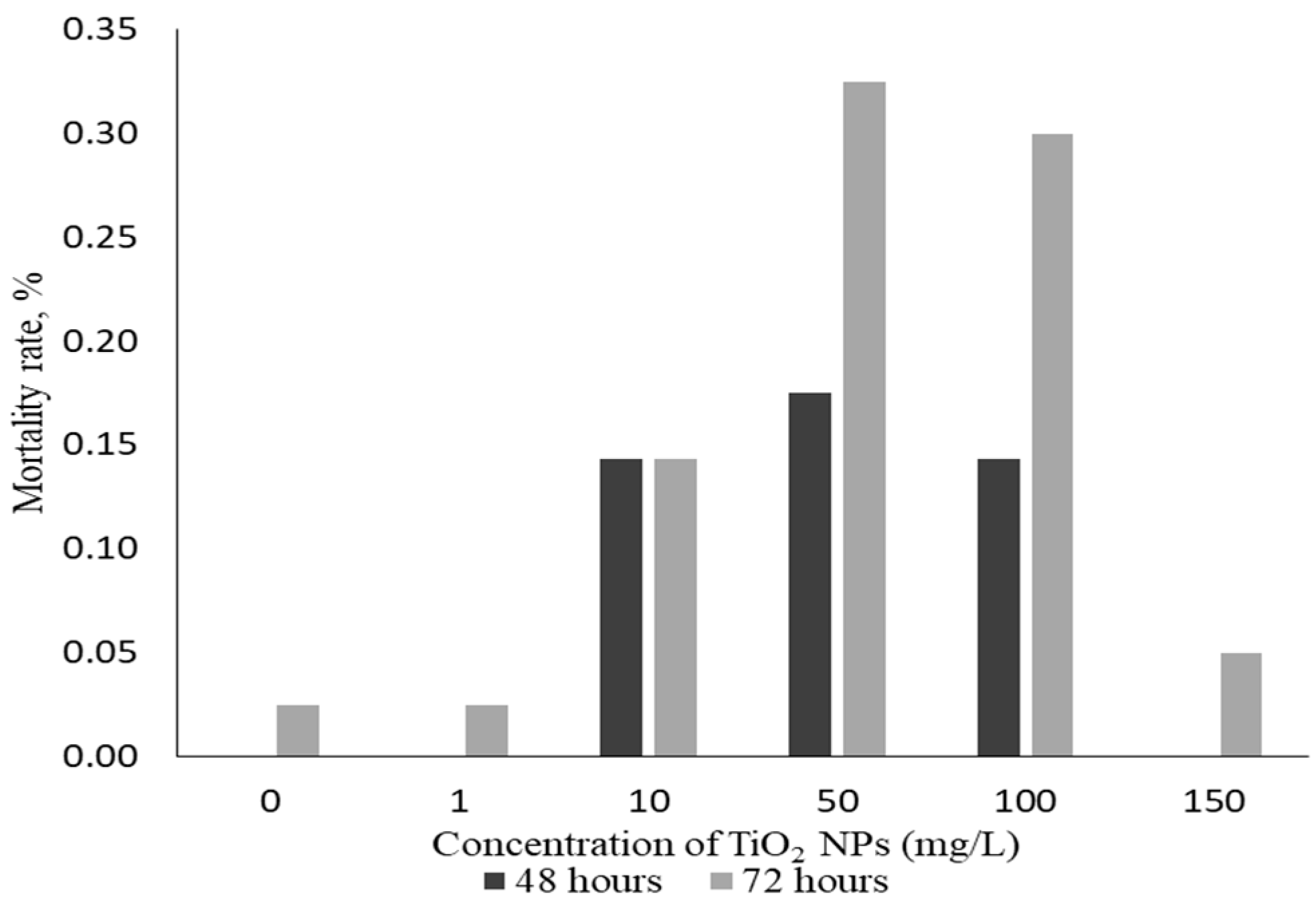
The mortality rate for each TiO2 NsP concentration at 24, 48, and 72 h exposure provided relevant information. At 24 h of exposure, no dead larvae were observed in any
TiO2 NPs concentration exposures or the control group. However, after 48 and 72 h, the mortality rate increased in the exposed group, specifically within 72 h of exposure (Figure 2). However, we did not see a consistent relationship between mortality and the concentrations of TiO2 NPs tested. The aforementioned is confirmed in the concentration of 150 mg/L where a decrease in mortality occurred rather than an increase for 48 and 72 h of exposure.
3.2. Movement Assessment
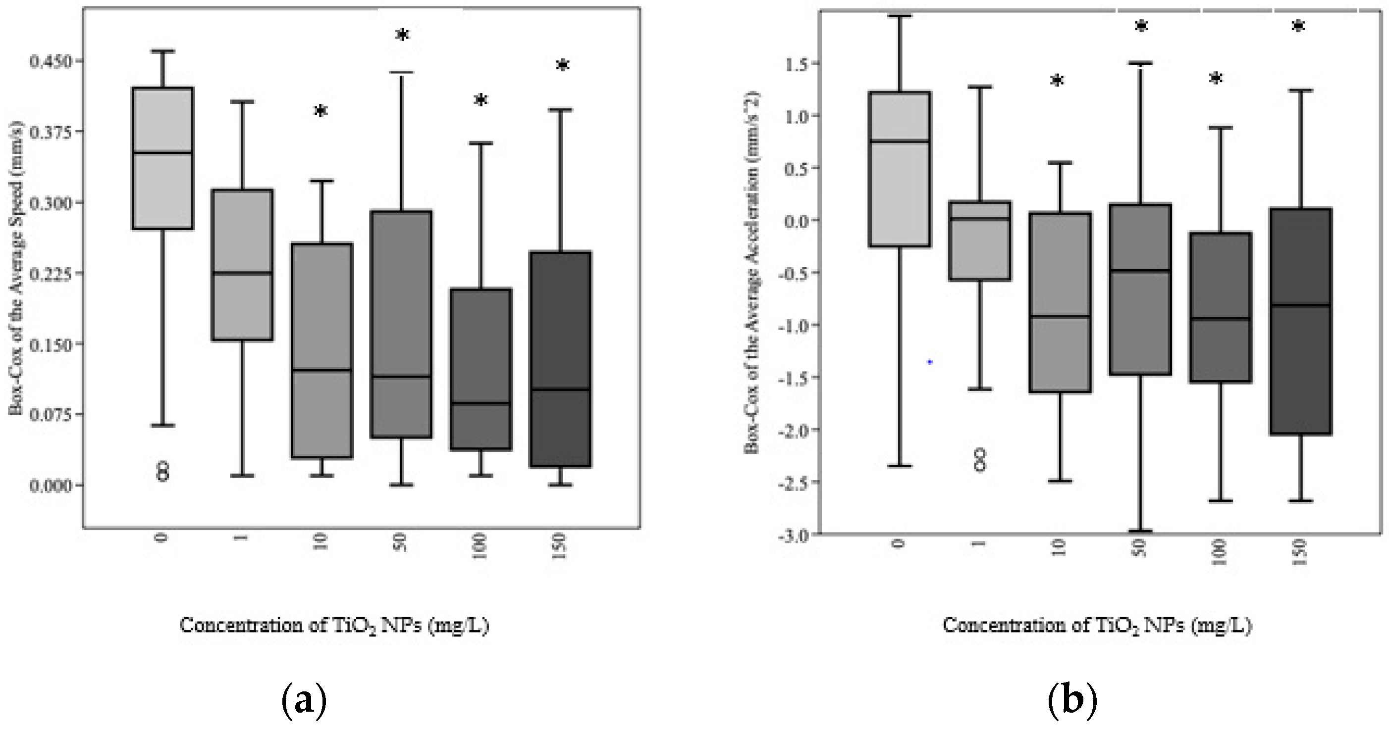
The analysis of movement after 24, 48, and 72 h of exposure to TiO2 NPs showed significant changes leading to hypoactivity behavior for both variables in the exposed groups. During the 24 h exposure, we observed a reduction in the average speed and average acceleration of the larvae in the exposed groups compared with the control group showing hypoactivity of movement (Figure 3).
The average speed (with a Box–Cox transformation; N = 25) observed by the larvae in the control group was 0.32 mm/s ± 0.03 with a minimum of 0.00009 mm/s and a maximum of 0.46 mm/s during the 24 h of exposure. For the different concentrations of TiO2 NPs in the exposed group, we observed average speeds of 0.22 ± 0.02, 0.14 ± 0.02, 0.16 ± 0.03, 0.13 ± 0.02, and 0.13 ± 0.03 mm/s for the concentrations of 1.0, 10.0, 50.0, 100.0, and 150.0 mg/L, respectively. Moreover, for the exposed groups, the speed of the larvae ranged between 0 mm/s and 0.34 mm/s. The one-way ANOVA for the comparison of the average speed variable for the 24 h of exposure of the larvae in the control group and the exposed larvae showed a significant difference (F(5,144) = 9.08; p < 0.01) among groups. Consequently, the Tukey test showed a p < 0.01 among the exposed groups from 10 to 150 mg/L of TiO2 NPs.
The average acceleration of the larvae (with a Box–Cox transformation; N = 25) was 0.38 ± 0.24 mm/s2 with a minimum of −2.35 mm/s2 and a maximum of 1.95 mm/s2 for the control group during the 24 h of exposure. The average acceleration in the exposure of the concentrations of TiO2 NPs were −0.27 ± 0.19, −0.82 ± 0.20, −0.65 ± 0.22, −0.89 ± 0.19, and −0.93 ± 0.23 mm/s2 for the concentrations of 1.0, 10.0, 50.0, 100.0, and 150.0 mg/L, respectively. Moreover, for the exposed groups, we observed a minimum average acceleration of −2.97 mm/s2 and a maximum of 1.50 mm/s2. The one-way ANOVA for the comparison of the average acceleration variable for the 24 h of exposure of the larvae in the control group and the exposed larvae showed a significant difference (F(5,144) = 5.61; p < 0.01) between groups. Consequently, Tukey test analysis showed values of p < 0.01 among exposed groups between 10 and 150 mg/L of TiO2 NPs, indicating similarities between exposed groups except for the control group.
In the exposure of larvae for 48 h to TiO2 NPs, we observed significant results for both locomotion variables. The one-way ANOVA to compare the effects of TiO2 NPs on the speed (F(5,144) = 7.7; p < 0.01) and acceleration (F(5,144) = 7.88; p < 0.01) of the larvae were highly significant. To analyze this exposure time, we used the Tukey test for significant differences among the concentrations of TiO2 NPs of 10.0, 50.0, 100.0, and 150.0 mg/L. No significant differences were found in comparison with the control group. Consequently, at 72 h of exposure, we observed no significant differences between the exposed groups and the control (speed: F(5,144) = 1.13; p = 0.35) (acceleration: F(5,144) = 1.37; p = 0.24).
3.3. Morphological Development
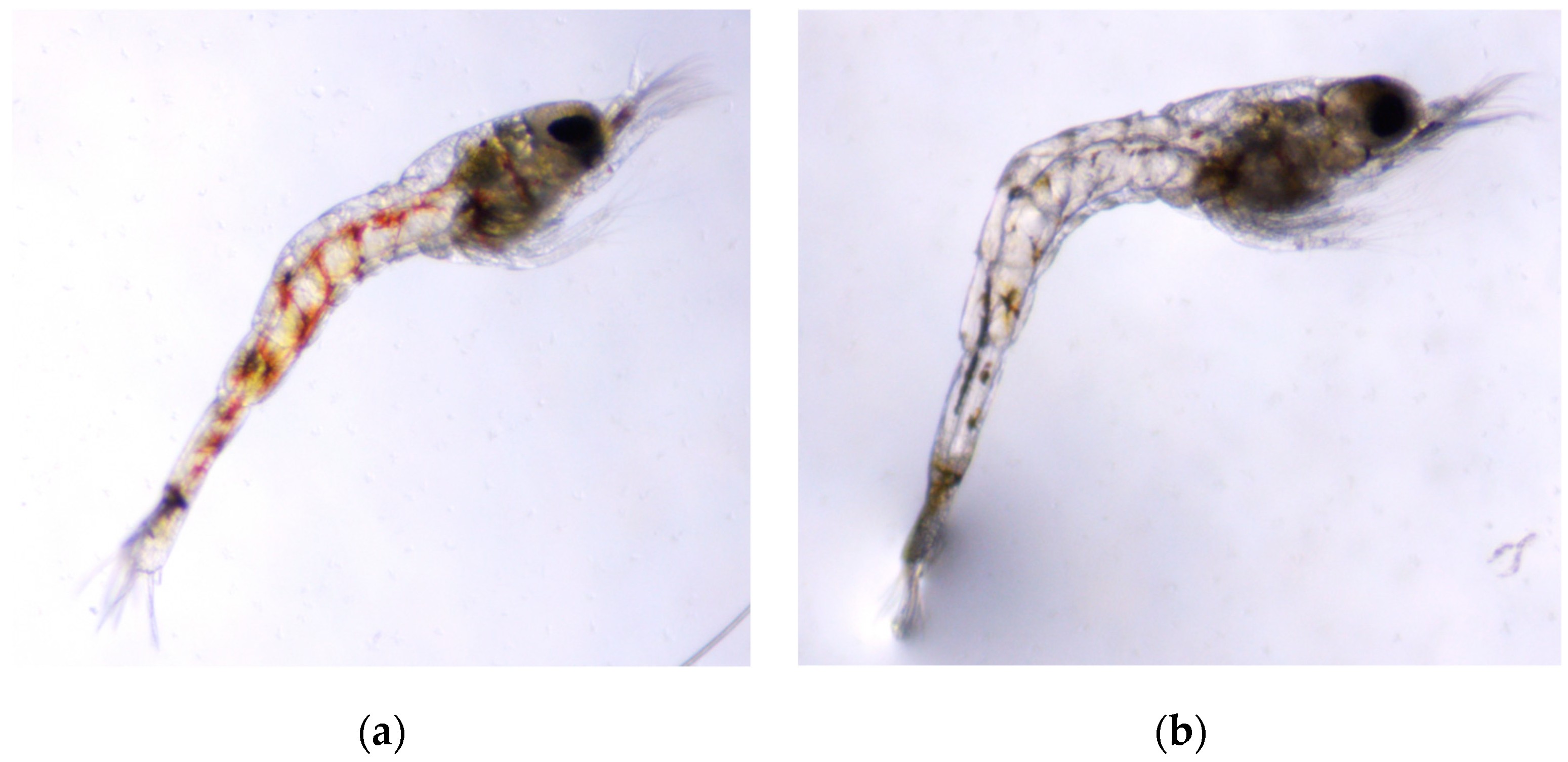
The morphological assessment consisted of total length measures, changes in the presence/absence of pigmentation, and edema in larvae at 24, 48, and 72 h of exposure to TiO2 NPs. For the larval pigmentation, we observed less pigmentation in the abdominal area in some exposed groups and with some exposure times (Figure 4).
In contrast, we found an edema in the abdominal area of the larvae during the 72 h exposure to the TiO2 NPs concentration of 150.0 mg/L (Figure 5). We did not find another case of edema in another period of exposure or concentration of TiO2 NPs.
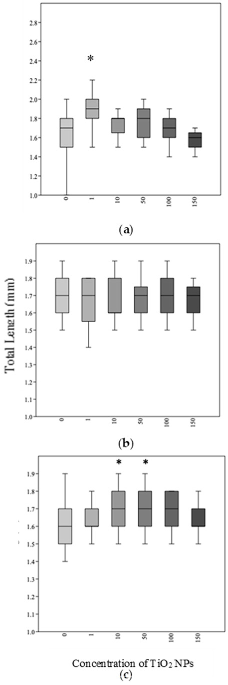
No significant differences in total length among the control and exposed groups were observed (Figure 6). Nevertheless, we found that, at 72 h of exposure, two concentrations (50.0 and 100.0 mg/L) of TiO2 NPs showed statistical significance compared to the control group means. The one-way ANOVA analysis showed total length: F(5,144) = 3.907; p < 0.01. Consequently, the Tukey test showed similarities between the 50.0 and 100.0 mg/L of TiO2 NPs (p < 0.05).
4. Discussion
Nanomaterials are an important emerging aquatic contaminant [24,25,26,27] that create potentially wide-ranging ecological impacts [28]. Studies with TiO2 NPs have found that these nanoparticles interact with other metals and are stored in sediments, acting as an adsorptive agent. Furthermore, it has been shown that TiO2 NPs can exacerbate the bioavailability and toxicity of organic pollutants and pesticides, serving as an adsorptive agent [29]. To date, several studies at a global scale are focused on using the photocatalysis (formation of oxidative agents) of TiO2 NPs for the remediation and treatment of wastewater. Many of these studies do not consider the possible toxicity to aquatic organisms [30,31]. Another action of TiO2 NPs in freshwater ecosystems is in the trophic transfer process. A recent study demonstrates that dietary TiO2 NPs exposure in some species may constitute a significant route for higher trophic level bioaccumulation [32]. It is essential to understand the biological impacts that these emerging pollutants can cause in benthic organisms because they interact mainly with the sediment and have complex life cycles (diadromy/amphidromy).
The study of the development of the A. lanipes larvae is not yet completely understood, but, as in other well-studied species, the early developmental stages are most often sensitive to environmental contaminants. For example, in the developing nervous system, the toxicant stress can have a detrimental impact in the early stage with persistent effects into adulthood [33]. Our study demonstrates that the TiO2 NPs can harm the nervous system in the larval stages of A. lanipes. During the first 24 h of exposure, we observed a reduction in the locomotion of the larvae evidencing hypoactivity movement similar to previous studies with zebrafish larvae and neotropical tadpoles where the individuals reduced their velocity and mobility behaviors after exposures to TiO2 NPs [33,34]. After 48 and 72 h of exposure, we observed variability in the hypoactive response of the shrimp larvae in the movement analyses. This result leads us to propose two possible explanations. The first is related to a possible change in the behavior of the TiO2 NPs. Nanomaterials change their physicochemical properties depending on the environment in which they are found (salinity, temperature, light, biological interaction, etc.). Therefore, they can also create environmental changes over time, resulting in a variable toxic effect for the zoea larvae of A. lanipes. Another possible response to this variability is that A. lanipes larvae can develop from one larval stage to another in a few days (in 2–3 days they can go from zoea I to zoea II stage). In a period of 48 and 72 h of exposure, we observed larvae in the zoea I stage and others in the zoea II stage, which was more common at 72 h of exposure. Thus, the larvae of A. lanipes could have differences in susceptibility to the toxicity of TiO2 NPs in different larval stages. In general, these changes in A. lanipes zoea larvae may represent a possible long-term impact on the development of adult shrimp and present undesirable ecological consequences. From upstream migrations after metamorphosis in the estuarine environment to adult activities, such as reproduction, food acquisition, and evasion of predation, can be affected by TiO2 NPs.
A limitation in the movement analysis was that the larvae were recorded in a different container than the cells in which they were exposed. Moreover, having recorded the larvae in a round container, the total distance variable could not be added because, in the data analysis, the program did not differentiate the distance travelled by the larvae when they were in the corner of the container due to the reflection of the water. Consequently, only the variables of velocity and acceleration were considered. The sample used for the movement analysis was only 25 larvae because they were recorded individually in a single microscope. Finally, the TiO2 NPs content on the cells in the culture dishes at the exposure time of each bioassay was not analyzed to determine the movement or final locations of the concentrations of the TiO2 NPs suspensions (agglomeration, dissolution, flocculation, etc.).
The results of this study demonstrated that an acute exposure to TiO2 NPs in the freshwater shrimp A. lanipes larvae represents a lethal effect. Mortality was evident after 48 and 72 h of exposure. The data showed different susceptibility to nanoparticles in different larval stages because, in A. lanipes larvae, their development is rapid. This may explain the variability in mortality between 48 and 72 h of exposure, especially when we observed a decrease in mortality in both exposure times in the 150 mg/L suspension concentration.
Regarding the sublethal effects of TiO2 NPs, we observed the loss of pigmentation in exposed larvae. Like other atyids shrimp, the pigment that develops in the larvae of A. lanipes is known as chromatophores [35]. These pigments are important for the development of the characteristic dark color of adult atyids that begins from the pre-larval stages [36]. These cells provide many important functions in atyids, such as controlling dissolved oxygen during periods of oxygen supersaturation and they protect the larva and the adult shrimp from the harmful effects of low oxygen concentrations [36]. However, when there are changes in the chromatophores, this is due to indicators of many factors, among which are homeostatic instability in response to low oxygen concentrations, pathological manifestations, and expressions that can indicate degrees of stress at a biological level [36]. For the pigment to disperse and express itself, the hormonal action of the red pigment-concentrating hormone (RPCH) is required, which induces pigment aggregation in shrimp chromatophores due to an internal or external increase in Ca2+ at the cellular level [36]. In oxidative stress, there is substantive evidence that the calcium homeostasis is altered, and some cells die (apoptosis) [37]. This explains why we found that larvae exposed to TiO2 NPs (reactive oxygen species generators) showed a change in pigmentation. There is a possibility of oxidative stress inducing changes in cellular Ca2+ flux homeostasis and an impact on pigment dispersion/aggregation.
The presence of edemas in the abdominal region in exposed larvae is common in studies related to the toxicity of TiO2 NPs, as is the case with zebrafish larvae [38,39]. However, in our study, for the three exposure periods 24, 48, and 72 h, we only observed one larva with an edema in the abdominal area close to the beginning of the telson. These data suggest that, for A. lanipes, unlike zebrafish, the development of edemas is not as relevant. We do not rule out the need for more repetition and validation of this analysis.
The data presented in this study are novel in helping to understand the toxicity levels of TiO2 NPs in an ecologically susceptible species that play key roles in the biofiltration of natural organic particles [40,41,42,43]. The most obvious sublethal effect was in the locomotion analysis. It was shown that acute exposure to TiO2 NPs can alter the normal locomotion of the larva, indicating neurobehavioral effects that could persist in other life stages of the shrimp. We demonstrated that this emerging contaminant could cause a significant biological impact by reducing locomotion, producing morphological changes, and, later, the death of the organism. A reduction in the number of larvae that metamorphose into juveniles and adults could lead to a reduction in the number of individuals in the population and to changes in their ecological role in the ecosystem.
This study provides new knowledge about the effect of TiO2 NPs in the larvae of a Caribbean freshwater shrimp that helps to keep flowing waters clear of suspended particles. Sustaining the biodiversity and ecosystem management is critical for maintaining ecological integrity. Moreover, our results contribute to the need in the scientific literature to use the most effective organisms that are ecologically susceptible to the presence of TiO2 NPs in the aquatic environments. These nanoecotoxicological studies need to include native benthic as well as pelagic organisms [44]. The A. lanipes is a shrimp that can be susceptible to the presence of engineered nanoparticles in the estuary during its larval stages.
Author Contributions
Conceptualization, S.C.-R. and O.P.-R.; methodology, S.C.-R.; software, S.C.-R.; validation, O.P.-R.; formal analysis S.C.-R. and O.P.-R.; investigation, S.C.-R. and O.P.-R.; resources, O.P.-R.; data curation, S.C.-R.; writing—original draft preparation, S.C.-R.; writing—review and editing, O.P.-R.; visualization, S.C.-R. and O.P.-R.; supervision, O.P.-R.; project administration, S.C.-R. and O.P.-R.; funding acquisition, S.C.-R. and O.P.-R. All authors have read and agreed to the published version of the manuscript.
Funding
This study was supported by the Puerto Rico Center for Environmental Neuroscience (PRCEN), grant # HRD-11736019 (PRCEN2) and the Shrimp and Fish Ecology Laboratory at the University of Puerto Rico—Río Piedras Campus. The authors declare no competing financial interests.
Institutional Review Board Statement
Not appliable.
Informed Consent Statement
Not appliable.
Data Availability Statement
Not appliable.
Acknowledgments
Fernando A. Villar-Fornés and Marla Santos for their appreciated help in the search for materials for the research methodology. We thank Alan Covich for his comments.
Conflicts of Interest
The authors declare no conflict of interest.
References
- Vance, M.E.; Kuiken, T.; Vejerano, E.P.; McGinnis, S.P.; Hochella, M.F., Jr.; Rejeski, D.; Hull, M.S. Nanotechnology in the real world: Redeveloping the nanomaterial consumer products inventory. Beilstein J. Nanotechnol. 2015, 6, 1769–1780. [Google Scholar] [CrossRef] [Green Version]
- Colvin, V.L. The potential environmental impact of engineered nanomaterials. Nat. Biotechnol. 2003, 21, 1166–1170. [Google Scholar] [CrossRef]
- Turan, N.B.; Erkan, H.S.; Engin, G.O.; Bilgili, M.S. Nanoparticles in the aquatic environment: Usage, properties, transformation and toxicity—A review. Process Saf. Environ. Prot. 2019, 130, 238–249. [Google Scholar] [CrossRef]
- Maurer-Jones, M.A.; Gunsolus, I.L.; Murphy, C.J.; Haynes, C.L. Toxicity of engineered nanoparticles in the environment. Anal. Chem. 2013, 85, 3036–3049. [Google Scholar] [CrossRef] [PubMed] [Green Version]
- Khan, I.; Saeed, K.; Khan, I. Nanoparticles: Properties, applications and toxicities. Arab. J. Chem. 2019, 12, 908–931. [Google Scholar] [CrossRef]
- Berardinelli, A.; Parisi, F. TiO2 in the food industry and cosmetics. In Titanium Dioxide (TiO₂) and Its Applications; Elsevier: Amsterdam, The Netherlands, 2021; pp. 353–371. [Google Scholar]
- Verma, S.K. Engineered Nanomaterials and Phytonanotechnology: Challenges for Plant Sustainability; Elsevier: Amsterdam, The Netherlands, 2019; Volume 87. [Google Scholar]
- Dedman, C.J.; King, A.M.; Christie-Oleza, J.A.; Davies, G.L. Environmentally relevant concentrations of titanium dioxide nanoparticles pose negligible risk to marine microbes. Environ. Sci. Nano 2021, 8, 1236–1255. [Google Scholar] [CrossRef]
- Musial, J.; Krakowiak, R.; Mlynarczyk, D.T.; Goslinski, T.; Stanisz, B.J. Titanium dioxide nanoparticles in food and personal care products—What do we know about their safety? Nanomaterials 2020, 10, 1110. [Google Scholar] [CrossRef]
- Gottschalk, F.; Sonderer, T.; Scholz, R.W.; Nowack, B. Possibilities and limitations of modeling environmental exposure to engineered nanomaterials by probabilistic material flow analysis. Environ. Toxicol. Chem. 2010, 29, 1036–1048. [Google Scholar] [CrossRef]
- Batley, G.; McLaughlin, M.J. Fate of Manufactured Nanomaterials in the Australian Environment; CSIRO Land and Water: Clayton, Australia, 2010. [Google Scholar]
- Gottschalk, F.; Sun, T.; Nowack, B. Environmental concentrations of engineered nanomaterials: Review of modeling and analytical studies. Environ. Pollut. 2013, 181, 287–300. [Google Scholar] [CrossRef]
- Titanium-Environmental Health Criteria 24. Available online: https://apps.who.int/iris/bitstream/handle/10665/37269/9241540842-eng.pdf (accessed on 13 August 2022).
- Song, B.; Liu, X.; Wei, L.; Shao, L. A review on potential neurotoxicity of titanium dioxide nanoparticles. Nanoscale Res. Lett. 2015, 10, 342. [Google Scholar] [CrossRef] [Green Version]
- Lin, D.; Ji, J.; Long, Z.; Yang, K.; Wu, F. The influence of dissolved and surface-bound humic acid on the toxicity of TiO2 nanoparticles to Chlorella sp. Water Res. 2012, 46, 4477–4487. [Google Scholar] [CrossRef]
- Chen, T.H.; Lin, C.Y.; Tseng, M.C. Behavioral effects of titanium dioxide nanoparticles on larval zebrafish (Danio rerio). Mar. Pollut. 2011, 63, 303–308. [Google Scholar] [CrossRef]
- Pérez-Reyes, O.; Crowl, T.A.; Covich, A.P. Comparison of decapod communities across an urban-forest land use gradient in Puerto Rican streams. Urban Ecosyst. 2016, 19, 181–203. [Google Scholar] [CrossRef]
- Benstead, J.P.; March, J.G.; Pringle, C.M. Estuarine larval development and upstream post-larval migration of freshwater shrimps in two tropical rivers of Puerto Rico. Biotropica 2000, 32, 545–548. [Google Scholar] [CrossRef]
- Bauer, R.T. Amphidromy in shrimps: A life cycle between rivers and the sea. Lat. Am. J. Aquat. Res. 2013, 41, 633–650. [Google Scholar] [CrossRef]
- Perez-Reyes, O.; Crowl, T.A.; Hernandez-Garcia, P.J.; Ledesma-Fuste, R.; Villar-Fornes, F.A.; Covich, A.P. Freshwater decapods of Puerto Rico: A checklist and reports of new localities. Zootaxa 2013, 3717, 329–344. [Google Scholar] [CrossRef] [Green Version]
- Solis, P.N.; Wright, C.W.; Anderson, M.M.; Gupta, M.P.; Phillipson, J.D. A microwell cytotoxicity assay using Artemia salina (brine shrimp). Planta Med. 1993, 59, 250–252. [Google Scholar] [CrossRef]
- Ingebretson, J.J.; Masino, M.A. Quantification of locomotor activity in larval zebrafish: Considerations for the design of high-throughput behavioral studies. Front. Neural Circuits 2013, 7, 109. [Google Scholar] [CrossRef] [Green Version]
- Anastasiadou, C.; Ntakis, A.; Leonardos, I.D. Larval development of the freshwater shrimp Atyaephyra desmarestii (Millet, 1831) sensu lato (Decapoda: Caridea: Atyidae) and morphological maturation from juveniles to adults. Zootaxa 2011, 2877, 41–54. [Google Scholar] [CrossRef]
- Farre, M.; Gajda-Schrantz, K.; Kantiani, L.; Barcelo, D. Ecotoxicity and analysis of nanomaterials in the aquatic environment. Anal. Bioanal. Chem. 2009, 393, 81–95. [Google Scholar] [CrossRef]
- Klaine, S.; Alvarez, P.; Batley, G.; Fernandes, T.; Handy, R.; Lyon, D.; Mahendra, S.; McLaughlin, M.J.; Lead, J.R. Nanomaterials in the environment: Behavior, fate, bioavailability, and effects. Environ. Toxicol. Chem. Int. J. 2008, 27, 1825–1851. [Google Scholar] [CrossRef]
- Navarro, E.; Baun, A.; Behra, R.; Hartmann, N.B.; Filser, J.; Miao, A.J.; Quigg, A.; Santschi, P.H.; Sigg, L. Environmental behavior and ecotoxicity of engineered nanoparticles to algae, plants, and fungi. Ecotoxicology 2008, 17, 372–386. [Google Scholar] [CrossRef] [Green Version]
- Nel, A.; Xia, T.; Madler, L.; Li, N. Toxic potential of materials at the nanolevel. Science 2006, 311, 622–627. [Google Scholar] [CrossRef] [Green Version]
- Miller, R.J.; Bennett, S.; Keller, A.A.; Pease, S.; Lenihan, H.S. TiO2 Nano-particles Are Phototoxic to Marine Phytoplankton. PLoS ONE 2012, 7, e30321. [Google Scholar]
- Fang, Q.; Shi, X.; Zhang, L.; Wang, Q.; Wang, X.; Guo, Y.; Zhou, B. Effect of titanium dioxide nanoparticles on the bioavailability, metabolism, and toxicity of pentachlorophenol in zebrafish larvae. J. Hazard. Mater. 2015, 283, 897–904. [Google Scholar] [CrossRef]
- Lazar, M.A.; Varghese, S.; Nair, S.S. Photocatalytic water treatment by titanium dioxide: Recent updates. Catalysts 2012, 2, 572–601. [Google Scholar] [CrossRef] [Green Version]
- Li, Z.; Deng, S.; Yu, G.; Huang, J.; Lim, V.C. As (V) and as (III) removal from water by a Ce–Ti oxide adsorbent: Behavior and mechanism. Chem. Eng. 2010, 161, 106–113. [Google Scholar] [CrossRef]
- Zhu, X.; Wang, J.; Zhang, X.; Chang, Y.; Chen, Y. Trophic transfer of TiO2 nano-particles from Daphnia to zebrafish in a simplified freshwater. Chemosphere 2010, 79, 928–933. [Google Scholar] [CrossRef]
- Fan, C.Y.; Cowden, J.; Simmons, S.O.; Padilla, S.; Ramabhadran, R. Gene expression changes in developing zebrafish as potential markers for rapid developmental neurotoxicity screening. Neurotoxicol. Teratol. 2010, 32, 91–98. [Google Scholar] [CrossRef]
- do Amaral, D.F.; Guerra, V.; Almeida, K.L.; Signorelli, L.; Rocha, T.L.; de Melo e Silva, D. Titanium dioxide nanoparticles as a risk factor for the health of Neotropical tadpoles: A case study of Dendropsophus minutus (Anura: Hylidae). Environ. Sci. Pollut. Res. 2022, 29, 50515–50529. [Google Scholar] [CrossRef]
- Hunte, W. The Complete Larval Development of the Freshwater Shrimp Atya innocous (Herbst) Reared in the Laboratory (Decapoda, Atyidae). Crustaceana, Supplement, no 5, 1979, 231–242. Available online: http://www.jstor.org/stable/25027505 (accessed on 13 August 2022).
- Ribeiro, M.; Campbell, J. Calcium movements during pigment aggregation in freshwater shrimp chromatophores. Pigment. Cell Res. 2007, 20, 70–77. [Google Scholar] [CrossRef] [PubMed]
- Mattson, M. Oxidative stress, perturbed calcium homeostasis, and immune dysfunction in Alzheimer’s disease. J. Neurovirol. 2002, 8, 539–550. [Google Scholar] [CrossRef] [PubMed] [Green Version]
- Ma, H.; Diamond, S. Phototoxicity of TiO2 nanoparticles to zebrafish (Danio rerio) is dependent on life stage. Toxicol. Chem. 2013, 32, 2139–2143. [Google Scholar] [CrossRef] [PubMed]
- Liu, J.; Fan, D.; Wang, L.; Shi, L.I.L.I.; Ding, J.; Chen, Y.; Shen, S. Effects of ZnO, CuO, Au, and TiO2 nanoparticles on Daphnia magna and early life stages of zebrafish Danio rerio. Environ. Prot. Eng. 2014, 40, 139–149. [Google Scholar]
- Covich, A.P. Atyid shrimp in the headwaters of the Luquillo Mountains, Puerto Rico: Filter feeding in natural and artificial streams. Verh. Int. Ver. Für Angew. Limnol. 1988, 23, 2108–2113. [Google Scholar] [CrossRef]
- Covich, A.P.; Palmer, M.A.; Crowl, T.A. The role of benthic invertebrate species in freshwater ecosystems—Zoobenthic species influence energy flows and nutrient cycling. BioScience 1999, 49, 119–127. [Google Scholar] [CrossRef] [Green Version]
- Covich, A.P.; Ewel, K.C.; Hall, R.O.; Giller, P.E.; Goedkoop, W.; Merritt, D.M. Ecosystem services provided by freshwater benthos. In Sustaining Biodiversity and Ecosystem Services in Soils and Sediments; Chapter 3; Wall, D.H., Ed.; Island Press: Washington, DC, USA, 2004; pp. 45–72. [Google Scholar]
- Crowl, T.A.; McDowell, W.H.; Covich, A.P.; Johnson, S. Species-specific responses in leaf litter processing in a tropical headwater stream (Puerto Rico). Ecology 2001, 82, 775–783. [Google Scholar] [CrossRef]
- Garric, J.; Thybaud, E. Toxicological Models Part B: Environmental Models. In Nanoethics and Nanotoxicology; Springer: Berlin/Heidelberg, Germany, 2011; pp. 379–396. [Google Scholar]
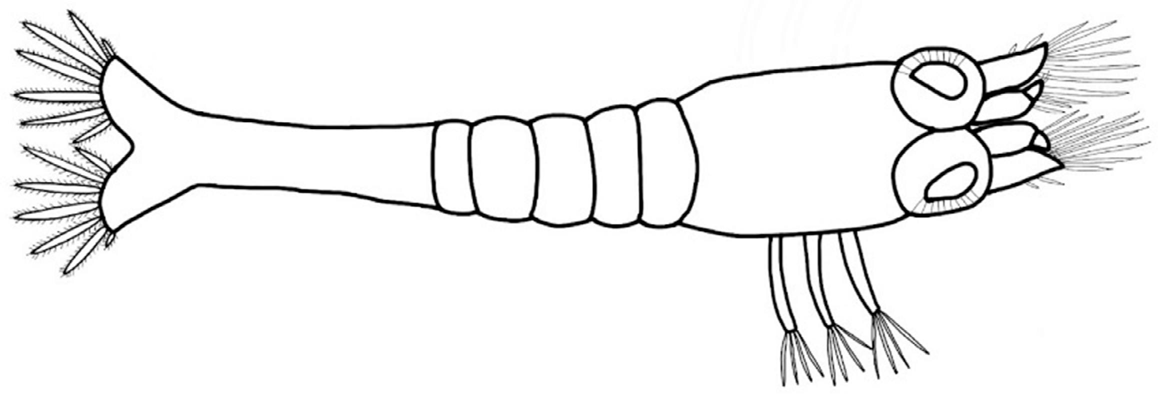
Figure 1.
Atya lanipes zoea larvae stage; the organism used in the study.

Figure 2.
The percentage of the mortality rate of A. lanipes larvae exposed to different concentrations of TiO2 NPs after 24, 48, and 72 h of exposure (N = 40 for each NP concentration). (Note: after 24 h of exposure, we observed no mortality in any exposed group or control).
Figure 2.
The percentage of the mortality rate of A. lanipes larvae exposed to different concentrations of TiO2 NPs after 24, 48, and 72 h of exposure (N = 40 for each NP concentration). (Note: after 24 h of exposure, we observed no mortality in any exposed group or control).

Figure 3.
Larvae movement behavior assessment (N = 25 for each concentration). (a) Average speed (mm/s) and (b) average acceleration (mm/s2) of A. lanipes larvae exposed to different concentrations of TiO2 NPs in an acute exposure of 24 h (* p < 0.01).
Figure 3.
Larvae movement behavior assessment (N = 25 for each concentration). (a) Average speed (mm/s) and (b) average acceleration (mm/s2) of A. lanipes larvae exposed to different concentrations of TiO2 NPs in an acute exposure of 24 h (* p < 0.01).

Figure 4.
A. lanipes larva exposed to a suspension of TiO2 NPs for 24 h showing changes in body pigmentation: (a) control A. lanipes larva, normal pigmentation along the abdomen; (b) A. lanipes larva exposed to 50 mg/L of TiO2 NPs, loss of pigmentation as a result of exposure to TiO2 NPs. The images were obtained using a stereo microscope (3.5 × -90 × LED Trinocular Zoom + 14 MP USB 3.0 Digital Camera). Both larvae were in the zoea I larval stage; the total length of the larvae is 1.3 mm.

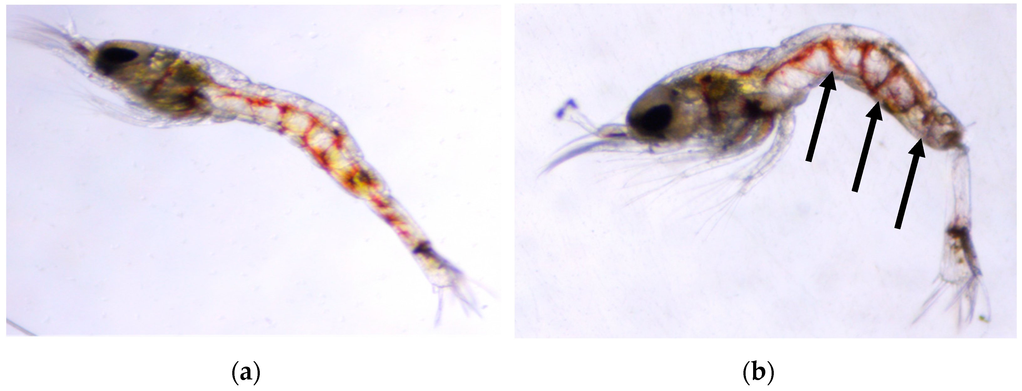
Figure 5.
A. lanipes larvae exposed to TiO2 NPs for 72 h showed edema development: (a) control A. lanipes larva; (b) A. lanipes larva exposed to 150 mg/L of TiO2 NPs. The images were obtained using a stereo microscope (3.5×-90× LED Trinocular Zoom + 14 MP USB 3.0 Digital Camera). Both larvae were in the zoea I larval stage; the total length of the larvae is 1.3 mm.

Figure 6.
Total length (mm) of A. lanipes larvae exposed to different TiO2 NPs concentrations for (a) 24 h (* p < 0.01), (b) 48 h (* p < 0.01), and (c) 72 h. (* p < 0.05).
Figure 6.
Total length (mm) of A. lanipes larvae exposed to different TiO2 NPs concentrations for (a) 24 h (* p < 0.01), (b) 48 h (* p < 0.01), and (c) 72 h. (* p < 0.05).

Disclaimer/Publisher’s Note: The statements, opinions and data contained in all publications are solely those of the individual author(s) and contributor(s) and not of MDPI and/or the editor(s). MDPI and/or the editor(s) disclaim responsibility for any injury to people or property resulting from any ideas, methods, instructions or products referred to in the content. |
© 2023 by the authors. Licensee MDPI, Basel, Switzerland. This article is an open access article distributed under the terms and conditions of the Creative Commons Attribution (CC BY) license (https://creativecommons.org/licenses/by/4.0/).
Share and Cite
MDPI and ACS Style
Cruz-Rosa, S.; Pérez-Reyes, O. Titanium Oxide Nanoparticles as Emerging Aquatic Pollutants: An Evaluation of the Nanotoxicity in the Freshwater Shrimp Larvae Atya lanipes. Ecologies 2023, 4, 141-151. https://doi.org/10.3390/ecologies4010011
AMA Style
Cruz-Rosa S, Pérez-Reyes O. Titanium Oxide Nanoparticles as Emerging Aquatic Pollutants: An Evaluation of the Nanotoxicity in the Freshwater Shrimp Larvae Atya lanipes. Ecologies. 2023; 4(1):141-151. https://doi.org/10.3390/ecologies4010011
Chicago/Turabian StyleCruz-Rosa, Stefani, and Omar Pérez-Reyes. 2023. "Titanium Oxide Nanoparticles as Emerging Aquatic Pollutants: An Evaluation of the Nanotoxicity in the Freshwater Shrimp Larvae Atya lanipes" Ecologies 4, no. 1: 141-151. https://doi.org/10.3390/ecologies4010011