Review

Developmental Pattern Formation: Spanish Contributions from a Biophysical Perspective

Institute for Integrative Systems Biology (I2SysBio), CSIC-UV, 46980 Valencia, Spain
*
Author to whom correspondence should be addressed.
Biophysica 2023, 3(2), 335-347; https://doi.org/10.3390/biophysica3020022
Submission received: 17 March 2023 / Revised: 25 April 2023 / Accepted: 26 April 2023 / Published: 6 May 2023
(This article belongs to the Special Issue State-of-the-Art Biophysics in Spain 2.0)

Abstract

:
During the last few decades, developmental pattern formation has evolved from being a descriptive discipline to a quantitative one. That process has been possible due to the implementation of multidisciplinary approaches where biophysicists and mathematicians have played a key role. In this review, we highlight relevant Spanish contributions and stress their biophysical approaches, as well as provide some historical context. Finally, this work also aimed at bridging the concepts from biology to physics/math (and back) and at shedding light on some directions for future research.

1. Introduction: Twenty Years of a Polemic Statement

Twenty years ago, the first chapter of the seventh edition of the developmental biology “bible” [1] teased readers with the following provocative comment: “Developmental biology has been described as the last refuge of the mathematically incompetent scientist.” Fortunately, S. Gilbert did not attribute authorship to this quote and added that “This phenomenon, however, is not going to last. While most embryologists have been content trying to analyze specific instances of development or even formulating some general principles of embryology, some researchers are now seeking the laws of development. The goal of these investigators is to base embryology on formal mathematical or physical principles […]. Pattern formation and growth are two areas in which such mathematical modeling has given biologists interesting insights into some underlying laws of animal development”. More recent versions of Gilbert’s book do not include the polemic statement (we do not know if he received complaints about it); however, what we do know is that his assessment about the future (now present) of the field was accurate. During these last twenty years, developmental biology has undergone a dramatic change. It has become a quantitative discipline where mathematical and physical models are routinely used/proposed to elucidate the laws that underlie development [2]. Such a change has been in part possible due to new generations of physicists and mathematicians that have shown interest in the field. In this short review, we address some relevant contributions made by scientists from Spain using a biophysical perspective.
In order to understand how part of the Spanish biophysics community became involved in the field of developmental pattern formation, a brief historical introduction is in order. In historical terms, the relation between patterning, developmental biology, and physical/mathematical models can be rooted in the seminal work of Alan Turing: The chemical basis of morphogenesis [3]. Turing published only a single work on that topic, which nevertheless has become deeply influential. Therein, he showed that interacting biological chemical species driven by a transport mechanism that is homogenizing in nature (diffusion) can create non-homogeneous ordered structures, i.e., patterns. Further publications by Wolpert [4] and Meinhart [5] and the publication of books such as Mathematical Biology [6] or the Biological Physics of the Developing Embryo [7], helped to popularize and crystallize in a decade (1995–2005) the field of biological pattern formation from a physical perspective.
In parallel, around that time, it became clear that there was a critical mass of researchers around the field of statistical physics in different Spanish institutions. The first national conference on the topic, FisEs, was held in Barcelona (FisEs has been organized since then every year and a half: 33 meetings so far), and the Group of Non-linear and Statistical Physics (GEFENOL), a specialized group within the Spanish Royal Society of Physics, was created in 2001. Taking a look at the logs and abstracts of the FisEs and GENEFOL meetings/workshops reveals that “pattern formation” was a major streamline of research. That included research about the role played by noise (i.e., fluctuations) and non-equilibrium effects during pattern formation [8,9]. All this seed activity led to the establishment of a number of groups in Spain addressing the pattern formation problem from a biophysical viewpoint during the past twenty years (thus confirming Gilbert’s forecast; see [10,11]). Still, as we discuss below, the concept of pattern may differ significantly depending on whether one approaches it from a physics of biology standpoint. We believe that this transition made by statistical physicists to the developmental biology problem has been a natural process. After all, statistical mechanics aims at understanding how macroscopic complexity, self-organization, and order arise from the interactions of individual “units” in environments that can be out of equilibrium and subjected to fluctuations, an idea that clearly fits with the objective of developmental biology. Finally, it is worth noticing that the Spanish Biophysics Society, traditionally focused on the biophysics of molecules and single cells, has been making an effort during the last few years to expand its interests and incorporate researchers working on tissue-level problems (including patterning). We believe that all these endeavors will help to strengthen even more the research carried out on developmental pattern formation in Spain from a biophysical perspective.

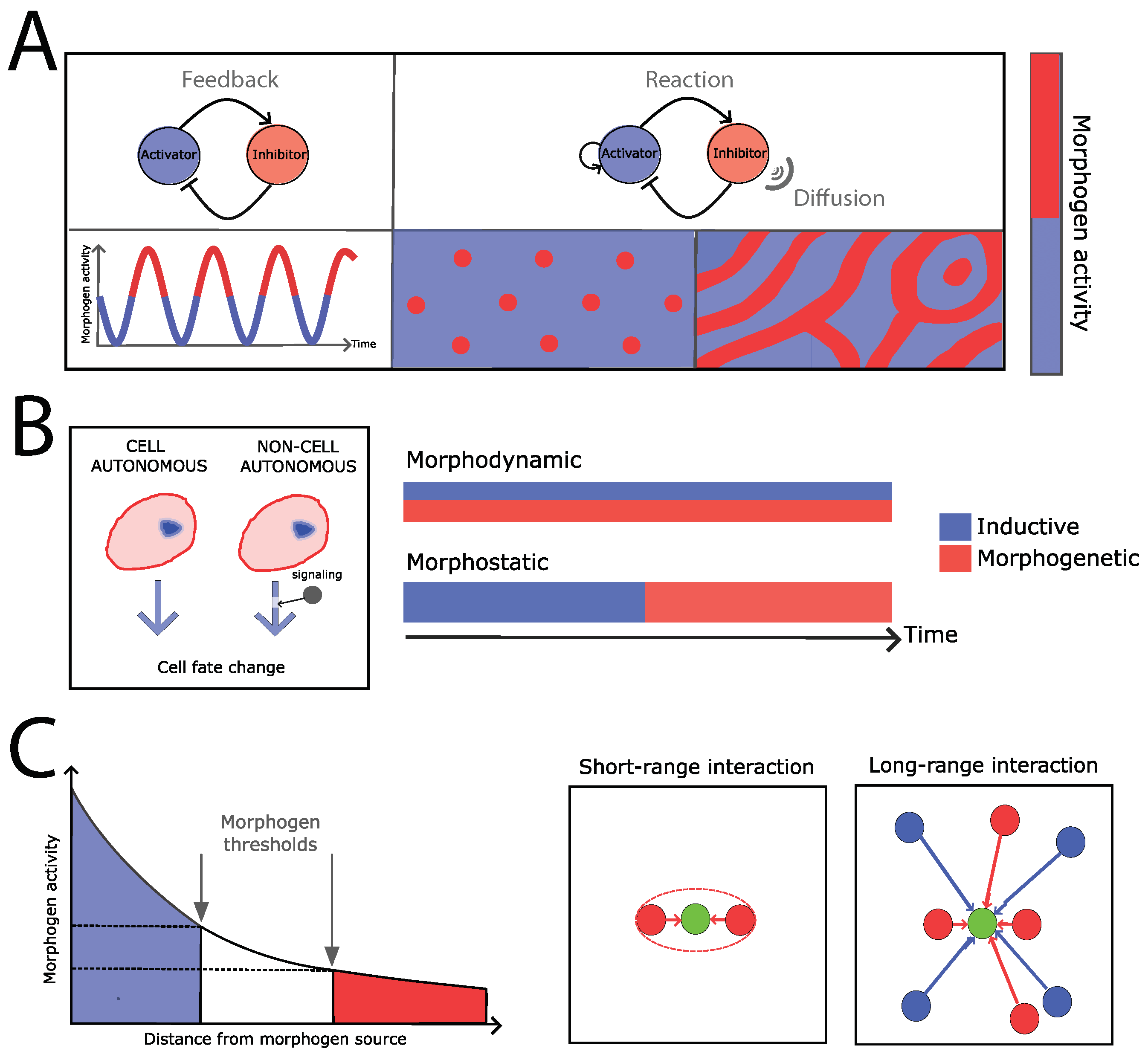
2. Developmental Pattern Formation: From Physics to Biology, from Models to Mechanisms

When facing multidisciplinary problems, one significant barrier for success is the ability to develop a common language. The field of developmental pattern formation is not an exception, and terms such as “model”, or “mechanism”, or even pattern may imply different things if one asks a biologist or a physicist. We introduce below different concepts and definitions shared in the field of developmental pattern formation for the sake of framing the problem correctly.
Patterning: physics and biology classifications. First of all, the meaning of spatiotemporal pattern in physics/math usually differs from that used in biology. In the former case, the idea of a spatiotemporal pattern suggests the existence of non-trivial symmetries in space and/or time. Those symmetries imply the appearance of a repeated behavior (order) that can be characterized, for example, by using a Fourier representation that highlights spatial and/or temporal modes/frequencies that are overrepresented. Thus, pattern classification in physics is linked to the instability mechanism that induces that some spatial and/or temporal modes/frequencies become unstable and generate stationary or oscillatory ordered structures in space and/or time [12]; see Figure 1A. Alternatively, from the viewpoint of their appearance, patterns can be loosely classified as oscillatory, spot-like, or stripe-/ring-like. We notice that this classification is valid from a 2D perspective, but can pose problems in a 3D context. On the other hand, in biology, the idea of a pattern is broader and, importantly, functional. In particular, in the context of a multicellular environment, patterning implies that groups of cells behave “differently” in terms of their “behavior” and/or the genes being expressed in space and/or time. Thus, roughly speaking, biological patterning refers to the complex spatiotemporal distribution of cells and their states, and it does not imply a “repeated behavior” necessarily. From that perspective, Salazar-Ciudad et al. classified patterning mechanisms into three categories [13,14]: Cell autonomous mechanisms imply that cells form patterns without interacting either mechanically or through signaling. Inductive mechanisms rely on cell–cell communication by means of diffusive molecules (e.g., morphogens), ligand–receptor interactions, or chemical coupling through gap junctions such that a pattern changes/arises due to changes in the cells’ state (Figure 1B). Finally, morphogenetic mechanisms use cellular behaviors other than signaling and alter the pattern by affecting form without affecting the cellular states. Moreover, it has been noticed that, during development, inductive and morphogenetic mechanisms often act together, either in a sequential way (morphostatic patterning) or simultaneously (morphodynamic patterning); see Figure 1B.
Patterning: models and mechanisms. The above classifications are focused on either the biological causation or the geometric (in a broad sense, temporal or spatial) features of patterns, but they lack providing information about the underlying mechanism from a mathematical/physical modeling standpoint. Going into the technicalities is out of the scope of this review, and we refer the reader to the work by Cross and Hohenberg and other references for details [5,6,7,9,12,15,16]. However, from a phenomenological standpoint, the mechanisms (sometimes used in biology with the same meaning as models) are usually simplified as follows. In threshold models, the cells modify their state (e.g., gene expression profile, cellular behavior) depending on the levels of an external signal. This mechanism is generically known as the “French flag” model and was proposed by Wolpert [17]: it consists on a source–sink mechanism, coupled with diffusion and degradation, which leads to a spatial gradient of morphogens that cells can “read” to obtain positional information (Figure 1C). In Turing models, several interacting diffusive species (activators and inhibitors) generate a pattern (either stationary or oscillatory) with some periodicity [18]. Turing patterns belong to a more general class, the so-called reaction–diffusion models, where the unstable mode (or modes) is (are) not necessarily generated by a Turing instability [6,19,20] (Figure 1A). Short-range interaction models that couple the local dynamics of cells (e.g., receptor–ligand models) can also induce local symmetry-breaking events and generate patterns such as those of due to Notch–Delta (receptor–ligand) interactions [21] (Figure 1C). Finally, a particular phenomenon relevant to cell and developmental biology refers to the propagation in a multi-cellular environment of a local behavior (oscillations, an excitatory dynamics, or local symmetry-breaking event) due to diffusive signals or short-range coupling, which generates traveling waves [22].

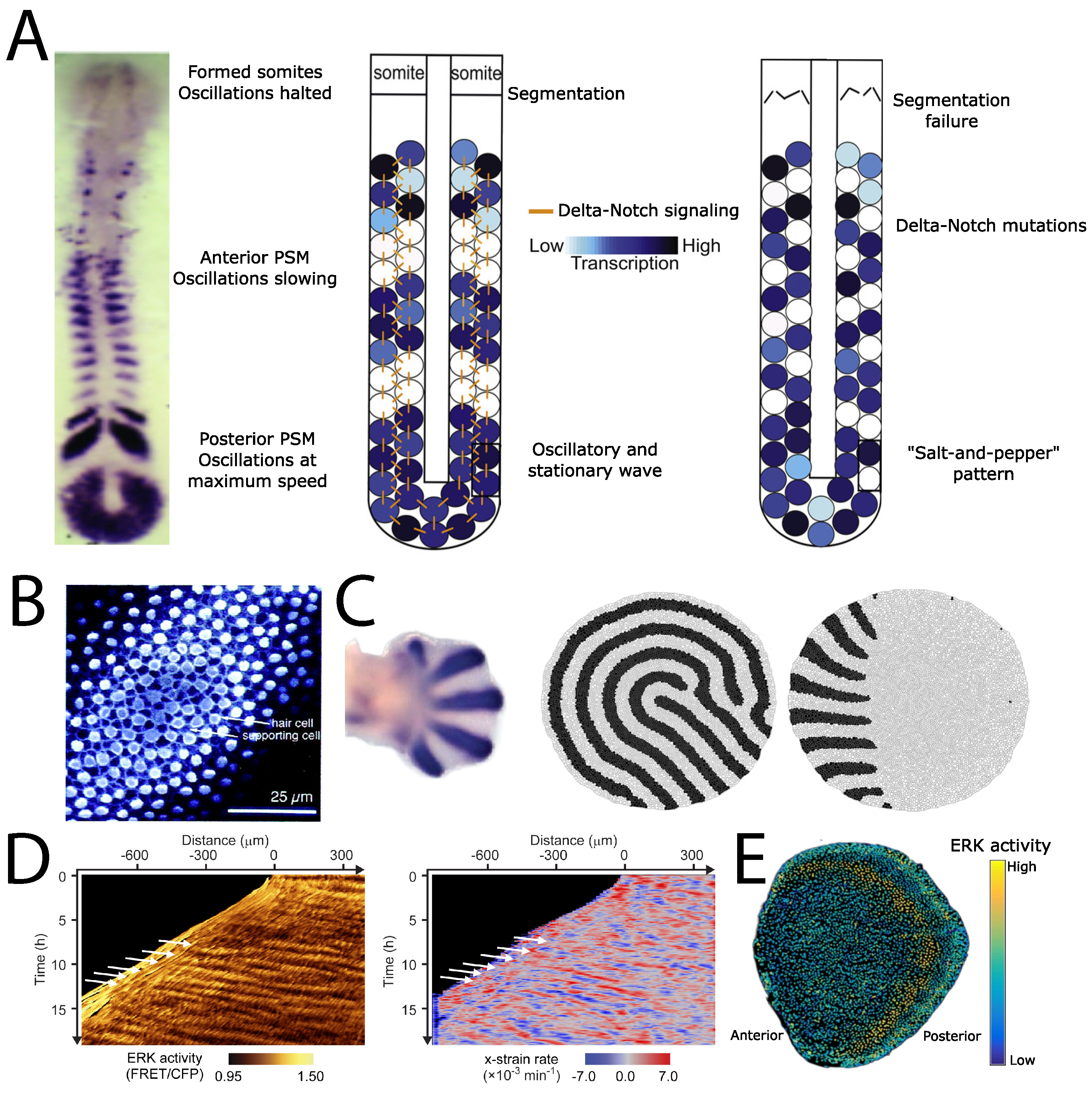
3. Examples of Developmental Patterning

Segmentation-like patterns. The so-called segmentation clock is an example of patterning during development that drives the sequential subdivision of the elongating body axis of vertebrate embryos into somites and, ultimately, leading to the segmented vertebral column [23,24] (Figure 2A). Interestingly, the segmentation clock comprises a cell autonomous mechanism and two inductive mechanisms, which, acting through different stages, produce an oscillatory and a stationary stripe pattern [25]. At the single-cell level, individual cells at the pre-somitic mesoderm produce an oscillatory gene expression pattern [25]. Synchronization of the cellular oscillations is achieved through a Delta–Notch (ligand–receptor) dynamics that generates a wave pattern of gene expression in the tissue [16]. The functional role of this pattern is evident when this inductive mechanism is perturbed: mutations in the Delta–Notch signaling network cause a salt-and-pepper (i.e., disorganized) pattern, and segmentation defects arise [26]. Finally, the cells of the pre-somitic mesoderm “read” concentration gradients of diffusing morphogens and, following the French flag model mechanism (Figure 1C), arrest the oscillations, and a stationary pattern develops. This pattern is subsequently translated into a specific cellular fate, thus producing the individual somites.
The segmentation, or compartmentalization, of tissues driven by morphogen gradients is, in fact, a common theme in biological patterning [27,28,29]. In that regard, on top of the segmentation clock, the most-celebrated example is the segmentation of the blastoderm-stage embryo of Drosophila melanogaster: a canonical example of the French flag model [30,31,32] (Figure 1C). In the first stage, the syncytium (a single cytoplasm that contains several nuclei) displays antiparallel gradients of Bicoid and Caudal proteins, which regulate the expression of the so-called gap genes in a concentration-dependent manner, which generates distinct, stripe-like, spatial domains. The interactions of gap genes generate a seven-stripe pattern (pair-rule genes), which in turn, activate the segment-polarity genes that create a pattern with fourteen stripes. Later during development, this pattern becomes the road map to segment the body of the adult fly. Interestingly, the boundaries of the domains (stripe-like pattern) of gap genes are not static, and yet, a robust segmentation pattern eventually arises [33]. Furthermore, it has been shown that each domain shares the same basic regulatory interactions between different genes following a combination between positive and negative feedback loops called the AC/DC circuit [34]. Notably, the AC/DC circuit was first described in the context of the neural tube patterning [35]. In that case, the neural tissue is patterned (segmented) into fourteen domains, which lead to different neuronal types [28,36]. The activity of two antiparallel morphogen gradients, BMPs and Shh, underlies also the establishment of the domains. However, there is one clear difference between the segmentation patterning of the blastoderm-stage embryo of Drosophila and that of the neural tube: the former qualifies as morphostatic, whereas in the latter, the patterning happens as the tube is growing (i.e., morphodynamic patterning); see Figure 1B. In that regard, it is important to notice that Shh plays a key role to regulate the growing dynamics of cells as a function of time in the tube domains [37]. It is also worth mentioning that the temporal patterning driven by morphogens of the Hh (Hedgehog) family seems to be relevant in other developmental patterning systems [38,39,40].
Figure 2. Developmental patterning examples. (A) The segmentation clock. The pre-somitic mesoderm (PSM) is patterned by oscillations that generate a stripe-like pattern. Right: the Delta–Notch (ligand-receptor) signaling dynamics generates a patterning wave of gene expression via lateral induction. Mutations in the Delta–Notch pathway cause a salt-and-pepper (i.e., disorganized) pattern, and segmentation defects arise (adapted from [41,42]). (B) The inner ear shows a short-range-signaling-induced pattern that generates an alternating pattern of hair and supporting cells (lateral inhibition mechanism; see the text; source: [43]). (C) Left: Digit patterning in vertebrates relies on a Turing mechanism (adapted from [44]). Right: Signaling properties and cell mechanismal properties determine the orientation of Turing patterns: simulation snapshots using constant cell–cell adhesion and diffusivity modulation (without/with: left/right); source: [45]. (D) The ERK pathway is a major regulator of the mechanical cell activity. During collective cell migration, traveling waves of mechanical activity (strain) appear due to ERK signaling events; source: [46]. (E) Signaling traveling waves pattern tissues during regeneration due to an excitatory mechanism (adapted from [47]).
Biophysica 03 00022 g002
The morphodynamic aspect of segmentation poses the intriguing question about how the maintenance of boundaries between patterning domains is achieved [48]. A hint to answer this question was obtained in the early 1970s when the induced recombination techniques enabled scientists to “label” single cells and their progeny (i.e., clones) [48,49,50]. Using this methodology, it has been shown that, for example, the wing imaginal disc is patterned and compartmentalized such that clones of cells in the tissue grow to certain boundaries, thereby preventing the intermingling of cellular domain populations [27]. These results promoted further research during the last few decades to understand how noise and fluctuations that originate from different sources, and that challenge the “straightness” of domain boundaries, become buffered (see the Section 4). In that regard, on top of the topology of gene regulatory networks, there is accumulating evidence that cellular mechanics is a key component during compartmentalization [51,52,53]. Furthermore, given the critical importance of setting effective barriers between cellular populations during developmental patterning, one might wonder about the existence of additional patterning mechanisms at the domains’ interface to ensure compartmentalization. In the particular case of the dorsal and ventral domains of the wing imaginal disc, short-range interactions between cells driven by Notch–Delta (receptor–ligand) are key to pattern the domain boundary and establish a signaling center from which cells receive positional information [54,55,56].
Short-range signaling induced patterns. The Notch–Delta system is indeed ubiquitous in developmental patterning processes by using either the lateral induction or the lateral inhibition mechanisms [57]. During lateral induction, Notch induces neighboring cells to adopt the same fate. This results in cellular differentiation waves (or synchronizing waves in the segmentation clock, as mentioned above; see [29]). During lateral inhibition, Notch inhibits neighboring cells from taking on a default cell state [58]. Interestingly, it has been shown that, during the patterning of the inner ear, both mechanisms are key for establishing the alternate pattern of hair cells and supporting cells and that switching between mechanisms depends on ligand competition [59]. Moreover, the non-linear dynamics of the Notch–Delta system leads, operating under the same conditions, to different possible stable patterns [60]; see Figure 2B. This poses the interesting question about the mechanisms of pattern selection. Palau-Ortin and colleagues addressed this problem computationally by studying how different trajectories in the parameter space (as a way to represent the sequence of biochemical signals) condition pattern selection [61]. Thus, they showed that, if signals act globally (whole tissue at the same time), in cell clusters, or through a propagating wave (i.e., sequentially), this leads to different types of patterns. The same group also explored other effects in the resulting Notch–Delta pattern such as those due to the so-called cis interactions (receptor–ligand binding dynamics within the same cell) and ligand diffusivity [62,63]. Further, in different contexts (pattern formation in plant development and neurogenesis), this group and collaborators have shown that the combination of both short-range interactions and long-range interactions is needed to determine and regulate segmentation patterning [64] and how Notch–Delta lateral inhibition becomes modified by neurogenic wavefronts [65,66].
Turing patterns. As mentioned above, the pioneering work of Alan Turing established, for the first time, a plausible mechanism of developmental pattern formation that connected the diffusive transport properties of “chemicals” and their interactions. Yet, the factual applicability of this mechanism at the most-fundamental level (genes and their products) remained elusive for decades. As crudely put by Koch and Meinhard forty years after Turing’s proposal: “So far, no biological system able to generate primary pattern formation has been completely characterized at the molecular level.” [5]. Around the time such an observation was made, a major breakthrough occurred when Turing patterns were produced for the first time in a controlled way in a chemical system (the CIMA reaction) [67]. Soon after that, experimental demonstrations that connected Turing’s ideas with the control of pattern formation during morphogenesis were published [68,69,70]. While still in the domain of chemical systems, these studies paved the way to understand, for example, the sequential formation of stationary stripes perpendicular to the growth axis of the pre-somitic mesoderm (i.e., the segmentation clock) [71]. To the best of our knowledge, the first genuine experimental demonstration of the applicability of the Turing mechanism in developmental patterning was proposed by the group of Kondo in the context of skin pigmentation [19,72,73]. Still, even if the molecular basis of the interactions between the pigment cells was identified, whether the mechanism belonged—canonically—to that of Turing or not was debatable since long-range interactions are not due to diffusive factors [74]. Later, in 2012, evidence was presented that the Turing mechanism was responsible of the generation of the regular pattern of the ridges of the palate of mammals [75], and simultaneously, the group of James Sharpe in Barcelona argued that a Turing-type mechanism underlies digit patterning [44]; see Figure 2C. Importantly, this study provided evidence that this patterning mechanism has been conserved in tetrapods, and moreover, the data suggested that the five fingers’ patterning was stabilized more than 360 million years ago. Finally, in 2014, Sharpe’s group demonstrated the applicability of Turing’s mechanism in limb patterning and identified the responsible genes and molecular species, thus closing a research quest that spanned more than half a century [76] (see also [77]). Further progress in the field from that group included a high-throughput analysis to determine a catalog of realistic Turing gene networks during developmental patterning and a study that clarified how Turing patterning relies on the gene network topology [18,78]. Other recent contributions have studied how the finite size of cells conditions the protein expression variability in Turing-patterned tissues [79], how non-linearities regulate the resulting pattern [80], and a topic that connects with the next subject of this review (mechanobiology): the interplay between cellular/tissue mechanics and Turing patterning [45], Figure 2C.
Mechanical patterns. As mentioned above, during development, inductive and morphogenetic mechanisms may act simultaneously (morphodynamic patterning; Figure 1B). This raises the interesting question of how the cell/tissue mechanical properties and patterning feed back to each other. This problem is one of the topics of interest of mechanobiology, a field that has flourished during the last few decades and tries to understand cells and tissues from the viewpoint of their “perception” of, and response to, the mechanical environment [81]. In this way, during morphogenesis, tissues expand and remodel and the constituent cells exert forces (along the direction of maximum stress [82]), and the cellular mechanics has been shown to play a key role in the self-organization of tissues [83,84]. These cellular forces propagate over the tissue due to cell–cell interactions, and mechanical patterns appear (e.g., spatiotemporal traveling waves of strain, traction, stress, etc.) to provide a “map” that coordinates growth, expansion, and replacement [85,86,87,88]. Importantly, mechanical patterns seems to play a key role for shaping the 3D structure and packing of tissues [84,89,90,91,92]. In fact, the role played by cellular movements and migration during pattern formation has been recently highlighted [93], and related to that, it has been shown that patterns of cell proliferation and migration display “memory effects” [94] that could, hence, condition the resulting gene expression pattern. Thus, these discoveries extended the idea of developmental patterning from the “chemical” to the “physical” realm and have opened the door towards the implementation of synthetic morphogenesis [95,96,97]. For example, in the specific context of Turing patterns, it has been recently studied how patterning (signaling) and cellular mechanics feed back to each other to achieve a robust tissue remodeling driven by self-sustained planar intercalations [45]. One interesting conclusion is the need for trade-offs between cellular/tissue strains (orchestrated through the cleavage orientation) and the resulting patterning that cooperate synergistically to achieve elongation. Connected to that, other works have clarified how cellular activities (mechanics, migration, growth/division) determine the elongation of the vertebrate body axis and generate mechanical patterns [98,99,100]. Importantly, these studies have shown that tissues undergo fluid-to-solid-like transitions, which help to sculpt and remodel organisms during morphogenesis. Furthermore, one particular example that illustrates the feedbacks and the connection between genetic regulation and cell/tissue mechanics during developmental patterning is the Extracellular signal-Regulated Kinase (ERK) pathway [101]. This pathway transduces an extracellular signal into the phosphorylation level of ERK, which regulates downstream targets linked to cellular proliferation, differentiation, and stress. The ERK pathway studies have shed light on how chemical cues drive cell mechanics to orchestrate tissue collective movement; namely, cell stretching triggers ERK activation, and conversely, ERK activation drives cellular contraction. These feedbacks result in spatiotemporal oscillatory patterns that instruct collective cell migration [46,102,103]; see Figure 2D. Still, it is worth noticing that the questions are open with respect to the right modeling framework that describes accurately the interplay between ERK signaling and the cellular mechanical activities for tissue patterning. Even though the proposed models lie within the common formalism of reaction–diffusion, some models claim the existence of an excitable dynamics mechanism that propagates due to diffusion [47] (Figure 2E), and others point towards an instability that leads to a wave pattern [104].

4. Discussion

Here, we provided a brief review of how contributions from biophysics, in particular emphasizing those from Spanish researchers, have helped quantitative approaches in developmental pattern formation flourish during the last few decades. As for some promising avenues of research during the coming years, we would like to spotlight the following ones. First, we believe that there is still much improvement to be made in regards of in silico approaches towards developmental pattern formation [16,105]. A number of methods that combine cell mechanics and, sometimes, also signaling have been proposed over the last few decades (e.g., vertex models, Voronoi-like models, Potts-like models, etc.) [84,90,91,105,106,107,108,109,110,111,112,113,114]. In terms of cellular architecture, these methods focused mainly on 2D approaches, i.e., representations of one surface of the tissue, but others tried to mimic the 3D packing of cells. Still, as of today, no method has been able to capture how the cellular shape changes dynamically in a realistic way (e.g., appearance/disappearance of apico-basal intercalations). This is also related to other relevant problems that need to be addressed. On the one hand, while progress has been made during the last few years [115], better computational tools are needed to extract 3D information from microscopy data (i.e., segmentation and tracking). On the other hand, the effects of the geometry of the 3D embedding space of the developing tissue, in particular the curvature, is becoming recognized as a major driving force in developmental patterning, where more research is required [91,116]. The combination of all these tools and ideas (novel/advanced in silico approaches, tools for 3D data analysis, and the analyses of the effects of the 3D embedding space) will allow tackling questions, and developing applications, with relevance in the field of biomedicine such as design and standardization in organoid research [117,118]. By standardization, here, we mean identifying the phenomena that cause the variability/stochasticity and lack of control during developmental patterning (from signaling to mechanics) and acting accordingly to correct them in vitro and in vivo [119,120,121,122]. Stochasticity is indeed a topic that has been deeply explored in pattern formation from a biophysical viewpoint, and it has been shown that noise and fluctuations, rather than being a nuisance, can be a driver of patterning [8,9,123,124,125,126,127,128,129,130]. Thus, a key question in the context of developmental patterning that is gaining interest is how living organisms achieve robustness and precision under different sources of stochasticity and, in some cases, even profit from fluctuations [32,131,132,133,134,135,136,137,138]. In closing, the input of biophysics, and in particular the Spanish contributions, have not only moved the field of developmental patterning forward, but have reversed the polemic statement from Gilbert that opened this review, and we can confidently affirm that developmental biology is now a refuge for mathematically competent scientists.

Funding

J.B. acknowledges support from the Spanish Ministry of Science and Innovation through Grant PID2019105566GB-I00 and from the LifeHUB Research Network (CSIC) PIE-202120E047-Conexiones-Life.

Acknowledgments

We do apologize to the many colleagues whose work/research was not cited due to space constraints.

Conflicts of Interest

The authors declare no conflict of interest.

References

  1. Gilbert, S.F. Developmental Biology, 7th ed.; Sinauer Associates: Sunderland, MA, USA, 2003. [Google Scholar]
  2. Oates, A.C.; Gorfinkiel, N.; González-Gaitán, M.; Heisenberg, C.P. Quantitative approaches in developmental biology. Nat. Rev. Genet. 2009, 10, 517–530. [Google Scholar] [CrossRef] [PubMed]
  3. Turing, A. The Chemical Basis of Morphogenesis. Philos. Trans. R. Soc. Lond. Ser. B 1990, 237, 37–72. [Google Scholar] [CrossRef]
  4. Wolpert, L. Positional information and pattern formation in development. Dev. Genet. 1994, 15, 485–490. [Google Scholar] [CrossRef] [PubMed]
  5. Koch, A.J.; Meinhardt, H. Biological pattern formation: From basic mechanisms to complex structures. Rev. Mod. Phys. 1994, 66, 1481–1507. [Google Scholar] [CrossRef]
  6. Murray, J.D. Mathematical Biology. 1: An Introduction, Softcover Reprint of the Hardcover 3rd edition 2002, corrected second printing ed.; Number 17 in Interdisciplinary applied mathematics; Springer: Berlin/Heidelberg, Germany, 2004. [Google Scholar]
  7. Forgacs, G.; Newman, S.A. Biological Physics of the Developing Embryo, 1st ed.; Cambridge University Press: Cambridge, UK, 2005. [Google Scholar] [CrossRef]
  8. García-Ojalvo, J.; Sancho, J.M. Noise in Spatially Extended Systems; Institute for nonlinear science; Springer: New York, NY, USA, 1999. [Google Scholar]
  9. Sagués, F.; Sancho, J.; García-Ojalvo, J. Spatiotemporal order out of noise. Rev. Mod. Phys. 2007, 79, 829–882. [Google Scholar] [CrossRef]
  10. Lewis, J. From signals to patterns: Space, time, and mathematics in developmental biology. Science 2008, 322, 399–403. [Google Scholar] [CrossRef]
  11. Kicheva, A.; Cohen, M.; Briscoe, J. Developmental pattern formation: Insights from physics and biology. Science 2012, 338, 210–212. [Google Scholar] [CrossRef]
  12. Cross, M.; Hohenberg, P. Pattern formation outside of equilibrium. Rev. Mod. Phys. 1993, 65, 851–1112. [Google Scholar] [CrossRef]
  13. Salazar-Ciudad, I.; Jernvall, J.; Newman, S.A. Mechanisms of pattern formation in development and evolution. Development 2003, 130, 2027–2037. [Google Scholar] [CrossRef]
  14. Salazar-Ciudad, I.; Jernvall, J. How different types of pattern formation mechanisms affect the evolution of form and development. Evol. Dev. 2004, 6, 6–16. [Google Scholar] [CrossRef]
  15. Buceta, J.; Lindenberg, K. Patterns in reaction-diffusion systems generated by global alternation of dynamics. Phys. Stat. Mech. Its Appl. 2003, 325, 230–242. [Google Scholar] [CrossRef]
  16. Morelli, L.G.; Uriu, K.; Ares, S.; Oates, A.C. Computational approaches to developmental patterning. Science 2012, 336, 187–191. [Google Scholar] [CrossRef] [PubMed]
  17. Wolpert, L. Positional information and the spatial pattern of cellular differentiation. J. Theor. Biol. 1969, 25, 1–47. [Google Scholar] [CrossRef]
  18. Diego, X.; Marcon, L.; Müller, P.; Sharpe, J. Key Features of Turing Systems are Determined Purely by Network Topology. Phys. Rev. X 2018, 8, 021071. [Google Scholar] [CrossRef]
  19. Kondo, S.; Miura, T. Reaction-diffusion model as a framework for understanding biological pattern formation. Science 2010, 329, 1616–1620. [Google Scholar] [CrossRef]
  20. Landge, A.N.; Jordan, B.M.; Diego, X.; Müller, P. Pattern formation mechanisms of self-organizing reaction-diffusion systems. Dev. Biol. 2020, 460, 2–11. [Google Scholar] [CrossRef]
  21. Collier, J.R.; Monk, N.A.; Maini, P.K.; Lewis, J.H. Pattern formation by lateral inhibition with feedback: A mathematical model of delta-notch intercellular signalling. J. Theor. Biol. 1996, 183, 429–446. [Google Scholar] [CrossRef] [PubMed]
  22. Talia, S.D.; Vergassola, M. Waves in Embryonic Development. Annu. Rev. Biophys. 2022, 51, 327–353. [Google Scholar] [CrossRef]
  23. Oates, A.C.; Morelli, L.G.; Ares, S. Patterning embryos with oscillations: Structure, function and dynamics of the vertebrate segmentation clock. Development 2012, 139, 625–639. [Google Scholar] [CrossRef]
  24. Kageyama, R. 25 years of the segmentation clock gene. Nature 2022, 611, 671–673. [Google Scholar] [CrossRef]
  25. Webb, A.B.; Lengyel, I.M.; Jörg, D.J.; Valentin, G.; Jülicher, F.; Morelli, L.G.; Oates, A.C. Persistence, period and precision of autonomous cellular oscillators from the zebrafish segmentation clock. eLife 2016, 5, e08438. [Google Scholar] [CrossRef] [PubMed]
  26. Lewis, J.; Hanisch, A.; Holder, M. Notch signaling, the segmentation clock, and the patterning of vertebrate somites. J. Biol. 2009, 8, 44. [Google Scholar] [CrossRef] [PubMed]
  27. Buceta, J. Multidisciplinary approaches towards compartmentalization in development: Dorsoventral boundary formation of the Drosophila wing disc as a case of study. Contrib. Sci. 2013, 9, 57–66. [Google Scholar] [CrossRef]
  28. Briscoe, J.; Small, S. Morphogen rules: Design principles of gradient-mediated embryo patterning. Development 2015, 142, 3996–4009. [Google Scholar] [CrossRef]
  29. Diaz-Cuadros, M.; Pourquié, O.; El-Sherif, E. Patterning with clocks and genetic cascades: Segmentation and regionalization of vertebrate versus insect body plans. PLoS Genet. 2021, 17, e1009812. [Google Scholar] [CrossRef]
  30. Jaeger, J.; Surkova, S.; Blagov, M.; Janssens, H.; Kosman, D.; Kozlov, K.N.; Manu; Myasnikova, E.; Vanario-Alonso, C.E.; Samsonova, M.; et al. Dynamic control of positional information in the early Drosophila embryo. Nature 2004, 430, 368–371. [Google Scholar] [CrossRef]
  31. Crombach, A.; Wotton, K.R.; Cicin-Sain, D.; Ashyraliyev, M.; Jaeger, J. Efficient reverse-engineering of a developmental gene regulatory network. PLoS Comput. Biol. 2012, 8, e1002589. [Google Scholar] [CrossRef]
  32. Jaeger, J.; Verd, B. Dynamic positional information: Patterning mechanism versus precision in gradient-driven systems. In Current Topics in Developmental Biology; Elsevier: Amsterdam, The Netherlands, 2020; Volume 137, pp. 219–246. [Google Scholar] [CrossRef]
  33. Verd, B.; Clark, E.; Wotton, K.R.; Janssens, H.; Jiménez-Guri, E.; Crombach, A.; Jaeger, J. A damped oscillator imposes temporal order on posterior gap gene expression in Drosophila. PLoS Biol. 2018, 16, e2003174. [Google Scholar] [CrossRef]
  34. Verd, B.; Monk, N.A.; Jaeger, J. Modularity, criticality, and evolvability of a developmental gene regulatory network. eLife 2019, 8, e42832. [Google Scholar] [CrossRef]
  35. Balaskas, N.; Ribeiro, A.; Panovska, J.; Dessaud, E.; Sasai, N.; Page, K.M.; Briscoe, J.; Ribes, V. Gene regulatory logic for reading the Sonic Hedgehog signaling gradient in the vertebrate neural tube. Cell 2012, 148, 273–284. [Google Scholar] [CrossRef]
  36. Kicheva, A.; Bollenbach, T.; Ribeiro, A.; Valle, H.P.; Lovell-Badge, R.; Episkopou, V.; Briscoe, J. Coordination of progenitor specification and growth in mouse and chick spinal cord. Science 2014, 345, 1254927. [Google Scholar] [CrossRef] [PubMed]
  37. Saade, M.; Gutiérrez-Vallejo, I.; Le Dréau, G.; Rabadán, M.A.; Miguez, D.G.; Buceta, J.; Martí, E. Sonic Hedgehog Signaling Switches the Mode of Division in the Developing Nervous System. Cell Rep. 2013, 4, 492. [Google Scholar] [CrossRef] [PubMed]
  38. Aguilar-Hidalgo, D.; Domínguez-Cejudo, M.A.; Amore, G.; Brockmann, A.; Lemos, M.C.; Córdoba, A.; Casares, F. A Hh-driven gene network controls specification, pattern and size of the Drosophila simple eyes. Development 2013, 140, 82–92. [Google Scholar] [CrossRef] [PubMed]
  39. García-Morales, D.; Navarro, T.; Iannini, A.; Míguez, D.G.; Casares, F. Dynamic Hh signaling can generate temporal information during tissue patterning. Development 2019, 146, dev.176933. [Google Scholar] [CrossRef]
  40. Míguez, D.G.; García-Morales, D.; Casares, F. Control of size, fate and time by the Hh morphogen in the eyes of flies. In Current Topics in Developmental Biology; Elsevier: Amsterdam, The Netherlands, 2020; Volume 137, pp. 307–332. [Google Scholar] [CrossRef]
  41. Giudicelli, F.; Özbudak, E.M.; Wright, G.J.; Lewis, J. Setting the Tempo in Development: An Investigation of the Zebrafish Somite Clock Mechanism. PLoS Biol. 2007, 5, e150. [Google Scholar] [CrossRef]
  42. Keskin, S.; Devakanmalai, G.S.; Kwon, S.B.; Vu, H.T.; Hong, Q.; Lee, Y.Y.; Soltani, M.; Singh, A.; Ay, A.; Özbudak, E.M. Noise in the Vertebrate Segmentation Clock Is Boosted by Time Delays but Tamed by Notch Signaling. Cell Rep. 2018, 23, 2175–2185.e4. [Google Scholar] [CrossRef]
  43. Eddison, M.; Le Roux, I.; Lewis, J. Notch signaling in the development of the inner ear: Lessons from Drosophila. Proc. Natl. Acad. Sci. USA 2000, 97, 11692–11699. [Google Scholar] [CrossRef]
  44. Sheth, R.; Marcon, L.; Bastida, M.F.; Junco, M.; Quintana, L.; Dahn, R.; Kmita, M.; Sharpe, J.; Ros, M.A. Hox Genes Regulate Digit Patterning by Controlling the Wavelength of a Turing-Type Mechanism. Science 2012, 338, 1476–1480. [Google Scholar] [CrossRef]
  45. Anbari, S.; Buceta, J. Self-sustained planar intercalations due to mechanosignaling feedbacks lead to robust axis extension during morphogenesis. Sci. Rep. 2020, 10, 10973. [Google Scholar] [CrossRef]
  46. Hino, N.; Rossetti, L.; Marín-Llauradó, A.; Aoki, K.; Trepat, X.; Matsuda, M.; Hirashima, T. ERK-Mediated Mechanochemical Waves Direct Collective Cell Polarization. Dev. Cell 2020, 53, 646–660. [Google Scholar] [CrossRef]
  47. De Simone, A.; Evanitsky, M.N.; Hayden, L.; Cox, B.D.; Wang, J.; Tornini, V.A.; Ou, J.; Chao, A.; Poss, K.D.; Di Talia, S. Control of osteoblast regeneration by a train of Erk activity waves. Nature 2021, 590, 129–133. [Google Scholar] [CrossRef] [PubMed]
  48. Dahmann, C.; Basler, K. Compartment boundaries: At the edge of development. Trends Genet. TIG 1999, 15, 320–326. [Google Scholar] [CrossRef] [PubMed]
  49. Morata, G.; Lawrence, P.A. Anterior and posterior compartments in the head of Drosophila. Nature 1978, 274, 473–474. [Google Scholar] [CrossRef]
  50. Lawrence, P.A. Developmental biology. Straight and wiggly affinities. Nature 1997, 389, 546–547. [Google Scholar] [CrossRef]
  51. Landsberg, K.P.; Farhadifar, R.; Ranft, J.; Umetsu, D.; Widmann, T.J.; Bittig, T.; Said, A.; Jülicher, F.; Dahmann, C. Increased Cell Bond Tension Governs Cell Sorting at the Drosophila Anteroposterior Compartment Boundary. Curr. Biol. 2009, 19, 1950–1955. [Google Scholar] [CrossRef] [PubMed]
  52. Umetsu, D.; Dahmann, C. Compartment boundaries: Sorting cells with tension. Fly 2010, 4, 241–245. [Google Scholar] [CrossRef] [PubMed]
  53. Canela-Xandri, O.; Sagués, F.; Casademunt, J.; Buceta, J. Dynamics and Mechanical Stability of the Developing Dorsoventral Organizer of the Wing Imaginal Disc. PLoS Comput. Biol. 2011, 7, e1002153. [Google Scholar] [CrossRef]
  54. Buceta, J.; Herranz, H.; Canela-Xandri, O.; Reigada, R.; Sagués, F.; Milán, M. Robustness and stability of the gene regulatory network involved in DV boundary formation in the Drosophila wing. PLoS ONE 2007, 2, e602. [Google Scholar] [CrossRef]
  55. Canela-Xandri, O.; Sagués, F.; Reigada, R.; Buceta, J. A spatial toggle switch drives boundary formation in development. Biophys. J. 2008, 95, 5111–5120. [Google Scholar] [CrossRef]
  56. Becam, I.; Milán, M. A permissive role of Notch in maintaining the DV affinity boundary of the Drosophila wing. Dev. Biol. 2008, 322, 190–198. [Google Scholar] [CrossRef]
  57. Bocci, F.; Onuchic, J.N.; Jolly, M.K. Understanding the Principles of Pattern Formation Driven by Notch Signaling by Integrating Experiments and Theoretical Models. Front. Physiol. 2020, 11, 929. [Google Scholar] [CrossRef] [PubMed]
  58. Sancho, J.M.; Ibañes, M. Landau theory for cellular patterns driven by lateral inhibition interaction. Phys. Rev. E 2020, 102, 032404. [Google Scholar] [CrossRef]
  59. Petrovic, J.; Formosa-Jordan, P.; Luna-Escalante, J.C.; Abelló, G.; Ibañes, M.; Neves, J.; Giraldez, F. Ligand-dependent Notch signaling strength orchestrates lateral induction and lateral inhibition in the developing inner ear. Development 2014, 141, 2313–2324. [Google Scholar] [CrossRef] [PubMed]
  60. Miller, A.C.; Lyons, E.L.; Herman, T.G. cis-Inhibition of Notch by Endogenous Delta Biases the Outcome of Lateral Inhibition. Curr. Biol. 2009, 19, 1378–1383. [Google Scholar] [CrossRef] [PubMed]
  61. Palau-Ortin, D.; Formosa-Jordan, P.; Sancho, J.M.; Ibañes, M. Pattern selection by dynamical biochemical signals. Biophys. J. 2015, 108, 1555–1565. [Google Scholar] [CrossRef] [PubMed]
  62. Formosa-Jordan, P.; Ibañes, M. Diffusible ligand and lateral inhibition dynamics for pattern formation. J. Stat. Mech. Theory Exp. 2009, 2009, P03019. [Google Scholar] [CrossRef]
  63. Formosa-Jordan, P.; Ibañes, M. Competition in notch signaling with cis enriches cell fate decisions. PLoS ONE 2014, 9, e95744. [Google Scholar] [CrossRef]
  64. Fàbregas, N.; Formosa-Jordan, P.; Confraria, A.; Siligato, R.; Alonso, J.M.; Swarup, R.; Bennett, M.J.; Mähönen, A.P.; Caño-Delgado, A.I.; Ibañes, M. Auxin Influx Carriers Control Vascular Patterning and Xylem Differentiation in Arabidopsis thaliana. PLoS Genet. 2015, 11, e1005183. [Google Scholar] [CrossRef]
  65. Formosa-Jordan, P.; Ibañes, M.; Ares, S.; Frade, J.M. Regulation of neuronal differentiation at the neurogenic wavefront. Development 2012, 139, 2321–2329. [Google Scholar] [CrossRef]
  66. Formosa-Jordan, P.; Ibañes, M.; Ares, S.; Frade, J.M. Lateral inhibition and neurogenesis: Novel aspects in motion. Int. J. Dev. Biol. 2013, 57, 341–350. [Google Scholar] [CrossRef]
  67. Ouyang, Q.; Swinney, H.L. Transition from a uniform state to hexagonal and striped Turing patterns. Nature 1991, 352, 610–612. [Google Scholar] [CrossRef]
  68. Rüdiger, S.; Míguez, D.G.; Muñuzuri, A.P.; Sagués, F.; Casademunt, J. Dynamics of Turing Patterns under Spatiotemporal Forcing. Phys. Rev. Lett. 2003, 90, 128301. [Google Scholar] [CrossRef] [PubMed]
  69. Míguez, D.G.; Nicola, E.M.; Muñuzuri, A.P.; Casademunt, J.; Sagués, F.; Kramer, L. Traveling-Stripe Forcing Generates Hexagonal Patterns. Phys. Rev. Lett. 2004, 93, 048303. [Google Scholar] [CrossRef] [PubMed]
  70. Kærn, M.; Míguez, D.G.; Muñuzuri, A.P.; Menzinger, M. Control of chemical pattern formation by a clock-and-wavefront type mechanism. Biophys. Chem. 2004, 110, 231–238. [Google Scholar] [CrossRef] [PubMed]
  71. Konow, C.; Dolnik, M.; Epstein, I.R. Insights from chemical systems into Turing-type morphogenesis. Philos. Trans. R. Soc. A Math. Phys. Eng. Sci. 2021, 379, 20200269. [Google Scholar] [CrossRef]
  72. Yamaguchi, M.; Yoshimoto, E.; Kondo, S. Pattern regulation in the stripe of zebrafish suggests an underlying dynamic and autonomous mechanism. Proc. Natl. Acad. Sci. USA 2007, 104, 4790–4793. [Google Scholar] [CrossRef]
  73. Nakamasu, A.; Takahashi, G.; Kanbe, A.; Kondo, S. Interactions between zebrafish pigment cells responsible for the generation of Turing patterns. Proc. Natl. Acad. Sci. USA 2009, 106, 8429–8434. [Google Scholar] [CrossRef]
  74. Kondo, S. Turing Pattern Formation without Diffusion. In How the World Computes; Springer: Berlin/Heidelberg, Germany, 2012; pp. 416–421. [Google Scholar] [CrossRef]
  75. Economou, A.D.; Ohazama, A.; Porntaveetus, T.; Sharpe, P.T.; Kondo, S.; Basson, M.A.; Gritli-Linde, A.; Cobourne, M.T.; Green, J.B.A. Periodic stripe formation by a Turing mechanism operating at growth zones in the mammalian palate. Nat. Genet. 2012, 44, 348–351. [Google Scholar] [CrossRef]
  76. Raspopovic, J.; Marcon, L.; Russo, L.; Sharpe, J. Modeling digits. Digit patterning is controlled by a Bmp-Sox9-Wnt Turing network modulated by morphogen gradients. Science 2014, 345, 566–570. [Google Scholar] [CrossRef]
  77. Cooper, K.L. Self-organization in the limb: A Turing mechanism for digit development. Curr. Opin. Genet. Dev. 2015, 32, 92–97. [Google Scholar] [CrossRef]
  78. Marcon, L.; Diego, X.; Sharpe, J.; Müller, P. High-throughput mathematical analysis identifies Turing networks for patterning with equally diffusing signals. eLife 2016, 5, e14022. [Google Scholar] [CrossRef] [PubMed]
  79. Buceta, J. Finite cell-size effects on protein variability in Turing patterned tissues. J. R. Soc. Interface 2017, 14, 20170316. [Google Scholar] [CrossRef] [PubMed]
  80. Chen, Y.; Buceta, J. A non-linear analysis of Turing pattern formation. PLoS ONE 2019, 14, e0220994. [Google Scholar] [CrossRef]
  81. Jacobs, C.R.; Huang, H.; Kwon, R.Y. Introduction to Cell Mechanics and Mechanobiology; OCLC: 1019679867; Garland Science: New York, NY, USA, 2013. [Google Scholar]
  82. Tambe, D.; Hardin, C.; Angelini, T.E.; Rajendran, K.; Park, C.Y.; Serra-Picamal, X.; Zhou, E.H.; Zaman, M.H.; Butler, J.P.; Weitz, D.A.; et al. Collective cell guidance by cooperative intercellular forces. Nat. Mat. 2011, 10, 469–475. [Google Scholar] [CrossRef] [PubMed]
  83. Fernandez-Gonzalez, R.; Zallen, J.A. Epithelial organization: May the force be with you. Curr. Biol. 2008, 18, R163–R165. [Google Scholar] [CrossRef]
  84. Gómez-Gálvez, P.; Anbari, S.; Escudero, L.M.; Buceta, J. Mechanics and self-organization in tissue development. Semin. Cell Dev. Biol. 2021, 120, 147. [Google Scholar] [CrossRef]
  85. Serra-Picamal, X.; Conte, V.; Vincent, R.; Anon, E.; Tambe, D.T.; Bazellieres, E.; Butler, J.P.; Fredberg, J.J.; Trepat, X. Mechanical waves during tissue expansion. Nat. Phys. 2012, 8, 628–634. [Google Scholar] [CrossRef]
  86. Pérez-González, C.; Alert, R.; Blanch-Mercader, C.; Gómez-González, M.; Kolodziej, T.; Bazellieres, E.; Casademunt, J.; Trepat, X. Active wetting of epithelial tissues. Nat. Phys. 2019, 15, 79–88. [Google Scholar] [CrossRef]
  87. Prat-Rojo, C.; Pouille, P.A.; Buceta, J.; Martin-Blanco, E. Mechanical coordination is sufficient to promote tissue replacement during metamorphosis in Drosophila. EMBO J. 2020, 39, e103594. [Google Scholar] [CrossRef]
  88. Alert, R.; Trepat, X. Physical Models of Collective Cell Migration. Annu. Rev. Condens. Matter Phys. 2020, 11, 77–101. [Google Scholar] [CrossRef]
  89. Latorre, E.; Kale, S.; Casares, L.; Gómez-González, M.; Uroz, M.; Valon, L.; Nair, R.V.; Garreta, E.; Montserrat, N.; del Campo, A.; et al. Active superelasticity in three-dimensional epithelia of controlled shape. Nature 2018, 563, 203–208. [Google Scholar] [CrossRef] [PubMed]
  90. Pérez-González, C.; Ceada, G.; Greco, F.; Matejčić, M.; Gómez-González, M.; Castro, N.; Menendez, A.; Kale, S.; Krndija, D.; Clark, A.G.; et al. Mechanical compartmentalization of the intestinal organoid enables crypt folding and collective cell migration. Nat. Cell Biol. 2021, 23, 745–757. [Google Scholar] [CrossRef] [PubMed]
  91. Gómez-Gálvez, P.; Vicente-Munuera, P.; Anbari, S.; Buceta, J.; Escudero, L.M. The Complex Three-Dimensional Organization of Epithelial Tissues. Development 2021, 148, dev195669. [Google Scholar] [CrossRef] [PubMed]
  92. Gómez-Gálvez, P.; Vicente-Munuera, P.; Anbari, S.; Tagua, A.; Gordillo-Vázquez, C.; Andrés-San Román, J.A.; Franco-Barranco, D.; Palacios, A.M.; Velasco, A.; Capitán-Agudo, C.; et al. A quantitative biophysical principle to explain the 3D cellular connectivity in curved epithelia. Cell Syst. 2022, 13, 631–643.e8. [Google Scholar] [CrossRef] [PubMed]
  93. Fulton, T.; Verd, B.; Steventon, B. The unappreciated generative role of cell movements in pattern formation. R. Soc. Open Sci. 2022, 9, 211293. [Google Scholar] [CrossRef] [PubMed]
  94. Heinrich, M.A.; Alert, R.; LaChance, J.M.; Zajdel, T.J.; Košmrlj, A.; Cohen, D.J. Size-dependent patterns of cell proliferation and migration in freely-expanding epithelia. eLife 2020, 9, e58945. [Google Scholar] [CrossRef] [PubMed]
  95. Salazar-Ciudad, I. Tooth morphogenesis in vivo, in vitro, and in silico. Curr. Top. Dev. Biol. 2008, 81, 341–371. [Google Scholar] [CrossRef]
  96. Gritti, N.; Oriola, D.; Trivedi, V. Rethinking embryology in vitro: A synergy between engineering, data science and theory. Dev. Biol. 2021, 474, 48–61. [Google Scholar] [CrossRef]
  97. Matejčić, M.; Trepat, X. Mechanobiological approaches to synthetic morphogenesis: Learning by building. Trends Cell Biol. 2022, 33, 95–111. [Google Scholar] [CrossRef]
  98. Mongera, A.; Rowghanian, P.; Gustafson, H.J.; Shelton, E.; Kealhofer, D.A.; Carn, E.K.; Serwane, F.; Lucio, A.A.; Giammona, J.; Campàs, O. A fluid-to-solid jamming transition underlies vertebrate body axis elongation. Nature 2018, 561, 401–405. [Google Scholar] [CrossRef]
  99. Kim, S.; Pochitaloff, M.; Stooke-Vaughan, G.A.; Campàs, O. Embryonic tissues as active foams. Nat. Phys. 2021, 17, 859–866. [Google Scholar] [CrossRef] [PubMed]
  100. Banavar, S.P.; Carn, E.K.; Rowghanian, P.; Stooke-Vaughan, G.; Kim, S.; Campàs, O. Mechanical control of tissue shape and morphogenetic flows during vertebrate body axis elongation. Sci. Rep. 2021, 11, 8591. [Google Scholar] [CrossRef] [PubMed]
  101. Samson, S.C.; Khan, A.M.; Mendoza, M.C. ERK signaling for cell migration and invasion. Front. Mol. Biosci. 2022, 9, 998475. [Google Scholar] [CrossRef] [PubMed]
  102. Aoki, K.; Kondo, Y.; Naoki, H.; Hiratsuka, T.; Itoh, R.E.; Matsuda, M. Propagating Wave of ERK Activation Orients Collective Cell Migration. Dev. Cell 2017, 43, 305–317.e5. [Google Scholar] [CrossRef] [PubMed]
  103. Hino, N.; Matsuda, K.; Jikko, Y.; Maryu, G.; Sakai, K.; Imamura, R.; Tsukiji, S.; Aoki, K.; Terai, K.; Hirashima, T.; et al. A feedback loop between lamellipodial extension and HGF-ERK signaling specifies leader cells during collective cell migration. Dev. Cell 2022, 57, 2290–2304.e7. [Google Scholar] [CrossRef]
  104. Boocock, D.; Hino, N.; Ruzickova, N.; Hirashima, T.; Hannezo, E. Theory of mechanochemical patterning and optimal migration in cell monolayers. Nat. Phys. 2020, 17, 267–274. [Google Scholar] [CrossRef]
  105. Fletcher, A.G.; Osborne, J.M. Seven challenges in the multiscale modeling of multicellular tissues. WIREs Mech. Dis. 2022, 14. [Google Scholar] [CrossRef]
  106. Graner, F.; Glazier, J. Simulation of biological cell sorting using a two-dimensional extended Potts model. Phys. Rev. Lett. 1992, 69, 2013–2016. [Google Scholar] [CrossRef]
  107. Izaguirre, J.A.; Chaturvedi, R.; Huang, C.; Cickovski, T.; Coffland, J.; Thomas, G.; Forgacs, G.; Alber, M.; Hentschel, G.; Newman, S.A.; et al. CompuCell, a multi-model framework for simulation of morphogenesis. Bioinformatics 2004, 20, 1129–1137. [Google Scholar] [CrossRef]
  108. Fletcher, A.G.; Osterfield, M.; Baker, R.E.; Shvartsman, S.Y. Vertex models of epithelial morphogenesis. Biophys. J. 2014, 106, 2291–2304. [Google Scholar] [CrossRef]
  109. Ishimoto, Y.; Morishita, Y. Bubbly vertex dynamics: A dynamical and geometrical model for epithelial tissues with curved cell shapes. Phys. Rev. E 2014, 90, 052711. [Google Scholar] [CrossRef] [PubMed]
  110. Sánchez-Gutiérrez, D.; Tozluoglu, M.; Barry, J.D.; Pascual, A.; Mao, Y.; Escudero, L.M. Fundamental physical cellular constraints drive self-organization of tissues. EMBO J. 2016, 35, 77–88. [Google Scholar] [CrossRef] [PubMed]
  111. Gómez-Gálvez, P.; Vicente-Munuera, P.; Tagua, A.; Forja, C.; Castro, A.M.; Letrán, M.; Valencia-Expósito, A.; Grima, C.; Bermúdez-Gallardo, M.; Serrano-Pérez-Higueras, Ó.; et al. Scutoids are a geometrical solution to three-dimensional packing of epithelia. Nat. Commun. 2018, 9, 2960. [Google Scholar] [CrossRef] [PubMed]
  112. Canela-Xandri, O.; Anbari, S.; Buceta, J. TiFoSi: An efficient tool for mechanobiology simulations of epithelia. Bioinformatics 2020, 36, 4525–4526. [Google Scholar] [CrossRef] [PubMed]
  113. Durney, C.H.; Feng, J.J. A three-dimensional vertex model for Drosophila salivary gland invagination. Phys. Biol. 2021, 18, 046005. [Google Scholar] [CrossRef] [PubMed]
  114. Rodríguez Cerro, Á.; Sancho, S.; Rodríguez, M.; Gamón, M.A.; Guitou, L.; Martínez, R.J.; Buceta, J. ANISE: An application to design mechanobiology simulations of planar epithelia. Bioinformatics 2022, 38, 4246–4247. [Google Scholar] [CrossRef]
  115. Andrés-San Román, J.A.; Gordillo-Vázquez, C.; Franco-Barranco, D.; Morato, L.; Tagua, A.; Vicente-Munuera, P.; Palacios, A.M.; Gavilán, M.P.; Annese, V.; Gómez-Gálvez, P.; et al. CartoCell, a high-throughput pipeline for accurate 3D image analysis, unveils cell morphology patterns in epithelial cysts. bioRxiv 2023. [Google Scholar] [CrossRef]
  116. Schamberger, B.; Roschger, A.; Ziege, R.; Anselme, K.; Amar, M.B.; Bykowski, M.; Castro, A.P.G.; Cipitria, A.; Coles, R.; Dimova, R.; et al. Curvature in Biological Systems: Its quantification, Emergence and Implications Across the Scales. Adv. Mater. 2023, 35, 2206110. [Google Scholar] [CrossRef] [PubMed]
  117. Kim, J.; Koo, B.K.; Knoblich, J.A. Human organoids: Model systems for human biology and medicine. Nat. Rev. Mol. Cell Biol. 2020, 21, 571–584. [Google Scholar] [CrossRef]
  118. Vives, J.; Batlle-Morera, L. The challenge of developing human 3D organoids into medicines. Stem Cell Res. Ther. 2020, 11, 72. [Google Scholar] [CrossRef]
  119. Huch, M.; Knoblich, J.A.; Lutolf, M.P.; Martinez-Arias, A. The hope and the hype of organoid research. Development 2017, 144, 938–941. [Google Scholar] [CrossRef]
  120. Moris, N.; Anlas, K.; van den Brink, S.C.; Alemany, A.; Schröder, J.; Ghimire, S.; Balayo, T.; van Oudenaarden, A.; Martinez Arias, A. An in vitro model of early anteroposterior organization during human development. Nature 2020, 582, 410–415. [Google Scholar] [CrossRef] [PubMed]
  121. Gjorevski, N.; Nikolaev, M.; Brown, T.E.; Mitrofanova, O.; Brandenberg, N.; DelRio, F.W.; Yavitt, F.M.; Liberali, P.; Anseth, K.S.; Lutolf, M.P. Tissue geometry drives deterministic organoid patterning. Science 2022, 375, eaaw9021. [Google Scholar] [CrossRef] [PubMed]
  122. Yamanaka, Y.; Hamidi, S.; Yoshioka-Kobayashi, K.; Munira, S.; Sunadome, K.; Zhang, Y.; Kurokawa, Y.; Ericsson, R.; Mieda, A.; Thompson, J.L.; et al. Reconstituting human somitogenesis in vitro. Nature 2023, 614, 509–520. [Google Scholar] [CrossRef] [PubMed]
  123. Parrondo, J.M.R.; van den Broeck, C.; Buceta, J.; de la Rubia, F.J. Noise-induced spatial patterns. Phys. A Stat. Mech. Its Appl. 1996, 224, 153–161. [Google Scholar] [CrossRef]
  124. Buceta, J.; Lindenberg, K.; Parrondo, J.M.R. Stationary and Oscillatory Spatial Patterns Induced by Global Periodic Switching. Phys. Rev. Lett. 2001, 88, 024103. [Google Scholar] [CrossRef]
  125. Buceta, J.; Lindenberg, K.; Parrondo, J.M.R. Pattern formation induced by nonequilibrium global alternation of dynamics. Phys. Rev. E Stat. Nonlinear Soft Matter Phys. 2002, 66, 036216. [Google Scholar] [CrossRef]
  126. Buceta, J.; Ibañes, M.; Sancho, J.M.; Lindenberg, K. Noise-Driven Mechanism for Pattern Formation. Phys. Rev. E Stat. Nonlinear Soft Matter Phys. 2002, 307. [Google Scholar] [CrossRef]
  127. Buceta, J.; Lindenberg, K. Switching-induced Turing instability. Phys. Rev. E Stat. Nonlinear Soft Matter Phys. 2002, 66, 046202. [Google Scholar] [CrossRef]
  128. Buceta, J.; Lindenberg, K. Spatial Patterns Induced Purely by Dichotomous Disorder. Phys. Rev. E Stat. Nonlinear Soft Matter Phys. 2003, 68, 011103. [Google Scholar] [CrossRef]
  129. Wood, K.; Buceta, J.; Lindenberg, K. Comprehensive study of pattern formation in relaxational systems. Phys. Rev. E Stat. Nonlinear Soft Matter Phys. 2006, 73, 022101. [Google Scholar] [CrossRef] [PubMed]
  130. Buceta, J.; Lindenberg, K.; Parrondo, J.M.R. Spatial patterns induced by random switching. In The Random and Fluctuating World; World Scientific: Singapore, 2022; pp. 203–211. [Google Scholar] [CrossRef]
  131. Bollenbach, T.; Kruse, K.; Pantazis, P.; González-Gaitán, M.; Jülicher, F. Robust formation of morphogen gradients. Phys. Rev. Lett. 2005, 94, 018103. [Google Scholar] [CrossRef] [PubMed]
  132. Gregor, T.; Tank, D.W.; Wieschaus, E.F.; Bialek, W. Probing the limits to positional information. Cell 2007, 130, 153–164. [Google Scholar] [CrossRef]
  133. Bollenbach, T.; Pantazis, P.; Kicheva, A.; Bökel, C.; González-Gaitán, M.; Jülicher, F. Precision of the Dpp gradient. Development 2008, 135, 1137–1146. [Google Scholar] [CrossRef] [PubMed]
  134. Rudge, T.; Burrage, K. Effects of intrinsic and extrinsic noise can accelerate juxtacrine pattern formation. Philos. Trans. R. Soc. Lond. Ser. B 2008, 70, 971–991. [Google Scholar] [CrossRef] [PubMed]
  135. Saunders, T.E.; Howard, M. Morphogen profiles can be optimized to buffer against noise. Phys. Rev. E Stat. Nonlinear Soft Matter Phys. 2009, 80, 041902. [Google Scholar] [CrossRef] [PubMed]
  136. Tkačik, G.; Dubuis, J.O.; Petkova, M.D.; Gregor, T. Positional Information, Positional Error, and Readout Precision in Morphogenesis: A Mathematical Framework. Genetics 2015, 199, 39–59. [Google Scholar] [CrossRef]
  137. Belousov, R.; Jacobo, A.; Hudspeth, A.J. Fluctuation theory in space and time: White noise in reaction-diffusion models of morphogenesis. Phys. Rev. E Stat. Nonlinear Soft Matter Phys. 2018, 98, 052125. [Google Scholar] [CrossRef]
  138. Exelby, K.; Herrera-Delgado, E.; Perez, L.G.; Perez-Carrasco, R.; Sagner, A.; Metzis, V.; Sollich, P.; Briscoe, J. Precision of tissue patterning is controlled by dynamical properties of gene regulatory networks. Development 2021, 148, dev197566. [Google Scholar] [CrossRef]
Figure 1. Concepts about patterning: models and mechanisms. (A) Symmetric-breaking events in space and/or time lead to pattern formation. Temporal patterns require feedback loops between “species” and spatial patterns (e.g., spot-like, stripe-like, etc.), reaction–diffusion interactions where the dynamics of diffusive signals (morphogens) are coupled with the regulation of species. (B) A broad, functional, classification of biological patterning mechanisms. Left: In cell autonomous/non-autonomous mechanisms, the cellular activity generates patterns without/through mechanical and/or biochemical signaling. Right: Inductive and morphogenetic mechanisms operate simultaneously (morphodynamic patterning) or sequentially (morphostatic patterning). (C) Mechanisms of patterning: long-range versus short-range interactions. The French flag model (left) provides a framework to understand how cells “read” their positional information depending on their distance from a diffusive signal source. Right: Short-range interactions rely on nearest-neighbors events (length-scales typically on the order of the cellular size: red dashed circle). In opposition, long-range interactions span distances typically larger than the cellular size.
Biophysica 03 00022 g001
Disclaimer/Publisher’s Note: The statements, opinions and data contained in all publications are solely those of the individual author(s) and contributor(s) and not of MDPI and/or the editor(s). MDPI and/or the editor(s) disclaim responsibility for any injury to people or property resulting from any ideas, methods, instructions or products referred to in the content.

